Embed Size (px)
Citation preview

J Biomater Sci Polymer Edn Vol 16 No 12 pp 1557ndash1574 (2005) VSP 2005Also available online - wwwvsppubcom
Corendashshell microspheres by dispersion polymerization aspromising delivery systems for proteins
KATIA SPARNACCI 1 MICHELE LAUS 1lowast LUISA TONDELLI 2lowastCINZIA BERNARDI 2 LAURA MAGNANI 2 FRANCO CORTICELLI 3MARCO MARCHISIO 4 BARBARA ENSOLI 5 ARIANNA CASTALDELLO 6
and ANTONELLA CAPUTO 6
1 Department of Environmental and Life Sciences (INSTM) University of Piemonte OrientaleSpalto Marengo 33 15100 Alessandria Italy
2 ISOF-CNR Via Gobetti 101 40129 Bologna Italy3 IMM-CNR Via Gobetti 101 40129 Bologna Italy4 Department of Morphology and Embriology University of Ferrara Via Fossato di Mortara 66
44100 Ferrara Italy5 Istituto Superiore Sanitagrave Viale Regina Elena 299 00161 Roma Italy6 Department of Histology Microbiology and Medical Biotechnology University of Padova
Via A Gabelli 63 35122 Padova Italy
Received 15 December 2004 accepted 27 May 2005
AbstractmdashFunctional poly(methyl methacrylate) corendashshell microspheres were prepared by disper-sion polymerization An appropriate selection of experimental parameters and in particular of theinitiator and stabilizer amount and of the medium solvency power allowed a monodisperse sampleas large as 600 nm to be prepared To this purpose low initiator concentration high steric stabilizeramount and a low solvency power medium were employed The microspheres present a corendashshellstructure in which the outer shell is constituted by the steric stabilizer which affords carboxylic groupsable to interact with basic proteins such as trypsin whose adsorption is essentially driven by the car-boxylic group density in the microsphere shell Finally fluorescent microspheres were prepared forbiodistribution studies and shown to be readily taken up by the cells both in vitro and in vivo Theseresults suggest that these microspheres are promising delivery systems for the development of novelprotein-based vaccines
Key words Corendashshell microspheres dispersion polymerization protein vaccine delivery
lowastTo whom correspondence should be addressed E-mail lausmfnunipmnit andtondelliisofcnrit
1558 K Sparnacci et al
INTRODUCTION
Due to recent advances in the area of biotechnology biochemistry and molecularbiology numerous bioactive peptides and proteins are available in large quantitiesfor therapeutic and vaccine purposes However most of them have short half-lives in vivo because they are easily degraded or denatured in biological fluids andsometimes not efficiently absorbed by the gastrointestinal tract due to their relativelyhigh molecular mass This implies that multiple administrations are required fortheir efficacy for these reasons there has been a growing need for suitable deliverysystems capable of stabilizing and increasing the shelf-life of these molecules andof efficiently delivering them to the desired location [1 2]
The use of microparticles as drugvaccine carriers to increase the bioavailabilityof peptide and protein drugs and vaccines is an expanding research field The mostpromising systems described up to now are based on encapsulation or entrapment ofproteins into biocompatible polymeric devices [3 4] In particular biodegradablepolymeric nano- and microparticles are widely investigated to produce controlledand sustained release of bioactive molecules (for a review see Ref [5]) Most of thework reported in literature is focused on protein encapsulation inside poly(lactide-co-glycolide) (PLG) matrices because of their good biocompatibility non-toxicityand favourable biodegradation pattern [6 7] These polymeric particles have alsoshown great potential as vaccine adjuvants due to their ability in optimizing antigenpresentation and hence humoral and cellular response [8ndash10] However someconcerns have arisen about the effects of both the encapsulation procedures andPLG degradation on protein stability [11ndash14]
As an alternative to the encapsulation method there is a completely differentconceptual approach in the development of delivery systems based on corendashshellmicrospheres or nanospheres in which the shell allows docking of the bioactivemolecule which can later be released in a biologically active form Modified PLGmicroparticles carrying antigens on their surface are able to increase the potencyof DNA and protein vaccines in several animal species [15ndash17] However theuse of chlorinated solvents andor surfactants during particle preparation may in-terfere with the reproducibility of particle size and size distribution thus affect-ing their biocompatibility To overcome these problems we designed and devel-oped a new type of poly(methyl methacrylate) (PMMA) corendashshell microsphereswhich were prepared by dispersion polymerization [18ndash21] using as the steric sta-bilizer a commercial statistical co-polymer of methacrylic acid and ethyl acrylate(Eudragit L 100-55) According to the polymerization mechanism they present acore mainly constituted of biocompatible PMMA and a shell of Eudragit L 100-55 which irrespective of its width strongly drives the microsphere behavior interms of protein adsorption propensity Actually these microspheres were specifi-cally designed for binding biologically active macromolecules on their shell with-out the need for added surfactants We also tested the feasibility of this approachby studying the adsorption behaviour of bovine serum albumin (BSA) as a modelprotein [22] PMMA is already known to be a well-tolerated polymeric material
PMMA corendashshell microspheres created by dispersion polymerization 1559
used in bone repair and surgery In addition it has been shown to be slowly degrad-able in the form of nanoparticles and to be an efficient vaccine adjuvant withoutany observable in vivo side effects or toxic reactions [23ndash25] On the other handEudragitreg co-polymers already approved for human use are also highly efficientas steric stabilizers in dispersion polymerization systems to prepare corendashshell mi-crospheres bearing functional groups covalently bound to the surface [26]
Recent studies indicate that the human immunodeficiency virus type 1 (HIV-1)Tat protein represents a promising candidate for a prophylactic andor therapeuticvaccine against HIV-1AIDS [27 28] and phase-I preventive and therapeutic clinicaltrials with the Tat protein vaccine have just been completed in Italy Due to its highcysteine content Tat is a very labile protein which oxidizes and consequentlyvery rapidly looses its biological activity when handled at room temperature and inlight [29] Since Tat contains a positively charged domain we have previouslystudied the adsorptionndashrelease behaviour and stability of HIV-1 Tat protein onsome of the above corendashshell microspheres with size comprised between 1 and5 microm We demonstrated that Tat adsorption onto the microsphere surface preventedits oxidation and loss of biological activity after exposure to light and roomtemperature thus greatly simplifying the handling and increasing the stability ofthis protein vaccine [30] Irrespective to the microsphere size the Tatndashmicrospherecomplexes were not toxic and elicited broad antigen-specific immune responsesin mice after intramuscular immunization (Ref [30] and unpublished results) Inaddition the immune responses were induced with a low amount of antigen and fewbooster shots indicating that these macromolecules are promising delivery systemsfor the development of protein-based vaccines with increased stability especiallywhen the native conformation of the molecule is required
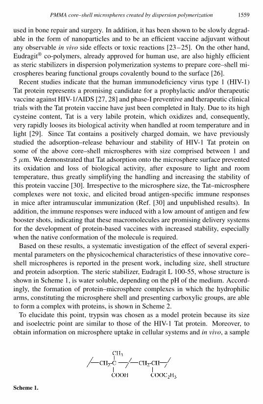
Based on these results a systematic investigation of the effect of several experi-mental parameters on the physicochemical characteristics of these innovative corendashshell microspheres is reported in the present work including size shell structureand protein adsorption The steric stabilizer Eudragit L 100-55 whose structure isshown in Scheme 1 is water soluble depending on the pH of the medium Accord-ingly the formation of proteinndashmicrosphere complexes in which the hydrophilicarms constituting the microsphere shell and presenting carboxylic groups are ableto form a complex with proteins is shown in Scheme 2
To elucidate this point trypsin was chosen as a model protein because its sizeand isoelectric point are similar to those of the HIV-1 Tat protein Moreover toobtain information on microsphere uptake in cellular systems and in vivo a sample
Scheme 1
1560 K Sparnacci et al
Scheme 2
of yellowgreen fluorescent corendashshell microspheres was prepared and employed asa fluorescent probe in vitro and in vivo
MATERIALS AND METHODS
Materials
Methanol (999 Carlo Erba) and 22prime-azobis (isobutyronitrile) (AIBN) (980Fluka) were used without further purification Methyl methacrylate (MMA) (99Aldrich) was distilled under vacuum immediately before use The poly(methacrylicacid ethyl acrylate) 1 1 statistical co-polymer (trade name Eudragit L 100-55)kindly supplied by Roumlhm Pharma as a powder sample is characterized by a numberaverage molar mass (Mn) of 250 kgmol Trypsin (pI = 974) was purchased fromSigma
Synthesis of fluorescent monomer 1
20 g fluoresceine (60 mmol) 20 g calcium carbonate and hydroquinone (trace)were dissolved in 100 DMF and the solution was heated at 60C Allyl chloridewas added dropwise and the reaction was allowed to proceed for 30 h in the darkAfter vacuum evaporation of the solvent the product was purified by flash columnchromatography (silica gel diethyl etherethyl acetate 80 20 as eluent) Maxλem = 528 nm (λexc = 488 nm) yield 53 (mp 123ndash125C) MS mz ()412 (M+ 100) 371 (10) 287 (20) 259 (15) 202 (7) 1H-NMR (CD3OD) δ 444(dd J = 59 and 1 Hz 2 H O CH2 CH ) 475 (dd J = 59 and 1 Hz 2 HO CH2 CH ) 508 (m 2H CH2 CH) 540 (m 2H CH2 CH) 558 (m 1HCH2 CH) 610 (m 1H CH2 CH) 660 (m 2H Ar) 698 (m 3H Ar) 725(d J = 1 Hz 1H Ar) 745 (dd J = 75 and 1 Hz 1H Ar) 785 (m 2H Ar) 830(dd J = 75 and 1Hz 1H Ar)
Dispersion polymerization procedure
Four series of samples 1andashe 2andashe 3andashc and 4andashc were prepared according to theexperimental conditions reported in Table 1
PMMA corendashshell microspheres created by dispersion polymerization 1561
Scheme 3
Table 1Experimental conditions for the preparation of sample series 1ndash5
Sample MeOH H2O MMA AIBN Eudragit Yield(wt) (wt) (wt) (wt) L100-55 ()
(wt)
1a 878 0 100 02 20 6421b 860 0 100 02 38 7621c 839 0 100 02 59 6951d 820 0 100 02 78 7011e 800 0 100 02 98 653
2a 667 211 100 02 20 6752b 654 206 100 02 38 6462c 638 201 100 02 59 7302d 623 197 100 02 78 7862e 599 191 100 02 98 731
3a 639 202 100 005 59 5663b 638 202 100 01 59 6013c 637 201 100 03 59 805
4a 468 394 100 005 38 7484b 468 393 100 01 38 8674c 467 393 100 02 38 881
5aa 638 201 100 02 59 663
Based on total recipe (1840 g) The dispersion polymerization reactions were performed at 60Cfor 24 h under continuous stirring nitrogen atmosphere and a reflux condenser For samples 2andashe3andashc and 5a the ratio between methanol and water in the solvent mixture is 76 24 wt whereas forsamples 4andashc it is 54 46 wt
a Reaction performed in presence of 50 mg fluoresceine derivative 1
As a typical example the polymerization of sample 1b is described 736 gEudragit L 100-55 was dissolved in 200 ml methanol and heated to 60C withmechanical stirring (stirring speed 300 rpm) under nitrogen atmosphere and refluxcondenser After 30 min 370 mg (225 mmol) AIBN dissolved in 183 g(183 mmol) MMA was added to the solution and the reaction was allowed toproceed for 24 h At the end of the reaction the latex was cooled filtered through afilter paper and then purified The following washing procedure was then employed
1562 K Sparnacci et al
(1) The latex was centrifuged at 3000 rpm for 15 min and the supernatant wasdischarged (2) The pellet was redispersed in fresh methanol and vigorously mixedfor 15 min (3) Steps 1 and 2 were repeated 4 times to eliminate any residualmonomer initiator and unbound Eudragit At the end the sample was redispersedin pure methanol (4) The obtained latex was centrifuged at 3000 rpm for 15 minand the supernatant was discharged (5) The pellet was redispersed in HPLC-gradewater and vigorously mixed for 15 min (6) Steps 4 and 5 were repeated 4 timesto eliminate any residual methanol The reaction yield was 762 as determinedgravimetrically
Fluorescent sample 5a was obtained by reacting fluorescent monomer 1 togetherwith MMA in the dispersion polymerization reaction In this preparation 110 gEudragit L 100-55 was dissolved in 200 ml methanolwater (76 24 ww) andheated at 60C under mechanical stirring (stirring speed 300 rpm) and nitrogenatmosphere and using a reflux condenser After 30 min 370 mg (225 mmol) AIBNand 50 mg (12 micromol) fluorescent monomer 1 dissolved in 183 g (183 mmol)MMA were added to the solution and the reaction was allowed to proceed for 24 hAt the end of the reaction the latex was purified as described above
Physico-chemical characterization
Microsphere size and size distribution were measured using a Jeol JEM 100CXscanning electron microscope (SEM) operating at an accelerating voltage of20ndash30 kV The samples were sputter-coated under vacuum with a thin layer(10ndash30 Aring) of gold The magnification was given by the scale on each micrographThe SEM photographs were digitalized and elaborated by the Scion Image process-ing program 150ndash200 individual microsphere diameters were measured for eachsample Table 2 reports the number-average diameter Dn the weight-average diam-eter Dw and the uniformity ratio U [31]
The amount of Eudragit L 100-55 linked to the microsphere surface was deter-mined by back titration of the excess NaOH after complete reaction of the acidgroups and microsphere removal by centrifugation To this purpose 06 g of eachmicrosphere sample was dispersed in 10 ml NaOH (20 mM) at room temperaturefor 24 h under nitrogen atmosphere Then the microsphere sample was collectedby centrifugation and washed twice with 25 ml distilled water The supernatantswere combined and the excess NaOH was titrated with 20 mM HCl under nitrogenatmosphere ζ -potential measurements were carried out at 25C on a Malvern Zeta-sizer 3000 HS instrument after dilution of microspheres in the presence of 10 mMNaCl solution and adjustment of pH by addition of HCl or NaOH All measurementswere run in triplicate
Absorption spectra were recorded on a UV Perkin-Elmer Lambda20 spectropho-tometer Luminescence and excitation spectra were obtained either from Perkin-Elmer LS 50 B instrument or a Spex Fluorolog II spectrofluorimeter equippedwith a Hamamatsu R928 phototube The experimental uncertainty in the band
PMMA corendashshell microspheres created by dispersion polymerization 1563
Table 2Number average diameter (Dn) weight average diameter (Dw) uniformity ratio (U ) amount of acidgroups per gram microspheres and surface charge density for sample series 1ndash5
Sample Dn Dw U COOHg microsphere Surface charge density(microm) (microm) (micromol) (nmolcm2)
1a 1018 1103 108 206 3691b 615 654 106 220 2351c 249 538 216 290 2011d 435 480 110 481 3781e 260 228 111 592 273
2a 242 389 161 272 1422b 238 249 105 427 1752c 236 245 104 542 2132d 169 173 102 621 1782e 177 183 104 653 198
3a 164 168 102 654 1343b 217 222 102 589 1943c 240 241 101 559 224
4a 080 072 103 687 0954b 065 067 103 602 0654c 078 080 102 573 075
5a 213 214 101 592 211
maximum for luminescence spectra was 2 nm Each sample was dissolved inmethanoldichloroethane 1 1 (HPLC grade Aldrich)
Protein adsorption
50 mg microspheres was incubated in 10 ml sodium phosphate buffer (20 mMpH 74) in the presence of different trypsin concentrations (from 10 to 250 microgml)for 2 h at room temperature in low-protein-binding tubes Then the microsphereswere collected by centrifugation at 15 times 103 rpm for 10 min and the amount ofthe residual protein in the supernatant was estimated after filtration on low proteinbinding filter units (Millex GV100 Millipore) using the bicinchoninic acid (BCA)assay [32] The amount of adsorbed protein was then calculated as the differencebetween the added and remaining Controls were represented by blank sampleswithout protein or microspheres Experiments were run in triplicate (SD 15)
Cellular uptake of fluorescent microspheres
Human HL3T1 cells (05 times 105) a HeLa derivative cell line containing anintegrated copy of the HIV-1 long-terminal repeat driving the expression of thechloramphenicol acetyl transferase reporter gene were seeded in 24-well platescontaining a 12-mm glass coverslip and incubated with 300 microgml of fluorescent5a microspheres in DMEM (Gibco) with 10 foetal bovine serum added After
1564 K Sparnacci et al
incubation cells were washed with phosphate-buffered saline (PBS) fixed with4 cold paraformaldehyde and observed at a confocal laser scanning microscopeLSM410 (Zeiss) Image acquisition recording and filtering were carried out usingan Indy 4400 graphic workstation (Silicon Graphics) as previously described [33]
In vivo inoculation of fluorescent microspheres
Animal use was according to national guidelines and institutional guidelinesSeven-week-old female BDF mice (n = 6) were injected in the quadriceps muscleof the left posterior leg with 1 mg of microspheres 5a resuspended in 100 microl PBSAs control mice were also injected in the quadriceps muscle of the right poster legwith 100 microl PBS alone Fifteen and 30 min after injection mice were anesthetizedintraperitoneally with 100 microl isotonic solution containing 1 mg Inoketan (Virbac)and 200 microg Rompun (Bayer) and killed Muscle samples at the site of injectionwere removed immediately submerged in liquid nitrogen for 1 min and storedat ndash80C Five-microm-thick frozen sections were prepared fixed with fresh 4paraformaldehyde for 10 min at room temperature washed with PBS and colouredwith DAPI (05 microgml Sigma) for 10 min which stains the nuclei After one washwith PBS the sections were dried with ethanol mounted in glycerolPBS containing14-diazabicyclo[222]octane to retard fading and observed through a fluorescencemicroscope (Axiophot 100 Zeiss) The green fluorescence (microspheres) wasobserved with a λ = 450ndash490 nm flow through λ = 510 nm and long passλ = 520 nm filter The blue fluorescence (DAPI) was observed with a band passλ = 365 nm flow through λ = 395 nm and long pass λ = 397 nm filter For thesame microscopic field green and blue images were taken with a Cool-Snapp CCDcamera (RS-Photometrics) and the images were then overlapped using the AdobePhotoshop 55 program
RESULTS AND DISCUSSION
In dispersion polymerization systems the main parameters affecting the microspheresize are the amount of the steric stabilizer the medium solvency power and theinitiator concentration [21 34ndash36] To get information concerning the effects ofthese parameters in the present dispersion polymerization system four sample se-ries were prepared and identified as 1andashe (5 runs) 2andashe (5 runs) 3andashc (3 runs) and4andashc (3 runs) In addition a sample of yellowgreen fluorescent microspheres iden-tified as 5a was synthesized using the fluoresceine derivative 1 All polymerizationreactions were performed using MMA as the monomer AIBN as the free radicalinitiator and Eudragit L 100-55 as the steric stabilizer either in methanol (series 1)or in methanolwater mixture (76 24 wt for series 2 and 3 and 54 46 wt forseries 4) All the reactions were carried out at 60C for 24 h In these conditionsyields ranged between 56 and 88 The reaction yield could be improved by in-creasing the reaction time but in some cases a decrease of the dispersion stability
PMMA corendashshell microspheres created by dispersion polymerization 1565
was observed leading to the formation of aggregates The amount of the stericstabilizer was varied in series 1 and 2 whereas the effect of the initiator concen-tration was studied in series 3 and 4 Moreover to better understand the effect ofthe medium solvency on the microsphere characteristics the composition of themethanolwater dispersion medium was changed Table 1 reports the reaction con-ditions for all samples
As both the monomer and the steric stabilizer are soluble in the reaction mediumthe polymerization start in a clear homogeneous system As soon as the polymeriza-tion begins primary radicals generated by decomposition of the initiator grow inthe continuous phase with the addition of the monomer units until they reach theircritical chain length and precipitate to form nuclei At this point which occurs aftera nucleation induction time of about 15ndash30 min a faint opalescence was observedThese nuclei grow further by capturing monomer oligo-radicals and stabilizer fromthe medium [36] At the end of the reaction a white and stable dispersion was al-ways obtained As typical examples two SEM micrographs of samples 2d and 5aare reported in Fig 1 Table 2 collects some physicochemical characteristics of thesamples including the number average diameter Dn and the uniformity ratio U aswell as the amount of carboxylic groups per gram of microspheres and the surfacecharge density In general the size distribution is quite narrow This indicates ashort particle-forming stage and a particle-growth stage free from both formation ofnew particles and coalescence of existing particles Figure 2 illustrates the trend ofthe number average diameter Dn for series 1 and 2 as a function of the steric stabi-lizer amount in the reaction medium For series 1 Dn is about 1018 microm for thesample with the lowest steric stabilizer concentration but it decreases steeply as thesteric stabilizer amount increases For series 2 the Dn values are definitely lowerthat those of the corresponding series 1 and only a slightly pronounced decrease ofDn can be observed
In both series the amount of carboxylic groups per gram of microspheresincreases regularly as the stabilizer amount in solution increases (Fig 3) Theobserved size decrease as the stabilizer concentration increases is related to theparallel increase in the grafting rate to the stabilizer which produces more nucleiand hence smaller final microspheres and more graft co-polymer in turn resultingin a greater amount of carboxylic groups at the microsphere surface Moreoverwater is a poorer solvent for PMMA than methanol Consequently the critical chainlength would decrease and the rate of adsorption of the Eudragit L 100-55-g-PMMAco-polymer would increase as the water content in the medium increases that isgoing from series 1 to series 2 Accordingly both the rate of nuclei formationand of Eudragit L 100-55-g-PMMA adsorption would increase thus resulting insmaller microspheres and in a greater amount of Eudragit L 100-55 located at themicrosphere surface
Figure 4 reports collectively the trends of Dn and the amount of carboxylicgroups per gram of microspheres for series 3 and 4 as a function of the initiatorconcentration For series 3 the microsphere diameter increases with the AIBN
1566 K Sparnacci et al
Figure 1 SEM micrographs of samples 2d (top) and 5a (bottom)
PMMA corendashshell microspheres created by dispersion polymerization 1567
Figure 2 Trend of number average diameter Dn for series 1 () and 2 () as a function of EudragitL 100-55 content (wt)
Figure 3 Trend of carboxylic group amount per gram microspheres for series 1 (2) and 2 (1) asa function of Eudragit L 100-55 content (wt)
Figure 4 Trend of number average diameter Dn ( series 3 series 4) and of the carboxylic groupamount per gram microspheres (2 series 3 1 series 4) as a function of AIBN content (wt)
1568 K Sparnacci et al
Figure 5 Trend of number average diameter Dn () and of carboxylic group amount per grammicrospheres (2) as a function of the water content (wt) for samples 1b 2b and 4c
content in agreement to the trend generally reported in the literature [19] whereasfor series 4 the size is unaffected by the initiator concentration For series 3 theincrease in microsphere size can be explained by considering that an increase in theinitiator concentration produces shorter PMMA chains which are more soluble sothat only a few of them would precipitate to form primary nuclei The aggregation ofmany but short macromolecules results in turn in few but large final microspheres[26 37] This effect is not observed for series 4 probably because the high watercontent (46 wt) reduces the critical chain length The amount of carboxylicgroups per gram of microspheres decreases slightly for both series as the AIBNconcentration increases
From the overall picture of the data it is clear that the main parameter affecting themicrosphere size is the solvency power of the reaction medium and consequentlythe amount of water To highlight the influence of this parameter on the microspheresize Fig 5 reports the trends of Dn and the amount of carboxylic groups at themicrosphere surface for samples 1b 2b and 4c These samples were preparedwith the same amount of monomer stabilizer and initiator the only variationbeing the water content As the water content increases the microsphere sizesubstantially decreases and the amount of carboxylic groups per gram microspheresincreases These effects are in agreement with those previously reported for theMMA dispersion polymerization in a watermethanol mixture using PVP as thesteric stabilizer [36] Water is a poorer solvent for PMMA than methanol and anincrease in water content reduces the oligomer solubility thus decreasing the criticalchain length for particles nucleation and increasing the rate of nuclei formationleading to smaller particles
Figure 6 illustrates the trend of the ionization degree of Eudragit as estimatedfrom the equilibrium constant (pKa approx 5) and the ζ -potential of sample 4bchosen as a typical one as a function of pH Although it is well known that theζ -potential decreases as pH increases [38] the similarity of the two sigmoidalcurves presenting very close inflection points clearly indicates that the Eudragit
PMMA corendashshell microspheres created by dispersion polymerization 1569
Figure 6 Trend of the ionization degree and ζ -potential as a function of pH for sample 4b
Figure 7 Adsorption isotherms of trypsin on microsphere samples 4a 4b and 2d in 20 mM sodiumphosphate buffer (pH 74)
is located at the microsphere surface thus further confirming that the dispersionpolymerization process is highly effective in providing microspheres endowed witha specific and tuneable surface
The protein adsorption behaviour on the above microspheres was investigatedusing trypsin a well known cheap and easily available model protein whosemolecular weight and pI (974) are similar to Tat (pI 1022) Microspheres 2d4a and 4b were chosen because of their different size and surface charge density
Figure 7 illustrates the trend of trypsin adsorption at physiological pH as afunction of the equilibrium trypsin amount The adsorption isotherms were fittedaccording to the LangmuirndashFreundlich equation (1) [35 36]
Cs = CmKC1n
b
1 + KC1n
b
(1)
by means of a non-linear regression method
1570 K Sparnacci et al
Figure 8 Trend of the LangmuirndashFreundlich parameters Cm and K as a function of the carboxylicgroups surface density on microsphere samples 4a 4b and 2d
In this equation Cs is the amount of adsorbed trypsin per unit surface areaCb is the bulk concentration of trypsin Cm is the plateau adsorbed amount(maximum amount of adsorption) and K is the equilibium adsorption constantIn the Langmuir isotherm K is the ratio of the adsorption to the desorptionrate constant In the LangmuirndashFreundlich this definition does not apply butits meaning is almost the same A large value of K represents a faster rate ofadsorption over desorption rate The value of n was fixed at 05 in the variouscalculations [39] Figure 8 illustrates the trend of Cm and K as functions of thecarboxylic group surface density The increase of Cm indicates that the adsorptionis mainly governed by polar interactions namely hydrogen bonds or electrostaticinteractions In contrast K decreases as the carboxylic group surface densityincreases A similar effect was also observed for microspheres bearing sulphonic orcarboxylic groups at the surface and it was explained considering a critical balancebetween hydrophobic and hydrophilic forces [40] In particular it was suggestedthat the adsorption rate due to hydrophobic forces is faster than the one due tohydrophilic forces Consequently the overall adsorption to desorption rate shoulddecrease as the hydrophilic contribution increases Although this explanation seemsquite reasonable other experiments should be performed to clarify this criticalpoint
To assess the capability of these microspheres to be taken up by the cells a flu-orescent corendashshell microsphere sample (5a) was also prepared Since fluorescentparticles obtained by simple dye adsorption at the surface can give rise to desorp-tion of dye and loss of fluorescent emission following exposure to light and duringin vitro experiments a sample was prepared using the reactive fluoresceine deriva-tive 1 Although allylic monomers do not undergo radical polymerization they areable to co-polymerize or at least to be included in the polymer chain as a terminalgroup Accordingly the microsphere sample 5a was prepared by running the disper-sion polymerization under the same experimental conditions as sample 2c with theaddition of a small amount of the fluorescent monomer 1 A microsphere sample
PMMA corendashshell microspheres created by dispersion polymerization 1571
(a) (b)
(c)
Figure 9 Confocal microscopy images of fluorescent microspheres 5a uptake by HL3T1 cellsafter 30 min (a) 6 h (b) and 24 h (c) incubation
with Dn = 213 microm and U = 101 was obtained After the conventional purifi-cation procedure a small amount of sample 5a was dissolved in chloroform andprecipitated in methanol The polymeric material appeared fluorescent whereas notrace of fluorescence was observed in the precipitation medium This demonstratesthat the fluorescent units deriving from 1 were covalently bound to the PMMA con-stituting the inner core of the microspheres Sample 5a presents an emission max-imum at 535 nm (λexc = 488 nm) and good photostability The photoemissionintensity of 5a in organic solution remained high and unaffected after exposure todaylight for 30 days whereas that of fluorescein was reduced by approximately 50under the same conditions This is in agreement with the fluorescence emissionbehaviour of commercially available particles with dyes covalently bound insidethe polymeric matrix [41] Cellular uptake experiments in human epithelial cells(HL3T1) were then carried out in the presence of sample 5a The analysis of thecells under a confocal microscope indicated that the fluorescent microspheres wererapidly and completely taken up in 24 h (Fig 9) Of note no significant differencein cellular uptake is detected when Tat protein is adsorbed onto the microspheresurface [30]
1572 K Sparnacci et al
Figure 10 Analysis at the site of injection of cellular uptake of 5a fluorescent microspheres15 min (panels A and C) and 30 min (panels B and D) after inoculation For the same microscopicfield green (5a fluorescent microspheres) and blue (DAPI) overlapped images are shown (A B)40times magnification (C D) 100times magnification of the images shown in the white square of panels Aand B respectively
Finally to determine whether the microspheres are taken up by the cells in vivomice were injected intramuscularly with sample 5a and killed 15 or 30 min afterinjection The muscle at the site of injection was then observed under a fluorescentmicroscope As shown in Fig 10 microspheres were taken up by muscle cells afterinjection confirming the in vitro internalization results described above These dataclearly indicate that the fluorescent sample 5a represents a useful tool for in vivostudies
CONCLUSIONS
In the present report corendashshell microspheres in the micron scale range were pre-pared by dispersion polymerization An appropriate selection of the experimentalparameters and in particular of the initiator and stabilizer amount and the mediumsolvency power allows a monodisperse sample as large as 600 nm to be prepared inwhich the shell is constituted by the steric stabilizer bearing carboxylic groups ableto interact with proteins mainly via ionic interactions To this purpose low initia-
PMMA corendashshell microspheres created by dispersion polymerization 1573
tor concentration high steric stabilizer amount and a low solvency power mediummust be employed The described microspheres are able to efficiently bind trypsinchosen as model protein for Tat The amount of adsorbed protein is linearly relatedto the carboxylic group amount Eventually these microspheres were shown to bereadily taken up by non-phagocytic cells in vitro and in vivo As a final remark thedispersion polymerization reaction is versatile in providing corendashshell microsphereswith highly differentiated characteristics [42] The synthetic procedure allows large-scale preparation of reproducible stable and homogeneous microsphere stocks anda wide modulation of the outer shell so that specific and reversible adsorption ofantigens with varying hydrophobicity molar mass and isoelectric point can be en-visaged Formation of the proteinndashmicrosphere complexes is easy and fast sincethey spontaneously assemble in aqueous solution after incubation of the two com-ponents at room temperature and no purification steps are required These featurestogether with the observation that the antigenndashmicrosphere complexes preserve theprotein native conformation and biological activity thus increasing the protein sta-bility [30] and induce potent antigen-specific immune responses in mice (data notshown) indicate that these novel microspheres represent suitable adjuvants for thedevelopment of novel and safe protein-based vaccines with increased shelf-life andcapable of triggering long-term specific immunity
Acknowledgements
This work was supported by the Italian Program on HIVAIDS Research and bythe Italian Concerted Action for the development of an HIVAIDS Vaccine (ICAV)grants 40D48 and 45D102 We are grateful to Marco Ballestri (CNR) for hisexpert technical assistance
REFERENCES
1 U B Kompella and V H L Lee Adv Drug Deliv Rev 46 211 (2001)2 U Bickel T Yoshikawa and W M Pardridge Adv Drug Deliv Rev 46 247 (2001)3 R Langer Acc Chem Res 33 94 (2000)4 S P Baldwin and W M Saltzman Adv Drug Deliv Rev 33 71 (1998)5 K S Soppimath T M Aminabhavi A R Kulkarni and W E Rudzinski J Control Rel 70 1
(2001)6 P Couvreur M J BlancoPrieto F Puisieux B Roques and E Fattal Adv Drug Deliv Rev 28
85 (1997)7 M L Hedley Expert Opin Biol Ther 13 903 (2003)8 J Hanes J L Cleland and R Langer Adv Drug Deliv Rev 28 97 (1997)9 P Johansen Y Men H P Merkle and B Gander Eur J Pharm Biopharm 50 129 (2000)
10 M J Alonso R K Gupta C Min G R Siber and R Langer Vaccine 12 299 (1994)11 D T OrsquoHagan M Singh and R K Gupta Adv Drug Deliv Rev 32 225 (1998)12 T Uchida A Yagi Y Oda Y Nakada and S Goto Chem Pharm Bull 44 235 (1996)13 G Crotts and T G Park J Control Rel 44 123 (1997)14 M Van de Weert W E Hennink and W Jiskoot Pharm Res 17 1159 (2000)
1574 K Sparnacci et al
15 J Kazzaz J Neidleman M Singh G Ott and D T OrsquoHagan J Control Rel 67 347 (2000)16 M Singh M Briones G Ott and D OrsquoHagan Proc Natl Acad Sci USA 97 811 (2000)17 M Singh J Kazzaz J Chesko E Soenawan M Ugozzoli M Giuliani M Pizza R Rappuoli
and D T OrsquoHagan J Pharm Sci 93 273 (2004)18 K E J Barret Dispersion Polymerization in Organic Media Wiley London (1975)19 P A Lovell and M S El-Aasser (Eds) in Emulsion Polymerization and Emulsion Polymers
p 743 Wiley New York NY (1997)20 M Laus C Dinnella G Lanzarini and A Casagrande Polymer 37 343 (1996)21 C K Ober and M L Hair J Polym Sci Polym Chem 25 1395 (1987)22 M Laus K Sparnacci AS Angeloni S Valenti and L Tondelli J Control Rel 72 280 (2001)23 J Kreuter U Tauber and V Illi J Pharm Sci 68 1443 (1979)24 J Kreuter and P P Spieser J Pharm Sci 65 1624 (1976)25 J Kreuter M Nefzger E Liehl R Czok and R Voges J Pharm Sci 72 1146 (1983)26 K Sparnacci M Laus L Tondelli L Magnani and C Bernardi Makromol Chem Phys 203
1364 (2002)27 A Cafaro A Caputo C Fracasso M T Maggiorella D Goletti S Baroncelli M Pace
L Sernicola M L Koanga-Mogtomo M Betti A Borsetti R Belli L Akerblom F CorriasS Buttograve J Heeney P Verani F Titti and B Ensoli Nature Med 5 643 (1999)
28 A Caputo R Gavioli and B Ensoli Curr HIV Res 2 357 (2004)29 E Fanales-Belasio A Cafaro A Cara D R M Negri V Fiorelli S Buttograve S Moretti
M T Maggiorella S Baroncelli Z Michelini A Tripiciano L Sernicola A ScoglioA Borsetti B Ridolfi R Bona P Ten Haaft I Macchia P Leone M Rosaria M R Pavone-Cossut F Nappi E Vardas M Magnani E Laguardia A Caputo F Titti and B Ensoli DNACell Biol 21 599 (2002)
30 A Caputo E Brocca-Cofano A Castaldello G Altavilla R De Michele M MarchisioR Gavioli U Rolen L Chiarantini A Cerasi S Dominici M Magnani K SparnacciM Laus L Tondelli and B Ensoli Vaccine 22 3258 (2004)
31 S A Chen and H S Chang J Polym Sci Polym Chem 23 2615 (1985)32 K J Wiechelman R D Braun and J D Fitzpatrick Anal Biochem 175 231 (1988)33 L M Neri A M Martelli and N M Maraldi Microsc Res Tech 36 179 (1997)34 K E J Barret and H R Thomas J Polym Sci Part A 7 2621 (1969)35 S Kobayashi H Uyama Y Matsumoto and I Yamamoto Makromol Chem 193 2355 (1992)36 S Shen E D Sudol and M S El-Aasser J Polym Sci Polym Chem 31 1393 (1993)37 M Yasuda H Seki H Yokoyama H Ogino K Ishimi and H Ishikawa Macromolecules 34
3261 (2001)38 R J Hunter Zeta Potential in Colloid Science mdash Principles and Applications Academic Press
London (1981)39 J Y Yoon J H Kimu and W S Kim Colloid Surf A 153 413 (1999)40 J Y Yoon H Y Park J H Kim and W S Kim J Colloid Interf Sci 177 613 (1996)41 M K Bhalgat R P Haugland J S Pollack S Swan and R P Haugland J Immunol Methods
219 57 (1998)42 B Ensoli A Caputo R Gavioli L Tondelli M Laus and K Sparnacci UK Patent
No 03256245 (2003)

1558 K Sparnacci et al
INTRODUCTION
Due to recent advances in the area of biotechnology biochemistry and molecularbiology numerous bioactive peptides and proteins are available in large quantitiesfor therapeutic and vaccine purposes However most of them have short half-lives in vivo because they are easily degraded or denatured in biological fluids andsometimes not efficiently absorbed by the gastrointestinal tract due to their relativelyhigh molecular mass This implies that multiple administrations are required fortheir efficacy for these reasons there has been a growing need for suitable deliverysystems capable of stabilizing and increasing the shelf-life of these molecules andof efficiently delivering them to the desired location [1 2]
The use of microparticles as drugvaccine carriers to increase the bioavailabilityof peptide and protein drugs and vaccines is an expanding research field The mostpromising systems described up to now are based on encapsulation or entrapment ofproteins into biocompatible polymeric devices [3 4] In particular biodegradablepolymeric nano- and microparticles are widely investigated to produce controlledand sustained release of bioactive molecules (for a review see Ref [5]) Most of thework reported in literature is focused on protein encapsulation inside poly(lactide-co-glycolide) (PLG) matrices because of their good biocompatibility non-toxicityand favourable biodegradation pattern [6 7] These polymeric particles have alsoshown great potential as vaccine adjuvants due to their ability in optimizing antigenpresentation and hence humoral and cellular response [8ndash10] However someconcerns have arisen about the effects of both the encapsulation procedures andPLG degradation on protein stability [11ndash14]
As an alternative to the encapsulation method there is a completely differentconceptual approach in the development of delivery systems based on corendashshellmicrospheres or nanospheres in which the shell allows docking of the bioactivemolecule which can later be released in a biologically active form Modified PLGmicroparticles carrying antigens on their surface are able to increase the potencyof DNA and protein vaccines in several animal species [15ndash17] However theuse of chlorinated solvents andor surfactants during particle preparation may in-terfere with the reproducibility of particle size and size distribution thus affect-ing their biocompatibility To overcome these problems we designed and devel-oped a new type of poly(methyl methacrylate) (PMMA) corendashshell microsphereswhich were prepared by dispersion polymerization [18ndash21] using as the steric sta-bilizer a commercial statistical co-polymer of methacrylic acid and ethyl acrylate(Eudragit L 100-55) According to the polymerization mechanism they present acore mainly constituted of biocompatible PMMA and a shell of Eudragit L 100-55 which irrespective of its width strongly drives the microsphere behavior interms of protein adsorption propensity Actually these microspheres were specifi-cally designed for binding biologically active macromolecules on their shell with-out the need for added surfactants We also tested the feasibility of this approachby studying the adsorption behaviour of bovine serum albumin (BSA) as a modelprotein [22] PMMA is already known to be a well-tolerated polymeric material
PMMA corendashshell microspheres created by dispersion polymerization 1559
used in bone repair and surgery In addition it has been shown to be slowly degrad-able in the form of nanoparticles and to be an efficient vaccine adjuvant withoutany observable in vivo side effects or toxic reactions [23ndash25] On the other handEudragitreg co-polymers already approved for human use are also highly efficientas steric stabilizers in dispersion polymerization systems to prepare corendashshell mi-crospheres bearing functional groups covalently bound to the surface [26]
Recent studies indicate that the human immunodeficiency virus type 1 (HIV-1)Tat protein represents a promising candidate for a prophylactic andor therapeuticvaccine against HIV-1AIDS [27 28] and phase-I preventive and therapeutic clinicaltrials with the Tat protein vaccine have just been completed in Italy Due to its highcysteine content Tat is a very labile protein which oxidizes and consequentlyvery rapidly looses its biological activity when handled at room temperature and inlight [29] Since Tat contains a positively charged domain we have previouslystudied the adsorptionndashrelease behaviour and stability of HIV-1 Tat protein onsome of the above corendashshell microspheres with size comprised between 1 and5 microm We demonstrated that Tat adsorption onto the microsphere surface preventedits oxidation and loss of biological activity after exposure to light and roomtemperature thus greatly simplifying the handling and increasing the stability ofthis protein vaccine [30] Irrespective to the microsphere size the Tatndashmicrospherecomplexes were not toxic and elicited broad antigen-specific immune responsesin mice after intramuscular immunization (Ref [30] and unpublished results) Inaddition the immune responses were induced with a low amount of antigen and fewbooster shots indicating that these macromolecules are promising delivery systemsfor the development of protein-based vaccines with increased stability especiallywhen the native conformation of the molecule is required
Based on these results a systematic investigation of the effect of several experi-mental parameters on the physicochemical characteristics of these innovative corendashshell microspheres is reported in the present work including size shell structureand protein adsorption The steric stabilizer Eudragit L 100-55 whose structure isshown in Scheme 1 is water soluble depending on the pH of the medium Accord-ingly the formation of proteinndashmicrosphere complexes in which the hydrophilicarms constituting the microsphere shell and presenting carboxylic groups are ableto form a complex with proteins is shown in Scheme 2
To elucidate this point trypsin was chosen as a model protein because its sizeand isoelectric point are similar to those of the HIV-1 Tat protein Moreover toobtain information on microsphere uptake in cellular systems and in vivo a sample
Scheme 1
1560 K Sparnacci et al
Scheme 2
of yellowgreen fluorescent corendashshell microspheres was prepared and employed asa fluorescent probe in vitro and in vivo
MATERIALS AND METHODS
Materials
Methanol (999 Carlo Erba) and 22prime-azobis (isobutyronitrile) (AIBN) (980Fluka) were used without further purification Methyl methacrylate (MMA) (99Aldrich) was distilled under vacuum immediately before use The poly(methacrylicacid ethyl acrylate) 1 1 statistical co-polymer (trade name Eudragit L 100-55)kindly supplied by Roumlhm Pharma as a powder sample is characterized by a numberaverage molar mass (Mn) of 250 kgmol Trypsin (pI = 974) was purchased fromSigma
Synthesis of fluorescent monomer 1
20 g fluoresceine (60 mmol) 20 g calcium carbonate and hydroquinone (trace)were dissolved in 100 DMF and the solution was heated at 60C Allyl chloridewas added dropwise and the reaction was allowed to proceed for 30 h in the darkAfter vacuum evaporation of the solvent the product was purified by flash columnchromatography (silica gel diethyl etherethyl acetate 80 20 as eluent) Maxλem = 528 nm (λexc = 488 nm) yield 53 (mp 123ndash125C) MS mz ()412 (M+ 100) 371 (10) 287 (20) 259 (15) 202 (7) 1H-NMR (CD3OD) δ 444(dd J = 59 and 1 Hz 2 H O CH2 CH ) 475 (dd J = 59 and 1 Hz 2 HO CH2 CH ) 508 (m 2H CH2 CH) 540 (m 2H CH2 CH) 558 (m 1HCH2 CH) 610 (m 1H CH2 CH) 660 (m 2H Ar) 698 (m 3H Ar) 725(d J = 1 Hz 1H Ar) 745 (dd J = 75 and 1 Hz 1H Ar) 785 (m 2H Ar) 830(dd J = 75 and 1Hz 1H Ar)
Dispersion polymerization procedure
Four series of samples 1andashe 2andashe 3andashc and 4andashc were prepared according to theexperimental conditions reported in Table 1
PMMA corendashshell microspheres created by dispersion polymerization 1561
Scheme 3
Table 1Experimental conditions for the preparation of sample series 1ndash5
Sample MeOH H2O MMA AIBN Eudragit Yield(wt) (wt) (wt) (wt) L100-55 ()
(wt)
1a 878 0 100 02 20 6421b 860 0 100 02 38 7621c 839 0 100 02 59 6951d 820 0 100 02 78 7011e 800 0 100 02 98 653
2a 667 211 100 02 20 6752b 654 206 100 02 38 6462c 638 201 100 02 59 7302d 623 197 100 02 78 7862e 599 191 100 02 98 731
3a 639 202 100 005 59 5663b 638 202 100 01 59 6013c 637 201 100 03 59 805
4a 468 394 100 005 38 7484b 468 393 100 01 38 8674c 467 393 100 02 38 881
5aa 638 201 100 02 59 663
Based on total recipe (1840 g) The dispersion polymerization reactions were performed at 60Cfor 24 h under continuous stirring nitrogen atmosphere and a reflux condenser For samples 2andashe3andashc and 5a the ratio between methanol and water in the solvent mixture is 76 24 wt whereas forsamples 4andashc it is 54 46 wt
a Reaction performed in presence of 50 mg fluoresceine derivative 1
As a typical example the polymerization of sample 1b is described 736 gEudragit L 100-55 was dissolved in 200 ml methanol and heated to 60C withmechanical stirring (stirring speed 300 rpm) under nitrogen atmosphere and refluxcondenser After 30 min 370 mg (225 mmol) AIBN dissolved in 183 g(183 mmol) MMA was added to the solution and the reaction was allowed toproceed for 24 h At the end of the reaction the latex was cooled filtered through afilter paper and then purified The following washing procedure was then employed
1562 K Sparnacci et al
(1) The latex was centrifuged at 3000 rpm for 15 min and the supernatant wasdischarged (2) The pellet was redispersed in fresh methanol and vigorously mixedfor 15 min (3) Steps 1 and 2 were repeated 4 times to eliminate any residualmonomer initiator and unbound Eudragit At the end the sample was redispersedin pure methanol (4) The obtained latex was centrifuged at 3000 rpm for 15 minand the supernatant was discharged (5) The pellet was redispersed in HPLC-gradewater and vigorously mixed for 15 min (6) Steps 4 and 5 were repeated 4 timesto eliminate any residual methanol The reaction yield was 762 as determinedgravimetrically
Fluorescent sample 5a was obtained by reacting fluorescent monomer 1 togetherwith MMA in the dispersion polymerization reaction In this preparation 110 gEudragit L 100-55 was dissolved in 200 ml methanolwater (76 24 ww) andheated at 60C under mechanical stirring (stirring speed 300 rpm) and nitrogenatmosphere and using a reflux condenser After 30 min 370 mg (225 mmol) AIBNand 50 mg (12 micromol) fluorescent monomer 1 dissolved in 183 g (183 mmol)MMA were added to the solution and the reaction was allowed to proceed for 24 hAt the end of the reaction the latex was purified as described above
Physico-chemical characterization
Microsphere size and size distribution were measured using a Jeol JEM 100CXscanning electron microscope (SEM) operating at an accelerating voltage of20ndash30 kV The samples were sputter-coated under vacuum with a thin layer(10ndash30 Aring) of gold The magnification was given by the scale on each micrographThe SEM photographs were digitalized and elaborated by the Scion Image process-ing program 150ndash200 individual microsphere diameters were measured for eachsample Table 2 reports the number-average diameter Dn the weight-average diam-eter Dw and the uniformity ratio U [31]
The amount of Eudragit L 100-55 linked to the microsphere surface was deter-mined by back titration of the excess NaOH after complete reaction of the acidgroups and microsphere removal by centrifugation To this purpose 06 g of eachmicrosphere sample was dispersed in 10 ml NaOH (20 mM) at room temperaturefor 24 h under nitrogen atmosphere Then the microsphere sample was collectedby centrifugation and washed twice with 25 ml distilled water The supernatantswere combined and the excess NaOH was titrated with 20 mM HCl under nitrogenatmosphere ζ -potential measurements were carried out at 25C on a Malvern Zeta-sizer 3000 HS instrument after dilution of microspheres in the presence of 10 mMNaCl solution and adjustment of pH by addition of HCl or NaOH All measurementswere run in triplicate
Absorption spectra were recorded on a UV Perkin-Elmer Lambda20 spectropho-tometer Luminescence and excitation spectra were obtained either from Perkin-Elmer LS 50 B instrument or a Spex Fluorolog II spectrofluorimeter equippedwith a Hamamatsu R928 phototube The experimental uncertainty in the band
PMMA corendashshell microspheres created by dispersion polymerization 1563
Table 2Number average diameter (Dn) weight average diameter (Dw) uniformity ratio (U ) amount of acidgroups per gram microspheres and surface charge density for sample series 1ndash5
Sample Dn Dw U COOHg microsphere Surface charge density(microm) (microm) (micromol) (nmolcm2)
1a 1018 1103 108 206 3691b 615 654 106 220 2351c 249 538 216 290 2011d 435 480 110 481 3781e 260 228 111 592 273
2a 242 389 161 272 1422b 238 249 105 427 1752c 236 245 104 542 2132d 169 173 102 621 1782e 177 183 104 653 198
3a 164 168 102 654 1343b 217 222 102 589 1943c 240 241 101 559 224
4a 080 072 103 687 0954b 065 067 103 602 0654c 078 080 102 573 075
5a 213 214 101 592 211
maximum for luminescence spectra was 2 nm Each sample was dissolved inmethanoldichloroethane 1 1 (HPLC grade Aldrich)
Protein adsorption
50 mg microspheres was incubated in 10 ml sodium phosphate buffer (20 mMpH 74) in the presence of different trypsin concentrations (from 10 to 250 microgml)for 2 h at room temperature in low-protein-binding tubes Then the microsphereswere collected by centrifugation at 15 times 103 rpm for 10 min and the amount ofthe residual protein in the supernatant was estimated after filtration on low proteinbinding filter units (Millex GV100 Millipore) using the bicinchoninic acid (BCA)assay [32] The amount of adsorbed protein was then calculated as the differencebetween the added and remaining Controls were represented by blank sampleswithout protein or microspheres Experiments were run in triplicate (SD 15)
Cellular uptake of fluorescent microspheres
Human HL3T1 cells (05 times 105) a HeLa derivative cell line containing anintegrated copy of the HIV-1 long-terminal repeat driving the expression of thechloramphenicol acetyl transferase reporter gene were seeded in 24-well platescontaining a 12-mm glass coverslip and incubated with 300 microgml of fluorescent5a microspheres in DMEM (Gibco) with 10 foetal bovine serum added After
1564 K Sparnacci et al
incubation cells were washed with phosphate-buffered saline (PBS) fixed with4 cold paraformaldehyde and observed at a confocal laser scanning microscopeLSM410 (Zeiss) Image acquisition recording and filtering were carried out usingan Indy 4400 graphic workstation (Silicon Graphics) as previously described [33]
In vivo inoculation of fluorescent microspheres
Animal use was according to national guidelines and institutional guidelinesSeven-week-old female BDF mice (n = 6) were injected in the quadriceps muscleof the left posterior leg with 1 mg of microspheres 5a resuspended in 100 microl PBSAs control mice were also injected in the quadriceps muscle of the right poster legwith 100 microl PBS alone Fifteen and 30 min after injection mice were anesthetizedintraperitoneally with 100 microl isotonic solution containing 1 mg Inoketan (Virbac)and 200 microg Rompun (Bayer) and killed Muscle samples at the site of injectionwere removed immediately submerged in liquid nitrogen for 1 min and storedat ndash80C Five-microm-thick frozen sections were prepared fixed with fresh 4paraformaldehyde for 10 min at room temperature washed with PBS and colouredwith DAPI (05 microgml Sigma) for 10 min which stains the nuclei After one washwith PBS the sections were dried with ethanol mounted in glycerolPBS containing14-diazabicyclo[222]octane to retard fading and observed through a fluorescencemicroscope (Axiophot 100 Zeiss) The green fluorescence (microspheres) wasobserved with a λ = 450ndash490 nm flow through λ = 510 nm and long passλ = 520 nm filter The blue fluorescence (DAPI) was observed with a band passλ = 365 nm flow through λ = 395 nm and long pass λ = 397 nm filter For thesame microscopic field green and blue images were taken with a Cool-Snapp CCDcamera (RS-Photometrics) and the images were then overlapped using the AdobePhotoshop 55 program
RESULTS AND DISCUSSION
In dispersion polymerization systems the main parameters affecting the microspheresize are the amount of the steric stabilizer the medium solvency power and theinitiator concentration [21 34ndash36] To get information concerning the effects ofthese parameters in the present dispersion polymerization system four sample se-ries were prepared and identified as 1andashe (5 runs) 2andashe (5 runs) 3andashc (3 runs) and4andashc (3 runs) In addition a sample of yellowgreen fluorescent microspheres iden-tified as 5a was synthesized using the fluoresceine derivative 1 All polymerizationreactions were performed using MMA as the monomer AIBN as the free radicalinitiator and Eudragit L 100-55 as the steric stabilizer either in methanol (series 1)or in methanolwater mixture (76 24 wt for series 2 and 3 and 54 46 wt forseries 4) All the reactions were carried out at 60C for 24 h In these conditionsyields ranged between 56 and 88 The reaction yield could be improved by in-creasing the reaction time but in some cases a decrease of the dispersion stability
PMMA corendashshell microspheres created by dispersion polymerization 1565
was observed leading to the formation of aggregates The amount of the stericstabilizer was varied in series 1 and 2 whereas the effect of the initiator concen-tration was studied in series 3 and 4 Moreover to better understand the effect ofthe medium solvency on the microsphere characteristics the composition of themethanolwater dispersion medium was changed Table 1 reports the reaction con-ditions for all samples
As both the monomer and the steric stabilizer are soluble in the reaction mediumthe polymerization start in a clear homogeneous system As soon as the polymeriza-tion begins primary radicals generated by decomposition of the initiator grow inthe continuous phase with the addition of the monomer units until they reach theircritical chain length and precipitate to form nuclei At this point which occurs aftera nucleation induction time of about 15ndash30 min a faint opalescence was observedThese nuclei grow further by capturing monomer oligo-radicals and stabilizer fromthe medium [36] At the end of the reaction a white and stable dispersion was al-ways obtained As typical examples two SEM micrographs of samples 2d and 5aare reported in Fig 1 Table 2 collects some physicochemical characteristics of thesamples including the number average diameter Dn and the uniformity ratio U aswell as the amount of carboxylic groups per gram of microspheres and the surfacecharge density In general the size distribution is quite narrow This indicates ashort particle-forming stage and a particle-growth stage free from both formation ofnew particles and coalescence of existing particles Figure 2 illustrates the trend ofthe number average diameter Dn for series 1 and 2 as a function of the steric stabi-lizer amount in the reaction medium For series 1 Dn is about 1018 microm for thesample with the lowest steric stabilizer concentration but it decreases steeply as thesteric stabilizer amount increases For series 2 the Dn values are definitely lowerthat those of the corresponding series 1 and only a slightly pronounced decrease ofDn can be observed
In both series the amount of carboxylic groups per gram of microspheresincreases regularly as the stabilizer amount in solution increases (Fig 3) Theobserved size decrease as the stabilizer concentration increases is related to theparallel increase in the grafting rate to the stabilizer which produces more nucleiand hence smaller final microspheres and more graft co-polymer in turn resultingin a greater amount of carboxylic groups at the microsphere surface Moreoverwater is a poorer solvent for PMMA than methanol Consequently the critical chainlength would decrease and the rate of adsorption of the Eudragit L 100-55-g-PMMAco-polymer would increase as the water content in the medium increases that isgoing from series 1 to series 2 Accordingly both the rate of nuclei formationand of Eudragit L 100-55-g-PMMA adsorption would increase thus resulting insmaller microspheres and in a greater amount of Eudragit L 100-55 located at themicrosphere surface
Figure 4 reports collectively the trends of Dn and the amount of carboxylicgroups per gram of microspheres for series 3 and 4 as a function of the initiatorconcentration For series 3 the microsphere diameter increases with the AIBN
1566 K Sparnacci et al
Figure 1 SEM micrographs of samples 2d (top) and 5a (bottom)
PMMA corendashshell microspheres created by dispersion polymerization 1567
Figure 2 Trend of number average diameter Dn for series 1 () and 2 () as a function of EudragitL 100-55 content (wt)
Figure 3 Trend of carboxylic group amount per gram microspheres for series 1 (2) and 2 (1) asa function of Eudragit L 100-55 content (wt)
Figure 4 Trend of number average diameter Dn ( series 3 series 4) and of the carboxylic groupamount per gram microspheres (2 series 3 1 series 4) as a function of AIBN content (wt)
1568 K Sparnacci et al
Figure 5 Trend of number average diameter Dn () and of carboxylic group amount per grammicrospheres (2) as a function of the water content (wt) for samples 1b 2b and 4c
content in agreement to the trend generally reported in the literature [19] whereasfor series 4 the size is unaffected by the initiator concentration For series 3 theincrease in microsphere size can be explained by considering that an increase in theinitiator concentration produces shorter PMMA chains which are more soluble sothat only a few of them would precipitate to form primary nuclei The aggregation ofmany but short macromolecules results in turn in few but large final microspheres[26 37] This effect is not observed for series 4 probably because the high watercontent (46 wt) reduces the critical chain length The amount of carboxylicgroups per gram of microspheres decreases slightly for both series as the AIBNconcentration increases
From the overall picture of the data it is clear that the main parameter affecting themicrosphere size is the solvency power of the reaction medium and consequentlythe amount of water To highlight the influence of this parameter on the microspheresize Fig 5 reports the trends of Dn and the amount of carboxylic groups at themicrosphere surface for samples 1b 2b and 4c These samples were preparedwith the same amount of monomer stabilizer and initiator the only variationbeing the water content As the water content increases the microsphere sizesubstantially decreases and the amount of carboxylic groups per gram microspheresincreases These effects are in agreement with those previously reported for theMMA dispersion polymerization in a watermethanol mixture using PVP as thesteric stabilizer [36] Water is a poorer solvent for PMMA than methanol and anincrease in water content reduces the oligomer solubility thus decreasing the criticalchain length for particles nucleation and increasing the rate of nuclei formationleading to smaller particles
Figure 6 illustrates the trend of the ionization degree of Eudragit as estimatedfrom the equilibrium constant (pKa approx 5) and the ζ -potential of sample 4bchosen as a typical one as a function of pH Although it is well known that theζ -potential decreases as pH increases [38] the similarity of the two sigmoidalcurves presenting very close inflection points clearly indicates that the Eudragit
PMMA corendashshell microspheres created by dispersion polymerization 1569
Figure 6 Trend of the ionization degree and ζ -potential as a function of pH for sample 4b
Figure 7 Adsorption isotherms of trypsin on microsphere samples 4a 4b and 2d in 20 mM sodiumphosphate buffer (pH 74)
is located at the microsphere surface thus further confirming that the dispersionpolymerization process is highly effective in providing microspheres endowed witha specific and tuneable surface
The protein adsorption behaviour on the above microspheres was investigatedusing trypsin a well known cheap and easily available model protein whosemolecular weight and pI (974) are similar to Tat (pI 1022) Microspheres 2d4a and 4b were chosen because of their different size and surface charge density
Figure 7 illustrates the trend of trypsin adsorption at physiological pH as afunction of the equilibrium trypsin amount The adsorption isotherms were fittedaccording to the LangmuirndashFreundlich equation (1) [35 36]
Cs = CmKC1n
b
1 + KC1n
b
(1)
by means of a non-linear regression method
1570 K Sparnacci et al
Figure 8 Trend of the LangmuirndashFreundlich parameters Cm and K as a function of the carboxylicgroups surface density on microsphere samples 4a 4b and 2d
In this equation Cs is the amount of adsorbed trypsin per unit surface areaCb is the bulk concentration of trypsin Cm is the plateau adsorbed amount(maximum amount of adsorption) and K is the equilibium adsorption constantIn the Langmuir isotherm K is the ratio of the adsorption to the desorptionrate constant In the LangmuirndashFreundlich this definition does not apply butits meaning is almost the same A large value of K represents a faster rate ofadsorption over desorption rate The value of n was fixed at 05 in the variouscalculations [39] Figure 8 illustrates the trend of Cm and K as functions of thecarboxylic group surface density The increase of Cm indicates that the adsorptionis mainly governed by polar interactions namely hydrogen bonds or electrostaticinteractions In contrast K decreases as the carboxylic group surface densityincreases A similar effect was also observed for microspheres bearing sulphonic orcarboxylic groups at the surface and it was explained considering a critical balancebetween hydrophobic and hydrophilic forces [40] In particular it was suggestedthat the adsorption rate due to hydrophobic forces is faster than the one due tohydrophilic forces Consequently the overall adsorption to desorption rate shoulddecrease as the hydrophilic contribution increases Although this explanation seemsquite reasonable other experiments should be performed to clarify this criticalpoint
To assess the capability of these microspheres to be taken up by the cells a flu-orescent corendashshell microsphere sample (5a) was also prepared Since fluorescentparticles obtained by simple dye adsorption at the surface can give rise to desorp-tion of dye and loss of fluorescent emission following exposure to light and duringin vitro experiments a sample was prepared using the reactive fluoresceine deriva-tive 1 Although allylic monomers do not undergo radical polymerization they areable to co-polymerize or at least to be included in the polymer chain as a terminalgroup Accordingly the microsphere sample 5a was prepared by running the disper-sion polymerization under the same experimental conditions as sample 2c with theaddition of a small amount of the fluorescent monomer 1 A microsphere sample
PMMA corendashshell microspheres created by dispersion polymerization 1571
(a) (b)
(c)
Figure 9 Confocal microscopy images of fluorescent microspheres 5a uptake by HL3T1 cellsafter 30 min (a) 6 h (b) and 24 h (c) incubation
with Dn = 213 microm and U = 101 was obtained After the conventional purifi-cation procedure a small amount of sample 5a was dissolved in chloroform andprecipitated in methanol The polymeric material appeared fluorescent whereas notrace of fluorescence was observed in the precipitation medium This demonstratesthat the fluorescent units deriving from 1 were covalently bound to the PMMA con-stituting the inner core of the microspheres Sample 5a presents an emission max-imum at 535 nm (λexc = 488 nm) and good photostability The photoemissionintensity of 5a in organic solution remained high and unaffected after exposure todaylight for 30 days whereas that of fluorescein was reduced by approximately 50under the same conditions This is in agreement with the fluorescence emissionbehaviour of commercially available particles with dyes covalently bound insidethe polymeric matrix [41] Cellular uptake experiments in human epithelial cells(HL3T1) were then carried out in the presence of sample 5a The analysis of thecells under a confocal microscope indicated that the fluorescent microspheres wererapidly and completely taken up in 24 h (Fig 9) Of note no significant differencein cellular uptake is detected when Tat protein is adsorbed onto the microspheresurface [30]
1572 K Sparnacci et al
Figure 10 Analysis at the site of injection of cellular uptake of 5a fluorescent microspheres15 min (panels A and C) and 30 min (panels B and D) after inoculation For the same microscopicfield green (5a fluorescent microspheres) and blue (DAPI) overlapped images are shown (A B)40times magnification (C D) 100times magnification of the images shown in the white square of panels Aand B respectively
Finally to determine whether the microspheres are taken up by the cells in vivomice were injected intramuscularly with sample 5a and killed 15 or 30 min afterinjection The muscle at the site of injection was then observed under a fluorescentmicroscope As shown in Fig 10 microspheres were taken up by muscle cells afterinjection confirming the in vitro internalization results described above These dataclearly indicate that the fluorescent sample 5a represents a useful tool for in vivostudies
CONCLUSIONS
In the present report corendashshell microspheres in the micron scale range were pre-pared by dispersion polymerization An appropriate selection of the experimentalparameters and in particular of the initiator and stabilizer amount and the mediumsolvency power allows a monodisperse sample as large as 600 nm to be prepared inwhich the shell is constituted by the steric stabilizer bearing carboxylic groups ableto interact with proteins mainly via ionic interactions To this purpose low initia-
PMMA corendashshell microspheres created by dispersion polymerization 1573
tor concentration high steric stabilizer amount and a low solvency power mediummust be employed The described microspheres are able to efficiently bind trypsinchosen as model protein for Tat The amount of adsorbed protein is linearly relatedto the carboxylic group amount Eventually these microspheres were shown to bereadily taken up by non-phagocytic cells in vitro and in vivo As a final remark thedispersion polymerization reaction is versatile in providing corendashshell microsphereswith highly differentiated characteristics [42] The synthetic procedure allows large-scale preparation of reproducible stable and homogeneous microsphere stocks anda wide modulation of the outer shell so that specific and reversible adsorption ofantigens with varying hydrophobicity molar mass and isoelectric point can be en-visaged Formation of the proteinndashmicrosphere complexes is easy and fast sincethey spontaneously assemble in aqueous solution after incubation of the two com-ponents at room temperature and no purification steps are required These featurestogether with the observation that the antigenndashmicrosphere complexes preserve theprotein native conformation and biological activity thus increasing the protein sta-bility [30] and induce potent antigen-specific immune responses in mice (data notshown) indicate that these novel microspheres represent suitable adjuvants for thedevelopment of novel and safe protein-based vaccines with increased shelf-life andcapable of triggering long-term specific immunity
Acknowledgements
This work was supported by the Italian Program on HIVAIDS Research and bythe Italian Concerted Action for the development of an HIVAIDS Vaccine (ICAV)grants 40D48 and 45D102 We are grateful to Marco Ballestri (CNR) for hisexpert technical assistance
REFERENCES
1 U B Kompella and V H L Lee Adv Drug Deliv Rev 46 211 (2001)2 U Bickel T Yoshikawa and W M Pardridge Adv Drug Deliv Rev 46 247 (2001)3 R Langer Acc Chem Res 33 94 (2000)4 S P Baldwin and W M Saltzman Adv Drug Deliv Rev 33 71 (1998)5 K S Soppimath T M Aminabhavi A R Kulkarni and W E Rudzinski J Control Rel 70 1
(2001)6 P Couvreur M J BlancoPrieto F Puisieux B Roques and E Fattal Adv Drug Deliv Rev 28
85 (1997)7 M L Hedley Expert Opin Biol Ther 13 903 (2003)8 J Hanes J L Cleland and R Langer Adv Drug Deliv Rev 28 97 (1997)9 P Johansen Y Men H P Merkle and B Gander Eur J Pharm Biopharm 50 129 (2000)
10 M J Alonso R K Gupta C Min G R Siber and R Langer Vaccine 12 299 (1994)11 D T OrsquoHagan M Singh and R K Gupta Adv Drug Deliv Rev 32 225 (1998)12 T Uchida A Yagi Y Oda Y Nakada and S Goto Chem Pharm Bull 44 235 (1996)13 G Crotts and T G Park J Control Rel 44 123 (1997)14 M Van de Weert W E Hennink and W Jiskoot Pharm Res 17 1159 (2000)
1574 K Sparnacci et al
15 J Kazzaz J Neidleman M Singh G Ott and D T OrsquoHagan J Control Rel 67 347 (2000)16 M Singh M Briones G Ott and D OrsquoHagan Proc Natl Acad Sci USA 97 811 (2000)17 M Singh J Kazzaz J Chesko E Soenawan M Ugozzoli M Giuliani M Pizza R Rappuoli
and D T OrsquoHagan J Pharm Sci 93 273 (2004)18 K E J Barret Dispersion Polymerization in Organic Media Wiley London (1975)19 P A Lovell and M S El-Aasser (Eds) in Emulsion Polymerization and Emulsion Polymers
p 743 Wiley New York NY (1997)20 M Laus C Dinnella G Lanzarini and A Casagrande Polymer 37 343 (1996)21 C K Ober and M L Hair J Polym Sci Polym Chem 25 1395 (1987)22 M Laus K Sparnacci AS Angeloni S Valenti and L Tondelli J Control Rel 72 280 (2001)23 J Kreuter U Tauber and V Illi J Pharm Sci 68 1443 (1979)24 J Kreuter and P P Spieser J Pharm Sci 65 1624 (1976)25 J Kreuter M Nefzger E Liehl R Czok and R Voges J Pharm Sci 72 1146 (1983)26 K Sparnacci M Laus L Tondelli L Magnani and C Bernardi Makromol Chem Phys 203
1364 (2002)27 A Cafaro A Caputo C Fracasso M T Maggiorella D Goletti S Baroncelli M Pace
L Sernicola M L Koanga-Mogtomo M Betti A Borsetti R Belli L Akerblom F CorriasS Buttograve J Heeney P Verani F Titti and B Ensoli Nature Med 5 643 (1999)
28 A Caputo R Gavioli and B Ensoli Curr HIV Res 2 357 (2004)29 E Fanales-Belasio A Cafaro A Cara D R M Negri V Fiorelli S Buttograve S Moretti
M T Maggiorella S Baroncelli Z Michelini A Tripiciano L Sernicola A ScoglioA Borsetti B Ridolfi R Bona P Ten Haaft I Macchia P Leone M Rosaria M R Pavone-Cossut F Nappi E Vardas M Magnani E Laguardia A Caputo F Titti and B Ensoli DNACell Biol 21 599 (2002)
30 A Caputo E Brocca-Cofano A Castaldello G Altavilla R De Michele M MarchisioR Gavioli U Rolen L Chiarantini A Cerasi S Dominici M Magnani K SparnacciM Laus L Tondelli and B Ensoli Vaccine 22 3258 (2004)
31 S A Chen and H S Chang J Polym Sci Polym Chem 23 2615 (1985)32 K J Wiechelman R D Braun and J D Fitzpatrick Anal Biochem 175 231 (1988)33 L M Neri A M Martelli and N M Maraldi Microsc Res Tech 36 179 (1997)34 K E J Barret and H R Thomas J Polym Sci Part A 7 2621 (1969)35 S Kobayashi H Uyama Y Matsumoto and I Yamamoto Makromol Chem 193 2355 (1992)36 S Shen E D Sudol and M S El-Aasser J Polym Sci Polym Chem 31 1393 (1993)37 M Yasuda H Seki H Yokoyama H Ogino K Ishimi and H Ishikawa Macromolecules 34
3261 (2001)38 R J Hunter Zeta Potential in Colloid Science mdash Principles and Applications Academic Press
London (1981)39 J Y Yoon J H Kimu and W S Kim Colloid Surf A 153 413 (1999)40 J Y Yoon H Y Park J H Kim and W S Kim J Colloid Interf Sci 177 613 (1996)41 M K Bhalgat R P Haugland J S Pollack S Swan and R P Haugland J Immunol Methods
219 57 (1998)42 B Ensoli A Caputo R Gavioli L Tondelli M Laus and K Sparnacci UK Patent
No 03256245 (2003)
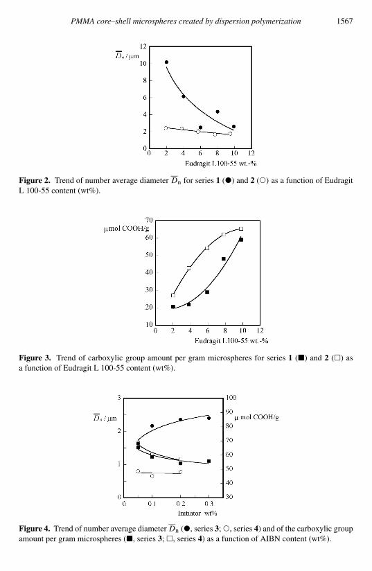
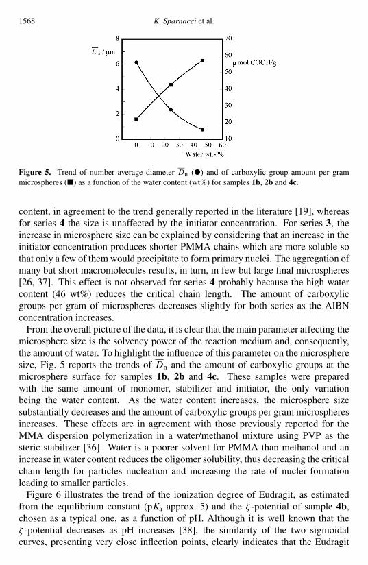
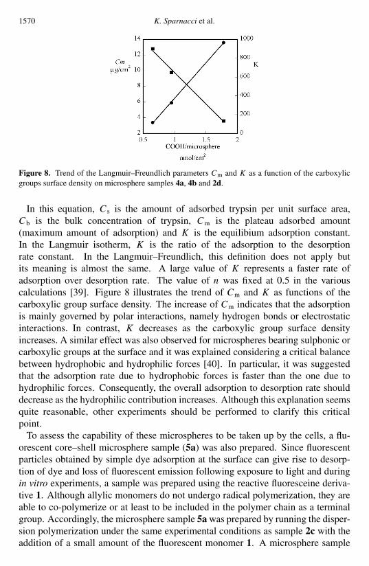


![Hirata et al., J Biotechnol Biomater 2015, 5:3 iotechnooy · PDF file · 2015-10-01Hirata et al., J Biotechnol Biomater 2015, 5:3 ... (TSEs) [2]. PrP is distinguished ... were sliced](https://img.pdfslide.us/doc/110x75/5aa2eba57f8b9a84398daca3/hirata-et-al-j-biotechnol-biomater-2015-53-iotechnooy-et-al-j-biotechnol.jpg)