Embed Size (px)
Citation preview
ISSN 1016-3573
VETERİNER KONTROL MERKEZARAŞTIRMA ENSTİTÜSÜ MÜDÜRLÜĞÜ
Etlik - ANKARA
Cilt/Volume 28 ♦ Sayı/Number 2 ♦ 2017
ETLİK VETERİNERMİKROBİYOLOJİ
DERGİSİ
JOURNAL OF ETLİK VETERINARY MICROBIOLOGYANKARA – TURKEY
Test
AB-0048-T

Adres / AddressVeteriner Kontrol Merkez Araştırma EnstitüsüAhmet Şefik Kolaylı Cad. No 21/21-A06020 Etlik - Ankara / TÜRKİYETel. : +90 312 326 00 90 (8 hat)Faks : +90 312 321 17 55
Web : http://vetkontrol.tarim.gov.tr/merkezE-posta : [email protected]
Yayın Kurulu / Publication Board
Sorumlu Yazı İşleri Müdürü / Managing EditorDr. A. Burak Güngör
Editör / Editor in ChiefDr. Tahsin Onur Kevenk
Bilimsel Kurul / Editorial BoardDr. Erhan AkçayDr. Asiye DakmanDr. Ali ErkurtDr. Elçin GünaydınDr. Filiz Şen
Etlik Veteriner Mikrobiyoloji DergisiCilt/Volume 28 ♦ Sayı/Number 2 ♦ 2017
Journal of Etlik Veterinary MicrobiologyYılda iki kez yayımlanır / Published two times per year
ISSN 1016-3573
SahibiVeteriner Kontrol Merkez Araştırma Enstitüsü Müdürlüğü Adına
Dr. Cevdet YaralıEnstitü Müdür V.
IV
Tasarım ve Baskı / Printing
MMEDİSAN
Medisan Yayinevi Ltd.Şti.Çankırı Cad. 45 / 347 Ulus - Ankara, Türkiye
Tel : +90 312 311 24 26 – 311 00 57 [email protected]
* İsimler soyada göre alfabetik dizilmiştir ve bu sayıda görev alanlar yazılmıştır.
ULAKBİM Yaşam Bilimleri ve Türkiye Atıf Dizini veritabanları kapsamında bulunan “çift hakemli” bir dergidir.
Copyright © Etlik Veteriner Mikrobiyoloji Dergisi 2017, Her hakkı saklıdır / All rights reservedBasım Tarihi / Publishing Date: Aralık / December 2017, Baskı adedi / Circulation: 500
Hakem Listesi / Referee List*
Prof.Dr. İbrahim BALKAYA Atatürk Üniversitesi Veteriner Fakültesi Parazitoloji Anabilim Dalı
Doç.Dr. Yunus Emre BEYHAN Yüzüncü Yıl Üniversitesi Tıp Fakültesi Parazitoloji Anabilim Dalı
Dr. Elvin ÇALIŞKAN GTHB Veteriner Kontrol Merkez Araştırma Enstitüsü Müdürlüğü
Doç.Dr. Yavuz ÇOKAL Bandırma Onyedi Eylül Üniversitesi Bandırma Meslek Yüksekokulu
Dr. Can ÇOKÇALIŞKAN GTHB Şap Enstitüsü Müdürlüğü
Doç.Dr. Alper ÇİFTCİ Ondokuz Mayıs Üniversitesi Veteriner Fakültesi Mikrobiyoloji Anabilim Dalı
Doç.Dr. Gülay ÇİFTÇİ Ondokuz Mayıs Üniversitesi Veteriner Fakültesi Biyokimya Anabilim Dalı
Yrd.Doç.Dr. Gülşen GONCAGÜL Uludağ Üniversitesi Veteriner Fakültesi Mikrobiyoloji Anabilim Dalı
Prof.Dr. Timur GÜLHAN Ondokuz Mayıs Üniversitesi Veteriner Fakültesi Biyokimya Anabilim Dalı
Dr. A. Burak GÜNGÖR GTHB Veteriner Kontrol Merkez Araştırma Enstitüsü Müdürlüğü
Prof.Dr. Ergün KÖROĞLU Fırat Üniversitesi Veteriner Fakültesi Parazitoloji Anabilim Dalı
Yrd.Doc.Dr. Duygu Neval SAYIN İPEK Dicle Üniversitesi Veteriner Fakültesi Parazitoloji Anabilim Dalı
Prof.Dr. Kezban Can ŞAHNA Fırat Üniversitesi Veteriner Fakültesi Viroloji Anabilim dalı
Yrd.Doç.Dr. Serap ÜNÜBOL AYPAK ADÜ Veteriner Fakültesi Bİyokimya Anabilim Dalı
Prof.Dr. Yakup YILDIRIM Mehmet Akif Ersoy Üniversitesi Veteriner Fakültesi Viroloji Anabilim Dalı

V
Etlik Vet Mikrobiyol Derg, http://vetkontrol.tarim.gov.tr/merkez Cilt 28, Sayı 2, Aralık 2017
İçindekiler / Contents
Olgu Sunumu / Case Report
Dual Infection of Sheep Aborted Foetus with Peste des Petits Ruminants Virus and Brucella melitensisAbort Olmuş Koyun Fötusunun Peste des Petits Ruminants Virus ve Brucella melitensis ile İkili EnfeksiyonuMurat Şevik, Yasin Gülcü, Müge Doğan .....................................................................................................61
Co-infection with Border Disease Virus and Brucella melitensis in an Aborted Sheep FoetusBir Koyun Abort Fötusunun Border Disease Virusu ve Brucella melitensis ile KoenfeksiyonuMurat Şevik, Yasin Gülcü, Müge Doğan .....................................................................................................65
Detection of a Mixed Infection of Lumpy Skin Disease Virus and Foot and Mouth Disease Virus in a CalfBir Buzağıda Lumpy Skin Disease Virusu ve Şap Haslığı Virusunun Miks Enfeksiyonunun TespitiMurat Şevik ..................................................................................................................................................69
Araştırma Makalesi / Research Article
İki Leylek (Ciconia ciconia) ve Bir Şahin (Buteo buteo)’de Lucilia sericata’nın Neden Olduğu Yara MiyazıWound Myiasis Caused by Lucilia Sericata in two White Storks (Ciconia ciconia) and in a Common Buzzard (Buteo buteo)Armağan Erdem Ütük, Cem Ecmel Şaki .....................................................................................................73
Comparision of Two Different Vaccination Shedule for Fight with Infectious Bursal Disease in Layer HensYumurtacı Tavuklarda Infectious Bursal Disease ile Mücadelede İki Farklı Aşılama Programının KarşılaştırılmasıAhmet Bildir, Sibel Gür ...............................................................................................................................76
Yaygın Salmonella Serovarlarının Moleküler Tekniklerle TiplendirilmesiMolecular Typing of Common Salmonella SerovarsElçin Günaydın, Selahattin Şen, K. Serdar Diker, Derya Karataş Yeni, Özlem Kardoğan, H. Kaan Müştak, Özlem Şahan ................................................................................................................................................85
Molecular Characterization and Phylogenetic Analysis of Canine Parvovirus 2 in Dogs, Mersin Province, TurkeyTürkiye’de Mersin İlinde Köpeklerde Canine Parvovirus 2’nin Moleküler Karakterizasyonu ve Filogenetik AnaliziEnder Dinçer ................................................................................................................................................96
VI
Etlik Vet Mikrobiyol Derg, http://vetkontrol.tarim.gov.tr/merkez Cilt 28, Sayı 2, Aralık 2017
İçindekiler / Contents
Türkiye’nin Nevşehir İlindeki Atlarda Kistik Ekinokokkoz SeroprevalansıSeroprevalance of Cystic Echinococcosis in Horses in Nevsehir Province of TurkeyArmağan Erdem Ütük, Selçuk Pekkaya, Fatih Kuzugüden, İbrahim Balkaya, Sami Şimşek ...................101
Serological Evidences of West Nile Virus in Domestic Bird Species in the Samsun ProvinceSamsun İlindeki Evcil Kanatlı Türlerinde Batı Nil Virusunun Serolojik BulgularıSinan Pir, Harun Albayrak .........................................................................................................................105
Derleme / Review Article
Ruminantlarda Mycobacterium avium subspecies paratuberculosis Enfeksiyonunun İmmunolojik ÖzellikleriImmunological Characteristics of Mycobacterium avium subspecies paratuberculosis Infection in RuminantsEzgi Şababoğlu, Hülya Türütoğlu ..............................................................................................................109
Viruslar ve TeratogenezTeratogenesis and VirusesCüneyt Tamer, Semra Okur Gümüşova .....................................................................................................115
VII
Etlik Vet Mikrobiyol Derg, http://vetkontrol.tarim.gov.tr/merkez Cilt 28, Sayı 2, Aralık 2017
Etlik Veteriner Mikrobiyoloji Dergisi Yayım Koşulları1. Dergi, T.C. Gıda, Tarım ve Hayvancılık Bakanlığı, Etlik Veteri-ner Kontrol Merkez Araştırma Enstitüsü Müdürlüğü’nün hakemli, bilimsel yayın organı olup, yılda iki defa yayımlanır. Derginin kı-saltılmış adı “Etlik Vet Mikrobiyol Derg” dir.2. Etlik Veteriner Mikrobiyoloji Dergisi’nde veteriner hekimlik alanında yapılan, başka bir yerde yayımlanmamış olan orijinal bi-limsel araştırmalar, güncel derleme, gözlem, kısa bilimsel çalışma-lar ve enstitüden haberler yayımlanır. Derleme şeklindeki yazılar; orijinal olması, en son yenilikleri içermesi, klasik bilgilerin tekrarı olmaması durumunda kabul edilir. Derlemeyi hazırlayan yazarın, o konuda ulusal ya da uluslararası düzeyde orijinal yayın ve araştır-malar yapmış olması koşulu aranır.3. Türkçe ve İngilizce olarak hazırlanacak metinler 12 punto Times New Roman yazı karakterinde, düz metin olarak, çift aralıklı ve kenarlarda 30 mm boşluk bırakılarak, A4 formundaki beyaz kağıda yazılmalıdır. Yazıların tamamı, şekil ve tablolar dahil olmak üzere orijinal bilimsel araştırmalarda 16, derlemelerde 10, gözlemlerde 6 ve kısa bilimsel çalışmalarda 4 sayfayı geçmemelidir.4. Microsoft Word formatındaki metin ile en az 300 dpi çözünürlükteki JPEG formatındaki resim/lerin tamamı [email protected] e-posta adresine gönderilmelidir.5. Türkçe orijinal çalışmalar konu başlığı, yazar/yazarların adları, adresleri, Türkçe özet ve anahtar sözcükler, İngilizce başlık, İngi-lizce özet ve anahtar sözcükler, giriş, materyal ve metot, bulgular, tartışma ve sonuç, teşekkür ve kaynaklar sırası ile hazırlanmalıdır. İngilizce orijinal çalışmalar konu başlığı, yazar/yazarların adları, adresleri, İngilizce özet ve anahtar sözcükler, Türkçe başlık, Türk-çe özet ve anahtar sözcükler, giriş, materyal ve metot, bulgular, tartışma ve sonuç, teşekkür ve kaynaklar şeklinde hazırlanmalıdır. Kısa bilimsel çalışmaların ve derlemelerin başlık ve özet bölümleri orijinal çalışma formatında, bundan sonraki bölümleri ise, derleme-lerde; giriş, metin ve kaynaklar şeklinde, kısa bilimsel çalışmalarda ise bölümlendirme yapılmadan hazırlanmalıdır.6. Orijinal çalışmalar ve gözlemler aşağıdaki sıraya göre düzenle-nerek yazılmalıdır.Başlık, kısa, konu hakkında bilgi verici olmalı ve küçük harflerle yazılmalıdır.Yazar(lar)ın, ad(lar)ı küçük, soyad(lar)ı büyük harflerle yazılmalı ve unvan belirtilmemelidir. ORCID numaraları yazılmalıdır.Özet, Türkçe ve İngilizce olarak, tek paragraf halinde ve en fazla 500 sözcük olmalıdır.Anahtar kelimeler, Türkiye Bilim Terimleri’nden seçilmeli, alfa-betik sıraya göre yazılmalı ve 5 sözcüğü geçmemelidir.Giriş, konu ile ilgili kısa literatür bilgisi içermeli, son paragrafında çalışmanın amacı vurgulanmalı ve iki sayfayı geçmemelidir.Materyal ve Metot, ayrıntıya girmeden, anlaşılır biçimde yazılma-lıdır. Başlıklar kalın, alt başlıklar italik yazı tipiyle belirtilmelidir.Bulgular bölümünde veriler, tekrarlama yapmadan açık bir şekilde belirtilmelidir. Tablo başlıkları tablonun üstünde, şekil başlıkları ise şeklin altında belirtilmelidir.Tartışma ve Sonuç bölümünde, araştırmanın sonucunda elde edi-len bulgular, diğer araştırıcıların bulguları ile karşılaştırılmalı ve literatüre olan katkısı kısaca belirtilmelidir.Teşekkür bölümü, gerekli görülüyorsa kaynaklardan hemen önce belirtilmelidir.Kaynaklar bölümünde, kaynaklar listesi alfabetik ve kronolojik olarak sıralanmalı ve numaralanmalıdır. Metin içerisindeki kaynak, yazar soyadı yazılıp sıra numarası ile; cümle sonunda ise sadece sıra numarası ile köşeli parantez içerisinde yazılmalıdır. Cümle sonunda birden çok kaynak belirtilecek ise kaynak numaraları kü-
çükten büyüğe doğru sıralanmalıdır. Metin içerisinde ikiden çok yazarlı kaynak kullanımlarında ilk yazarın soyadı yazılmalı diğer yazarlar ise “ve ark.” (İngilizce metinlerde “et al.”) kısaltması ile belirtilmelidir. Dergi adlarının kısaltılmasında “Periodical Title Abbreviations: By Abbreviation” son baskısı esas alınmalıdır. Kay-naklar listesinde yazar(lar)ın aynı yıla ait birden fazla yayını varsa, yayın tarihinin yanına “a” ve “b” şeklinde belirtilmelidir.Kaynak yazımı ve sıralaması aşağıdaki gibi yapılmalıdır;Süreli Yayın:Dubey JP, Lindsay DS, Anderson ML, Davis SW, Shen SK, (1992). Induced transplacental transmission of N. caninum in cattle. J Am Vet Med Ass. 201, 709-713.Yazarlı Kitap:Fleiss Jl, (1981). Statistical methods for rates and proportions. Second edition. New York: John Willey and Sons, p.103.Editörlü Kitap:Balows A, Hausler WJ, Herramann Kl, eds., (1990). Manual of Clinical Microbiology. Fifth edition. Washington DC: IRL Press, p.37.Editörlü Kitapta Bölüm:Bahk J, Marth EH, (1990). Listeriosis and Listeria monocytogenes. Cliver DD. eds. Foodborne Disease. Academic press Inc, San Di-ego. p.248-256.Kongre Bildirileri:Çetindağ M, (1994). Pronoprymna ventricosa, a new digenic trem-atoda from the Alosa fallax in Turkey. Eighth International Con-gress of Parasitology (ICOPA VIII), October, 10-14, İzmir-Turkey.Tezler:Aksoy E, (1997). Sığır Vebası hastalığının histolojik ve immuno-peroksidaz yöntemle tanısı üzerine çalışmalar. Doktora Tezi, AÜ Sağlık Bilimleri Enstitüsü, Ankara.Anonim:Anonim, (2009). Contagious equine metritis. Erişim adresi: http://www.cfsph.iastate.edu/Factsheets/pdf, Erişim tarihi: 17.10.2009.Peter AT (2009). Abortions in dairy cows. Erişim adresi: http://www.wcds.afns.ualberta.ca.htm, Erişim tarihi: 14.11.2009.Yazışma adresi, çok yazarlı çalışmalarda yazışma adresi olarak yazarlardan sadece birinin adı/soyadı, adresi ve e-posta adresi ça-lışmanın sonunda belirtilmelidir.7. Latince cins ve tür isimleri italik yazı tipi ile yazılmalıdır. Tüm ölçüler SI (Systeme Internationale)’ye göre verilmelidir.8. Dergide yayımlanmak üzere gönderilen makaleler tüm yazarlar tarafından imzalanan “Yayın Hakkı Devri Sözleşmesi” ve başvu-ruya ilişkin bir dilekçe ile birlikte gönderilmelidir. Yayımlanması uygun görülen çalışmalar, istendiğinde Yayım Komitesi’nin basıma ilişkin kararı, yazar(lar)ına bildirilir.9. Etlik Veteriner Mikrobiyoloji Dergisi’nde yayımlanacak olan, hayvan deneylerine dayalı bilimsel çalışmalarda “Etik Kurul Onayı Alınmıştır” ifadesi aranır.10. Gönderilen yazıların basım düzeltmeleri orijinal metne göre ya-pıldığından, yazıların her türlü sorumluluğu yazarlara aittir.11. Ürünlerin ticari adları ile karşılaştırılmalarına yönelik araştır-malar derginin ilgi kapsamı dışındadır.12. Araştırmaya konu olan maddelerin ve ürünlerin ticari adları kullanılmamalıdır.13. Şayet varsa araştırmanın desteklendiği kurum adı ve proje nu-marası belirtilmelidir. 14. Dergiye gönderilen yazılar geliş tarihine göre yayımlanır.15. Yayımlanmayan yazılar, yazarına iade edilmez.
Yazarlara Bilgi / Instructions for Authors
VIII
Etlik Vet Mikrobiyol Derg, http://vetkontrol.tarim.gov.tr/merkez Cilt 28, Sayı 2, Aralık 2017
Journal of Etlik Veterinary Microbiology Publication Conditionsnumbers at the end of sentence. If the reference is more than two authors, the surname of the first author should be written and other authors should be mentioned with the abbreviation of “et al.”. For the abbreviation of journals, the latest edition of the “Periodical Title Abbreviations: By Abbreviation” should be taken as basis. If the author(s) have more than one publication within the same year, besides the publication date, it should be mentioned as “a” and “b” in the list of references.The writing of the references and their alignment should be as in the following examples.For articles:Dubey JP, Lindsay DS, Anderson ML, Davis SW, Shen SK, (1992). Induced transplacental transmission of N. caninum in cattle. J Am Vet Med Ass. 201, 709-713.For books:Fleiss Jl, (1981). Statistical methods for rates and proportions. Second edition. New York: John Willey and Sons, p.103.For edited books:Balows A, Hausler WJ, Herramann Kl, eds., (1990). Manual of Clinical Microbiology. Fifth edition. Washington DC: IRL Press, p.37.For chapter in edited books:Bahk J, Marth EH, (1990). Listeriosis and Listeria monocytogenes. Cliver DD. eds. Foodborne Disease. Academic press Inc, San Di-ego. p.248-256.For congress papers:Çetindağ M, (1994). Pronoprymna ventricosa, a new digenic trem-atoda from the Alosa fallax in Turkey. Eighth International Con-gress of Parasitology (ICOPA VIII), October, 10-14, İzmir-Turkey.For dissertations:Aksoy E, (1997). Sığır Vebası hastalığının histolojik ve İmmuno-peroksidaz yöntemle tanısı üzerine çalışmalar. PhD Thesis, Ankara University Institute of Health Sciences, Ankara.Corresponding address, in multiple-author studies, as a corre-spondence address, only one of the authors’ name/surname, address and e-mail should be mentioned at the end.7. Genus and species names in Latin should be written in italic. All measures should be given according to the SI (Systeme Internatio-nale) units.8. The articles that are sent to be published in the journal should be sent with a covering letter and “Publication Rights Transfer Agree-ment” signed by all of the authors. The selected articles for the publication, and if asked for, the decision of the editorial commit-tee concerning the publication, are declared to the article’s author/authors.9. The wording of “Ethical Commission Permission is obtained” should appear in scientific studies based on animal experiments, which will be published in the Journal of Etlik Veterinary Micro-biology.10. As the edition of the sent articles are done in accordance with the original text, all responsibility of the articles bear on the au-thors.11. Researches that aim at comparisons of the products with their commercial names are out of the journal’s theme scope.12. The trademarks of materials and products that are subject of the research should not be mentioned.13. If the research is supported by a foundation, name of the foun-dation and project number must be mentioned.14. The articles that are sent to the journal are published in line with their coming date.15. Unpublished papers are not returned to their author.
1. The Journal is a refereed, scientific publication of Republic of Turkey Ministry of Food, Agriculture and Livestock, Directorate of Etlik Veterinary Control Central Research Institute and is published two issues in a year. The abbreviation of the journal is “J Etlik Vet Microbiol”.2. In the Journal of Etlik Veterinary Microbiology, original research articles, actual reviews, case reports, short communications on the issue of veterinary medicine whose one part or whole have not been published in any other place before, and news from the institute are published. The review articles will be accepted only if they are original, actual and not repeating the classical knowledge. The au-thor of the review is asked to possess original publications or re-searches on the subject at national or international levels.3. Manuscripts that will be prepared in Turkish and English should be typed as a full text, on A4 paper with 12 pt, in Times New Ro-man typing character, double-spaced and with 30 mm space in both sides of the paper. Manuscripts including figures and tables should not exceed 16 pages for original research articles, 10 pages for re-views, 6 pages for case reports and 4 pages for short communica-tions.4. Manuscript written in Microsoft Word format and figures in JPEG format at minimum 300 dpi resolution should be submitted to [email protected]. Original research articles and case reports should include in fol-lowing rank: title, name(s) of the author(s), their addresses, abstract and key words in English, title, abstract and key words in Turkish, introduction, material and method, findings, discussion and conclu-sion, acknowledgements and references. In short communications and reviews, divisions except summaries should be omitted.6. Original research articles and case reports should be arranged and composed as in the following.Title should be brief, explanatory and written in small caps. Explanation(s) about the study should be written as footnotes.Author(s) should be mentioned by their names and surnames; their surnames should be written in capital letters and author(s) title should not be mentioned. ORCID numbers should be written.Summary should be in Turkish and English, single paragraph and composed of at most about 500 words.Key words must be selected from Medical Subject Headings, should be written in alphabetical order and should not exceed 5 words. Introduction not exceeding two pages should include a short re-view of the literature related with the subject and in the end para-graph; the aim of the study should be mentioned.Material and Method should be written in an essential and com-prehensible manner without getting into details. Subtopics should be mentioned first in bold and after in italic type.Findings should be shortly explained and data should not be re-peated within the text. Legends should be indicated at the top of each table, whereas should be indicated at the bottom of each figure and print. Vertical lines are not allowed in tables.Discussion and Conclusion must include the evaluation and com-parison of results with other researchers’ findings. The study’s con-tributions to the existing literature should also be explained briefly.Acknowledgements must be indicated before references if neces-sary.References should be listed alphabetically and chronologically by numbers. In the body of text, reference must be shown by author’s surname and list number or only by list number within square pa-renthesis. If there is more than one reference that refers to the same issue, these should be arranged by smallest to biggest reference list
Yazarlara Bilgi / Instructions for Authors
IX
Etlik Vet Mikrobiyol Derg, http://vetkontrol.tarim.gov.tr/merkez Cilt 28, Sayı 2, Aralık 2017
Yayın Hakkı Devri SözleşmesiEtlik Veteriner Mikrobiyoloji Dergisi - Ankara
Aşağıda başlığı bulunan ve yazarları belirtilen makalenin tüm sorumluluğu Etlik Veteriner Mikrobiyoloji Dergisi Yayın Komisyonu Başkanlığı’na ulaşıncaya kadar yazar/larına aittir.Yayının adı: ...............................................................................................................................................................................................................................................................................................................................Yazar/ların ad/ları: .....................................................................................................................................................................................................................................................................................................................Aşağıda isim ve imzaları bulunan yazarlar; yayınlamak üzere gönderdikleri makalenin orijinal olduğunu, daha önce başka bir dergiye yayınlanmak üzere gönderilmediğini ve kısmen ya da tamamen yayınlanma-dığını, gerekli düzeltmelerle birlikte her türlü yayın hakkının, yazının yayımlanmasından sonra Etlik Vete-riner Mikrobiyoloji Dergisi’ne devrettiklerini kabul ederler. Yayımlanmak üzere gönderilen bu makalenin tüm sorumluluğunu da yazar/lar üstlenmektedir.Yukarıdaki makalenin tüm hakları Etlik Veteriner Mikrobiyoloji Dergisi’ne devredilmiştir.Yazar ad/ları İmza Tarih........................................................................................................................................................................................................................................................................................................................................................................................................................................................................................................................................................................................................................................................................................................Yazışma Adresi: ........................................................................................................................................................................................................................................................................................................................
Copyright ReleaseJournal of Etlik Veterinary Microbiology Ankara - TURKEY
The undersigned authors release Journal of Etlik Veterinary Microbiology from all responsibility concern-ing the manuscript entitled;Title of paper: ............................................................................................................................................................................................................................................................................................................................Authors names: .........................................................................................................................................................................................................................................................................................................................Upon its submission to the publishing commission of the Journal of Etlik Veterinary Microbiology.The undersigned author/s warrant that the article is original, is not under consideration by another journal, has not been previously published or that if has been published in whole or in part, any permission neces-sary to publish it in the above mentioned journal has been obtained and provided to the Journal of Etlik Veterinary Microbiology. We sign for and accept responsibility for releasing this material.Copyright to the above article is hereby transferred to the Journal of Etlik Veterinary Microbiology, effec-tive upon acceptance for publication.To be signed by all author/sAuthors names Signature Date........................................................................................................................................................................................................................................................................................................................................................................................................................................................................................................................................................................................................................................................................................................Correspondence Address: ..........................................................................................................................................................................................................................................................................................................
Yazarlara Bilgi / Instructions for Authors

Yazışma adresi / Correspondence: Murat Şevik, Department of Molecular Microbiology, Veterinary Control Institute, Konya, Turkey E-posta: [email protected]
Etlik Vet Mikrobiyol Derg, 2017; 28 (2): 61-64 Olgu Sunumu / Case Report
Dual Infection of Sheep Aborted Foetus with Peste des Petits Ruminants Virus and Brucella melitensis
Murat Şevik1, Yasin Gülcü2, Müge Doğan1
1 Department of Molecular Microbiology, Veterinary Control Institute, Konya, Turkey2 Department of Bacteriology, Veterinary Control Institute, Konya, Turkey
Geliş Tarihi / Received: 18.09.2017, Kabul Tarihi / Accepted: 01.11.2017
Abstract: In this study, we investigated the potential roles of Brucella melitensis and peste des petits ruminants virus (PPR) virus (PPRV) infections in a case of sheep abortion. Samples were collected from PPR-suspected ewe and its aborted foetus from a sheep flock in the Antalya Province in the Mediterranean region of Turkey in 2016. The presence of Brucella spp. directly assessed by bacterial isolation and detection of PPRV was carried by real time RT-PCR. Genetic characterization of the PPRV field isolates was conducted by sequencing the fusion (F) gene of PPRV. Brucella strain was isolated from the samples of aborted sheep foetus, and it was identified as Brucella melitensis by biochemical char-acteristics, agglutination with monospecific A and M sera. PPRV RNA was detected in samples of PPR-suspected ewe and its foetus. Phylogenetic analysis showed that the field isolate of PPRV obtained in this study was clustered within lineage IV. To the best of our knowledge, this is the first report on the dual infection of aborted sheep foetus with PPRV and Brucella melitensis.Key words: Sheep foetus, Peste des petits ruminants virus, Genetic characterization, F gene, Brucella melitensis
Abort Olmuş Koyun Fötusunun Peste des Petits Ruminants Virus ve Brucella melitensis ile İkili Enfeksiyonu
Özet: Bu çalışmada bir koyun abort vakasında, koyun ve keçi vebası virusu (PPRV) ile Brucella melitensis’in potansi-yel rolleri araştırılmıştır. Çalışmada kullanılan örnekler, 2016 yılında Türkiye’nin Akdeniz Bölgesinde yer alan Antalya İlindeki bir koyun işletmesindeki PPR şüpheli bir koyun ve bu koyunun fötusundan elde edilmiştir. Brucella spp. varlığı bakteriyel izolasyon, PPRV ise real time RT-PCR yöntemi ile araştırılmıştır. PPRV’unun genetik karakterizasyonu, PPRV’unun füzyon (F) geninin sekans analizi ile gerçekleştirilmiştir. Abort koyun fötusundan izole edilen Brucella suşu biyokimyasal karakteri ve monospesifik A ve M serumları ile aglütinasyonuna bağlı olarak Brucella melitensis olarak identifiye edilmiştir. PPR şüpheli koyun ve fötusunda PPRV tespit edilmiştir. Filogenetik analiz sonucu, bu çalışmada izole edilen PPRV saha suşunun lineage IV’de yer aldığı belirlenmiştir. Bizim bildiğimiz kadarıyla, bu çalışma koyun fötusunun PPRV ve Brucella melitensis ile ikili enfeksiyonu hakkındaki ilk rapordur.Anahtar Kelimler: Koyun fötus, Koyun ve keçi vebası virusu, Genetik karakterizasyon, F gen, Brucella melitensis
IntroductionPeste des petits ruminants (PPR) is a highly conta-gious disease of small ruminants, which is charac-terised by high fever, pneumonia and enteritis. The causative agent, peste des petits ruminants virus (PPRV) belongs to the Morbillivirus genus of the Paramyxoviridae family [6]. Transmission of PPRV mainly occurs during close contact [3]. However, vertical transmission of PPRV has been reported [9,16].
B. melitensis, the main etiological agent of bru-cellosis in small ruminants, is the most important and pathogenic Brucella spp. with a worldwide distribution [14]. B. melitensis is usually transmit-ted both vertically and horizontally. It can cause
abortions and stillbirths [11]. Furthermore, border disease virus (BDV) can be one of the causes of abortion in small ruminants [12]. The current study was conducted to investigate the potential roles of B. melitensis, PPRV and BDV in a case of sheep abortion.
Material and MethodsCollection of samplesAn aborted sheep foetus was submitted to the Konya Veterinary Control Institute from a sheep flock in the Antalya Province in the Mediterranean region of Turkey in 2016. According to farmer’ report, fe-ver, ocular and nasal discharge and nodular lesions around the mouth were observed in the ewe before

62 Şevik M. et al. Dual Infection of Sheep Aborted Foetus with Peste des Petits Ruminants Virus and B. melitensis
Etlik Vet Mikrobiyol Derg, http://vetkontrol.tarim.gov.tr/merkez Cilt 28, Sayı 2, 2017, 61-64
abortion, and abortions occurred at 2 months of ges-tation. The rate of abortion in this flock was 25% (20/80). Foetal stomach contents and liver of the aborted foetus were collected. Furthermore, nodular lesions of ewe that had aborted and internal organ specimens (spleen, lung and liver) of aborted foetus were collected for PPRV detection.
Bacteriological examinationsSamples from stomach contents and liver of the aborted foetus were inoculated onto Farrell’s me-dium (5-10% v/v sterile inactivated horse serum) (Oxoid, SR0035) supplemented with Brucella se-lective supplement (Oxoid, SR083A). After incuba-tion of the plate at 37°C and 5% CO2 conditions for 7 days, the observed colonies were investigated and identified as Brucella spp. by morphological, cul-tural and characteristics. The strain was biotyped by agglutination with monospecific A and M antisera [11].
Samples were also inoculated onto Campylobacter agar base with selective supplement (Oxoid, SR069E) and 7% defibrinated sheep blood and MacConkey agar for isolation of other bacterial agents.
RNA extraction and real-time RT-PCRViral RNA extraction was carried out from the nodular lesions of ewe that had aborted and organ specimens of aborted foetus using a QIAamp Cador Pathogen Mini Kit (Qiagen, Hilden, Germany) in a QIAcube (Qiagen, Hilden, Germany). Real-time RT-PCR was performed using PPRV nucleocapsid protein (N) gene specific primers and probe de-signed by Batten et al. [1].
Furthermore, aborted foetus samples were also tested by real-time RT-PCR for detection of BDV. The protocol described by La Rocca and Sandvik [10] was used for detection of BDV RNA.
RT-PCR and sequencing of PCR productsOne-step RT-PCR was performed with primers that amplified 448 bp of the fusion (F) protein gene of PPRV [5]. PCR products were purified from gels and sequenced. Sequence analysis was performed by using ChromasPro software (Version 1.7.5, Technolysium Ltd.). Phylogenetic tree was con-
structed for the F gene of PPRV with additional se-quences from GenBank.
ResultsBacteriological isolationBrucella was isolated from stomach contents and liver of the aborted foetus. Brucella strain was iden-tified as B. melitensis by biochemical characteristics and agglutination with monospecific A and M anti-sera. Other bacterial agents were not detected in the investigated foetus.
Detection of PPRVPPRV RNA was detected in spleen, lung and liver samples from foetus and nodular lesions of ewe that had aborted. However, BDV RNA was not detected in the investigated foetus samples.
Sequence analysesAnalysis of the PPRV F gene sequences revealed the homology between the two isolates in the pres-ent study was 100%, whereas the similarity among the field isolate in this study and previously char-acterized Turkish isolates ranged from 87.6% to 100%. The deduced amino acid homology among the field isolate and previously characterised PPRV isolates ranged between 96% and 100%.
Discussion
B. melitensis is the main aetiological agent of sheep and goat brucellosis in Turkey. Previous investiga-tions of abortion cases in sheep in different regions of Turkey have shown that B. melitensis is responsible for about 20-31% of sheep abortions [2,8]. Stomach contents, spleen, liver, lung and foetal membranes are useful for diagnosis of B. melitensis in aborted foetuses [11]. In this study, Brucella was isolated from stomach contents and liver of the aborted foe-tus, and identified as B. melitensis. However, Ilhan et al. [8] reported that using stomach contents for diagnosis is better than using other foetal materials. The rate of abortion in B. melitensis positive flock was 25% (20/80). This rate is consistent with the findings of previous studies in which it has been re-ported that rate of abortion in B. melitensis positive flocks ranged between 6% and 45% [4,7].
Şevik M. et al. Dual Infection of Sheep Aborted Foetus with Peste des Petits Ruminants Virus and B. melitensis 63
Etlik Vet Mikrobiyol Derg, http://vetkontrol.tarim.gov.tr/merkez Cilt 28, Sayı 2, 2017, 61-64
PPRV can cause abortion in pregnant ani-mals [16]. In this study PPRV RNA was detected in spleen, lung and liver samples from foetus. This finding in agreement with previous report that sug-gest lung, liver, spleen and mesenteric lymph node samples can equally be used for PPR virus detec-tion [15]. To the best of our knowledge, this is the first report on the dual infection of aborted sheep
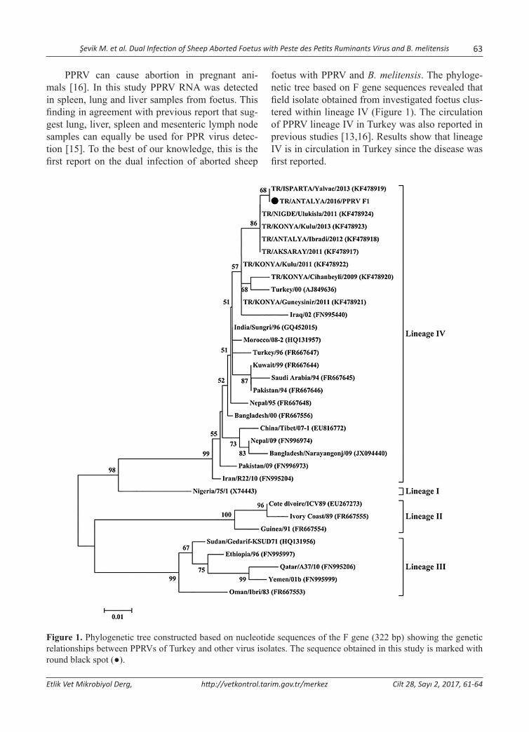
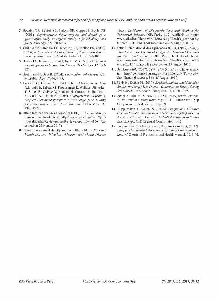
foetus with PPRV and B. melitensis. The phyloge-netic tree based on F gene sequences revealed that field isolate obtained from investigated foetus clus-tered within lineage IV (Figure 1). The circulation of PPRV lineage IV in Turkey was also reported in previous studies [13,16]. Results show that lineage IV is in circulation in Turkey since the disease was first reported.
Figure 1. Phylogenetic tree constructed based on nucleotide sequences of the F gene (322 bp) showing the genetic relationships between PPRVs of Turkey and other virus isolates. The sequence obtained in this study is marked with round black spot (●).
64 Şevik M. et al. Dual Infection of Sheep Aborted Foetus with Peste des Petits Ruminants Virus and B. melitensis
Etlik Vet Mikrobiyol Derg, http://vetkontrol.tarim.gov.tr/merkez Cilt 28, Sayı 2, 2017, 61-64
The results of this study indicate that dual in-fection with PPRV and B. melitensis can occur in abortion cases. Therefore, PPRV should be taken into consideration in abortion cases in endemic ar-eas.
References1. Batten CA, Banyard AC, King DP, Henstock MR, Edwards
L, Sanders A, Buczkowski H, Oura CC, Barrett T, (2011). A real time RT-PCR assay for the specific detection of Peste des petits ruminants virus. J Virol Methods. 171, 401-404.
2. Büyükcangaz E, Şen A, Kahya S, (2009). Isolation and bio-typing of Brucella melitensis from aborted sheep and goat foetuses. Turk. J Vet Anim Sci. 33, 311-316.
3. Couacy-Hymann E, Bodjo SC, Koffi MY, Kouakou C, Danho T, (2009). The early detection of peste-des-petits-ruminants (PPR) virus antigens and nu-cleic acid from experimentally infected goats using RT-PCR and immunocapture ELISA techniques. Res Vet Sci. 87, 332-335.
4. European Commission, Health & Consumers Directorate-General (2012). Eradication programme for Sheep and Goat Brucellosis (B. Melitensis). Erişim adresi: https://ec.europa.eu/food/sites/food/files/safety/docs/cff_animal_vet-progs_2012_dec-2011-807-ec_ov-cap-brucellosis_grc.pdf, Erişim tarihi: 08.08.2017
5. Forsyth MA, Barrett T, (1995). Evaluation of polymerase chain reaction for the detection and characterisation of rinderpest and peste des petits ruminants viruses for epide-miological studies. Virus Res. 39, 151-163.
6. Gibbs PJE, Taylor WP, Lawman MP, Bryant J, (1979). Classification of the peste des petits ruminants virus as the fourth member of the genus Morbillivirus. Intervirology 11, 268-274.
7. Hawari AD, (2012). Epidemiological Studies, Seroprevalance and Some Risk Factors of Brucellosis in Sheep and Goats
in the South Province of West Bank. Asian J Anim Vet Adv. 7, 535-539.
8. Ilhan Z, Solmaz H, Aksakal A, Gülhan T, Ekin IH, Boynukara B, (2007). Comparison of PCR assay and bacteriological culture method for the detection of Brucella melitensis in stomach content samples of aborted sheep fetuses. Dtsch Tierarztl Wochenschr. 114, 460-464.
9. Kul O, Kabakci N, Ozkul A, Kalender H, Atmaca HT, (2008). Concurrent peste des petits ruminants virus and pestivirus infection in stillborn twin lambs. Vet Pathol. 45, 191-196.
10. La Rocca SA, Sandvik T, (2009). A short target real-time RT-PCR assay for detection of pestiviruses infecting cattle. J Virol Methods. 161, 122-127.
11. Office International des Epizooties (OIE) (2016). Brucellosis (Brucella abortus, B. melitensis and B. suis). Erişim ad-resi: http://www.oie.int/fileadmin/Home/eng/Health_stan-dards/tahm/2.01.04_BRUCELLOSIS.pdf, Erişim tarihi: 24.10.2017
12. Office International des Epizooties (OIE) (2017). Border Disease. Erişim adresi: http://www.oie.int/fileadmin/Home/eng/Health_standards/tahm/2.07.01_BORDER_DIS.pdf, Erişim tarihi: 24.10.2017
13. Ozkul A, Akca Y, Alkan F, Barrett T, Karaoglu T, Dagalp SB, Anderson J, Yesilbag K, Cokcaliskan C, Gencay A, Burgu I, (2002). Prevalence, distribution, and host range of peste des petits ruminants virus, Turkey. Emerg Infect Dis. 8, 708-712.
14. Pappas G, Akritidis N, Bosilkovski M, Tsianos E, (2005). Medical progress Brucellosis. N Engl J Med. 352, 2325-2367.
15. Şevik M, (2014). Molecular Detection of Peste des Petits Ruminants Virus from Different Organs/Tissues of Naturally Infected Animals. Kafkas Univ Vet Fak Derg. 20, 165-168.
16. Şevik M, Sait A, (2015). Genetic characterization of peste des petits ruminants virus, Turkey, 2009-2013. Res Vet Sci. 101, 187-195.
Yazışma adresi / Correspondence: Murat Şevik, Department of Molecular Microbiology, Veterinary Control Institute, Konya, Turkey E-posta: [email protected]
Etlik Vet Mikrobiyol Derg, 2017; 28 (2): 65-68 Olgu Sunumu / Case Report
Co-infection with Border Disease Virus and Brucella melitensis in an Aborted Sheep Foetus
Murat Şevik1, Yasin Gülcü2, Müge Doğan1
1 Department of Molecular Microbiology, Veterinary Control Institute, Konya, Turkey2 Department of Bacteriology, Veterinary Control Institute, Konya, Turkey
Geliş Tarihi / Received: 18.09.2017, Kabul Tarihi / Accepted: 10.11.2017
Abstract: In this study, we investigated the potential roles of border disease virus (BDV) and Brucella melitensis infec-tions in a case of sheep abortion. Internal organ specimens from aborted sheep foetus and EDTA whole blood sample from mother of the foetus were collected from a sheep flock in the Konya Province in the Central Anatolia region of Turkey in 2017. The presence of Brucella spp. directly assessed by bacterial isolation and detection of BDV was car-ried by real time RT-PCR. Genetic characterization of the BDV field isolate was conducted by sequencing the 5’- end untranslated region (UTR) region of BDV. Brucella strain was isolated from the samples of aborted sheep foetus, and it was identified as Brucella melitensis by biochemical characteristics, agglutination with monospecific A and M sera. BDV RNA was detected in EDTA whole blood sample and aborted sheep foetus. Phylogenetic analysis in 5’-UTR re-gion allocated the field isolate of BDV obtained in this study into BDV-7 genotype. To the best of our knowledge, this is the first report on the dual infection of aborted sheep foetus with BDV and Brucella melitensis.Key words: Abortion, Border disease virus, Genetic characterization, Brucella melitensis, Sheep
Bir Koyun Abort Fötusunun Border Disease Virusu ve Brucella melitensis ile Koenfeksiyonu
Özet: Bu çalışmada, bir koyun abort vakasında border disease virusu (BDV) ve Brucella melitensis enfeksiyonlarının potansiyel rolleri araştırılmıştır. İki bin on yedi yılında Türkiye’nin İç Anadolu Bölgesinde yer alan Konya İlindeki bir koyun işletmesinden, bir abort koyun fötusuna ait iç organ örnekleri ve fötusun annesinden EDTA’lı tam kan örneği elde edilmiştir. Brucella spp. varlığı bakteriyel izolasyon, BDV ise real time RT-PCR yöntemi ile araştırılmıştır. BDV’unun genetik karakterizasyonu, 5’- translate olmayan bölge sonunun (UTR) sekans analizi ile gerçekleştirilmiştir. Abort ko-yun fötusundan izole edilen Brucella suşu biyokimyasal karakteri ve monospesifik A ve M serumları ile aglütinasyonu-na bağlı olarak Brucella melitensis olarak identifiye edilmiştir. Koyun abort fötusunda ve EDTA’lı kan örneğinde BDV tespit edilmiştir. Filogenetik analiz sonucu, bu çalışmada izole edilen BDV saha suşunun BDV-7 genotipinde olduğu belirlenmiştir. Bizim bildiğimiz kadarıyla, bu çalışma koyun fötusunun BDV ve Brucella melitensis ile birlikte enfek-siyonu hakkındaki ilk rapordur.Anahtar Kelimler: Yavru atma, Border disease virus, Genetik karakterizasyon, Brucella melitensis, Koyun
Introduction
Border disease (BD) is a reproductive disease of sheep, and occasionally seen in goats. The clinical manifestations of the disease are infertility, abor-tion, mummified foetuses, stillbirths, and the birth of ‘hairy-shaker’ lambs and persistent infections of the offspring [9]. The causative agent of disease, border disease virus (BDV), classified in the genus Pestivirus of the Flaviviridae family, and is close-ly related to bovine viral diarrhea viruses (BVDV 1, 2) and classical swine fever virus (CSFV) [6]. BDV can also infect cattle, chamois and pigs [1,8]. Transmission of BDV mainly occurs by horizontal
and vertical routes, and weak lambs can be persis-tently infected (PI) [2].
Ovine brucellosis is another economically im-portant disease of small ruminants that causes re-productive problems such as infertility and abor-tions. B. melitensis is the main etiological agent of brucellosis in small ruminants. The main clinical signs of B. melitensis infection in small ruminants are abortion and stillbirths, which usually occur during the last two months of gestation following infection [3]. The current study was conducted to investigate occurrence of BDV and B. melitensis in the case of small ruminant abortion.
66 Şevik M et al. Co-infection with Border Disease Virus and Brucella melitensis in an Aborted Sheep Foetus
Etlik Vet Mikrobiyol Derg, http://vetkontrol.tarim.gov.tr/merkez Cilt 28, Sayı 2, 2017, 65-68
Material and MethodsCollection of samplesAn aborted sheep foetus was submitted to the Konya Veterinary Control Institute from a sheep flock in the Konya Province in the Central Anatolia region of Turkey in 2017. According to farmer’ report, flock had a history of barren ewes, birth of small weak lambs with hairy fleeces, and abortions occurred at 2 to 3 months of gestation. The rate of abortion in this flock was 15% (24/160). Foetal stomach con-tents and liver of the aborted foetus were collected. Furthermore, whole blood sample from mother of the foetus and internal organ specimens of aborted foetus were collected for BDV detection.
Bacteriological examinationsSamples from stomach contents and liver of the aborted foetus were inoculated onto Farrell’s me-dium (5-10% v/v sterile inactivated horse serum) (Oxoid, SR0035) supplemented with Brucella se-lective supplement (Oxoid, SR083A). After incu-bation of the plate at 37°C and 5% CO2 conditions for 7 days, the observed colonies were investigated and identified as Brucella spp. by morphological, cultural and characteristics. The strain was bio-typed by agglutination with monospecific A and M antisera. Samples were also inoculated onto Campylobacter agar base with selective supplement (Oxoid, SR069E) and 7% defibrinated sheep blood and MacConkey agar for isolation of other bacterial agents.
RNA extraction and reverse transcription-polymerase chain reaction (RT-PCR)Viral RNA extracted from the buffy coat cells from whole blood sample and organ specimens of abort-ed foetus using a QIAamp Cador Pathogen Mini Kit (Qiagen, Hilden, Germany). A quantitative real-time RT-PCR described by La Rocca and Sandvik [7] was used to detect BDV RNA. Amplification of part of the 5’- end untranslated region (UTR) was carried out for samples in one step RT-PCR using primers 324 and 326 [11].
Sequence and phylogenetic analysisPCR products were purified from gels with a High Pure PCR Product Purification Kit (Roche Diagnostics, Indianapolis, USA), and sequenced on
an ABI 3130xl DNA Analyser (Applied Biosystems, USA). Phylogenetic tree was constructed with the programme MEGA software version 6, based on the evolutionary distances between different sequences calculated by Kimura two-parameter model. The confidence of the neighbour-joining tree was as-sessed by bootstrapping, using 1000 replicates, and only values above 50% are reported.
ResultsBacteriological isolationIn this study, Brucella was isolated from aborted sheep foetus. Brucella strain was identified as B. melitensis by biochemical characteristics and ag-glutination with monospecific A and M antisera. Other bacterial agents were not detected in the in-vestigated foetus.
Detection of BDVBDV RNA was detected in the investigated foetus and EDTA whole blood sample from mother of the foetus
Sequence analysesA 100% level of identity was observed between the deduced amino acid sequences of the two isolates, from foetus and its mother, in the present study, whereas the similarity with sequences from differ-ent regions ranged from 67.8% to 96%, lowest with United States isolate (890) highest with Turkish iso-late (Aydin-04).
Discussion
In this study, Brucella was isolated from aborted sheep foetus, and identified as B. melitensis. The rate of abortion in B. melitensis positive flock was 15% (24/160). This rate is consistent with the find-ings of previous studies in which it has been re-ported that rate of abortion in B. melitensis positive flocks ranged between 6% and 45% [4,5].
Border disease virus can cause abortion in pregnant small ruminants [9]. In this study BDV RNA was detected in the investigated foetus. To the best of our knowledge, this is the first report on the dual infection of aborted sheep foetus with BDV and B. melitensis. It has been reported that foetal death may occur at any stage of gestation, but is
Şevik M et al. Co-infection with Border Disease Virus and Brucella melitensis in an Aborted Sheep Foetus 67
Etlik Vet Mikrobiyol Derg, http://vetkontrol.tarim.gov.tr/merkez Cilt 28, Sayı 2, 2017, 65-68
more common during the first 2 months of gesta-tion [12]. In this study abortion occurred at 2 to 3 months of gestation. This situation can be explained by the period of infection, immune status of the host and the virulence of virus.
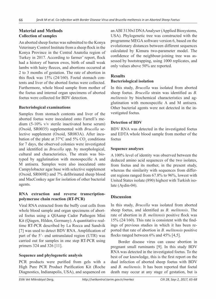
Phylogenetic analysis has been used to deter-mine the subgenotypes of field isolates from differ-ent areas of the world. The most frequent genetic
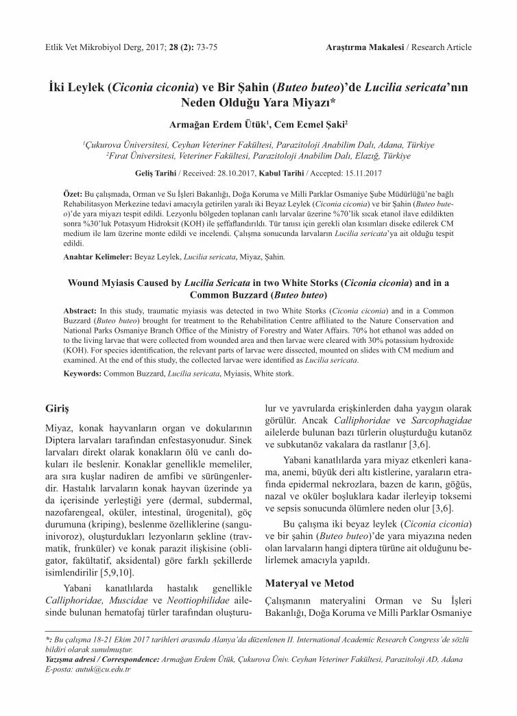
classification is based on a comparison of nucleo-tide sequences from the 5’UTR [1,10]. The phylo-genetic analysis of 5’UTR sequences typed the field isolate in this study as BDV and clustered within the BDV-7 isolates together previously characterized Turkish isolates (Figure 1). The circulation of BDV-7 genotype in Turkey was also reported in previous study [10]. Results show that BDV-7 genotype is in circulation in Turkey.
Figure 1. Phylogenetic tree constructed based on nucleotide sequences of the 5’UTR region (246 bp) showing the genetic relationships between BDVs of Turkey and other virus isolates. The sequence obtained in this study is marked with round black spot (●) and previous Turkish isolates are marked with black triangle (▲).
The results of this study indicate that dual in-fection with BDV and B. melitensis can occur in small ruminant abortion cases. BDV and B. meli-tensis infections cause important economic losses
due to reproductive failure in affected animals. Therefore, abortion cases should be examined for these two diseases.

68 Şevik M et al. Co-infection with Border Disease Virus and Brucella melitensis in an Aborted Sheep Foetus
Etlik Vet Mikrobiyol Derg, http://vetkontrol.tarim.gov.tr/merkez Cilt 28, Sayı 2, 2017, 65-68
References1. Becher P, Orlich M, Shannon AD, Horner G, König M, Thiel
HJ, (1997). Phylogenetic analysis of pestiviruses from do-mestic and wild ruminants. J Gen Virol. 78, 1357-1366.
2. Cabezón O, Rosell R, Velarde R, Mentaberre G, Casas-Díaz E, Lavín S, Marco I, (2010). Border disease virus shed-ding and detection in naturally infected Pyrenean chamois (Rupicapra pyrenaica). J Vet Diagn Invest. 22, 744-747.
3. Díaz Aparicio E, (2013). Epidemiology of brucellosis in do-mestic animals caused by Brucella melitensis, Brucella suis and Brucella abortus. Rev Sci Tech. 32, 43-51, 53-60.
4. European Commission, Health & Consumers Directorate-General (2012). Eradication programme for Sheep and Goat Brucellosis (B. Melitensis). Erişim adresi: https://ec.europa.eu/food/sites/food/files/safety/docs/cff_animal_vet-progs_2012_dec-2011-807-ec_ov-cap-brucellosis_grc.pdf, Erişim tarihi: 08.08.2017
5. Hawari AD, (2012). Epidemiological Studies, Seroprevalance and Some Risk Factors of Brucellosis in Sheep and Goats in the South Province of West Bank. Asian J Anim Vet Adv. 7, 535-539.
6. Heinz FX, Collett MS, Purcell RH, Gould EA, Howard CR, Houghton M, Moormann RJM, Rice CM, Tiehl HJ, (2000). Family Flaviviridae, Virus Taxonomy. Seventh Report of the International Committee on Taxonomy of Viruses. San Diego: Academic Pres, p. 859-878.
7. La Rocca SA, Sandvik T, (2009). A short target real-time RT-PCR assay for detection of pestiviruses infecting cattle. J Virol Methods. 161, 122-127.
8. Marco I, Lopez-Olvera JR, Rosell R, Vidal E, Hurtado A, Juste R, Pumarola M, Lavin S, (2007). Severe outbreak of disease in the southern chamois (Rupicapra pyrenaica) as-sociated with border disease virus infection. Vet Microbiol. 120, 33-41.
9. Nettleton PF, Gilray JA, Russo P, Dlissi E, (1998). Border disease of sheep and goats. Vet Res. 29, 327-340.
10. Oguzoglu TC, Tan MT, Toplu N, Demir AB, Bilge-Dagalp S, Karaoglu T, Ozkul A, Alkan F, Burgu I, Haas L, Greiser-Wilke I, (2009). Border disease virus (BDV) infections of small ruminants in Turkey: a new BDV subgroup? Vet Microbiol. 135, 374-379.
11. Vilcek S, Herring AJ, Herring JA, Nettleton PF, Lowings JP, Paton DJ, (1994). Pestiviruses isolated from pigs, cattle and sheep can be allocated into at least three genogroups using polymerase chain reaction and restriction endonucle-ase analysis. Arch Virol. 136, 309-323.
12. Office International des Epizooties (OIE), (2017). Border Disease. In Manual of Diagnostic Tests and Vaccines for Terrestrial Animals. OIE, Paris, 1-13. Available at: http://www.oie.int/fileadmin/Home/eng/Health_standards/tahm/2.07.01_BORDER_DIS.pdf (accessed on 25 August 2017).
Yazışma adresi / Correspondence: Murat Şevik, Department of Molecular Microbiology, Veterinary Control Institute, Konya, Turkey E-posta: [email protected]
Etlik Vet Mikrobiyol Derg, 2017; 28 (2): 69-72 Olgu Sunumu / Case Report
Detection of a Mixed Infection of Lumpy Skin Disease Virus and Foot and Mouth Disease Virus in a Calf
Murat Şevik1
1 Department of Molecular Microbiology, Veterinary Control Institute, Konya, Turkey
Geliş Tarihi / Received: 18.09.2017, Kabul Tarihi / Accepted: 08.11.2017
Abstract: In this study, I investigated the possibility of dual infection with lumpy skin disease virus (LSDV) and foot and mouth disease virus (FMDV) in a 6-month-old Holstein calf which had fever, limping, nasal secretions, and lesions on the skin and vesicles on mucous membranes of the mouth. To assess presence of LSDV DNA in skin lesions, swab samples from skin were collected, and were analysed by real time PCR. Epithelium samples were collected from vesic-ular lesions, and were tested by the Sap Institute, Ankara for FMDV infection. Genetic characterization of the LSDV field isolate was conducted by sequencing the G-protein-coupled chemokine receptor gene segment. LSDV DNA was detected in swab samples and calf was diagnosed with FMD, serotype A. Phylogenetic analysis showed that the field isolate in this study was clustered together with other Africa, Europe and Middle East isolates. To the best of my knowl-edge, this is the first report on the dual infection of a calf with LSDV and FMDV.Key words: Lumpy skin disease virus, Foot and mouth disease virus, Dual infection, calf
Bir Buzağıda Lumpy Skin Disease Virusu ve Şap Haslığı Virusunun Miks Enfeksiyonunun Tespiti
Özet: Bu çalışmada ateş, burun salgıları, topallık, deride lezyonlar ve ağız mukoz membranlarında veziküller olan 6 aylık bir Holştayn buzağıda lumpy skin disease virus (LSDV) ve şap haslığı virusu (FMDV)’nin dual enfeksiyon ola-sılığı araştırılmıştır. Deri lezyonlarında LSDV varlığını araştırmak için svap örnekleri toplanmış ve real time PCR yön-temi ile analiz edilmiştir. Veziküller lezyonlardan epitelyum örnekleri toplanmış ve Şap Enstitüsü (Ankara) tarafından şap hastalığı yönünden test edilmiştir. LSDV’unun genetik karakterizasyonu, G-proteine bağlı kemokin reseptörü gen segmentinin sekans analizi ile gerçekleştirilmiştir. LSDV DNA’sı svap örneklerinde tespit edilmiş olup, buzağıya şap hastalığı (serotip A) tanısı konmuştur. Filogenetik analiz sonucu, bu çalışmada izole edilen LSDV saha suşunun, Afrika, Avrupa ve Orta Doğu izolatları ile birlikte gruplandığı görülmüştür. Bildiğim kadarıyla, bu çalışma bir buzağının LSDV ve FMDV’ları ile dual enfeksiyonu hakkındaki ilk rapordur.Anahtar Kelimler: Lumpy skin disease virusu, Şap hastalığı virusu, Dual enfeksiyon, Buzağı
Introduction
Lumpy skin disease (LSD) is an emerging viral dis-ease of cattle, which is characterised by fever, en-larged lymph nodes, and nodules on the skin, mu-cous membrane and internal organs [5,10]. LSD is caused by a double stranded DNA virus, classified in the genus Capripoxvirus of the family Poxviridae, and is antigenically closely related to sheep and goat poxviruses [10]. The most effective route of LSDV transmission is mechanical via biting flies [4].
Foot and mouth disease (FMD) is highly con-tagious disease of cloven-hoofed livestock includ-ing cattle, swine, sheep and goats, and wild animals [1]. Foot-and-mouth disease virus (FMDV), the
etiological agent, is a single-stranded RNA virus belonging to the genus Aphthovirus in the family Picornaviridae, and it has seven immunologically distinct serotypes, O, A, C, SAT 1, SAT 2, SAT 3 and Asia 1 with a large number of subtypes [6]. Infection with one serotype does not confer immu-nity against another [9]. Transmission of FMDV mainly occurs via respiratory aerosols and direct or indirect contact with infected animals and contami-nated fomites [2]. Serotypes A, O, and Asia-1 have predominated in Turkey [11]. The current study was conducted to investigate the possibility of dual in-fection with LSDV and FMDV in a calf which had clinical symptoms of LSD and FMD.

70 Şevik M. Detection of a Mixed Infection of Lumpy Skin Disease Virus and Foot and Mouth Disease Virus in a Calf
Etlik Vet Mikrobiyol Derg, http://vetkontrol.tarim.gov.tr/merkez Cilt 28, Sayı 2, 2017, 69-72
Material and MethodsCollection of samplesA six month old Holstein calf was submitted to the Konya Veterinary Control Institute from a herd in the Antalya Province in the Mediterranean region of Turkey in 2016. The observed clinical signs were fever (40°C), nasal secretions, limping, and lesions on the skin and vesicles on mucous membranes of the mouth. According to farmer’ report, calf was not vaccinated against LSD and FMD. Firstly, epithe-lium samples were collected from vesicular lesions of calf, and were sent to Sap Institute, Ankara for confirmatory diagnosis of FMD and serotype de-termination. Furthermore, swab samples from skin were collected for LSDV detection.
DNA extraction and real-time PCRViral DNA extraction was carried out from the swab samples using a QIAamp Cador Pathogen Mini Kit (Qiagen, Hilden, Germany) in a QIAcube (Qiagen, Hilden, Germany). Real-time PCR was performed using P32 protein, encoded by open reading frame (ORF) 074, specific primers and probe designed by Bowden et al. [3].
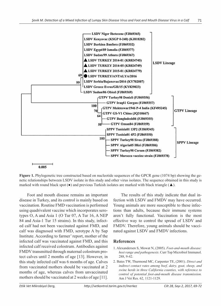
PCR and sequencing of PCR productsPCR was performed with primers that amplified 1158 bp of the G-protein-coupled chemokine re-ceptor (GPCR) gene of Capripoxviruses [7]. PCR product was purified from gels with a High Pure PCR Product Purification Kit (Roche Diagnostics, Indianapolis, USA) and sequenced with the BigDye Terminator v3.1 Cycle Sequencing Kit (Applied Biosystems, USA) on an ABI 3130xl DNA Analyzer (Applied Biosystems, USA). Sequence analysis was performed by using ChromasPro software (Version 1.7.5, Technolysium Ltd.). Phylogenetic tree was constructed for the GPCR gene of LSDV with ad-ditional sequences from GenBank. The confidence of the neighbour-joining tree was assessed by boot-strapping, using 1000 replicates, and only values above 50% are reported.
ResultsConfirmatory diagnosis of FMDClinical submission of the epithelium samples was identified as FMDV serotype A by Sap Institute.
Detection of LSDVLSDV DNA was detected in swab samples.
Sequence analysesAnalysis of the GPCR gene sequences revealed that the homology between the field isolate in the pres-ent study and LSDV isolates from different regions ranged from 98.8% to 100%. The deduced amino acid homology among the field isolate and previ-ously characterised LSDV isolates ranged between 98.5% and 100%.
Discussion
Lumpy skin disease was first reported in Turkey in 2013, and then the disease becomes endemic in Turkey [8]. LSDV is thought to be transmitted primarily by blood-feeding vectors [4]. Antalya Province is located in the Mediterranean region; this region has a subtropical Mediterranean climate characterised by mild and rainy winters and hot, dry summers that are suitable for the sustenance of blood-feeding insect activity and spread of arbovi-ral infections. Therefore, the Antalya Province has a higher risk of LSDV infection.
In this study, LSDV was detected in a calf. According to farmer’ report, infected calf was not vaccinated against LSD, whereas other animals in the herd were vaccinated against LSD and clini-cal symptoms were not observed in vaccinated animals. In Turkey, vaccination was carried out us-ing the sheep and goat pox vaccine. Vaccination is the most effective way to control further spread of LSDV. Calves from vaccinated mothers should be vaccinated between 3 to 4 months of age [14]. The characteristic clinical signs of LSD are fever, nasal and pharyngeal secretions, loss of appetite, nodules in the skin and enlarged lymph nodes [5,10]. In this study, fever of 40°C, nasal secretions, and lesions on the skin were observed in infected calf. Clinical signs can change depend on age, breed of cattle and immune status at the time of infection [15].
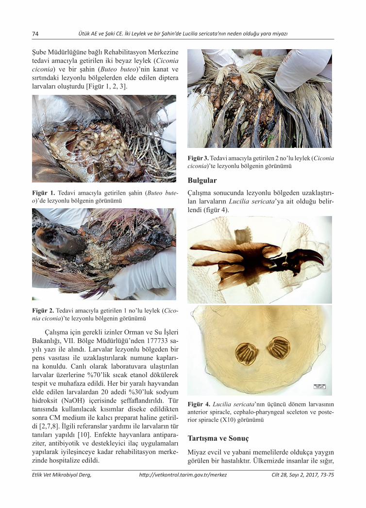
The phylogenetic tree based on the GPCR gene sequences revealed that the field isolates in this study clustered together with other isolates from Africa, Europe and the Middle East (Fig. 1). This result is in agreement with previous report [12].
Şevik M. Detection of a Mixed Infection of Lumpy Skin Disease Virus and Foot and Mouth Disease Virus in a Calf 71
Etlik Vet Mikrobiyol Derg, http://vetkontrol.tarim.gov.tr/merkez Cilt 28, Sayı 2, 2017, 69-72
Figure 1. Phylogenetic tree constructed based on nucleotide sequences of the GPCR gene (1074 bp) showing the ge-netic relationships between LSDV isolate in this study and other virus isolates. The sequence obtained in this study is marked with round black spot (●) and previous Turkish isolates are marked with black triangle (▲).
Foot and mouth disease remains an important disease in Turkey, and its control is mainly based on vaccination. Routine FMD vaccination is performed using quadrivalent vaccine which incorporates sero-types O, A and Asia 1 (O Tur 07, A Tur 16, A NEP 84 and Asia-1 Tur 15 strains). In this study, infect-ed calf had not been vaccinated against FMD, and calf was diagnosed with FMD, serotype A by Sap Institute. According to farmer’ report, mother of the infected calf was vaccinated against FMD, and this infected calf received colostrum. Antibodies against FMDV transmitted through maternal colostrum pro-tect calves until 2 months of age [13]. However, in this study infected calf was 6 months of age. Calves from vaccinated mothers should be vaccinated at 2 months of age, whereas calves from unvaccinated mothers should be vaccinated at 2 weeks of age [11].
The results of this study indicate that dual in-fection with LSDV and FMDV may have occurred. Young animals are more susceptible to these infec-tions than adults, because their immune systems aren’t fully functional. Vaccination is the most effective way to control the spread of LSDV and FMDV. Therefore, young animals should be vacci-nated against LSDV and FMDV infections.
References1. Alexandersen S, Mowat N, (2005). Foot-and-mouth disease:
host range and pathogenesis. Curr Top Microbiol Immunol. 288, 9-42.
2. Bates TW, Thurmond MC, Carpenter TE, (2001). Direct and indirect contact rates among beef, dairy, goat, sheep, and swine herds in three California counties, with reference to control of potential foot-and-mouth disease transmission. Am J Vet Res. 62, 1121-1129.
72 Şevik M. Detection of a Mixed Infection of Lumpy Skin Disease Virus and Foot and Mouth Disease Virus in a Calf
Etlik Vet Mikrobiyol Derg, http://vetkontrol.tarim.gov.tr/merkez Cilt 28, Sayı 2, 2017, 69-72
3. Bowden TR, Babiuk SL, Parkyn GR, Copps JS, Boyle DB, (2008). Capripoxvirus tissue tropism and shedding: A quantitative study in experimentally infected sheep and goats. Virology. 371, 380-393.
4. Chihota CM, Rennie LF, Kitching RP, Mellor PS, (2003). Attempted mechanical transmission of lumpy skin disease virus by biting insects. Med Vet Entomol. 17, 294-300.
5. Davies FG, Krauss H, Lund J, Taylor M, (1971). The labora-tory diagnosis of lumpy skin disease. Res Vet Sci. 12, 123-127.
6. Grubman MJ, Baxt B, (2004). Foot-and-mouth disease. Clin Microbiol Rev. 17, 465-493.
7. Le Goff C, Lamien CE, Fakhfakh E, Chadeyras A, Aba-Adulugba E, Libeau G, Tuppurainen E, Wallace DB, Adam T, Silber R, Gulyaz V, Madani H, Caufour P, Hammami S, Diallo A, Albina E, (2009). Capripoxvirus G-protein-coupled chemokine receptor: a host-range gene suitable for virus animal origin discrimination. J Gen Virol. 90, 1967-1977.
8. Office International des Epizooties (OIE), 2013: OIE disease information. Available at: http://www.oie.int/wahis_2/pub-lic/wahid.php/Reviewreport/Review?reportid=14106 (ac-cessed on 25 August 2017).
9. Office International des Epizooties (OIE), (2017). Foot and Mouth Disease (Infection with Foot and Mouth Disease
Virus). In Manual of Diagnostic Tests and Vaccines for Terrestrial Animals. OIE, Paris, 1-32. Available at: http://www.oie.int/fileadmin/Home/eng/Health_standards/tahm/2.01.08_FMD.pdf (accessed on 25 August 2017).
10. Office International des Epizooties (OIE), (2017). Lumpy skin disease. In Manual of Diagnostic Tests and Vaccines for Terrestrial Animals. OIE, Paris, 1-13. Available at: www.oie.int/fileadmin/Home/eng/Health_standards/tahm/2.04.14_LSD.pdf (accessed on 25 August 2017).
11. Şap Enstitüsü, (2017). Türkiye’de Şap Hastalığı. Available at: http://vetkontrol.tarim.gov.tr/sap/Menu/34/Turkiyede-Sap-Hastaligi (accessed on 25 August 2017).
12. Şevik M, Doğan M, (2017). Epidemiological and Molecular Studies on Lumpy Skin Disease Outbreaks in Turkey during 2014-2015. Transbound Emerg Dis. 64, 1268-1279.
13. Şenel E, Ulutürk S, Boz C, (1989). Buzağılarda şap aşı-sı ile aşılama zamanının tespiti. 1. Uluslararası Şap Sempozyumu, Ankara. pp. 191-194.
14. Tuppurainen E, Galon N, (2016). Lumpy Skin Disease: Current Situation in Europe and Neighbouring Regions and Necessary Control Measures to Halt the Spread in South-East Europe. OIE Regional Commission. 1-12.
15. Tuppurainen E, Alexandrov T, Beltrán-Alcrudo D, (2017). Lumpy skin disease field manual -A manual for veterinar-ians. FAO Animal Production and Health Manual. 20, 1-60.
*: Bu çalışma 18-21 Ekim 2017 tarihleri arasında Alanya’da düzenlenen II. International Academic Research Congress’de sözlü bildiri olarak sunulmuştur. Yazışma adresi / Correspondence: Armağan Erdem Ütük, Çukurova Üniv. Ceyhan Veteriner Fakültesi, Parazitoloji AD, Adana E-posta: [email protected]
Etlik Vet Mikrobiyol Derg, 2017; 28 (2): 73-75 Araştırma Makalesi / Research Article
İki Leylek (Ciconia ciconia) ve Bir Şahin (Buteo buteo)’de Lucilia sericata’nın Neden Olduğu Yara Miyazı*
Armağan Erdem Ütük1, Cem Ecmel Şaki2
1Çukurova Üniversitesi, Ceyhan Veteriner Fakültesi, Parazitoloji Anabilim Dalı, Adana, Türkiye2Fırat Üniversitesi, Veteriner Fakültesi, Parazitoloji Anabilim Dalı, Elazığ, Türkiye
Geliş Tarihi / Received: 28.10.2017, Kabul Tarihi / Accepted: 15.11.2017
Özet: Bu çalışmada, Orman ve Su İşleri Bakanlığı, Doğa Koruma ve Milli Parklar Osmaniye Şube Müdürlüğü’ne bağlı Rehabilitasyon Merkezine tedavi amacıyla getirilen yaralı iki Beyaz Leylek (Ciconia ciconia) ve bir Şahin (Buteo bute-o)’de yara miyazı tespit edildi. Lezyonlu bölgeden toplanan canlı larvalar üzerine %70’lik sıcak etanol ilave edildikten sonra %30’luk Potasyum Hidroksit (KOH) ile şeffaflandırıldı. Tür tanısı için gerekli olan kısımları diseke edilerek CM medium ile lam üzerine monte edildi ve incelendi. Çalışma sonucunda larvaların Lucilia sericata’ya ait olduğu tespit edildi.Anahtar Kelimeler: Beyaz Leylek, Lucilia sericata, Miyaz, Şahin.
Wound Myiasis Caused by Lucilia Sericata in two White Storks (Ciconia ciconia) and in a Common Buzzard (Buteo buteo)
Abstract: In this study, traumatic myiasis was detected in two White Storks (Ciconia ciconia) and in a Common Buzzard (Buteo buteo) brought for treatment to the Rehabilitation Centre affiliated to the Nature Conservation and National Parks Osmaniye Branch Office of the Ministry of Forestry and Water Affairs. 70% hot ethanol was added on to the living larvae that were collected from wounded area and then larvae were cleared with 30% potassium hydroxide (KOH). For species identification, the relevant parts of larvae were dissected, mounted on slides with CM medium and examined. At the end of this study, the collected larvae were identified as Lucilia sericata.Keywords: Common Buzzard, Lucilia sericata, Myiasis, White stork.
Giriş
Miyaz, konak hayvanların organ ve dokularının Diptera larvaları tarafından enfestasyonudur. Sinek larvaları direkt olarak konakların ölü ve canlı do-kuları ile beslenir. Konaklar genellikle memeliler, ara sıra kuşlar nadiren de amfibi ve sürüngenler-dir. Hastalık larvaların konak hayvan üzerinde ya da içerisinde yerleştiği yere (dermal, subdermal, nazofarengeal, oküler, intestinal, ürogenital), göç durumuna (kriping), beslenme özelliklerine (sangu-inivoroz), oluşturdukları lezyonların şekline (trav-matik, frunküler) ve konak parazit ilişkisine (obli-gator, fakültatif, aksidental) göre farklı şekillerde isimlendirilir [5,9,10].
Yabani kanatlılarda hastalık genellikle Calliphoridae, Muscidae ve Neottiophilidae aile-sinde bulunan hematofaj türler tarafından oluşturu-
lur ve yavrularda erişkinlerden daha yaygın olarak görülür. Ancak Calliphoridae ve Sarcophagidae ailelerde bulunan bazı türlerin oluşturduğu kutanöz ve subkutanöz vakalara da rastlanır [3,6].
Yabani kanatlılarda yara miyaz etkenleri kana-ma, anemi, büyük deri altı kistlerine, yaraların etra-fında epidermal nekrozlara, bazen de karın, göğüs, nazal ve oküler boşluklara kadar ilerleyip toksemi ve sepsis sonucunda ölümlere neden olur [3,6].
Bu çalışma iki beyaz leylek (Ciconia ciconia) ve bir şahin (Buteo buteo)’de yara miyazına neden olan larvaların hangi diptera türüne ait olduğunu be-lirlemek amacıyla yapıldı.
Materyal ve MetodÇalışmanın materyalini Orman ve Su İşleri Bakanlığı, Doğa Koruma ve Milli Parklar Osmaniye
74 Ütük AE ve Şaki CE. İki Leylek ve bir Şahin’de Lucilia sericata’nın neden olduğu yara miyazı
Etlik Vet Mikrobiyol Derg, http://vetkontrol.tarim.gov.tr/merkez Cilt 28, Sayı 2, 2017, 73-75
Şube Müdürlüğüne bağlı Rehabilitasyon Merkezine tedavi amacıyla getirilen iki beyaz leylek (Ciconia ciconia) ve bir şahin (Buteo buteo)’nin kanat ve sırtındaki lezyonlu bölgelerden elde edilen diptera larvaları oluşturdu [Figür 1, 2, 3].
Figür 1. Tedavi amacıyla getirilen şahin (Buteo bute-o)’de lezyonlu bölgenin görünümü
Figür 2. Tedavi amacıyla getirilen 1 no’lu leylek (Cico-nia ciconia)’te lezyonlu bölgenin görünümü
Çalışma için gerekli izinler Orman ve Su İşleri Bakanlığı, VII. Bölge Müdürlüğü’nden 177733 sa-yılı yazı ile alındı. Larvalar lezyonlu bölgeden bir pens vasıtası ile uzaklaştırılarak numune kapları-na konuldu. Canlı olarak laboratuvara ulaştırılan larvalar üzerlerine %70’lik sıcak etanol dökülerek tespit ve muhafaza edildi. Her bir yaralı hayvandan elde edilen larvalardan 20 adedi %30’luk sodyum hidroksit (NaOH) içerisinde şeffaflandırıldı. Tür tanısında kullanılacak kısımlar diseke edildikten sonra CM medium ile kalıcı preparat haline getiril-di [2,7,8]. İlgili referanslar yardımı ile larvaların tür tanıları yapıldı [10]. Enfekte hayvanlara antipara-ziter, antibiyotik ve destekleyici ilaç uygulamaları yapılarak iyileşinceye kadar rehabilitasyon merke-zinde hospitalize edildi.
Figür 3. Tedavi amacıyla getirilen 2 no’lu leylek (Ciconia ciconia)’te lezyonlu bölgenin görünümü
BulgularÇalışma sonucunda lezyonlu bölgeden uzaklaştırı-lan larvaların Lucilia sericata’ya ait olduğu belir-lendi (figür 4).
Figür 4. Lucilia sericata’nın üçüncü dönem larvasının anterior spiracle, cephalo-pharyngeal sceleton ve poste-rior spiracle (X10) görünümü
Tartışma ve Sonuç
Miyaz evcil ve yabani memelilerde oldukça yaygın görülen bir hastalıktır. Ülkemizde insanlar ile sığır,
Ütük AE ve Şaki CE. İki Leylek ve bir Şahin’de Lucilia sericata’nın neden olduğu yara miyazı 75
Etlik Vet Mikrobiyol Derg, http://vetkontrol.tarim.gov.tr/merkez Cilt 28, Sayı 2, 2017, 73-75
koyun, keçi, kedi ve köpek gibi memelilerde miyaz vakalarına sıklıkla rastlanır [4]. Hastalık kanatlılar-da memeliler kadar yaygın değildir [9]. Dünyada yapılan çalışmalarda kartal, doğan, şahin, kerkenez, baykuş, turna, beyaz leylek, kaz, bazı ördek türleri, ağaç kakan, bülbül ve makavlarda miyaz vakaları tespit edilmiştir [1,6]. Ülkemizde ise şahin, baykuş ve kargalarda hastalık bildirilmiştir [4,8].
Dünyaya genelinde yapılan çalışmalarda kanat-lılarda tespit edilen miyaz etkenleri Calliphora vici-na, Lucilia sericata, Wohlfahrtia vigil, W.magnifica, W.opaca, Cuterebra buccata ve Dermatobia homi-nis’tir [1,3,6]. Ülkemizde ise yabani kanatlılarda tespit edilen tür L.sericata’dır [4,8].
Bu çalışmada iki beyaz leylek (Ciconia ciconia) ve bir şahin (Buteo buteo)’in kanat ve sırtındaki lez-yonlu bölgelerden elde edilen larvaların L.sericata olduğu tespit edildi. Dünyada ve ülkemizde kanat-lıların miyaz etkenleri ile ilgili fazla literatür bilgi-sine rastlanılmadığından mevcut vakaların konu ile ilgilenen araştırmacıların bilgisine sunulması uygun görüldü.
Miyaz vakaları sığır, koyun ve keçilerde et, süt, yapağı ve deri kayıplarına, kedi ve köpeklerde teda-vi ve hospitalizasyon masraflarına bağlı olarak ciddi ekonomik kayıplara neden olur [4, 5,9]. Özellikle kesimhanelerin atıklarını usulüne uygun olarak imha etmemesi, belediyelerin topladıkları çöpleri açık alanlarda biriktirmesi, hala birçok belediyenin hayvan barınaklarının bulunmaması, olanların ise fiziksel koşullarının ve veteriner hizmetlerinin ye-tersiz oluşu, halkımızın, pet sahiplerinin, yetiştirici-lerin bilinçsizliği ve ekolojik dengelerin bozulması gibi birçok faktör miyaz vakaları ile birlikte birçok enfeksiyöz hastalığa zemin hazırlayarak hayvan sağlığını, halk sağlığını ve yaban hayatını tehdit et-mektedir [5,7,9,10].
Bu gibi olumsuzlukları en düşük seviyeye in-dirmek için toplumun her kesiminden insanın bi-linçlendirilmesi, belediyelerin barınak ve kesim-hanelerinin fiziksel koşullarını düzeltmesi, bu gibi
ortamlarda yeterli sayıda veteriner hekim ve yar-dımcı personel istihdam edilmesi gerekmektedir. Ayrıca çok geniş bir coğrafya, farklı iklim bölgeleri ve zengin bir biyoçeşitliliğe sahip olan ülkemizde yaban hayvanlarının paraziter hastalıkları ile ilgili daha fazla çalışma yapılması, bu hastalıkların epi-demiyolojisinin daha iyi anlaşılmasına ve daha et-kin mücadele yöntemleri geliştirilmesine katkı sağ-layacaktır.
TeşekkürKatkılarından dolayı Veteriner Hekim Bünyamin AKIN’a teşekkür ederiz.
Kaynaklar1. Araghi MP, Eskandari F, Gilasian E, (2015). Avian Wound
Myiasis Caused by Calliphora vicina Robineau-Desvoidy (Diptera: Calliphoridae) in an Immature Migrating Eastern Imperial Eagle (Aquila Heliaca Savigny) (Aves: Accipitridae) in Southwestern Iran. J Veterinar Sci Technolo. 6, 1-3.
2. Clark EW, Morishita F, (1950). C-M Medium: A Mounting Medium Smal Insects, Mites and Other Whole Months. Science, 112, 789.
3. Cooper JE, Cooper ME, Krone O, Newton I, Peakal DB, Zucca P, (2002). Birds of Prey: Health and Diseases. 3rd ed, Blackwell Science.
4. Dik B, Uslu U, Işık N, (2012). Myiasis in Animals and Humanbeings in Turkey. Kafkas Univ Vet Fak Derg. 18, 37-42.
5. Dinçer Ş, (1997). İnsan ve Hayvanlarda Myiasis. Özcel MA, Daldal N. eds. Parazitolojide Artropod Hastalıkları ve Vektörler. İzmir. Turkiye Parazitol Dern, Yay No: 13, Ege Ünivesitesi Basımevi, Türkiye.
6. Little SE, (2008). Myiasis in Wild Birds. Atkinson CT, Thomas NJ, Hunter DB .eds. Parasitic Diseases of Wild Birds. 1st ed, Wiley-Blackwell.
7. Ütük AE, (2006). Bir köpekte travmatik miyazis olgusu. Fırat Üniv Sağlık Bil Dergisi, 20, 97-99.
8. Ütük AE, Şaki CE, (2016). Bir Baykuşta Travmatik Miyaz Olgusu. İMSEC2016, Ekim, 16-28, Adana, Türkiye.
9. Wall R, Shearer D, (2001). Veterinary Ectoparasites: Biology, Pathology, and Control. 2nd ed, Blackwell Science, Oxford.
10. Zumpt, F, (1965). Myiasis in Man and Animals in the Old World. Butterwoths & Co. ltd., London.
*Aynı isimli Yüksek Lisans Tezinden Özetlenmiştir Yazışma adresi / Correspondance: Sibel Gür, Department of Virology, Faculty of Veterinary Medicine, Afyon Kocatepe University, ANS Campus, Afyonkarahisar, Turkey E-mail: [email protected]
Etlik Vet Mikrobiyol Derg, 2017; 28 (2): 76-84 Araştırma Makalesi / Research Article
Comparision of Two Different Vaccination Shedule for Fight with Infectious Bursal Disease in Layer Hens
Ahmet Bildir1, Sibel Gür2
1 Kartallar Limited Company, Afyonkarahisar, Turkey.2Faculty of Veterinary Medicine, Department of Virology, Afyon Kocatepe University, Afyonkarahisar, Turkey.
Geliş Tarihi / Received: 29.09.2017, Kabul Tarihi / Accepted: 10.11.2017
Abstract: Infectious Bursal Disease Virus (IBDV) is one of the most important immunosuppressive diseases of poultry which classified in Avibirnavirus genus belonging to Birnaviridae family. In this study, two different vaccination sched-ules were compared in the aspect of protectivity. For this purpose, blood serum samples was collected from 6 flocks of layer chickens in the postnatal period on days 0, 7, 14, 21, 28, 35, 42, 49, 56, 63 and 70. Samples were randomly selected from 10 chicks in every sampling. Flock 1 was sampled two more times due to acute clinical infection. The total of 680 serum samples was tested for IBDV specific Antibody (Ab) presence and titers using ELISA. Maternal derived antibody (MDA) was found to be protective for 6-8 days. Standard vaccination schedule (one inactive and three live intermediate vaccines) was carried out at three flocks (2, 3 and 5). In other flocks, one live, one inactive and additional more live vaccine were administered to the chicks into water. Despite regular vaccination, IBD epidemic was seen in flocks 1 and 2. In flock 1, an outbreak was reported with 23% mortality in previous breeding period. This proportion was reduced to 4.5% in this enterprise by applying initially live vaccine, and then any sign of clinical infection has not been detected. Antibody titers were entered augmentation trend in flocks 1 and 2 until day 21. probably due to viral load, this turning point was detected on day 14 in other flocks. Statistical analysis clearly showed that MDA titers showed a sharp decrease in the initially live vaccine given flock. In conclusion, to stimulate cellular immune response using initial live dose as soon as possible is more influential. It means, changing standard vaccination schedule was found to be more effective on protection of animals in the critical period of infection, especially in the presence of viral load in the field.Keywords: Infectious bursal disease, Maternal derived antibody, Virus, Vaccination.
Yumurtacı Tavuklarda Infectious Bursal Disease ile Mücadelede İki Farklı Aşılama Programının Karşılaştırılması
Özet: Infectious Bursal Disease Virus (IBDV) Birnaviridae familyasının Avibirnavirus genusunda sınıflandırılmış olan kanatlıların en önemli immunsupresif hastalıklarından biridir. Bu çalışmada, iki farklı aşılama programı koruyuculuğu açısından karşılaştırıldı. Bunun için, 6 yumurtacı sürüden portnatal 0, 7, 14, 21, 28, 35, 42, 49, 56, 63 ve 70. günlerde kan örnekleri alındı. Her örneklemede rastgele seçilmiş 10 civciv örneklendi. Akut klinik enfeksiyon gelişmesi üzerine sürü 1 iki kez daha örneklendi. Toplam olarak 680 serum örneği IBDV spesifik Ab varlığı ve titresi açısından ELISA ile test edildi. Maternal Antikorların (MA) 6-8. günlere kadar koruyucu olduğu belirlendi. Standart aşılama program (1 inaktif ve 3 canlı intermediate aşı) 3 sürüde uygulandı (2, 3 and 5). Diğer 3 sürüde, civcivlere bir canlı bir inaktif ve ek olarak bir canlı aşı içme suyuna karıştırılarak verildi. Düzenli aşılama yapılmasına rağmen sürü 1 ve 2’de IBD salgını görüldü. Bir nolu sürüde bir önceki yetiştirme döneminde %23 mortaliteli bir salgın kayıt edilmişti. Bu oran aynı sürüde canlı aşı uygulamasıyla %4.5’a düşürüldü ve hiçbir klinik enfeksiyon tespit edilmedi. Antikor titreleri 1 ve 2 nolu sürüde muhte-melen viral yük nedeniyle 21. güne kadar artış eğiliminde iken diğer sürülerde bu dönüş noktasının 14. gün olduğu tespit edildi. İlk olarak canlı aşı uygulanan sürülerde maternal antikor titrelerinde ani bir düşüş gerçekleştiği istatistiksel ana-lizlerle açıkça gösterilmiştir. Sonuç olarak, aşı takvimini değiştirmek anlamına gelen, hücresel immun yanıtı mümkün olduğunca erken bir şekilde uyarmak için ilk dozun canlı aşı olarak kullanılmasının, enfeksiyonun kritik dönemlerinde ve özellikle de sahada akut viral enfeksiyon söz konusu olduğunda etkili bir koruma sağlayabileceği belirlendi.Anahtar Kelimeler: Aşılama, Infectious bursal disease, Maternal Antikor, Virus
Introduction
Infectious Bursal Disease Virus (IBDV) was classified in Avibirnavirus genus belonging to
Birnaviridae family. There are two viruses in this group; IBDV in chicken and Infectious pancreatic necrosis virus in fish [24]. Birnaviruses have ico-
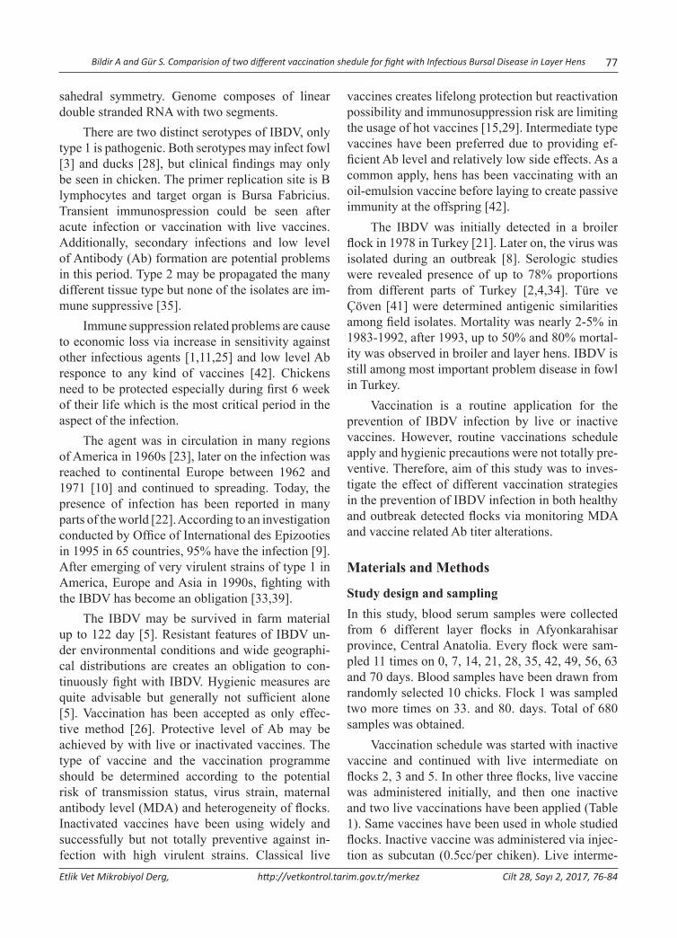
Bildir A and Gür S. Comparision of two different vaccination shedule for fight with Infectious Bursal Disease in Layer Hens 77
Etlik Vet Mikrobiyol Derg, http://vetkontrol.tarim.gov.tr/merkez Cilt 28, Sayı 2, 2017, 76-84
sahedral symmetry. Genome composes of linear double stranded RNA with two segments.
There are two distinct serotypes of IBDV, only type 1 is pathogenic. Both serotypes may infect fowl [3] and ducks [28], but clinical findings may only be seen in chicken. The primer replication site is B lymphocytes and target organ is Bursa Fabricius. Transient immunospression could be seen after acute infection or vaccination with live vaccines. Additionally, secondary infections and low level of Antibody (Ab) formation are potential problems in this period. Type 2 may be propagated the many different tissue type but none of the isolates are im-mune suppressive [35].
Immune suppression related problems are cause to economic loss via increase in sensitivity against other infectious agents [1,11,25] and low level Ab responce to any kind of vaccines [42]. Chickens need to be protected especially during first 6 week of their life which is the most critical period in the aspect of the infection.
The agent was in circulation in many regions of America in 1960s [23], later on the infection was reached to continental Europe between 1962 and 1971 [10] and continued to spreading. Today, the presence of infection has been reported in many parts of the world [22]. According to an investigation conducted by Office of International des Epizooties in 1995 in 65 countries, 95% have the infection [9]. After emerging of very virulent strains of type 1 in America, Europe and Asia in 1990s, fighting with the IBDV has become an obligation [33,39].
The IBDV may be survived in farm material up to 122 day [5]. Resistant features of IBDV un-der environmental conditions and wide geographi-cal distributions are creates an obligation to con-tinuously fight with IBDV. Hygienic measures are quite advisable but generally not sufficient alone [5]. Vaccination has been accepted as only effec-tive method [26]. Protective level of Ab may be achieved by with live or inactivated vaccines. The type of vaccine and the vaccination programme should be determined according to the potential risk of transmission status, virus strain, maternal antibody level (MDA) and heterogeneity of flocks. Inactivated vaccines have been using widely and successfully but not totally preventive against in-fection with high virulent strains. Classical live
vaccines creates lifelong protection but reactivation possibility and immunosuppression risk are limiting the usage of hot vaccines [15,29]. Intermediate type vaccines have been preferred due to providing ef-ficient Ab level and relatively low side effects. As a common apply, hens has been vaccinating with an oil-emulsion vaccine before laying to create passive immunity at the offspring [42].
The IBDV was initially detected in a broiler flock in 1978 in Turkey [21]. Later on, the virus was isolated during an outbreak [8]. Serologic studies were revealed presence of up to 78% proportions from different parts of Turkey [2,4,34]. Türe ve Çöven [41] were determined antigenic similarities among field isolates. Mortality was nearly 2-5% in 1983-1992, after 1993, up to 50% and 80% mortal-ity was observed in broiler and layer hens. IBDV is still among most important problem disease in fowl in Turkey.
Vaccination is a routine application for the prevention of IBDV infection by live or inactive vaccines. However, routine vaccinations schedule apply and hygienic precautions were not totally pre-ventive. Therefore, aim of this study was to inves-tigate the effect of different vaccination strategies in the prevention of IBDV infection in both healthy and outbreak detected flocks via monitoring MDA and vaccine related Ab titer alterations.
Materials and Methods
Study design and samplingIn this study, blood serum samples were collected from 6 different layer flocks in Afyonkarahisar province, Central Anatolia. Every flock were sam-pled 11 times on 0, 7, 14, 21, 28, 35, 42, 49, 56, 63 and 70 days. Blood samples have been drawn from randomly selected 10 chicks. Flock 1 was sampled two more times on 33. and 80. days. Total of 680 samples was obtained.
Vaccination schedule was started with inactive vaccine and continued with live intermediate on flocks 2, 3 and 5. In other three flocks, live vaccine was administered initially, and then one inactive and two live vaccinations have been applied (Table 1). Same vaccines have been used in whole studied flocks. Inactive vaccine was administered via injec-tion as subcutan (0.5cc/per chiken). Live interme-
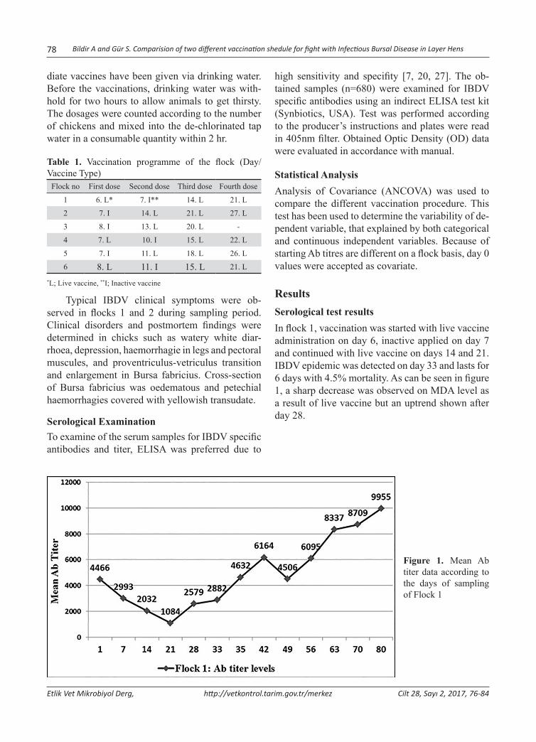
78 Bildir A and Gür S. Comparision of two different vaccination shedule for fight with Infectious Bursal Disease in Layer Hens
Etlik Vet Mikrobiyol Derg, http://vetkontrol.tarim.gov.tr/merkez Cilt 28, Sayı 2, 2017, 76-84
diate vaccines have been given via drinking water. Before the vaccinations, drinking water was with-hold for two hours to allow animals to get thirsty. The dosages were counted according to the number of chickens and mixed into the de-chlorinated tap water in a consumable quantity within 2 hr.
Table 1. Vaccination programme of the flock (Day/Vaccine Type)
Flock no First dose Second dose Third dose Fourth dose
1 6. L* 7. I** 14. L 21. L
2 7. I 14. L 21. L 27. L
3 8. I 13. L 20. L -
4 7. L 10. I 15. L 22. L
5 7. I 11. L 18. L 26. L
6 8. L 11. I 15. L 21. L
*L; Live vaccine, **I; Inactive vaccine
Typical IBDV clinical symptoms were ob-served in flocks 1 and 2 during sampling period. Clinical disorders and postmortem findings were determined in chicks such as watery white diar-rhoea, depression, haemorrhagie in legs and pectoral muscules, and proventriculus-vetriculus transition and enlargement in Bursa fabricius. Cross-section of Bursa fabricius was oedematous and petechial haemorrhagies covered with yellowish transudate.
Serological ExaminationTo examine of the serum samples for IBDV specific antibodies and titer, ELISA was preferred due to
high sensitivity and specifity [7, 20, 27]. The ob-tained samples (n=680) were examined for IBDV specific antibodies using an indirect ELISA test kit (Synbiotics, USA). Test was performed according to the producer’s instructions and plates were read in 405nm filter. Obtained Optic Density (OD) data were evaluated in accordance with manual.
Statistical AnalysisAnalysis of Covariance (ANCOVA) was used to compare the different vaccination procedure. This test has been used to determine the variability of de-pendent variable, that explained by both categorical and continuous independent variables. Because of starting Ab titres are different on a flock basis, day 0 values were accepted as covariate.
ResultsSerological test resultsIn flock 1, vaccination was started with live vaccine administration on day 6, inactive applied on day 7 and continued with live vaccine on days 14 and 21. IBDV epidemic was detected on day 33 and lasts for 6 days with 4.5% mortality. As can be seen in figure 1, a sharp decrease was observed on MDA level as a result of live vaccine but an uptrend shown after day 28.
Figure 1. Mean Ab titer data according to the days of sampling of Flock 1
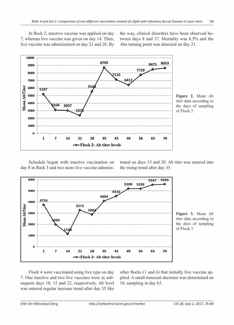
Bildir A and Gür S. Comparision of two different vaccination shedule for fight with Infectious Bursal Disease in Layer Hens 79
Etlik Vet Mikrobiyol Derg, http://vetkontrol.tarim.gov.tr/merkez Cilt 28, Sayı 2, 2017, 76-84
In flock 2, inactive vaccine was applied on day 7, whereas live vaccine was given on day 14. Then, live vaccine was administered on day 21 and 28. By
the way, clinical disorders have been observed be-tween days 8 and 37. Mortality was 8.5% and the Abs turning point was detected on day 21.
Figure 2. Mean Ab titer data according to the days of sampling of Flock 2
Schedule began with inactive vaccination on day 8 in flock 3 and two more live vaccine adminis-
trated on days 13 and 20. Ab titer was entered into the rising trend after day 35.
Figure 3. Mean Ab titer data according to the days of sampling of Flock 3
Flock 4 were vaccinated using live type on day 7. One inactive and two live vaccines were in sub-sequent days 10, 15 and 22, respectively. Ab level was entered regular increase trend after day 35 like
other flocks (1 and 6) that initially live vaccine ap-plied. A small transient decrease was determined on 10. sampling in day 63.
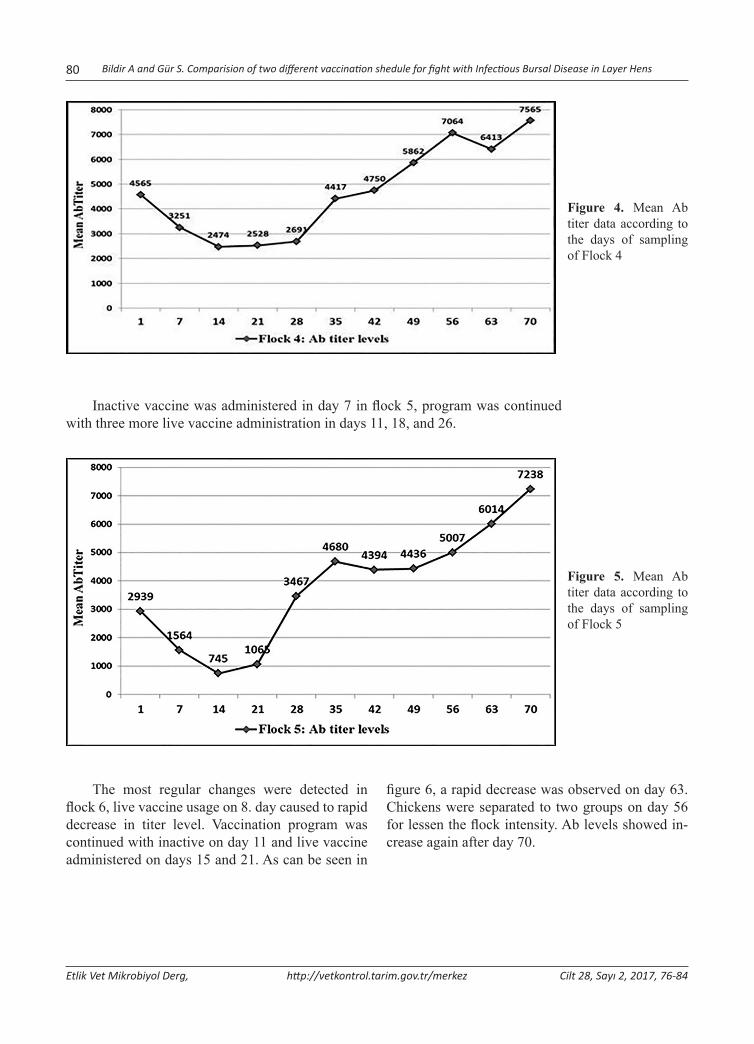
80 Bildir A and Gür S. Comparision of two different vaccination shedule for fight with Infectious Bursal Disease in Layer Hens
Etlik Vet Mikrobiyol Derg, http://vetkontrol.tarim.gov.tr/merkez Cilt 28, Sayı 2, 2017, 76-84
Figure 4. Mean Ab titer data according to the days of sampling of Flock 4
Inactive vaccine was administered in day 7 in flock 5, program was continued with three more live vaccine administration in days 11, 18, and 26.
Figure 5. Mean Ab titer data according to the days of sampling of Flock 5
The most regular changes were detected in flock 6, live vaccine usage on 8. day caused to rapid decrease in titer level. Vaccination program was continued with inactive on day 11 and live vaccine administered on days 15 and 21. As can be seen in
figure 6, a rapid decrease was observed on day 63. Chickens were separated to two groups on day 56 for lessen the flock intensity. Ab levels showed in-crease again after day 70.
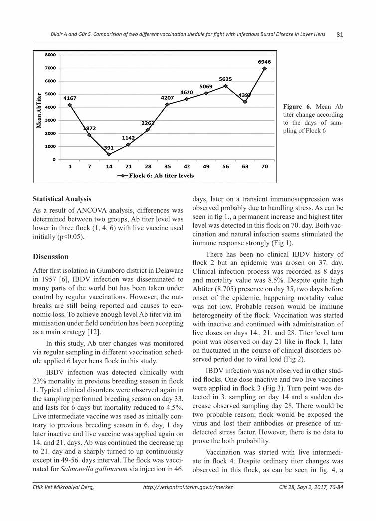
Bildir A and Gür S. Comparision of two different vaccination shedule for fight with Infectious Bursal Disease in Layer Hens 81
Etlik Vet Mikrobiyol Derg, http://vetkontrol.tarim.gov.tr/merkez Cilt 28, Sayı 2, 2017, 76-84
Figure 6. Mean Ab titer change according to the days of sam-pling of Flock 6
Statistical AnalysisAs a result of ANCOVA analysis, differences was determined between two groups, Ab titer level was lower in three flock (1, 4, 6) with live vaccine used initially (p<0.05).
Discussion
After first isolation in Gumboro district in Delaware in 1957 [6], IBDV infection was disseminated to many parts of the world but has been taken under control by regular vaccinations. However, the out-breaks are still being reported and causes to eco-nomic loss. To achieve enough level Ab titer via im-munisation under field condition has been accepting as a main strategy [12].
In this study, Ab titer changes was monitored via regular sampling in different vaccination sched-ule applied 6 layer hens flock in this study.
IBDV infection was detected clinically with 23% mortality in previous breeding season in flock 1. Typical clinical disorders were observed again in the sampling performed breeding season on day 33. and lasts for 6 days but mortality reduced to 4.5%. Live intermediate vaccine was used as initially con-trary to previous breeding season in 6. day, 1 day later inactive and live vaccine was applied again on 14. and 21. days. Ab was continued the decrease up to 21. day and a sharply turned to up continuously except in 49-56. days interval. The flock was vacci-nated for Salmonella gallinarum via injection in 46.
days, later on a transient immunosuppression was observed probably due to handling stress. As can be seen in fig 1., a permanent increase and highest titer level was detected in this flock on 70. day. Both vac-cination and natural infection seems stimulated the immune response strongly (Fig 1).
There has been no clinical IBDV history of flock 2 but an epidemic was arosen on 37. day. Clinical infection process was recorded as 8 days and mortality value was 8.5%. Despite quite high Abtiter (8.705) presence on day 35, two days before onset of the epidemic, happening mortality value was not low. Probable reason would be immune heterogeneity of the flock. Vaccination was started with inactive and continued with administration of live doses on days 14., 21. and 28. Titer level turn point was observed on day 21 like in flock 1, later on fluctuated in the course of clinical disorders ob-served period due to viral load (Fig 2).
IBDV infection was not observed in other stud-ied flocks. One dose inactive and two live vaccines were applied in flock 3 (Fig 3). Turn point was de-tected in 3. sampling on day 14 and a sudden de-crease observed sampling day 28. There would be two probable reason; flock would be exposed the virus and lost their antibodies or presence of un-detected stress factor. However, there is no data to prove the both probability.
Vaccination was started with live intermedi-ate in flock 4. Despite ordinary titer changes was observed in this flock, as can be seen in fig. 4, a

82 Bildir A and Gür S. Comparision of two different vaccination shedule for fight with Infectious Bursal Disease in Layer Hens
Etlik Vet Mikrobiyol Derg, http://vetkontrol.tarim.gov.tr/merkez Cilt 28, Sayı 2, 2017, 76-84
transient decrease was determined in day 63 of sam-pling just after injectable vaccination apply in day 58 for Salmonella gallinarum.
Ab level changes were detected as expected in flock 5 until day 35 (Fig 5). However, inadequate Ab increase observed from this point. On the con-trolling farm records, rise in temperature was de-tected in the middle of sampling period (end of the July). Heat stress would have been created immu-nosuppression.
Live vaccine was applied as first vaccination in flock 6, after a short rapid decrease, titer entered the augmentation trend from 14. days but a sharp tran-sient downturn shown in 63. day sampling (Fig 6). This decrease was eventuated after dividing flock as two distinct groups to make rarefy. It is obvious that handling stress was effected the immune response.
Main factors of the epidemics in poultry farm-ing are inappropriate disinfection, deficiency in management practises, wrong vaccine type and wrong timing applies are The infection may be seen more severe and earlier in next breeding period [5,36].
The most feasible method for prevention of IBDV infection in the field is vaccination. However a unique vaccination programme cannot be propos-ing due to variability of MDA, and other factors related to management and environmental condi-tions. Vaccination would be unnecessary in broiler flocks keeping in strictly controlled closed breeding management conditions. Additionally, have high level MDA seems sufficient in their short life span. However, active immunoprotection is quite neces-sary in layer hens. To determine the right vaccina-tion schedule is the most critical decision and has vital importance. The most important parameter on the successful immunisation is to get obtain ho-mogeny in MDA titer at the beginning. In the field, controlling of Ab titer level is a routine work but, in most of the time, not gives enough information in the aspect of flock homology due to arbitrarily sampling style.
Main aim of the vaccination is to supply opti-mal Ab titer level in the critical period of IBDV in-fection (25-45 days). MDA is protective in the first 6-8 days of life, and end of first week of life was regarded as suitable time for initial vaccination.
Inactive vaccines have been preferred as first vaccination due to in case of no viral challenge risks. Live vaccines are stimulates the cellular immune system and stronger Ab level may be achieved. Commonly using schedule is 1 inactive and 2 or 3 doze live vaccine apply but this schedule may not be feasible in the presence of the virus in and around the farm. In the flock 1, 23% mortality was recorded in previous breeding period despite stan-dard vaccination program. After changing schedule, this proportion was reduced to 4.5% in the sampling performed course and no infection was aroused in the subsequent breeding period. Considering pres-ence of the virus in the farm and resistant character of the agent under field conditions, rearranging of vaccination style seems effective on prevention of economic looses. IBDV Ab titer decrease has been continued to day 14 of postnatal period on 4 flocks (3, 4, 5 and 6). After live vaccine apply in day 14, trend was entered the increase. Turn point was de-tected as 21. day on the flocks 1 and 2, probably due to environmental viral load. Two dose live vaccine has been administration in days 15 and 26. resulted with fast and ordinary titer increase in all of the flocks. In this period, statsistical difference was not determined between different schedule used flocks.
Goddard et al. [13] was vaccinated the four group of chicks with different combinations of com-mercial inactivated and live vaccines. Only a dose of live vaccine administered group was detected as susceptible to the viral challenge up to seven week of life. Besides, bot vaccination schedule were found to be protective against experimental viral challenge.
However, initially live vaccine administered groups were found to be safer in this study. Decrease in mortality was observed in flocks 1 and 2 that clin-ical infection emerged accidentally.
The most of the experimental studies for de-termining optimal vaccination period were carried out in broiler. Half life of MDA has been evaluated differently according to breeding aim and manage-ment conditions. To determine the right schedule for vaccinations is not easy under field conditions. Interference of vaccine virus by MDA is the greatest risk and cause to retardation or prevention of im-mune response to the vaccine [40,43]. MDA level, half life of MDA, breeding aim, sampling days, the

Bildir A and Gür S. Comparision of two different vaccination shedule for fight with Infectious Bursal Disease in Layer Hens 83
Etlik Vet Mikrobiyol Derg, http://vetkontrol.tarim.gov.tr/merkez Cilt 28, Sayı 2, 2017, 76-84
Ab titer uniformity of the flock and vaccine type like factors have to be considered to determine the most suitable vaccination schedule using Deventer formula [7].
Correlation among stress factors and immune response is a well known phenomenon. Cortisone is one of the best known immune suppressor sub-stances. Fear stimulates the amygdala and leads to cortisone secretion. This correlation was proved in more than 20 species. As can be obviously seen in this study, human manipulations have transient but strong negative effect on the immune response. Even though there was no clear description on this issue, stress related growth retardation [17,18], im-pairment in immune response [14,16,30], productiv-ity problems were reported before [17,38]. To avoid from manipulative performances after the vaccina-tions is a necessity to obtain expected results.
Epidemics could be emerged despite routine vaccination programme and disinfection precau-tions. Any risk factors would not be detected in some cases. In this kind of situations, management condi-tions and other less important factors for transmis-sion should be taken into consideration like birds, fly (Aedes vexans) [19], mealworm (Alphitobius diaperinus) [37], dogs and rodents [25,31,32].
In conclusion, IBDV Ab titer changes were monitored in two different vaccination programs applied groups of flocks. Prevention of epidemics and eliminate the economic losses could be more feasible via stimulating cellular immune response as soon as possible with initially live vaccine ad-ministration considering half-life of MDA.
AcknowledgementThe authors thanks to Dr. Nurhan Doğan for contri-bution to study with statistical evaluation.
References1. Allan WH, Faragher JT, Cullen GA, (1972). Immunosupression
by the infectious bursal agent in chickens immunized against Newcastle disease. Vet Rec. 90, 511-512.
2. Babila A, Ası Y, Akçadağ B, Gürel A, (1988). İstanbul ve Trakya Bölgesi kümes hayvanlarında IB, ILT, IBD, EDS, AE ve adenovirus enfeksiyonlarının epizootiyolojik araştır-ması ve izolasyon çalışmaları. Pendik Hay Hast Arş Enst Derg. 19, 6-77.
3. Barnes HJ, Wheeler J, Reed D, (1982). Serological evidence of infectious bursal disease virus infection in Iowa turkeys. Avian Dis. 26, 560-565.
4. Baysal T, Bozkır M. (1989). Konya Bölgesi kümes hayvanla-rında IBD, ILT, EDS, IBD, AE, ve adenovirus enfeksiyonla-rının epizootiyolojik araştırılması ve izolasyon çalışmaları. Etlik Vet Mikrobiyol Derg. 6, 23-30.
5. Benton WJ, Cover MS, Rosenberger JK, Lake RS, (1967). Physicochemical properties of the infectious bursal disease agent (IBA). Avian Dis. 11, 430-438.
6. Cosgrove AS, (1962). An apparently new disease of chick-ens- Avian Nephrosis. Avian Dis. 6, 385-389.
7. De Wit JJ, (1999). Gumboro disease: optimising vaccination. Int Poult Prod. 7, 19-21.
8. Ergün A, (1989). Tavukların bazı viral hastalıklarının epi-zootiyolojik taramasında kullanılmak üzere antijen ve an-tiserum hazırlanması. Etlik Vet Mikrobiyol Derg. 6, 35-54.
9. Eterradossi N, (1995). Progress in the diagnosis and pro-phylaxis of infectious bursal disease in poultry. OIE, Paris, France, p.75-82.
10. Faragher JT, (1972). Infectious bursal disease of chicken. Vet Bull. 42, 361-369.
11. Faragher JT, Allan WH, Wyeth CJ, (1974). Immunosupressive effect of infectious bursal disease agent on vaccination against Newcastle disease. Vet Rec. 95, 385-388.
12. Fussel LW, (1998). Poultry industry strategies for control of immunosuppressive diseases. Poult Sci. 77, 1193-1196.
13. Goddard RD, Wyeth PJ, Varney WC, (1994). Vaccination of commercial layer chicks against infectious bursal disease with maternally derived antibodies. Vet Rec. 135, 273-274.
14. Grandin T, (1997). Assessment of stress during handling and transport. J Anim Sci. 75, 249-257.
15. Guittet M, Le Coq H, Picault JP, Eterradossi N, Bennejean G, (1992). Safety of infectious bursal disease vaccines: as-sesment of an acceptability threshold. Dev Biol Stand.79, 147-152.
16. Hastings BE, Abbott DE, George LM, (1992). Stress fac-tors influencing plasma cortisol levels and adrenal weights in Chinese water deer (Hydropotesinermis). Res Vet Sci. 53, 375-80.
17. Hemsworth PH, Barnett JL, (1989). Relationships between-fear of human productivity and cage position of laying hens. Br Poult Sci. 30, 505-518.
18. Hemsworth PH, Coleman GL, Barnett JL, Borg S, (2000). Relationships between human-animal interactions and pro-ductivity of commertial dairy cows. J Anim Sci. 78, 2821-2831, 2000.
19. Howie RI, Thorsen J, (1981). Identification of a strain of in-fectious bursal disease virus isolated from mosquitoes. Can J Comp Med. 45, 315-320.
20. Jackwood DJ, Sommer SE, (1999). Restriction fragment length polymorphisms in the VP2 gene of infectious bursal disease viruses from outside the United States. Avian Dis, 43, 310-314.
21. Kandil M, (1978). Hastalıklı piliçlerin bursa Fabricilerinde bir enfeksiyöz bursitis virusun izolasyonu ve bazı özel-
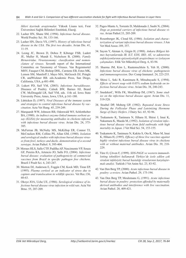
84 Bildir A and Gür S. Comparision of two different vaccination shedule for fight with Infectious Bursal Disease in Layer Hens
Etlik Vet Mikrobiyol Derg, http://vetkontrol.tarim.gov.tr/merkez Cilt 28, Sayı 2, 2017, 76-84
likleri üzerinde araştırmalar. Yüksek Lisans tezi, Fırat Üniversitesi Sağlık Bilimleri Enstitüsü, Elazığ.
22. Lasher HN, Shane SM, (1994). Infectious bursal disease. World Poultry Sci. 50, 133-166.
23. Lasher HN, Davis VS, (1997). History of infectious bursal disease in the USA. The first two decades. Avian Dis. 41, 11-19.
24. Leong JC, Brown D, Dobos P, Kibenge FSB, Ludert JE, Muller H, Mundt E, Nicholson B, (2000). Family Birnaviridae. Virustaxonomy: classification and nomen-clature of viruses. Seventh report of the International Committee on Taxonomy of Viruses. Van Regenmortel MHV, Fauquet CM, Bishop DHL, Carstens EB, Estes MK, Lemon SM, Maniloff J, Mayo MA, McGeoch DJ, Pringle CR, andWickner RB. eds.Academic Press, San Diego, California, USA, p.481-490.
25. Lukert PD, Saif YM, (2003). Infectious bursaldisease. Diseases of Poultry. Calnek BW, Barnes HJ, Beard CW, McDougald LR, Saif YM, eds. 11th ed. Iowa State University Press, Ames, Iowa, USA, p.161-180.
26. Lütticken D, (1997). Viral Diseases of the immune system and strategies to control infectious bursal disease by vac-cination. Acta Vet Hung. 45, 239-249.
27. Marquardt WW, Johnson RB, Odenwald WF, Schlotthober BA, (1980). An indirect enzyme-linked immuno sorbent as-say (ELISA) for measuring antibodies in chickens infected with infectious bursal disease virus. Avian Dis. 24, 375-385.
28. McFerran JB, McNulty MS, McKillop ER, Connor TJ, McCracken RM, Collins PS, Allan GM, (1980). Isolation and serological studies with infectious bursal disease virus-es from fowl, turkeys and ducks, demonstration of a second serotype. Avian Pathol. 9, 395-404.
29. Moraes HLS, Salle CTP, Padilha AP, Nascimento VP, Souza GF, Pereira RA, Artencio JO, Salle FO, (2004). Infectious bursal disease: evaluation of pathogenicity of commercial vaccines from Brazil in specific pathogen free chichens. Brazil J Poult Sci. 6, 243-247.
30. Morton DJ, Anderson E, Foggin CM, Kock MD, Tiran EP, (1995). Plasma cortisol as an indicator of stress due to capture and translocation in wildlife species. Vet Rec.136, 60-63.
31. Okoye JOA, Uche UE, (1986). Serological evidence of in-fectious bursal disease virus infection in wild rats. Acta Vet Brno. 55, 207-209.
32. Pages-Mante A, Torrents D, Maldonado J, Saubi N, (2004). Dogs as potential carriers of infectious bursal disease vi-rus. Avian Pathol.33, 205-209.
33. Rosenberger JK, Cloud SS, (1986). Isolation and charac-terization of variant infectious bursal disease viruses. J Am Vet Med Assoc.189, 357.
34. Sayım Y, Akman A, Girgin H, (1988). Ankara Bölgesi kü-mes hayvanlarında IB, ILT, EDS, IBD, AE, ve adenovirus enfeksiyonlarının epizootiyolojik araştırılması ve izolasyon çalışmaları. Etlik Vet Mikrobiyol Derg. 6, 83-94.
35. Sharma JM, Kim L, Rautemschlein S, Yeh H, (2000). Infections bursal disease virus of chickens: pathogenesis and immunosupression. Dev Comp Immunol. 24, 223-235.
36. Shirai L, Seki R, Kamimura R, Mitsubayashi S, (1994). Effects of invert soap with 0.05% sodium hydroxide on in-fectious bursal disease virus. Avian Dis. 38, 240-243.
37. SnedekerC, Wills FK, Moulthrop IM, (1967). Some stud-ies on the infectious bursal disease agent. Avian Dis. 11, 519-528.
38. Stoebel DP, Moberg GP, (1982). Repeated Acute Stress During the Follicular Phase and Luteinizing Hormone Surge of Dairy Heifers. J Dairy Sci. 65, 92-96.
39. Tsukamoto K, Tanimura N, Hihara H, Shirai J, Imai K, Nakamura K, Maeda M, (1992). Isolation of virulent infec-tious bursal disease virus from field outbreaks with high mortality in Japan. J Vet Med Sci. 54, 153-155.
40. Tsukamoto K, Tanimura N, Kakita S, Ota K, Mase M, Imai K, Hihara H, (1995). Efficacy of three live vaccines against highly virulent infectious bursal disease virus in chickens with or without maternal antibodies. Avian Dis. 39, 218-229.
41. Türe O, Çöven F, (1999). SDS-PAGE ve western immunob-lotting teknikleri kullanarak Türkiye’de izole edilen çok virulent infeksiyöz bursal hastalığı viruslarının karşılaştır-malı analizi. Turkish J Vet Anim Sci. 23, 83-92.
42. Van Den Berg TP, (2000). Acute infectious bursal disease in poultry: a review. Avian Pathol. 29, 175-194.
43. Van Den Berg TP, Meulemans G, (1991). Acute infectious bursal disase in poultry: protection afforded by maternally derived antibodies and interference with live vaccination. Avian Pathol. 20, 409-421.
Yazışma adresi / Correspondence: Elçin Günaydın, Veteriner Kontrol Merkez Araştırma Enstitüsü, Yetiştirme Hastalıkları Teşhis Laboratuvarı, Ankara, Türkiye E-posta: [email protected]
Etlik Vet Mikrobiyol Derg, 2017; 28 (2): 85-95 Araştırma Makalesi / Research Article
Yaygın Salmonella Serovarlarının Moleküler Tekniklerle Tiplendirilmesi
Elçin Günaydın1, Selahattin Şen2, K. Serdar Diker3, Derya Karataş Yeni4, Özlem Kardoğan5, H. Kaan Müştak3, Özlem Şahan6
1 Veteriner Kontrol Merkez Araştırma Enstitüsü, Yetiştirme Hastalıkları Teşhis Laboratuvarı, Ankara2 Veteriner Kontrol Merkez Araştırma Enstitüsü, Bakteriyolojik Teşhis Laboratuvarı, Ankara
3 Ankara Üniversitesi Veteriner Fakültesi, Mikrobiyoloji Anabilim Dalı, Ankara4 Veteriner Kontrol Merkez Araştırma Enstitüsü, Viral Aşı Üretim Laboratuvarı, Ankara
5 Veteriner Kontrol Merkez Araştırma Enstitüsü, Kanatlı hayvan Hastalıkları Teşhis Laboratuvarı, Ankara6 Mehmet Akif Ersoy Üniversitesi Veteriner Fakültesi, Mikrobiyoloji Anabilim Dalı, Burdur
Geliş Tarihi / Received: 10.09.2017, Kabul Tarihi / Accepted: 16.10.2017
Özet: Bu çalışmanın amacı, Veteriner Kontrol Merkez Araştırma Enstitüsü (VKMAE), Bakteriyolojik Teşhis Laboratuvarı sorumluluğunda 2009-2011 yılları arasında Salmonella Prevalans çalışması kapsamında, Kauffmann-White serotiplendirme şeması ile tiplendirmesi yapılan toplam 907 Salmonella izolatının, serogrup Salmonella multip-lex- PZR, Faz 1 Salmonella multiplex-PZR ve Faz 2 Salmonella multiplex-PZR kullanarak sahada yaygın olarak sirküle olan Salmonellla serovarları için dizayn edilen primerler ile maksimum 2-3 gün gibi kısa sürede, düşük maliyetle tiplen-dirmesini yapabilmektir. Bu çalışmayla, konvansiyonel yöntemle faz 1 ve faz 2 antijenlerinin eş zamanlı sergilenmemesi sonucunda ekstra laboratuvar iş yükü, olası çarpraz reaksiyonlar sebepli ekstra antiserum harcanması dolayısıyla yaşanı-lan zaman kaybı ve yüksek maliyetin üstesinden gelineceği öngörüldü. Ayrıca, 2 testin (multiplex-PZR ve konvansiyel serotiplendirme) örtüşmediği durumlar bakterilerin 6.5 kb operonunu inceleyen ribotiplendirme desteğiyle çözülmesi yoluna gidilmiştir. Çalışmada kullanılacak primerler ile 907 izolatın % 29.32’sinin (266 izolat; 15 farklı serovar) üç an-tijenik yapısı da tam olarak identifiye edilip, isimlendirilebilmiştir. İncelenen izolatların somatik, flagellar 1 ve flagellar 2 antijenlerinin sırasıyla; %54.56 (490), %90.95 (825) ve %78.94 (716)’ü tespit edilebilmiştir. İzolatların en azından % 50’sinin tam olarak isimlendirilebileceği öngörülmüştür ancak Salmonella Infantis (S. Infantis) ve S. Mbandaka suşa-rında somatik antijeni amplifiye eden primer olmasına rağmen somatik antijen tespit edilememiştir. Bu suşlar ribotip-lendirmeye alınmıştır. Konvansiyonel seroloji, multiplex-PZR ve ribotiplendirme değerlendirildiğinde; ribotiplendirme ve multiplex-PZR’ların gold standart metoda destek metodlar olarak kullanabileceği tespit edilmiştir. Özet olarak; çalışmada mevcut olan primerler sadece sahada yaygın olan serovarların somatik, faz 1 ve faz 2 antijenlerine spesifik oldukları için PZR tüm serovarları identifiye etmekte yetersiz kalmıştır. Sonuç olarak ucuz, hızlı ve duyarlı metot olan multiplex-PZR’ların belirli somatik, flagellar 1 ve 2 antijenlerinin tespitinde konvansiyonel serolojik yönteme destek olarak kullanılabileceği kanısına varılmıştır.Anahtar kelimeler: Salmonella serovarları, Multiplex-PZR
Molecular Typing of Common Salmonella SerovarsAbstract: The purpose of this study was to identify total 907 Salmonella isolates had been formerly identified by us-ing Kauffmann-White serotype scheme on the scope of Salmonella Prevalance Study between 2009-2011 under the responsibility of Veterinary Control Central Research Institute, Bacteriological Diagnosis Laboratory; by performing serogroup Salmonella multiplex-PCR, phase-1 Salmonella multiplex-PCR, phase-2 Salmonella multiplex-PCR with the specific primers designed for the common Salmonella serovars circulating around the field, in maximum 2-3 days, relatively short time, with a low cost. With this study, extra laboratory work load due to phase 1 and phase 2 flagellar antigens not being expressed synchronously, dilution of time and high cost due to probable cross-reactions requiring extra antiserum usage are envisaged to have been overcomed. Moreover, the occasions where 2 tests (multiplex-PCRs and conventional serotyping) are not overlapped was resolved with the support of ribotyping which examine the 6.5 kb operone of bacteriae. Consequently, identification by both methods, and evaluating the results comparatively is thought to be beneficialWith the primers used in the study, three antigenic structures of 29.32% (266 isolates; 15 different se-rovars) of the 907 isolates were fully identified. Somatic, phase 1 and phase 2 antigens were deterrmined with a percent-age of 54.56 (490 isolates), 90.95 (825 isolates) and 78.94 (716 isolates), respectively. At least 50% of the isolates were preasumed to be fully identified. However, the somatic antigen of Salmonella Infantis (S. Infantis) ve S. Mbandaka were not detected altough the primers amplfying somatic antigen were present. These two sereovars were ribotyped. When conventional serology, multiplex-PCRs and ribotyping were evaluated, multiplex-PCRs and ribotyping were found to be supportive to conventional serology. In a summary, due to present primers, specific to somatic, phase 1 and phase 2 of
86 Günaydın E ve ark. Yaygın Salmonella Serovarlarının Moleküler Tekniklerle Tiplendirilmesi
Etlik Vet Mikrobiyol Derg, http://vetkontrol.tarim.gov.tr/merkez Cilt 28, Sayı 2, 2017, 85-95
the only common Salmonella serovars in the field, PCR has deficiencies for identifying whole serovars. Consequently, the multiplex-PCRs termed cheap, sensitive, and rapid were decided to be supportive to conventional serology for defin-ing specific somatic, phase 1 and 2 antigens.Key words: Salmonella serovars, Multiplex-PCR
GirişSalmonella, hayvanların ve insanların en önemli patojenlerinden biri olup, aynı zamanda zoonozdur. Günümüze kadar tiplendirilen toplam 2700’ e ya-kın Salmonella serovarı mevcuttur. Bunlardan 2587 adedi Salmonella enterica, 23 adedi Salmonella bongori alt grubunda bulunmaktadır (8). Salmonella enterica altı alt türden oluşmaktadır; Salmonella en-terica subsp. enterica, Salmonella enterica subsp. salamae, Salmonella enterica subsp. arizonae, Salmonella enterica subsp. diarizonae, Salmonella enterica subsp. houtenae ve Salmonella enterica subsp. indica veya sırasıyla Salmonella enterica subsp. I, II, IIIa, IIIb, IV ve VI (17). Salmonella en-terica subsp. I genellikle insanlardan ve sıcak-kanlı hayvanlardan izole edilir. Klinik laboratuvarlarda izole edilen serotiplerin büyük çoğunluğu; 1547 adedi bu alt türe aittir (8, 17).
Salmonella serovarlarının isimlendirir-ken, Kauffmann-White serotiplendirme şema-sı, Salmonella izolatlarının tiplendirmesinde birçok laboratuvar tarafından kullanılmaktadır. Serotiplendirme, Salmonella’nın hücre duvarında bulunan somatik (O) ve flagellar (H) faktör antijen-leri temel alınarak yapılır. H faktörü Salmonella su-şunun serotip kimliğini belirlerken, O faktörü grubu tespit eder (8). O antijenleri, gram negatif bakteri-lerin önemli komponentlerinden olan lipopolisak-kariti (LPS) oluşturan spesifik polisakkaritlerdir. O antijen sentezinden sorumlu genler, rfb diye isim-lendirilen bir gen kümesinde, kromozom üzerinde grup halinde bulunurlar (2). rfb genleri nükleotid şeker biyosentez yolunu ve tekrarlayan ünitelerin bir arada bulunması için gerekli olan transferazla-rı kodlamaktadır. Oniki potansiyel transmembran segmentini içeren proteini kodlayan ve daha ön-celeri rfbX, şimdi ise wzx olarak adlandırılan gen, tüm Salmonella O antijen gen kümelerinde bulun-maktadır. Farklı O antijen kümelerinden wzx pro-teinin yapısal homoloji gösterdiği fakat aminoasit sekans seviyesinde çok az bir benzerliğin olduğu öngörülmektedir (15). Wzx’in sitoplazmik memb-ranın sitoplazmik kısmından periplazmik kısmına
O-ünitelerinin transferinden sorumlu olduğu bildi-rilmiştir (20).
Faz 1 ve faz 2 flagellar H antijenleri alternatif olarak, sırasıyla fliC ve fliB genleri sorumluluğun-da faz varyasyon mekanizması tarafından eksprese edilirler. Bu genler kromozom üzerinde farklı iki lokalizasyonda bulunurlar. fljBA operonu, Hin re-kombinazı kodlayan hin genini içerir; fljB geni pha-se 2 flagellinlerini, fljA geni de fliC için reprösörü kodlamaktadır. Hin rekombinaz geni, fljBA operonu için promotör içeren kromozomun 993 bp’lık seg-mentinin reversible inversiyonunu katalize eder. Bir oryantasyonda, promotör fliC geninin represyonu-na yol açarak, fljA ve fljB genlerinin transkripsiyo-nunu sağlar. Hin’in diğer oryantasyonunda fljA ve fljB genleri eksprese edilmez, faz 2 flagellinin üstü kapanır, bu da faz 1 flagellinin eksprese edilmesini olanaklı hale getirir. (13, 25). Bu allelerden bazıları tek bir faktörle (i, d veya r), diğerleri ise birçok alt faktörle (l,v, g,m, e,n,x) tanımlanır. Salmonella fla-gellinlerinin aminoasit sekanslarının karşılaştırması ile 8 farklı bölge tanımlanmıştır. Amino ve karboksi terminal sekansları (sırasıyla bölge I ve II ve böl-ge VIII) oldukça korunmuştur. Polimerizasyon ve transport için çok önemli oldukları düşünülmekte-dir. Bölge IV, V ve VI’dan oluşan merkez bölge ise hem sekanslarda hem de flagellar antijenler arası uzunluklarda çok fazla çeşitlilik gösterir ve genelde H antijeninin epitopunu belirlediği düşünülmekte-dir (16, 23).
Yukarıda Salmonella somatik ve flagellar an-tijenlerinin ekspresyonu ile ilgili açıklamalar ya-pılmıştır. Flagellar 1 ve flagellar 2 antijenlerinin eş zamanlı sergilenmemesi dolayısıyla konvansiyonel yöntemde, flagellar antijenlerin ayrı ayrı açığa çıka-rılmasıyla Salmonelların karakterizasyonu yapıla-bilmektedir (17). Bu çerçevede, konvansiyonel yön-temle bu işlem, her şey yolunda giderse, minimum 5 iş günü gibi bir zaman dilimini, çapraz reaksiyonlar sebebiyle bazen tekrarları ve gerek besiyeri gerekse antiserum harcamaları dolayısıyla yüksek bir mali-yeti gerektirmektedir.
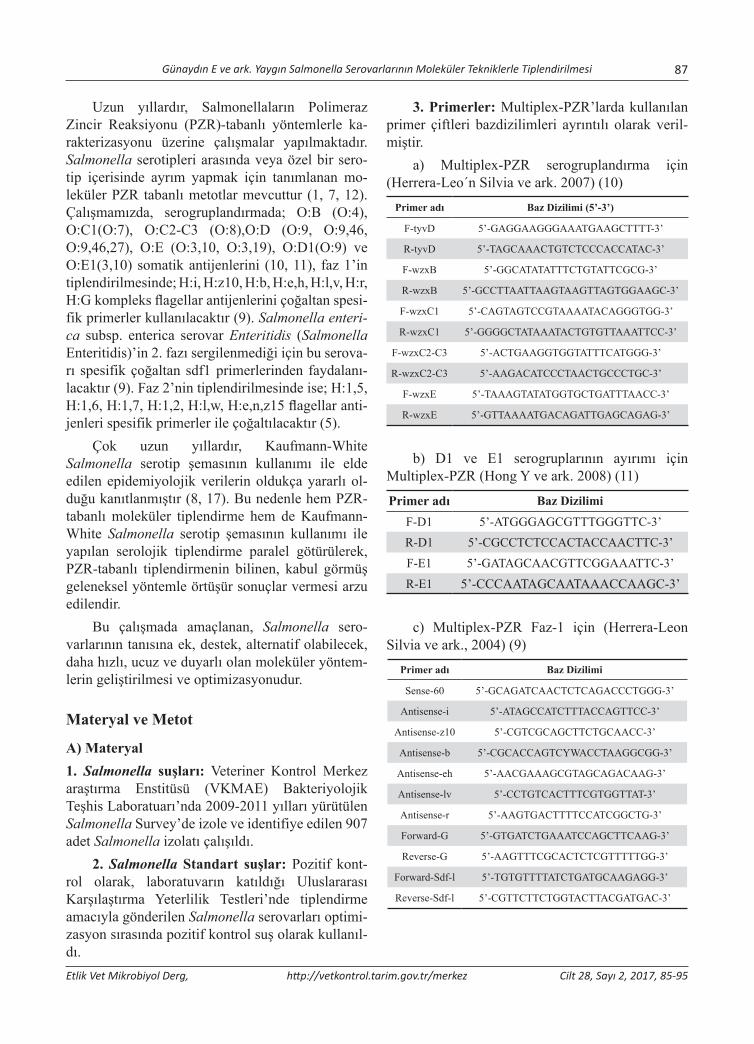
Günaydın E ve ark. Yaygın Salmonella Serovarlarının Moleküler Tekniklerle Tiplendirilmesi 87
Etlik Vet Mikrobiyol Derg, http://vetkontrol.tarim.gov.tr/merkez Cilt 28, Sayı 2, 2017, 85-95
Uzun yıllardır, Salmonellaların Polimeraz Zincir Reaksiyonu (PZR)-tabanlı yöntemlerle ka-rakterizasyonu üzerine çalışmalar yapılmaktadır. Salmonella serotipleri arasında veya özel bir sero-tip içerisinde ayrım yapmak için tanımlanan mo-leküler PZR tabanlı metotlar mevcuttur (1, 7, 12). Çalışmamızda, serogruplandırmada; O:B (O:4), O:C1(O:7), O:C2-C3 (O:8),O:D (O:9, O:9,46, O:9,46,27), O:E (O:3,10, O:3,19), O:D1(O:9) ve O:E1(3,10) somatik antijenlerini (10, 11), faz 1’in tiplendirilmesinde; H:i, H:z10, H:b, H:e,h, H:l,v, H:r, H:G kompleks flagellar antijenlerini çoğaltan spesi-fik primerler kullanılacaktır (9). Salmonella enteri-ca subsp. enterica serovar Enteritidis (Salmonella Enteritidis)’in 2. fazı sergilenmediği için bu serova-rı spesifik çoğaltan sdf1 primerlerinden faydalanı-lacaktır (9). Faz 2’nin tiplendirilmesinde ise; H:1,5, H:1,6, H:1,7, H:1,2, H:l,w, H:e,n,z15 flagellar anti-jenleri spesifik primerler ile çoğaltılacaktır (5).
Çok uzun yıllardır, Kaufmann-White Salmonella serotip şemasının kullanımı ile elde edilen epidemiyolojik verilerin oldukça yararlı ol-duğu kanıtlanmıştır (8, 17). Bu nedenle hem PZR-tabanlı moleküler tiplendirme hem de Kaufmann-White Salmonella serotip şemasının kullanımı ile yapılan serolojik tiplendirme paralel götürülerek, PZR-tabanlı tiplendirmenin bilinen, kabul görmüş geleneksel yöntemle örtüşür sonuçlar vermesi arzu edilendir.
Bu çalışmada amaçlanan, Salmonella sero-varlarının tanısına ek, destek, alternatif olabilecek, daha hızlı, ucuz ve duyarlı olan moleküler yöntem-lerin geliştirilmesi ve optimizasyonudur.
Materyal ve Metot
A) Materyal1. Salmonella suşları: Veteriner Kontrol Merkez araştırma Enstitüsü (VKMAE) Bakteriyolojik Teşhis Laboratuarı’nda 2009-2011 yılları yürütülen Salmonella Survey’de izole ve identifiye edilen 907 adet Salmonella izolatı çalışıldı.
2. Salmonella Standart suşlar: Pozitif kont-rol olarak, laboratuvarın katıldığı Uluslararası Karşılaştırma Yeterlilik Testleri’nde tiplendirme amacıyla gönderilen Salmonella serovarları optimi-zasyon sırasında pozitif kontrol suş olarak kullanıl-dı.
3. Primerler: Multiplex-PZR’larda kullanılan primer çiftleri bazdizilimleri ayrıntılı olarak veril-miştir.
a) Multiplex-PZR serogruplandırma için (Herrera-Leo´n Silvia ve ark. 2007) (10)
Primer adı Baz Dizilimi (5’-3’)
F-tyvD 5’-GAGGAAGGGAAATGAAGCTTTT-3’
R-tyvD 5’-TAGCAAACTGTCTCCCACCATAC-3’
F-wzxB 5’-GGCATATATTTCTGTATTCGCG-3’
R-wzxB 5’-GCCTTAATTAAGTAAGTTAGTGGAAGC-3’
F-wzxC1 5’-CAGTAGTCCGTAAAATACAGGGTGG-3’
R-wzxC1 5’-GGGGCTATAAATACTGTGTTAAATTCC-3’
F-wzxC2-C3 5’-ACTGAAGGTGGTATTTCATGGG-3’
R-wzxC2-C3 5’-AAGACATCCCTAACTGCCCTGC-3’
F-wzxE 5’-TAAAGTATATGGTGCTGATTTAACC-3’
R-wzxE 5’-GTTAAAATGACAGATTGAGCAGAG-3’
b) D1 ve E1 serogruplarının ayırımı için Multiplex-PZR (Hong Y ve ark. 2008) (11)
Primer adı Baz Dizilimi
F-D1 5’-ATGGGAGCGTTTGGGTTC-3’R-D1 5’-CGCCTCTCCACTACCAACTTC-3’F-E1 5’-GATAGCAACGTTCGGAAATTC-3’R-E1 5’-CCCAATAGCAATAAACCAAGC-3’
c) Multiplex-PZR Faz-1 için (Herrera-Leon Silvia ve ark., 2004) (9)
Primer adı Baz Dizilimi
Sense-60 5’-GCAGATCAACTCTCAGACCCTGGG-3’
Antisense-i 5’-ATAGCCATCTTTACCAGTTCC-3’
Antisense-z10 5’-CGTCGCAGCTTCTGCAACC-3’
Antisense-b 5’-CGCACCAGTCYWACCTAAGGCGG-3’
Antisense-eh 5’-AACGAAAGCGTAGCAGACAAG-3’
Antisense-lv 5’-CCTGTCACTTTCGTGGTTAT-3’
Antisense-r 5’-AAGTGACTTTTCCATCGGCTG-3’
Forward-G 5’-GTGATCTGAAATCCAGCTTCAAG-3’
Reverse-G 5’-AAGTTTCGCACTCTCGTTTTTGG-3’
Forward-Sdf-l 5’-TGTGTTTTATCTGATGCAAGAGG-3’
Reverse-Sdf-l 5’-CGTTCTTCTGGTACTTACGATGAC-3’
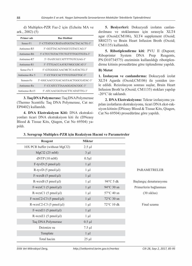
88 Günaydın E ve ark. Yaygın Salmonella Serovarlarının Moleküler Tekniklerle Tiplendirilmesi
Etlik Vet Mikrobiyol Derg, http://vetkontrol.tarim.gov.tr/merkez Cilt 28, Sayı 2, 2017, 85-95
d) Multiplex-PZR Faz-2 için (Echeita MA ve ark., 2002) (5)
Primer adı Baz Dizilimi
Sense-F1 5’-CTTATGCCRATAATGGTACTACACTG-3’
Antisense-R5 5’-GGTTACAGVAGCCGTACCAG-3’
Antisense-R6 5’-CTCCTGTACTTCTGTTTTGGTTGTA-3’
Antisense-R7 5’-TAATCGCCATTTTTGTCGAG-3’
Antisense-R1 5’-TTTGACCAAYKYMGCGSCAT-3’
Sense-Fw 5 5’-GTGGGGCAACMCTCAATACTG-3’
Antisense-Rw 5 5’-CCTGCCACTTTCGTGGTTGC-3’
Sensen-Fe 5’-GGCAACCCGACAGTAACTGGCGATAC-3’
Antisense-Rx 5’-CCATCCTTAAAGGATACGGC-3’
Antisense-Rz15 5’-ATCAACGGTAACTTCATATTTG-3’
3. Taq DNA Polymerase: Taq DNA Polymerase (Thermo Scentific Taq DNA Polymerase, Cat no: EP0402) kullanıldı.
4. DNA Ekstraksiyon Kiti: DNA ekstraksi-yonları ticari DNA ekstraksiyon kiti ile (DNeasy Blood & Tissue Kits, Qiagen, Cat No 69504) ya-pıldı.
5. Besiyerleri: Dokuzyedi izolatın canlan-dırılması ve stoklanması için sırasıyla XLT4 agar (Oxoid;CM106), XLT4 supplement (Oxoid; SR0237) ve Brain Heart Infusion Broth (Oxoid; CM1135) kullanıldı.
5. Ribotiplendirme kiti: PVU II (Dupont; Riboprinter System DNA Prep Reagents, PN:D10734575) enziminin kullanıldığı ribotiplen-dirme kitinin prosedürüne göre tiplendirme yapıldı.B) Metot1. İzolasyon ve canlandırma: Dokuzyedi izolat XLT4 Agarda (Oxoid;CM106) ile yeniden izo-le edildi. Reizolasyon sonrası suşlar, Brain Heart Infusion Broth’ta (Oxoid; CM1135) stokları yapılıp -20°C’de saklandı.
2. DNA Ekstraksiyonu: Tekrar izolasyonu ya-pılan izolatların ekstraksiyonu, ticari DNA ekst-rak-siyon kitinin (DNeasy Blood & Tissue Kits, Qiagen, Cat No 69504) prosedürüne göre yapıldı.
3. Serogrup Multiplex-PZR için Reaksiyon Hacmi ve Parametreler
Reagent Miktar
10X PCR buffer (without MgCl2) 2.5 µl
MgCl2 (25 mM) 3 µl
dNTP (10 mM) 0.5µl
F-tyvD (5 pmol/µl) 1 µl
R-tyvD (5 pmol/µl) 1 µl PARAMETRELER
F-wzxB (5 pmol/µl) 1 µl
R-wzxB (5 pmol/µl) 1 µl 94°C 5 dk Başlangıç denaturasyonu
F-wzxC1 (5 pmol/µl) 1 µl 94°C 30 sn Primerlerin bağlanması
R-wzxC1 (5 pmol/µl) 1 µl 57°C 40 sn (30 siklus)
F-wzxC2-C3 (5 pmol/µl) 1 µl 72°C 30 sn
R-wzxC2-C3 (5 pmol/µl) 1 µl 72°C 10 dk Final uzama
F-wzxE1 (5 pmol/µl) 1 µl
R-wzxE1 (5 pmol/µl) 1 µl
Taq DNA Polymerase 0.5 µl
Deionize su 7.5 µl
Template 1 µl
Total hacim 25 µl
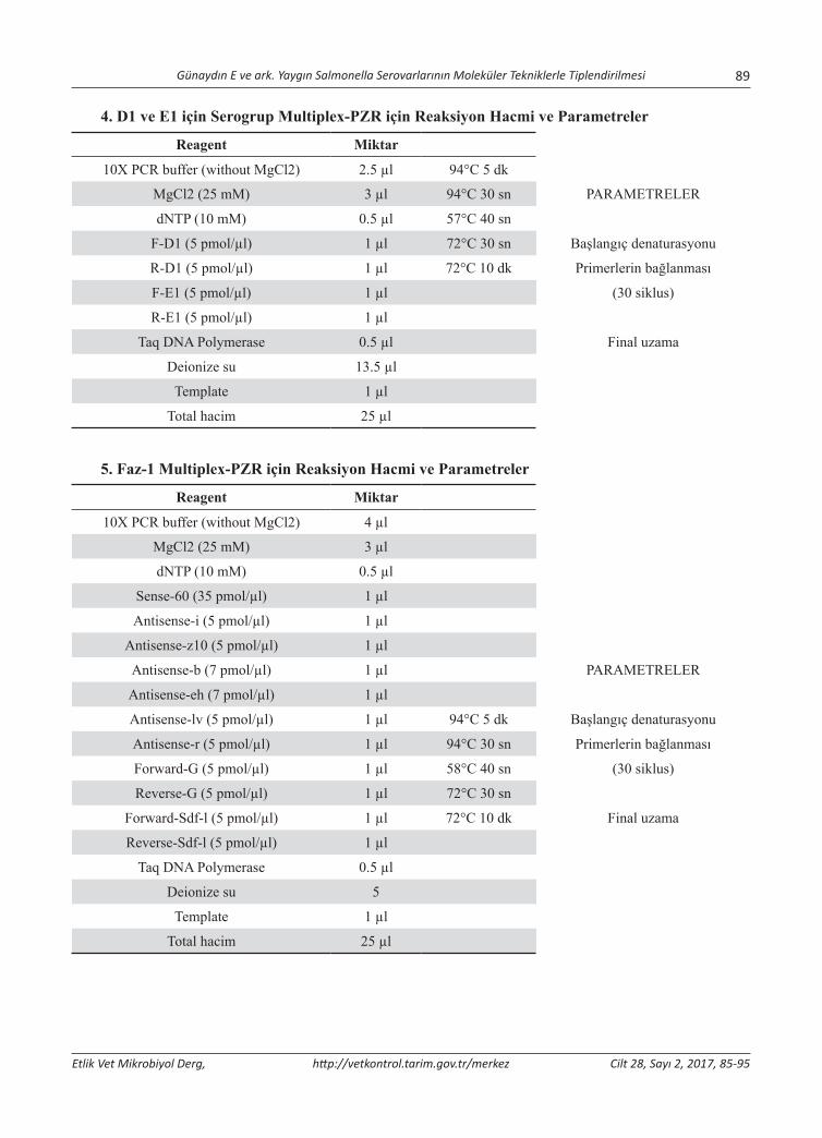
Günaydın E ve ark. Yaygın Salmonella Serovarlarının Moleküler Tekniklerle Tiplendirilmesi 89
Etlik Vet Mikrobiyol Derg, http://vetkontrol.tarim.gov.tr/merkez Cilt 28, Sayı 2, 2017, 85-95
4. D1 ve E1 için Serogrup Multiplex-PZR için Reaksiyon Hacmi ve Parametreler
Reagent Miktar
10X PCR buffer (without MgCl2) 2.5 µl 94°C 5 dk
MgCl2 (25 mM) 3 µl 94°C 30 sn PARAMETRELER
dNTP (10 mM) 0.5 µl 57°C 40 sn
F-D1 (5 pmol/µl) 1 µl 72°C 30 sn Başlangıç denaturasyonu
R-D1 (5 pmol/µl) 1 µl 72°C 10 dk Primerlerin bağlanması
F-E1 (5 pmol/µl) 1 µl (30 siklus)
R-E1 (5 pmol/µl) 1 µl
Taq DNA Polymerase 0.5 µl Final uzama
Deionize su 13.5 µl
Template 1 µl
Total hacim 25 µl
5. Faz-1 Multiplex-PZR için Reaksiyon Hacmi ve Parametreler
Reagent Miktar
10X PCR buffer (without MgCl2) 4 µl
MgCl2 (25 mM) 3 µl
dNTP (10 mM) 0.5 µl
Sense-60 (35 pmol/µl) 1 µl
Antisense-i (5 pmol/µl) 1 µl
Antisense-z10 (5 pmol/µl) 1 µl
Antisense-b (7 pmol/µl) 1 µl PARAMETRELER
Antisense-eh (7 pmol/µl) 1 µl
Antisense-lv (5 pmol/µl) 1 µl 94°C 5 dk Başlangıç denaturasyonu
Antisense-r (5 pmol/µl) 1 µl 94°C 30 sn Primerlerin bağlanması
Forward-G (5 pmol/µl) 1 µl 58°C 40 sn (30 siklus)
Reverse-G (5 pmol/µl) 1 µl 72°C 30 sn
Forward-Sdf-l (5 pmol/µl) 1 µl 72°C 10 dk Final uzama
Reverse-Sdf-l (5 pmol/µl) 1 µl
Taq DNA Polymerase 0.5 µl
Deionize su 5
Template 1 µl
Total hacim 25 µl
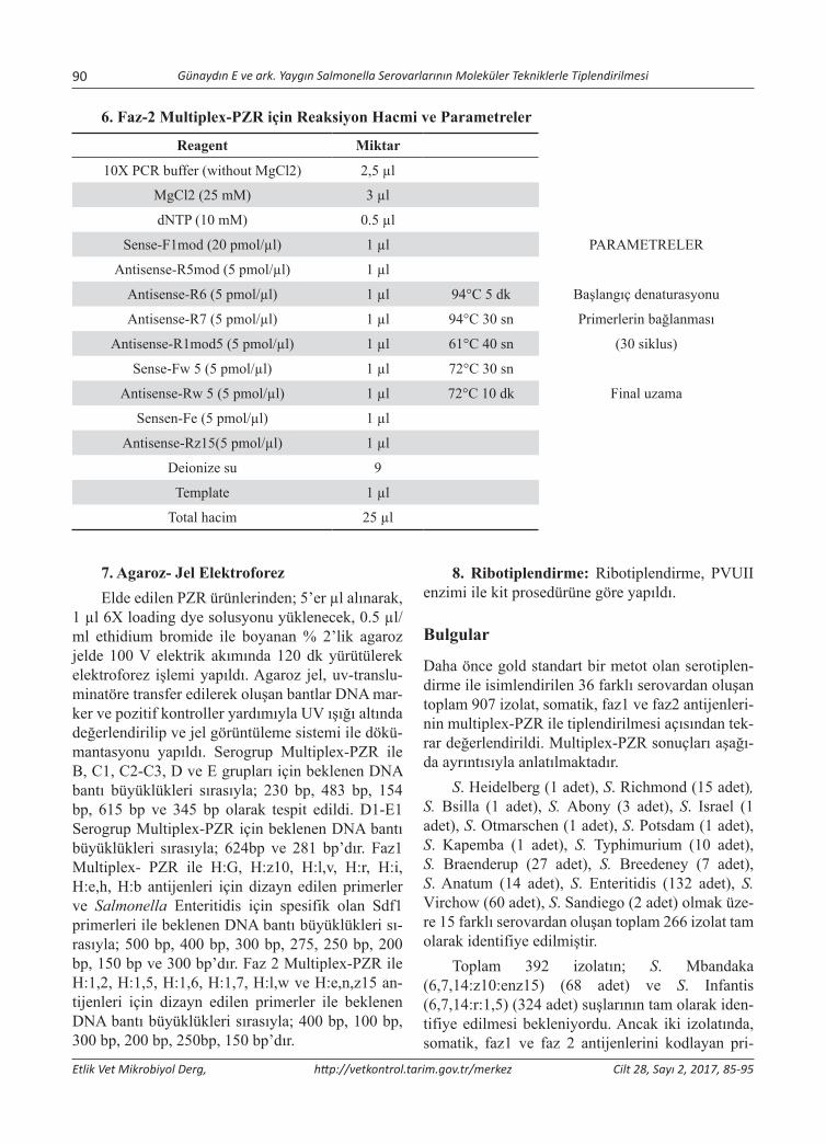
90 Günaydın E ve ark. Yaygın Salmonella Serovarlarının Moleküler Tekniklerle Tiplendirilmesi
Etlik Vet Mikrobiyol Derg, http://vetkontrol.tarim.gov.tr/merkez Cilt 28, Sayı 2, 2017, 85-95
6. Faz-2 Multiplex-PZR için Reaksiyon Hacmi ve Parametreler
Reagent Miktar
10X PCR buffer (without MgCl2) 2,5 µl
MgCl2 (25 mM) 3 µl
dNTP (10 mM) 0.5 µl
Sense-F1mod (20 pmol/µl) 1 µl PARAMETRELER
Antisense-R5mod (5 pmol/µl) 1 µl
Antisense-R6 (5 pmol/µl) 1 µl 94°C 5 dk Başlangıç denaturasyonu
Antisense-R7 (5 pmol/µl) 1 µl 94°C 30 sn Primerlerin bağlanması
Antisense-R1mod5 (5 pmol/µl) 1 µl 61°C 40 sn (30 siklus)
Sense-Fw 5 (5 pmol/µl) 1 µl 72°C 30 sn
Antisense-Rw 5 (5 pmol/µl) 1 µl 72°C 10 dk Final uzama
Sensen-Fe (5 pmol/µl) 1 µl
Antisense-Rz15(5 pmol/µl) 1 µl
Deionize su 9
Template 1 µl
Total hacim 25 µl
7. Agaroz- Jel ElektroforezElde edilen PZR ürünlerinden; 5’er µl alınarak,
1 µl 6X loading dye solusyonu yüklenecek, 0.5 µl/ml ethidium bromide ile boyanan % 2’lik agaroz jelde 100 V elektrik akımında 120 dk yürütülerek elektroforez işlemi yapıldı. Agaroz jel, uv-translu-minatöre transfer edilerek oluşan bantlar DNA mar-ker ve pozitif kontroller yardımıyla UV ışığı altında değerlendirilip ve jel görüntüleme sistemi ile dökü-mantasyonu yapıldı. Serogrup Multiplex-PZR ile B, C1, C2-C3, D ve E grupları için beklenen DNA bantı büyüklükleri sırasıyla; 230 bp, 483 bp, 154 bp, 615 bp ve 345 bp olarak tespit edildi. D1-E1 Serogrup Multiplex-PZR için beklenen DNA bantı büyüklükleri sırasıyla; 624bp ve 281 bp’dır. Faz1 Multiplex- PZR ile H:G, H:z10, H:l,v, H:r, H:i, H:e,h, H:b antijenleri için dizayn edilen primerler ve Salmonella Enteritidis için spesifik olan Sdf1 primerleri ile beklenen DNA bantı büyüklükleri sı-rasıyla; 500 bp, 400 bp, 300 bp, 275, 250 bp, 200 bp, 150 bp ve 300 bp’dır. Faz 2 Multiplex-PZR ile H:1,2, H:1,5, H:1,6, H:1,7, H:l,w ve H:e,n,z15 an-tijenleri için dizayn edilen primerler ile beklenen DNA bantı büyüklükleri sırasıyla; 400 bp, 100 bp, 300 bp, 200 bp, 250bp, 150 bp’dır.
8. Ribotiplendirme: Ribotiplendirme, PVUII enzimi ile kit prosedürüne göre yapıldı.
Bulgular
Daha önce gold standart bir metot olan serotiplen-dirme ile isimlendirilen 36 farklı serovardan oluşan toplam 907 izolat, somatik, faz1 ve faz2 antijenleri-nin multiplex-PZR ile tiplendirilmesi açısından tek-rar değerlendirildi. Multiplex-PZR sonuçları aşağı-da ayrıntısıyla anlatılmaktadır.
S. Heidelberg (1 adet), S. Richmond (15 adet), S. Bsilla (1 adet), S. Abony (3 adet), S. Israel (1 adet), S. Otmarschen (1 adet), S. Potsdam (1 adet), S. Kapemba (1 adet), S. Typhimurium (10 adet), S. Braenderup (27 adet), S. Breedeney (7 adet), S. Anatum (14 adet), S. Enteritidis (132 adet), S. Virchow (60 adet), S. Sandiego (2 adet) olmak üze-re 15 farklı serovardan oluşan toplam 266 izolat tam olarak identifiye edilmiştir.
Toplam 392 izolatın; S. Mbandaka (6,7,14:z10:enz15) (68 adet) ve S. Infantis (6,7,14:r:1,5) (324 adet) suşlarının tam olarak iden-tifiye edilmesi bekleniyordu. Ancak iki izolatında, somatik, faz1 ve faz 2 antijenlerini kodlayan pri-
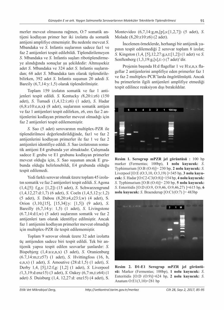
Günaydın E ve ark. Yaygın Salmonella Serovarlarının Moleküler Tekniklerle Tiplendirilmesi 91
Etlik Vet Mikrobiyol Derg, http://vetkontrol.tarim.gov.tr/merkez Cilt 28, Sayı 2, 2017, 85-95
merler mevcut olmasına rağmen, O:7 somatik an-tijeni kodlayan primer her iki izolatta da somatik antijeni amplifiye etmemiştir. Bu nedenle mevcut S. Mbandaka ve S. Infantis suşlarının sadece faz1 ve faz 2 antijenleri tespit edilebildi. Tiplendirilemeyen S. Mbandaka ve S. Infantis suşları ribotiplendirme-ye alındığında sonuçlar şu şekildedir: Altmışsekiz adet S. Mbandaka ve 324 adet S. Infantis suşların-dan; 68 adet S. Mbandaka tam olarak tiplendirile-bilirken, 392 adet S. Infantis suşunun 20 adedi S. Bareily (6,7,14:y:1,5) olarak tiplendirilmiştir.
Toplam 159 izolatın somatik ve faz 1 anti-jenleri tespit edildi. S. Kentucky (8,20:i:z6) (150 adet), S. Tumadi (1,4,12:i:z6) (1 adet), S. Hadar (6,8:z10:e,n,x) (8 adet), suşlarının somatik antijen ve faz 1 antijenleri tespit edilirken, z6, enx faz 2 an-tijenlerini kodlayan primerler mevcut olmadığı için faz 2 antijenleri tespit edilememiştir.
S. Sao (5 adet) serovarının multiplex-PZR ile tiplendirilmesi değerlendirildiğinde, faz1 ve faz 2 antijenlerini kodlayan primerler ile faz 1 ve faz 2 antijenleri identifiye edildi. S. Sao izolatoının soma-tik antijeni E4 grubunda yer almaktadır. Çalışmada sadece E grubu ve E1 grubunu kodlayan primerler mevcut olduğu için, S. Sao suşunun ancak E gru-bunda olduğu belirlenebildi, E4 grubunda olduğu tespit edilemedi.
Yedi farklı serovar olmak üzere toplam 45 izola-tın somatik ve faz 2 antijenleri tespit edildi. S. Agona (1,4,[5]: f,g,s: [1,2]) (15 adet), S. Schwarzengrund (1,4,12,27:d:1,7) (6 adet), S. Coeln (1,4,5,12:y:1,2) (5 adet), S. Dabou (8,20:z4,z23:l,w) (4 adet), S. Orion (3,10,[15], [15,34]:y: [1,5]) (9 adet), S. Bareilly (6,7,14:y: 1,5) (1 adet), S. Livingstone (6.7,14:d:l,w) (5 adet) suşlarının somatik ve faz 2 antijenleri tam olarak identifiye edilmiştir. Ancak faz 1 antijenini kodlayan primerler mevcut olmadığı için multiplex-PZR ile tespit edilememiştir.
Toplam 9 serovar olmak üzere 32 adet izolatta üç antijenden sadece biri tespit edildi. Tek bir an-tijenik yapısı tespit edilen seovarlar şunlardır: S. Bispebjerg (1,4:a:e,n,x) (3 adet), S. Oranienburg (6.7,14:m,t:z57) (1 adet), S. Hvittingfoss (16, b, e,n,x) (1 adet), S. Amoutive (28:d:1,5) (1 adet), S. Derby 1,4, [5],12:f,g: [1,2] (1 adet), S. Liverpool (1,3,19:d:enz15) (3 adet), S. Oakey (6,7:m,t:z64) (1 adet) S. Duisburg (1,4, 12,27:d: enz15) (4 adet), S.
Montevideo (6,7,14:g,m,[p],s:[1,2,7]) (5 adet), S. Molade (8,20:z10:z6) (2 adet).
İncelenen örneklerde, herhangi bir antijenik ya-pının tespit edilemediği 2 serovar toplam 8 izolat; S. Kingston (1,4, [5],12,27:g,s,t:[1,2]) (1 adet) ve S. Senftenberg (1,3,19:g,[s],t:-) (7 adet)’dir.
Projenin başında H:d flagellar 1 ve H:e,n,x fla-gellar 2 antijenlerini amplifiye eden primerler faz 1 ve faz 2 multiplex-PCR’larda öngörülmüştü. Ancak bu primerlerin ilgili antijenleri amplifiye etmediği tespit edilince reaksiyon dışı bırakıldılar.
Resim 1. Serogrup mPZR jel görüntüsü : 100 bp marker (Fermentas; 100bp), 1 nolu kuyucuk: S. Typhimurium [O:B (O:4)]= 230 bp, 2 nolu kuyucuk: S. Liverpool [O:E (O:3,10, O:3,19) ]=345 bp, 3 nolu kuyu-cuk: S. Hadar [O:C2-C3(O:8)]=154 bp, 4 nolu kuyucuk: S. Typhimurium [O:B (O:4)]= 230 bp, 5 nolu kuyucuk: S. Enteritidis [O:D (O:9, O:9,46, O:9,46,27) ]=615 bp, 6 nolu kuyucuk: S. Braenderup [O:C1(O:7) ]= 483bp
Resim 2. D1-E1 Serogrup mPZR jel görüntü-sü: Marker (Fermentas; 100bp), 1 nolu kuyucuk: S. Enteritidis [O:D (O:9)]=624 bp, 2 nolu kuyucuk: S. Anatum O:E1(3,10)=281 bp
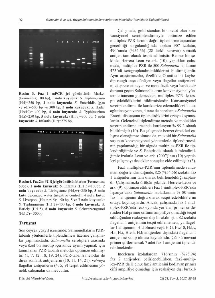
92 Günaydın E ve ark. Yaygın Salmonella Serovarlarının Moleküler Tekniklerle Tiplendirilmesi
Etlik Vet Mikrobiyol Derg, http://vetkontrol.tarim.gov.tr/merkez Cilt 28, Sayı 2, 2017, 85-95
Resim 3. Faz 1 mPCR jel görüntüsü: Marker (Fermentas; 100 bp), 1 nolu kuyucuk: S. Typhimurium (H:i)=250 bp, 2 nolu kuyucuk: S. Enteritidis (g,m ve sdf)=500 bp ve 300 bp, 3 nolu kuyucuk: S. Hadar (H:z10)= 400 bp, 4 nolu kuyucuk: S. Typhimurium (H:i)=250 bp, 5 nolu kuyucuk: (H:l,v)=300 bp, 6 nolu kuyucuk: S. Infantis (H:r)=275 bp,
Resim 4. Faz 2 mPCR jel görüntüsü: Marker (Fermentas; 50bp), 1 nolu kuyucuk: S. Infantis (H:1,5)=100bp, 2 nolu kuyucuk: S. Livingstone (H:l,w)=250 bp, 3 nolu kutu:deionized water (negative control), 4 nolu kutu: S. Liverpool (H:e,n,z15): 150 bp, 5 ve 7 nolu kuyucuk: S. Typhimurium (H:1,2)=400 bp, 6 nolu kuyucuk: S. Bariely (H:1,5), 8 nolu kuyucuk: S. Schwarzengrund (H:1,7)= 300bp
TartışmaSon çeyrek yüzyıl içerisinde; Salmonellaların PZR-tabanlı yöntemlerle tiplendirmesi üzerine çalışma-lar yapılmaktadır. Salmonella serotipleri arasında veya özel bir serotip içerisinde ayrım yapmak için tanımlanan PZR-tabanlı metotlar optimize edilmiş-tir. (1, 7, 12, 18, 19, 24). PCR-tabanlı metotlar ile direk somatik antijenlerin (10, 11, 14, 21), ve/veya flagellar antijenlerin (4, 5, 9) tespit edilmesine yö-nelik çalışmalar da mevcuttur.
Çalışmada, gold standart bir metot olan kon-vansiyonel serotiplendirmeyle optimize edilen multiplex-PZR’larının doğru tiplendirme açısından geçerliliği sorgulandığında toplam 907 izolatın, 490’nında (%54.56) (28 farklı serovar) somatik antijen tam olarak tespit edilmiştir. Benzer bir şe-kilde, Herrera-Leon ve ark. (10), yaptıkları çalış-mada, multiplex-PZR ile 500 Salmonella izolatının 423’nü serogruplandırabildiklerini bildirmişlerdir. Aynı araştırmacılar, özellikle O-antijenini kaybe-dip rough suşa dönüşen veya flagellar antijenleri-ni eksprese etmeyen ve monofazik veya hareketsiz duruma geçen Salmonellalarıın konvansiyonel yön-temle tanısına gidemezken, multiplex-PZR ile tes-pit edebildiklerini bildirmişlerdir. Konvansiyonel serotiplendirme ile karakterize edemedikleri 1 oto-aglutinasyon veren, 4 tane de hareketsiz Salmonella Enteritidis suşunu tiplendirdiklerini ortaya koymuş-lardır. Geleneksel tiplendirme metodu ve moleküler serotiplendirme arasında korelasyon % 99.2 olarak bildirlmiştir (10). Bu çalışmada benzer örnekleri ça-lışma olanağımız olmasa da, mukoid bir Salmonella suşunun konvansiyonel yöntemlerle tiplendirmesi-nin yapılamadığı bir olguda multiplex-PZR ile tip-lendirdiğimiz ve S. Enteritidis olarak isimlendirdi-ğimiz izolatla Leon ve ark. (2007)’nın (10) yaptık-leri çalışmayı destekler sonuçlar elde edilmiştir (3).
Faz1 multiplex-PZR’nun tiplendirmede randı-manı değerlendirildiğinde, 825 (%54.56) izolatın faz 1 antijenlerinin tam olarak belirlenebildiği saptan-dı. Çalışmamızla örtüşür şekilde; Herrera-Leon ve ark. (9), optimize ettikleri Faz 1 multiplex-PZR’nda İspanya’daki Salmonella izolatlarının % 80’ninin faz 1 antijenini doğru olarak tespit edebildiklerini ortaya koymuşlardır. Ancak, çalışmada faz-1 mul-tiplex-PZR’nda reaksiyonda yer alan primer çiftle-rinden H:d primer çiftinin amplifiye olmadığı tespit edildiğinden reaksiyon dışı bırakılmıştır. 82 izolatta flagellar 1 antijeninin tespit edilememesi, ya flagel-lar 1 antijeninin H:d olması veya H:G, H:z10, H:l,v, H:r, H:i, H:e,h, H:b antijenleri dışındaki flagellar 1 antijenine sahip olması kaynaklıdır. Çünkü mevcut primer çiftleri ancak 7 adet faz 1 antijenini tiplendi-rebilmektedir.
İncelenen izolatlardan 716’sının (%78.94) faz 2 antijenleri belirlenebilirken, faz2-multip-lex-PZR’da H:e,n,x faz 2 antijenini kodlayan primer çifti amplifiye olmadığı için reaksiyon dışı bırakıl-
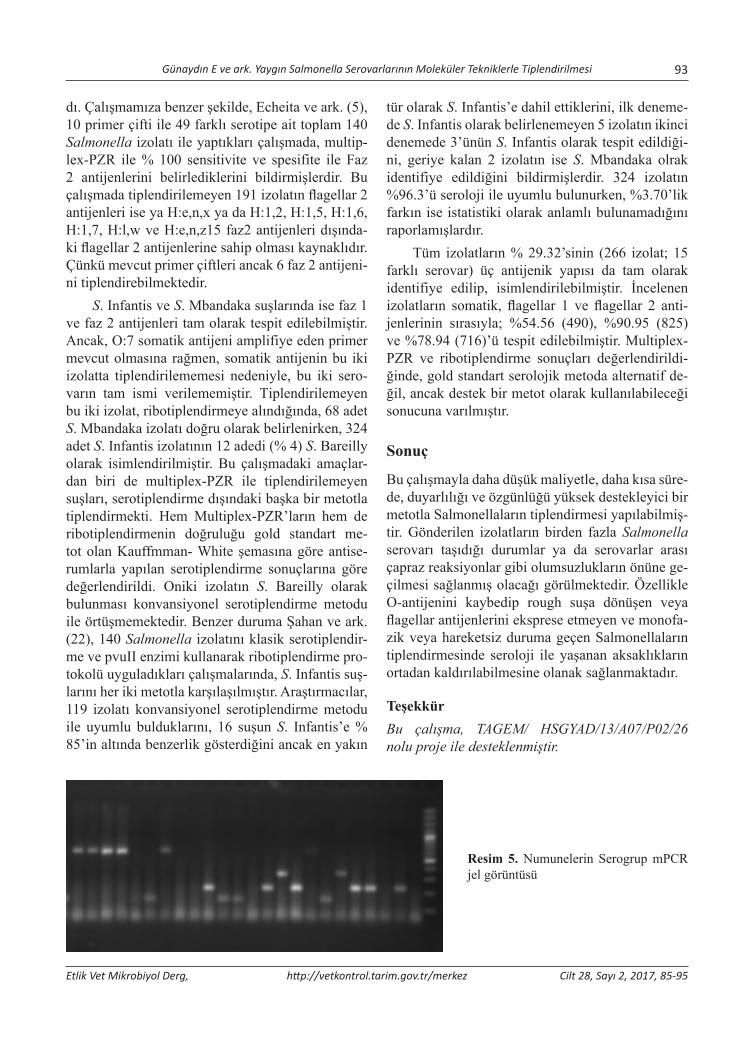
Günaydın E ve ark. Yaygın Salmonella Serovarlarının Moleküler Tekniklerle Tiplendirilmesi 93
Etlik Vet Mikrobiyol Derg, http://vetkontrol.tarim.gov.tr/merkez Cilt 28, Sayı 2, 2017, 85-95
dı. Çalışmamıza benzer şekilde, Echeita ve ark. (5), 10 primer çifti ile 49 farklı serotipe ait toplam 140 Salmonella izolatı ile yaptıkları çalışmada, multip-lex-PZR ile % 100 sensitivite ve spesifite ile Faz 2 antijenlerini belirlediklerini bildirmişlerdir. Bu çalışmada tiplendirilemeyen 191 izolatın flagellar 2 antijenleri ise ya H:e,n,x ya da H:1,2, H:1,5, H:1,6, H:1,7, H:l,w ve H:e,n,z15 faz2 antijenleri dışında-ki flagellar 2 antijenlerine sahip olması kaynaklıdır. Çünkü mevcut primer çiftleri ancak 6 faz 2 antijeni-ni tiplendirebilmektedir.
S. Infantis ve S. Mbandaka suşlarında ise faz 1 ve faz 2 antijenleri tam olarak tespit edilebilmiştir. Ancak, O:7 somatik antijeni amplifiye eden primer mevcut olmasına rağmen, somatik antijenin bu iki izolatta tiplendirilememesi nedeniyle, bu iki sero-varın tam ismi verilememiştir. Tiplendirilemeyen bu iki izolat, ribotiplendirmeye alındığında, 68 adet S. Mbandaka izolatı doğru olarak belirlenirken, 324 adet S. Infantis izolatının 12 adedi (% 4) S. Bareilly olarak isimlendirilmiştir. Bu çalışmadaki amaçlar-dan biri de multiplex-PZR ile tiplendirilemeyen suşları, serotiplendirme dışındaki başka bir metotla tiplendirmekti. Hem Multiplex-PZR’ların hem de ribotiplendirmenin doğruluğu gold standart me-tot olan Kauffmman- White şemasına göre antise-rumlarla yapılan serotiplendirme sonuçlarına göre değerlendirildi. Oniki izolatın S. Bareilly olarak bulunması konvansiyonel serotiplendirme metodu ile örtüşmemektedir. Benzer duruma Şahan ve ark. (22), 140 Salmonella izolatını klasik serotiplendir-me ve pvuII enzimi kullanarak ribotiplendirme pro-tokolü uyguladıkları çalışmalarında, S. Infantis suş-larını her iki metotla karşılaşılmıştır. Araştırmacılar, 119 izolatı konvansiyonel serotiplendirme metodu ile uyumlu bulduklarını, 16 suşun S. Infantis’e % 85’in altında benzerlik gösterdiğini ancak en yakın
tür olarak S. Infantis’e dahil ettiklerini, ilk deneme-de S. Infantis olarak belirlenemeyen 5 izolatın ikinci denemede 3’ünün S. Infantis olarak tespit edildiği-ni, geriye kalan 2 izolatın ise S. Mbandaka olrak identifiye edildiğini bildirmişlerdir. 324 izolatın %96.3’ü seroloji ile uyumlu bulunurken, %3.70’lik farkın ise istatistiki olarak anlamlı bulunamadığını raporlamışlardır.
Tüm izolatların % 29.32’sinin (266 izolat; 15 farklı serovar) üç antijenik yapısı da tam olarak identifiye edilip, isimlendirilebilmiştir. İncelenen izolatların somatik, flagellar 1 ve flagellar 2 anti-jenlerinin sırasıyla; %54.56 (490), %90.95 (825) ve %78.94 (716)’ü tespit edilebilmiştir. Multiplex-PZR ve ribotiplendirme sonuçları değerlendirildi-ğinde, gold standart serolojik metoda alternatif de-ğil, ancak destek bir metot olarak kullanılabileceği sonucuna varılmıştır.
Sonuç
Bu çalışmayla daha düşük maliyetle, daha kısa süre-de, duyarlılığı ve özgünlüğü yüksek destekleyici bir metotla Salmonellaların tiplendirmesi yapılabilmiş-tir. Gönderilen izolatların birden fazla Salmonella serovarı taşıdığı durumlar ya da serovarlar arası çapraz reaksiyonlar gibi olumsuzlukların önüne ge-çilmesi sağlanmış olacağı görülmektedir. Özellikle O-antijenini kaybedip rough suşa dönüşen veya flagellar antijenlerini eksprese etmeyen ve monofa-zik veya hareketsiz duruma geçen Salmonellaların tiplendirmesinde seroloji ile yaşanan aksaklıkların ortadan kaldırılabilmesine olanak sağlanmaktadır.
TeşekkürBu çalışma, TAGEM/ HSGYAD/13/A07/P02/26 nolu proje ile desteklenmiştir.
Resim 5. Numunelerin Serogrup mPCR jel görüntüsü

94 Günaydın E ve ark. Yaygın Salmonella Serovarlarının Moleküler Tekniklerle Tiplendirilmesi
Etlik Vet Mikrobiyol Derg, http://vetkontrol.tarim.gov.tr/merkez Cilt 28, Sayı 2, 2017, 85-95
Resim 6. Numunelerin Faz 1 mPCR jel görüntüsü
Resim 7. Numunelerin Faz 2 mPCR jel görüntüsü
Kaynaklar1. Aarts HJ, LA Van Lith, J. Keijer, (1998). High resolution
genotyping of Salmonella strains by AFLP-fingerprinting. Lett Appl Microbiol, 26, 131-135.
2. Bastin DA, Reeves PR, (1995). Sequence and Analysis of the O antigen gene (rfb) cluster of Escherichia coli O111. Gene, 16, 17-23.
3. Bayindir Bilman F, Gunaydin E, Turhanoglu M, Akkoc A, (2014). The Value of PCR Method in the Species-Level Identification of a Mucoid Salmonella sp. Strain Isolated from the Urine Culture of a Case with Asymptomatic Nephrolithiasis. Mikrobiol Bul, 48(1), 151-159.
4. Echeita MA and Usera MA, (1998). Rapid Identification of Salmonella spp. phase 2 antigens of the H1 antigenic com-plex using ‘multiplex PCR’. Res Microbiol, 149, 757-761.
5. Echeita MA, Herrera S, Garazier J and Usera MA, (2002). Multiplex PCR- based detection and identification of the most common Salmonella-second-phase flagellar antigens. Research in Microbiol, 153, 107-113.
6. Fitzgerald C, Sherwood R, Gheesling LL, Brenner FW, Fields PI, (2003). Molecular analysis of the rfb O anti-gen gene cluster of Salmonella enterica serogroup O:6,14 and development of a serogroup-specific PCR assay. Appl Environ Microbiol, 69(10), 6099-105.
7. Grazier J, Lopez-Molina N, Laconcha I, Bagessen DL, Rementeria A, Vivanco A, Audicana I, Perales A, (2000). Suitable of PCR fingerprinting, Infrequent-Restriction-Site PCR, and Pulsed-Field Gel Electrophoresis, combined with computerized gel analysis, in library typing of Salmonella enterica serovar Enteritidis. Appl Environ Microbiol, 66, 5273-5281.
8. Guibourdenche M, Roggentin, P Mikoleit M, Fields PI, Bockemühl J, Grimont PA.D, Weill François-Xavier, (2010). Supplement 2003-2007 (No. 47) to the White-Kauffmann-Le Minor scheme. Res Microbiol, 161(1), 26-9.
9. Herrera-Leon S, McQuiston JR, Usera MA, Fields PI, Garazier J and Echeita MA, (2004). Multiplex PCR for Distinguishing the Most Common Phase-1 Flagellar Antigens of Salmonella spp. J Clin Microbiol, 42(6), 2581-2586.
10. Herrera-Leon S, Ramiro R, Arroyo M, Diez R, Usera MA, Echeita MA, (2007). Blind comparison of traditional se-rotyping with three multiplex PCRs for the identification of Salmonella serotypes. Research in Microbiology, 158, 122-127.
11. Hong Y, Liu T, Lee MD, Hofacre CL, Maier M, White DG, Ayers S, Wang L, Berghaus R, Maurer JJ, (2008). Rapid screening of Salmonella enterica serovars Enteritidis, Hadar, Heidelberg and Typhimurium using a serologically-correlative allelotyping PCR targeting the O and H antigen alleles, BMC Microbiol, 8, 178.
12. Lindstedt BA, Heir E, Vardund T, Kapperud G, (2000). Fluorescent amplified-fragment length polymorphism ge-notyping of Salmonella enterica subsp. enterica serovars and comparison with pulsed-field gel electrophoresis typ-ing. J Clin Microbiol, 38, 1623-1627.
13. Lino T, (1997). Genetics of structure and function of bacte-rial flagella. Anu Rev Genet, 11, 161-182.
14. Luk JM, Kongmuang U, Reeves PR, Lindberg AA, (1993). Selective amplification of abequose and paratose synthase genes (rfb) by polymerase chain reaction for identification of Salmonella major serogroups (A, B, C2, and D). J Clin Microbiol, 31, 2118-2123.
15. Marolda CL, Vicarioli J, Valvano MA, (2004). Wzx proteins involved in biosynthesis of O antigen function inassociation with the first sugar of O specific lipopolisaccharide subunit. Microbiology, 150, 4095-4105.
16. Newton S, MRD Wasley, A Wilson, LT Rosenberg, JF Miller, and BA Stocker, (1991). Segment IV of a Salmonella flagellin gene specifies flagellar antigen epitopes. Mol Microbiol, 5, 419-425.
17. Patrick AD Grimont & François-Xavier Weill, (2007). Kauffmann-White Scheme Antigenic formulae of the Salmonella serovars, 9th edition, Institut Pasteur, 28 rue du Dr. Roux, 75724 Paris Cedex 15, France.
Günaydın E ve ark. Yaygın Salmonella Serovarlarının Moleküler Tekniklerle Tiplendirilmesi 95
Etlik Vet Mikrobiyol Derg, http://vetkontrol.tarim.gov.tr/merkez Cilt 28, Sayı 2, 2017, 85-95
18. Ranjbar R, Mortazavi SM, Mehrabi Tavana A, Sarshar M, Najafi A, Soruri Zanjani R, (2017), Simultaneous Molecular Detec-tion of Salmonella enterica Serovars Typhi, Enteritidis, Infantis, and Typhimurium. Iran J Public Health, 46(1),103-111.
19. Reeves MW, Evins GM, Heiba AA, Plikaytis BD, Farmer JJ, (1989). Clonal nature of Salmonella typhi and its genet-ic relatedness to other salmonellae as shown by multilocus enzyme electrophoresis, and proposal of S. bongori comb. J Clin Microbiol, 35, 2786-2790.
20. Samuel G, Reeves PR, (2003). Biosynthesis of O-antigens: genes and pathways involved in nucleotide sugar precur-sor synthesis and O-antigen assembly, Carbohydr Res, 338, 2503-2519.
21. Shah DH, Park JH, Cho MR, Kim MC, Chae JS, (2005). Allele-specific PCR method based on rfbS sequence for dis-tinquishing Salmonella gallinarum from Salmonella pullo-rum: serotype-specific rfbS sequence polymorphism. J Clin Microbiol, Methods, 60, 169-177.
22. Şahan Ö, Müştak K, Torun E, Akan M, Diker S. Salmonella Infantis tanısında ribtiplendirme ve serotiplendirme yön-temlerinin karşılaştırması. 10. Uluslararası Veteriner Hekimleri Mikrobiyoloji Kongresi, 24-27 Eylül 2012, Kuşadası, AYDIN
23. Wei LN, and TM Joys, (1995). Covalent structure of three phase-1 flagellae filament proteins of Salmonella spp. J Mol Biol, 186, 791-803.
24. Zappelini L, Martone-Rocha S, Dropa M, Matté MH, Tiba MR, Breternitz BS, Razzolini MT, (2017). Effective char-acterization of Salmonella Enteritidis by most probable number (MPN) followed by multiplex polymerase chain reaction (PCR) methods, Environ Sci Pollut Res Int, 24(5), 4828-4834.
25. Zieg J, Silverman M, Hilmen M and Simon M, (1977). Recombinational switch for gene expression. Science, 196, 170-172.

Yazışma adresi / Correspondence: Ender Dinçer, Mersin University, Advanced Technology Education, Research and Application Center, Çiftlikköy, Mersin, Turkey E-posta:[email protected]
Etlik Vet Mikrobiyol Derg, 2017; 28 (2): 96-100 Araştırma Makalesi / Research Article
Molecular Characterization and Phylogenetic Analysis of Canine Parvovirus 2 in Dogs, Mersin Province, Turkey
Ender Dinçer
Mersin University, Advanced Technology Education, Research and Application Center, Mersin
Geliş Tarihi / Received: 10.10.2017, Kabul Tarihi / Accepted: 02.11.2017
Abstract: Canine parvovirus type 2 (CPV-2) is a highly contagious and lethal viral disease with acute gastroenteritis in young dogs. After it first appeared in USA in 1978, continuous evolution of virus genome has arisen new genetic and antigenic variants such as CPV-2a, CPV-2b, new CPV-2a, new CPV-2b and CPV-2c throughout the world. The aim of the present study was to detect CPV-2 genetic variants from blood samples of clinically ill dogs and to characterize CPV-2 genetic variants circulating in Mersin province (Turkey), by partial sequence analysis of VP2 gene. A total of 20 blood samples were collected from dogs with gastroenteritis from local veterinary clinics between April 2017 to September 2017. Out of 20, 7 samples were positive for CPV-2 genomic DNA using conventional PCR. Amino acid comparisons showed that Ile-324 mutation was detected in the VP2 of clinical CPV-2b samples, as differ previously studies Turkish genetic variants. A phylogenetic tree constructed based on VP2 gene sequences indicated that CPV-2b sequences from Mersin province were related to Indian genetic variants. Moreover, CPV-2b sequences from Mersin have clustered sepa-rately from other Turkish CPV-2b genetic variants previously reported. The results show that CPV-2b genetic variant is circulating among in dogs in Mersin province. Key words: Canine parvovirus, dog, phylogenetic analysis, VP2 gene.
Türkiye’de Mersin İlinde Köpeklerde Canine Parvovirus 2’nin Moleküler Karakterizasyonu ve Filogenetik Analizi
Özet: Canine parvovirus 2 (CPV-2) genç köpeklerde akut kanlı gastroenteritis ile seyreden oldukça bulaşıcı ve ölümcül bir viral hastalıktır. 1978’de ABD’de ilk ortaya çıktıktan sonra, virus genomunun sürekli evrimi, tüm dünyada CPV-2a, CPV-2b, new CPV-2a, new CPV-2b ve CPV-2c gibi yeni genetik ve antijenik varyantların ortaya çıkmasına yol açmıştır. Bu çalışmanın amacı, Mersin (Türkiye) ilinde klinik olarak hasta köpeklerin kan örneklerinde CPV -2’yi tespit etmek ve dolaşımda olan CPV- 2 genetik varyatlarının karakterizasyonunu sağlamaktır. Bunun için 2017 Nisan ve 2017 Eylül ayları arasında, yerel kliniklerden 20 adet kan örneği gastroenteritisli köpeklerden toplandı. 20 örneğin 7’si PZR ile CPV- 2 yönünden pozitif olarak test edildi. Klinik CPV-2b örneklerinin VP2’sindeki amino asit karşılaştırmaları sonucunda daha önce çalışılan Türk soylarından farklı olarak Ile-324 mutasyonu tespit edildi. VP2 geni dizinlerinden köken alarak yapılan filogenetik ağaç, Mersin ilinden gelen CPV-2b dizilerinin Hindistan genetik varyantlarıyla ilişkili olduğunu göstermiştir. Aynı zamanda Mersin ilinden elde edilen CPV-2b dizinleri daha önce tespit edilen Türk genetik varyantlarından farklı bir dal şekillendirmiştir. Bu sonuçlar Mersin ilinde CPV-2b genetik varyantının köpekler arasında dolaştığını göstermiştir. Anahtar kelimeler: Canine parvovirus, köpek, filogenetik analiz, VP2 gen.
Introduction
Canine parvovirus 2 (CPV-2) is small, non-envel-oped, single-stranted DNA virus, belonging to genus Parvovirus, the family Parvoviridea with genome 0f 5, 2 Kb in lenght. CPV-2 has two open reading frames (ORFs). One of them encodes non-structural proteins (NS1, NS2) and the other encodes struc-tural proteins (VP1, VP2). Amino acid changes in the VP2 protein, main viral protein of capsid, alter the antigenic properties of the virus in determining the viral host distribution and tissue tropism [6, 13].
After the emergence of the CPV-2 in 1978, two new genetic variants (CPV-2a and CPV-2b) were charac-terized in 1979 and 1984, respectively. Monoclonal antibodies can distinguish as CPV-2a (426Asn) and CPV-2b (426Asp) by amino acid substitution in the capsid protein gene. In 2000, CPV-2c (Asp-426 to Glu) is a new genetic variant was detected in Italy [6, 11]. These three genetic variants occur in dogs throughout the world; CPV-2a was found in Nigeria, South Africa, Turkey, Greece and oth-er European countries [14]. CPV-2b was reported from USA, Turkey and China [6, 23]. Morever,
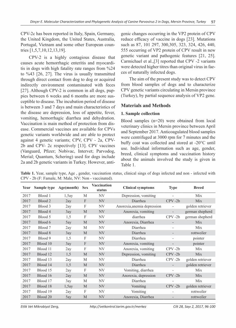
Dinçer E. Molecular Characterization and Phylogenetic Analysis of Canine Parvovirus 2 in Dogs, Mersin Province, Turkey 97
Etlik Vet Mikrobiyol Derg, http://vetkontrol.tarim.gov.tr/merkez Cilt 28, Sayı 2, 2017, 96-100
CPV-2c has been reported in Italy, Spain, Germany, the United Kingdom, the United States, Australia, Portugal, Vietnam and some other European coun-tries [1,5,7,10,12,13,19].
CPV-2 is a highly contagious disease that causes acute hemorrhagic enteritis and myocardi-tis in dogs with high fatality rate ranges from %24 to %43 [26, 27]. The virus is usually transmitted through direct contact from dog to dog or acquired indirectly environment contaminated with feces [27]. Although CPV-2 is common in all dogs, pup-pies between 6 weeks and 6 months are more sus-ceptible to disease. The incubation period of disease is between 3 and 7 days and main characteristics of the disease are depression, loss of appetite, fever, vomiting, hemorrhagic diarrhea and dehydration. Vaccination is main method of protection from dis-ease. Commercial vaccines are available for CPVs genetic variants worldwide and are able to protect against 4 genetic variants; CPV, CPV - 2a, CPV- 2b and CPV- 2c respectively [13]. CPV vaccines (Vanguard, Pfizer; Nobivac, Intervet; Parvodog, Merial; Quantum, Schering) used for dogs include 2a and 2b genetic variants in Turkey. However, anti-
genic changes occurring in the VP2 protein of CPV reduce efficacy of vaccine in dogs [23]. Mutations such as 87, 101 297, 300,305, 323, 324, 426, 440, 555 occurring of VP2 protein of CPV result in new genetic variant and pathogenic features [21, 25]. Carmichael et al.,[3] reported that CPV -2 variants were detected higher titres than original virus in fae-ces of naturally infected dogs.
The aim of the present study was to detect CPV from blood samples of dogs and to characterize CPV genetic variants circulating in Mersin province (Turkey), by partial sequence analysis of VP2 gene.
Materials and Methods1. Sample collectionBlood samples (n=20) were obtained from local veterinary clinics in Mersin province between April and September 2017. Anticoagulated blood samples were centrifuged at 3000 rpm for 7 minutes and the buffy coat was collected and stored at -20°C until use. Individual information such as age, gender, breed, clinical symptoms and vaccination history about the animals involved the study is given in Table 1.
Table 1. Year, sample type, Age , gender, vaccination status, clinical sings of dogs infected and non - infected with CPV - 2b (F: Famale, M: Male, NV: Non - vaccinated).
Year Sample type Age(month) Sex Vaccinationstatus Clinical symptoms Type Breed
2017 Blood 1 1,5ay M NV Depression, vomiting - Mix2017 Blood 2 2ay F NV Diarrhea CPV -2b Mix2017 Blood 3 2ay F NV Anorexia,anemia depression - golden retriever2017 Blood 4 3ay M NV Anorexia, vomiting - german shepherd2017 Blood 5 1,5 F NV diarrhea CPV -2b german shepherd2017 Blood 6 5ay M NV Anorexia, Diarrhea - Mix2017 Blood 7 2ay M NV Diarrhea - Mix2017 Blood 8 3ay M NV Diarrhea - rottweiler2017 Blood 9 1,5 F NV Diarrhea - pointer2017 Blood 10 3ay F NV Anorexia, vomiting - pointer2017 Blood 11 2ay F NV Anorexia, vomiting CPV -2b Mix2017 Blood 12 1,5 M NV Depression, vomiting CPV -2b Mix2017 Blood 13 2ay M NV Diarrhea CPV -2b golden retriever2017 Blood 14 1,5 M NV Diarrhea - golden retriever2017 Blood 15 2ay F NV Vomiting, diarrhea - Mix2017 Blood 16 2ay M NV Anorexia, depression CPV -2b Mix2017 Blood 17 3ay M NV Diarrhea - Mix2017 Blood 18 1,5ay M NV Vomiting CPV -2b golden retriever2017 Blood 19 2ay F NV Vomiting - rottweiler2017 Blood 20 5ay M NV Anorexia, Diarrhea - rottweiler

98 Dinçer E. Molecular Characterization and Phylogenetic Analysis of Canine Parvovirus 2 in Dogs, Mersin Province, Turkey
Etlik Vet Mikrobiyol Derg, http://vetkontrol.tarim.gov.tr/merkez Cilt 28, Sayı 2, 2017, 96-100
2. Template DNA Preparation and PCRNucleic acids were extracted from 200µL blood samples using High Pure Viral Nucleic Acid Kit (Roche Diagnostics, Mannhaeim, Germany) follow-ing the manufacturer’s recommendations. Purified DNAs were stored at -20°C until PCR. The PCR targeting partial VP2 gene (629bp) was performed as described elsewhere [2]. Amplified PCR products were visualized under ultraviolet light via ethid-ium bromide staining after electrophoresis using in %1 agarose gel in TAE (Figure 1). Consequently, 100µL amplicon from each sample was purified us-ing a commercial kit (GeneJET, Thermo Scientific, Lithuania) according to manufacturer’s instruc-tions. Purified DNA was used for DNA sequencing and subsequent phylogenetic analysis.
Figure 1. Agarose gel image of the PCR products (629 bp) of Canine parvovirus DNA using by Hfor and Hrev prim-ers. Lane M: 100bp DNA ladder (Thermoscientific, USA); Lane: 1 - 7 positive samples; Lane 8: Negative control.
3. DNA sequencing and phylogenetic analysisSequencing was performed in ABI PRISM 310 Genetic Analyzer (Applied Biosystem, CA) us-ing same primers that were used in the PCR. The
obtained DNA sequences were compared to other CPV sequences available from GenBank Database (http://www.ncbi.nlm.nih.gov). Alignment and pairwise comparisons were carried out in CLC Main Workbench v7.7 (CLCBio, Aarhus, Denmark). Phylogenetic tree was drawn in MEGA software v.6.06 using neighbor-joining (NJ) method and bootstrap values were calculated with 1,000 repli-cates [19].
Results and DiscussionTwenty dogs with clinically defined gastroenteritis (especially vomiting and diarrhea) signs were test-ed with conventional PCR assay for the presence of CPV-2. Seven (35%) of the dogs were CPV-2 positive. Samples from negative dogs were not re-investigated for the other etiological causes of gan-troenteritis. CPV- 2 positive dogs were between 1, 5 and 5 months old. Four of the positive dogs were male and three were female. According to the infor-mation received from the pet clinics, no vaccination was administrated to dogs. Breeds of dogs involved the study were recorded as Rottweiler, Golden Retriever, Pointer, German Shepherd and mix breed (Table 1).
Conventional PCR was carried out using Hfor and Hrev primers. Sequence analysis of PCR am-plicons were performed with same primer sets. The obtained sequence results were compared to gene bank reference sequences (Figure 2). Phylogenetic (NJ) analysis revealed that all CPV-2 detected in the study were found to be CPV-2b. Other genetic variants (CPV-2a and CPV-2c) were not identified in the study. In order to investigate the variations occurring on VP2 gene, reference strain (GenBank Accession # M38245) was compared to amino acid residues obtained from clinical samples (Table 2).
Table 2. Amino acid changes in VP2 partial gene of CPV - 2b typeAa no. 297 300 305 324 336 375 426 440
M38245 S A D Y A N N TP5 A G Y I G D D AP12 A G Y I G D D AP2 A G Y I G D D AP13 A G Y I G D D AP18 A G Y I G D D AP11 A G Y I G D D AP16 A G Y I G D D A
Aa change TCT→GCTS→A*
GCT→GGTA→G*
GAT→TATD→Y*
TAT→ATTY→I*
GTA→GTGV→V
AAT→ GATN→D*
ATT→GATN→D*
ACA→GCAT→A*
Amino acid substitutions are indicated by asterisks (*).
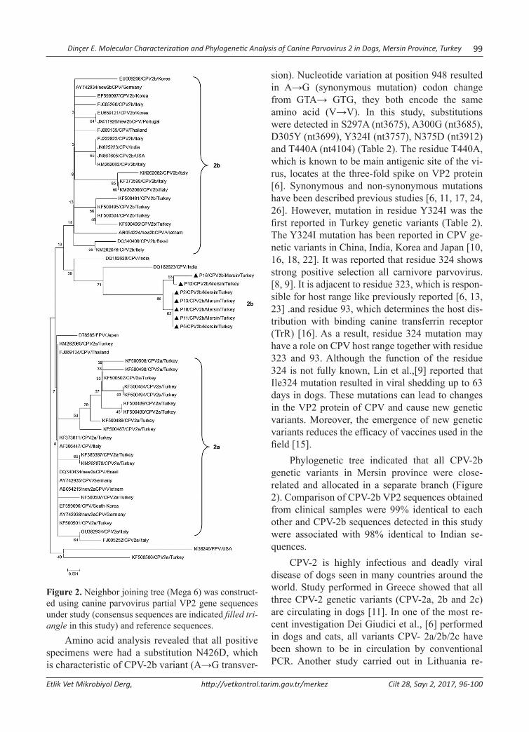
Dinçer E. Molecular Characterization and Phylogenetic Analysis of Canine Parvovirus 2 in Dogs, Mersin Province, Turkey 99
Etlik Vet Mikrobiyol Derg, http://vetkontrol.tarim.gov.tr/merkez Cilt 28, Sayı 2, 2017, 96-100
Figure 2. Neighbor joining tree (Mega 6) was construct-ed using canine parvovirus partial VP2 gene sequences under study (consensus sequences are indicated filled tri-angle in this study) and reference sequences.
Amino acid analysis revealed that all positive specimens were had a substitution N426D, which is characteristic of CPV-2b variant (A→G transver-
sion). Nucleotide variation at position 948 resulted in A→G (synonymous mutation) codon change from GTA→ GTG, they both encode the same amino acid (V→V). In this study, substitutions were detected in S297A (nt3675), A300G (nt3685), D305Y (nt3699), Y324I (nt3757), N375D (nt3912) and T440A (nt4104) (Table 2). The residue T440A, which is known to be main antigenic site of the vi-rus, locates at the three-fold spike on VP2 protein [6]. Synonymous and non-synonymous mutations have been described previous studies [6, 11, 17, 24, 26]. However, mutation in residue Y324I was the first reported in Turkey genetic variants (Table 2). The Y324I mutation has been reported in CPV ge-netic variants in China, India, Korea and Japan [10, 16, 18, 22]. It was reported that residue 324 shows strong positive selection all carnivore parvovirus. [8, 9]. It is adjacent to residue 323, which is respon-sible for host range like previously reported [6, 13, 23] .and residue 93, which determines the host dis-tribution with binding canine transferrin receptor (TrR) [16]. As a result, residue 324 mutation may have a role on CPV host range together with residue 323 and 93. Although the function of the residue 324 is not fully known, Lin et al.,[9] reported that Ile324 mutation resulted in viral shedding up to 63 days in dogs. These mutations can lead to changes in the VP2 protein of CPV and cause new genetic variants. Moreover, the emergence of new genetic variants reduces the efficacy of vaccines used in the field [15].
Phylogenetic tree indicated that all CPV-2b genetic variants in Mersin province were close-related and allocated in a separate branch (Figure 2). Comparison of CPV-2b VP2 sequences obtained from clinical samples were 99% identical to each other and CPV-2b sequences detected in this study were associated with 98% identical to Indian se-quences.
CPV-2 is highly infectious and deadly viral disease of dogs seen in many countries around the world. Study performed in Greece showed that all three CPV-2 genetic variants (CPV-2a, 2b and 2c) are circulating in dogs [11]. In one of the most re-cent investigation Dei Giudici et al., [6] performed in dogs and cats, all variants CPV- 2a/2b/2c have been shown to be in circulation by conventional PCR. Another study carried out in Lithuania re-
100 Dinçer E. Molecular Characterization and Phylogenetic Analysis of Canine Parvovirus 2 in Dogs, Mersin Province, Turkey
Etlik Vet Mikrobiyol Derg, http://vetkontrol.tarim.gov.tr/merkez Cilt 28, Sayı 2, 2017, 96-100
vealed CPV-2a and 2b genetic variants are present in dogs [26]. Moreover, CPV-2a has been reported in rectal swaps obtained from dogs in India [17]. In Turkey, there is limited number of studies on CPV genetic variants in dogs [21, 22, 23]. Timurkan et al., [20] carried out the first molecular characterization of CPV-2a and CPV-2b genetic variants in Ankara province. Results of the study showed presence of CPV-2b genetic variant in dogs in Mersin province. All of these investigations show that the CPV-2a and CPV-2b are predominant strains in dog popula-tion in Turkey and CPV-2c variant has not yet been identified in Turkey so far. Further molecular and epidemiological studies are needed to determine on CPV infections and to characterize circulating types in dogs.
In conclusion, the presence of amino acid mu-tations suggests that the virus has evolved continu-ously. Although parvovirus is a DNA virus, it has high genetic substitution rate, which causes emerg-ing new genetic variants as well as RNA viruses [5]. Differences between field and vaccine strains are the most important reason for some immune fail-ures [17]. Periodic epidemiological survey and mo-lecular studies can be identified new CPV-2 genetic variants and mutations. In addition, comparison of strains obtained from different geographical areas can be provided the development of used vaccines and CPV 2 infections can be effectively controlled in the country.
References1. Apaa TT, Daly JM, Tarlinton RE, (2016). Canine parvovirus (CPV-
2) variants circulating in Nigerian dogs. Vet Rec Open. 3, 1 -4.2. Buonavoglia C, Martella V, Pratelli A, Tempesta M, Cavalli A,
Buonavoglia D, Bozzo G, Elia G, Decaro N, & Carmichael L, (2001). Evidence for evoluation of canine parvovirus type 2 in Italy. J Gen Virol. 82, 3021 - 3025.
3. Charmichael LE, (1994). Canine parvovirus type -2.An evolving pathogen of dogs.Ann Vet Med. 135, 459- 464.
4. Decaro N, Martella V, Desario C, Bellacicco AL, Camero M, Manna L, D’Aloja D, Buonavoglia C, (2006). First detection of canine parvovirus type C in pups with haemorragica enteritis in Spain. J Vet Med. 53, 468-472.
5. Decaro N, Desario C, Addie DD, Martella V, Vieira JM, Elia G, Zicola A, Davis C, Thompson G, Thiry E, Truyen U, and Buonavoglia C, (2007). Molecular Epidemiology of Canine Parvovirus, Europe. Emerg Infect Dis. 13, 1222 - 124.
6. Dei Giudici S, Cubeddu T, Giagu A, Sanna G, Rocca S, Oggiano A, (2017). First molecular characterization of canine parvovirus strains in Sardinia, Italy. Arch Virol. 017, 3457-3.
7. Hong C, Decaro N, Desario C, Tanner P, Pardo CM, Sanchez S, Buonavoglia C, Saliki TJ, (2007). Occurrence of canine parvovi-rus type 2c in the United States. J Vet Diagn Invest. 19, 535-539.
8. Horiuchi M, Goto N, Ishiguro M, Shinagawa J,(1994). Mapping of determinants of the host range fort he canine cells in the genome of the canine parvovirus using canine parvovirus/mink enteritis virus chimeric viruses.J Gen Virol.75, 1319 - 1328.
9. Lin CN, Chien CH, Chio MT, Chueh LL, Hung MY, Hsu HS, (2014). Genetic characterization of type 2a canine parvovirus from Twaiwan reveals the emergence of an ıle324 mutation in VP2. Virol J. 11, 39.
10. Jeoung S, Ahn S, Kim D, (2008). Genetic analysis of VP2 gene canine parvovirus isolates in Korea. J Vet Med Sci. 70, 719 - 722.
11. Ntafis V, Xylouri E, Kalli I, Desario C, Mari V, Decaro N, Buonavoglia C,(2010). Characterization of Canine parvovirus 2 variants circulating in Greece J Vet Diagn Invest. 22, 737-740.
12. Martella V, Decaro N, Elia G, Buonavoglia C, (2005). Surveillance activity for canine parvovirus in Italy. J Vet Med. 52, 312-315.
13. Meers J, Kyaw-Tanner M, Benzink Z, Zwijnenberg R, (2007). Genetic analysis of canine parvovirus from dogs in Australia. Aust Vet J. 85, 392-396.
14. Miranda C, and Thompson G, (2016). Canine parvovirus: the worldwide occurrence of antigenic variants. J Gen Virol, 97, 2043 - 2057.
15. Mittal M, Chakravarti S, Mohapatra KJ, Chug KP, Dubey R, Upmanuyu V, Narwal PS, Kumar A, Churamani CP, Kanwar SN,(2014). Molecular typing of canine parvovirus strains circu-lating from 2008 to 2012 in an organized kennel in India reveals the possibility of vaccination failure. Infec Genet Evol. 23, 1- 6.
16. Mukhopadhyay HK, Matta SL, Amsaveni S, Antony PX, Thanislass J, Pillai RM, (2014). Phylogenetic analysis of canine parvovirus partial VP2 gene in India. Virus Genes. 48, 89 - 95.
17. Raj MJ, Mukhopadhyay KH, Thanislass J, Antony XP, Pillai MR, (2010). Isolation, molecular characterization and phylogenetic analysis if canineparvovirus. Infec Genet Evol. 10, 1237 - 1241.
18. Soma T, Taharaguchi S, Ohinata T, Ishii H, Hara M, (2013). Analysis of the VP2 protein gene of canine parvovirus strains from affected dogs in Japan. Res Vet Sci. 94, 368 - 371.
19. Tamura K, Stecher G, Peterson D, Filipski A, and Kumar S, (2013). MEGA6: Molecular Evolutionary Genetics Analysis ver-sion 6.0. Mol Biol Evol. 30, 2725 - 2729.
20. Timurkan M, Oguzoglu T, (2015). Molecular characterization of canine parvovirus (CPV) infection in Turkey. Vet Ital. 51, 39 - 44.
21. U. Truyen, (2006). Evolution of canine parvovirus-a need for new vaccines? Vet Microbiol. 117, 9-13
22. Yesilbag K, Yılmaz Z, Ozkul A, & Pratelli A, (2007). Aetiological role of viruses in puppies with diarrhoea. Vet Rec. 161, 169 - 170.
23. Yılmaz Z, Pratelli A, &Torun S, (2005). Distribution of antigen types of canine parvovirus type 2 in dogs with hemorrhagic en-teritis in Turkey. Turk J. Vet Anim Sci. 29, 1073 - 1076.
24. Zhang R, Yang S, Zhang W, Zhang T, Xie Z, Feng H, Wang S, Xia X, (2010). Phylogenetic analysis of the VP2 gene of canine parvoviruses circulating in China.Virus Genes. 40, 397- 402.
25. Zhao Z, Liu H, Ding K, Peng C, Xue Q, Yu Z, Xue Y, (2016). Occurance of canine parvovirus in dogs from Henan province of China. Vet Res. 12, 138 - 145.
26. Zienius D, Lelesius R, Kavaliauskis H, Stankevicius A, Salomskas A, (2016). Pol J Vet Sci. 19, 187-196.
27. Zourkas E, Ward MP, Kelman M, (2015). Canine parvovirus in Australia: A comperative study of reported rural and urban cas-es.Vet Microbiol. 121, 198 - 203.
*: Bu çalışma 18-21 Ekim 2017 tarihleri arasında Alanya’da düzenlenen II. International Academic Research Congress’de sözlü bildiri olarak sunulmuştur. Yazışma adresi / Correspondence: Armağan Erdem Ütük, Çukurova Üniversitesi, Ceyhan Veteriner Fakültesi, Parazitoloji AD, Adana, Türkiye E posta: [email protected]
Etlik Vet Mikrobiyol Derg, 2017; 28 (2): 101-104 Araştırma Makalesi / Research Article
Türkiye’nin Nevşehir İlindeki Atlarda Kistik Ekinokokkoz Seroprevalansı*
Armağan Erdem Ütük1, Selçuk Pekkaya2, Fatih Kuzugüden3, İbrahim Balkaya4, Sami Şimşek5
1Çukurova Üniversitesi, Ceyhan Veteriner Fakültesi, Parazitoloji Anabilim Dalı, Adana, Türkiye2Veteriner Kontrol Merkez Araştırma Enstitüsü, Ankara, Türkiye
3İl, Gıda Tarım ve Hayvancılık Müdürlüğü, Nevşehir, Türkiye4Atatürk Üniversitesi, Veteriner Fakültesi, Parazitoloji Anabilim Dalı, Erzurum, Türkiye
5Fırat Üniversitesi, Veteriner Fakültesi, Parazitoloji Anabilim Dalı, Elazığ, Türkiye
Geliş Tarihi / Received: 28.10.2017, Kabul Tarihi / Accepted: 11.11.2017
Özet: Kistik ekinokokkoz (KE) son derece önemli bir paraziter zoonoz olup insan ve hayvanlarda ciddi ekonomik kayıplara neden olur. Türkiye’de hastalığın sığır, koyun, keçi gibi çiftlik hayvanlarında oldukça yaygın olduğu tespit edilmiştir. Ancak atlarda KE konusunda çok az çalışma bulunmaktadır. Bu çalışmanın amacı Nevşehir ilinde halk elinde yetiştirilen atlarda KE seroprevalansını belirlemekti. Bu amaçla yaşları 3-24 arasında değişen, farklı ırklardan, 105 dişi attan kan alınarak serumları çıkarıldı. Enfekte koyun kistlerinden Antijen-B bakımından zengin kısmi purufiye kist sı-vısı antijeni hazırlanarak enzyme-linked immunosorbent assay testinde (ELISA) kullanıldı. Tarama sonucunda 7 yaş ve altı atlarda %4,54 (2/44), 7 yaş üstü atlarda %6,55 (4/61) oranında anti-Echinococcus granulosus antikorları tespit edil-di. Ki-kare (X2) testi ile yaş grupları ile seroprevalans arasında istatistiksel açıdan önemli bir fark bulunmadı (p ˃ 0,05). Genel seroprevalans ise %5,71 (6/105) olarak belirlendi. Elde edilen veriler bu bölgede atların da son konak köpekgiller için enfeksiyon kaynağı olabileceğini ve hastalığın epidemiyolojisi açısından önemli olduğunu ortaya koymuştur. Bu çalışma ile Nevşehir ilindeki atlarda ilk defa KE seroprevalansı belirlenirken, ülkemizde konuyla ilgili çok sınırlı sayıda bulunan literatür bilgiye bir yenisi eklenmiştir. Anahtar kelimerler: At, ELISA, Kistik Ekinokokkoz, Nevşehir.
Seroprevalance of Cystic Echinococcosis in Horses in Nevsehir Province of TurkeyAbstract: Cystic Echinococcosis (CE) is a highly significant parasitic zoonosis and can cause dramatic economic losses in humans and animals. In Turkey, the disease has been detected and highly prevalent in farm animals such as cattle, sheep and goat; however, there is very little research on CE in horses. The aim of this study is to detect the seropreva-lance of CE in horses bred by the locals in Nevsehir province. For this purpose, blood samples were taken from 105 female horses of different races, aged from 3 to 24 and their sera were obtained. Partially purified cyst fluid antigen from sheep hydatid cysts was used as antigen in enzyme-linked immunosorbent assay (ELISA). At the end of the screening, anti-Echinococcus granulosus antibodies were detected in %4,54 (2/44) of horses aged 7 and under, and in %6,55 (4/61) of horses above 7 years of age. With the use of Chi-Square Test (X2), no statistically significant difference was found between age groups and seroprevalance (p ˃ 0,05). The overall seroprevalance rate was detected as %5,71 (6/105). The data obtained indicate that the horses in this region may be a source of infection for the final host, the canidae and are significant for the disease epidemiology. This study detects for the first time the seraprevalance of cystic echinoccocus in horses in Nevsehir province and contributes to the limited amount of literature related to this topic in Turkey.Key words: Cystic Echinococcosis, ELISA, Horse, Nevsehir.
Giriş
Echinococcus granulosus son konakları köpek, til-ki, çakal kurt gibi kanideler ara konakları ise insan, sığır, koyun, keçi, deve, geyik, domuz, at ve eşek gibi memeliler olan taenid bir sestodtur. Ara konak-lardaki metasestodlar hidatik kist, bu metasestod-ların oluşturduğu hastalık ise kistik ekinokokkoz
(KE) adını alır. Kistik ekinokokoz kozmopolit bir yayılışa sahiptir. Hastalık çiftlik hayvanlarında cid-di ekonomik kayıplara neden olur. İnsanlarda ise anaflaktik reaksiyonlar, çeşitli organ ve dokularda yapısal ve fonksiyonel bozukluklar ve bazen ölüm-ler görülür. Bu nedenle önemli bir helmintik zoonoz ve halk sağlığı problemidir [4,7].
102 Ütük AE ve ark. Türkiye’nin Nevşehir İlindeki Atlarda Kistik Ekinokokkoz Seroprevalansı
Etlik Vet Mikrobiyol Derg, http://vetkontrol.tarim.gov.tr/merkez Cilt 28, Sayı 2, 2017, 101-104
Hidatidozun ara konaklardaki yaygınlığı ke-simhane kontrolleri ve serolojik testler ile belir-lenmektedir. Türkiye’de hastalığın sığır, koyun ve keçilerdeki yaygınlığı üzerine çok sayıda çalışma olmasına rağmen, at hidatidozu ile ilgili sınırlı sayı-da veri bulunmaktadır [2,6]. Ekinokokkozun belirli bir bölgede ara konaklardaki yaygınlığının belir-lenmesi hastalığın epidemiyolojisi ve etkin kontrol yöntemlerinin geliştirilmesi açısından önem arz eder. Bu çalışmanın amacı Nevşehir ilindeki atlarda kistik ekinokokkozun prevalansını indirekt ELISA yöntemi ile belirlemekti.
Materyal ve Metod
Çalışmanın materyalini halk elinde bulunan ve tu-ristik amaçlarla yetiştirilen, yaşları 3-24 arasında değişen, farklı ırklardan 105 dişi at oluşturdu. Atlar 7 yaş ve altı (≤7) ile 7 yaş üstü (˃7) olarak iki gruba ayrıldı. Yaş grupları ile seropozitiflik arasında ista-tistiksel açıdan ilişki bulunup bulunmadığını belir-lemek için Ki-kare testi (X2) yapıldı.
Çalışma için Etlik Veteriner Kontrol Merkez Araştırma Enstitüsü Müdürlüğü, Yerel Etik Kurulundan 21.11.2015 tarih ve 2015/07 sayılı yazı ile izin alındı. Kan örnekleri atların Vena jugula-ris’inden vakumlu serum tüplerine alındı ve oda ısı-sında 2-3 saat pıhtılaşması beklendi. Pıhtılaşan kan 5000 rpm’de 5 dk santrifüj edildi. Serumlar mikro-santrifüj tüplerine ayrıldı ve kullanılıncaya kadar -20 °C’de muhafaza edildi [16].
Mezbahada kesimler takip edilerek ağır enfekte olduğu tespit edilen bir koyundan elde edilen kara-ciğer laboratuara getirildi ve hidatik kist sıvısından antijen B (AgB) bakımından zengin kist sıvısı an-tijeni hazırlandı. Bu amaçla kist sıvısı steril koşul-larda aspire edildi ve mikroskopta protoskoleklerin varlığı yönünden incelendi. Fertil olduğu belirlenen kistlerin sıvısı 2000 g’de 15 dk santrifüj edilerek protoskoleksler ve diğer katı materyal çöktürüldü. 100 ml süpernatant bir gece +4°C’de 0.005 M asetat buffer (pH 5) içerisinde diyaliz edildi. Diyaliz sonu-cunda elde edilen kist sıvısı +4°C’de 15,000 g’de 30 dk santrifüj edildi. Pelet toplanarak 10 ml 0.2 M fosfat tamponunda (pH 8) çözdürüldü ve 15 dk kay-natılarak +4°C’de 20,000 g’de 1 saat santrifüj edil-di. AgB bakımından zengin süpernatant porsiyon-
lanarak kullanılıncaya kadar -20 °C’de muhafaza edildi. Protein konsantrasyonu spektrofotometre ile ölçülerek (NanoDrop ND-1000 Spectrophotometer) μg /μl olarak belirlendi [6,10].
Test küçük modifikasyonlarla Şimsek ve ark. [14]’a göre yapıldı. ELISA plaklarının (Dynatech Laboratories, IA, USA) her bir kuyucuğuna 5 μg/ml antijeni ihtiva eden 0.1 M karnonat/bikarbonat tamponundan (pH 9.6) 100 μl eklendi ve +4°C’de bir gece inkube edildi. İnkubasyondan sonra plaklar 2 kere içerisinde 0.01% Tween-20 bulunan 130 μl PBS (yıkama solüsyonu) ile yıkandı. Her kuyucuğa 5% oranında yağsız süt tozu ihtiva eden 0.01 M PBS (pH 7.4)’den 130μl eklendi ve 37°C’de 1.5 saat in-kube edilerek bloklama işlemi yapıldı. Bloklamadan sonra 3 kere yıkama işlemi yapıldı. Serum örnekleri 0.05% Tween-20 ihtiva eden PBS ile 1:200 sulan-dırıldı ve her kuyucuğa 100 μl eklenerek 37°C’de 1.5 saat inkube edildi. Plaklar 5 kere PBS/Tween ile yıkandıktan sonra her kuyucuğa 100 μl konjugat eklendi [1:5000’lik goat antihorse IgG HRP (Santa Cruz Biotechnology, sc2906, Lot: B212)] ve pleyt-ler 37°C’de 2 saat inkube edildi. İnkubasyon sonu-cunda pleyler 5 kez yıkandı ve her kuyucuğa 100 μl substrat eklendi (sitrat/fosfat tamponu içerisinde O-fenilen diamin ve hidrojen peroksit) ve oda ısı-sında 15 dk inkube edildi. İnkubasyon sonucunda her kuyucuğa 50 μl durdurma solüsyonu (1 N sül-fürik asit) eklendi. Plaklar ELISA okuyucuda (Bio-Tek instruments, USA) 450 nm dalga boyunda oku-tuldu. Her örnek çift olarak çalışıldı. Her örnek için sonuçlar örneklerin optik dansitelerinin ortalaması alınarak değerlendirildi. Aralarında %10’dan faz-la optik dansite farkı olan örnekler tekrar çalışıldı. Negatif kontrollerin absorbans değerinin aritmetik ortalaması + 3 standart sapma (X+3SD) değerinin (cut-off) üstü pozitif olarak kabul edildi [6,14 ].
Bulgular
Çalışma sonucunda 7 yaş ve altı atlarda %4,54 (2/44), 7 yaş üstü atlarda %6,55 (4/61) oranında an-ti-Echinococcus granulosus antikorları tespit edildi. Ki-kare testi ile yaş grupları ile seropozitiflik ara-sında istatistiksel açıdan önemli bir fark bulunmadı (p˃0,05). Genel seroprevalans ise %5,71 (6/105) olarak belirlendi.
Ütük AE ve ark. Türkiye’nin Nevşehir İlindeki Atlarda Kistik Ekinokokkoz Seroprevalansı 103
Etlik Vet Mikrobiyol Derg, http://vetkontrol.tarim.gov.tr/merkez Cilt 28, Sayı 2, 2017, 101-104
Tartışma ve Sonuç
Postmortem incelemeye dayalı kontrollerde İtalya’nın Sicilya bölgesinde kesilen 360 atın %1,7’sinde [11] Şili’de 9391 atın %9’unda [1] Çin’in Xinjiang bölgesinde kesilen 352 atın %4,3’ünde [12] ve Fas’ta incelenen 455 tek tırnaklının %17,18’inde [5] hidatidoz tespit edilmiştir. Seroprevalans çalış-malarında ise Yunaninstan’da 753 atın %0,1’inde ve İran’da 193 atın %3,11’inde anti-E.granulosus antikorları tespit edilmiştir [8,13].
Ülkemizde Ankara Üniversitesi Veteriner Fakültesi Patoloji Anabilim Dalında 1933-1961 yılları arasında nekropsisi yapılan 1352 atın %0,15’inde [3] ve Atatürk Orman çiftliğinde kar-nivorların beslenmesi amacıyla kesilen 80 tek tır-naklının %2,5’inde hidatidoz tespit edilmiştir [9]. Erzurum’da hidatidozun tek tırnaklılardaki seropre-valansını belirlemek amacıyla yapılan bir çalışmada 250 hayvanın %31,2’sinde, atların %20,4, eşeklerin ise %33,5’inde seropozitiflik belirlenmiştir [6].
Bu çalışmada incelenen 105 dişi atın %5,71’inde, 7 yaş ve altı atların %4,54’ünde, 7 yaş üstü atların ise %6,55 ’inde anti-E. granulosus an-tikorları tespit edildi. Yaş grupları ve seropozitiflik arasında ise istatistiki açıdan bir ilişki bulunmadı (p˃0,05).
Dünyadaki postmortem incelemeye dayalı ça-lışmalar ile karşılaştırıldığında ülkemizde tek tır-naklılarda hidatidoz prevalansı dünya ortalamasın-dan daha yüksek değildir. Ancak serolojik çalışma-lara bakıldığında ülkemizdeki atlardaki hidatidoz seroprevalansının Yunanistan ve İran’dan daha yük-sek olduğu görülmektedir [6,8,13]. Hidatidoz se-roprevalansını belirlemek amacıyla Yunanistan’da ELISA, İran’da ise Lateks Aglutinasyon Testi (LAT) kullanılmıştır [8,13]. Serolojik çalışmalarda kullanılan testlerin duyarlılıklarının farklı olması, aynı test kullanılsa dahi cut-off değerlerindeki fark-lılıkların sonuçları değiştirebileceği bilinmektedir [17]. Dolayısı ile Yunanistan, İran ve Türkiye’deki seroprevalans farklılıkları kullanılan test metodları ve cut-off değerlerindeki farklılıklardan kaynakla-nabileceği gibi hastalığın Türkiye’de daha endemik olmasından ya da bu çalışma kullanılan hayvanların yetiştirme ve beslenme özelliklerinden de kaynak-lanabilir. Ancak bu çalışmalarda kullanılan antijen, test ve cut-off değerlerinin Balkaya ve Şimşek [6]’in
çalışması ile aynı olduğu göz önüne alındığında hi-datidozunun Erzurum yöresinde Nevşehir’den daha endemik olduğu söylenebilir. Bu seroprevalans far-kı atların beslenme özelliklerine bağlı olabilir nite-kim Balkaya ve Şimşek [6], atların en az bir dönem merada otladığını belirtmiştir. Bu çalışmada kulla-nılan atlar daha çok turistik amaçlarla kullanılmakta ve merada otlama imkânı bulamamaktadır. Bu da parazit konak temasının azalmasına bağlı olarak prevalansın düşmesine neden olmuş olabilir.
Atlarda hidatidozun genellikle asemptoma-tik seyretmesi, modern görüntüleme tekniklerinin hayvanlarda çok sık kullanılmaması ve kesimhane kontrollerine dayalı monitöring çalışmalarının yapı-lamaması atlarda hidatidozun yaygınlığı konusun-daki verilerin yetersiz kalmasına neden olmaktadır. Bu gibi durumlarda serolojik testler hastalığın at-lardaki yaygınlığını belirleme noktasında ön plana çıkmaktadır [15]. Bu çalışmadan elde edilen veri-ler Nevşehir yöresinde diğer çiftlik hayvanları gibi atların da son konak köpekgiller için enfeksiyon kaynağı olabileceğini ve hastalığın epidemiyolojisi açısından önemli olduğunu ortaya koymuştur. Bu çalışma ile Nevşehir ilindeki atlarda ilk defa kistik ekinokkokkoz seroprevalansı belirlenirken, ülke-mizde konuyla ilgili çok sınırlı sayıda bulunan lite-ratür bilgiye bir yenisi daha eklenmiştir.
Kaynaklar1. Acosta-Jamett G, Cleaveland S, Cunningham AA, Bronsvoort
BM, Craig PS, (2010). Echinococcus granulosus infection in humans and livestock in the Coquimbo region, north-central Chile. Vet Parasitol. 169, 102-110.
2. Akyol ÇV, (2004). Echinococ Türlerinin Epidemiyolojisi. Altıntaş N, Tınar R, Çoker A. Eds. Echinococcosis. Hidatidoloji Derneği, Yayın No:1, Bornova, İzmir.
3. Alibasoglu A, Yalciner S, (1965). 1933-1961 yılları arasında Ankara ve yöresinde atlarda görülen hastalıklara toplu bir bakış. Ankara Univ Vet Fak Derg. 12, 98-111.
4. Amman RW, Eckert J, (1995). Clinical diagnosis and treat-ment of echinococcosis in humans. Thompson RCA, Lymbery AJ. Eds. Echinococcus and Hydatid Disease. Wallingford, Oxon: CAB International. p. 411-463.
5. Azlaf R,Dakkak A, (2006). Epidemiological study of the cys-tic echinococcosis in Morocco. Vet Parasitol. 137, 83-93.
6. Balkaya I, Simsek S, (2011). A Serological Survey of Cystic Echinococcosis in Equids in East of Turkey. Iranian J Parasitol. 6, 46-50.
7. Güralp N, 1981. Helmintoloji. İkinci baskı. Ank. Üniv. Vet. Fak. Yayın. No:368. Ankara
104 Ütük AE ve ark. Türkiye’nin Nevşehir İlindeki Atlarda Kistik Ekinokokkoz Seroprevalansı
Etlik Vet Mikrobiyol Derg, http://vetkontrol.tarim.gov.tr/merkez Cilt 28, Sayı 2, 2017, 101-104
8. Kouam MK, Diakou A, Kanzoura V, Papadopoulos E, Gajadhar AA, Theodoropoulos G, (2010). A seroepide-miological study of exposure to Toxoplasma, Leishmania, Echinococcus and Trichinella in equids in Greece and analysis of risk factors. Vet Parasitol. 170, 170–175.
9. Oge S, Kircali F, Yildirim A, Oge H, (2004). Hydatidosis (Hydatid cyst) in equines. Ankara Univ Vet Fak Derg. 51, 75-76.
10. Oriol R, Williams JF, Perez-Esandi MV, Oriol C (1971). Purification of lipoprotein antigens of Echinococcus granu-losus from sheep hydatid fluid. Am J Trop Med Hyg. 20, 569-574.
11. Pierotti P, (1954). Echinococcosis in equine liver. Ann Fac Med Vet Pisa. 7, 37-42.
12. Qingling M, Guanglei W Jun Q, Xinquan Z, Tianli L, Xuemei S, Jinsheng Z, Huisheng W, Kuojun C, Chuangfu C, (2014). Prevalence of Hydatid Cysts in Livestock Animals in Xinjiang, China. Korean J Parasitol. 52, 331-334.
13. Sakhaee E, Golchin M, Amir H, Fayed MR, Eydi J, (2016). First serological study of equine hydatidosis in Iran. J Parasit Dis. 40, 1567–1570.
14. Simsek S, Risvanli A, Utuk AE, Yuksel M, Saat N, Koroglu E (2007). Evaluation of relationship between repeat breed-ing and Fasciola hepatica and hydatid cyst infections in cows in Elazig district of eastern Turkey. Res Vet Sci. 83, 102-104.
15. Şimşek S, (2016). Hidatidosis. Özcel MA. Ed. Veteriner Hekimliğinde Parazit Hastalıkları, Türkiye Parazitoloji Derneği Yayınları, No: 24, Cilt:2, İzmir.
15. Şenlik B, (2011). Teşhis yöntemleri. Tınar R. Ed. Veteriner Helmintoloji. 1.Baskı. Dora Yayınları, Bursa.
16. Utuk AE, Eski F, (2017). Detection of Anti-Neospora cani-num Antibodies in a Goat Flock in Kilis Province of Turkey. Inter J Vet Sci. 6, 114-117.
* This study was summarized from the MSc thesis of first author. Yazışma adresi / Correspondence: Sinan Pir, Department of Poultry Disease, Veterinary Control Institute, Samsun, Turkey E-posta: [email protected]
Etlik Vet Mikrobiyol Derg, 2017; 28 (2): 105-108 Araştırma Makalesi / Research Article
Serological Evidences of West Nile Virus in Domestic Bird Species in the Samsun Province
Sinan Pir1 , Harun Albayrak2*
1 Department of Poultry Disease, Veterinary Control Institute, 55400, Samsun, Turkey (Corresponding Author)2Department of Virology, Faculty of Veterinary Medicine, Ondokuz Mayis University, Atakum, Samsun, Turkey
Geliş Tarihi / Received: 13.10.2017, Kabul Tarihi / Accepted: 10.11.2017
Serological Evidences of West Nile Virus in Domestic Bird Species in the Samsun Province Abstract: West Nile fever (WNF) is a zoonotic disease, occurring nearly globally. Birds are important host species of West nile virus. West nile virus (WNV) is a mosquito-borne viral disease. Although West Nile Virus causes an asymp-tomatic infection, especially it can cause neurologic disorders in humans, birds and horses. In this study, blood samples were randomly collected from domestic birds (chicken, duck, goose and turkey). The material consisted of 736 domestic birds, including 394 geese, 129 ducks, 117 turkeys and 96 chickens, from Samsun province in northern Turkey. The se-rum samples were analysed for the presence of antibodies to WNV using a competitive enzyme-linked immunosorbent assay (C-ELISA). Seropositivity rates in chickens, ducks, geese and turkeys were detected as 3.1%, 0.8%, 1.8% and 17.9% for WNV. Out of 736 serum samples examined, 32 (4.3%) were positive for WNV. The results, recorded for the first time for WNV in Turkey, supported the hypothesis that domestic bird act as a potential reservoir of WNV, and thus have a role in the epidemiology of these diseases.Keywords: Domestic birds, ELISA, Turkey, West Nile virus.
Samsun İlindeki Evcil Kanatlı Türlerinde Batı Nil Virusunun Serolojik BulgularıÖzet: Batı Nil ateşi hemen hemen küresel düzeyde görülen zoonotik bir hastalıktır. Kuşlar batı nil virusunun önemli konakçılarıdır. Batı nil virüsü (BNV) sineklerle taşınan bir virüstür. Batı Nil virüsü asemptomatik bir enfeksiyona sebep olmasına rağmen, özellikle at, kuş ve insanlarda nörolojik bozukluklara neden olur. Bu çalışmada, evcil kanatlı türlerin-den(tavuk, ördek, kaz ve hindi) rastgele örnekleme yoluyla kan örnekleri toplandı. Samsun ilinde 96 tavuk, 117 hindi, 129 ördek ve 394 kaz olmak üzere toplam 736 kanatlı kan serum örneği toplandı. Serum örnekleri c-ELISA metodu kullanılarak Batı nil virusu antikorları yönünden test edildi. Seropozitiflik oranı Batı Nil virüsü için tavuklarda %3,1, ördeklerde %0,8, kazlarda %1,8 ve hindilerde %17,9 olarak tespit edildi. Toplamda 736 serum örneğinin 32’si (%4,3) Batı nil virusu antikorları yönünden pozitif bulundu. Sonuçlar, Türkiye’de evcil kanatlılarda Batı Nil virusu için ilk veriler olup, kuşların BNV yönünden potansiyel rezervuar olduğu ve hastalıkların epidemiyolojisinde önemli bir role sahip olduğu hipotezini desteklemektedir.Anahtar kelimeler: Batı Nil virüsü, ELISA, Evcil kuşlar, Türkiye.
Introduction
Domestic birds are important to public health be-cause they can be infected by a number of patho-genic microorganisms that are transmissible to hu-mans. Among the most important of these diseases are emerging infectious diseases that are newly recognized or previously known diseases that ap-pear in a new population or are rapidly increasing in incidence or geographical area.
West Nile virus has a wide geographical range that includes portions of Europe, Asia, Africa,
Australia (Kunjin virus) and North America [4,14]. West Nile virus (WNV), genus Flavivirus, family Flaviviridae, is an “old-world” arbovirus, trans-mitted mainly by infected mosquitoes. Wild birds are the primary amplifying hosts of the virus, but a number of species (amphibians, domestic poultry, and mammals, including humans and horses) can be infected through bites from WNV-infected mosqui-toes, and disease will develop in some [11,14]. The diagnosis of WNV infection is commonly achieved using serological assays. While plaque reduction neutralization tests are still considered the gold stan-
106 Pir S and Albayrak H. Serological Evidences of West Nile Virus in Domestic Bird Species in the Samsun Province
Etlik Vet Mikrobiyol Derg, http://vetkontrol.tarim.gov.tr/merkez Cilt 28, Sayı 2, 2017, 105-108
dard for specific diagnosis, ELISA is now routinely used [5], as it is less laborious and more suited to high-throughput screening. WNV infection and an-tibodies have been detected in humans and animals in Turkey [10,19].
The objective of this study was to perform se-rological investigation of WNV in domestic birds (chicken, duck, goose and turkey) reared in rural areas of the northern Turkey.
Materials and Methods
Ethical Committee ApprovalThe study protocols and experimental procedures were approved by the Samsun Veterinary Control Institute Scientific Ethics Committee (No: 20).
Sample collection and processingBlood samples were randomly collected from do-mestic birds, including 394 geese (Anser anser), 129 ducks (Anas platyrhynchos domesticus), 117 turkeys (Melagris gallopavo) and 96 chickens (Gallus gallus domesticus), without clinical sig-ns of the diseases from Samsun province and their towns (Bafra, Kavak, Ladik, 19 Mayis and Terme) in the northern part of Turkey between January and May 2013 (Figure 1.). The age of the animals varied from 11 months to 9 years. Blood samples were ta-ken from the wing veins of the birds. Blood tubes were centrifuged at 3,000×g for 10 min, and sera were transferred to sterile tubes and stored at −20°C until investigation.
ELISA (Enzyme Linked Immunosorbent Assay)The commercial ELISA kits were obtained from ID.VET, Montpellier, France, and the tests were performed according to the producer’s description. This diagnostic kit is designed to detect avian IgG antibodies directed against the West Nile Virus en-velope protein (pr-E) by competetive ELISA. Plates were read with an ELISA reader at 450 nm and re-sults were calculated.
Statistical Method
The chi-square test was used to determine the sig-nificance of obtained results.
ResultsA total of 736 serum samples (chicken, duck, goose and turkey) were examined for antibodies against WNV as detailed in Table 1. The overall seropreva-lence was found to be 4.3% (32/736) for WNV.
No anti-WNV antibodies were detected in all domestic birds in Bafra, Kavak and Ladik towns. Seropositivity rates in chicken, duck, goose and tur-key were detected as 3.1%, 0.8%, 1.8% and 17.9% for WNV. The provincial distribution of WNV sero-prevalence in this study was as follows: 19 Mayıs 23.1% and Terme 4.1% . The highest rate of sero-positivity (27.3%) was detected in 19 Mayıs town of Samsun province from turkey for WNV. The turkey was more susceptible for WNV infection than other domestic birds (chicken, duck and goose) (p<0.01).
Discussion and Conclusion
West Nile virus has a wide geographical range that includes portions of Europe, Asia, Africa, Australia and America [14]. Many serological test methods were used in the diagnosis of WNV such as plaque reduction neutralization test (PRNT) and ELISA. While PRNT is still considered the gold standard for specific diagnosis, ELISA is now routinely used [6]. The C-ELISA has a higher specificity (99.4%) and sensitivity (84.9%) for WNV infection [16].
West Nile virus has not affected commercial chickens (Gallusgallus domesticus) or turkeys (Meleagridis gallopavo), which are predominantly raised indoors with low potential for exposure to mosquito vectors [13]. However, natural WN virus infections were associated with severe neurolog-ic signs and death in 8- to 10-week-old domestic geese from a flock in Israel [12]. The role of do-mestic geese as a WN virus reservoir in the Israel outbreak is unknown, but goose infection rates in the Sindbis District of the northern Nile Valley were 27%, suggesting that geese may have a role in local WN virus ecology [17]. West Nile virus antibod-ies have been detected in humans and animals in Turkey Albayrak and Ozan [2], Ergunay et al. [8], Ozkul et al. [15] and antibodies and viruses have been detected among mammals and vectors in the neighbouring countries of Balkan peninsula [11]. In addition, mosquito species known to transmit mos-
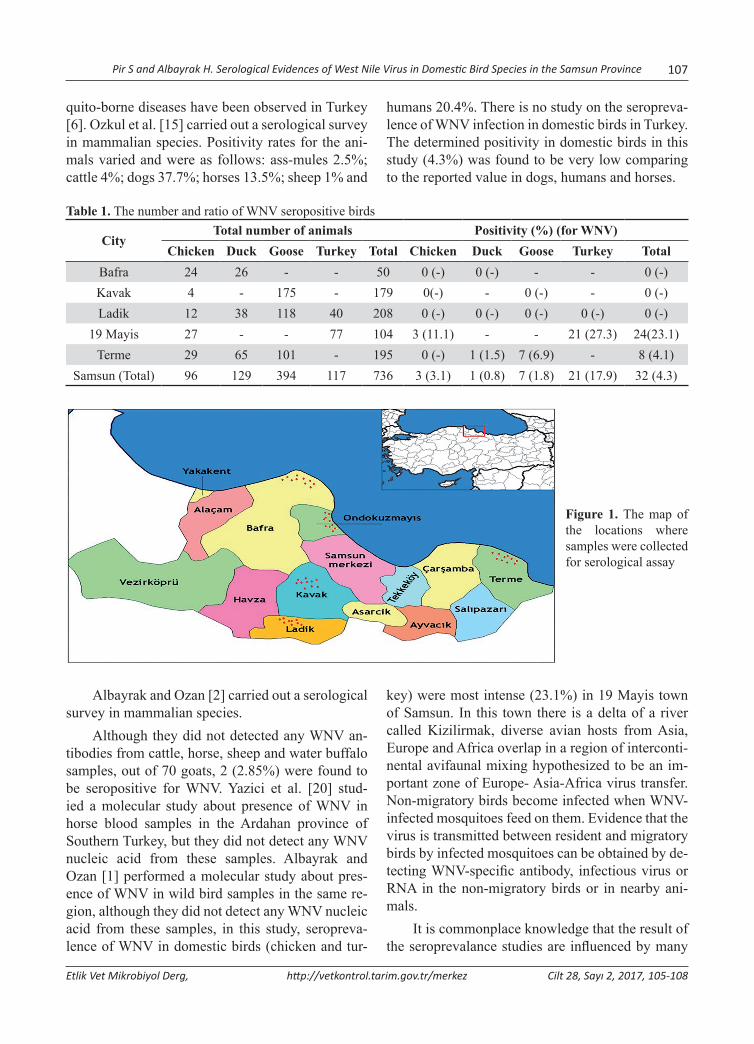
Pir S and Albayrak H. Serological Evidences of West Nile Virus in Domestic Bird Species in the Samsun Province 107
Etlik Vet Mikrobiyol Derg, http://vetkontrol.tarim.gov.tr/merkez Cilt 28, Sayı 2, 2017, 105-108
quito-borne diseases have been observed in Turkey [6]. Ozkul et al. [15] carried out a serological survey in mammalian species. Positivity rates for the ani-mals varied and were as follows: ass-mules 2.5%; cattle 4%; dogs 37.7%; horses 13.5%; sheep 1% and
humans 20.4%. There is no study on the seropreva-lence of WNV infection in domestic birds in Turkey. The determined positivity in domestic birds in this study (4.3%) was found to be very low comparing to the reported value in dogs, humans and horses.
Table 1. The number and ratio of WNV seropositive birds
CityTotal number of animals Positivity (%) (for WNV)
Chicken Duck Goose Turkey Total Chicken Duck Goose Turkey TotalBafra 24 26 - - 50 0 (-) 0 (-) - - 0 (-)Kavak 4 - 175 - 179 0(-) - 0 (-) - 0 (-)Ladik 12 38 118 40 208 0 (-) 0 (-) 0 (-) 0 (-) 0 (-)
19 Mayis 27 - - 77 104 3 (11.1) - - 21 (27.3) 24(23.1)Terme 29 65 101 - 195 0 (-) 1 (1.5) 7 (6.9) - 8 (4.1)
Samsun (Total) 96 129 394 117 736 3 (3.1) 1 (0.8) 7 (1.8) 21 (17.9) 32 (4.3)
Figure 1. The map of the locations where samples were collected for serological assay
Albayrak and Ozan [2] carried out a serological survey in mammalian species.
Although they did not detected any WNV an-tibodies from cattle, horse, sheep and water buffalo samples, out of 70 goats, 2 (2.85%) were found to be seropositive for WNV. Yazici et al. [20] stud-ied a molecular study about presence of WNV in horse blood samples in the Ardahan province of Southern Turkey, but they did not detect any WNV nucleic acid from these samples. Albayrak and Ozan [1] performed a molecular study about pres-ence of WNV in wild bird samples in the same re-gion, although they did not detect any WNV nucleic acid from these samples, in this study, seropreva-lence of WNV in domestic birds (chicken and tur-
key) were most intense (23.1%) in 19 Mayis town of Samsun. In this town there is a delta of a river called Kizilirmak, diverse avian hosts from Asia, Europe and Africa overlap in a region of interconti-nental avifaunal mixing hypothesized to be an im-portant zone of Europe- Asia-Africa virus transfer. Non-migratory birds become infected when WNV-infected mosquitoes feed on them. Evidence that the virus is transmitted between resident and migratory birds by infected mosquitoes can be obtained by de-tecting WNV-specific antibody, infectious virus or RNA in the non-migratory birds or in nearby ani-mals.
It is commonplace knowledge that the result of the seroprevalance studies are influenced by many
108 Pir S and Albayrak H. Serological Evidences of West Nile Virus in Domestic Bird Species in the Samsun Province
Etlik Vet Mikrobiyol Derg, http://vetkontrol.tarim.gov.tr/merkez Cilt 28, Sayı 2, 2017, 105-108
factors such as the number of sampled animals, the age of the animals, the time of sampling, the condi-tions of care and feeding, individual differences.
It was concluded that, the results, recorded for the first time for WNV in domestic birds in Turkey, supported the hypothesis that domestic bird act as a potential reservoir of WNV, and thus have a role in the epidemiology of these diseases. To understand better acute infection rates of WNV in domestic bir-ds, additional studies focusing on field observations are needed.
AcknowledgementsFunding for this research was provided by the Ondokuz Mayis University Scientific Research Council (Project Number: PYO.VET. 1904. 13.002)
References1. Albayrak H., Ozan E., 2010. Molecular detection of avian in-
fluenza virus but not West Nile virus in wild birds in north-ern Turkey. Zoonoses Public Health, 57, e71-e75.
2. Albayrak H., Ozan E., 2013. Seroepidemiological study of West Nile virus and Rift Valley fever virus in some of mammalian species (Herbivores) in northern Turkey. J Arthropod-Borne Dis, 7, 90-93.
3. Bagheri M., Terenius O., Oshaghi MA., Motazakker M., Asgari S., Dabiri F., Vatandoost H., Mohammadi BM., Chavshin AR., 2015. West Nile virus in mosquitoes of Iranian Wetlands. Vector-Borne Zoonot Dis, 15, 750-754.
4. Burke DS., Monath TP., 2001. Flaviviruses. In “Fields Virology”, Eds., DM Knipe, PM Howley, 4th ed., 1043–1126, Lippincott Williams & Wilkins, Philadelphia.
5. Dauphin G., Zientara S., 2007. West Nile virus: recent trends in diagnosis and vaccine development. Vaccine, 25, 5563-5576.
6. Dik B., Yagcı S., Linton YM., 2006. A review of species diver-sity and distribution of Culicoides Latreille, 1809 (Diptera: Ceratopogonidae) in Turkey. J Nat Hist, 40, 1947-1967.
7. Ergunay K., Saygan MB., Aydogan S., Menemenlioglu D., Turan HM., Ozkul A., Us D., 2007a. West Nile virus sero-prevalance in blood donors from central Anatolia, Turkey. Vector Borne Zoonot Dis, 7, 157-161.
8. Ergunay K., Ozer N., Us D., Ozkul A., Simsek F., Kaynas S., Ustacelebi S., 2007b Seroprevalance of West Nile virus and tick-borne encephalitis virus in southeastern Turkey: First
evidence for tick-borne encephalitis virus infections. Vector Borne Zoonot Dis, 10, 771-775.
9. Ergunay K., Bakonyi T., Nowotny N., Ozkul A., 2015. Close relationship between West Nile virus from Turkey and line-age 1 strain from Central African Republic. Emerg Infect Dis, 21, 352-355.
10. Gazi H., Özkütük N., Ecemis T., Atasoylu G., Köroglu G., Kurutepe S., Horasan GD., 2016. Seroprevalence of west nile virus, crimean-congo hemorrhagic fever virus, Francisella tularensis and Borrelia burgdorferi in rural population of manisa, western Turkey. J Vector Borne Dis, 53, 112-117.
11. Hubalek Z., Halouzka J., 1999. West Nile fever – a reemerg-ing mosquito-borne viral disease in Europe. Emerg Infect Dis, 5, 643-650.
12. OIE, 1999. Office International des Epizooties. West Nile fever in Israel in geese. Disease Information, 12, 166
13. OIE, 2000. Office International des Epizooties. West Nile fever in the United States of America: in horses. Disease Information, 13, 150–151
14. OIE, 2015. The World Organisation for Animal Health. West Nile fever. Manual of Diagnostic Tests and Vaccines for Terrestrial Animals 2013.Volume 1, Part 2
15. Ozkul A., Yıldırım Y., Pinar D., Akcali A., Yilmaz V., Colak D., 2006. Serological evidence of West Nile virus (WNV) in mammalian species in Turkey. Epidemiol Infect, 134, 826–829.
16. Padilla JA., Rubio EL., Romero EE., Cordoba L., Cuevas S., Mejia F., Calderon R., Milian F., Rosa ATD., Weaver SC., Franco JGE., Saiz JC., 2009. The continous spread of west nile virus (WNV): seroprevalance in asymptomatic horses. Epidemiol Infect, 137, 1163-1168.
17. Swayne DE., Beck JR., Smith CS., Shieh WJ., Zaki SR., 2001. Fatal encephalitis and myocarditis in young domestic geese (Anser anser domesticus) caused by West Nile virus. Emerg Infect Dis, 7, 751-753.
18. Thomas-Bachli AL., Pearl DL., Berke O., Parmley EJ., Barker IK., 2017. A geographic study of West Nile virus in humans, dead corvids and mosquitoes in Ontario us-ing spatial scan statistics with a survival time application. Zoonoses Public Health, 00, 1-9.
19. Toplu N., Oğuzoğlu TÇ., Ural K., Albayrak H., Ozan E., Ertürk A., Epikmen ET., 2015. West Nile virus infection in horses: Detection by immunohistochemistry, in situ hybri-dization, and ELISA. Vet Pathol, 52, 1073-1076.
20. Yazici Z., Albayrak H., Ozan E., Gumusova S., 2012. The first investigation of West Nile virus in horses using real time RT-PCR in Middle Black Sea Region in Turkey. J Arthropod-Borne Dis, 6, 151-155.
Yazışma adresi / Correspondence: Ezgi Şababoğlu, Mehmet Akif Ersoy Üniversitesi, Veteriner Fakültesi, Mikrobiyoloji Anabilim Dalı, İstiklal Yerleşkesi, Burdur, Türkiye. E posta:[email protected]
Etlik Vet Mikrobiyol Derg, 2017; 28 (2): 109-114 Derleme / Review Article
Ruminantlarda Mycobacterium avium subspecies paratuberculosis Enfeksiyonunun İmmunolojik Özellikleri
Ezgi Şababoğlu, Hülya Türütoğlu
Mehmet Akif Ersoy Üniversitesi, Veteriner Fakültesi, Mikrobiyoloji Anabilim Dalı, Burdur
Geliş Tarihi / Received: 10.04.2017, Kabul Tarihi / Accepted: 23.10.2017
Özet: Mycobacterium avium subspecies paratuberculosis, hayvan sağlığını etkileyen ve ciddi ekonomik kayıplara se-bep olan önemli bir patojendir. Etkenin mevcut immun sistemden kaçma stratejileri nedeniyle bugüne kadar paratüber-küloza karşı etkili bir aşı geliştirilememiş ve uygun bir koruma ve kontrol stratejisi belirlenememiştir. M. avium subsp. paratuberculosis’e karşı doğru kontrol stratejilerinin geliştirilebilmesi için konakçı-patojen etkileşimini düzenleyen hücresel ve moleküler mekanizmalar ile hastalığın patolojik temelinin detaylı olarak anlaşılması gerekmektedir. Bu der-lemede; M. avium subsp. paratuberculosis ile konakçı ilişkisi ve konakçıda gelişen immunolojik reaksiyonlar üzerine kapsamlı bilgiler sunuldu.Anahtar kelimeler: İmmünoloji, konakçı savunması, Mycobacterium avium subspecies paratuberculosis
Immunological Characteristics of Mycobacterium avium subspecies paratuberculosis Infection in Ruminants
Abstract: Mycobacterium avium subspecies paratuberculosis is an important pathogen that affects animal health and causes serious economic loss. Due to the current strategy of escape from the immunity system, so far, an effective vaccine against paratuberculosis has not been developed and no appropriate protection and control strategy has been determined. In order to develop correct control strategies against M avium subsp paratuberculosis, cellular and molecu-lar mechanisms regulating the host-pathogen interaction and the pathological basis of the disease must be understood in detail. In this review M. avium subsp. paratuberculosis-host interactions and the information on the immunological reactions developed in the host were presented.Keywords: Host defense, immunology, Mycobacterium avium subspecies paratuberculosis
Giriş
Paratüberküloz (Johne hastalığı), Mycobacterium avium subspecies paratuberculosis (Map) tarafın-dan oluşturulan, kronik granülomatöz enterokoli-tis, bölgesel lenfanjitis ve lenfadenitis ile karakte-rize yavaş gelişen hatta ölümle sonuçlanabilen bir hastalıktır [36, 41]. Evcil ve yabani ruminantlar-da (sığır, koyun, keçi, bizon, geyik, alpaka, lama, deve, manda) ve ruminant olmayan yabani türler-de (tavşan, tilki, maymun, porsuk, çakal, kuş, kedi, rakun) [3, 20, 23] hastalığa neden olmaktadır. Çok eskiden beri bilinen bir hastalık olmasına rağmen, patogenezi henüz tam olarak ortaya konulamamış ve dolayısıyla hastalığın kontrolü için etkili bir aşı veya uygun bir tedavi protokolü öngörülememiştir. Hayvan sağlığını tehdit eden ve büyük ekonomik kayıplara yol açan bu hastalığın önemi günümüzde halen devam etmektedir [20]. Ekonomik zararının
yanı sıra ruminant paratüberkülozu ile insanların Crohn hastalığı arasında yakın bir ilişkinin tespit edilmesi, son zamanlarda paratüberküloz hasta-lığının daha çok ilgi çekmesine yol açmıştır [24]. Crohn hastalarının ince barsaklarındaki histopatolo-jik bulguların paratüberküloza büyük benzerlik gös-terdiği ve vakaların çoğunda paratüberküloz hasta-lığının etkeni olan Map’in izole edildiği bildirilme-sine rağmen, Map’in hastalığın oluşumundaki rolü kesin olarak ortaya konulamamıştır [4, 9]. Etkenin enfekte hayvanların dışkısının yanı sıra sütleriyle de atıldığı tespit edilmiş ve enfekte hayvanların süt ve süt ürünlerinin insanlar için potansiyel bir bulaşma kaynağı olabileceği ileri sürülmüştür [4, 13, 16].
Hastalığın Gelişim Evreleri ve Konakçı YanıtıRuminantlarda paratüberküloz farklı evrelerden oluşmakta ve konakçının patolojik durumlara kar-

110 Şababoğlu E and Türütoğlu H. Ruminantlarda M. avium subs. paratuberculosis Enfeksiyonunun İmmunolojik Özellikleri
Etlik Vet Mikrobiyol Derg, http://vetkontrol.tarim.gov.tr/merkez Cilt 28, Sayı 2, 2017, 109-114
şı gösterdiği immun yanıta bağlı olarak bu evreler erken, subklinik ve klinik enfeksiyon evresi olarak sınıflandırılmaktadır [23].
Erken Evre
Hastalığın erken evresi, 2 yaştan küçük hayvan-larda ya da Map’in düşük konsantrasyonları-nın (501.0x10³CFU) sindirim [20] veya vertikal [39] yol ile bulaştığı hayvanlarda görülmektedir. Konakçı tarafından sindirim yoluyla alınan Map’in, bağırsak ile ilişkili lenfoid dokuya (gut-associated lymphoid tissue, GALT) yerleştiği ve Payer plak-larında bulunan M hücreleri ile fibronektin köp-rüsü kurarak konakçıya girdiği bilinmektedir [20, 32]. Fibronektin köprüsünün yanısıra, Map’in epi-tel hücrelerine invaze olabileceği de bildirilmiştir [20, 30]. Map’in, vücuda girdikten sonra intestinal makrofajlara invaze olma yeteneğine sahip olduğu ve çeşitli stratejilerle konakçı savunmasını engelle-yerek makrofajların içlerinde yaşayıp replike olabil-diği açıklanmıştır [20, 32]. Bakterinin makrofajlar içinde hayatta kalma stratejisini farklı mekanizma-lar oluşturmaktadır. Bakterinin kullandığı bu meka-nizmalar arasında; stres koşullarında spor benzeri yapı oluşturarak ısı, lizozim ve proteinaz K’dan ko-runması [20], fagozom olgunlaşmasını inhibe etme-si, makrofaj apoptozisini ve fagozom asitleşmesini engellemesi [3, 10, 32], MHC molekülünün eks-presyonunu azaltması [32] ve sinyal iletim yollarını etkilemesi [1, 33] sayılabilir.
Map’e karşı konakçı tarafından oluşturulan doğal savunma mekanizması: Birçok mikroorga-nizmada bulunan ve oldukça korunmuş patojen ile ilişkili moleküler yapıları (pathogen associa-ted molecular patterns, PAMP) tanıyan reseptörler (pattern-recognition receptors, PRRs), PAMP’ları bağlayarak hücrelerarası sinyal yollarını oluştu-rur ve böylece immun sistem hücrelerinin yangıyı uyaran molekülleri salgılamasına ve immun yanıtın uyarılmasına yol açarlar [29, 35]. Map’in endosito-zunda görevli fagositik hücrelerin üzerinde Map’i bağlayan önemli PRR’ler bulunmaktadır. Bu re-septörler (PRR) arasında komplement reseptörleri (complement receptor, CR1, CR3, CR4), mannoz reseptörü (MR), membran proteinini bağlayan bir fosfatidilinositol olan CD14 [32], CD11a, CD18 gibi β-integrin reseptörleri [23], sürfaktan protein-
ler, mannoz bağlayan lektinler (mannose-binding lectins, MBLs), C1q, IgG ile kaplı mikobakteriler için Fcγ reseptörü [32], transferrin reseptörleri, toll benzeri reseptör (toll-like receptors, TLR) 2, TLR 4 [23] ve TLR 9 [2] bulunmaktadır. En önemli PRR’lerden olan TLR’ler ile immun sistem uyarıla-rak sitokin yanıtı başlatılır [29]. Fagozomal olgun-laşma gibi mikobakterilerin patogenezisinde önemli hücresel işlemler TLR aracılığıyla şekillenmektedir [29]. TLR üretimindeki artışın Map’e karşı koruyu-cu olduğu gösterilmiştir [3]. TLR’lerin farklı etki-ye sahip olmaları nedeniyle paratüberküloza karşı gelişen immun yanıttaki rollerinin oldukça karışık olduğu bildirilmiş [3], TLR’lerde meydana gelen mutasyonların mikobakterilere karşı duyarlılığı art-tırdığı ileri sürülmüştür [23].
Mikobakterilerin öldürülmesinde etkili me-kanizmalar: Mikobakterilerin öldürülmesinde en önemli mekanizmanın makrofaj aktivasyonu oldu-ğu, makrofaj aktivasyonunda ise interferon gama (IFN-γ) ve tümör nekrozis faktör-beta (TNF-β) gibi proenflamatuar sitokinlerin rol oynadığı bildi-rilmiştir [32]. Lipopolisakkaritleri tanıyan TLR’ler tarafından aktive olan monosit ve makrofaj gibi mo-nonüklear fagositler tarafından mikroorganizmala-rın sindirildiği, hücre içinde ise otofaji uyarılarak antimikrobiyal peptidler, reaktif oksijen/nitrojen ara ürünleri aracılığı ile mikroorganizmanın öldü-rüldüğü açıklanmıştır [31, 40]. D vitamininin aktif metaboliti olan 1, 25- dihidroksikolekalsiferol (kal-sitriol, [1, 25 (OH)2D3])’ün ise CD14 ve mannoz reseptörü gibi yüzey reseptörlerinin üretimini art-tırarak makrofajların fagositoz yeteneğini güçlen-dirdiği, katelisidin ve defensinlerin ekspresyonunu arttırarak makrofajların antimikrobiyal etkisine kat-kıda bulunduğu ve ayrıca antijen sunumunun yanı-sıra bazı kemokin, sitokin ve makrofaj faktörlerinin sekresyonunu da düzenlediği açıklanmıştır [31]. Serbest yağ asitlerinin ve granülizinin de antimik-robiyal aktiviteye sahip olduğu ve Mycobacterium tuberculosis (Mtb)’in makrofajlar tarafından öldü-rülmesinde önemli rol oynadığı açıklanmıştır [32]. Bunun yanısıra apoptozisin de mikobakterinin yayı-lımını engelleyen diğer bir mekanizma olduğu be-lirtilmiştir [32].
Konakçı tarafından oluşturulan kazanılmış im-mun yanıt: Mikobakteri enfeksiyonlarının erken ev-resinde hücresel immun yanıtın çok önemli olduğu
Şababoğlu E and Türütoğlu H. Ruminantlarda M. avium subs. paratuberculosis Enfeksiyonunun İmmunolojik Özellikleri 111
Etlik Vet Mikrobiyol Derg, http://vetkontrol.tarim.gov.tr/merkez Cilt 28, Sayı 2, 2017, 109-114
bilinmektedir. Ancak Map, hayatta kalabilmek için erken gelişen bu hücresel immun yanıtı engelle-yen stratejiler geliştirmektedir [23]. Örneğin, Map makrofajları enfekte ettiğinde fagozom lizozom füzyonunu engeller ve mitojen ile aktive edilmiş protein kinaz (mitogen activated protein kinase, MAPK) düzeyini arttırır. MAPK, IL-10 sentezini arttırarak ve fagozom-lizozom füzyonunu engel-leyerek bakterinin yaşamasına yardımcı olur [38]. Ayrıca TLR’lerde (TLR2 ve TLR4) de artış olur. Aktive edilen makrofajlar interleukin-1 (IL-1), tümör nekrozis faktör-α (TNF-α ) ve IL-12 gibi pro-enflamatuar sitokinleri üretir. IL-6, IL-8 ve IL-10’da artış gözlenir. IL-1’in artışı sonucunda IL-2 üreten T hücreleri aktive olur ve CD4+ yardımcı T hücrelerinin ve CD8+ sitozolik T hücrelerinin klonal çoğalmasına yol açar [32].
Paratüberkülozun erken aşamasında CD4+T hücreleri oldukça önemlidir. Map makrofajlar ta-rafından fagosite edildikten sonra makrofajlar ta-rafından antijen sunumu gerçekleşir ve sitokinler aracılığıyla CD4+T hücreleri aktive olarak hücresel immun yanıt şekillenir [6]. CD4+T hücrelerinin alt tipleri intrasellüler Map’e karşı hücresel immun yanıtı aktive etmede görevlidir. Aktive edilmiş makrofajlar enfeksiyon bölgesine CD4+T hücreleri-ni toplayan IL-12 ve kemokinleri salgılamaktadır. CD4+T hücreleri ya Th1 ya da Th2 hücrelerine fark-lılaşır. Th1 hücrelerinin aktivasyonu IFN-γ, IL-2 ve TNF-α sentezine neden olur [32]. Sentezlenen IFN-γ da makrofajlara IL-12 salgılanması ve yangı-sal değişiklikleri teşvik etmek üzere etki eder [23]. Ayrıca, B hücreleri de CD4+T hücrelerine antijen sunarak daha fazla IFN-γ sentezlenmesini sağlar [36]. IFN-γ, Th1 hücreleri aracılığıyla gelişen hüc-resel immun yanıtı da uyarır [23]. Diğer taraftan makrofajlar ve intestinal epitel hücreleri tarafından üretilen IL-18’in de IFN-γ üretimini arttırdığı belir-tilmiştir [32].
Son yıllarda paratüberkülozun erken aşamasın-da gama-delta T (γδT) hücrelerinin rolü üzerinde ça-lışılmaktadır. γδT hücreleri 6 aylıktan küçük buza-ğılarda baskın olan T hücreleridir [32]. Buzağılarda sirkülasyondaki periferal kan mononüklear hücrele-rinin yaklaşık %40’ını, yetişkin sığırlarda ise %10-15’ini oluşturmaktadır [23]. Deneysel yapılan bir çalışmada; canlı Map içeren inokulumların enjekte edildiği buzağılarda γδT hücrelerinin hastalığın er-
ken döneminde enfeksiyon bölgesine göç ettikleri gösterilmiştir [26]. γδT hücrelerinin antijenlerin CD4+T hücrelerine sunulmasında [23], canlı miko-bakteri veya mikobakteriyel hücre duvarına karşı IFN-γ üretiminde [15], IFN-γ etkisi altında TNF-α salgılanmasında [32], intrasellüler ve ekstrasellü-ler Mtb etkenlerini granülizin salgılayarak öldü-rülmesinde, enfekte dendritik hücrelerden IL-17A salgılanmasında [21], IL-10 ve TGF-β üretiminde [5], Th1 immun yanıtının düzenlenmesi ve diğer dokulardaki hasarı engellemek için granülasyon oluşumunda [32] görevli olduğu rapor edilmiştir. IL-15’in ise IL-12 reseptörlerini etkilediği ve intra-epitelyal γδT hücrelerini stimüle ettiği açıklanmıştır [32]. Diğer taraftan γδT hücrelerinin, sistemik sir-külasyonda strese oldukça duyarlı oldukları ve bu nedenle strese maruz kalan hayvanların paratüber-küloza daha duyarlı olabilecekleri ileri sürülmüştür [32].
Bazı araştırmacılar [10, 17], dışkı, süt, doku gibi örneklerde az miktarda basil bulunduran pa-ratüberkülozlu hayvanlarda Th1 (IFN-γ) yanıtının, çok miktarda basil içeren hayvanlarda ise Th2 tip yanıtının daha ağırlıkta olduğunu bildirmiştir. Fazla sayıda basil bulunan sığırlarda IL-2, IL-4 ve IL-10 düzeylerinde artış gözlendiği belirtilmiştir [22]. Son yıllarda yapılan bazı çalışmalarda [27, 37], ise deneysel olarak enfekte edilen hayvanlarda IFN-γ yanıtı gibi kısa bir sürede antikor yanıtının gelişti-ği saptanmıştır. Begg ve ark. [8] koyunlar üzerin-de yürüttükleri deneysel çalışmalarında; enfekte koyunların %39’unda immun yanıtın güçlü IFN-γ yanıtından antikor yanıtına dönüştüğünü, %50’sin-de enfeksiyonun erken aşamasında IFN-γ ve antikor yanıtının birlikte olduğunu, %16’sında ise antijene spesifik antikor yanıtının engellediğini saptamışlar ve bu bulgulara göre enfekte koyunlarda Th1 yanı-tının Th2 yanıtına dönüşmesinin yaygın olmadığını ileri sürmüşlerdir.
Subklinik Evre
Subklinik evre, paratüberkülozun 2-5 yıla kadar sü-rebilen uzun bir latent fazıdır [23]. Bu fazda enfek-te hayvan klinik bulgu göstermez, sağlıklı görünür. Fakat bu fazın geç aşamalarında hayvanlar aralıklı olarak dışkı ile etkeni çıkarır ve antikor düzeyi ge-nellikle düşüktür [23, 36]. Bu evredeki hayvanla-
112 Şababoğlu E and Türütoğlu H. Ruminantlarda M. avium subs. paratuberculosis Enfeksiyonunun İmmunolojik Özellikleri
Etlik Vet Mikrobiyol Derg, http://vetkontrol.tarim.gov.tr/merkez Cilt 28, Sayı 2, 2017, 109-114
rın yaklaşık %10’unda IFN-γ ve antikor düzeyleri tespit edilebilir bir seviyeye yükselir [7, 11]. Tanı testlerinin yetersizliği veya Map’in aralıklı olarak çıkarılmasından dolayı subklinik enfekte hayvanla-rın tespit edilmesi zordur ve bu durum Johne hasta-lığının kontrolünü zorlaştırmaktadır [23, 36]. IgG1 sınıfı antikorların subklinik enfeksiyonun özellikle orta ve ileri aşamalarında tespit edilebileceği bildi-rilmiştir [23].
Paratüberkülozun bilinen en önemli özelliği ince bağırsağın orta ve alt segmentlerinde granülom oluşturmasıdır. Subklinik evredeki hayvanlarda kü-çük granülomlar gözlenmektedir [34]. Bu granüla-matöz lezyonların, Map’in bağırsağın diğer bölüm-leri ile lenf düğümlerine yerleşmesini engellemek ve kontrol etmek için konakçı tarafından oluşturulan geç immun yanıtla ilişkili olabileceği düşünülmüş-tür [14]. Ancak bu granülomların mikobakteriler ta-rafından enfeksiyon yerine yeni makrofajları topla-mak ve yer değiştirmek için kullanabileceği de ileri sürülmüştür [14]. Kısacası Map’in bağırsağın başka bölümleri ile meme bezleri ve mezenterik lenf dü-ğümleri gibi diğer organları enfekte etmek için gra-nülomlardan köprü olarak yararlanabileceği belirtil-miştir [20]. Granülomların içinde Map bulunduran makrofajlar ve dev hücreler bulunmaktadır. CD8+T hücreleri ve CD4+T hücreleri bu granülomların et-rafını çevreler. Bu aşamada en önemli sitokin, γδT hücreleri ve CD4+T hücreleri tarafından salınan TNF-α’dır. TNF-α, makrofajları toplayarak enfek-siyonun kontrol altına alınmasına yardımcı olur. Ancak doku hasarına da neden olabilir. Makrofajlar tarafından salınan IL-10, CD4+T hücrelerini bas-kılayarak IFN-γ üretimini azaltır. IFN-γ düzeyinin azalması nedeniyle yeni toplanan makrofajlar kalıcı enfekte olma eğilimindedir. Bu aşamada sitotoksik T hücre popülasyonu (CD8+ ve γδT hücreleri) fark-lı roller oynamaktadır. γδT hücreleri CD4+T hücre proliferasyonunu baskılarken, CD8+T hücre prolife-rasyonunu korur. Ayrıca γδT hücreleri granülasyon oluşumuna neden olur ve yangısal yanıtı kısıtlar. Humoral immun yanıt sınırlıdır. γδT hücrelerinden ve CD4+T hücrelerinden salınan IFNγ, makrofajla-rın hücre içi Map’i öldürmesi için yeterli değildir. IFN-γ yanıtı yeterli olmadığı için IgG1, IgG2’ye dönüşemez ve IgG1 tip immunglobulin yanıtı de-vam eder [32].
Erken subklinik aşamada olan hayvanların ile-al dokularında IFN-γ, TNF-α, IL-1α ve IL-6 gibi pro-enflamatuar sitokinlerin aşırı sentezine bağlı olarak CD4+T hücrelerinin oldukça fazla çoğaldı-ğı Th1 tip hücresel immun yanıtının baskın olduğu gözlenmiştir [12, 19]. Fakat hastalıkta Th1 hücresel yanıtın, artan antikor yanıtı ile birlikte azaldığı bil-dirilmiş, ancak bu değişikliğin arkasındaki nedenin ve ne zaman olduğunun kesin olarak bilinmediği belirtilmiştir [23]. Subklinik aşamada düzenleyici T (regulatory T, Treg) hücrelerinin düşük düzeyde Map antijenleri ile uyarıldığı ve Map’e karşı geli-şecek etkili bir T hücre yanıtını kısıtladığı açıklan-mıştır [15]. Ayrıca bu dönemin hastalığın subklinik formdan klinik forma dönüştüğünün de göstergesi olduğu ifade edilmiştir [28]. Treg hücrelerinin im-munomodülatör etkilerini IL-10 ve TGF-β üreterek gösterdikleri bilinmektedir [15]. Roussey ve ark. [28], sığır Treg hücrelerinin Map’e karşı spesifik olmadığını ve non-spesifik etkili bu Treg hücrele-rinin in vitro koşulda Th1 sitokinlerinden IFN-γ ile sitotoksik T hücrelerinin sitotoksik fonksiyonun-da görevli ve perforini kodlayan önemli bir prote-in olan profilin-1’in üretimini baskıladığını ortaya koymuşlardır.
Klinik Evre Map ile enfekte hayvanlarda subklinik fazın geç dö-nemi ile erken klinik faz döneminde sürekli ishal, yem yemede azalma, halsizlik, kas erimesi ve süt üretiminde azalma gibi klinik belirtiler gözlenmekte ve hastalık en son aşamada ölüme neden olmakta-dır [23]. Klinik bulguların ilerlediği dönemde, aşı-rı ishal, zayıflama ve kaşeksi görülmektedir [20]. Hayvanlar aşırı zayıftır ve bağırsakta fonksiyon kaybı nedeniyle özellikle albümin başta olmak üze-re serum proteinlerinin konsantrasyonunun düşme-si sonucu submandibular ödem ortaya çıkmaktadır [10, 20]. Subklinik evrede olduğu gibi bu evrede de süt verimi azalır [23]. Ayrıca klinik aşamada Map, sürekli olarak dışkı, süt ve kolostrum ile etrafa sa-çılmaktadır. Bu durum ise Map’in sürü içinde yayı-lım riskini arttırmaktadır [23].
Klinik belirti gösteren hayvanlarda intestinal mukozada Map yükü yüksek konsantrasyona ulaş-tığı için dışkı kültürü ile polimeraz zincir reaksiyo-nu pozitif sonuç verir ve bu hayvanlarda antikorlar
Şababoğlu E and Türütoğlu H. Ruminantlarda M. avium subs. paratuberculosis Enfeksiyonunun İmmunolojik Özellikleri 113
Etlik Vet Mikrobiyol Derg, http://vetkontrol.tarim.gov.tr/merkez Cilt 28, Sayı 2, 2017, 109-114
ELİZA testi ile de tespit edilebilir [18]. Ayrıca bu dönemde süt ve kolostrumda da etkenin tespit edi-lebildiği [25] ve gebe hayvanların bakteriyi uterusta yavruya geçirebildiği [39] rapor edilmiştir. Lenfosit, epiteloid makrofaj ve dev hücre infiltrasyonu ile ka-lınlaşan intestinal mukozada villus ve mikrovillus-ların fonksiyonlarını kaybettiği saptanmıştır [20]. İntestinal mukozadaki kalınlaşma ile intestinal du-varda granülomatöz yangı gibi patolojik lezyonla-rın varlığı protein kaybına neden olan enteropatinin kanıtı olarak belirtilmiştir [23, 36]. Ayrıca Map’in, supramammar, karaciğer ve akciğer lenf düğümleri de dâhil olmak üzere bağırsak dışında diğer organ-lara da yayılabildiği bildirilmiştir [20].
Klinik bulguların görülmesiyle birlikte immun yanıtın Th1’den Th2 aracılığıyla gelişen immun ya-nıta dönüştüğü gözlenmiştir [7]. Antikor üreten B hücrelerinin arttığı ve bununla orantılı bir şekilde serum antikor düzeylerinin de tespit edilebilebilir şekilde yükseldiği ortaya konulmuştur [23]. İmmun yanıtta bu değişiklikler belirlenmiş olmasına rağ-men, bu değişime neden olan faktörler henüz ay-dınlatılamamıştır [23]. İmmun yanıtın değişimi ile periferal mononüklear kan hücrelerinde, ileum ve ilgili lenf nodüllerinde IL-10, TGF-β, IL-4 gibi anti-enflamatuar sitokinlerin baskın olduğu gözlen-miştir [12, 23]. Ayrıca Treg hücreleri (CD4+CD25+) tarafından IL-10 ve TGF-β sentezinin sağlandığı ve böylece CD4+T hücre popülasyonun daha çok bas-kılandığı, Th2 hücre yanıtının baskılanması nede-niyle B hücre yanıtının azaldığı bildirilmiş, ancak B hücre yanıtının diğer deyişle IgG1 yanıtının bu aşamada hala ölçülebilir düzeyde kaldığı açıklan-mıştır [36].
Sonuç olarak, bugüne kadar paratüberküloz için etkenin mevcut immun sistemden kaçma strate-jileri nedeniyle uygun bir koruma ve kontrol strate-jisi belirlenememiş, etkili bir aşı geliştirilememiştir. Spesifik bir aşı veya tedavinin bulunabilmesi için Map’in, immun sistemden kaçış mekanizmalarının ve patogenezisinin anlaşılması gerekmektedir. Bu derlemede, Map’in enfeksiyon oluşturma mekaniz-maları ile immun sistemi nasıl yanılttığı ve kalıcı enfeksiyonun nasıl oluştuğu açıklandı. Yapılan li-teratür taramalarında detaylı çalışmalarla karşıla-şılmasına rağmen, hastalığın patogenezinde hala açıklanamayan mekanizmaların bulunduğu ve has-talığın oluşum mekanizmasının henüz tam olarak
anlaşılamadığı görülmektedir. Bu nedenle, Map’in virülens faktörleri ile hastalığın patogenezi ve ko-nakçının genetik duyarlılığı gibi konuların daha iyi anlaşılabilmesi için daha ayrıntılı çalışmalara ge-reksinim olduğu söylenebilir. Ancak bu detaylı ça-lışmalar sonucunda, etkili bir tedavi veya aşı bulu-narak ya da yeni tanı testleri geliştirilerek hastalığın kontrolü yapılabilir ve böylece hayvan sağlığının yanısıra insanlar için de gıda güvenliği sağlanabilir.
Kaynaklar1. Arsenault RJ, Li Y, Bell K, Doig K, Potter A, Griebel PJ,
Kusalik A, Napper S, (2012). Mycobacterium avium subsp. paratuberculosis inhibits interferon gamma induced signal-ing in bovine monocytes: insights into the cellular mecha-nisms of Johne’s disease. Infect Immun. 80, 3039-3048.
2. Arsenault RJ, Li Y, Maattanen P, Scruten E, Doig K, Potter A, Griebel P, Kusalik A, Napper S, (2013). Altered Toll-like receptor 9 signaling in Mycobacterium avium subsp. paratu-berculosis-infected bovine monocytes reveals potential thera-peutic targets. Infect Immun. 1, 226-237.
3. Arsenault RJ, Maattanen P, Daigle J, Potter A, Griebel P, Napper S, (2014). From mouth to macrophage: mechanisms of innate immune subversion by Mycobacterium avium subsp. paratuberculosis. Vet Res. 45, 54.
4. Ayele WY, Machackova M, Pavlik I, (2001). The transmission and impact of paratuberculosis infection in domestic and wild ruminants. Vet Med-Czech. 46, 205-224.
5. Baquero MM, Plattner BL, (2016). Bovine WC1(+) γδ T lym-phocytes modify monocyte-derived macrophage responses during early Mycobacterium avium subspecies paratubercu-losis infection. Vet Immunol Immunopathol. 170, 65-72.
6. Basler T, Geffers R, Weiss S, Valentin-Weigand P, Goethe R, (2008). Mycobacterium avium subspecies induce differen-tial expression of proinflammatory mediators in a murine macrophage model: evidence for enhanced pathogenic-ity of Mycobacterium avium subspecies paratuberculosis. Immunobiology. 213, 879-888.
7. Bassey EO, Collins MT, (1997). Study of T-lymphocyte subsets of healthy and Mycobacterium avium subsp. paratuberculo-sis-infected cattle. Infect Immun. 65, 4869-4872.
8. Begg DJ, de Silva K, Carter N, Plain KM, Purdie A, Whittington RJ, (2011). Does a Th1 over Th2 dominancy really exist in the early stages of Mycobacterium avium subspecies paratuber-culosis infections? Immunobiology. 216, 840-846.
9. Behr MA, Kapur V (2008): The evidence for Mycobacterium paratuberculosis in Crohn’s disease. Curr Opin Gastroenterol. 24, 17-21.
10. Clarke CJ (1997): The pathology and pathogenesis of para-tuberculosis in ruminants and other species. J Comp Pathol. 116, 217-261.
11. Collins MT, Wells SJ, Petrini KR, Collins JE, Schultz RD, Whitlock RH, (2005). Evaluation of five antibody detection tests for diagnosis of bovine paratuberculosis. Clin Diagn Lab Immunol. 12, 685-692.
114 Şababoğlu E and Türütoğlu H. Ruminantlarda M. avium subs. paratuberculosis Enfeksiyonunun İmmunolojik Özellikleri
Etlik Vet Mikrobiyol Derg, http://vetkontrol.tarim.gov.tr/merkez Cilt 28, Sayı 2, 2017, 109-114
12. Coussens PM, Verman N, Coussens MA, Elftman MD, McNulty AM, (2004). Cytokine gene expression in peripheral blood mononuclear cells and tissues of cattle infected with Mycobacterium avium subsp. paratuberculosis: evidence for an inherent proinflammatory gene expression pattern. Infect Immun. 72, 1409-1422.
13. Çetinkaya B, Muz A, Ertaş B, Öngör H, Sezen Y, Gülcü B, (2000). Süt ineklerinde paratüberküloz prevalansının polime-raz zincir reaksiyonu (PZR) ile saptanması. Turk J Vet Anim Sci. 24, 371-379.
14. Davis JM, Ramakrishnan L, (2009). The role of the granu-loma in expansion and dissemination of early tuberculous infection. Cell. 136, 37-49.
15. de Almeida DE, Colvin CJ, Coussens PM, (2008). Antigen-specific regulatory T cells in bovine paratuberculosis. Vet Immunol Immunopathol. 125, 234-245.
16. Ellingson JLE, Anderson JL, Koziczkowski JJ, Radcliff RP, Sloan, SJ, Allen SE, Sullivan NM, (2005). Detection of vi-able Mycobacterium avium subsp. paratuberculosis in retail pasteurized whole milk by two culture methods and PCR. J Food Prot. 68, 966-972.
17. Gillan S, O’Brien R, Hughes AD, Griffin JF, (2010). Identification of immune parameters to differentiate disease states among sheep infected with Mycobacterium avium subsp. paratuberculosis. Clin Vaccine Immunol. 17, 108-117.
18. Kalis CH, Barkema HW, Hesselink JW, van Maanen C, Collins MT, (2002). Evaluation of two absorbed enzyme-linked immunosorbent assays and a complement fixation test as replacements for fecal culture in the detection of cows shedding Mycobacterium avium subspecies paratuberculosis. J Vet Diagn Invest. 14, 219-224.
19. Khalifeh MS, Stabel JR, (2004). Effects of gamma interferon, interleukin-10, and transforming growth factor beta on the survival of Mycobacterium avium subsp. paratuberculosis in monocyte-derived macrophages from naturally infected cattle. Infect Immun. 72, 1974-82.
20. Lamont EA, (2012). Survival strategies of Mycobacterium avium subsp. paratuberculosis in a variety of microenviron-ments. PhD Thesis, The University of Minnesota, Minnesota, United States.
21. Lockhart E, Green AM, Flynn JL, (2006). IL-17 produc-tion is dominated by T cells rather than CD4 T cells dur-ing Mycobacterium tuberculosis infection. J Immunol. 177, 4662-4669.
22. Magombedze G, Eda S, Stabel J, (2015). Predicting the role of IL-10 in the regulation of the adaptive immune responses in Mycobacterium avium subsp. paratuberculosis infections using mathematical models. PLoS One. 10, e0141539.
23. Mallikarjunappa S, (2013). Cytokine gene expression in Holstein-Friesian and Jersey Calves infected with Mycobacterium avium subsp. paratuberculosis. Master Thesis. The University of Guelph, Ontario, Canada.
24. Nielsen SS, (2008a). Transtions in diagnostic tests used for detection of Mycobacterium avium subsp. paratuberculosis infections in cattle. Vet Microbiol. 132, 274-282.
25. Nielsen SS, Bjerre H, Toft N, (2008b). Colostrum and milk as risk factors for infection with Mycobacterium avium subspe-cies paratuberculosis in dairy cattle. J Dairy Sci. 91, 4610-4615.
26. Plattner BL, Doyle RT, Hostetter JM, (2009). Gamma-delta T cell subsets are differentially associated with granuloma de-velopment and organization in a bovine model of mycobacte-rial disease. Int J Exp Pathol. 90, 587-597.
27. Robinson M, O’Brien R, Mackintosh C, Griffin F, (2008). Differential immune responses of red deer (Cervus elaphus) following experimental challenge with Mycobacterium avium subsp. paratuberculosis. Clin Vaccine Immunol. 15, 963-969.
28. Roussey JA, Steibel JP, Coussens PM, (2014). Regulatory T cell activity and signs of T cell unresponsiveness in bovine paratuberculosis. Front Vet Sci. 1, 20.
29. Sanjuan MA, Milasta S, Green DR, (2009). Toll-like recep-tor signaling in the lysosomal pathways. Immunol Rev. 227, 203-220.
30. Secott TE, Lin TL, Wu CC, (2002). Fibronectin attachment protein is necessary for efficient attachment and invasion of epithelial cells by Mycobacterium avium subsp. paratubercu-losis. Infect Immun. 70, 2670-2675.
31. Selvaraj P, Afsal K, Harishankar M, (2015). Vitamin D and macrophage functions in tuberculosis. Macrophage. 2, e756.
32. Sohal JS, Singh SV, Tyagi P, Subhodh S, Singh PK, Singh AV, Narayanasamyc K, Sheoran N, Komal Singh Sandhu, (2008). Immunology of mycobacterial infections: with special refer-ence to Mycobacterium avium subspecies paratuberculosis. Immunobiology. 213, 585-598.
33. Sommer S, Pudrith CB, Colvin CJ, Coussens PM, (2009). Mycobacterium avium subspecies paratuberculosis sup-presses expression of IL-12p40 and iNOS genes induced by signalling through CD40 in bovine monocyte-derived macro-phages. Vet Immunol Immunopathol. 128, 44-52.
34. Tiwari A, VanLeeuwen JA, McKenna SL, Keefe GP, Barkema HW, (2006). Johne’s disease in Canada Part I: clinical symp-toms, pathophysiology, diagnosis, and prevalence in dairy herds. Can Vet J. 47, 874-882.
35. Tizard IR, (2009). Veterinary Immunology, 8th edition, Saunders Elsevier, St. Louis, Missouri, p: 11-16.
36. Wadhwa A, Kumar N, Velasco-Villa A, Eda S, (2013). Overview of Johne’s disease immunology. Veterinary World. 6, 901-904.
37. Waters WR, Miller JM, Palmer MV, Stabel JR, Jones DE, Koistinen KA, Steadham EM, Hamilton J, Davis WC, Bannantine JP, (2003). Early induction of humoral and cel-lular immune responses during experimental Mycobacterium avium subsp. paratuberculosis infection of calves. Infect Immun. 71, 5130-5138.
38. Weiss DJ, Souza CD, (2008). Modulation of mononuclear phagocyte function by Mycobacterium avium subsp. paratu-berculosis. Vet Pathol. 45, 829-841.
39. Whittington RJ, Windsor PA, (2009). In utero infection of cattle with Mycobacterium avium subsp. paratuberculosis: A critical review and meta-analysis. Vet J. 179, 60-69.
40. Woo SR, Czuprynski CJ, (2008). Tactics of Mycobacterium avium subsp. paratuberculosis for intracellular survival in mononuclear phagocytes. J Vet Sci. 9, 1-8.
41. Yardımcı H, (2006). Mycobacterium infeksiyonları. Editörler: Aydın N, Paracıkoğlu J. Veteriner Mikrobiyoloji, İlke-Emek Yayınları, Ankara, s: 87-107.

Yazışma adresi / Correspondence: Cüneyt Tamer, Ondokuz Mayıs Üniversitesi, Veteriner Fakültesi, Viroloji Anabilim Dalı, Atakum/SAMSUN E posta: [email protected]
Etlik Vet Mikrobiyol Derg, 2017; 28 (2): 115-122 Derleme / Review Article
Viruslar ve Teratogenez
Cüneyt Tamer, Semra Okur Gümüşova
Ondokuz Mayıs Üniversitesi Veteriner Fakültesi Viroloji AD.
Geliş Tarihi / Received: 05.01.2017, Kabul Tarihi / Accepted: 17.10.2017
Özet: Hayvancılıkta reprodüksiyon sorunları işletmelerin mücadele edeceği önemli sorunlardan birisi olarak kabul edilir ve erken teşhisi edilmesi önem taşır. Teratojen ajanlar ise, gebelik döneminde gelişimsel bozukluklara neden olarak reprodüktif sorunlar oluşturan önemli etkenlerdendir. Enfeksiyöz teratojen ajanlar içerisinde sınıflandırılan viral etkenler de reprodüktif sorunların önemli nedenlerinden birisi olarak kabul edilir. Teratojen virusların birçoğu gebelik döneminde asemptomatik seyreder ve latentlik periyodu nedeniyle teşhisi zordur. Bu durum virusların saçılmasını ko-laylaştırdığından sürü sağlığını tehdit etmektedir. Bu derleme ile önemli viral teratojenler hakkında yapılmış çalışmala-rın verileri toparlanıp özetlenerek bu konudaki bilgilerin güncellenmesi amaçlanmıştır.Anahtar kelimeler: Patogenez, teratogenez, virus,
Teratogenesis and VirusesAbstract: Reproduction problems is one of the important problem for dairy farming and early diagnosis is important. Teratogen agents are creates reproductive problems as the cause of developmental disorders in the pregnancy period. Also, viruses classified as teratogen infectious agent are considered to be one of the major causes of reproductive prob-lems. Many teratogen viruses are asymptomatic and is difficult to diagnose due to the latency period. This situation is threatens herd health. In this review, we aimed to update the datas of the studies about the important viral teratogens. Key Words: Pathogenesis, teratogenesis, virus.
Giriş
Teratojenite, gebelik süresince yavruda meydana gelen gelişimsel bozuklukları (malformasyonları) ifade ederken, teratojen terimi ise bu bozukluk-ları meydana getiren etkenleri tanımlamaktadır. Hayvanlarda teratojenik olguların önlenmesi repro-düksiyonun sorunsuz devam edebilmesi için gerek-lidir.
Malformasyonlar etiyolojlerine göre; kendi-liğinden veya genetik olarak gelişen kromozomal bozukluklara bağlı şekillenen farklı protein kodlan-malarının neden oldukları gelişim bozuklukları ile transplasental olarak yavruyu etkiyen enfeksiyonlar ve toksik kimyasallar gibi teratojenlerin oluşturdu-ğu gelişim bozuklukları olarak ikiye ayrılır. Viral etkenler, teratojen enfeksiyöz ajanlar içerisinde sı-nıflandırılırlar ve daha çok sinir ve iskelet kas sis-temini etkileyerek malformasyonlar meydana getir-dikleri ortaya konmuştur [20].
Fötal Sinir Sistemi Gelişimi ve Viral Terato-jenlerin Etki Mekanizmaları: Embriyogenezisin erken dönemlerinde ektodermin üst orta kısımları
kalınlaşıp içeri doğru çökerek mezodermden ayrılıp nöral tüpü şekillendirirken, nöral tüpü şekillendiren nöroektodermal hücreler ise dışa doğru göç ederek serebral ve serebellar dokuları şekillendirmektedir. Daha sonra, nöral tüpün rostral kısmından serebrum ve serebellum oluşurken, kaudal kısmı ise, omurili-ği oluşturmak için farklılaşarak, ventrikuluslara ve kanalis spinalise dönüşmektedir [13]. Her hayvan türünde nöral tüpün gelişimi, myelinizasyonu ve sinir hücrelerinin göçü farklılık göstermektedir. Bu nedenle sinir sistemi gelişimini koyun, sığır, at ve domuzlarda prenatal dönemde tamamladığı, kedi ve köpekte ise postnatal dönemde de gelişimin devam ettiği bilinmektedir. Dolayısı ile teratojenlerin he-def aldığı hücrelerin, hayvan türlerine göre farklılık gösterdiği bildirilmiştir [20]. Enfeksiyöz etkenlerin gebelik sırasında fötusa bulaşmasından sonra, yav-runun nötralizan antikorları üretebilme ya da an-neden alabilme durumuna bağlı olarak teratojenik defektin büyüklüğünün şekillenebildiği de bilin-mektedir. Bazı hayvan virusları bovine viral diyare (BVD), border disease (BD) ve klasik domuz vebası (CSF), sinir sistemi dokularındaki teratojenik etki-
116 Tamer C and Okur Gümüşova S. Viruslar ve Teratogenez
Etlik Vet Mikrobiyol Derg, http://vetkontrol.tarim.gov.tr/merkez Cilt 28, Sayı 2, 2017, 115-122
lerini genellikle selektif hücre ölümü ile gerçekleş-tirmektedir. Bu mekanizma, virusun hücreyi nekro-za uğratıp nonspuratif yangı oluşturarak hücrelerin göçünü engellenmesi ve nekroza uğrayan dokuda boşluklar oluşması sonucu porensafali şekillenmesi ile açıklanmıştır. Hidranensefali gibi malformas-yonların ise zamanla bu boşlukların beyin sıvısıyla dolması sonucu şekillendiği bildirilmiştir. Viral et-kenler sinir sisteminde en çok hidranensefali, poren-sefali, hipomiyelinongenezis, demiyelinogenezis, serebellar hipoplazi, serebellar displazi ve hidrose-falusa, iskelet kas sisteminde ise nöral tüp defektine bağlı oluşan iskelet kası atrofisine ve artrogrippozis sendromuna sebep olmaktadırlar. Teratojen virus-lar hücre ölümü olmadan da hücrenin fonksiyonu-nu bozabilmektedirler. Örneğin BVD, BD ve CSF hücrelerde yetersiz miyelin sentezlemesine neden olarak hipomiyelinogenezis oluşturdukları bildiril-miştir [13].
Hayvanlar İçin Önemli Teratojen Virus Aileleri
1. Bunyaviridae ailesi: Bunyavirdae ailesi içinde sınıflandırılan iki genusda önemli teratojen viruslar yer alır. Bunlar; phlebovirus genusuna ait rift vadisi humması virusu (RVFV) ile orthobunyavirus genu-suna ait akabane virus (AKAV), cache vadisi virusu (CVV), aino virus (AIV) ve schmallenberg virus (SMBV) olarak bildirilmiştir [7, 12] Bunlardan, akabane virus enfeksiyonlarının sıklıkla kuzu, bu-zağı ve oğlaklarda, aino virus enfeksiyonlarının daha çok buzağılarda, cache vadisi virusu’nun ne-den olduğu enfeksiyonların ise daha çok kuzularda meydana geldiği belirlenmiştir [12]. 2011 yılından itibaren bir çok avrupa ülkesinde enfeksiyonlara neden olan schmallanberg virusun ise sığır, koyun ve keçilerde non spesifik semptomlarla seyrederek abort ve anormal doğumlara sebep olmaktadır [7].
Akabane virusun sığırlarda gebeliğin ikinci ya-rısında (3 - 6. ay arasında), koyun ve keçilerin ise gebeliğin erken dönemlerinde (30 - 50. gün ara-sında) alınması durumunda, virusun subventrikü-ler çizgilerde nekroza yol açtığı ve ölen nöronların gebeliğin ilerleyen döneminde beyinin gri madde-sine doğru göç edememesi nedeniyle de beyin do-kusunun tamamı ya da tamamına yakınının ortadan kalkarak sadece zarla çevrili içi sıvı dolu bir kese-
ye dönüşerek hidranensefaliye neden olduğu bil-dirilmiştir. Ayrıca, gebeliğin daha geç döneminde şekillenen enfeksiyonlarda beyindeki lezyonların şiddetinin azaldığı ve porensefali şekillendiği, geç şekillenen enfeksiyonlar sırasında ise mikroensefali ve serebellar hipoplazinin meydana gelebileceği de bildirilmiştir [13].
Schmallanberg virus enfeksiyonlarında ise transplesental enfeksiyona az rastlandığı, sığırların immun sistemi gebeliğin 40 - 170. günleri arasın-da geliştiğinden merkezi sinir sistemi lezyonları-nın genellikle gebeliğin 60 - 180. günleri arasında oluştuğu belirlenmiştir [6]. Ayrıca, gebeliğin erken dönemindeki enfeksiyonlarda şiddetli merkezi sinir sistemi bulgularının, geç dönemindeki enfeksiyon-larda ise ensefalomiyelitisin meydana geldiği de bildirilmiştir [17]. Akabane virus ile birlikte sch-mallanberg virusu’nun oluşturduğu önemli başka bir sendrom ise virusun nöral tüpün kaudal kısmı-nındaki bozukluklarının (Disrafizm) sebep olduğu artrogrippozistir. Artrogrippotik hayvanlarda omur-gada kifozis, lordozis ya da tortikolliz şekillendiği ve ayak eklemlerinin ise fleksiyon durumda kaldığı belirlenmiştir. Ayrıca medulla spinalisin ventral kol-larındaki nöronların ölümünün sebep olduğu dener-vasyona bağlı iskelet kası atrofisinin meydana gel-diği ve kas dokusunun yerini yağ ve fibröz dokunun doldurduğu da bildirilmiştir [3]. 2. Flaviviridae ailesi: Flaviviridae ailesinin pes-tivirus cinsine ait ve birbiri ile genetik yakınlığa sahip 3 önemli virus olan BVDV, BDV ve CSFV’ nin ve flavivirus cinsinde yer alan wesselbron virus (WBV)’ un da teratojenik etkiye sahip viruslar oldu-ğu bilinmektedir. Sığırlarda gebeliğin yaklaşık 100. gününe kadar şekillenen BVDV enfeksiyonlarında mumifikasyon veya abortlar şekillenir. 100 ile 125. günler arası şekillenen BVDV enfeksiyonlarında ise immun sistem gelişmesini henüz tamamlamadığı için, yavru doğsa bile zayıf ve immun tolere persiste enfeksiyon ile doğmaktadır. Malformasyonların ise gebeliğin yaklaşık olarak 6. ayından sonraki BVDV enfeksiyonunu takiben ortaya çıktığı belirlenmiştir. BDV ve CSFV enfeksiyonlarındaki malformasyon-ların ise, sırasıyla küçük ruminantların ve domuzla-rın gebeliklerinin geç döneminde (özellikle 30-80. günde) oluşan enfeksiyonlar sonucu şekillendiği tespit edilmiştir [12]. Bu ailedeki viruslar diğer tera-tojenik viruslara benzer malformasyonlara da sebep

Tamer C and Okur Gümüşova S. Viruslar ve Teratogenez 117
Etlik Vet Mikrobiyol Derg, http://vetkontrol.tarim.gov.tr/merkez Cilt 28, Sayı 2, 2017, 115-122
olmaktadırlar. Örneğin, BVDV sığırlarda, hastalık için karakteristik olan serebellar hipoplazinin yanı sıra hidranansefali, hidrosefalus, microensefali, perosomus elumbus, kistik septum pellisidum, hi-pomiyelinogenezis ve retinopatiye sebep olabildiği belirlenmiştir [2, 10, 13]. Koyunlarda BDV enfek-siyonunda ise bunlara ek olarak vertebra defektleri tespit edilmiştir [12].3. Reoviridae ailesi: Bluetongue virusu (BTV), ruminantlarda, özellikle koyun ve keçilerde vaskü-litis, oral kavitede hiperemi, perikarditis, abort ve anomali doğumlara sebep olan Orbivirus genusuna ait bir virusdur. Virus, sığırlarda ve vahşi ruminant-larda genelde asemptomatik seyreder [12]. Virusun dış kapsit proteini-2 (VP2)’ nin yapısına bağlı ola-rak 27 farklı serotip tespit edilmiştir [9]. BTV saha suşlarının çok az bir kısmının transplesental enfek-siyona yol açtığı bildirilse de laboratuvar koşulların-da hazırlanan canlı attenüe aşıların ve virusun bazı suşlarının (özellikle serotip 8) fötal enfeksiyona ne-den olduğu bilinmektedir [12]. Sığırlarda gebeliğin ilk yarısında (70 - 130. günler arasında) oluşturulan deneysel enfeksiyonlar sonucunda buzağılarda mal-formasyonlar gözlemlenirken [11], yine koyunlar-da 50 ile 55. günler arasında oluşturulan deneysel enfeksiyon sonucu, ensefalopati ve retinopati şekil-lendiği saptanmıştır. Ayrıca virusun neden olduğu beyin lezyonlarının hemen hepsinde serebellar hi-poplazi de bildirilmiştir [22]. BTV ile enfekte olan gebe hayvanlarda beyindeki öncü glia hücrelerinin ve nöronların enfeksiyonu sonucu hidranensefali ve porensefali şekillenmektedir. Çalışmalar porensefa-linin hidranensefaliye göre daha az şekillendiğini de ortaya koymuştur [11]. Ayrıca virusun sebep olduğu vaskülitisin beyin lezyonlarını arttığı ve geç dönem-deki enfeksiyonların herhangi bir malformasyon yapmadan sadece ensefalitise neden olduğu saptan-mıştır [21]. BTV ile enfekte gebe hayvanlarda bas-
kın semptom olarak ise hidranensefali görülmekle birlikte periventriküler doku kaybının olduğu bazı olgularda hidrosefalus da bildirilmiştir [18]. Şuana kadar saptanan BTV-8’ in neden olduğu enfeksiyon-larda ise beyin sapı, medulla ve spinal kord boyun-ca bir bulgu saptanmamış ve bu enfeksiyonlar da artrogrippozis ve vertebral kanal malformasyonları gibi iskelet kas sistemini etkileyen lezyonlar belir-lenmemiştir [1]. 4. Parvoviridae ailesi: Feline Panlökopeni virus (FPV), Parvoviridae ailesinin Parvovirinae alt ai-lesinin Parvovirus cinsinde yer alır [8]. FPV, çok bulaşıcı ve tüm dünyada oldukça yaygın enfeksi-yonlara sebep olmaktadır. Hastalık direk kontak yolla bulaşır. İnsan ve fomitlerin ise enfeksiyonun taşınmasında mekanik vektör rolü oynayabildikleri bildirilmiştir [12].
Parvoviruslar enfekte ettikleri hücreleri bölün-mesini S fazında yakaladıklarından yüksek mitotik aktivite gösteren hücrelere tropizmleri oldukları düşünülmüştür [4]. Karnivorların sinir sistemi ge-lişimi doğumdan sonra da devam ettiği için, daha önce bu virusla perinatal dönemde enfekte olan ke-dilerde serebellar hipoplazi lezyonları ve beyindeki nöronlarda virusun varlığı immunohistokimaysal yöntemlerle tespit edilmiştir [5]. Gebelik süresin-ce plesentayı geçen virus, fötusun serebellumdaki extragranüler tabakada nekroz oluşturup serebellar hipoplaziye sebep olmaktadır Ayrıca, serebellum-daki purkinje hücrelerinde de virus tespit edilmiş ve enfekte fötuslarda hafif derecede renal hipoplazi şekillendiği de bildirilmiştir [16]. FPV ile enfekte kedi yavrularında kifozis de tespit edilmiştir ama bunun virus enfeksiyonu ile ilgili olup olmadığı tam olarak ortaya konulamamıştır [13]. Tablo 1’ de hav-yanlar için teratojen olan viruslar ve neden oldukları malformasyonlar gösterilmiştir
BVDV BDV AKAV BTV SMBV FPVHidranensefali + + + + + +
Porensefali + + + + +Hidrosefali + + + +
Microensefali + + + + +Serebellar hipoplazi + + + + +Vertebra defektleri + + +?
Arthrogripozis + +
Tablo 1. Teratojen virusların fö-tusta meydana getirdiği malfor-masyonlar.
118 Tamer C and Okur Gümüşova S. Viruslar ve Teratogenez
Etlik Vet Mikrobiyol Derg, http://vetkontrol.tarim.gov.tr/merkez Cilt 28, Sayı 2, 2017, 115-122
BulaşmaTeratojen viruslar genel olarak vertikal bulaşmaya sahip olsalar da horizontal olarak bireyden bireye farklı şekilde bulaşabilmektedirler. Flaviviridae ve Bunyaviridae ailesine ait teratojenler birer ar-bovirustur. AKAV, SMB ve BTV sokucu sinekler-le aktarıldığı bildirilmiştir. BTV için özellikle su-lak bölgelerde, bahar aylarında sokucu sineklerin (Culicoides spp.) artmasıyla birlikte hayvanlar için risk artmaktadır. BTV özellikle ruminantların hari-cinde, son yıllarda köpek gibi diğer memelilerde de bildirilmiştir [12, 15]. Bu yüzden bu virusun rezer-vuar hayvan türünün arttığı göz önünde bulundurul-malıdır. BVDV, BDV, CSFV, FPV ve AEV ise direk kontak yolu ile bulaşmaktadır. Sığırlarla koyunlar arasında BVDV ve BDV enfeksiyonlarının türler arası nakli gözlemlenmiştir ama keçilerden diğer hayvan türlerine aktarım tespit edilememiştir [12].
TeşhisBTV virusunun damar endotel duyarlılığından dolayı direk teşhisin de, kandan, yüksek oranda Polimaraz zincir reaksiyonu (PCR) ile virus tespit edilebilir. Ayrıca yine dalak, akciğer, lenf yumrula-rından antijen tespiti yapılabilir. BTV, indirek ola-rak ise antikor ELİSA kitleri ile de tespit edilebilir [12]. BVDV, BDV ve CSFV virusları hücre kültü-ründe sitopatojenik etki yapmadan da üreyebileceği için identifikasyonda immunperoksidaz ve immunf-loresan testleri hala “gold” yöntem olarak kabul edilmektedir [19]. Bununla birlikte antijen ELİSA kitleri ile de virus tespiti yapılmaktadır. Antikor tespitinde ise en sık serum nötralizasyon ve antikor ELİSA testleri kullanılmaktadır [12].
SMBV ve AKAV enfeksiyonlarının yavru atık-ları, amniyon sıvısı ve kandan tespitinde real-time ve konvansiyonel PCR teknikleri kullanılabilmekte-dir. AKAV için ayrıca virus nötralizasyon ve ELİSA testleri sayesinde antikor tespiti de yapılabilmekte-dir. SMBV için ise yine ELİSA, virus nötralizasyon ve immunflorasan antikor tekniği ile antikor varlığı saptanmaktadır [14]
Korunma ve MücadeleTürkiye’de BVDV ve BDV hastalığı ile mücade-le için üretilen aşı bulunmamakla birlikte iki has-talığa karşı da ithal inaktif aşılar kullanılmaktadır.
Sığırlarda BVD hastalığı için uygulanan aşı takvi-mi, gebelikten 2 ay önce, kuru dönemde bir doz ve antikor titresinin gebelikte koruyucu seviyeye gele-bilmesi için ise birinci dozdan 4 hafta sonra rapel doz uygulanmasıdır. BDV aşısı ise Türkiye’de rutin olarak gebelikten önce uygulanmaktadır. Koyunlar yılda iki defa gebelik oluşturulabileceği için birinci gebelikte aşılama uygulandıysa, ikinci gebelikten önce serum antikor titresi ölçülerek aşı uygulama-sına buna göre karar verilmelidir.
Türkiye’de görülen ve vektörler aracılığı ile aktarılan diğer teratojen viruslar için ise korun-mada mevsimsel aşılama önemli yer tutmaktadır. SMBV ve AKAV’ a karşı da ithal ya da yerli bir aşı bulunmazken, BTV ile mücadele için Türkiye’de üretilen serotip 4’e karşı canlı aşı bulunmaktadır. Koyunlarda BTV aşılamasının yılda bir kez ilkba-har aylarında koç katımından 1 ay önce uygulanma-sı uygundur.
Ülkemizde kedilerde görülen FPV için korun-ma ise, uygulanmakta olan karma aşılarla sağlana-bilmekte ve enfeksiyonla mücadelede başarı şansını arttırmak için aşılamanın sokakta ve barınakta yaşa-yan kedilerede uygulanması önerilmektedir.
Sonuç
Hayvancılıkta özellikle süt ve damızlık işletmeler-de, anaçlardan düzenli yavru alımı önemli bir konu-dur. Bu nedenle reprodüksiyon sorunları işletmele-rin mücadele edeceği en önemli sorunlardan birisi olarak kabul edilir.
Teratojen virusların birçoğunun annelerde asemptomatik seyretmesi ve bu enfeksiyonlardaki latentlik periyodu nedeniyle hasta hayvanların ye-tişkin dönemlerinde teşhis edilememeleri bu virus-ların saçılmasını kolaylaştırıp sürü sağlığını tehdit etmelerine neden olmaktadır. Bu nedenle gebelik-ten önce viral teratojenlerin teşhisi ve bunlardan ko-runmanın sağlanması ve enfekte hayvanların tespit edilip sürüden çıkartılması bu viruslar ile mücadele için önem taşımaktadır. Bunun yanı sıra viral tera-tojenlerin neden olduğu enfeksiyonlar ortak semp-tomlarla seyrettiğinden malformasyonlu doğan yavrulara ve virusun neden olduğu makroskobik lezyonlara bakarak enfeksiyonun etiyolojisi hakkın-da tahmin yürütmek zordur. Bu nedenle sürülerde belirli aralıklarla viral teratojenlerin varlığının araş-
Tamer C and Okur Gümüşova S. Viruslar ve Teratogenez 119
Etlik Vet Mikrobiyol Derg, http://vetkontrol.tarim.gov.tr/merkez Cilt 28, Sayı 2, 2017, 115-122
tırılması ve bu enfeksiyonlar için etkili yerli aşıların geliştirilmesi de teratojen viruslar ile mücadele ve ülke ekonomisi açısından önem taşımaktadır.
Kaynaklar1. Agerholm JS, Hewicker-Trautwein M, Peperkamp K,
Windsor PA, (2015) Virus-induced congenital malforma-tions in cattle. Acta Vet Scand. 57: 54.
2. Bahr C, Kuiper H, Distl O, (2003) Bilateral anophthalmia associated with further anomalies of the head in German Holstein calves. Dtsch Tierarztl Wochenschr. 110 (11): 454-456.
3. Bayrou C, Garigliany MM, Sarlet M, Sartelet A, Cassart D, Desmecht D, (2014) Natural intrauterine infection with Schmallenberg virus in malformed newborn calves. Emerg Infect Dis. 20: 1327-1330.
4. Deleu L, Pujol A, Faisst S, Rommelaere J, (1999) Activation of promoter P4 of the autonomous parvovirus minute virus of mice at early S phase is required for productive infection. J Virol. 73:3877–3885.
5. Garigliany M, Gilliaux G, Jolly S, Casanova T, Bayrou C, Gommeren K, Fett T, Mauroy A, Lévy E, Cassart D, Peeters D, Poncelet L, Desmecht D, (2016) Feline panleukopenia virus in cerebral neurons of young and adult cats. BMC Vet Res. 12(1):28.
6. Herder V, Hansmann F, Wohlsein P, Peters M, Varela M, Palmarini M, Baumgartner W, (2013) Immunophenotyping of inflammatory cells associated with Schmallenberg virus infection of the central nervous system of ruminants. PLoS One. 8(5):1-11.
7. Hoffmann B, Scheuch M, Hoper D, Jungblut R, Holsteg M, Schirrmeier H, Eschbaumer M, Goller KV, Wernike K, Fischer M, Breithaupt A, Mettenleiter TC, Beer M, (2011) Novel orthobunyavirus in Cattle, Europe. Emerg Infect Dis. 18(3):469–479.
8. International Committee on Taxonomy of Viruses. (2012). http://www.ictvonline.org/taxonomyHistory.asp?taxnode_id=20142920&taxa_name=Parvoviridae Erişim tarihi: 03.04.2016.
9. Jenckel M, Bréard E, Schulz C, Sailleau C, Viarouge C, Hoffmann B, Höper D, Beer M, Zientara S, (2015) Complete coding genome sequence of putative novel blue-tongue virus serotype 27. Genome Announc. 3(2):1-2.
10. Karakaya E, Alpay G, Yilmazbas-Mecitoglu G, Alasonyalilar-Demirer A, Akgül B, Inan-Ozturkoglu S,
Ozyigit MO, Seyrek-Intas D, Seyrek-Intas K, Yesilbag K, Gumen A, Keskin A, (2013) Perosomus elumbis in a Holstein calf infected with bovine viral diarrhea virus. Tierarztl Prax Ausg G Grosstiere Nutztiere. 41(6):387-391.
11. MacLachlan NJ, Conley AJ, Kennedy PC, (2000) Bluetongue and equine viralarteritis viruses as models of virus-induced fetal injury and abortion. Anim Reprod Sci. 60-61: 643-651.
12. MacLachlan NJ, Dubovi EJ, (2011) Fenner’s Veterinary Virology. 4. Baskı, Kalifornia. Elsevier. 62-380
13. Maxie MG, (2015) Kennedy & Palmer’s Pathology of Domestic Animals, 6. Baskı, Ontario, 3:283-432.
14. OIE, Bunyaviral Diseases of Animals. http://www.oie.int/fileadmin/Home/fr/Health_ standards/tahm /2.09.01_bun-yaviral_diseases.pdf Erişim Tarihi: 07.05.2016
15. Oura CA, E Harrak M, (2011) Midge-transmitted blue-tongue in domestic dogs. Epidemiol Infect. 139 (9): 1396-1400.
16. Parrish CR, (1995) Pathogenesis of feline panleukopenia virus and canine parvovirus. Baillieres Clin Haematol. 8:57-71.
17. Peperkamp NH, Luttikholt SJ, Dijkman R, Vos JH, Junker K, Greijdanus S, Roumen MP, van Garderen E, Meertens N, van Maanen C, Lievaart K, van Wuyckhuise L, Wouda W, (2015) Ovine and Bovine Congenital Abnormalities Associated With Intrauterine Infection With Schmallenberg Virus. Vet Pathol. 52(6):1057-1066.
18. Peters M, Mösenfechtel S, Jacobsen B, Beineke A, Wohlsein P, (2008) Bluetongue virus serotype 8 (BTV-8)-associated brain malformations in two calves. Dtsch Tierarztl Wochenschr. 115(8):298-303.
19 Schelp C, Greiser-Wilke I, (2003) BVD diagnosis: an over-view. Berl Munch Tierarztl Wochenschr. 116:227-233.
20. Vandevelde M, Higgins RJ, Oevermann A, (2012) Veterinary Neuropathology. 1. Baskı, Oxford. 92-94.
21. Williamson SM, Scholes SF, Welchman D. de B, Dennison M, Batten CA, Williams DL, Mertens PPC, Mellor PS, (2010) Bluetongue virus serotype 8-associated hydranen-cephaly in two calves in south-eastern England. Vet Rec. 167:216-218.
22. Wouda W, Peperkamp NH, Roumen MP, Muskens J, van Rijn A, Vellema P, (2010) Epizootic congenital hydran-encephaly and abortion in cattle due to bluetongue virus serotype 8 in the Netherlands. Tijdschr Diergeneeskd. 134:422–427.