Embed Size (px)
Citation preview
African Horse Sickness
Center for Food Security and Public Health 2012 1
Slide 1
African Horse Sickness
Perdesiekte,
Pestis Equorum,
La Peste Equina,
Peste Equina Africana
African horse sickness (AHS), also known as Perdesiekte, Pestis Equorum,
La Peste Equina, and Peste Equina Africana, is a serious, often fatal,
disease that affects members of the Equidae family. The disease is spread
by arthropod vectors (primarily Culicoides species– biting midges), with
mortality in horses as high as 95%. Currently the disease is endemic in
Africa, however infected animals or vectors may carry the virus into AHS-
free regions. AHS is considered as one of the most lethal of horse diseases
and is an OIE reportable disease (http://www.oie.int/animal-health-in-the-
world/oie-listed-diseases-2011/).
Slide 2
Overview
• Etiology
• Species Affected
• Epidemiology
• Economic Importance
• Clinical Signs
• Diagnosis and Treatment
• Prevention and Control
• Actions to TakeCenter for Food Security and Public Health, Iowa State University, 2011
This presentation will discuss African horse sickness, its etiology, species
affected, and transmission. It will also cover the epidemiology (e.g.,
geographic distribution, morbidity, mortality) of the disease as well as
outline the economic impact the disease has had in the past and could have
in the future. Additionally, we will overview the clinical signs and
necropsy findings, as well as the diagnosis and treatment of the disease.
Finally, we will address prevention and control measures for the disease,
as well as actions to take if AHS is suspected.
[This photo shows a horse standing in a field. Source: www-flickr-
com_creative-commons]
Slide 3
ETIOLOGY
Center for Food Security and Public Health, Iowa State University, 2011
Slide 4
African Horse Sickness Virus
• Non-enveloped RNA
• Family Reoviridae–Genus Orbivirus
• Nine serotypes (1-9)–All viscerotropic
–Serotype 9• Endemic areas
• Outbreaks outside of Africa
–Serotypes 1-8• Limited geographical areas
Center for Food Security and Public Health, Iowa State University, 2011
African horse sickness (AHS) is caused by the African horse sickness
virus (AHSV), a non-enveloped, double-stranded RNA virus from the
family Reoviridae, genus Orbivirus. The AHS virus is comparable in
morphology and molecular structure to bluetongue virus (which is
considered the prototype virus of the Genus Orbivirus). There are nine
serotypes of the AHS virus, all of which are viscerotropic (having a
predilection for the abdominal and thoracic viscera). Serotype 9 is
widespread in endemic regions, while serotypes 1-8 are found only in
limited geographic areas. Serotype 9 has also been responsible for the
majority of AHS outbreaks outside of Africa. However serotype 4 caused
one outbreak in Spain and Portugal between 1987 and 1990.
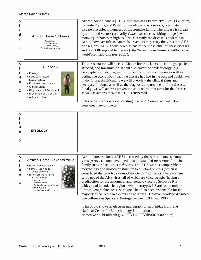
[This photo shows an electron micrograph of Reoviridae from The
National Center for Biotechnology Information at
http://www.ncbi.nlm.nih.gov/ICTVdb/ICTVdB/60000000.htm]
African Horse Sickness
Center for Food Security and Public Health 2012 2
Slide 5
African Horse Sickness Virus
• Inactivated by
–Heat (temps greater than 140oF)
–pH less than 6, or 12 or greater
–Acidic disinfectants
• Rapidly destroyed in carcasses that have undergone rigor mortis
Center for Food Security and Public Health, Iowa State University, 2011
The AHSV can be inactivated in the laboratory with formalin, β-
propriolactone, acetylethyleneimine derivatives or radiation. It is also
destroyed at a pH less than 6, or pH 12 or greater. Acidic disinfectants
such as 2% acetic or citric acid have been recommended for
decontamination when it is warranted. AHSV can survive in frozen meat,
but is inactivated at temperatures greater than 60oC) 140
oF. Optimum pH
for virus survival is pH 7-8.5. The virus is destroyed at a pH less than 6, or
pH 12 or greater; the virus is rapidly destroyed in carcasses that have
undergone rigor mortis due to pH fluctuations.
Slide 6
EPIDEMIOLOGY
Center for Food Security and Public Health, Iowa State University, 2011
Slide 7
Species Affected
• Equidae
–Horses, donkeys, mules
–Zebras
• Other
–Camels
–Dogs
Center for Food Security and Public Health, Iowa State University, 2011
The African horse sickness virus can infect horses, donkeys, mules, zebras,
camels and dogs. The most serious infections occur in horses and mules,
which appear to be accidental hosts. Zebras and donkeys rarely develop
serious clinical signs. Zebras, which are often asymptomatic, are thought
to be the natural reservoir hosts in most regions of Africa. Infections in
camels have been reported, but appear to be uncommon and asymptomatic.
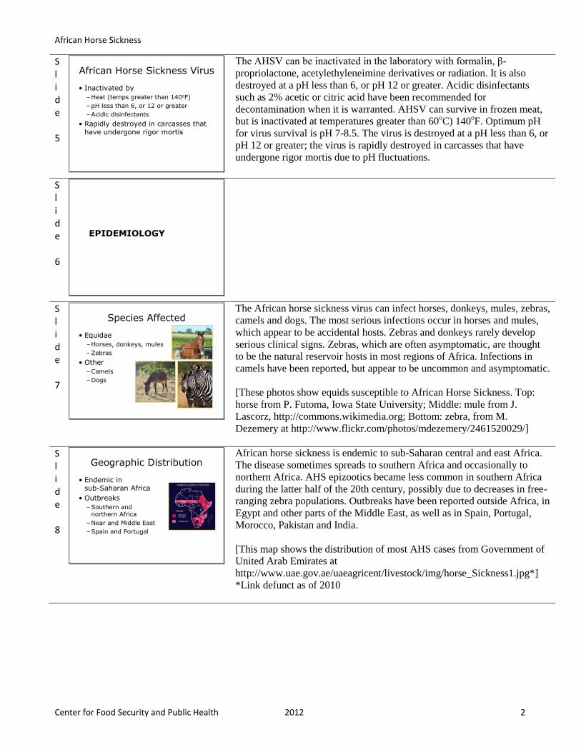
[These photos show equids susceptible to African Horse Sickness. Top:
horse from P. Futoma, Iowa State University; Middle: mule from J.
Lascorz, http://commons.wikimedia.org; Bottom: zebra, from M.
Dezemery at http://www.flickr.com/photos/mdezemery/2461520029/]
Slide 8
Geographic Distribution
• Endemic in sub-Saharan Africa
• Outbreaks
–Southern and northern Africa
–Near and Middle East
–Spain and Portugal
Center for Food Security and Public Health, Iowa State University, 2011
African horse sickness is endemic to sub-Saharan central and east Africa.
The disease sometimes spreads to southern Africa and occasionally to
northern Africa. AHS epizootics became less common in southern Africa
during the latter half of the 20th century, possibly due to decreases in free-
ranging zebra populations. Outbreaks have been reported outside Africa, in
Egypt and other parts of the Middle East, as well as in Spain, Portugal,
Morocco, Pakistan and India.
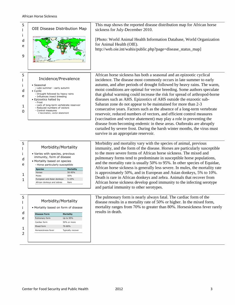
[This map shows the distribution of most AHS cases from Government of
United Arab Emirates at
http://www.uae.gov.ae/uaeagricent/livestock/img/horse_Sickness1.jpg*]
*Link defunct as of 2010
African Horse Sickness
Center for Food Security and Public Health 2012 3
Slide 9
OIE Disease Distribution Map
Center for Food Security and Public Health, Iowa State University, 2011
This map shows the reported disease distribution map for African horse
sickness for July-December 2010.
[Photo: World Animal Health Information Database, World Organization
for Animal Health (OIE).
http://web.oie.int/wahis/public.php?page=disease_status_map]
Slide 10
Incidence/Prevalence
• Seasonal– Late summer - early autumn
• Cyclic– Drought followed by heavy rains– Influences insect breeding
• Epizootics halted by– Frost– Lack of long-term vertebrate reservoir– Reduced numbers of vectors– Control measures
• Vaccination, vector abatement
Center for Food Security and Public Health, Iowa State University, 2011
African horse sickness has both a seasonal and an epizootic cyclical
incidence. The disease most commonly occurs in late summer to early
autumn, and after periods of drought followed by heavy rains. The warm,
moist conditions are optimal for vector breeding. Some authors speculate
that global warming could increase the risk for spread of arthropod-borne
diseases such as AHS. Epizootics of AHS outside the enzootic sub-
Saharan zone do not appear to be maintained for more than 2-3
consecutive years. Factors such as the absence of a long-term vertebrate
reservoir, reduced numbers of vectors, and efficient control measures
(vaccination and vector abatement) may play a role in preventing the
disease from becoming endemic in these areas. Outbreaks are abruptly
curtailed by severe frost. During the harsh winter months, the virus must
survive in an appropriate reservoir.
Slide 11
Morbidity/Mortality
• Varies with species, previous immunity, form of disease
• Mortality based on species
–Horse particularly susceptible
Center for Food Security and Public Health, Iowa State University, 2011
Species Mortality
Horses 50-95%
Mules 50%
European and Asian donkeys 5-10%
African donkeys and zebras Rare
Morbidity and mortality vary with the species of animal, previous
immunity, and the form of the disease. Horses are particularly susceptible
to the more severe forms of African horse sickness. The mixed and
pulmonary forms tend to predominate in susceptible horse populations,
and the mortality rate is usually 50% to 95%. In other species of Equidae,
African horse sickness is generally less severe. In mules, the mortality rate
is approximately 50%, and in European and Asian donkeys, 5% to 10%.
Death is rare in African donkeys and zebra. Animals that recover from
African horse sickness develop good immunity to the infecting serotype
and partial immunity to other serotypes.
Slide 12
Morbidity/Mortality
• Mortality based on form of disease
Center for Food Security and Public Health, Iowa State University, 2011
Disease Form Mortality
Pulmonary form Up to 95%
Cardiac form 50% or more
Mixed form 70-80%
Horsesickness fever Typically recover
The pulmonary form is nearly always fatal. The cardiac form of the
disease results in a mortality rate of 50% or higher. In the mixed form,
mortality ranges from 70% to greater than 80%. Horsesickness fever rarely
results in death.
African Horse Sickness
Center for Food Security and Public Health 2012 4
Slide 13
TRANSMISSION
Center for Food Security and Public Health, Iowa State University, 2011
Slide 14
Transmission
• Not contagious
• Vector-borne: Culicoides spp.
–Culicoides imicola – principal vector
–C. bolitinos
–C. variipennis
–Other potential arthropods
• Viremia in Equidae
–Horses: 12 to 40 days
–Zebras, African donkeys: up to 6 weeksCenter for Food Security and Public Health, Iowa State University, 2011
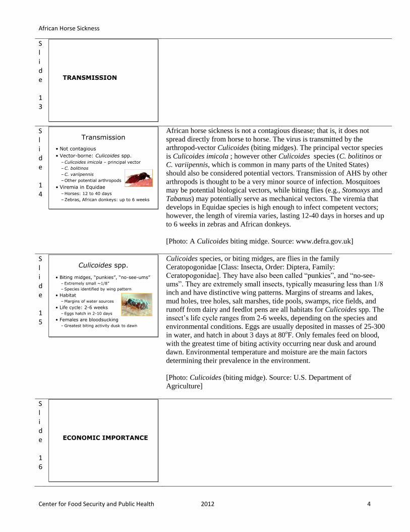
African horse sickness is not a contagious disease; that is, it does not
spread directly from horse to horse. The virus is transmitted by the
arthropod-vector Culicoides (biting midges). The principal vector species
is Culicoides imicola ; however other Culicoides species (C. bolitinos or
C. variipennis, which is common in many parts of the United States)
should also be considered potential vectors. Transmission of AHS by other
arthropods is thought to be a very minor source of infection. Mosquitoes
may be potential biological vectors, while biting flies (e.g., Stomoxys and
Tabanus) may potentially serve as mechanical vectors. The viremia that
develops in Equidae species is high enough to infect competent vectors;
however, the length of viremia varies, lasting 12-40 days in horses and up
to 6 weeks in zebras and African donkeys.
[Photo: A Culicoides biting midge. Source: www.defra.gov.uk]
Slide 15
Culicoides spp.
• Biting midges, “punkies”, “no-see-ums”
– Extremely small ~1/8”
– Species identified by wing pattern
• Habitat
– Margins of water sources
• Life cycle: 2-6 weeks
– Eggs hatch in 2-10 days
• Females are bloodsucking
– Greatest biting activity dusk to dawn
Center for Food Security and Public Health, Iowa State University, 2011
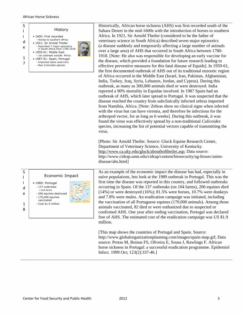
Culicoides species, or biting midges, are flies in the family
Ceratopogonidae [Class: Insecta, Order: Diptera, Family:
Ceratopogonidae]. They have also been called “punkies”, and “no-see-
ums”. They are extremely small insects, typically measuring less than 1/8
inch and have distinctive wing patterns. Margins of streams and lakes,
mud holes, tree holes, salt marshes, tide pools, swamps, rice fields, and
runoff from dairy and feedlot pens are all habitats for Culicoides spp. The
insect’s life cycle ranges from 2-6 weeks, depending on the species and
environmental conditions. Eggs are usually deposited in masses of 25-300
in water, and hatch in about 3 days at 80oF. Only females feed on blood,
with the greatest time of biting activity occurring near dusk and around
dawn. Environmental temperature and moisture are the main factors
determining their prevalence in the environment.
[Photo: Culicoides (biting midge). Source: U.S. Department of
Agriculture]
Slide 16
ECONOMIC IMPORTANCE
Center for Food Security and Public Health, Iowa State University, 2011
African Horse Sickness
Center for Food Security and Public Health 2012 5
Slide 17
History
• 1600: First recorded– Horses to southern Africa
• 1921: Sir Arnold Theiler– Described 7 major epizootics
in South Africa from 1780-1918
• 1959-61: Middle East– 1st outbreak outside Africa
• 1987-91: Spain, Portugal– Imported zebra reservoirs
– New Culicoides species
Center for Food Security and Public Health, Iowa State University, 2011
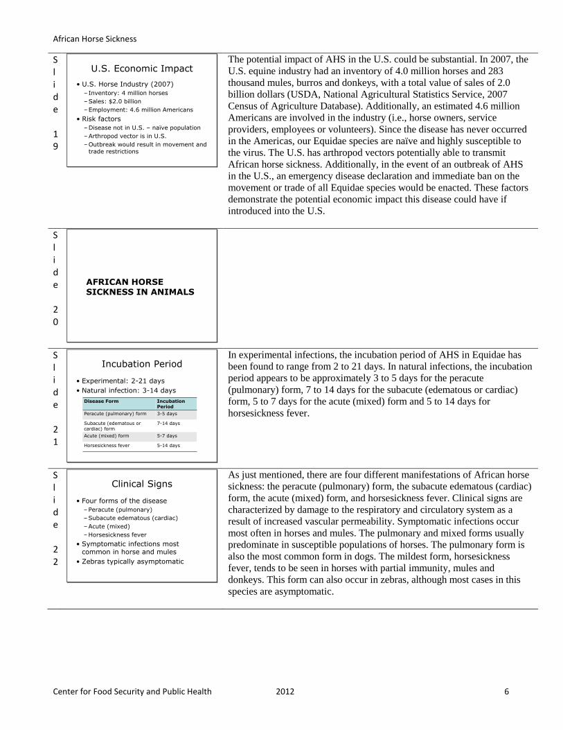
Historically, African horse sickness (AHS) was first recorded south of the
Sahara Desert in the mid-1600s with the introduction of horses to southern
Africa. In 1921, Sir Arnold Theiler (considered to be the father of
veterinary science in South Africa) described seven major epizootics
(a disease suddenly and temporarily affecting a large number of animals
over a large area) of AHS that occurred in South Africa between 1780-
1918. [Note: He also was responsible for developing an early vaccine for
the disease, which provided a foundation for future research leading to
effective preventive measures for this fatal disease of Equids]. In 1959-61,
the first documented outbreak of AHS out of its traditional enzootic region
of Africa occurred in the Middle East (Israel, Iran, Pakistan, Afghanistan,
India, Turkey, Iraq, Syria, Lebanon, Jordan, and Cyprus). During this
outbreak, as many as 300,000 animals died or were destroyed. India
reported a 90% mortality in Equidae involved. In 1987 Spain had an
outbreak of AHS, which later spread to Portugal. It was suspected that the
disease reached the country from subclinically infected zebras imported
from Namibia, Africa. [Note: Zebras show no clinical signs when infected
with the virus but can have viremia, and therefore be infectious for the
arthropod vector, for as long as 6 weeks]. During this outbreak, it was
found the virus was effectively spread by a non-traditional Culicoides
species, increasing the list of potential vectors capable of transmitting the
virus.
[Photo: Sir Arnold Theiler. Source: Gluck Equine Research Center,
Department of Veterinary Science, University of Kentucky.
http://www.ca.uky.edu/gluck/abouthoftheiler.asp; Data source:
http://www.cidrap.umn.edu/cidrap/content/biosecurity/ag-biosec/anim-
disease/ahs.html]
Slide 18
Economic Impact
• 1989: Portugal
–137 outbreaks
• 104 farms
–206 equines destroyed
–170,000 equinesvaccinated
–Cost $1.9 million
Center for Food Security and Public Health, Iowa State University, 2011
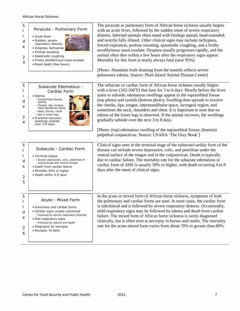
SPAIN
As an example of the economic impact the disease has had, especially in
naïve populations, lets look at the 1989 outbreak in Portugal. This was the
first time the disease was reported in this country, and followed outbreaks
occurring in Spain. Of the 137 outbreaks (on 104 farms), 206 equines died
(14%) or were destroyed (16%); 81.5% were horses, 10.7% were donkeys
and 7.8% were mules. An eradication campaign was initiated, including
the vaccination of all Portuguese equines (170,000 animals). Among those
animals vaccinated, 82 died or were euthanized due to suspected or
confirmed AHS. One year after ending vaccination, Portugal was declared
free of AHS. The estimated cost of the eradication campaign was US $1.9
million.
[This map shows the countries of Portugal and Spain. Source:
http://www.globalorganizationplanning.com/images/spain-map.gif; Data
source: Protas M, Boinas FS, Oliveira E, Sousa J, Rawlings F. African
horse sickness in Portugal: a successful eradication programme. Epidemiol
Infect. 1999 Oct; 123(2):337-46.]
African Horse Sickness
Center for Food Security and Public Health 2012 6
Slide 19
U.S. Economic Impact
• U.S. Horse Industry (2007)
– Inventory: 4 million horses
–Sales: $2.0 billion
–Employment: 4.6 million Americans
• Risk factors
–Disease not in U.S. – naïve population
–Arthropod vector is in U.S.
–Outbreak would result in movement and trade restrictions
Center for Food Security and Public Health, Iowa State University, 2011
The potential impact of AHS in the U.S. could be substantial. In 2007, the
U.S. equine industry had an inventory of 4.0 million horses and 283
thousand mules, burros and donkeys, with a total value of sales of 2.0
billion dollars (USDA, National Agricultural Statistics Service, 2007
Census of Agriculture Database). Additionally, an estimated 4.6 million
Americans are involved in the industry (i.e., horse owners, service
providers, employees or volunteers). Since the disease has never occurred
in the Americas, our Equidae species are naïve and highly susceptible to
the virus. The U.S. has arthropod vectors potentially able to transmit
African horse sickness. Additionally, in the event of an outbreak of AHS
in the U.S., an emergency disease declaration and immediate ban on the
movement or trade of all Equidae species would be enacted. These factors
demonstrate the potential economic impact this disease could have if
introduced into the U.S.
Slide 20
AFRICAN HORSE SICKNESS IN ANIMALS
Center for Food Security and Public Health, Iowa State University, 2011
Slide 21
Incubation Period
• Experimental: 2-21 days
• Natural infection: 3-14 days
Center for Food Security and Public Health, Iowa State University, 2011
Disease Form Incubation Period
Peracute (pulmonary) form 3-5 days
Subacute (edematous or cardiac) form
7-14 days
Acute (mixed) form 5-7 days
Horsesickness fever 5-14 days
In experimental infections, the incubation period of AHS in Equidae has
been found to range from 2 to 21 days. In natural infections, the incubation
period appears to be approximately 3 to 5 days for the peracute
(pulmonary) form, 7 to 14 days for the subacute (edematous or cardiac)
form, 5 to 7 days for the acute (mixed) form and 5 to 14 days for
horsesickness fever.
Slide 22
Clinical Signs
• Four forms of the disease
–Peracute (pulmonary)
–Subacute edematous (cardiac)
–Acute (mixed)
–Horsesickness fever
• Symptomatic infections most common in horse and mules
• Zebras typically asymptomatic
Center for Food Security and Public Health, Iowa State University, 2011
As just mentioned, there are four different manifestations of African horse
sickness: the peracute (pulmonary) form, the subacute edematous (cardiac)
form, the acute (mixed) form, and horsesickness fever. Clinical signs are
characterized by damage to the respiratory and circulatory system as a
result of increased vascular permeability. Symptomatic infections occur
most often in horses and mules. The pulmonary and mixed forms usually
predominate in susceptible populations of horses. The pulmonary form is
also the most common form in dogs. The mildest form, horsesickness
fever, tends to be seen in horses with partial immunity, mules and
donkeys. This form can also occur in zebras, although most cases in this
species are asymptomatic.
African Horse Sickness
Center for Food Security and Public Health 2012 7
Slide 23
Peracute - Pulmonary Form
• Acute fever
• Sudden, severerespiratory distress
• Dyspnea, tachypnea
• Profuse sweating
• Spasmodic coughing
• Frothy serofibrinous nasal exudate
• Rapid death (few hours)
Center for Food Security and Public Health, Iowa State University, 2011
Foam from the nares due
to pulmonary edema
The peracute or pulmonary form of African horse sickness usually begins
with an acute fever, followed by the sudden onset of severe respiratory
distress. Infected animals often stand with forelegs spread, head extended,
and nostrils fully dilated. Other clinical signs may include tachypnea,
forced expiration, profuse sweating, spasmodic coughing, and a frothy
serofibrinous nasal exudate. Dyspnea usually progresses rapidly, and the
animal often dies within a few hours after the respiratory signs appear.
Mortality for this form is nearly always fatal (near 95%).
[Photo: Abundant froth draining from the nostrils reflects severe
pulmonary edema. Source: Plum Island Animal Disease Center]
Slide 24
Subacute Edematous -Cardiac Form
• Edema–Supraorbital fossae,
eyelids
–Cheeks, lips, tongue, intermandibular space
–Neck, thorax, chest
–Not in lower legs
• If animal recovers, swellings subsideover 3-8 days
Center for Food Security and Public Health, Iowa State University, 2011
The subacute or cardiac form of African horse sickness usually begins
with a fever (102-106oF) that lasts for 3 to 6 days. Shortly before the fever
starts to subside, edematous swellings appear in the supraorbital fossae
(top photo) and eyelids (bottom photo). Swelling then spreads to involve
the cheeks, lips, tongue, intermandibular space, laryngeal region, and
sometimes the neck, shoulders and chest. It is important to note that no
edema of the lower legs is observed. If the animal recovers, the swellings
gradually subside over the next 3 to 8 days.
[Photo: (top) edematous swelling of the supraorbital fossae; (bottom)
palpebral conjunctivae. Source: USAHA ‘The Grey Book’]
Slide 25
Subacute - Cardiac Form
• Terminal stages
–Severe depression, colic, petechiae of conjunctivae and ventral tongue
• Death from cardiac failure
• Mortality 50% or higher
• Death within 4-8 days
Center for Food Security and Public Health, Iowa State University, 2011
Clinical signs seen in the terminal stage of the subacute/cardiac form of the
disease can include severe depression, colic, and petechiae under the
ventral surface of the tongue and in the conjunctivae. Death is typically
due to cardiac failure. The mortality rate for the subacute edematous or
cardiac form of AHS is usually 50% or higher, with death occurring 4 to 8
days after the onset of clinical signs.
Slide 26
Acute - Mixed Form
• Pulmonary and cardiac forms
• Cardiac signs usually subclinical
–Followed by severe respiratory distress
• Mild respiratory signs
–Followed by edema and death
• Diagnosed by necropsy
• Mortality 70-80%
Center for Food Security and Public Health, Iowa State University, 2011
In the acute or mixed form of African horse sickness, symptoms of both
the pulmonary and cardiac forms are seen. In most cases, the cardiac form
is subclinical and is followed by severe respiratory distress. Occasionally,
mild respiratory signs may be followed by edema and death from cardiac
failure. The mixed form of African horse sickness is rarely diagnosed
clinically, but is often seen at necropsy in horses and mules. The mortality
rate for the acute-mixed form varies from about 70% to greater than 80%.
African Horse Sickness
Center for Food Security and Public Health 2012 8
Slide 27
Horsesickness Fever
• Mild clinical signs
• Characteristic fever (3 to 8 days)–Morning remission (undetectable)
–Afternoon exacerbation
• Other signs–Mild anorexia or depression
–Congested mucous membranes
– Increased heart rate
• Rarely fatalCenter for Food Security and Public Health, Iowa State University, 2011
In horsesickness fever, the clinical signs are mild. The characteristic fever
usually lasts for 3 to 8 days; morning remissions and afternoon
exacerbations are often seen. Other symptoms are generally mild and may
include mild anorexia or depression, edema of the supraorbital fossae,
congested mucous membranes and an increased heart rate. Animals almost
always recover from horse-sickness fever. This form of the disease is
rarely fatal.
Slide 28
Post Mortem Lesions
• Pulmonary form
–Severe, diffusepulmonary edema
–Hydrothorax
–Fluid in abdominal and thoracic cavity
–Enlarged endematous lymph nodes
–Hyperemia and petechial hemorrhages in intestines
Center for Food Security and Public Health, Iowa State University, 2011
In the pulmonary form of African horse sickness, interlobular edema of the
lungs and hydrothorax are the characteristic lesions. In the most acute
cases, frothy fluid flows from the nostrils and the cut surface of the lungs,
which are mottled red, noncollapsed and heavy. In more prolonged cases,
there may be extensive interstitial and subpleural edema, and hyperemia
may be less apparent. Fluid accumulation can occur in the abdominal and
thoracic cavities. Occasionally, extensive fluid accumulation may be noted
in the thoracic cavity (hydrothorax), with near normal appearance of the
lungs. The lymph nodes, particularly the nodes in the thoracic and
abdominal cavities, are usually enlarged and edematous. Less often, there
may be subcapsular hemorrhages in the spleen, congestion in the renal
cortex or gastric fundus, and edematous infiltration around the aorta and
trachea. The stomach mucosa may be hyperemic and edematous.
Hyperemia and petechial hemorrhages may also be apparent in the small
and large intestines and the pericardium may contain petechiae.
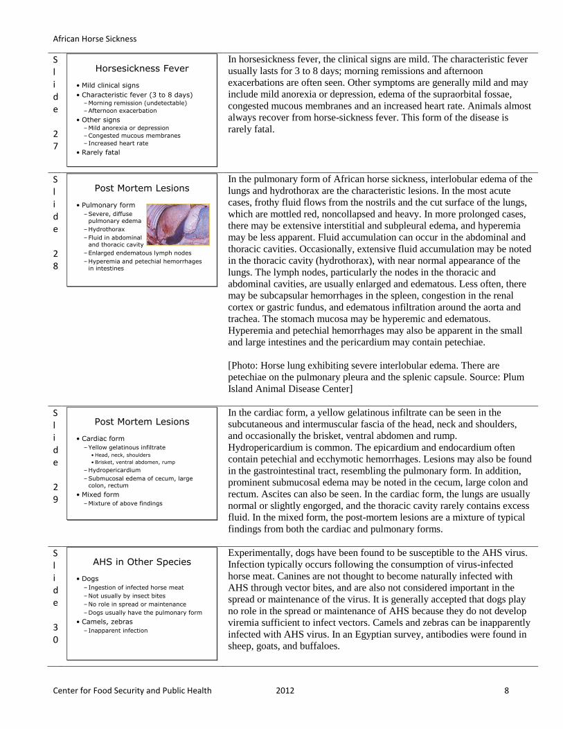
[Photo: Horse lung exhibiting severe interlobular edema. There are
petechiae on the pulmonary pleura and the splenic capsule. Source: Plum
Island Animal Disease Center]
Slide 29
Post Mortem Lesions
• Cardiac form
–Yellow gelatinous infiltrate
• Head, neck, shoulders
• Brisket, ventral abdomen, rump
–Hydropericardium
–Submucosal edema of cecum, large colon, rectum
• Mixed form
–Mixture of above findings
Center for Food Security and Public Health, Iowa State University, 2011
In the cardiac form, a yellow gelatinous infiltrate can be seen in the
subcutaneous and intermuscular fascia of the head, neck and shoulders,
and occasionally the brisket, ventral abdomen and rump.
Hydropericardium is common. The epicardium and endocardium often
contain petechial and ecchymotic hemorrhages. Lesions may also be found
in the gastrointestinal tract, resembling the pulmonary form. In addition,
prominent submucosal edema may be noted in the cecum, large colon and
rectum. Ascites can also be seen. In the cardiac form, the lungs are usually
normal or slightly engorged, and the thoracic cavity rarely contains excess
fluid. In the mixed form, the post-mortem lesions are a mixture of typical
findings from both the cardiac and pulmonary forms.
Slide 30
AHS in Other Species
• Dogs
– Ingestion of infected horse meat
–Not usually by insect bites
–No role in spread or maintenance
–Dogs usually have the pulmonary form
• Camels, zebras
– Inapparent infection
Center for Food Security and Public Health, Iowa State University, 2011
Experimentally, dogs have been found to be susceptible to the AHS virus.
Infection typically occurs following the consumption of virus-infected
horse meat. Canines are not thought to become naturally infected with
AHS through vector bites, and are also not considered important in the
spread or maintenance of the virus. It is generally accepted that dogs play
no role in the spread or maintenance of AHS because they do not develop
viremia sufficient to infect vectors. Camels and zebras can be inapparently
infected with AHS virus. In an Egyptian survey, antibodies were found in
sheep, goats, and buffaloes.
African Horse Sickness
Center for Food Security and Public Health 2012 9
Slide 31
DIAGNOSIS AND TREATMENT
Center for Food Security and Public Health, Iowa State University, 2011
Slide 32
Differential Diagnosis
• Equine viral arteritis
• Equine infectious anemia
• Hendra virus infection
• Purpura hemorrhagica
• Equine piroplasmosis
• Equine encephalosis virus
• Anthrax
• ToxinsCenter for Food Security and Public Health, Iowa State University, 2011
The differential diagnosis includes equine viral arteritis, equine infectious
anemia, Hendra virus infection, purpura hemorrhagica and equine
piroplasmosis. In Africa, equine encephalosis virus, another orbivirus
transmitted by Culicoides, causes a syndrome resembling horsesickness
fever. Toxins, anthrax and other causes of sudden death, as well as
diseases that result in severe respiratory distress, should also be
considered.
Slide 33
Diagnosis
• Clinical signs
–Supraorbital swelling is characteristic
• History
–Prevalence or exposure to competent vectors
–Travel from enzootic area
• Laboratory tests - definitive diagnosis
• Serotype needed for control measures
Center for Food Security and Public Health, Iowa State University, 2011
African horse sickness should be suspected in Equidae demonstrating the
previously mentioned clinical signs. Supraorbital swellings are particularly
characteristic of this disease. Additionally, a history of prevalence or
exposure to competent vectors or of travel from an enzootic area can be
important factors. Tentative diagnosis of AHS may be obtained by
serology if blood is taken during the febrile stage; however, laboratory
confirmation is essential for definitive diagnosis, and serotype
determination will be important for control measures. Virus isolation and
identification may be done from a number of tissues; samples taken should
include small (2-4 g) sections of the spleen, lung, and lymph nodes.
Samples for virus isolation should be stored and transported at 4oC. AHS
viral antigens can be detected with enzyme-linked immunosorbent assays
(ELISAs), as well as with a reverse-transcription polymerase chain
reaction (RT-PCR), which can be particularly useful for rapid serotyping
of viral RNA. Currently there is no efficient treatment for African horse
sickness. Surviving Equidae develop solid immunity to the homologous
serotype but remain susceptible to heterologous serotypes. Vaccines have
been developed for all 9 serotypes of the virus, but are not currently
available in the U.S.
Slide 34
Laboratory Diagnosis
• Laboratory tests
–Virus isolation
–ELISA, RT-PCR
–Serology (tentative)
–Necropsy: spleen, lung, lymph node
• More than one test should be used
• AHSV does not cross-react with other known orbiviruses
Center for Food Security and Public Health, Iowa State University, 2011
African horse sickness can be diagnosed by isolating the virus or detecting
its nucleic acids or antigens. More than one test should be used to diagnose
an outbreak (particularly the index case) whenever possible. AHSV
antigens can be detected with enzyme-linked immunosorbent assays
(ELISAs). A reverse-transcription polymerase chain reaction (RT-PCR)
technique is used to detect viral RNA. A recently developed type-specific
RT-PCR assay can be used for rapid serotyping. Serology can also be used
to diagnose African horse sickness. Antibodies can be detected within 8 to
14 days after infection, and may persist for one to four years. The indirect
ELISA and complement fixation tests are the prescribed tests for
international trade. AHSV does not cross-react with other known
orbiviruses.
African Horse Sickness
Center for Food Security and Public Health 2012 10
Slide 35
Sampling
• Before collecting or sending any samples, the proper authorities should be contacted.
• Samples should only be sent under secure conditions and to authorized laboratories to prevent the spread of the disease.
Center for Food Security and Public Health, Iowa State University, 2011
Before collecting or sending any samples from animals with a suspected
foreign animal disease, the proper authorities should be contacted.
Samples should only be sent under secure conditions and to authorized
laboratories to prevent the spread of the disease.
Slide 36
Samples To Collect
• For virus isolation
–Blood samples
–Necropsy samples
• Spleen, lung, lymph nodes
• Paired serum samples are recommended
• Store and transport samples at 39oF
Center for Food Security and Public Health, Iowa State University, 2011
In live animals, blood samples collected into anticoagulant should be taken
for virus isolation. Success is most likely if these samples are collected
early during the febrile stage. Necropsy samples for virus isolation should
include samples of the spleen, lung and lymph nodes. The samples for
virus isolation should be stored and transported at 4°C (39°F). Serum
should also be taken for serology. Paired serum samples are recommended,
and are particularly important in areas where the disease is endemic.
Slide 37
AFRICAN HORSE SICKNESS IN HUMANS
Center for Food Security and Public Health, Iowa State University, 2011
Slide 38
AHS in Humans
• No natural infection in humans
• Neurotropic vaccine strains
–Transnasal infection can lead to encephalitis or retinitis
• Handle modified live AHS vaccine strains with caution
Center for Food Security and Public Health, Iowa State University, 2011
There is no evidence that humans can become infected with field strains of
AHS virus through contact with infected animals, nor from working in
laboratories; however, it has been shown that certain neurotropic vaccine
strains may cause encephalitis and retinitis in humans following transnasal
infection. Modified live vaccine strains of AHS should be handled with
caution.
Slide 39
PREVENTION AND CONTROL
Center for Food Security and Public Health, Iowa State University, 2011

African Horse Sickness
Center for Food Security and Public Health 2012 11
Slide 40
Recommended Actions
• IMMEDIATELY notify authorities
–OIE reportable disease
• In the U.S. notify
–Federal Area Veterinarian in Charge (AVIC) www.aphis.usda.gov/animal_health/area_offices/
–State Veterinarian www.usaha.org/Portals/6/StateAnimalHealthOfficials.pdf
• Quarantine premises
Center for Food Security and Public Health, Iowa State University, 2011
African horse sickness is reportable to the World Organization for Animal
Health (OIE). Disease notification requirements for OIE member nations
and import/export guidelines can be found in the OIE Terrestrial Animal
Health Code [http://www.oie.int/international-standard-setting/terrestrial-
code/access-online/].
If you suspect a case or outbreak of African horse sickness, contact your
state and/or federal veterinarian immediately and quarantine the premises.
If AHS is detected in a non-endemic country, a strict quarantine zone
should be established. Individual veterinarians who encounter a case of
African horse sickness should follow their national and/or local guidelines
for disease reporting and diagnostic testing.
Slide 41
Disinfection
• Disinfectants
–Sodium hypochlorite (bleach)
–2% acetic or citric acid
• Killed
–pH less than 6
–pH 12 or greater
• Rapidly destroyed in carcasses that have undergone rigor mortis
Center for Food Security and Public Health, Iowa State University, 2011
The AHS virus can be inactivated in the laboratory by formalin, -
propiolactone, acetyl-ethyleneimine derivatives, or radiation. Sodium
hypochlorite (bleach) is an effective disinfectant against the virus. The
virus is also destroyed at a pH less than 6 or greater than 12, so acidic
disinfectants, such as 2% acetic or citric acid have been recommended for
decontamination when warranted. Due to pH fluctuations, the AHS virus is
rapidly destroyed in carcasses that have undergone rigor mortis; however
the virus can survive in frozen meat, but is inactivated at temperatures
greater than 60˚C (140˚F).
Slide 42
Control
• Quarantine
–Equidae from endemic areas
• Asia, Africa, Mediterranean
–Minimum 60 days at point of entry
• Vector control and protection
– Insect repellants
–Stable in insect-proof housingfrom dusk to dawn
Center for Food Security and Public Health, Iowa State University, 2011
Horses cannot enter the US from an African horse sickness endemic
country unless they have been in an AHS-free country or area of the world
for at least 60 days. Upon entering the US, horses are then subject to the
regular 3 or 7 day quarantine period at the point of entry. If African horse
sickness is detected in a country where it is not endemic, a strict quarantine
zone and movement controls should be established. Euthanasia of infected
and exposed animals may be considered. Whenever possible, all Equidae
should be stabled in insect-proof housing. At a minimum, stabling from
dusk to dawn, the period when Culicoides are most active, is
recommended. Vector control measures such as modifications of
Culicoides breeding areas, insect repellents, and targeted applications of
insecticides or larvicides may also be useful.
Slide 43
Control
• Monitor temperature of all equids
– If febrile
• Euthanize or isolate in an insect-free stable until cause is determined
• Vaccination
– In endemic areas
–Surrounding protection zone
–Not available in the U.S.
Center for Food Security and Public Health, Iowa State University, 2011
Monitoring for fever may be helpful for the early detection of African
horse sickness. Each susceptible animal should have its temperature taken
regularly (optimally, twice daily). Those animals that develop a fever
should be kept in insect-free stables until the cause of the fever has been
established, or killed to prevent potential virus transmission to the vector.
In endemic areas, vaccination is strongly recommended for susceptible
Equidae. Additionally, areas around the affected area should vaccinate, as
well, to produce a surrounding protection zone.
African Horse Sickness
Center for Food Security and Public Health 2012 12
Slide 44
Vaccination
• Attenuated live vaccine available
–Horses, mules, donkeys
–Not in U.S.
–Reassortment possible
–Teratogenic
• No killed or subunit vaccine available
• Recovering animals
– Lifelong immunity post-infection to the infecting serotype
Center for Food Security and Public Health, Iowa State University, 2011
Vaccines have been developed for the 9 serotypes of AHS virus.
Attenuated (monovalent and polyvalent) live (Vero cell) vaccines for use
in horses, mules, and donkeys are currently available in some countries
(but not in the U.S. at this time). These vaccines result in viremia, and the
viruses could theoretically reassort with an outbreak virus. Attenuated
vaccines may not be safe in AHS-free countries. They are also teratogenic.
No killed or subunit vaccines are currently manufactured commercially.
Animals that recover from the disease develop solid life-long immunity
against the infecting viral serotype.
Slide 45
Additional Resources
• World Organization for Animal Health (OIE)
– www.oie.int
• Center for Food Security and Public Health
– www.cfsph.iastate.edu
• USAHA Foreign Animal Diseases (“The Gray Book”)
– www.aphis.usda.gov/emergency_response/downloads/nahems/fad.pdf
• Center for Infectious Disease Research and Policy
– www.cidrap.umn.edu/cidrap/content/biosecurity/ag-biosec/anim-disease/ahs.htm
• African Horse Sickness Trust
– www.africanhorsesickness.co.za
Center for Food Security and Public Health, Iowa State University, 2011
For more information, see these listed resources.
References used for this presentation:
• Center for Food Security and Public Health (CFSPH). African horse
sickness. Technical Fact Sheet. 30 Nov 2006.
http://www.cfsph.iastate.edu/Factsheets/pdfs/african_horse_sickness.p
df
• U.S. Animal Health Association (USAHA). Foreign Animal Diseases,
7th edition. 2008. Guthrie, AJ. African Horse Sickness. p103-109.
www.aphis.usda.gov/emergency_response/downloads/nahems/fad.pdf
• Center for Infectious Disease Research and Policy (CIDRAP). African
horse sickness. 26 Sept 2004.
http://www.cidrap.umn.edu/cidrap/content/biosecurity/ag-biosec/anim-
disease/ahs.html
• U.S. Department of Agriculture, Animal and Plant Health Inspection
Service (USDA APHIS). Horse Reports and Highlights – African
Horsesickness.
http://www.aphis.usda.gov/animal_health/emergingissues/byspeciescat
egory/horse.shtml
Slide 46
Acknowledgments
Development of this presentation was made possible through grants provided to
the Center for Food Security and Public Health at Iowa State University, College of Veterinary Medicine from
the Centers for Disease Control and Prevention, the U.S. Department of Agriculture,
the Iowa Homeland Security and Emergency Management Division, and the
Multi-State Partnership for Security in Agriculture.
Authors: Glenda Dvorak, DVM, MPH, DACVPM; Anna Rovid Spickler, DVM, PhD
Reviewers: James A. Roth, DVM, PhD; Bindy Comito, BA; Katie Spaulding, BS; Meghan Blankenship, BS; Kerry Leedom Larson, DVM, MPH, PhD, DACVPM
Center for Food Security and Public Health, Iowa State University, 2011
Last reviewed: October 2011