Embed Size (px)
Citation preview

I.J.A.B.R., VOL. 2(3) 2012: 477-482 ISSN 2250 - 3579
477
ESTIMATION THE LEVELS OF CONTRACTILE AND COLLAGENOUSPROTEINS OF BACK SKELETAL MUSCLE DURING FETAL
DEVELOPMENT IN RATS
R.R. Al-Saadi A.R.Al-SalihiHigh Institute for Infertility Diagnosis and Assisted Reproductive Technologies
ABSTRACTBiochemical determinations of contractile and collagenous proteins made on the skeletal muscle tissues of rat fetus andnew born rats. The biochemical change of contractile and collagenous proteins of muscle tissue that takes place during theintrauterine life is an essential part in the development and organization of skeletal muscles. The study was performed onthe albino rat, as a mammalian model. The specimen of muscle tissue from 29 fetuses at different age starting from day 17and 5 new born rats was including in this study. The biochemical assays were performed at the medical city teachinglaboratories/ Baghdad. The estimation levels of contractile and collagenous proteins in the tissue sample were done byusing Double Beam Spectrophotometer. As a result, there was an increase in tissue level of contractile proteins begin evenbefore fetal life and also there was decrease in collagenous proteins that start during fetal life .It was concluded, the relativeproportion between contractile and collagenous proteins in developing muscle tissue is inverted in new born rats whencompared with those rats at day 17 of embryonic development.
KEY WORDS: Skeletal Muscle, Development, Contractile proteins, collagenous protein
INTRODUCTIONProteins are the basic material of tissue structure, They arethe most important component of striated skeletal muscles,their classification is correlated with the histologicalstructure of muscle tissue .Muscle tissue is a compositetissue which is composed of, muscle cells (muscle fibers)specialized for contraction, connective tissue, nerve fibersand blood vessels [1]. Muscle growth occurs duringembryonic development and continues in adult life asregeneration [2,3]. During embryonic muscle growth andregeneration in mature muscle, single nucleated myoblastfuse to each other to form myotubes, within the myotubes,bundles of contractile proteins are organized intomyofibrils giving the tissue a striated appearance [3]. Themyofibrils are basically composed of two filamentous,contractile proteins, actin and myosin. Contractile proteinsare an important cellular component, critical tomaintaining cell shape, mass, and other cellularfunctions[4]. The mass of a muscle is made up of 75%water and more than 20% protein. The muscle proteins canbe divided into contractile, regulatory, sarcoplasmic andextracellular forms; the most important are the contractileproteins actin and myosin[5]. The contractile proteins wasfirst shown by [6] and include troponin, tropomysin, M-protein, beta actin, gamma actin, C–protein which are ofgreat importance. Sarcolemma is also protein component,and others proteins such as elastin, collagen and reticulinare also found in the muscle, there are also myoglobin, andothers[5,7]. Striated myofibers held together by connectivetissue, the muscle connective tissue layer is composed ofcollagen and elastine, but collagen is the more abundant ofthese two proteins, representing about 2% to 6% or moreof dry weight of muscle [8,9]. This layer contains andsupports the contractile tissues of the muscle and at its
extremities provides the connection to the skeleton,connective tissue of muscle is almost as important as themuscle fibers [10] for without it there would be no structureto the muscle belly and no way for the movement andforces produced in the fibers to be transmitted to thetendon. There are variations in amount of collagenbetween different muscles of the same animal [11].
MATERIALS AND METHODSThe study was performed on the albino rat (Rattus rattusnorvegicus albinus), as a mammalian model. Male andfemale couples were kept together in mating cages .Acareful examination of the cage was done each morningand the presence of vaginal plug was considered anindication of copulation. The day when the vaginal plugwas found was considered as “day one post coitum” (dpc),the occurrence of vaginal plug considered as the first dayof pregnancy .Pregnancies were dated by appearance of acopulatory plug. In this study we used the E-designation[12].This method is used to standardize the embryologicalmaterial, although this designation includes severalparameters in combination.This parameter includes, dpc, Witschi stage, Thieler’sstage. The day plug was found being E1.The subsequentdays were sequentially numbered. At each dpc, startingfrom day 17 to day 21, two to three pregnant rat femaleswere anesthetized to obtain embryos of that conceptionage, the embryos were washed with normal saline anddelivered into Petri dishes, and the embryo was examinedunder magnifying lens for features of staging. Twopregnant rat females were allowed to complete pregnancyand have normal vaginal delivery. The new born litterswere immediately separated from their mother andincluded in this work. Witschi staging system[13] was

Estimation the levels of proteins of back skeletal muscle during fetal development in rats
478
correlated with the Theiler’s stages [14], based ondiagnostic features of the rat embryos. All rat embryoswere sacrificed humanly by general anesthesia beforeobtaining muscle tissue sample. For tissue samplesemployed in biochemical assay, back skin was incised,carefully opening from occipital region caudally to thepelvic region. The muscles of the back were cleaned offsubcutaneous material; the muscles around the vertebralcolumn. The muscles were easily identified by theirpinkish color, location and texture .Using blunt dissectionby forceps and with aid of a probe, the muscles wereteased away from the vertebral column and the skin waspeeled away. The tissue sample was eventually freed awayfrom its place by sharp dissection. The numbers ofspecimen for biochemical study are 29 and 5 new born rat.The tissue samples were trimmed under dissectingmicroscope to ensure inclusion of relevant parts of thesample. A small piece of back skeletal muscle tissue wasfirst weighted then minced by chopping using a sharprazor blade. The minced pieces were put intoappendrofetube, containing normal saline (50mg wettissue per 1 ml of normal saline).The biochemical assayswere performed at the medical city teaching laboratories/Baghdad, 2010.The biochemical assays for determinations contractileproteins were made according to Lowery assay [15],whereas collagenous proteins determined according toReddy and Enwemeka assay [16] .Lowry Method for Contractile Protein DeterminationThe Lowry assay[15], is an often cited general use proteinassay. For a long time it is the method of choice foraccurate protein determination for tissue and cell fractions.PrincipleUnder alkaline conditions, the divalent copper ion (Cu+2)forms a complex with peptide bonds of proteins, and it isreduced to a monovalent copper ion (Cu+1). Monovalentcopper ion (Cu+1) and the radical groups of tyrosine,tryptophan and cysteine react with Folin reagent. This isan oxidation reduction reaction, in which the folin reagentis reduced to heteropolymolybdenum blue and oxidationof copper and aromatic acids.The Lowry method is sensitive to low concentrations ofprotein.Lowry method is sensitive to pH changes and therefore,the pH range of assay solution should be maintained at 10-10.5. However, if the assay involves using very smallvolumes of sample, there will be little or no effect on pHof the reaction mixture.ProcedureReagents1. Reagent AIt consists of 2mg sodium potassium tartarate , hydrated(4H20) (BDH); 100mg sodium bicarbonate (BDH); 500ml 1N sodium hydroxide (BDH); water is added to oneliter.This will give (7mM Na-K tartarate, 0.81 sodiumbicarbonate, 0.5N NaOH) final concentration. This reagentkeeps for 2-3 months2. Reagent B:It consists of 2 mg sodium potassium tartarate, hydrated(4H20) (BDH); 1g copper sulphate (CuSO4.5H20)(Fluka), 90 ml H2O, 1N sodium hydroxide.
This will give (70mM Na-K tartarate, 40mM coppersulphate) final concentration. This reagent keeps for 2-3months.3. Reagent C:It consists of one volume of Folin reagent (BDH) dilutedwith 15 volumes water.4. Bovine serum albumin standard: (1.0 mg/ml).Assay1. A series of dilutions was prepared from bovine serum
albumin standardl.0 mg/ml, to give solution that concentrations 0.3-1.0
mg/ml.2. Ads 1.0 ml of each dilution of the standard, protein
containing unknown (test samples), to 0.9m1 of ReagentA in a separate test tubes and mix.
3. Incubates the tubes for 10 minutes in water bath at50°C, and then cool to room temperature.
4. Ads 0.1ml Reagent B, mix, incubates for 10 minutes atroom temperature.
5. Rapidly adds 3.0 ml Reagent C to each tube, mix,incubate for 10 minutes in water bath at 50°C, thencool to room temperature. The final assay volume isabout 5.0 ml.
6. Measures absorbance at 650nm in 1cm cuvettes usingCentre 5 UV-Vis Double beam spectrophotometer.
AnalysisStandard curve of absorbance versus micrograms protein
(or vice versa) and determines and the amounts of proteinin test sample was obtained from the curve.Reddy and Enwemeka Assay for HydroxyprolineDeterminatioThe simplified method for analysis of hydroxyproline inbiological tissue developed by Reddy and Enwemeka[16] ,
was used to quantify the collagen content throughhydroxyproline determination.PrincipleThe method is based on alkaline hydrolysis of the tissuehomogenate and subsequent determination of freehydroxyproline in the hydrolyzates. Chloramines-T wasused to oxidize the free hydroxyproline for production ofpyrrole. The addition of Ehrlich's reagent resulted in theformation of a chromophore that can be measured at550nm. The method is highly sensitive and reproduciblewhen used to measure the amino acid in tissuehomogenate.
ProcedureAssay1- Prepares standard collagenous protein (mammalian,
Fluka) samples (0, 2.4.6.8.10µl) and test sample oftissue homogenate (l0µl, 50 mg wet tissue/ml saline)in high temperature polypropylene tubes of 2mlcapacity.
2- Adds 50µl of 2N sodium hydroxide (BDH) to each tubeof standard and test samples and mixs at roomtemperature for 20 minutes
3-The tubes are hydrolysed (alkali hydrolysis in 2NNaOH) by putting on open flam fire of benzeneburner for 20 min.
1. Chloramines-T (BDH) 50ul is added to each tubeand mixed with the hydrolyzate; oxidation isallowed to proceed for 25 min at room temperature.

I.J.A.B.R., VOL. 2(3) 2012: 477-482 ISSN 2250 - 3579
479
2. 50µl of 1M Ehrlich's reagent (BDH) is added toeach tube and mixed, then incubated for 20 min at65°C water bath.
3. Finally, the absorbance of the samples is read at550nm in 1cm cuvattes using Centro 5 UV-VisDouble Beam spectrophotometer.
Analysis:Prepares standard curve of absorbance versus microgramsprotein (or vice versa) and determines the unknownamounts from the standard curve.
RESULTSEach of the experimental muscle tissue was observed toundergo a significant (P [ANOVA]).The evaluation of thelevels of contractile proteins in tissue samples forms the
foundation of the biochemical part of this work. Theestimation is all standardized as µg /ml of wet tissuehomogenate obtained from (34) tissue samples recoveredfrom rat embryos and new born litters.Contractile proteinsThe result showed that there is a highly statisticalsignificant among all embryonic stages [P (ANOVA)<0.001].The highest mean value of contractile proteins was 577.00µg/ml of wet tissue homogenate of skeletal muscle foundin group of the new born rat. While the lowest mean valueof contractile proteins was 356.33 µg/ml wet tissuehomogenate found in E17stage as it is shown in Table (1),Fig. (1).
TABLE 1: Descriptive statistics for contractile proteins contents of skeletal muscles tissue, value are in µg/ml of wettissue
FIGURE 1: Histogram of Mean Values of Contractile Proteins Estimated at Various Embryonic Stages andNew Born Rats.
Collagenous proteinThe results showed that there is a highly statisticalsignificant among all embryonic age [P (ANOVA)<0.001].
The highest mean value of collagenous proteins was 5.07µg/ml of wet tissue homogenate of skeletal muscle at E17Whereas the minimal mean value of collagenous proteinsestimated was 3.83 µg/ml of wet tissue homogenate innew born rats as it is shown in Table (2), Fig.(2).
P(ANOVA)µg/ml of WetTissue
Homogenate
VariablesEmbryonic age
0.001<
356.333MeanE17 3.777SD
6N376.833Mean
E18 2.639SD6N
446.000MeanE19 4.359SD
5N496.714Mean
E20 0.951SD7N
554.200MeanE21 2.588SD
5N577.000Mean
New born rats 4.183SD5N

Estimation the levels of proteins of back skeletal muscle during fetal development in rats
480
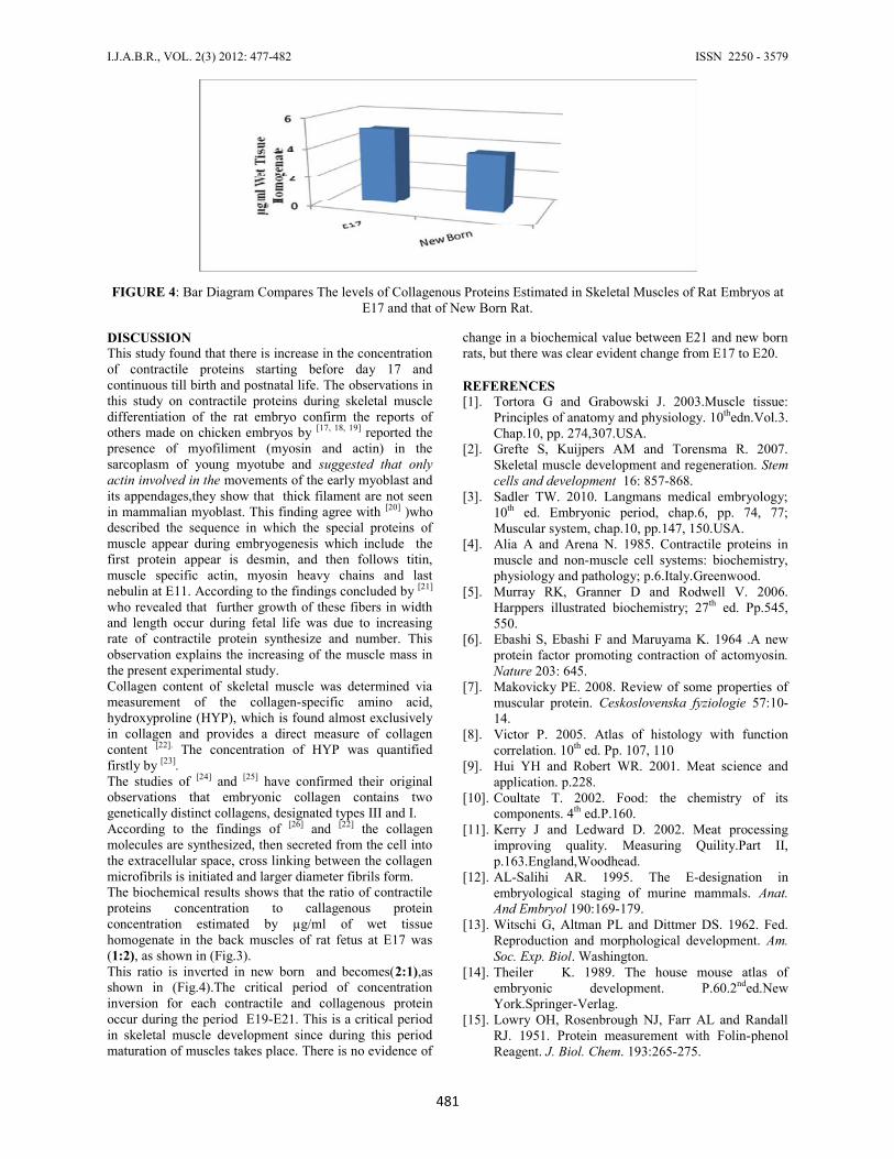
The biochemical results shows that the ratio of contractileproteins concentration to callagenous proteinconcentration estimated by µg/ml of wet tissuehomogenate in the back muscles of rat fetus at E17 was(1:2), as shown in (Fig.3).
This ratio is inverted in new born and becomes (2:1),asshown in (Fig.4).
TABLE 2: Descriptive statistics for collagenous proteins content of skeletal muscle tissue, value are in µg/ml of wet tissueHomogenate
FIGURE 2: Histogram of Mean Values of Collagenous Proteins Estimated at Various Embryonic Stages andNew Born Rats
P(ANOVA)µg/ml of Wet TissueHomogenate
VariableEmbryonic age
0.001<
5.073MeanE17 0,059SD
6N4.867Mean
E18 0.103SD6N
4.332MeanE19 0.024SD
5N3.749Mean
E20 0.043SD7N
3.758MeanE21 0.088SD
5N3.826Mean
New born rats 0.043SD5N
FIGURE 3: Bar Diagram Compares The levels of Contractile Proteins Estimated in Skeletal Muscles of RatEmbryos at E17 and that of New Born Rat
I.J.A.B.R., VOL. 2(3) 2012: 477-482 ISSN 2250 - 3579
481
FIGURE 4: Bar Diagram Compares The levels of Collagenous Proteins Estimated in Skeletal Muscles of Rat Embryos atE17 and that of New Born Rat.
DISCUSSIONThis study found that there is increase in the concentrationof contractile proteins starting before day 17 andcontinuous till birth and postnatal life. The observations inthis study on contractile proteins during skeletal muscledifferentiation of the rat embryo confirm the reports ofothers made on chicken embryos by [17, 18, 19] reported thepresence of myofiliment (myosin and actin) in thesarcoplasm of young myotube and suggested that onlyactin involved in the movements of the early myoblast andits appendages,they show that thick filament are not seenin mammalian myoblast. This finding agree with [20] )whodescribed the sequence in which the special proteins ofmuscle appear during embryogenesis which include thefirst protein appear is desmin, and then follows titin,muscle specific actin, myosin heavy chains and lastnebulin at E11. According to the findings concluded by [21]
who revealed that further growth of these fibers in widthand length occur during fetal life was due to increasingrate of contractile protein synthesize and number. Thisobservation explains the increasing of the muscle mass inthe present experimental study.Collagen content of skeletal muscle was determined viameasurement of the collagen-specific amino acid,hydroxyproline (HYP), which is found almost exclusivelyin collagen and provides a direct measure of collagencontent [22]. The concentration of HYP was quantifiedfirstly by [23].The studies of [24] and [25] have confirmed their originalobservations that embryonic collagen contains twogenetically distinct collagens, designated types III and I.According to the findings of [26] and [22] the collagenmolecules are synthesized, then secreted from the cell intothe extracellular space, cross linking between the collagenmicrofibrils is initiated and larger diameter fibrils form.The biochemical results shows that the ratio of contractileproteins concentration to callagenous proteinconcentration estimated by µg/ml of wet tissuehomogenate in the back muscles of rat fetus at E17 was(1:2), as shown in (Fig.3).This ratio is inverted in new born and becomes(2:1),asshown in (Fig.4).The critical period of concentrationinversion for each contractile and collagenous proteinoccur during the period E19-E21. This is a critical periodin skeletal muscle development since during this periodmaturation of muscles takes place. There is no evidence of
change in a biochemical value between E21 and new bornrats, but there was clear evident change from E17 to E20.
REFERENCES[1]. Tortora G and Grabowski J. 2003.Muscle tissue:
Principles of anatomy and physiology. 10thedn.Vol.3.Chap.10, pp. 274,307.USA.
[2]. Grefte S, Kuijpers AM and Torensma R. 2007.Skeletal muscle development and regeneration. Stemcells and development 16: 857-868.
[3]. Sadler TW. 2010. Langmans medical embryology;10th ed. Embryonic period, chap.6, pp. 74, 77;Muscular system, chap.10, pp.147, 150.USA.
[4]. Alia A and Arena N. 1985. Contractile proteins inmuscle and non-muscle cell systems: biochemistry,physiology and pathology; p.6.Italy.Greenwood.
[5]. Murray RK, Granner D and Rodwell V. 2006.Harppers illustrated biochemistry; 27th ed. Pp.545,550.
[6]. Ebashi S, Ebashi F and Maruyama K. 1964 .A newprotein factor promoting contraction of actomyosin.Nature 203: 645.
[7]. Makovicky PE. 2008. Review of some properties ofmuscular protein. Ceskoslovenska fyziologie 57:10-14.
[8]. Victor P. 2005. Atlas of histology with functioncorrelation. 10th ed. Pp. 107, 110
[9]. Hui YH and Robert WR. 2001. Meat science andapplication. p.228.
[10]. Coultate T. 2002. Food: the chemistry of itscomponents. 4th ed.P.160.
[11]. Kerry J and Ledward D. 2002. Meat processingimproving quality. Measuring Quility.Part II,p.163.England,Woodhead.
[12]. AL-Salihi AR. 1995. The E-designation inembryological staging of murine mammals. Anat.And Embryol 190:169-179.
[13]. Witschi G, Altman PL and Dittmer DS. 1962. Fed.Reproduction and morphological development. Am.Soc. Exp. Biol. Washington.
[14]. Theiler K. 1989. The house mouse atlas ofembryonic development. P.60.2nded.NewYork.Springer-Verlag.
[15]. Lowry OH, Rosenbrough NJ, Farr AL and RandallRJ. 1951. Protein measurement with Folin-phenolReagent. J. Biol. Chem. 193:265-275.
Estimation the levels of proteins of back skeletal muscle during fetal development in rats
482
[16]. Reddy GK and Enwemeka CS. 1996. A simplifiedmethod for analysis of hydroxyproline in biologicaltissue. Clini. Biochem. 29: 225-229.
[17]. Bennett MR and Pettigrew AG. 1974. The formationof synapses in striated muscle during development.J. Physiol. 241: 515-545
[18]. Maclntosh BR, Phillip F, Gardiner AJ and ComasMC. 2006. Skeletal muscle structure and function.2nded.Pp.52, 71.
[19]. Wang H. 1968. Human embryology. WilliamHeinemann. Pp.190, 191. London.
[20]. Frust DO, Osborn M and Weber K. 1989.Myogenesis in the mouse embryo: differential onsetof expression of myogenic proteins and theinvolvement of titin in myofibril assembly. J.CellBiol.109:517-527.
[21]. Jones TC, Hunt RD and King NW. 1997. Veterinarypathology. Skeletal muscle, chap.18,6thed. p.873.Wiliam and wilkins,USA.
[22]. Champ PC and Harvey RA. 2005. Lippncott'sIllustrated Reviews.In: Biochemistry. 3th ed. Pp. 40,43.USA.
[23]. Woessner JF. 1996. The determination ofhydroxyproline in tissue and protein samplescontaining small proportions of this amino acid.Arch.Biochem.Biophys. 39:440.
[24]. Chung E and Miller EJ. 1974. Science 183:1200-1201.
[25]. Epstein EH. 1974. Human skin collagen: Release bypepsin digestion and preponderance in fetal life. J.Biol. Chem. 249: 3225-3231.
[26]. Reiser k, Cormick RJ and Rucker RB. 1992. Theenzymatic and non-enzymatic cross linking ofcollagen and elastin. Federation of Americansocieties for experimental biology 6(7):2439–2449.