Embed Size (px)
Citation preview

1
Establishment of Proximity-dependent Biotinylation Approaches in Different Plant 1
Model Systems 2
3
Deepanksha Arora1,2,*, Nikolaj B. Abel3,*, Chen Liu4,*, Petra Van Damme1,2,5,*, Lam Dai 4
Vu1,2, Anna Tornkvist4, Francis Impens6,7,8, Dominique Eeckhout1,2, Alain Goossens1,2, 5
Geert De Jaeger1,2,#, Thomas Ott3,9,#, Panagiotis Moschou4,10,11,#, Daniel Van Damme1,2,# 6
1 Ghent University, Department of Plant Biotechnology and Bioinformatics, Technologiepark 7
71, 9052 Ghent University, Ghent, Belgium. 8 2 VIB Center for Plant Systems Biology, Technologiepark 71, 9052 Ghent, Belgium. 9 3 Faculty of Biology, Cell Biology, University of Freiburg, Germany. 10 4 Department of Plant Biology, Uppsala BioCenter, Swedish University of Agricultural 11
Sciences and Linnean Center for Plant Biology, Uppsala, Sweden. 12 5 Department of Biochemistry and Microbiology, Ghent University, Ghent, Belgium 13 6 Department of Biochemistry, Ghent University, Ghent, Belgium. 14 7 VIB Center for Medical Biotechnology, Ghent, Belgium. 15 8 VIB Proteomics Core, Ghent, Belgium. 16 9 CIBSS – Centre for Integrative Biological Signalling Studies, University of Freiburg, 17
Germany 18 10 Department of Biology, University of Crete, Heraklion, Greece. 19 11 Institute of Molecular Biology and Biotechnology, Foundation for Research and 20
Technology - Hellas, Heraklion, Greece. 21
22
* joint first authors 23
# joint senior and corresponding authors 24
25
Running title: Proximity-dependent biotinylation in plants 26
.CC-BY-NC-ND 4.0 International licenseauthor/funder. It is made available under aThe copyright holder for this preprint (which was not peer-reviewed) is the. https://doi.org/10.1101/701425doi: bioRxiv preprint

2
Abstract 27
The use of proximity-dependent biotin labelling (PDL) approaches coupled with mass 28
spectrometry recently greatly advanced the identification of protein-protein interactions and 29
study of protein complexation. PDL is based on the expression of a promiscuous biotin ligase 30
(PBL), e.g. BirA* or a peroxidase fused to a bait protein of interest. In the presence of biotin 31
as substrate, PBL enables covalent biotin labelling of proteins in the vicinity of the PBL-fused 32
bait in vivo, allowing the subsequent capture and identification of interacting and neighbouring 33
proteins without the need for the protein complex to remain intact during purification. To date, 34
PDL has not been extensively used in plants. Here we present the results of a systematic multi-35
lab study applying a variety of PDL approaches in several plant systems under various 36
conditions and bait proteins. We show that TurboID is the most promiscuous variant for PDL 37
in plants and establish protocols for its efficient application. We demonstrate the applicability 38
of TurboID in capturing membrane protein interactomes using the Lotus japonicus 39
symbiotically active receptor kinases RLKs NOD FACTOR RECEPTOR 5 (NFR5) and LRR-40
RLK SYMBIOTIC RECEPTOR-KINASE (SYMRK) as test-cases. Furthermore, we 41
benchmark the efficiency of various PBLs using the octameric endocytic TPLATE complex 42
and compare PDL with one-step AP-MS approaches. Our results indicate that different PDL 43
approaches in plants may differ in signal-to-noise ratio and robustness. We present a 44
straightforward strategy to identify both non-biotinylated as well as biotinylated proteins in 45
plants in a single experimental setup. Finally, we provide initial evidence that this technique 46
has potential to infer structural information of protein complexes. Our methods, tools and 47
adjustable pipelines provide a useful resource for the plant research community. 48
49
.CC-BY-NC-ND 4.0 International licenseauthor/funder. It is made available under aThe copyright holder for this preprint (which was not peer-reviewed) is the. https://doi.org/10.1101/701425doi: bioRxiv preprint

3
INTRODUCTION 50
Protein-protein interaction (PPI) studies often fail to capture low affinity interactions as these 51
are usually not maintained following cell lysis and protein extraction. This is in particular the 52
case for PPI’s with or among integral membrane proteins because of the harsh conditions 53
during protein extraction and purification. Proximity-dependent biotin labelling (PDL) on the 54
contrary, uses covalent biotinylation of proteins that are interactors or near-neighbours of a 55
certain bait protein in vivo [1]. Hence, in order to identify interactions, they do not need to 56
remain intact during purification. Biotin is an essential cofactor for a small number of 57
omnipresent biotin-dependent enzymes involved mainly in the transfer of CO2 during HCO3--58
dependent carboxylation reactions. Biotinylation is therefore a relatively rare in vivo protein 59
modification. Moreover, biotinylated proteins can be selectively isolated with high avidity 60
using streptavidin-biotin pairing. PDL therefore permits the identification of both high and low 61
affinity interactions. 62
Analogues to DamID in which a prokaryotic Dam methylase is fused to a protein of 63
interest to monitor DNA-protein interactions in eukaryotes [2], the principle of PDL allows the 64
capture of protein-protein interactions. More specifically, PDL is based on the fact that native 65
biotin ligases, e.g. the Escherichia coli BirA catalyzes a two-step reaction: first, the generation 66
of reactive biotinyl-AMP (biotinoyl-5’-AMP or bioAMP) from biotin and ATP, and second, 67
the attachment of that bioAMP to a specific lysine of the target protein. Engineered PBLs have 68
a significantly reduced affinity for the reactive bioAMP intermediate [3, 4]. This intermediate 69
is prematurely released and, due to its high reactivity, will interact with neighbouring primary 70
amines (e.g., lysine). Therefore, these variants lead to promiscuous labelling despite their lower 71
affinity for biotin compared to native biotin ligases. 72
There are several variations of PDL. The first-generation enzymes used for PDL are 73
based on the E. coli biotin ligase BirA [5]. The mutant BirA, designated BirA* (R118G) [6], 74
referred hereafter as BioID, a monomeric protein of 35.3 kDa, was the first PBL variant used 75
for PDL [3, 4, 7]. A second-generation PBL, called BioID2, was derived from the Aquifex 76
aeolicus biotin ligase [3]. BioID2, which naturally lacks a DNA-binding domain, is 77
approximately one-third smaller than BioID, potentially reducing sterical hindrance of the bait 78
protein [8]. The third-generation PBLs, called TurboID and mini TurboID (mTurboID), are 79
derived from directed evolution of BirA* expressed in yeast. These two variants showed 80
maximal activity at 30oC, while the previous variants show maximal activity at higher 81
temperatures [9]. TurboID has the same size as the original BirA* tag, albeit with 14 amino 82
acid mutations that greatly increase its labelling efficiency. mTurbo has 12 out of 14 of those 83
.CC-BY-NC-ND 4.0 International licenseauthor/funder. It is made available under aThe copyright holder for this preprint (which was not peer-reviewed) is the. https://doi.org/10.1101/701425doi: bioRxiv preprint

4
mutations. The N-terminal DNA-binding domain was deleted to reduce its size (28 versus 35 84
kDa), which also slightly impacted on its labelling efficiency by reducing it ~2-fold. The first 85
and second-generation PBLs required approximately 18 to 24 h of labelling (and sometimes 86
even much longer) to produce detectable levels of protein biotinylation, while the TurboID 87
variants required a labelling time only in the range of 1 h or less in the various eukaryotic, non-88
plant systems tested so far [9]. 89
PDL has its own intrinsic advantages and limitations. In the presence of biotin, the bait-90
PBL fusion protein labels proximal proteins without the activation by a conditional trigger, 91
thereby keeping track of all interactions that occurred over a time-period. The ability for 92
selective capture makes the method generally insensitive to protein solubility or protein 93
complexation, with obvious applicability to membrane proteins and cytoskeletal constituents, 94
a major advantage over alternative approaches. Nevertheless, the identity of a candidate 95
interactor does not immediately imply a direct or indirect interaction with the bait but could 96
merely reflect close proximity [estimated to be ~10 to 15 nm [10]]. Furthermore, true 97
interactors (false negatives) are missed if they lack accessible primary amines. 98
So far PBLs have successfully been used in yeast [11], protozoa [12], amoebae [13], 99
embryonic stem cells [14], and xenograft tumors [15] to map a wide range of interactomes in 100
both small-scale (i.e. single bait protein) and large-scale network mapping approaches (i.e. the 101
protein interaction landscape of the centrosome-cilium interface and the organization of 102
mRNA-associated granules and bodies (mRNP complexes)) [16, 17]. 103
In plants, PBLs have not been extensively used. So far, four papers describe the 104
application of PDL in plants [18-21]. In these first trials, overexpression of a first-generation 105
BirA* was used combined with long labelling times, very high biotin levels and relatively poor 106
labelling efficiencies. These results, combined with the so far non-extensive use of PDL in 107
plants could suggest that BioID variants used so far show reduced activity in plant tissues due 108
to suboptimal working temperatures with reference to their temperature-activity profiles. 109
Here, we report a systematic multi-lab study of different PDL approaches in various 110
plant systems. We provide guidelines for the use of PDL in various frequently used plant 111
models suggesting most relevant shortcomings and contingencies. Furthermore, we benchmark 112
our PDL methods studying the TPLATE protein complex. We foresee that the methods, tools 113
and produced materials will greatly benefit the research community. 114
115
116
117
.CC-BY-NC-ND 4.0 International licenseauthor/funder. It is made available under aThe copyright holder for this preprint (which was not peer-reviewed) is the. https://doi.org/10.1101/701425doi: bioRxiv preprint

5
RESULTS 118
Increased PBL-mediated biotin labelling efficiencies upon biotin administration in planta 119
In non-plant systems, supplementation of biotin is important for efficient proximity biotin 120
ligation with all the PBLs tested so far. In contrast, plants synthesize biotin endogenously and 121
thus the intracellular pool of biotin might be high enough for the PBL. In fact, free biotin has 122
been shown to accumulate in the cytosol of plant mesophyll cells to a high concentration of ca. 123
11 μM [22], while for example in yeast this is more than 10-fold lower [23]. Considering that 124
the Km of BirA* for biotin is 0.3 µM, this could, in theory, lead to efficient PDL even in the 125
absence of exogenous biotin supplementation. 126
Figure S1. GFP expression in tomato hairy root cultures produced with rhizogenic Agrobacterium. Fluorescence
micrograph images of eGFP expression were obtained from primary hairy roots (~14 days) transformed rhizogenic
Agrobacterium with the following expression constructs; Pro35S::eGFP-BirA*, Pro35S::eGFP-BioID2, Pro35S::eGFP-
TurboID, and Pro35S::eGFP-miniTurboID. The scale bars are 500 μm. Images are representative for the four to ten
independent roots selected for subcultivation and showing expression of the marker per construct.
.CC-BY-NC-ND 4.0 International licenseauthor/funder. It is made available under aThe copyright holder for this preprint (which was not peer-reviewed) is the. https://doi.org/10.1101/701425doi: bioRxiv preprint

6
We thus tested biotinylation efficiency in the presence or absence of biotin using 127
different tagged PBLs as fusion proteins, either codon-optimized for plants or non-codon 128
optimized (Supplemental Table 1 and Supplemental File 1). More specifically, we 129
comparatively screened the potential applicability of enzyme-catalyzed proximity labelling 130
when using the PBL BirA* [5, 8], BioID2 [8], TurboID or mTurboID [9]. We first tested these 131
PBLs in stable hairy root lines of Solanum lycopersicum (see Materials and Methods). For 132
protein visualisation, we fused the engineered PBL to FLAG and enhanced green fluorescent 133
protein (eGFP) under control of the constitutive cauliflower mosaic virus (CaMV) 35S 134
promoter (Supplemental Figure 1). 135
Figure 1. Characterization of enzyme-catalysed proximity labelling in hairy root cultures. (A)
Experimental setup. (B) Comparison of biotinylation activity in four PBL-expressing hairy root cultures.
Addition of 50 μM exogenous biotin to two-weeks old hairy root cultures for 2 or 24 h was used for labelling.
Arrowheads indicate the expected size of the cis-biotinylation signal. (B) Comparison of biotinylation activity
in four PBL hairy root cultures from wild-type tomato expressing eGPF.BirA* (~66 kDa), eGPF.BioID2 (~56
kDa), eGPF.Turbo (~64 kDa) and eGPF.miniTurbo (~57 kDa). Gray regions in intense black areas represent
saturation of the streptavidin-s680 signal and is most prominent in case of self-biotinylation activity. This is a
representative experiment repeated twice and two independent root cultures were analyzed per combination.
.CC-BY-NC-ND 4.0 International licenseauthor/funder. It is made available under aThe copyright holder for this preprint (which was not peer-reviewed) is the. https://doi.org/10.1101/701425doi: bioRxiv preprint

7
As a control for non-bait specific biotinylation, PBL-fused eGFP was used. 136
Biotinylation, was evident as smears in streptavidin-HRP Western blot. This smear depicts 137
biotinylation of other proteins than PBLs, and will be referred to as “trans-biotinylation”. As a 138
proxy of PBL activity, we used the cis-biotinylation efficiency (i.e. auto- or self-biotinylation 139
level of PBL fusions) as readout (Figure 1). Manifold faster kinetics for TurboID and 140
mTurboID over BioID and BioID2 could be observed (Figure 1). This is in line with the 141
previously reported lower catalytic activities of the latter PBLs, especially at the growth 142
conditions used (i.e. cultivation of hairy roots was performed at 22-25°C) [9]. Noteworthy, 143
only residual trans-biotinylation was observed when no exogenous biotin was added. 144
Therefore, the addition of surplus of (free) biotin seems to function as an inducing agent of 145
PDL in this system. This observation indicates that PDL in plants to some extend might also 146
have the capacity to identify the spatiotemporal dynamics of interactome composition. 147
148
149
150
PDL-efficiency depends on growth temperatures and PBL can facilitate trans-biotinylation 151
in Nicotiana benthamiana 152
We used transient transformation of Nicotiana benthamiana leaf mesophyll cells to test the 153
applicability of PBL in an alternative system commonly used for protein expression in planta 154
Figure S2 Characterization of PBL-catalysed proximity labelling in N. benthamiana. (A) Experimental setup.
(B) Comparison of biotinylation activity in N. benthamiana expressing eGPF.-BirA* (~66 kDa), eGPF-BioID2
(~56 kDa), eGPF-Turbo (~64 kDa) and eGPF-miniTurbo (~57 kDa). Overlapping signal as indicated with a black
arrow denote enzyme-catalysed cis-biotinylation. Gray bands in intense black areas represent saturation of the
streptavidin-s680 signal and is most prominent in case of auto-biotinylation activity. Two infiltrated tobacco leaf
segments/leaves were analyzed per setup and the experiment was repeated twice with similar results.
.CC-BY-NC-ND 4.0 International licenseauthor/funder. It is made available under aThe copyright holder for this preprint (which was not peer-reviewed) is the. https://doi.org/10.1101/701425doi: bioRxiv preprint

8
under various conditions. In this case, biotin was infiltrated directly into leaf tissue 24 h after 155
transfection and harvested 24 h post-biotin infiltration (Supplemental Figure 2A). We 156
confirmed that also in this system, the highest cis-biotinylation level was observed in the case 157
of TurboID, and supplementation of biotin was important for the efficient detection of cis-158
biotinylation (Supplemental Figure 2B). Furthermore, the overall biotinylation output in 159
tobacco leaves increased when biotin concentration increased from 50 μM to 1 mM 160
(Supplemental Figure 2B). 161
We confirmed that the R118G mutation is responsible for promiscuous labelling in 162
plants, as the wild-type BirA showed no trans-biotinylation (in the presence of 50 µM 163
exogenous biotin; Supplemental Figure 3A). Furthermore, a temperature shift from 22°C to 164
28oC increased cis- and trans-biotinylation for both BioID and TurboID, suggesting that 165
temperature control, at least to some extent, could also be used to modulate PDL in plants 166
(Supplemental Figure 3A, see also below). 167
168
Noteworthy, the effect of temperature on TurboID activity was less apparent compared to that 169
of BioID, consistent with the temperature-dependency of the two enzymes [9]. Interestingly, 170
similar to GFP-TurboID expressed in the hairy root cultures, biotinylation reaches its highest 171
cis-biotinylation level already 2 h after biotin administration in N.benthamiana (Supplemental 172
Figure S3. Biotinylation of BirA∗ increases at elevated growth temperature and biotin concentration in
Nicotiana benthamiana. (A) Upper blot: The wild-type version of BirA shows residual cis-biotinylation activity
at both 22oC and 28oC. BirA* (BioID) shows promiscuous biotinylation which increases with temperature.
Bottom blot: GFP-TurboID-HF activity was ~2 fold increased at higer temperature (i.e. when going from 22°C
to 28°C). Two biological replicates are depicted. (B) Increasing biotin levels elevated biotinylation efficiency
and efficient cis-biotinylation was observed already 2 h after biotin administration. Green arrowheads show the
corresponding BL. N.c. negative control.
.CC-BY-NC-ND 4.0 International licenseauthor/funder. It is made available under aThe copyright holder for this preprint (which was not peer-reviewed) is the. https://doi.org/10.1101/701425doi: bioRxiv preprint

9
Figure 3B). TurboID and mTurboID were the only PBLs in plants with biotinylation efficiency 173
occurring in the range of few hours, as other PBLs did not show any visible sign of trans-174
biotinylation in that time (Figure 1 and Supplemental Figure 2). 175
176
TurboID can be used for the efficient capture of plasma membrane interactomes in 177
Nicotiana benthamiana 178
Next, we tested whether we could achieve biotinylation of protein interactors using PDL under 179
the conditions established for N. benthamiana. We observed that the prey proteins used in 180
plants for BioID so far were relatively small proteins (i.e. HopG, 25 kDa [20], OsFD2 18 kDa 181
[18], AvrPto 16 kDa [19]). Interestingly, all these proteins have a smaller predicted radius 182
(assuming a globular shape) than all four PBLs used in the PDL approaches so far. 183
We tested our conditions for PDL using as test-cases plasma membrane-localized 184
protein complexes with radii within a range of a few nm. First, we used a known membrane 185
receptor complex from Lotus japonicus comprised of two symbiotically active receptor 186
kinases: The LysM-type RLKs NOD FACTOR RECEPTOR 5 (NFR5) and the LRR-RLK 187
SYMBIOTIC RECEPTOR-KINASE (SYMRK). These proteins assemble within the same 188
complex in L. japonicus roots [24] as well as in N. benthamiana upon heterologous expression 189
[25]. In contrast, the brassinosteroid receptor BRASSINOSTEROID INSENSITIVE 1 (BRI1) 190
was shown not to be involved in this interaction as co-IP results failed to identify BRI1 in these 191
experiments [25]. To further extend the set of control proteins, we additionally cloned the A. 192
thaliana innate immune pattern recognition receptors FLAGELLIN SENSING 2 (FLS2) and 193
the EF-TU RECEPTOR (EFR), both belonging to the LRR-family, as well as the LOW 194
TEMPERATURE INDUCED PROTEIN LTI6b that is commonly used as a bona fide plasma 195
membrane marker in plant cell biology [26]. 196
.CC-BY-NC-ND 4.0 International licenseauthor/funder. It is made available under aThe copyright holder for this preprint (which was not peer-reviewed) is the. https://doi.org/10.1101/701425doi: bioRxiv preprint

10
We first co-expressed the symbiotic receptors NFR5 and SYMRK as C-terminal GFP 197
fusion proteins with a cytosolic TurboID-GFP in N. benthamiana. Immunoprecipitation and 198
Western Blot analysis revealed the successful expression and pull-down of all three proteins 199
(Figure 2A). When probing for biotinylation levels, we exclusively detected self-biotinylation 200
in case of cytosolic TurboID-GFP (Figure 2A). However, prolonged exposure of the blot 201
resulted in weak detectable bands in case of NFR5-GFP and SYMRK-GFP indicating the 202
presence of some background trans-biotinylation. To test whether trans-biotinylation of known 203
interacting proteins can be observed, we generated an NFR5-TurboID and co-expressed it with 204
SYMRK-GFP. Here, we not only detected a clear cis-biotinylation of NFR5-TurboID but also 205
trans-biotinylation of SYMRK (Figure 2B). These data show that specific trans-biotinylation 206
occurs within a known receptor complex. 207
Figure 2. Trans-biotinylation within membrane-resident receptor complexes. (A) TurboID-GFP was co-
expressed with symbiotic RLKs to test for unspecific trans-biotinylation. While all proteins were detected before
(middle panel) and after immunoprecipitation (lower panel), no trans-biotinylation of the receptors was observed
under these conditions (upper panel). The activity of TurboID is indicated by self-biotinylation of TurboID-GFP
(70 kDa). (B) Fusing TurboID to NFR5 (120 kDa) resulted in strong trans-biotinylation of the known interaction
partner SYMRK (150 kDa), weak signals were detected in case of the PM-resident receptors BRI1 and FLS2,
whileno trans-biotinylation of EFR and the PM-marker LTI6b were detected. IP= immunoprecipitation; WB=
Western Blot. *= unclassified band.
.CC-BY-NC-ND 4.0 International licenseauthor/funder. It is made available under aThe copyright holder for this preprint (which was not peer-reviewed) is the. https://doi.org/10.1101/701425doi: bioRxiv preprint

11
To test for specificity in the assay, we co-expressed NFR5-TurboID additionally with 208
the functionally unrelated transmembrane RLK BRI1, which was previously shown to not 209
interact with the NFR5/SYMRK complex using co-IP [25]. While we detected strong BRI1 210
expression, this protein was only weakly trans-biotinylated by NFR5-TurboID indicating some 211
unspecific labelling or that the protein is in proximity to the complex (Figure 2B). To broaden 212
this, we also co-expressed the transmembrane proteins FLS2, EFR and LTI6b with NFR5-213
TurboID. While no bands were detected for EFR and LTI6b, we observed some weak signal 214
for FLS2 indicating that this receptor may locate in close proximity to NFR5 (Figure 2B). 215
Prolonged exposure of the blots yielded some weak signal for all membrane proteins as also 216
observed for GFP-TurboID (data not shown). However, these signals were orders of 217
magnitude lower than those detected for NFR5/SYMRK. This assay was further optimized by 218
temporally limiting the reaction. We could show that trans-biotinylation efficiently occurs 219
within 15 min after applying exogenous biotin, demonstrating that specificity is maintained by 220
minimizing the availability of the substrate (Supplemental Figure 4). 221
It should also be considered that in addition to ectopic expression of the constructs, the weak 222
dimerization potential of GFP here or other protein tags with similar properties may result in 223
potentially unspecific trans-biotinylation. 224
Taken together these data clearly show that TurboID-mediated PDL can be efficiently 225
used for membrane proteins. It can be advantageous over other methods such as co-226
immunoprecipitation as it does not require any optimization of the solubilization conditions 227
and it provides the possibility to detected transiently protein complex constituents. 228
Figure S4. Temporally limiting the reaction results in weak but specifically detectable bands in case of
NFR5-TurboID and SYMRK-GFP. Biotin was applied for 15 or 30 min. IP= immunoprecipitation; WB=
Western Blot.
.CC-BY-NC-ND 4.0 International licenseauthor/funder. It is made available under aThe copyright holder for this preprint (which was not peer-reviewed) is the. https://doi.org/10.1101/701425doi: bioRxiv preprint

12
Application of PDL in Arabidopsis thaliana cell cultures using the TPLATE complex as a 229
case study 230
Next, we surveyed the efficiency of trans-biotinylation for a stable multi-subunit plant protein 231
complex. As a test case, we took the plasma membrane-associated octameric TPLATE 232
complex (TPC) [27] and used stably transformed A. thaliana culture cells as a third model 233
system. 234
Given the higher biotinylation level observed in N. benthamiana at 28oC 235
(Supplemental Figure 3) we tested different labelling conditions using A. thaliana suspension 236
cell cultures. We grew the cells expressing TPLATE-BioID and GFP-BioID, i.e. proteins fused 237
to the initial version of BirA*, at various temperatures in the presence of 50 µM biotin for 24 238
h. We subsequently isolated the complex using streptavidin affinity purification (see Materials 239
and Methods), performed tryptic on-bead digest and analyzed the released non-biotinylated 240
peptides using LC-MS/MS. 241
242
243
244
In order to compare the effect of temperature on the biotinylation efficiency to 245
identification of proteins from isolated protein complexes, we focused on the other seven 246
TPLATE complex members and compared their abundance and fold changes after streptavidin-247
purification as deduced from label-free protein quantification (LFQ; [28]) to the control setup 248
(35S::GFP-BioID) (Figure 3A). The fold change difference with respect to the control for the 249
Figure 3. Detection of TPC subunits with TPLATE-BioID is optimal at 28° C. (A) Experimental setup to
look for enriched TPC subunits in biotin treated transformed Arabidopsis cell cultures. (B) Fold change
abundance of the TPC subunits and statistical significance (-log(p-value)) compared to control. Fold change and
p-values were calculated from the average LFQ intensities for 3 technical replicates of TPLATE-BioID
w.r.t.versus GFP-BioID at similar temperature. Cell cultures were incubated with 50µM biotin at 25°-35°C for
24 h before harvesting. The TPC subunits are detected at all 4 temperatures without major differences. At 28°C
and 30°C, the overall detection of several of the other subunits shows increased robustness (p-value) compared
to both the lower (25°C) and higher (35°C) temperatures.
.CC-BY-NC-ND 4.0 International licenseauthor/funder. It is made available under aThe copyright holder for this preprint (which was not peer-reviewed) is the. https://doi.org/10.1101/701425doi: bioRxiv preprint

13
other TPC subunits was subunit dependent and not dramatically different between the different 250
temperatures. The highest overall fold change difference for the different subunits, combined 251
with the optimal robustness of the identification (p-value) was detected at 28oC (Figure 3B, 252
Supplemental Data Set 1), indicating that this temperature presents the optimal trade-off 253
between biotinylation efficiency of BioID and perturbation of physiological processes due to 254
elevated temperatures. 255
256
Various PBLs affect biotinylation of TPC subunits differently 257
The introduction of a flexible linker has been successfully used to extend the labelling radius 258
of PBLs [8], which is estimated to be about 10 to 15 nm [10]. This increased labelling radius 259
may be desirable when the protein of interest is significantly larger than the labelling radius of 260
the PBL alone, and/or when the goal is to map the constituency of a larger protein complex or 261
discrete subcellular region. We thus compared the efficiencies of various PBLs and assessed 262
their biotinylation radius by inserting a long flexible linker. For this, Arabidopsis cultures 263
expressing C-terminal fusions of TPLATE with BioID or BioID2 were assessed, with and 264
without a 65 aa linker similar to the one that was reported before [5]. As controls, we generated 265
GFP fused to BioID or BioID2 without additional linker (Supplemental Figure 5). 266
267
268
269
Anti HA-HRP
200
150 kDa
50
37 kDa
- +Biotin
GFP-BioID (62 kDa)
PSB-D TPLATE-BioID(166 kDa)
TPLATE-linker BioID (170kDa)
TPLATE-BioID2(157 kDa)
TPLATE-linker BioID2 (160 kDa)
A.
B.
- + - + - + - + - +
Streptavidin-HRP
250
150
100
75
50
37kDa
250
150
100
75
50
37kDa
Biotin
*
* * **
GFP-BioID (62 kDa)
PSB-D TPLATE-BioID(166 kDa)
TPLATE-linker BioID (170kDa)
TPLATE-BioID2(157 kDa)
TPLATE-linker BioID2 (160 kDa)
C. D.
Streptavidin-HRP
250
150
100
75
50
37
25
20
15 kDa
Biotin - 1hr 24hrs - 1hr 24hrs
GFP-linker TurboID(64 kDa)
TPLATE-linker TurboID (170 kDa)
*
*
250
150
100
50
37
25kDa
GFP-linker TurboID(64 kDa)
TPLATE-linker TurboID (170 kDa)
GFP-BioID2(62 kDa)
250
150
100
50
37
25kDa
Biotin - 1hr 24hrs - 1hr 24hrs
250
150
100
75
50
37kDa
GFP-BioID2(62 kDa)
24hrs
24hrs
*
Figure S5. Different PBL cause different self- and trans- biotinylation. Cell cultures expressing different
TPLATE-PBL were incubated with 50µM biotin at 28°C for 1 h and 24 h before harvesting. (A and B) Anti-
HA HRP western blotting was performed to visualize expression levels of the different cultures. (C and D)
Streptavidin-HRP western blotting of different TPLATE-PBLs, GFP-BioID and control cell cultures (PSB-D).
Self-biotinylation of the bait and trans-biotinylation can .clearly be observed. * indicates self-biotinylation of
the bait.
.CC-BY-NC-ND 4.0 International licenseauthor/funder. It is made available under aThe copyright holder for this preprint (which was not peer-reviewed) is the. https://doi.org/10.1101/701425doi: bioRxiv preprint

14
Next, we tested the activity of different PBLs in this system. We grew the cultures for 270
24h at 28oC, with and without exogenous biotin, and assessed expression and biotinylation via 271
Western blotting (Supplemental Figure 5). Protein abundance of the BioID and BioID2 272
constructs was comparable to their respective controls in our cell cultures and not affected by 273
the addition of biotin. Only the levels of TPLATE-BioID2 were somewhat lower. At the level 274
of cis- and trans-biotinylation, we observed different patterns for each of the enzymes used. 275
As several of the detected bands which increased significantly in the presence of biotin, did not 276
correspond to bands in the control (PSB-D) or GFP-BioID culture, they likely represent 277
different trans-biotinylated interactors and suggest that the outcome of a BioID-based 278
interaction assay might (partially) depend on the PBL used. TPLATE-linker PBL showed the 279
most diverse biotinylation pattern when comparing to the other setups expressing BioID and 280
BioID2 fusions (Supplemental Figure 5) suggesting that adding a linker may be used to to 281
optimize proximity labelling. Consistent with the results described for tobacco, linkerTurboID 282
constructs showed some biotinylation without the addition of exogenous biotin, increased 283
biotinylation after 1 h incubation with biotin and extensive biotinylation after 24 h incubation 284
with biotin in both control and bait cultures suggesting it is highly promiscuous. 285
286
As observed in N. benthamiana (Supplemental Figure 2), BioID outperformed BioID2 287
using TPLATE as bait in this system, although this might be skewed due to lower expression 288
levels of the latter. Adding a flexible linker increased self-biotinylation levels of the bait 289
compared to the constructs without linker (Supplemental Figure 5A and C). Our results are 290
consistent with previous observations in non-plant systems suggesting that linkers increase the 291
biotinylation output [8]. 292
Following the positive effect of exogenous biotin supplementation (Supplemental 293
Figures 2 and 3), we tested the effect of increasing biotin concentrations on cis-biotinylation 294
efficiency. Cell cultures expressing TPLATE-linkerBioID were grown at 28oC in the presence 295
of increasing concentrations of biotin (50 µM to 4 mM) and processed for Western blotting. 296
Supplementing the culture with concentrations of biotin in the range of 50 uM to 1 mM 297
increased cis-biotinylation output up to a maximum of ~2-fold (Supplemental Figure 6). 298
Increasing biotin concentration >2mM did not further increase the cis-biotinylation efficiency. 299
.CC-BY-NC-ND 4.0 International licenseauthor/funder. It is made available under aThe copyright holder for this preprint (which was not peer-reviewed) is the. https://doi.org/10.1101/701425doi: bioRxiv preprint

15
300
We took advantage of the increased biotinylation observed by including a long linker 301
sequence and generated Arabidopsis cultures expressing GFP-linkerTurboID and TPLATE-302
linkerTurboID. Similar to other systems, 24 h post-biotin addition, TurboID efficiency strongly 303
outperformed all other PBLs tested as evident from the high biotinylation levels observed with 304
and without the addition of exogenous biotin and for both the control (GFP) as well as the 305
TPLATE expressing cultures (Supplemental Figure 5B and D). 306
In order to compare the different PBL modules, we processed the cell cultures for LC-307
MS/MS and focused on the relative levels of the various TPC subunits compared to the control 308
setup. Our first mass spec results following streptavidin pull down and on-bead digestion 309
identified all known subunits of the TPC. Given that this is a robust multi-subunit complex [27] 310
and that we identify only non-biotinylated peptides with our on-bead digestion protocol, we 311
assumed that the subunits we detect are a combination of direct biotinylation as well as co-312
immunoprecipitation of the complex as a whole. To test this, we adapted our protocol (Figure 313
4A) and included protein extraction and stringent washing steps with a buffer containing 8M 314
urea and 2% SDS to unfold proteins captured by the beads and to be able to remove unspecific 315
protein binders. We also included the TPLATE-linkerBioID setup treated with 2 mM biotin for 316
24 h to assess if increased biotin concentration improves TPC subunit detection. 317
Figure S6. Cis-biotinylation of TPLATE-linkerBioID increases at higher concentration of exogenous
biotin. Cell cultures expressing TPLATE-linkerBioID were incubated with biotin (50µM to 4mM) at 28°C for
24 h. (A) Anti-HA HRP western immunostaining was performed to check protein expression while streptavidin-
HRP western immunostaining was used to assess the biotinylation levels of TPLATE-BioID. (B) Quantification
of the percentage of biotinylation (orange) as well as the expression (blue) for each biotin concentration
compared to the maximum biotinylation efficiency (2mM) using ImageJ.
.CC-BY-NC-ND 4.0 International licenseauthor/funder. It is made available under aThe copyright holder for this preprint (which was not peer-reviewed) is the. https://doi.org/10.1101/701425doi: bioRxiv preprint

16
318
In agreement with the higher stringency of the isolation procedure, the smallest TPC subunit, 319
LOLITA, which could be robustly detected using AP-MS [27] and could be detected without 320
being denatured prior to binding to streptavidin beads, was no longer detected (Figure 4, 321
Supplemental Data Set 2). LFQ revealed that the remaining seven TPC subunits, including 322
the bait TPLATE, could be detected using BioID, linkerBioID, linkerBioID2 and 323
linkerTurboID. The TASH3 and TWD40-2 subunits could however hardly be detected using 324
BioID2, which might be caused by the reduced expression level of the bait observed in these 325
cultures (Supplemental Figure 5). Increasing the concentration of biotin to 2mM had an 326
adverse effect on the detection of the TPC subunits as only the bait itself could be identified. It 327
is likely that increasing biotin concentrations causes residual free biotin to accumulate in the 328
protein extract, even after protein desalting to deplete free biotin, thereby occupying the 329
streptavidin binding sites on the beads (saturated at >9 µM of biotin). We tested this “saturation 330
hypothesis” using N. benthamiana leaves and protein precipitation to completely remove 331
residual biotin, showing that even low concentration of residual biotin can saturate the 332
streptavidin beads and incapacitate detection (Supplemental Figure 7). Hence, special care 333
should be taken to avoid excess of residual free biotin during streptavidin-capture. 334
Figure 4. Different TPLATE-PBLs affect biotinylation of TPC subunits differently. (A) Experimental
setup. (B) Comparison of the relative fold change and statistical significance (-log(p-value)) compared to control
samples for the various PBLs and conditions. Relative fold change and p-values were calculated from the
average LFQ intensities of 3 technical replicates of the test compared to the control. GFP-BioID served as a
control for TPLATE-BioID and TPLATE-linkerBioID while GFP-BioID2 is a control for TPLATE-BioID2 and
TPLATE-linkerBioID2 and GFP-linkerTurboID is the control for TPLATE-linkerTurboID.
.CC-BY-NC-ND 4.0 International licenseauthor/funder. It is made available under aThe copyright holder for this preprint (which was not peer-reviewed) is the. https://doi.org/10.1101/701425doi: bioRxiv preprint

17
335
336
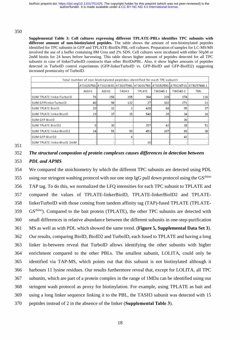
It should be noticed that the fold change by which the other TPC subunits were detected with 337
TurboID was only similar or even sometimes lower (e.g. AtEH2) compared to the other BioID 338
forms (Figure 4). This was caused by the fact that TPC subunits were identified more in the 339
TurboID control samples, resulting in the lower relative fold changes. All individual TPC 340
subunits were detected with more than 20 unique peptides using the GFP-linkerTurboID 341
whereas TWD40-2 was the only TPC subunit detected in other GFP-PBLs, which explains its 342
overall low fold change (Supplemental Table 3). Nevertheless, TurboID identified the other 343
TPC subunits more robustly compared to the other PBLs. So, although in our case, TurboID 344
showed to be superior to all others in identifying the other TPC subunits, the lower signal/noise 345
ratio of TurboID, due to its increased activity, might work as a disadvantage to observe 346
differences between bait proteins and control samples, which might even be enhanced if the 347
proteins are targeted to specific subcellular locations. 348
349
Figure S7. Exogenous application of biotin can exceed the binding capacity of streptavidin beads. Blot on
the left: input and IP with streptavidin using 25 or 50 ul of beads. Note that 2 x more beads increased the recovery
of the input signal, suggesting that the beads are saturated. Blot on the right: IP with 25 ul of streptavidin beads
but in this case the supernatant was precipitated using ammonium acetate to remove excess biotin. Green
arrowheads mark the position of the BL.
.CC-BY-NC-ND 4.0 International licenseauthor/funder. It is made available under aThe copyright holder for this preprint (which was not peer-reviewed) is the. https://doi.org/10.1101/701425doi: bioRxiv preprint
18
350
351
The structural compostion of protein complexes causes differences in detection between 352
PDL and APMS 353
We compared the stoichiometry by which the different TPC subunits are detected using PDL 354
using our stringent washing protocol with our one step IgG pull down protocol using the GSrhino 355
TAP tag. To do this, we normalized the LFQ intensities for each TPC subunit to TPLATE and 356
compared the values of TPLATE-linkerBioID, TPLATE-linkerBioID2 and TPLATE-357
linkerTurboID with those coming from tandem affinity tag (TAP)-fused TPLATE (TPLATE-358
GSrhino). Compared to the bait protein (TPLATE), the other TPC subunits are detected with 359
small differences in relative abundance between the different subunits in one-step-purification 360
MS as well as with PDL which showed the same trend. (Figure 5, Supplemental Data Set 3). 361
Our results, comparing BioID, BioID2 and TurboID, each fused to TPLATE and having a long 362
linker in-between reveal that TurboID allows identifying the other subunits with higher 363
enrichment compared to the other PBLs. The smallest subunit, LOLITA, could only be 364
identified via TAP-MS, which points out that this subunit is not biotinylated although it 365
harbours 11 lysine residues. Our results furthermore reveal that, except for LOLITA, all TPC 366
subunits, which are part of a protein complex in the range of 1MDa can be identified using our 367
stringent wash protocol as proxy for biotinylation. For example, using TPLATE as bait and 368
using a long linker sequence linking it to the PBL, the TASH3 subunit was detected with 15 369
peptides instead of 2 in the absence of the linker (Supplemental Table 3). 370
Supplemental Table 3: Cell cultures expressing different TPLATE-PBLs identifies TPC subunits with
different amount of non-biotinylated peptides. The table shows the amount of non-biotinylated peptides
identified for TPC subunits in GFP and TPLATE-BioIDs PBL cell cultures. Preparation of samples for LC-MS/MS
involved the use of a buffer containing 8M Urea and 2% SDS. Cell cultures were incubated with either 50µM or
2mM biotin for 24 hours before harvesting. This table shows higher amount of peptides detected for all TPC
subunits in case of linkerTurboID constructs than other BioIDsPBL. Also, it show higher amounts of peptides
detected in TurboID control experiments (GFP-linkerTurboID vs. GFP-BioID and GFP-BioID2) suggesting
increased promiscuity of TurboID.
.CC-BY-NC-ND 4.0 International licenseauthor/funder. It is made available under aThe copyright holder for this preprint (which was not peer-reviewed) is the. https://doi.org/10.1101/701425doi: bioRxiv preprint

19
371
Identification of biotinylated peptides allows identifying structural relationships between 372
complex subunits 373
The interaction between biotin-streptavidin is strong enough to be retained even under harsh 374
conditions, e.g, in reductive buffers (Supplemental Figure 7). Thus, biotinylated peptides are 375
expected to be retained on the streptavidin beads even under stringent washing. Following 376
stringent washing under denaturing conditions, on bead digest will release non-biotinylated 377
proteins, which can subsequently be identified using LC-MS. This approach, however, does 378
not provide direct evidence for biotinylation and it relies on the assumption that only 379
biotinylated proteins remain bound to the beads after the washing steps. To acquire direct proof 380
of biotinylation MS-based identification of biotinylated peptides is required. 381
382
383
384
385
386
387
388
389
Figure 5. Comparison of relative abundance of TPC subunits in pull down and BioID. Pull down and
LinkerBioIDs experiments were performed in triplicate, using TPLATE as bait. Per set of experiments, average
LFQ intensities were calculated for each of the TPC subunits. Values were normalized versus the bait,
TPLATE, taking into account the molecular weight of the TPC subunit. Normalized LFQ values show small
differences in relative abundance of the TPC subunits, both in pull down and BioID.
.CC-BY-NC-ND 4.0 International licenseauthor/funder. It is made available under aThe copyright holder for this preprint (which was not peer-reviewed) is the. https://doi.org/10.1101/701425doi: bioRxiv preprint

20
390
391
Thus, we expanded the protocol to also be able to identify biotinylated peptides. For this, we 392
included a second elution step (see Materials and Methods) to release the biotinylated 393
peptides from the beads using an adapted protocol based on [29]. This approach allows 394
detecting both non-biotinylated as well as biotinylated peptides in the same experimental setup. 395
We performed the analysis on the TPLATE-linkerTurboID setup and again specifically focused 396
on peptide identification of the TPC subunits (Figure 6 and Supplemental Data Set 4). The 397
highest number of biotinylated peptides were identified for TPLATE (44 biotinylated 398
peptides), followed by TWD40-1 (18), EH2 (16), EH1 (12), TWD40-2 (9) and TML (3). No 399
biotinylated peptides could be detected for LOLITA, correlating with our previous results. 400
Mapping non-biotinylated and biotinylated peptides, taking into account their relative 401
abundance, on the different TPC subunits revealed differences in the number of detected 402
peptides as well as differences in the distribution of the biotinylated peptides along the length 403
of the subunits. Whereas the bait, TPLATE, shows a relatively even distribution of biotinylated 404
peptides along the protein sequence, there is a clear tendency of the EH1, EH2 and TML 405
subunits towards biotinylation at their C-terminal parts (Figure 6). It is tempting to speculate 406
that the observed distribution of biotinylated peptides, as well as their absence, reflect the 407
proximity of the domains as well as structural constraints with respect to the bait protein and 408
Figure S8. The biotin-streptavidin interaction is retained under harsh conditions. Different extraction
buffers were used for testing the binding affinity of biotin-labelled proteins with streptavidin from equal amount
of plant protein material: 1. 50mM HEPES, 150 mM NaCl, 0.5% NP40, 10% Glycerol, 1mM PMSF; 2. 50mM
Tris/HCl, 150 mM NaCl, 0.5% NP40, 10% Glycerol, 1mM PMSF; 3. 1x PBST, 1mM PMSF; 4. 2x Laemmli
sample buffer (65 mM Tris-HCl, pH 6.8, 20% (w/v) glycerol, 2% SDS, 0.01% bromophenol blue, 10mM DTT).
Con: control. M: marker. Note that the lack of enrichment in the pulldown (right panel) is due to the presence of
biotin (see also Supplemental Figure 7).
.CC-BY-NC-ND 4.0 International licenseauthor/funder. It is made available under aThe copyright holder for this preprint (which was not peer-reviewed) is the. https://doi.org/10.1101/701425doi: bioRxiv preprint

21
that proximity biotinylation harnesses the potential to help deduce structural insight into protein 409
complexes as well as topology information in case of transmembrane proteins. 410
411
412
Discussion 413
We provide a comprehensive comparison of various PBL based proximity labelling strategies 414
in plants and show that TurboID is the most promiscuous one, and that this also sometimes 415
leads to a lower signal to noise ratio. We also provide guidelines and approaches for 416
interactome capture in various plant systems specifically focusing on the ones that interact with 417
the plasma membrane. Furthermore, we show that for each bait/system conditions need to be 418
optimized independently. 419
We observed that in all three plant systems, using exogenous application of biotin 420
enhances PDL output but might not be a strict requirement for the successful application of 421
PDL. This result seems to contradict with what has been reported for a related method called 422
INTACT (isolation of nuclei tagged in specific cell types) in plants. This method allows for 423
affinity-based isolation of nuclei from individual cell types of tissue. INTACT relies on the 424
endogenous pool of biotin as no exogenous supplementation is required [30]. In INTACT, 425
nuclei are affinity-labelled through transgenic expression of the wild-type variant of BirA 426
which biotinylates a nuclear envelope protein carrying biotin ligase recognition peptide from 427
ACC1. This tag acts as a native substrate for the E. coli biotin ligase BirA [31]. The use of 428
wild-type BirA along with its preferable substrate could explain the higher affinity for the free 429
biotin pool in INTACT, and the peptide used as fusion is an optimal substrate for the bioAMP 430
Figure 6. Comparison of biotinylated versus non-biotinylated peptide identification of the various TPC
subunits using TPLATE-linkerTurboID as bait. (A) Experimental setup. (B) Overview of the identified
peptides and their abundance, mapped onto the different TPC subunits.
.CC-BY-NC-ND 4.0 International licenseauthor/funder. It is made available under aThe copyright holder for this preprint (which was not peer-reviewed) is the. https://doi.org/10.1101/701425doi: bioRxiv preprint

22
intermediate. We assume that various proteins may show variability in functioning as acceptors 431
of bioAMP (e.g. depending on the presence of accessible lysine residues). 432
PDL utilizing bacterial enzymes poses the question of whether these enzymes could 433
perform adequately in plants [8]. The activity optimum for BioID2 is 50ºC, whereas for BioID 434
this is 37ºC and thus BioID2 may be most adequate for use at higher temperature conditions. 435
Both temperatures are however far-off from the usual growth temperatures of most plant 436
species grown in temperate regions (e.g. Arabidopsis sp.). Both BioID2 and BioID show 437
reduced activity below 37ºC ([8] and our results herein). Furthermore, the lower temperature 438
optimum of TurboID (and mTurboID) [9] would imply that may function better at normal plant 439
growth temperature. In fact, we observed that TurboID activity is only increased by 2-fold from 440
22oC to 28oC. We, however, cannot rule out that the optimal temperature for PDL may vary 441
depending on the bait protein. At all tested temperatures, we observed that TurboID (and 442
mTurboID) outperforms other PBLs in terms of speed and promiscuity. Hence, TurboID might 443
be preferable when it comes to initial study of (transient) complex composition where the 444
generation of as much as possible specific biotinylation output in short time might be desirable. 445
However, the strong promiscuity of the control might also work as a disadvantage in 446
revealing specific interactions in cases where the reaction cannot be controlled that easily in 447
time or when both the bait and the control would be targeted to a confined intracellular space. 448
We provide evidence that our methods and conditions are applicable to plasma-449
membrane complexes. We showed that the interaction of the symbiotic RLKs NFR5 and 450
SYMRK can be identified by exploiting PDL and particularly the PBL TurboID. Furthermore, 451
the use of proper negative controls is imperative. However, even though the brassinosteroid 452
receptor BRI1 was not co-immunoprecipitated with the symbiotic receptors in a previously 453
published dataset [25], we detected weak biotinylation of this RLK and the immune-receptor 454
FLS2. While it could be interpreted as unspecificity within the PBL system, it should also be 455
considered, that PBL allows labelling of transient interactions or proximal proteins. As a 456
consequence, continuous unstable interactions accumulate to detectable amounts of proteins 457
and would thus allow their identification. As PDL using TurboID is capable of trans-458
biotinylation in the range of minutes (15 minutes under our experimental conditions), the 459
enrichment of unstable interactions would thus be more prominent. Therefore, putative 460
interactions identified by PBL still need to be verified using independent experimental systems 461
but comparisons between the different experimental systems should always reflect the technical 462
limitations of each approach. 463
.CC-BY-NC-ND 4.0 International licenseauthor/funder. It is made available under aThe copyright holder for this preprint (which was not peer-reviewed) is the. https://doi.org/10.1101/701425doi: bioRxiv preprint

23
By expanding our protocols and PBLs into Arabidopsis cell cultures, we could 464
reproduce the composition of the TPC except for one subunit. We show that the use of linkers 465
can be advantageous when it comes to identifying protein-protein interactions of multi-subunit 466
complexes. Furthermore, TPLATE-linkerBioID2 shows reduced cis-biotinylation compared to 467
TPLATE-linkerBioID in the presence of exogenous biotin but seems to function in the absence 468
of biotin suggesting that in plants, BioID2 can function in tissues where exogenous 469
supplementation of biotin may be slower, e.g. the vasculature. Furthermore, increased biotin 470
applications can lead to serious impediments when it comes to the identification of TPC 471
subunits as this can interfere with biotinylated protein binding on streptavidin slurries. Caution 472
is warrented to assure sufficient capture capacity of biotinylated proteins’, since the amount of 473
beads needed for capture should be tested for each experimental setup/protocol. 474
Finally, by establishing a strategy for simultaneous identification of biotinylated and 475
non-biotinylated peptides we could provide evidence for the accessibility of different protein 476
parts to PDL. We show that EH1, EH2 and TML subunits are preferentially biotinylated at 477
their C-terminal parts, suggesting that their C-termini are in closer proximity to the C-terminal 478
end of TPLATE and/or some domains (even complex subunits) are not available for 479
biotinylation. We thus provide evidence that PDL approaches in plants may be able to provide 480
structural information of multi-subunit protein complexes and that this may be extended to the 481
topology of membrane proteins. 482
While this manuscript was on preparation, two additional works appeared in BioRxiv 483
making use of TurboID in plants. These two works complement our work providing further 484
evidence that TurboID can be used to capture cell-specific interactomes [32] and transient 485
signalling components [33]. Taken together, these three studies provide a new arena for the 486
identification of novel protein-protein interactions in plants. 487
488
Supplemental Table 1: list of constructs used 489
Name Vector Promoter Gene PBL used
GFP-BioID pK7m34Gw p35s GFP BioID
GFP-BioID2 pK7m34Gw p35s GFP BioID2
TPLATE-BioID pK7m34Gw p35s TPLATE BioID
TPLATE-BioID2 pH7m34Gw-R p35s TPLATE BioID2
TPLATE-linkerBioID pH7m34Gw-R p35s TPLATE (GGGGS)13 BioID
TPLATE-linkerBioID2 pH7m34Gw-R p35s TPLATE (GGGGS)13 BioID
GFP linkerTurboID pK7m34Gw p35s GFP (GGGGS)13 TurboID
attL2-BioID-attL3 pG9m-2 - - bioID
.CC-BY-NC-ND 4.0 International licenseauthor/funder. It is made available under aThe copyright holder for this preprint (which was not peer-reviewed) is the. https://doi.org/10.1101/701425doi: bioRxiv preprint

24
attL2-BioID2-attL3 pG9m-2 - - bioID2
attL2-linkerBioID-attL3 pUC54 - - (GGGGS)13 BioID
attL2-linkerBioID2-attL3 pG9m-2 - - (GGGGS)13 BioID2
attL2-linkerTurboID-attL3 pUC54 - - (GGGGS)13 TurboID
TPLATE-linkerTurboID pK7m34Gw p35s TPLATE (GGGGS)13 TurboID
BirA-myc pICSL86900 p35s BirA-myc BirA-myc
BirA*-myc pICSL86900 p35s BirA*-myc BioID-myc
HF-BioID2-HA pICSL86900 p35s BioID2-HA BioID2-HA
GFP-TurboID-HF pICSL86922 p35s TurboID-HF TurboID
GFP-TurboID-His Xpre2-S (pCAMBIA) P35S GFP TurboID
NFR5-TurboID Xpre2-S (pCAMBIA) P35S NFR5 TurboID
490
Supplemental Table 2: list of primers used 491
Name Primer
BirA GTGGTCTC A T TCG GGA AAC GCGGCT ATT AGA TCA AAG GAT AAC ACC GTG CCA CTT A
BirA GTGGTCTC A AAGC CTA CAG ATC CTC TTC TGA GAT GAG TTT TTG TTC TTT TTC TGC ACT ACG AAG G
BirA GTGGTCTC A CGTAGAGGTCGTAAATGGTTTTC
BirA GTGGTCTC C TACGTCCACGACCAGCCTGCT
HF-Module-Fw GTGGTCTC ACCATGGGTTCCGGAAGAGGATCGCA
HF-Module-Rv GTGGTCTC ACATTCCCTTGTCATCGTCATCCTTG
BioID2 with long-linker-Module-Fw agGAAGACaaTTCGGGATCTGGAGGTGGCGGAAG
BioID2 without linker-Module-Fw agGAAGACaaTTCGTTTAAGAACTTGATATGGCTGAAAG
ScFv-superfolder-GFP-Module-Fw GTGGTCTC A ATGG GCCCCGACATCGT
ScFv-superfolder-GFP-Module-Fw GTGGTCTC A CGAA CCACCTTTGTAGAGCTC
492
MATERIAL AND METHODS 493
Bacterial strains 494
For cloning, Escherichia coli strain DH10B or Top10 was used using standard chemical 495
transformation protocols. Electrocompetent Agrobacterium tumefaciens C58C1 RifR (pMP90), 496
AGL1 RifR or GV3101 RifR bacterial cells (i.e. a cured nopaline strain commonly used for 497
tobacco infiltration [34] were used for electroporation and tobacco infiltration. 498
Electrocompetent rhizogenic Agrobacterium (RAB) ATCC15834 (ATCC® 15834™)[35] 499
bacterial cells were used for electroporation and for hairy root transformation. 500
.CC-BY-NC-ND 4.0 International licenseauthor/funder. It is made available under aThe copyright holder for this preprint (which was not peer-reviewed) is the. https://doi.org/10.1101/701425doi: bioRxiv preprint

25
Electrocompetent Agrobacterium tumefaciens C58C1 RifR (pMP90) or GV3101 RifR bacterial 501
cells (i.e. a cured nopaline strain commonly used for tobacco infiltration [34] were used for 502
Arabidopsis cell culture transformation. For cloning, Escherichia coli strain DH10B was used 503
using standard chemical transformation protocols. Electrocompetent Agrobacterium 504
tumefaciens C58C1 RifR (pMP90) or GV3101 RifR bacterial cells (i.e. a cured nopaline strain 505
commonly used for tobacco infiltration [34] were used for Arabidopsis cell culture 506
transformation. 507
508
Cloning of the proximity label-tagged control constructs 509
For constructs used in hairy roots: Constructs encoding the full-length ORF of the PBL (e.g. 510
BirA* (pDEST-pcDNA5-BirA*-Flag C-term, a kind gift from the Gingras laboratory 511
(Couzens, Knight et al. 2013)), BioID2 (MCS-BioID2-HA, Addgene, Plasmid #74224 (Kim, 512
Jensen et al. 2016)), TurboID (V5-TurboID-NES_pCDNA3, Addgene, Plasmid #107169 513
(Branon, Bosch et al. 2018)), miniTurboID (V5-miniTurbo-NES_pCDNA3, Addgene, Plasmid 514
#107170 [9] and Apex2 (linear DNA sequence synthesis) were PCR amplified using Q5® 515
High-Fidelity DNA Polymerase (New England Biolabs, Cat n° M0491) with oligonucleotide 516
primers containing attB recombination sequences. The forward and reverse primer additionally 517
encoded the GGGGS linker and the Flag-tag (DYKDDDDK) followed by a stop codon, 518
respectively. The primer sequences are depicted in Table S2. The resultant attB-flanked PCR 519
products were used in a Gateway® BP recombination reaction with the pDONR™ P2r-P3 520
vector (Life Technologies, Carlsbad, CA, USA) according to the manufacturer’s instructions, 521
thereby creating an entry clone. The construct was transformed in DH5α chemical competent 522
cells and verified by sequencing (i.e. Sanger sequencing). Using a standard multisite (3-523
fragment) Gateway® LR cloning strategy as described by [36], the entry clones together with 524
pEN-L1-F-L2 encoding eGFP [37] (https://gateway.psb.ugent.be/search) and pEN-L4-2-R1 525
encoding the constitutive cauliflower mosaic virus (CaMV) 35S promoter [37], were 526
recombined with the multisite Gateway destination vector pKm43GW [37] to generate 527
expression constructs. More specifically, the multisite LR Gateway reaction resulted in 528
translational fusions between the eGFP and the proximity labels, driven by the 35S promoter. 529
This way, the following expression constructs were created; Pro35S::eGFP-BirA*, 530
Pro35S::eGFP-BioID2, Pro35S::eGFP-TurboID and Pro35S::eGFP-miniTurboID and 531
Pro35S::eGFP-BirA*(Deep) construct (in pKm43GW), with a C-terminally triple HA-tagged 532
BirA* fused to eGFP. 533
.CC-BY-NC-ND 4.0 International licenseauthor/funder. It is made available under aThe copyright holder for this preprint (which was not peer-reviewed) is the. https://doi.org/10.1101/701425doi: bioRxiv preprint

26
For constructs used in N. benthamiana: original BioID, BioID2 and TurboID DNA 534
sequences were taken from [5, 9, 10], codon optimized to Arabidopsis. The GOLDENGATE 535
compatible BirA, BirA*, BioID2 and TurboID were synthesized and codon optimized using 536
the codon optimization tool of Integrated DNA Technologies, Inc. The ORFs were synthesized 537
with BsaI overhands and were ligated to the Level1/2 vector pICSL86900 and pICSL86922, 538
as previously described [38]. The following expression vectors were used: Pro35S::BirA-Myc, 539
Pro35S::BirA*-myc, Pro35S::HF-BioID2-HA and Pro35S::superfolderGFP-TurboID-FLAG. 540
The genomic sequence of NFR5 and the coding sequence of BRI1 was synthesized with 541
BsaI overhangs for Golden Gate as Level1 vector [39]. Pro35S::NFR5-TurboID and 542
Pro35S::BRI1-GFP were created by Golden Gate cloning in Xpre2-S (pCAMBIA) vectors 543
(Binder et al 2014). Pro35S::FLS2-GFP was kindly provided by Hemsley lab, University of 544
Dundee, Scotland. Pro35S::EFR-GFP [40] and Pro35S::SymRK-GFP/ Pro35S::NFR5-GFP 545
[41, 42] were kindly provided by Cyril Zipfel (University of Zurich, Switzerland) and Jens 546
Stougaard (Aarhus University, Denmark). 547
548
For constructs used in A. thaliana: BioID and BioID2 DNA sequences were taken from [5, 10], 549
codon optimized to Arabidopsis using the codon optimization tool of Integrated DNA 550
Technologies, Inc. The BioID and BioID2 with and without linker (GGGGS)13 with stop codon, 551
flanked by attB2 and attB3 sites [43] were synthesized by Gen9. The TurboID sequence (Tess 552
et al., 2018) was codon optimized to Arabidopsis using the codon optimization tool of 553
Integrated DNA Technologies, Inc. TurboID with linker (GGGGS)13 with stop codons, flanked 554
by attB2 and attB3 sites [43], was synthesized by GenScript. Entry clones of eGFP [44], TML 555
(At5g57460) [27] and TPLATE (At3g01780) [45] without stop codon were used in a triple 556
Gateway LR reaction, combining pK7m34GW or pH7m34GW [43], pDONRP4-P1R-35sp and 557
pDONRP2-P3R-BioID/BioID2/(GGGGS)13 BioID/(GGGGS)13 BioID2/(GGGGS)13 TurboID 558
to yield pK7m34GW,35sp: GFP/TPLATE/TML-BioID, pK7m34GW,35sp:: GFP/ TML-559
BioID2, pH7m34GW,35sp:: TPLATE-BioID2, pK7m34GW 35sp:: TML-(GGGGS)13 560
BioID/BioID2, pK7m34GW 35sp:: TPLATE-(GGGGS)13 BioID/BioID2 and pK7m34GW 561
35sp:: GFP/TPLATE-(GGGGS)13 TurboID. 562
563
Plant transformations 564
Hairy roots: Seeds of tomato (Solanum spp.) cv. Moneymaker were surface-sterilized in 70% 565
ethanol for 10 min and in 3% NaOCl for 20 min (rinsing with sterile deionized water was 566
performed in between the two sterilization steps), and then rinsed 3 times 5 min each with 567
.CC-BY-NC-ND 4.0 International licenseauthor/funder. It is made available under aThe copyright holder for this preprint (which was not peer-reviewed) is the. https://doi.org/10.1101/701425doi: bioRxiv preprint

27
sterile deionized water. The seeds were germinated on Murashige and Skoog (MS) tissue 568
culture medium [46] containing 4.3 g/L MS medium (Duchefa; catalog no. M0221.0050), 0.5 569
g/L MES, 20 g/L sucrose, pH 5.8, and 8 g/L agar (Difco; catalog no. 214530) in magenta boxes 570
(~50 ml). The pH of the medium was adjusted to 5.8 with KOH and autoclaved at 121°C for 571
20 min. The boxes were covered and placed in the dark at 4°C in a cold room for two days. 572
Subsequently, the boxes were transferred to a 24°C growth chamber (16 h light/8 h 573
photoperiod) for ~10 days until cotyledons were fully expanded and the true leaves just 574
emerged. Rhizogenic Agrobacterium (RAB) transformation was essentially performed as 575
described previously [47] with some minor modifications. More specifically, competent 576
rhizogenic Agrobacterium cells were transformed by electroporation (Shen and Forde 1989) 577
with the desired binary vector, plated on YEB medium plates with the appropriate antibiotics 578
(100 mg/L spectinomycin), and incubated for 3 to 4 d at 28°C. A transformed rhizogenic 579
Agrobacterium culture was inoculated from fresh plates into YEB liquid medium with the 580
appropriate antibiotics added and grown overnight at 28°C with shaking at 200 rpm. The RAB 581
culture was used to transform 20 to 40 tomato cotyledon halves. Using a scalpel, the cotyledons 582
were cut in half from ~10 days old tomato seedlings, transferred (adaxial side down) onto MS 583
liquid medium. The MS liquid was subsequently removed and the cotyledon halves 584
immediately immersed in a bacterial suspension at an optical density at 600 nm of 0.3 in MS 585
liquid medium for 20 min, then blotted on sterile Whatman filter paper and transferred (adaxial 586
side down) onto MS agar plates without antibiotics (4.3 g/L MS medium, 0.5 g/L MES, 30 g/L 587
sucrose, pH 5.8, and 8 g/L agar). The co-cultivation culture plates were closed with aeropore 588
tape. After 3 to 4 days of incubation at 22-25°C in the dark (Oberpichler, Rosen et al. 2008), 589
the cotyledons were transferred to MS agar plates with 200 mg/L cefotaxime (Duchefa; 590
catalogue no. c0111.0025) and 50 mg/L kanamycin and returned to 22-25°C. Typically, three 591
to five independent roots arise from each cotyledon. The expression of the eGFP marker of 592
antibiotic-resistant roots that emerged was monitored by means of fluorescent microscopic 593
imaging (Leica stereomicroscope and imaging DFC7000 T Leica microscope camera) and four 594
to ten independent roots showing expression of the marker were subcloned for each construct. 595
These roots were subsequently transferred to new selection plates with the same antibiotic 596
concentration for 3 rounds of subcultivation (~6 weeks) before antibiotics-free cultivation of 597
the hairy root cultures in liquid MS (in 50 ml Falcon tubes containing 10 to 30 ml MS medium 598
at 22-25°C and shaking at 300 rpm) and downstream analysis. After 3 rounds of cultivation, 599
root cultures were maintained and grown in antibiotics-free half-strength (½) Murashige and 600
Skoog (MS) medium supplemented with 3% sucrose at 22-25°C. 601
.CC-BY-NC-ND 4.0 International licenseauthor/funder. It is made available under aThe copyright holder for this preprint (which was not peer-reviewed) is the. https://doi.org/10.1101/701425doi: bioRxiv preprint

28
N. benthamiana: Wild-type tobacco (Nicotiana benthamiana) plants were grown under normal 602
light and dark regime at 25°C and 70% relative humidity1. 3- to 4-weeks old N. benthamiana 603
plants were watered from the bottom ~2h prior infiltration. Transformed Agrobacterium 604
tumefaciens strain C58C1 RifR (pMP90), AGL1 RifR) or GV3101 RifR harbouring the 605
constructs of interest were used to infiltrate tobacco leaves and used for transient expression of 606
binary constructs by Agrobacterium tumefaciens-mediated transient transformation of lower 607
epidermal leaf cells essentially as described previously [48]. Transformed Agrobacterium 608
tumefaciens were grown for ~20h in a shaking incubator (200 rpm) at 28°C in 5 mL of LB-609
medium (Luria/Miller) (Carl Roth) or yeast extract broth (YEB) medium (5 g/L beef extract, 1 610
g/L yeast extract, 5 g/L peptone, 0.5 g/L MgCl2, and 15 g/L bacterial agar), supplemented with 611
appropriate antibiotics (i.e. 100 g/L spectinomycin). After incubation, the bacterial culture was 612
transferred to 15 ml Falcon tubes and centrifuged (10 min, 5,000 rpm). The pellets were washed 613
with 5 mL of the infiltration buffer (10 mM MgCl2, 10 mM MES pH 5.7) and the final pellet 614
resuspended in the infiltration buffer supplemented with 100-150 μM acetosyringone. The 615
bacterial suspension was diluted with supplemented infiltration buffer to adjust the inoculum 616
concentration to a final OD600 value of 0.025-1.0. The inoculum was incubated for 2-3 h at 617
room temperature before injecting and delivered to tobacco by gentle pressure infiltration of 618
the lower epidermis leaves (fourth and older true leaves were used; and about 4/5-1/1 of their 619
full size) with a 1-mL hypodermic syringe without needle [49]. 620
Arabidopsis cell suspension: The PSB-D Arabidopsis thaliana cell suspension cultures were 621
transformed with the POI: 35sp::GFP/TPLATE/TML-BioID/BioID2, 35sp::TPLATE/TML-622
(GGGGS)13 BioID/BioID2 and 35sp:: GFP/TPLATE-(GGGGS)13 TurboID and selected 623
without callus screening, grown and subcultured as described by [36]. 624
Biotin treatments 625
Hairy roots: For assessing self-biotinylation, 2 weeks old 25 ml liquid cultures were added 5 626
ml fresh MS medium with or w/o supplemented biotin (i.e. 50 μM f.c.; stock solution dissolved 627
in water) for 2h or 24h and samples collected. Two independent root cultures were analyzed 628
per combination and the experiment repeated twice with similar results. 629
N. benthamiana leaves: Plants were kept under normal growing conditions 22oC, re-infiltrated 630
with infiltration buffer (no biotin) or alternatively, infiltration buffer supplemented with biotin 631
(stock solution dissolved in DMSO or water) and samples collected at the indicated times 632
points. Two infiltrated tobacco leaf segments/leaves were analyzed per combination. 633
.CC-BY-NC-ND 4.0 International licenseauthor/funder. It is made available under aThe copyright holder for this preprint (which was not peer-reviewed) is the. https://doi.org/10.1101/701425doi: bioRxiv preprint

29
Arabidopsis cell cultures: were grown under normal conditions, at 25°C at 130 rpm in the dark. 634
48 h after subculturing, the required amount of biotin was added and the cell culture was 635
transferred to the desired temperature for 24 h at 130 rpm in the dark in an INCLU-line IL56 636
(VWR) incubator. After 24 h, cell cultures were harvested and flash frozen in liquid nitrogen 637
and stored at -70° till used. 638
639
Protein extractions 640
Hairy roots: The tissue samples were flash frozen and crushed using a liquid cooled mortar and 641
pestle and the crushed material transferred to a 1.5 ml Eppendorf in homogenization buffer (25 642
mM Tris-HCl pH 7.6, 15 mM MgCl2, 5 mM EGTA, 150 mM NaCl, 15mM pNO2PhenylPO4, 643
15 mM β-glycerolphosphate, 1mM DTT, 0.1% NP-40, 0.1 mM Na3VO4, 1mM NaF, 1mM 644
PMSF, 10 μg/ml leupeptin, 10 μg/ml aprotinin, 10 μg/ml SBTI, 0.1 mM benzamidine, 5 μg/ml 645
antipain, 5 μg/ml pepstatin, 5 μg/ml chymostatin, 1μM E64, 5% ethylene glycol) was added 646
with volumes according to the dry weight of the recovered material (1/1 w/v) and protein 647
material extracted by three repetitive freeze-thaw cycles in liquid nitrogen and the lysate 648
transferred to a 1.5 ml Eppendorf. The lysates were cleared by centrifugation for 15 min at 649
16,100 x g (4 °C) and the supernatant transferred to a new 1.5 ml Eppendorf. This step was 650
repeated two times and the protein concentration was determined by the DC Protein Assay Kit 651
(Bio-Rad, Munich, Germany) according to the manufacturer’s instructions. 652
N. benthamiana leaves: The tissue samples were crushed using a liquid cooled mortar and 653
pestle and the crushed material transferred to a 1.5 ml Eppendorf in homogenization buffer. 654
Leaves were harvested and directly frozen in liquid nitrogen. Proteins were extracted with 655
buffer containing 50 mM Tris-HCl (pH 7.5), 150 mM NaCl, 10 % glycerol, 2 mM EDTA, 5 656
mM DTT, 1 mM PMSF, Protease inhibitor Cocktail (Roche) and 1 % (v/v) IGEPAL CA-630 657
(Sigma-Aldrich). Extraction buffer was added at 2 ml/g tissue. Extracts were incubated at 4 °C 658
for 1 h and then centrifuged at 4 °C, 13000 rpm for 30min. Supernatants were used directly or 659
filtered through PD-10 columns (GE Healthcare) and incubated with streptavidin (Roche) or 660
GFP (Chromotek) beads for 1 h. For ammonium acetate protein precipitation, supernatants 661
were precipitated using 5x v/v pre-cold 0.1 M ammonium acetate in methanol at -20 °C for 2h 662
and then centrifuged at 4 °C, 13,000 rpm for 15min. The pellet was washed with pre-cold 0.1 663
M ammonium acetate and dissolved in the same extraction buffer plus 1% SDS. Magnetic 664
separation was done using Dynabeads™ M-280 Streptavidin (Thermo Fisher Scientific) 665
followed by 5 times washing in buffer containing 50 mM Tris-HCl (pH 7.5), 150 mM NaCl, 666
.CC-BY-NC-ND 4.0 International licenseauthor/funder. It is made available under aThe copyright holder for this preprint (which was not peer-reviewed) is the. https://doi.org/10.1101/701425doi: bioRxiv preprint

30
10 % glycerol, 2 mM EDTA, Protease inhibitor Cocktail (Roche) and 0.5 % (v/v) IGEPAL 667
CA-630 (Sigma-Aldrich) and one time in buffer containing 50 mM Tris-HCl (pH 7.5), 1M 668
NaCl, 10 % glycerol, 2 mM EDTA, Protease inhibitor Cocktail (Roche) and 0.5 % (v/v) 669
IGEPAL CA-630 (Sigma-Aldrich) at 4°C. To release the proteins, 100 μl 2x NuPAGE LDS 670
sample buffer (Invitrogen) was added and samples were heated for 5 min at 95 °C 671
Arabidopsis cell cultures: Total protein extracts were obtained from liquid nitrogen retched (20 672
Hz, 1 min), biotin treated and harvested, Arabidopsis cell suspension cultures using double the 673
volume (w/2v) of extraction buffer containing 150mM Tris-HCl pH 7.5; 150 mM NaCl; 10 % 674
glycerol; 10 mM EDTA; 1mM sodium molybdate; 1 mM NaF and freshly added 10 mM DTT; 675
1 % (v/v) protease inhibitor cocktail (P9599, Sigma (1 tablet/10ml elution buffer) and 1 % (v/v) 676
NP-40. Cell debris was removed by two rounds of centrifugation at 14,000 rpm for 20 min at 677
4°C and the supernatant was collected. 678
SDS-PAGE and western blots 679
Hairy roots: Sample loading buffer was added and equivalent amounts of protein (~ 30 μg) 680
separated by SDS-PAGE (1.0 mm thick 4 to 12% polyacrylamide Criterion Bis-Tris XT- gels, 681
Bio-Rad or equivalent) in MOPS buffer (Bio-Rad) at 150 V. Subsequently, proteins were 682
transferred onto PVDF membranes with 0.2 um porous size. Membranes were blocked for 30 683
min in a 1:1 Tris‐buffered saline (TBS)/Odyssey Blocking solution (cat n° 927-40003, LI-684
COR, Lincoln, NE, USA) and probed by Western blotting. Following overnight incubation of 685
primary antibody in TBS‐T/Odyssey blocking buffer and three 10 min washes in TBS‐T (0.1% 686
Tween-20), membranes were incubated with secondary antibody for 30 min in TBS‐T/Odyssey 687
blocking buffer followed by 3 washes in TBS‐T or TBS (last wash step). The following 688
antibodies were used: streptavidin-S680 (Invitrogen, S32358, 1/10000), mouse anti-Flag 689
(Sigma, F3165; 1/5000), mouse anti-actin (plant) (Sigma, A0480, 1/2000), rabbit anti-GFP 690
(Invitrogen, A11122, 1/1000), anti-mouse (IRDye 800 CW goat anti-mouse antibody IgG, LI-691
COR, cat n° 926-32210, 1/10000) and anti-rabbit (IRDye 800 CW goat anti-rabbit IgG, LI-692
COR, cat n° 926-3221, 1/10000). The bands were visualized using an Odyssey infrared 693
imaging system (LI-COR) and the intensity of bands assessed using the LICOR Odyssey 694
software for Western Blot image processing. 695
N. benthamiana: Extracted proteins were loaded to 12% SDS-PAGE gels and separated for 2 696
h at 90-110V. SDS-PAGE gels were blotted via wet transfer on PVDF membranes (Carl Roth) 697
overnight at 30V. Membrane blocking was performed with 3%BSA in PBS-t buffer for 1 h at 698
.CC-BY-NC-ND 4.0 International licenseauthor/funder. It is made available under aThe copyright holder for this preprint (which was not peer-reviewed) is the. https://doi.org/10.1101/701425doi: bioRxiv preprint

31
room temperature followed by incubation with Mouse-anti-GFP (TaKaRa) (1/5,000) for 2 h 699
followed by Anti-Mouse-HRP (Sigma-Aldrich) (1/5,000) for 2 h or directly Strep-Tactin-HRP 700
(iba-Life Sciences) (1/5,000) for 2 h. Chemiluminescence was detected with Clarity Western 701
ECL (Bio-rad). 702
N. benthamiana: Input and eluted proteins were loaded to 12% SDS-PAGE gels and separated 703
for 1-2 h at 120 V. SDS-PAGE gels were blotted via wet transfer on PVDF membranes (Bio-704
rad) 3h at 300 mA in a cool room. The membrane was blocked with 3% BSA in PBS-T buffer 705
for 1 h at room temperature followed by incubation with Streptavidin-HRP (Sigma-Aldrich) 706
(1/25,000) for 2 h. Chemiluminescence was detected with ECL Prime Western Blotting 707
Detection Reagent (GE healthcare). 708
Arabidopsis cell cultures: The extracts were heated in sample buffer for 10 min at 70°C and 709
loaded in equal amounts (20 μg) on a 4-20% SDS-PAGE gel. SDS-PAGE separated proteins 710
were blotted on PVDF membrane (Thermo Fischer). Membranes were blocked overnight at 711
RT in 5% (v/v) BSA dissolved in 25 mM Tris-HCl, pH8, 150 mM NaCl and 0.1% Tween20. 712
The blots were then incubated at room temperature with the Pierce High Sensitivity 713
Streptavidin-HRP Thermo Fisher scientific (1/2,000) or Abcam Anti-HA-HRP tag antibody 714
(ab1190) (1/5,000) for 2 h. Antigen-antibody complexes were detected using 715
chemiluminescence (Perkin-Elmer). 716
717
Protein Extraction and Pull-Down for mass spectrometry analysis 718
Arabidopsis cell cultures expressing different POI were ground in 0.67 volume of extraction 719
buffer containing 100mM Tris (pH7.5), 2% SDS and 8M Urea. The extract was mechanically 720
disrupted using three repetitive freeze-thaw cycles followed by 2 cycles of sonication at output 721
level 4 with a 40% duty cycle for 50 secs. Cell debris was removed by two rounds of 722
centrifugation at 20,000 rpm for 20 min and the supernatant was buffer exchanged using pre-723
equilibrated PD-10 columns and eluted in binding buffer containing 100mM Tris (pH7.5), 2% 724
SDS and 7.5M Urea. Pull-downs were performed in triplicate. For each pull-down, 1/3 of the 725
soluble protein extract was incubated with 200 µl slurry of streptavidin sepharose high-726
performance beads (Amersham) overnight on a rotating wheel at RT. The unbound fraction 727
was removed after brief centrifugation at 1,500 rpm for 1 min. Beads were transferred to a 728
Mobicol column and washed with 4 ml binding buffer incubating for 5 min, followed by a wash 729
with high salt buffer containing 1M NaCl, 100 mM Tris-HCl pH 7.5 and incubated for 30 min. 730
The beads were washed once with ultrapure water, incubated for five min, and finally washed 731
.CC-BY-NC-ND 4.0 International licenseauthor/funder. It is made available under aThe copyright holder for this preprint (which was not peer-reviewed) is the. https://doi.org/10.1101/701425doi: bioRxiv preprint

32
with 3.2ml of 50mM ammonium bicarbonate pH8.0. Proteins were digested on beads with 732
Trypsin/LysC (Promega) overnight followed by zip-tip cleanup using C-18 Omix tips 733
(Agilent). Digests containing the unbiotinylated peptides were dried in a Speedvac and stored 734
at -20 °C until LC-MS/MS analyses. Biotinylated peptides were eluted first by adding 300µl 735
of the elution buffer (0.2% TFA, 0.1% formic acid and 80% acetonitrile in water) [29] and 736
collecting the eluted proteins by centrifuging at 1500 rpm for 1 min and a second elution by 737
adding 300µl of the elution buffer and heating at 95°C for 5 min for maximum release of 738
peptides. A short spin at 1500 rpm for 1 min was done to collect the eluted peptides. The two 739
elutes were pooled together and dried in a Speedvac. The dried peptides were dissolved in 1% 740
TFA solution to perform zip-tip cleanup using C-18 Omix tips (Agilent). Digests were dried in 741
a Speedvac as Elution-2 and stored at -20 °C until LC-MS/MS analysis. 742
743
Mass Spectrometry and Data Analysis 744
Pull-down experiments were analyzed on a Q Exactive (ThermoFisher Scientific) as previously 745
reported [50]. Proteins were identified with MaxQuant using standard parameters. The relative 746
abundance of the TPC subunits in pull-down and BioID experiments was determined by 747
average LFQ intensities of the TPC subunits per set of three experiments, normalized versus 748
the bait, TPLATE or TML, and taking into account the MW of the TPC subunit. LFQ values 749
in case of non-detection were imputed using the Perseus software package. For each of the TPC 750
subunits, the identified peptides were mapped to the protein sequence. Non-biotinylated and 751
biotinylated peptides of TPC subunits were mapped to the protein sequence by using the Draw 752
Map tool in the MSTools package (http://peterslab.org/MSTools/ [51]) and put together using 753
Inkscape v 0.92.4 (www.inkscape.org). Domain annotation of TPC subunits was retrieved 754
using InterPro protein sequence analysis (https://www.ebi.ac.uk/interpro/) [52]. 755
756
REFERENCES 757
1. Varnaite, R. and S.A. MacNeill, Meet the neighbors: Mapping local protein interactomes by 758 proximity-dependent labeling with BioID. Proteomics, 2016. 16(19): p. 2503-2518. 759
2. van Steensel, B. and S. Henikoff, Identification of in vivo DNA targets of chromatin proteins 760 using tethered dam methyltransferase. Nat Biotechnol, 2000. 18(4): p. 424-8. 761
3. Kim, D.I. and K.J. Roux, Filling the Void: Proximity-Based Labeling of Proteins in Living Cells. 762 Trends Cell Biol, 2016. 26(11): p. 804-817. 763
4. Choi-Rhee, E., H. Schulman, and J.E. Cronan, Promiscuous protein biotinylation by Escherichia 764 coli biotin protein ligase. Protein Sci, 2004. 13(11): p. 3043-50. 765
5. Roux, K.J., et al., A promiscuous biotin ligase fusion protein identifies proximal and 766 interacting proteins in mammalian cells. J Cell Biol, 2012. 196(6): p. 801-10. 767
.CC-BY-NC-ND 4.0 International licenseauthor/funder. It is made available under aThe copyright holder for this preprint (which was not peer-reviewed) is the. https://doi.org/10.1101/701425doi: bioRxiv preprint

33
6. Kwon, K. and D. Beckett, Function of a conserved sequence motif in biotin holoenzyme 768 synthetases. Protein Sci, 2000. 9(8): p. 1530-9. 769
7. Cronan, J.E., Targeted and proximity-dependent promiscuous protein biotinylation by a 770 mutant Escherichia coli biotin protein ligase. J Nutr Biochem, 2005. 16(7): p. 416-8. 771
8. Kim, D.I., et al., An improved smaller biotin ligase for BioID proximity labeling. Mol Biol Cell, 772 2016. 27(8): p. 1188-96. 773
9. Branon, T.C., et al., Efficient proximity labeling in living cells and organisms with TurboID. Nat 774 Biotechnol, 2018. 36(9): p. 880-887. 775
10. Kim, D.I., et al., Probing nuclear pore complex architecture with proximity-dependent 776 biotinylation. Proc Natl Acad Sci U S A, 2014. 111(24): p. E2453-61. 777
11. Opitz, N., et al., Capturing the Asc1p/Receptor for Activated C Kinase 1 (RACK1) 778 Microenvironment at the Head Region of the 40S Ribosome with Quantitative BioID in Yeast. 779 Mol Cell Proteomics, 2017. 16(12): p. 2199-2218. 780
12. Opitz, N., et al., Capturing the Asc1p/Receptor for Activated C Kinase 1 (RACK1) 781 Microenvironment at the Head Region of the 40S Ribosome with Quantitative BioID in Yeast. 782 Molecular & Cellular Proteomics, 2017. 16(12): p. 2199-2218. 783
13. Batsios, P., I. Meyer, and R. Graf, Proximity-Dependent Biotin Identification (BioID) in 784 Dictyostelium Amoebae. Methods Enzymol, 2016. 569: p. 23-42. 785
14. Gu, B., et al., AIRE is a critical spindle-associated protein in embryonic stem cells. Elife, 2017. 786 6. 787
15. Dingar, D., et al., BioID identifies novel c-MYC interacting partners in cultured cells and 788 xenograft tumors. J Proteomics, 2015. 118: p. 95-111. 789
16. Gupta, G.D., et al., A Dynamic Protein Interaction Landscape of the Human Centrosome-790 Cilium Interface. Cell, 2015. 163(6): p. 1484-99. 791
17. Youn, J.Y., et al., High-Density Proximity Mapping Reveals the Subcellular Organization of 792 mRNA-Associated Granules and Bodies. Mol Cell, 2018. 69(3): p. 517-532.e11. 793
18. Lin, Q., et al., Screening of Proximal and Interacting Proteins in Rice Protoplasts by Proximity-794 Dependent Biotinylation. Front Plant Sci, 2017. 8: p. 749. 795
19. Conlan, B., et al., Development of a Rapid in planta BioID System as a Probe for Plasma 796 Membrane-Associated Immunity Proteins. Frontiers in plant science, 2018. 9: p. 1882-1882. 797
20. Khan, M., et al., In planta proximity dependent biotin identification (BioID). Scientific Reports, 798 2018. 8(1): p. 9212. 799
21. Das, P.P., et al., In planta proximity-dependent biotin identification (BioID) identifies a TMV 800 replication co-chaperone NbSGT1 in the vicinity of 126 kDa replicase. Journal of Proteomics, 801 2019. 204: p. 103402. 802
22. Alban, C., D. Job, and R. Douce, BIOTIN METABOLISM IN PLANTS. Annu Rev Plant Physiol 803 Plant Mol Biol, 2000. 51: p. 17-47. 804
23. Pirner, H.M. and J. Stolz, Biotin sensing in Saccharomyces cerevisiae is mediated by a 805 conserved DNA element and requires the activity of biotin-protein ligase. Journal of Biological 806 Chemistry, 2006. 281(18): p. 12381-12389. 807
24. Ried, M.K., M. Antolin-Llovera, and M. Parniske, Spontaneous symbiotic reprogramming of 808 plant roots triggered by receptor-like kinases. Elife, 2014. 3. 809
25. Antolin-Llovera, M., M.K. Ried, and M. Parniske, Cleavage of the SYMBIOSIS RECEPTOR-LIKE 810 KINASE ectodomain promotes complex formation with Nod factor receptor 5. Curr Biol, 2014. 811 24(4): p. 422-7. 812
26. Grebe, M., et al., Arabidopsis sterol endocytosis involves actin-mediated trafficking via ARA6-813 positive early endosomes. Curr Biol, 2003. 13(16): p. 1378-87. 814
27. Gadeyne, A., et al., The TPLATE adaptor complex drives clathrin-mediated endocytosis in 815 plants. Cell, 2014. 156(4): p. 691-704. 816
.CC-BY-NC-ND 4.0 International licenseauthor/funder. It is made available under aThe copyright holder for this preprint (which was not peer-reviewed) is the. https://doi.org/10.1101/701425doi: bioRxiv preprint

34
28. Cox, J., et al., Accurate proteome-wide label-free quantification by delayed normalization 817 and maximal peptide ratio extraction, termed MaxLFQ. Mol Cell Proteomics, 2014. 13(9): p. 818 2513-26. 819
29. Schiapparelli, L.M., et al., Direct detection of biotinylated proteins by mass spectrometry. J 820 Proteome Res, 2014. 13(9): p. 3966-78. 821
30. Deal, R.B. and S. Henikoff, The INTACT method for cell type-specific gene expression and 822 chromatin profiling in Arabidopsis thaliana. Nat Protoc, 2011. 6(1): p. 56-68. 823
31. Beckett, D., E. Kovaleva, and P.J. Schatz, A minimal peptide substrate in biotin holoenzyme 824 synthetase-catalyzed biotinylation. Protein Sci, 1999. 8(4): p. 921-9. 825
32. Mair, A., et al., Proximity labeling of protein complexes and cell type-specific organellar 826 proteomes in Arabidopsis enabled by TurboID. BioRxiv 2019: p. 629675. 827
33. Kim, T.-W., et al., Application of TurboID-mediated proximity labeling for mapping a GSK3 828 kinase signaling network in Arabidopsis. BioRxiv 2019: p. 636324. 829
34. Ashby, A.M., et al., Ti plasmid-specified chemotaxis of Agrobacterium tumefaciens C58C1 830 toward vir-inducing phenolic compounds and soluble factors from monocotyledonous and 831 dicotyledonous plants. J Bacteriol, 1988. 170(9): p. 4181-7. 832
35. Kajala, K., D.A. Coil, and S.M. Brady, Draft Genome Sequence of Rhizobium rhizogenes Strain 833 ATCC 15834. Genome Announc, 2014. 2(5). 834
36. Van Leene, J., et al., A tandem affinity purification-based technology platform to study the 835 cell cycle interactome in Arabidopsis thaliana. Mol Cell Proteomics, 2007. 6(7): p. 1226-38. 836
37. Karimi, M., et al., Building Blocks for Plant Gene Assembly. 2007. 145(4): p. 1183-1191. 837 38. Patron, N.J., et al., Standards for plant synthetic biology: a common syntax for exchange of 838
DNA parts. New Phytol, 2015. 208(1): p. 13-9. 839 39. Binder, A., et al., A Modular Plasmid Assembly Kit for Multigene Expression, Gene Silencing 840
and Silencing Rescue in Plants. PLOS ONE, 2014. 9(2): p. e88218. 841 40. Schwessinger, B., et al., Phosphorylation-Dependent Differential Regulation of Plant Growth, 842
Cell Death, and Innate Immunity by the Regulatory Receptor-Like Kinase BAK1. PLOS 843 Genetics, 2011. 7(4): p. e1002046. 844
41. Madsen, E.B., et al., Autophosphorylation is essential for the in vivo function of the Lotus 845 japonicus Nod factor receptor 1 and receptor-mediated signalling in cooperation with Nod 846 factor receptor 5. Plant J, 2011. 65(3): p. 404-17. 847
42. Wong, J., et al., A Lotus japonicus cytoplasmic kinase connects Nod factor perception by the 848 NFR5 LysM receptor to nodulation. Proc Natl Acad Sci U S A, 2019. 849
43. Karimi, M., B. De Meyer, and P. Hilson, Modular cloning in plant cells. Trends Plant Sci, 2005. 850 10(3): p. 103-5. 851
44. Mylle, E., et al., Emission spectra profiling of fluorescent proteins in living plant cells. 2013. 852 9(1): p. 10. 853
45. Van Damme, D., et al., Somatic Cytokinesis and Pollen Maturation in <em>Arabidopsis</em> 854 Depend on TPLATE, Which Has Domains Similar to Coat Proteins. 2006. 18(12): p. 3502-3518. 855
46. Murashige, T. and F. Skoog, A Revised Medium for Rapid Growth and Bio Assays with 856 Tobacco Tissue Cultures. Physiologia Plantarum, 1962. 15(3): p. 473-497. 857
47. Harvey, J.J.W., J.E. Lincoln, and D.G. Gilchrist, Programmed cell death suppression in 858 transformed plant tissue by tomato cDNAs identified from an Agrobacterium rhizogenes-859 based functional screen. Molecular Genetics and Genomics, 2008. 279(5): p. 509-521. 860
48. Boruc, J., et al., Functional modules in the Arabidopsis core cell cycle binary protein-protein 861 interaction network. Plant Cell, 2010. 22(4): p. 1264-80. 862
49. Moschou, P.N., et al., Separase Promotes Microtubule Polymerization by Activating CENP-E-863 Related Kinesin Kin7. Dev Cell, 2016. 37(4): p. 350-361. 864
50. Nelissen, H., et al., Dynamic Changes in ANGUSTIFOLIA3 Complex Composition Reveal a 865 Growth Regulatory Mechanism in the Maize Leaf. Plant Cell, 2015. 27(6): p. 1605-19. 866
.CC-BY-NC-ND 4.0 International licenseauthor/funder. It is made available under aThe copyright holder for this preprint (which was not peer-reviewed) is the. https://doi.org/10.1101/701425doi: bioRxiv preprint
35
51. Kavan, D. and P. Man, MSTools—Web based application for visualization and presentation of 867 HXMS data. International Journal of Mass Spectrometry, 2011. 302(1): p. 53-58. 868
52. Mitchell, A.L., et al., InterPro in 2019: improving coverage, classification and access to 869 protein sequence annotations. Nucleic Acids Research, 2018. 47(D1): p. D351-D360. 870
871
872
.CC-BY-NC-ND 4.0 International licenseauthor/funder. It is made available under aThe copyright holder for this preprint (which was not peer-reviewed) is the. https://doi.org/10.1101/701425doi: bioRxiv preprint