Embed Size (px)
Citation preview
In Vivo Biotinylation of the Toxoplasma Parasitophorous VacuoleReveals Novel Dense Granule Proteins Important for Parasite Growthand Pathogenesis
Santhosh M. Nadipuram,a,b Elliot W. Kim,a Ajay A. Vashisht,c Andrew H. Lin,a Hannah N. Bell,a Isabelle Coppens,d
James A. Wohlschlegel,c,e Peter J. Bradleya,e
Department of Microbiology, Immunology and Molecular Genetics, University of California, Los Angeles, Los Angeles, California, USAa; Department of Pediatrics, DavidGeffen School of Medicine at the University of California, Los Angeles, Los Angeles, California, USAb; Department of Biological Chemistry and Institute of Genomics andProteomics, University of California, Los Angeles, Los Angeles, California, USAc; Department of Molecular Microbiology and Immunology, The Johns Hopkins UniversityBloomberg School of Public Health, Baltimore, Maryland, USAd; Molecular Biology Institute, University of California Los Angeles, Los Angeles, California, USAe
ABSTRACT Toxoplasma gondii is an obligate intracellular parasite that invades host cells and replicates within a unique parasi-tophorous vacuole. To maintain this intracellular niche, the parasite secretes an array of dense granule proteins (GRAs) into thenascent parasitophorous vacuole. These GRAs are believed to play key roles in vacuolar remodeling, nutrient uptake, and im-mune evasion while the parasite is replicating within the host cell. Despite the central role of GRAs in the Toxoplasma life cycle,only a subset of these proteins have been identified, and many of their roles have not been fully elucidated. In this report, we uti-lize the promiscuous biotin ligase BirA* to biotinylate GRA proteins secreted into the vacuole and then identify those proteins byaffinity purification and mass spectrometry. Using GRA-BirA* fusion proteins as bait, we have identified a large number ofknown and candidate GRAs and verified localization of 13 novel GRA proteins by endogenous gene tagging. We proceeded tofunctionally characterize three related GRAs from this group (GRA38, GRA39, and GRA40) by gene knockout. While �gra38 and�gra40 parasites showed no altered phenotype, disruption of GRA39 results in slow-growing parasites that contain striking lipiddeposits in the parasitophorous vacuole, suggesting a role in lipid regulation that is important for parasite growth. In addition,parasites lacking GRA39 showed dramatically reduced virulence and a lower tissue cyst burden in vivo. Together, the findingsfrom this work reveal a partial vacuolar proteome of T. gondii and identify a novel GRA that plays a key role in parasite replica-tion and pathogenesis.
IMPORTANCE Most intracellular pathogens reside inside a membrane-bound vacuole within their host cell that is extensivelymodified by the pathogen to optimize intracellular growth and avoid host defenses. In Toxoplasma, this vacuole is modified by ahost of secretory GRA proteins, many of which remain unidentified. Here we demonstrate that in vivo biotinylation of proximaland interacting proteins using the promiscuous biotin ligase BirA* is a powerful approach to rapidly identify vacuolar GRA pro-teins. We further demonstrate that one factor identified by this approach, GRA39, plays an important role in the ability of theparasite to replicate within its host cell and cause disease.
Received 18 May 2016 Accepted 19 June 2016 Published 2 August 2016
Citation Nadipuram SM, Kim EW, Vashisht AA, Lin AH, Bell HN, Coppens I, Wohlschlegel JA, Bradley PJ. 2016. In vivo biotinylation of the Toxoplasma parasitophorous vacuolereveals novel dense granule proteins important for parasite growth and pathogenesis. mBio 7(4):e00808-16. doi:10.1128/mBio.00808-16.
Editor John C. Boothroyd, Stanford University
Copyright © 2016 Nadipuram et al. This is an open-access article distributed under the terms of the Creative Commons Attribution 4.0 International license.
Address correspondence to Peter J. Bradley, [email protected].
Toxoplasma gondii is an intracellular parasite that is capable ofinfecting virtually all warm-blooded animals and nearly any
mammalian cell type (1). Human infection is estimated at approx-imately 30% of the world’s population, although rates vary widely,depending on geographical location (2). Most humans have nomanifestation of chronic disease (asymptomatic infection), al-though in the acute phase, many will develop flu-like symptoms,including fever, lymphadenitis, and fatigue. Immunocompro-mised individuals, such as those with HIV infection, solid organ orhematopoietic stem cell transplant, or those on high-dose steroidtherapy, are subject to dire end organ diseases, including eye dis-ease (retinitis) and central nervous system disease (encephalitis)(3). Fetuses of mothers with acute or reactivated toxoplasmosisare also at risk of congenital infection, with manifestations rang-
ing from retinitis to devastating cerebritis and obstructive hydro-cephalus with consequent global mental and physical disability(3). Despite our knowledge of this disease over the last 100 years,there is still much to learn about how Toxoplasma invades hostcells, establishes a replication-competent niche, and acquires nu-trients from its host cell.
Toxoplasma invasion is mediated by three specialized secretoryorganelles, named the micronemes, rhoptries, and dense granules,which contribute to the parasite’s ability to initiate and sustaininfection within its host (4). The micronemes first secrete an arrayof adhesins that facilitate parasite attachment to the surface of thehost cell (5). Second, the club-shaped rhoptries secrete proteinsthat enable host cell penetration and vacuole formation, as well ashijacking of host immune functions (6). Finally, proteins from the
RESEARCH ARTICLE
crossmark
July/August 2016 Volume 7 Issue 4 e00808-16 ® mbio.asm.org 1
on July 27, 2018 by guesthttp://m
bio.asm.org/
Dow
nloaded from
dense granules are secreted which are implicated in the remodel-ing and maintenance of the nascent parasitophorous vacuole (PV)for intracellular survival (7). Although the dense granule pro-teome is hypothesized to be composed of hundreds of proteins,only about 30 of these proteins have been discovered, and theirprecise roles in intracellular parasite survival and growth are notwell elucidated (8). Thus far, it is known that some dense granuleproteins (GRAs) are integral for the formation and maintenanceof a lipid-based intravacuolar network (IVN), while others areresponsible for the uptake of nutrients from the host cell (9–13).Recently, a newly discovered class of GRAs has been discoveredthat are exported beyond the vacuolar membrane into the hostcytoplasm and modulate host immune and cell cycle activities(14–17).
To date, most GRAs have been discovered individually by sub-cellular fractionation of organelles and antibody production,analysis of excreted or secreted fractions, screening for immuno-genic peptides, or bioinformatics searches for proteins containingsecretory signal peptides (8, 18). Many of these previously identi-fied GRAs are extremely abundant or immunogenic, which aidedin their discovery. The more recently discovered class of host cell-exported GRAs were found in silico by screening for secreted pro-teins that also contain nuclear localization sequences suggestive oftrafficking to the host cell nucleus or examining for secreted pro-teins with predicted interaction with the host immune machinery(14). Although these approaches have been successful, we sought amethod to more rapidly identify a large number of PV compo-nents. This has been previously difficult as there is no effectivemethod for purifying the PV without substantial parasite and/orhost contamination.
To address this need, we applied the BioID technique to labeldense granule proteins secreted into the PV. This biochemical
approach utilizes a promiscuous biotin ligase that, when fused to a“bait” protein, can traffic to an organellar subcompartment andbiotinylate multiple interacting and proximal proteins of the bait(19). Once biotinylated, the parasites are lysed, and the biotinyl-ated proteins are purified using streptavidin affinity chromatog-raphy and identified by mass spectrometry. We have recently usedthis approach to identify a number of novel components of theToxoplasma inner membrane complex (IMC) (20).
In this report, we show that the BioID system with dense gran-ule bait proteins can successfully be used to biotinylate and iden-tify a large number of known and candidate GRAs. From a subsetof this candidate pool, we were able to thus far identify 13 novelGRAs (GRAs 28 to 40) using C-terminal endogenous gene taggingand immunofluorescence assays (IFAs), greatly expanding theknown GRA proteome of T. gondii. We then conducted a func-tional analysis on a group of three related novel GRA proteins(GRAs 38, 39, and 40) and demonstrate that disruption of GRA39results in the accumulation of lipid deposits in the parasito-phorous vacuole and plays a key role in parasite replication, viru-lence, and cyst burden.
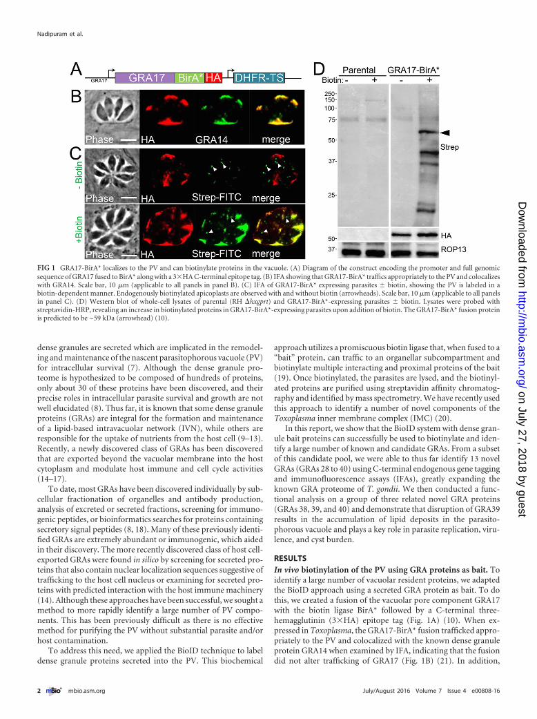
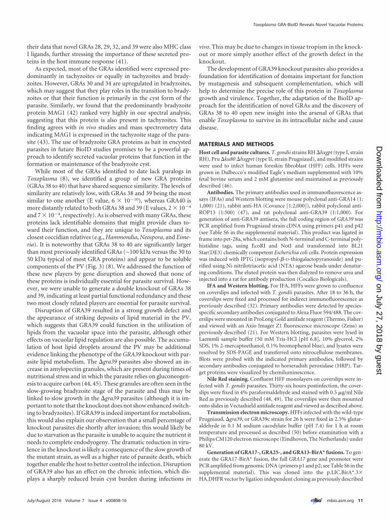
RESULTSIn vivo biotinylation of the PV using GRA proteins as bait. Toidentify a large number of vacuolar resident proteins, we adaptedthe BioID approach using a secreted GRA protein as bait. To dothis, we created a fusion of the vacuolar pore component GRA17with the biotin ligase BirA* followed by a C-terminal three-hemagglutinin (3�HA) epitope tag (Fig. 1A) (10). When ex-pressed in Toxoplasma, the GRA17-BirA* fusion trafficked appro-priately to the PV and colocalized with the known dense granuleprotein GRA14 when examined by IFA, indicating that the fusiondid not alter trafficking of GRA17 (Fig. 1B) (21). In addition,
FIG 1 GRA17-BirA* localizes to the PV and can biotinylate proteins in the vacuole. (A) Diagram of the construct encoding the promoter and full genomicsequence of GRA17 fused to BirA* along with a 3�HA C-terminal epitope tag. (B) IFA showing that GRA17-BirA* traffics appropriately to the PV and colocalizeswith GRA14. Scale bar, 10 �m (applicable to all panels in panel B). (C) IFA of GRA17-BirA* expressing parasites � biotin, showing the PV is labeled in abiotin-dependent manner. Endogenously biotinylated apicoplasts are observed with and without biotin (arrowheads). Scale bar, 10 �m (applicable to all panelsin panel C). (D) Western blot of whole-cell lysates of parental (RH �hxgprt) and GRA17-BirA*-expressing parasites � biotin. Lysates were probed withstreptavidin-HRP, revealing an increase in biotinylated proteins in GRA17-BirA*-expressing parasites upon addition of biotin. The GRA17-BirA* fusion proteinis predicted to be ~59 kDa (arrowhead) (10).
Nadipuram et al.
2 ® mbio.asm.org July/August 2016 Volume 7 Issue 4 e00808-16
on July 27, 2018 by guesthttp://m
bio.asm.org/
Dow
nloaded from
GRA17-BirA* labeled the PV only when biotin was supplemented(as assessed by streptavidin-fluorescein isothiocyanate [FITC]staining), demonstrating that the fusion is catalytically active inthis compartment (Fig. 1C). By Western blotting, we found a dra-matic increase in biotinylated proteins in GRA17-BirA* parasitelysates, confirming the ability of the GRA17-BirA* fusion to bio-tinylate a range of targets in the PV (Fig. 1D). Large-scale GRA17-BioID experiments were then performed on intracellular parasiteswith a high multiplicity of infection (~5), which were grown for~36 h to obtain large vacuoles to maximize labeling of the PV.These intracellular parasites were harvested and lysed, and biotin-ylated proteins were purified via streptavidin chromatography.
Compared to a control lysate derived from wild-type parasitessupplemented with biotin, the GRA17-BioID purified fraction re-vealed a large number of known dense granule proteins (e.g.,GRAs, MAG1, MAF1, cathepsin, and nucleoside triphosphatase II[NTPase II]) (7, 8), all of which ranked highly by normalizedspectral abundance factor (NSAF) (Table 1; see Table S1 in thesupplemental material). We therefore concluded that GRA17-BirA* appropriately labeled PV-resident proteins. In total, theGRA17-BioID experiment yielded 279 proteins (see Table S1).
While the highest-ranking proteins were mostly GRAs, the lower-ranked proteins also included other secreted proteins, such asrhoptry body proteins (ROPs) and rhoptry neck proteins (RONs),as well as some proteins from the micronemes (MICs) and para-site surface antigens (SAGs) that would be exposed to the PV.
To expand our studies beyond labeling with GRA17, we gen-erated two additional BioID constructs—a GRA25-BirA* fusionand a previously unpublished GRA13 (TgME49_237880)-BirA*fusion (see Fig. 2 below for IFA of GRA13-HA). These constructswere expressed in Toxoplasma (see Fig. S1 in the supplementalmaterial), and then the in vivo biotinylation and purification wereperformed as in the GRA17-BirA* experiment. We again found bytandem mass spectrometry (MS/MS) analysis that many of theknown components of the PV were purified and scored high byspectral count and unique peptide count (Table 2 and Table 3; seeTables S2 and S3 in the supplemental material). When we exam-ined our collective data sets for common proteins, we found sub-stantial (but not complete) overlap between these data sets (seeTable S4 in the supplemental material). Between the three GRA-BioID experiments, we identified most of the known numberedGRAs and named dense granule proteins, including TgPSD1, NT-
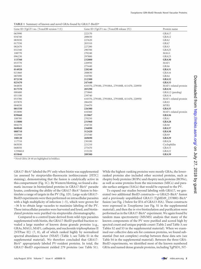
TABLE 1 Summary of known and novel GRAs found by GRA17-BioIDa
Gene ID (TgGT1 no. [ToxoDB version 7.3]) Gene ID (TgGT1 no. [ToxoDB release 25]) Protein name
065990 222170 GRA17034740 288650 GRA12083030 227620 GRA2017550 203310 GRA7082670 227280 GRA3032340 290700 GRA25108770 270240 MAG1096230 297880 GRA23115760 232000 GRA30053770 220950 MAF1068065 275440 GRA6014920 220240 GRA31021860 208830 GRA16087090 310780 GRA4072150 212300 GRA32025470 247440 GRA33064850 410370, 279100, 279100A, 279100B, 411470, 220950 MAF1-related protein017570 203290 GRA34000480 275860 GRA12 (paralog)051100 239740 GRA14053780 410370, 279100, 279100A, 279100B, 411470, 220950 MAF1-related protein037870 286450 GRA5004030 254470 MYR1081550 226380 GRA35053890 410360 MAF1-related protein039660 213067 GRA36108780 270250 GRA1115800 231960 GRA28004270 254720 GRA8069040 236890 GRA37088710 312420 GRA38102940 251540 GRA9108120 269690 GRA29117710 230180 GRA24065030 221210 Cyclophilin068050 275470 GRA15125960.0 215220 GRA22033840 289380 GRA39a Novel GRAs 28-40 are highlighted in boldface.
Toxoplasma GRA-BioID Reveals Novel Vacuolar Proteins
July/August 2016 Volume 7 Issue 4 e00808-16 ® mbio.asm.org 3
on July 27, 2018 by guesthttp://m
bio.asm.org/
Dow
nloaded from
Pase II, PI-I, cyclophilin, several MAF1 proteins, and MYR1 (Ta-bles 1, 2, and 3; see Tables S1 to S4). We were not able to detectGRAs 10, 19, 20, and 21 nor the recently described TgLCAT (22).
We also analyzed the data from the GRA-BioID data sets forpotential interacting or proximal host proteins. In each of theexperiments, a relatively small number of host proteins were iden-tified (21 for GRA17, 43 for GRA25, and 93 for GRA13). Thiscould be due to the relative abundance of parasite versus hosttargets within the vacuole or the fact that few proteins are actuallyin close contact with the BirA* fusions. Only a single host protein,programmed cell death 6-interacting protein, was in common be-tween the all three GRA-BioID experiments. Other common hu-man hits between pairs of BioID experiments are shown in Ta-ble S5 in the supplemental material.
Identification of novel dense granule proteins from BioIDdata sets. Analysis of the GRA-BioID data sets also yielded a largenumber of hypothetical proteins (see Tables S1, S2, and S3 in thesupplemental material), which were filtered for likely GRAs byselecting proteins that contained a predicted signal peptide, wereconstitutively expressed, and lacked a C-terminal endoplasmic re-ticulum (ER) retention signal (K/HDEL) (7, 8). As most GRAsidentified to date also lack similarity to known proteins, we alsoselected candidates that lacked obvious functional domains forverification. This resulted in a list of �100 candidates, of which wechose 15 for localization studies using endogenous gene tagging(see Tables S1, S2, and S3).
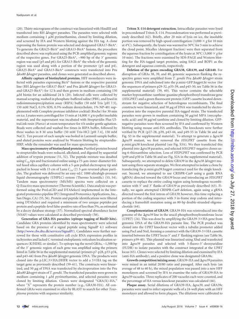
To assess localization of these candidate proteins, we engi-neered constructs that would recombine a sequence encoding a
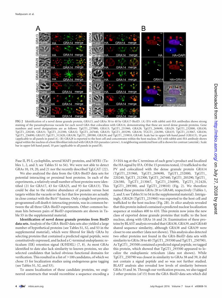
3�HA tag at the C terminus of each gene’s product and localizedthe HA signal by IFA. Of the 15 proteins tested, 13 trafficked to thePV and colocalized with the dense granule protein GRA14(TgGT1_231960, TgGT1_269690, TgGT1_232000, TgGT1_220240, TgGT1_212300, TgGT1_247440, TgGT1_203290, TgGT1_226380, TgGT1_213067, TgGT1_236890, TgGT1_312420,TgGT1_289380, and TgGT1_219810) (Fig. 2). We thereforenamed these proteins GRAs 28 to GRA40, respectively (Tables 1,2, and 3; see Tables S1 to S4 in the supplemental material). Intrigu-ingly, GRA28 (TgGT1_231960) was exported to the host cell andtrafficked to the host nucleus (Fig. 2B). In silico analysis revealedthat this protein indeed contained a predicted nuclear localizationsequence at residues 400 to 410. This protein now joins the newclass of exported dense granule proteins that traffic to the hostnucleus, along with GRAs 16 and 24. Examination of these pro-teins by BLAST analysis revealed that GRA38, GRA39, and GRA40shared sequence similarity, although GRA38 and GRA39 werecloser to one another (data not shown). This analysis also detectedtwo other proteins not found in the GRA-BioID data sets withsimilarity to GRAs 38 to 40 (TgGT1_293500 and TgGT1_250790).As TgGT1_293500 contained a predicted signal peptide, we taggedthis protein, which showed that TgGT1_293500 appeared to lo-calize the endoplasmic reticulum (data not shown). WhileTgGT1_250790 was closest in similarity to GRAs 38 and 39, it didnot contain a signal peptide and so was not further studied.BLAST analysis also revealed low levels of similarity betweenGRAs 35 and 36. Through our verification process, we also tagged2 other proteins (of 15) from the GRA-BioID data sets which did
FIG 2 Identification of a novel dense granule protein, GRA13, and GRAs 30 to 40 by GRA17-BioID. (A) IFA with rabbit anti-HA antibodies shows strongstaining of the parasitophorous vacuole for each novel GRA that colocalizes with GRA14, demonstrating that these are novel dense granule proteins. Genenumbers and novel designations are as follows: TgGT1_237880, GRA13; TgGT1_231960, GRA28; TgGT1_269690, GRA29; TgGT1_232000, GRA30;TgGT1_220240, GRA31; TgGT1_212300, GRA32; TgGT1_247440, GRA33; TgGT1_203290, GRA34; TGGT1_226380, GRA35; TgGT1_213067, GRA36;TgGT1_236890, GRA37; TgGT1_312420, GRA38; TgGT1_289380, GRA39; and TgGT1_219810, GRA40. Scale bar in upper left-hand panel (GRA13), 10 �m(applicable to all panels in panel A). (B) GRA28 is exported to the host cell and concentrates within the host nucleus. IFA with rabbit anti-HA antibody showssignal within the nucleus of a host fibroblast infected with GRA28-HA parasites (arrow). A neighboring uninfected host cell is shown for contrast (asterisk). Scalebar in upper left hand panel, 10 �m (applicable to all panels in panel B).
Nadipuram et al.
4 ® mbio.asm.org July/August 2016 Volume 7 Issue 4 e00808-16
on July 27, 2018 by guesthttp://m
bio.asm.org/
Dow
nloaded from
not localize to the PV: TgGT1_209720 localized to the mitochon-drion, while TgGT1_304990 localized to the Golgi apparatus (seeTable S1) (data not shown). The remaining candidate proteinsthat match our criteria for potential GRAs have yet to be localized.However, the high frequency of successful GRA proteins in ourverification analysis suggests that many of the remaining candi-dates are likely to be novel GRAs.
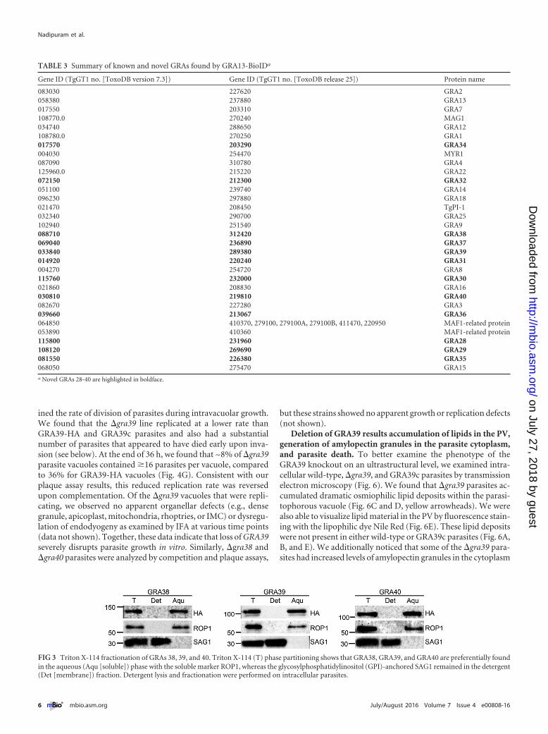
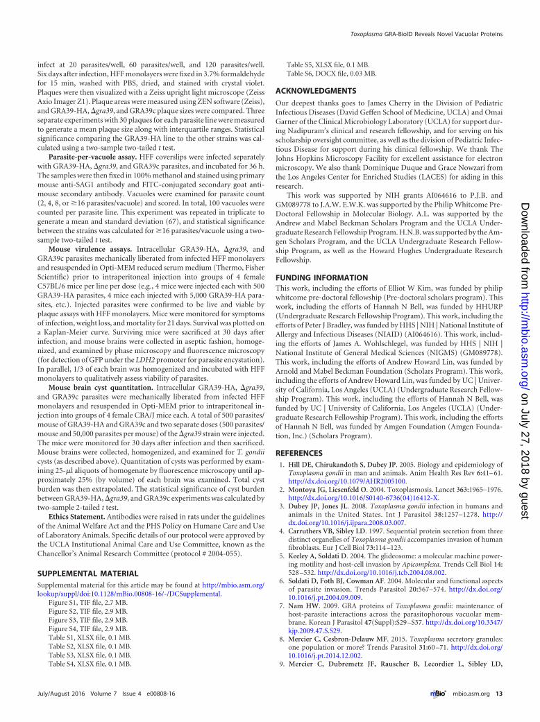
GRAs 38, 39, and 40 are not tightly associated with PV mem-branes. We chose to analyze the group of GRAs 38, 39, and 40, asGRA38 was previously found to be antigenic in a human serolog-ical screen that was performed in collaboration with our lab (23).Most of the previously described GRA proteins are associated withthe parasitophorous vacuolar membrane or the intravacuolar net-work in Toxoplasma infections. GRAs 38, 39, and 40 lack predictedtransmembrane domains or other readily identifiable sequencesfor membrane association, suggesting they are soluble compo-nents of the vacuole. To assess this experimentally, we performedTriton X-114 partitioning of intracellular parasites and found thateach of these family members fractionated in the aqueous fraction(as opposed to the detergent/membrane fraction), demonstratingthat they are not firmly associated with the IVN or PV membrane(PVM) (Fig. 3). While we cannot exclude the possibility of weakassociations with vacuolar membranes, these data indicate thatthis new family of GRA proteins are soluble proteins of the PV.
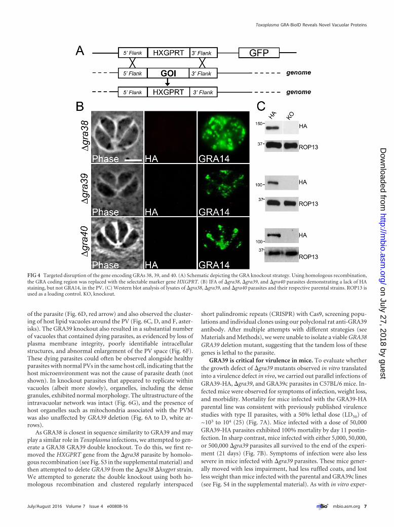
Targeted disruption of GRAs 38, 39, and 40. To understandthe function of the newly characterized GRAs, we disruptedGRA38, GRA39, and GRA40 by homologous recombination in
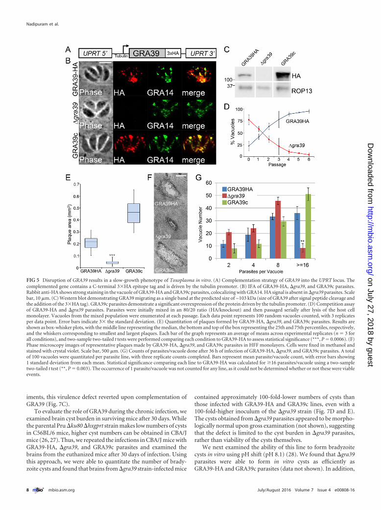
their respective 3�HA-tagged parental strains (Fig. 4A). Knock-out clones were identified that lacked the 3�HA tag by IFA(Fig. 4B) and Western blotting (Fig. 4C) and were verified by PCR(see Fig. S2 in the supplemental material). Intriguingly, �gra39clones were initially difficult to isolate from a transfected popula-tion as they were quickly outcompeted by nonhomologous re-combinants, suggesting these parasites possessed a growth pheno-type in vitro. However, we were able to obtain a knockout clonethat we subsequently complemented with a construct containingHA-tagged GRA39 driven from the tubulin promoter (Fig. 5A).This transgene was targeted to the uracil phosphoribosyltrans-ferase locus (UPRT) (24), and its expression was confirmed by IFAand Western blotting using both anti-HA antibodies and anti-GRA39 polyclonal antibodies. (The complemented strain wasdesignated GRA39c [Fig. 5B and C; see Fig. S3 in the supplementalmaterial].)
GRA39 is critical for efficient growth in vitro in type IIT. gondii. Next, we analyzed the overall fitness of our GRA knock-out parasites using competition growth assays and plaque assays.Competition growth assays showed that �gra39 parasites wererapidly outcompeted by their GRA39-HA parental line after 7serial passages (Fig. 5D). Plaque assays similarly revealed that�gra39 mutants produced significantly smaller plaques (~80%reduction in size) than the GRA39-HA line in human foreskinfibroblast (HFF) monolayers (Fig. 5E and F). This poor-growthphenotype in knockout parasites was reversed upon complemen-tation of GRA39. To investigate this phenotype further, we exam-
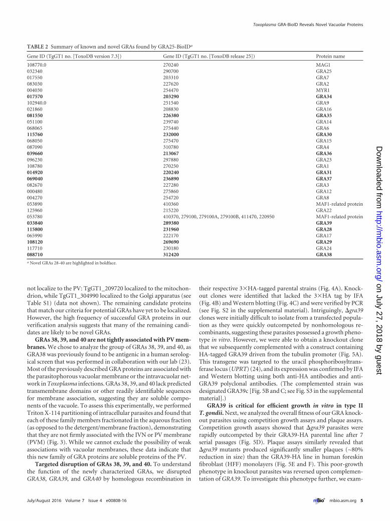
TABLE 2 Summary of known and novel GRAs found by GRA25-BioIDa
Gene ID (TgGT1 no. [ToxoDB version 7.3]) Gene ID (TgGT1 no. [ToxoDB release 25]) Protein name
108770.0 270240 MAG1032340 290700 GRA25017550 203310 GRA7083030 227620 GRA2004030 254470 MYR1017570 203290 GRA34102940.0 251540 GRA9021860 208830 GRA16081550 226380 GRA35051100 239740 GRA14068065 275440 GRA6115760 232000 GRA30068050 275470 GRA15087090 310780 GRA4039660 213067 GRA36096230 297880 GRA23108780 270250 GRA1014920 220240 GRA31069040 236890 GRA37082670 227280 GRA3000480 275860 GRA12004270 254720 GRA8053890 410360 MAF1-related protein125960 215220 GRA22053780 410370, 279100, 279100A, 279100B, 411470, 220950 MAF1-related protein033840 289380 GRA39115800 231960 GRA28065990 222170 GRA17108120 269690 GRA29117710 230180 GRA24088710 312420 GRA38a Novel GRAs 28-40 are highlighted in boldface.
Toxoplasma GRA-BioID Reveals Novel Vacuolar Proteins
July/August 2016 Volume 7 Issue 4 e00808-16 ® mbio.asm.org 5
on July 27, 2018 by guesthttp://m
bio.asm.org/
Dow
nloaded from
ined the rate of division of parasites during intravacuolar growth.We found that the �gra39 line replicated at a lower rate thanGRA39-HA and GRA39c parasites and also had a substantialnumber of parasites that appeared to have died early upon inva-sion (see below). At the end of 36 h, we found that ~8% of �gra39parasite vacuoles contained �16 parasites per vacuole, comparedto 36% for GRA39-HA vacuoles (Fig. 4G). Consistent with ourplaque assay results, this reduced replication rate was reversedupon complementation. Of the �gra39 vacuoles that were repli-cating, we observed no apparent organellar defects (e.g., densegranule, apicoplast, mitochondria, rhoptries, or IMC) or dysregu-lation of endodyogeny as examined by IFA at various time points(data not shown). Together, these data indicate that loss of GRA39severely disrupts parasite growth in vitro. Similarly, �gra38 and�gra40 parasites were analyzed by competition and plaque assays,
but these strains showed no apparent growth or replication defects(not shown).
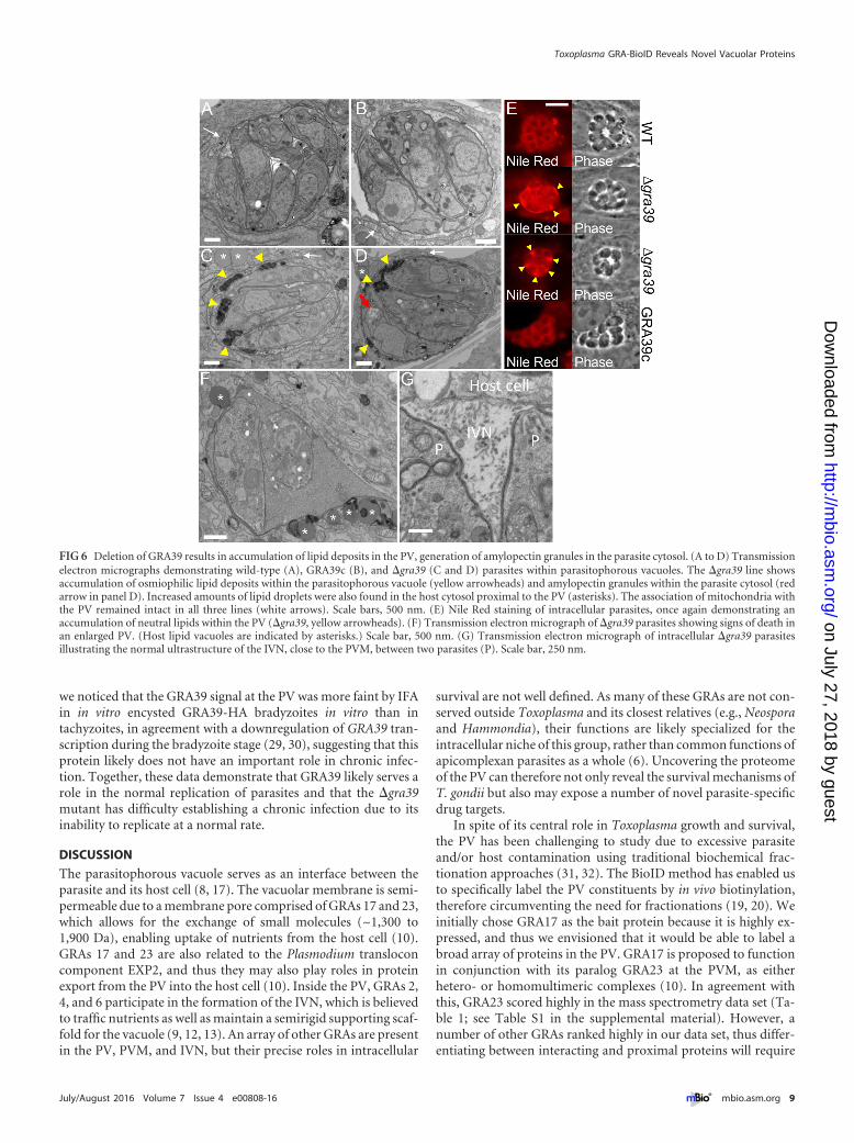
Deletion of GRA39 results accumulation of lipids in the PV,generation of amylopectin granules in the parasite cytoplasm,and parasite death. To better examine the phenotype of theGRA39 knockout on an ultrastructural level, we examined intra-cellular wild-type, �gra39, and GRA39c parasites by transmissionelectron microscopy (Fig. 6). We found that �gra39 parasites ac-cumulated dramatic osmiophilic lipid deposits within the parasi-tophorous vacuole (Fig. 6C and D, yellow arrowheads). We werealso able to visualize lipid material in the PV by fluorescence stain-ing with the lipophilic dye Nile Red (Fig. 6E). These lipid depositswere not present in either wild-type or GRA39c parasites (Fig. 6A,B, and E). We additionally noticed that some of the �gra39 para-sites had increased levels of amylopectin granules in the cytoplasm
TABLE 3 Summary of known and novel GRAs found by GRA13-BioIDa
Gene ID (TgGT1 no. [ToxoDB version 7.3]) Gene ID (TgGT1 no. [ToxoDB release 25]) Protein name
083030 227620 GRA2058380 237880 GRA13017550 203310 GRA7108770.0 270240 MAG1034740 288650 GRA12108780.0 270250 GRA1017570 203290 GRA34004030 254470 MYR1087090 310780 GRA4125960.0 215220 GRA22072150 212300 GRA32051100 239740 GRA14096230 297880 GRA18021470 208450 TgPI-1032340 290700 GRA25102940 251540 GRA9088710 312420 GRA38069040 236890 GRA37033840 289380 GRA39014920 220240 GRA31004270 254720 GRA8115760 232000 GRA30021860 208830 GRA16030810 219810 GRA40082670 227280 GRA3039660 213067 GRA36064850 410370, 279100, 279100A, 279100B, 411470, 220950 MAF1-related protein053890 410360 MAF1-related protein115800 231960 GRA28108120 269690 GRA29081550 226380 GRA35068050 275470 GRA15a Novel GRAs 28-40 are highlighted in boldface.
FIG 3 Triton X-114 fractionation of GRAs 38, 39, and 40. Triton X-114 (T) phase partitioning shows that GRA38, GRA39, and GRA40 are preferentially foundin the aqueous (Aqu [soluble]) phase with the soluble marker ROP1, whereas the glycosylphosphatidylinositol (GPI)-anchored SAG1 remained in the detergent(Det [membrane]) fraction. Detergent lysis and fractionation were performed on intracellular parasites.
Nadipuram et al.
6 ® mbio.asm.org July/August 2016 Volume 7 Issue 4 e00808-16
on July 27, 2018 by guesthttp://m
bio.asm.org/
Dow
nloaded from
of the parasite (Fig. 6D, red arrow) and also observed the cluster-ing of host lipid vacuoles around the PV (Fig. 6C, D, and F, aster-isks). The GRA39 knockout also resulted in a substantial numberof vacuoles that contained dying parasites, as evidenced by loss ofplasma membrane integrity, poorly identifiable intracellularstructures, and abnormal enlargement of the PV space (Fig. 6F).These dying parasites could often be observed alongside healthyparasites with normal PVs in the same host cell, indicating that thehost microenvironment was not the cause of parasite death (notshown). In knockout parasites that appeared to replicate withinvacuoles (albeit more slowly), organelles, including the densegranules, exhibited normal morphology. The ultrastructure of theintravacuolar network was intact (Fig. 6G), and the presence ofhost organelles such as mitochondria associated with the PVMwas also unaffected by GRA39 deletion (Fig. 6A to D, white ar-rows).
As GRA38 is closest in sequence similarity to GRA39 and mayplay a similar role in Toxoplasma infections, we attempted to gen-erate a GRA38 GRA39 double knockout. To do this, we first re-moved the HXGPRT gene from the �gra38 parasite by homolo-gous recombination (see Fig. S3 in the supplemental material) andthen attempted to delete GRA39 from the �gra38 �hxgprt strain.We attempted to generate the double knockout using both ho-mologous recombination and clustered regularly interspaced
short palindromic repeats (CRISPR) with Cas9, screening popu-lations and individual clones using our polyclonal rat anti-GRA39antibody. After multiple attempts with different strategies (seeMaterials and Methods), we were unable to isolate a viable GRA38GRA39 deletion mutant, suggesting that the tandem loss of thesegenes is lethal to the parasite.
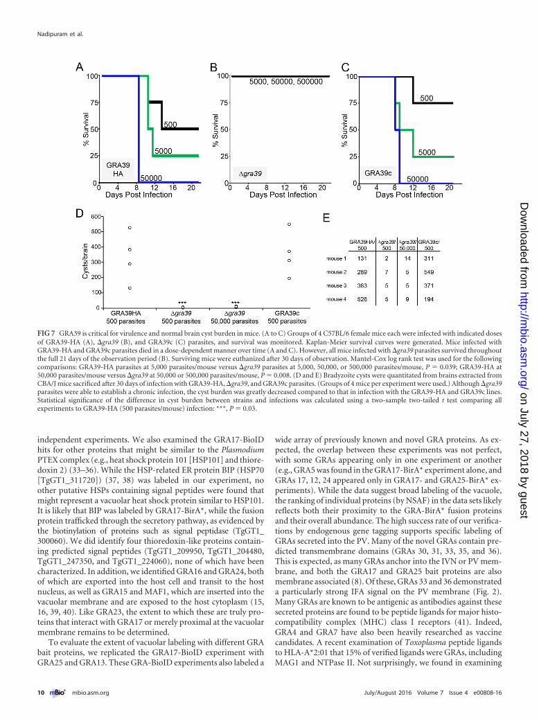
GRA39 is critical for virulence in mice. To evaluate whetherthe growth defect of �gra39 mutants observed in vitro translatedinto a virulence defect in vivo, we carried out parallel infections ofGRA39-HA, �gra39, and GRA39c parasites in C57BL/6 mice. In-fected mice were observed for symptoms of infection, weight loss,and morbidity. Mortality for mice infected with the GRA39-HAparental line was consistent with previously published virulencestudies with type II parasites, with a 50% lethal dose (LD50) of~103 to 104 (25) (Fig. 7A). Mice infected with a dose of 50,000GRA39-HA parasites exhibited 100% mortality by day 11 postin-fection. In sharp contrast, mice infected with either 5,000, 50,000,or 500,000 �gra39 parasites all survived to the end of the experi-ment (21 days) (Fig. 7B). Symptoms of infection were also lesssevere in mice infected with �gra39 parasites. These mice gener-ally moved with less impairment, had less ruffled coats, and lostless weight than mice infected with the parental and GRA39c lines(see Fig. S4 in the supplemental material). As with in vitro exper-
FIG 4 Targeted disruption of the gene encoding GRAs 38, 39, and 40. (A) Schematic depicting the GRA knockout strategy. Using homologous recombination,the GRA coding region was replaced with the selectable marker gene HXGPRT. (B) IFA of �gra38, �gra39, and �gra40 parasites demonstrating a lack of HAstaining, but not GRA14, in the PV. (C) Western blot analysis of lysates of �gra38, �gra39, and �gra40 parasites and their respective parental strains. ROP13 isused as a loading control. KO, knockout.
Toxoplasma GRA-BioID Reveals Novel Vacuolar Proteins
July/August 2016 Volume 7 Issue 4 e00808-16 ® mbio.asm.org 7
on July 27, 2018 by guesthttp://m
bio.asm.org/
Dow
nloaded from
iments, this virulence defect reverted upon complementation ofGRA39 (Fig. 7C).
To evaluate the role of GRA39 during the chronic infection, weexamined brain cyst burden in surviving mice after 30 days. Whilethe parental Pru �ku80 �hxgprt strain makes low numbers of cystsin C56BL/6 mice, higher cyst numbers can be obtained in CBA/Jmice (26, 27). Thus, we repeated the infections in CBA/J mice withGRA39-HA, �gra39, and GRA39c parasites and examined thebrains from the euthanized mice after 30 days of infection. Usingthis approach, we were able to quantitate the number of brady-zoite cysts and found that brains from �gra39 strain-infected mice
contained approximately 100-fold-lower numbers of cysts thanthose infected with GRA39-HA and GRA39c lines, even with a100-fold-higher inoculum of the �gra39 strain (Fig. 7D and E).The cysts obtained from �gra39 parasites appeared to be morpho-logically normal upon gross examination (not shown), suggestingthat the defect is limited to the cyst burden in �gra39 parasites,rather than viability of the cysts themselves.
We next examined the ability of this line to form bradyzoitecysts in vitro using pH shift (pH 8.1) (28). We found that �gra39parasites were able to form in vitro cysts as efficiently asGRA39-HA and GRA39c parasites (data not shown). In addition,
FIG 5 Disruption of GRA39 results in a slow-growth phenotype of Toxoplasma in vitro. (A) Complementation strategy of GRA39 into the UPRT locus. Thecomplemented gene contains a C-terminal 3�HA epitope tag and is driven by the tubulin promoter. (B) IFA of GRA39-HA, �gra39, and GRA39c parasites.Rabbit anti-HA shows strong staining in the vacuole of GRA39-HA and GRA39c parasites, colocalizing with GRA14. HA signal is absent in �gra39 parasites. Scalebar, 10 �m. (C) Western blot demonstrating GRA39 migrating as a single band at the predicted size of ~103 kDa (size of GRA39 after signal peptide cleavage andthe addition of the 3�HA tag). GRA39c parasites demonstrate a significant overexpression of the protein driven by the tubulin promoter. (D) Competition assayof GRA39-HA and �gra39 parasites. Parasites were initially mixed in an 80/20 ratio (HA/knockout) and then passaged serially after lysis of the host cellmonolayer. Vacuoles from the mixed population were enumerated at each passage. Each data point represents 100 random vacuoles counted, with 3 replicatesper data point. Error bars indicate 3� the standard deviation. (E) Quantitation of plaques formed by GRA39-HA, �gra39, and GRA39c parasites. Results areshown as box-whisker plots, with the middle line representing the median, the bottom and top of the box representing the 25th and 75th percentiles, respectively,and the whiskers corresponding to smallest and largest plaques. Each bar of the graph represents an average of means across experimental replicates (n � 3 forall conditions), and two-sample two-tailed t tests were performed comparing each condition to GRA39-HA to assess statistical significance (***, P � 0.0006). (F)Phase microscopy images of representative plaques made by GRA39-HA, �gra39, and GRA39c parasites in HFF monolayers. Cells were fixed in methanol andstained with crystal violet. Scale bar, 500 �m. (G) Counts of parasites/vacuole done after 36 h of infection of GRA39-HA, �gra39, and GRA39c parasites. A totalof 100 vacuoles were quantitated per parasite line, with three replicate counts completed. Bars represent mean parasite/vacuole count, with error bars showing1 standard deviation from each mean. Statistical significance comparing each line to GRA39-HA was calculated for �16 parasites/vacuole using a two-sampletwo-tailed t test (**, P � 0.003). The occurrence of 1 parasite/vacuole was not counted for any line, as it could not be determined whether or not these were viableevents.
Nadipuram et al.
8 ® mbio.asm.org July/August 2016 Volume 7 Issue 4 e00808-16
on July 27, 2018 by guesthttp://m
bio.asm.org/
Dow
nloaded from
we noticed that the GRA39 signal at the PV was more faint by IFAin in vitro encysted GRA39-HA bradyzoites in vitro than intachyzoites, in agreement with a downregulation of GRA39 tran-scription during the bradyzoite stage (29, 30), suggesting that thisprotein likely does not have an important role in chronic infec-tion. Together, these data demonstrate that GRA39 likely serves arole in the normal replication of parasites and that the �gra39mutant has difficulty establishing a chronic infection due to itsinability to replicate at a normal rate.
DISCUSSION
The parasitophorous vacuole serves as an interface between theparasite and its host cell (8, 17). The vacuolar membrane is semi-permeable due to a membrane pore comprised of GRAs 17 and 23,which allows for the exchange of small molecules (~1,300 to1,900 Da), enabling uptake of nutrients from the host cell (10).GRAs 17 and 23 are also related to the Plasmodium transloconcomponent EXP2, and thus they may also play roles in proteinexport from the PV into the host cell (10). Inside the PV, GRAs 2,4, and 6 participate in the formation of the IVN, which is believedto traffic nutrients as well as maintain a semirigid supporting scaf-fold for the vacuole (9, 12, 13). An array of other GRAs are presentin the PV, PVM, and IVN, but their precise roles in intracellular
survival are not well defined. As many of these GRAs are not con-served outside Toxoplasma and its closest relatives (e.g., Neosporaand Hammondia), their functions are likely specialized for theintracellular niche of this group, rather than common functions ofapicomplexan parasites as a whole (6). Uncovering the proteomeof the PV can therefore not only reveal the survival mechanisms ofT. gondii but also may expose a number of novel parasite-specificdrug targets.
In spite of its central role in Toxoplasma growth and survival,the PV has been challenging to study due to excessive parasiteand/or host contamination using traditional biochemical frac-tionation approaches (31, 32). The BioID method has enabled usto specifically label the PV constituents by in vivo biotinylation,therefore circumventing the need for fractionations (19, 20). Weinitially chose GRA17 as the bait protein because it is highly ex-pressed, and thus we envisioned that it would be able to label abroad array of proteins in the PV. GRA17 is proposed to functionin conjunction with its paralog GRA23 at the PVM, as eitherhetero- or homomultimeric complexes (10). In agreement withthis, GRA23 scored highly in the mass spectrometry data set (Ta-ble 1; see Table S1 in the supplemental material). However, anumber of other GRAs ranked highly in our data set, thus differ-entiating between interacting and proximal proteins will require
FIG 6 Deletion of GRA39 results in accumulation of lipid deposits in the PV, generation of amylopectin granules in the parasite cytosol. (A to D) Transmissionelectron micrographs demonstrating wild-type (A), GRA39c (B), and �gra39 (C and D) parasites within parasitophorous vacuoles. The �gra39 line showsaccumulation of osmiophilic lipid deposits within the parasitophorous vacuole (yellow arrowheads) and amylopectin granules within the parasite cytosol (redarrow in panel D). Increased amounts of lipid droplets were also found in the host cytosol proximal to the PV (asterisks). The association of mitochondria withthe PV remained intact in all three lines (white arrows). Scale bars, 500 nm. (E) Nile Red staining of intracellular parasites, once again demonstrating anaccumulation of neutral lipids within the PV (�gra39, yellow arrowheads). (F) Transmission electron micrograph of �gra39 parasites showing signs of death inan enlarged PV. (Host lipid vacuoles are indicated by asterisks.) Scale bar, 500 nm. (G) Transmission electron micrograph of intracellular �gra39 parasitesillustrating the normal ultrastructure of the IVN, close to the PVM, between two parasites (P). Scale bar, 250 nm.
Toxoplasma GRA-BioID Reveals Novel Vacuolar Proteins
July/August 2016 Volume 7 Issue 4 e00808-16 ® mbio.asm.org 9
on July 27, 2018 by guesthttp://m
bio.asm.org/
Dow
nloaded from
independent experiments. We also examined the GRA17-BioIDhits for other proteins that might be similar to the PlasmodiumPTEX complex (e.g., heat shock protein 101 [HSP101] and thiore-doxin 2) (33–36). While the HSP-related ER protein BIP (HSP70[TgGT1_311720]) (37, 38) was labeled in our experiment, noother putative HSPs containing signal peptides were found thatmight represent a vacuolar heat shock protein similar to HSP101.It is likely that BIP was labeled by GRA17-BirA*, while the fusionprotein trafficked through the secretory pathway, as evidenced bythe biotinylation of proteins such as signal peptidase (TgGT1_300060). We did identify four thioredoxin-like proteins contain-ing predicted signal peptides (TgGT1_209950, TgGT1_204480,TgGT1_247350, and TgGT1_224060), none of which have beencharacterized. In addition, we identified GRA16 and GRA24, bothof which are exported into the host cell and transit to the hostnucleus, as well as GRA15 and MAF1, which are inserted into thevacuolar membrane and are exposed to the host cytoplasm (15,16, 39, 40). Like GRA23, the extent to which these are truly pro-teins that interact with GRA17 or merely proximal at the vacuolarmembrane remains to be determined.
To evaluate the extent of vacuolar labeling with different GRAbait proteins, we replicated the GRA17-BioID experiment withGRA25 and GRA13. These GRA-BioID experiments also labeled a
wide array of previously known and novel GRA proteins. As ex-pected, the overlap between these experiments was not perfect,with some GRAs appearing only in one experiment or another(e.g., GRA5 was found in the GRA17-BirA* experiment alone, andGRAs 17, 12, 24 appeared only in GRA17- and GRA25-BirA* ex-periments). While the data suggest broad labeling of the vacuole,the ranking of individual proteins (by NSAF) in the data sets likelyreflects both their proximity to the GRA-BirA* fusion proteinsand their overall abundance. The high success rate of our verifica-tions by endogenous gene tagging supports specific labeling ofGRAs secreted into the PV. Many of the novel GRAs contain pre-dicted transmembrane domains (GRAs 30, 31, 33, 35, and 36).This is expected, as many GRAs anchor into the IVN or PV mem-brane, and both the GRA17 and GRA25 bait proteins are alsomembrane associated (8). Of these, GRAs 33 and 36 demonstrateda particularly strong IFA signal on the PV membrane (Fig. 2).Many GRAs are known to be antigenic as antibodies against thesesecreted proteins are found to be peptide ligands for major histo-compatibility complex (MHC) class I receptors (41). Indeed,GRA4 and GRA7 have also been heavily researched as vaccinecandidates. A recent examination of Toxoplasma peptide ligandsto HLA-A*2:01 that 15% of verified ligands were GRAs, includingMAG1 and NTPase II. Not surprisingly, we found in examining
FIG 7 GRA39 is critical for virulence and normal brain cyst burden in mice. (A to C) Groups of 4 C57BL/6 female mice each were infected with indicated dosesof GRA39-HA (A), �gra39 (B), and GRA39c (C) parasites, and survival was monitored. Kaplan-Meier survival curves were generated. Mice infected withGRA39-HA and GRA39c parasites died in a dose-dependent manner over time (A and C). However, all mice infected with �gra39 parasites survived throughoutthe full 21 days of the observation period (B). Surviving mice were euthanized after 30 days of observation. Mantel-Cox log rank test was used for the followingcomparisons: GRA39-HA parasites at 5,000 parasites/mouse versus �gra39 parasites at 5,000, 50,000, or 500,000 parasites/mouse, P � 0.039; GRA39-HA at50,000 parasites/mouse versus �gra39 at 50,000 or 500,000 parasites/mouse, P � 0.008. (D and E) Bradyzoite cysts were quantitated from brains extracted fromCBA/J mice sacrificed after 30 days of infection with GRA39-HA, �gra39, and GRA39c parasites. (Groups of 4 mice per experiment were used.) Although �gra39parasites were able to establish a chronic infection, the cyst burden was greatly decreased compared to that in infection with the GRA39-HA and GRA39c lines.Statistical significance of the difference in cyst burden between strains and infections was calculated using a two-sample two-tailed t test comparing allexperiments to GRA39-HA (500 parasites/mouse) infection: ***, P � 0.03.
Nadipuram et al.
10 ® mbio.asm.org July/August 2016 Volume 7 Issue 4 e00808-16
on July 27, 2018 by guesthttp://m
bio.asm.org/
Dow
nloaded from
their data that novel GRAs 28, 29, 32, and 39 were also MHC classI ligands, further stressing the importance of these secreted pro-teins in the host immune response (41).
As expected, most of the GRAs identified were expressed pre-dominantly in tachyzoites or equally in tachyzoites and brady-zoites. However, GRAs 30 and 34 are upregulated in bradyzoites,which may suggest that they play roles in the transition to brady-zoites or that their function is primarily in the cyst form of theparasite. Similarly, we found that the predominantly bradyzoiteprotein MAG1 (42) ranked very highly in our spectral analysis,suggesting that this protein is also present in tachyzoites. Thisfinding agrees with in vivo studies and mass spectrometry dataindicating MAG1 is expressed in the tachyzoite stage of the para-site (43). The use of bradyzoite GRA proteins as bait in encystedparasites in future BioID studies promises to be a powerful ap-proach to identify secreted vacuolar proteins that function in theformation or maintenance of the bradyzoite cyst.
While most of the GRAs identified to date lack paralogs inToxoplasma (8), we identified a group of new GRA proteins(GRAs 38 to 40) that have shared sequence similarity. The levels ofsimilarity are relatively low, with GRAs 38 and 39 being the mostsimilar to one another (E value, 6 � 10�10), whereas GRA40 ismore distantly related to both GRAs 38 and 39 (E values, 2 � 10�4
and 7 � 10�4, respectively). As is observed with many GRAs, theseproteins lack identifiable domains that might provide clues to-ward their function, and they are unique to Toxoplasma and itsclosest coccidian relatives (e.g., Hammondia, Neospora, and Eime-ria). It is noteworthy that GRAs 38 to 40 are significantly largerthan most previously identified GRAs (~100 kDa versus the 30 to50 kDa typical of most GRA proteins) and appear to be solublecomponents of the PV (Fig. 3) (8). We addressed the function ofthese new players by gene disruption and showed that none ofthese proteins is individually essential for parasite survival. How-ever, we were unable to generate a double knockout of GRAs 38and 39, indicating at least partial functional redundancy and thesetwo most closely related players are essential for parasite survival.
Disruption of GRA39 resulted in a strong growth defect andthe appearance of striking deposits of lipid material in the PV,which suggests that GRA39 could function in the utilization oflipids from the vacuolar space into the parasite, although othereffects on vacuolar lipid regulation are also possible. The accumu-lation of host lipid droplets around the PV may be additionalevidence linking the phenotype of the GRA39 knockout with par-asite lipid metabolism. The �gra39 parasites also showed an in-crease in amylopectin granules, which are present during times ofnutritional stress and in which the parasite relies on gluconeogen-esis to acquire carbon (44, 45). These granules are often seen in theslow-growing bradyzoite stage of the parasite and thus may belinked to slow growth in the �gra39 parasites (although it is im-portant to note that the knockout does not show enhanced switch-ing to bradyzoites). If GRA39 is indeed important for metabolism,this would also explain our observation that a small percentage ofknockout parasites die shortly after invasion; this would likely bedue to starvation as the parasite is unable to acquire the nutrient itneeds to complete endodyogeny. The dramatic reduction in viru-lence in the knockout is likely a consequence of the slow growth ofthe mutant strain, as well as a higher rate of parasite death, whichtogether enable the host to better control the infection. Disruptionof GRA39 also has an effect on the chronic infection, which dis-plays a sharply reduced brain cyst burden during infections in
vivo. This may be due to changes in tissue tropism in the knock-out or more simply another effect of the growth defect in theknockout.
The development of GRA39 knockout parasites also provides afoundation for identification of domains important for functionby mutagenesis and subsequent complementation, which willhelp to determine the precise role of this protein in Toxoplasmagrowth and virulence. Together, the adaptation of the BioID ap-proach for the identification of novel GRAs and the discovery ofGRAs 38 to 40 open new insight into the arsenal of GRAs thatenable Toxoplasma to survive in its intracellular niche and causedisease.
MATERIALS AND METHODSHost cell and parasite cultures. T. gondii strains RH �hxgpt (type I, strainRH), Pru �ku80 �hxgprt (type II, strain Prugniaud), and modified strainswere used to infect human foreskin fibroblast (HFF) cells. HFFs weregrown in Dulbecco’s modified Eagle’s medium supplemented with 10%fetal bovine serum and 2 mM glutamine and maintained as previouslydescribed (46).
Antibodies. The primary antibodies used in immunofluorescence as-says (IFAs) and Western blotting were mouse polyclonal anti-GRA14 (1:1,000) (21), rabbit anti-HA (Covance [1:2,000]), rabbit polyclonal anti-ROP13 (1:500) (47), and rat polyclonal anti-GRA39 (1:1,000). Forgeneration of anti-GRA39 antisera, the full coding region of GRA39 wasPCR amplified from Prugniaud strain cDNA using primers p41 and p42(see Table S6 in the supplemental material). This product was ligated inframe into pet-28a, which contains both N-terminal and C-terminal poly-histidine tags, using EcoRI and NotI and transformed into BL21Star(DE3) chemically competent Escherichia coli cells. Protein expressionwas induced with IPTG (isopropyl-�-D-thiogalactopyranoside) and pu-rified using Ni-nitrilotriacetic acid (NTA) agarose beads under denatur-ing conditions. The eluted protein was then dialyzed to remove urea andinjected into a rat for antibody production (Cocalico Biologicals).
IFA and Western blotting. For IFA, HFFs were grown to confluenceon coverslips and infected with T. gondii parasites. After 18 to 36 h, thecoverslips were fixed and processed for indirect immunofluorescence aspreviously described (32). Primary antibodies were detected by species-specific secondary antibodies conjugated to Alexa Fluor 594/488. The cov-erslips were mounted in ProLong Gold antifade reagent (Thermo, Fisher)and viewed with an Axio Imager Z1 fluorescence microscope (Zeiss) aspreviously described (21). For Western blotting, parasites were lysed inLaemmli sample buffer (50 mM Tris-HCl [pH 6.8], 10% glycerol, 2%SDS, 1% 2-mercaptoethanol, 0.1% bromophenol blue), and lysates wereresolved by SDS-PAGE and transferred onto nitrocellulose membranes.Blots were probed with the indicated primary antibodies, followed bysecondary antibodies conjugated to horseradish peroxidase (HRP). Tar-get proteins were visualized by chemiluminescence.
Nile Red staining. Confluent HFF monolayers on coverslips were in-fected with T. gondii parasites. Thirty-six hours postinfection, the cover-slips were fixed in 4% paraformaldehyde and stained with 0.5 �g/ml NileRed as previously described (48, 49). The coverslips were then mountedonto slides in Vectashield antifade reagent and viewed as described above.
Transmission electron microscopy. HFFs infected with the wild-typePrugniaud, �gra39, or GRA39c strain for 26 h were fixed in 2.5% glutar-aldehyde in 0.1 M sodium cacodylate buffer (pH 7.4) for 1 h at roomtemperature and processed as described (50) before examination with aPhilips CM120 electron microscope (Eindhoven, The Netherlands) under80 kV.
Generation of GRA17-, GRA25-, and GRA13-BirA* fusions. To gen-erate the GRA17-BirA* fusion, the full GRA17 gene and promoter werePCR amplified from genomic DNA (primers p1 and p2; see Table S6 in thesupplemental material). This was cloned into the p.LIC.BirA*.3�HA.DHFR vector by ligation independent cloning as previously described
Toxoplasma GRA-BioID Reveals Novel Vacuolar Proteins
July/August 2016 Volume 7 Issue 4 e00808-16 ® mbio.asm.org 11
on July 27, 2018 by guesthttp://m
bio.asm.org/
Dow
nloaded from
(20). Thirty micrograms of the construct was linearized with HindIII andtransfected into RH �hxgprt parasites. The parasites were selected withmedium containing 1 �M pyrimethamine, cloned by limiting dilution,and screened by IFA and Western blotting against the HA tag. A cloneexpressing the fusion protein was selected and designated GRA17-BirA*.To generate the GRA25-BirA* and GRA13-BirA* fusions, the proceduredescribed above was replicated using the PCR-amplified genomic regionsof the respective genes. For GRA25-BirA*, ~900 bp of the 3= genomicregion was used (p5 and p6); for GRA13-BirA* the whole of the genomicregion was used along with a portion of the promoter (p3 and p4).GRA25-BirA* and GRA13-BirA* constructs were transfected into Pru�ku80 �hxgprt parasites, and clones were generated as described above.
Affinity capture of biotinylated proteins. HFF monolayers were in-fected with parasites expressing BirA* fusions or the respective parentalline (RH �hxgprt for GRA17-BirA* and Pru �ku80 �hxgprt for GRA13-and GRA25-BirA*) for 12 h and then grown in medium containing 150�M biotin for an additional 24 h (20). Infected cells were collected bymanual scraping, washed in phosphate-buffered saline (PBS), and lysed inradioimmunoprecipitation assay (RIPA) buffer (50 mM Tris [pH 7.5],150 mM NaCl, 0.1% SDS, 0.5% sodium deoxycholate, 1% NP-40) sup-plemented with Complete protease inhibitor cocktail (Roche) for 30 minon ice. Lysates were centrifuged for 15 min at 14,000 � g to pellet insolublematerial, and the supernatant was incubated with Streptavidin Plus Ul-traLink resin (Pierce) at room temperature for 4 h under gentle agitation.Beads were collected and washed five times in RIPA buffer, followed bythree washes in 8 M urea buffer (50 mM Tris-HCl [pH 7.4], 150 mMNaCl). Ten percent of each sample was boiled in Laemmli sample buffer,and eluted proteins were analyzed by Western blotting by streptavidin-HRP, while the remainder was used for mass spectrometry.
Mass spectrometry of biotinylated proteins. Purified proteins boundto streptavidin beads were reduced, alkylated, and digested by sequentialaddition of trypsin protease (51, 52). The peptide mixture was desaltedusing C18 tips and fractionated online using a 75-�m-inner-diameter frit-ted fused silica capillary column with a 5-�m pulled electrospray tip andpacked in house with 15 cm of Luna C18 (2) 3-�m reversed-phase parti-cles. The gradient was delivered by an easy-nLC 1000 ultrahigh-pressureliquid chromatography (UHPLC) system (Thermo Scientific) (53, 54).Tandem mass spectrometry (MS/MS) spectra were collected on aQ-Exactive mass spectrometer (Thermo Scientific). Data analysis was per-formed using the ProLuCID and DTASelect2 implemented in the Inte-grated proteomics pipeline IP2 (Integrated Proteomics Applications, Inc.,San Diego, CA) (55, 56). Protein and peptide identifications were filteredusing DTASelect and required a minimum of two unique peptides perprotein and a peptide-level false-positive rate of less than 5%, as estimatedby a decoy database strategy (57). Normalized spectral abundance factor(NSAF) values were calculated as described previously (58).
Generation of GRA-HA parasites (epitope tagging of BioID hits).Candidate GRA proteins identified by mass spectrometry were selectedbased on the presence of a signal peptide using SignalP 4.1 software(http://www.cbs.dtu.dk/services/SignalP/). Candidates were further nar-rowed for those with constitutive cell cycle RNA expression profiles intachyzoites and lacked C-terminal endoplasmic reticulum localization se-quences (K/HDEL or similar). To epitope tag the novel GRAs, ~1,500 bpof the 3= genomic region of each gene was amplified using the primerslisted in Table S6 in the supplemental material (primers p7-p28, p33-p34,and p43-44) from Pru �ku80 �hxgprt genomic DNA. The products werecloned into the p.LIC.3�HA.DHFR vector to add a 3�HA tag on thetarget gene as previously described (59–61). The constructs were linear-ized, and 50 �g of DNA was transfected by electroporation into the Pru�ku80 �hxgprt strain of T. gondii. The transfected parasites were grown inmedium containing 1 �M pyrimethamine, and selected parasites werecloned by limiting dilution. Clones were designated “GRA(X)-HA,”where “X” represents the protein number (e.g., GRA30-HA). All con-firmed GRAs were examined in silico by BLAST to search for other Toxo-plasma proteins with sequence similarity.
Triton X-114 detergent extraction. Intracellular parasites were lysedin precondensed Triton X-114. Precondensation was performed as previ-ously described (62). Briefly, after 20 min of lysis on ice, the insolubleportion was removed by high-speed centrifugation (3,000 � g for 10 minat 4°C). Subsequently, the lysate was warmed to 30°C for 5 min to achievethe cloud point. Micelles (detergent fraction) were then separated fromthe aqueous fraction by centrifugation of the lysate at 30°C (3,000 � g for10 min). The fractions were examined by SDS-PAGE and Western blot-ting for the HA-tagged target proteins, using SAG1 and ROP1 as thedetergent and aqueous controls, respectively.
Deletion of the genes encoding GRA38, GRA39, and GRA40. Fordisruption of GRAs 38, 39, and 40, genomic sequences flanking the re-spective genes were amplified from T. gondii Pru �ku80 �hxgprt straingenomic DNA and subcloned into the p.mini-GFP.hxgprt.ht vector (forthe sequences of primers p29-32, p35-39, and p45-50, see Table S6 in thesupplemental material) (59, 60). This vector contains the selectablemarker hypoxanthine-xanthine-guanine phosphoribosyl transferase gene(HXGPRT) and a green fluorescent protein (GFP) cassette located down-stream for negative selection of heterologous recombinants. The finalconstructs were linearized, and 50 �g of DNA was transfected by electro-poration into the respective parental HA-tagged strain. The transfectedparasites were grown in medium containing 50 �g/ml MPA (mycophe-nolic acid) and 50 �g/ml xanthine and cloned by limiting dilution. GFP-negative parasites were then screened by IFA and confirmed by Westernblotting using mouse anti-HA antibodies. The knockouts were furtherverified by PCR (p27-28, p39, p43-44, and p49-55 in Table S6 and seeFig. S3 in the supplemental material). To attempt to generate a �gra38�gra39 mutant, we first removed the HXGPRT cassette from thep.mini.gra38 knockout plasmid (see Fig. S3A). We then transfected thisplasmid into �gra38 parasites, and selected HXGPRT-negative clones us-ing 6-thioxanthine selection. Loss of HXPGPRT was confirmed by PCR(p49 and p50 in Table S6 and see Fig. S2A in the supplemental material).Subsequently, we attempted to delete GRA39 in the �gra38 �hxgprt mu-tant using three separate strategies. We first attempted to isolate a clone bytransfecting the p.mini.gra39.hxgprt construct used for the single knock-out. Second, we attempted to use CRISPR-Cas9 using a guide RNA(gRNA) directed toward the GRA39 locus and introducing an HXGPRTcassette to replace the entire coding region using homologous recombi-nation with 5= and 3= flanks of GRA39 as previously described (63). Fi-nally, we again attempted CRISPR-Cas9 deletion, again using a gRNAdirected toward the first intron of GRA39; however, this time replacing aportion of the coding sequence with 3 in-frame stop codons and intro-ducing a frameshift mutation using an 80-bp double-stranded oligonu-cleotide (64).
Complementation of GRA39. GRA39-HA was reintroduced into thegenome of the �gra39 line in the uracil phosphoribosyltransferase locus(UPRT) (24). This was done by amplifying the GRA39-3�HA gene fromgenomic DNA of the GRA39-HA parasite line. The PCR product wascloned into the UPRT knockout vector with a tubulin promoter addedusing PacI and NsiI, forming a construct with the GRA39-3�HA cassetteinserted between the UPRT locus 5= and 3= flanking regions (see Table S6,primers p39-40). This plasmid was linearized using XbaI and transfectedinto �gra39 parasites and selected with 5-fluoro-5=-deoxyuridine(FUDR) to isolate parasites with the construct integrated at the UPRTlocus (65). Clones were selected by limiting dilution and examined by IFA(anti-HA antibody), and a positive clone was designated GRA38c.
Growth competition/mixing assay. GRA39-HA and �gra39 parasiteswere mixed in an initial 20/80 ratio and passaged. After each lysis (anaverage of 48 to 60 h), the mixed population was passed into a new HFFmonolayers and screened by IFA to examine the ratio of GRA39-HA to�gra39 vacuoles. Three replicates of 200 vacuoles each were counted, andthe percentage of HA versus knockout parasites was calculated (66).
Plaque assay. Serial dilutions of GRA39-HA, �gra39, and GRA39cparasites were used to infect separate wells of a 24-well plate with an HFFmonolayer and allowed to form plaques. The dilutions were calibrated to
Nadipuram et al.
12 ® mbio.asm.org July/August 2016 Volume 7 Issue 4 e00808-16
on July 27, 2018 by guesthttp://m
bio.asm.org/
Dow
nloaded from
infect at 20 parasites/well, 60 parasites/well, and 120 parasites/well.Six days after infection, HFF monolayers were fixed in 3.7% formaldehydefor 15 min, washed with PBS, dried, and stained with crystal violet.Plaques were then visualized with a Zeiss upright light microscope (ZeissAxio Imager Z1). Plaque areas were measured using ZEN software (Zeiss),and GRA39-HA, �gra39, and GRA39c plaque sizes were compared. Threeseparate experiments with 30 plaques for each parasite line were measuredto generate a mean plaque size along with interquartile ranges. Statisticalsignificance comparing the GRA39-HA line to the other strains was cal-culated using a two-sample two-tailed t test.
Parasite-per-vacuole assay. HFF coverslips were infected separatelywith GRA39-HA, �gra39, and GRA39c parasites, and incubated for 36 h.The samples were then fixed in 100% methanol and stained using primarymouse anti-SAG1 antibody and FITC-conjugated secondary goat anti-mouse secondary antibody. Vacuoles were examined for parasite count(2, 4, 8, or �16 parasites/vacuole) and scored. In total, 100 vacuoles werecounted per parasite line. This experiment was repeated in triplicate togenerate a mean and standard deviation (67), and statistical significancebetween the strains was calculated for �16 parasites/vacuole using a two-sample two-tailed t test.
Mouse virulence assays. Intracellular GRA39-HA, �gra39, andGRA39c parasites mechanically liberated from infected HFF monolayersand resuspended in Opti-MEM reduced serum medium (Thermo, FisherScientific) prior to intraperitoneal injection into groups of 4 femaleC57BL/6 mice per line per dose (e.g., 4 mice were injected each with 500GRA39-HA parasites, 4 mice each injected with 5,000 GRA39-HA para-sites, etc.). Injected parasites were confirmed to be live and viable byplaque assays with HFF monolayers. Mice were monitored for symptomsof infection, weight loss, and mortality for 21 days. Survival was plotted ona Kaplan-Meier curve. Surviving mice were sacrificed at 30 days afterinfection, and mouse brains were collected in aseptic fashion, homoge-nized, and examined by phase microscopy and fluorescence microscopy(for detection of GFP under the LDH2 promoter for parasite encystation).In parallel, 1/3 of each brain was homogenized and incubated with HFFmonolayers to qualitatively assess viability of parasites.
Mouse brain cyst quantitation. Intracellular GRA39-HA, �gra39,and GRA39c parasites were mechanically liberated from infected HFFmonolayers and resuspended in Opti-MEM prior to intraperitoneal in-jection into groups of 4 female CBA/J mice each. A total of 500 parasites/mouse of GRA39-HA and GRA39c and two separate doses (500 parasites/mouse and 50,000 parasites per mouse) of the �gra39 strain were injected.The mice were monitored for 30 days after infection and then sacrificed.Mouse brains were collected, homogenized, and examined for T. gondiicysts (as described above). Quantitation of cysts was performed by exam-ining 25-�l aliquots of homogenate by fluorescence microscopy until ap-proximately 25% (by volume) of each brain was examined. Total cystburden was then extrapolated. The statistical significance of cyst burdenbetween GRA39-HA, �gra39, and GRA39c experiments was calculated bytwo-sample 2-tailed t test.
Ethics Statement. Antibodies were raised in rats under the guidelinesof the Animal Welfare Act and the PHS Policy on Humane Care and Useof Laboratory Animals. Specific details of our protocol were approved bythe UCLA Institutional Animal Care and Use Committee, known as theChancellor’s Animal Research Committee (protocol # 2004-055).
SUPPLEMENTAL MATERIALSupplemental material for this article may be found at http://mbio.asm.org/lookup/suppl/doi:10.1128/mBio.00808-16/-/DCSupplemental.
Figure S1, TIF file, 2.7 MB.Figure S2, TIF file, 2.9 MB.Figure S3, TIF file, 2.9 MB.Figure S4, TIF file, 2.9 MB.Table S1, XLSX file, 0.1 MB.Table S2, XLSX file, 0.1 MB.Table S3, XLSX file, 0.1 MB.Table S4, XLSX file, 0.1 MB.
Table S5, XLSX file, 0.1 MB.Table S6, DOCX file, 0.03 MB.
ACKNOWLEDGMENTS
Our deepest thanks goes to James Cherry in the Division of PediatricInfectious Diseases (David Geffen School of Medicine, UCLA) and OmaiGarner of the Clinical Microbiology Laboratory (UCLA) for support dur-ing Nadipuram’s clinical and research fellowship, and for serving on hisscholarship oversight committee, as well as the division of Pediatric Infec-tious Disease for support during his clinical fellowship. We thank TheJohns Hopkins Microscopy Facility for excellent assistance for electronmicroscopy. We also thank Dominique Duque and Grace Nowzari fromthe Los Angeles Center for Enriched Studies (LACES) for aiding in thisresearch.
This work was supported by NIH grants AI064616 to P.J.B. andGM089778 to J.A.W. E.W.K. was supported by the Philip Whitcome Pre-Doctoral Fellowship in Molecular Biology. A.L. was supported by theAndrew and Mabel Beckman Scholars Program and the UCLA Under-graduate Research Fellowship Program. H.N.B. was supported by the Am-gen Scholars Program, and the UCLA Undergraduate Research Fellow-ship Program, as well as the Howard Hughes Undergraduate ResearchFellowship.
FUNDING INFORMATIONThis work, including the efforts of Elliot W Kim, was funded by philipwhitcome pre-doctoral fellowship (Pre-doctoral scholars program). Thiswork, including the efforts of Hannah N Bell, was funded by HHURP(Undergraduate Research Fellowship Program). This work, including theefforts of Peter J Bradley, was funded by HHS | NIH | National Institute ofAllergy and Infectious Diseases (NIAID) (AI064616). This work, includ-ing the efforts of James A. Wohlschlegel, was funded by HHS | NIH |National Institute of General Medical Sciences (NIGMS) (GM089778).This work, including the efforts of Andrew Howard Lin, was funded byArnold and Mabel Beckman Foundation (Scholars Program). This work,including the efforts of Andrew Howard Lin, was funded by UC | Univer-sity of California, Los Angeles (UCLA) (Undergraduate Research Fellow-ship Program). This work, including the efforts of Hannah N Bell, wasfunded by UC | University of California, Los Angeles (UCLA) (Under-graduate Research Fellowship Program). This work, including the effortsof Hannah N Bell, was funded by Amgen Foundation (Amgen Founda-tion, Inc.) (Scholars Program).
REFERENCES1. Hill DE, Chirukandoth S, Dubey JP. 2005. Biology and epidemiology of
Toxoplasma gondii in man and animals. Anim Health Res Rev 6:41– 61.http://dx.doi.org/10.1079/AHR2005100.
2. Montoya JG, Liesenfeld O. 2004. Toxoplasmosis. Lancet 363:1965–1976.http://dx.doi.org/10.1016/S0140-6736(04)16412-X.
3. Dubey JP, Jones JL. 2008. Toxoplasma gondii infection in humans andanimals in the United States. Int J Parasitol 38:1257–1278. http://dx.doi.org/10.1016/j.ijpara.2008.03.007.
4. Carruthers VB, Sibley LD. 1997. Sequential protein secretion from threedistinct organelles of Toxoplasma gondii accompanies invasion of humanfibroblasts. Eur J Cell Biol 73:114 –123.
5. Keeley A, Soldati D. 2004. The glideosome: a molecular machine power-ing motility and host-cell invasion by Apicomplexa. Trends Cell Biol 14:528 –532. http://dx.doi.org/10.1016/j.tcb.2004.08.002.
6. Soldati D, Foth BJ, Cowman AF. 2004. Molecular and functional aspectsof parasite invasion. Trends Parasitol 20:567–574. http://dx.doi.org/10.1016/j.pt.2004.09.009.
7. Nam HW. 2009. GRA proteins of Toxoplasma gondii: maintenance ofhost-parasite interactions across the parasitophorous vacuolar mem-brane. Korean J Parasitol 47(Suppl):S29 –S37. http://dx.doi.org/10.3347/kjp.2009.47.S.S29.
8. Mercier C, Cesbron-Delauw MF. 2015. Toxoplasma secretory granules:one population or more? Trends Parasitol 31:60 –71. http://dx.doi.org/10.1016/j.pt.2014.12.002.
9. Mercier C, Dubremetz JF, Rauscher B, Lecordier L, Sibley LD,
Toxoplasma GRA-BioID Reveals Novel Vacuolar Proteins
July/August 2016 Volume 7 Issue 4 e00808-16 ® mbio.asm.org 13
on July 27, 2018 by guesthttp://m
bio.asm.org/
Dow
nloaded from
Cesbron-Delauw MF. 2002. Biogenesis of nanotubular network in Toxo-plasma parasitophorous vacuole induced by parasite proteins. Mol BiolCell 13:2397–2409. http://dx.doi.org/10.1091/mbc.E02-01-0021.
10. Gold DA, Kaplan AD, Lis A, Bett GC, Rosowski EE, Cirelli KM,Bougdour A, Sidik SM, Beck JR, Lourido S, Egea PF, Bradley PJ,Hakimi MA, Rasmusson RL, Saeij JP. 2015. The Toxoplasma densegranule proteins GRA17 and GRA23 mediate the movement of small mol-ecules between the host and the parasitophorous vacuole. Cell Host Mi-crobe 17:642– 652. http://dx.doi.org/10.1016/j.chom.2015.04.003.
11. Coppens I, Dunn JD, Romano JD, Pypaert M, Zhang H, Boothroyd JC,Joiner KA. 2006. Toxoplasma gondii sequesters lysosomes from mamma-lian hosts in the vacuolar space. Cell 125:261–274. http://dx.doi.org/10.1016/j.cell.2006.01.056.
12. Sibley LD, Niesman IR, Parmley SF, Cesbron-Delauw MF. 1995. Reg-ulated secretion of multi-lamellar vesicles leads to formation of a tubulo-vesicular network in host-cell vacuoles occupied by Toxoplasma gondii. JCell Sci 108:1669 –1677.
13. Travier L, Mondragon R, Dubremetz JF, Musset K, Mondragon M,Gonzalez S, Cesbron-Delauw MF, Mercier C. 2008. Functional domainsof the Toxoplasma GRA2 protein in the formation of the membranousnanotubular network of the parasitophorous vacuole. Int J Parasitol 38:757–773. http://dx.doi.org/10.1016/j.ijpara.2007.10.010.
14. Bougdour A, Tardieux I, Hakimi MA. 2014. Toxoplasma exports densegranule proteins beyond the vacuole to the host cell nucleus and rewiresthe host genome expression. Cell Microbiol 16:334 –343. http://dx.doi.org/10.1111/cmi.12255.
15. Bougdour A, Durandau E, Brenier-Pinchart MP, Ortet P, Barakat M,Kieffer S, Curt-Varesano A, Curt-Bertini RL, Bastien O, Coute Y,Pelloux H, Hakimi MA. 2013. Host cell subversion by ToxoplasmaGRA16, an exported dense granule protein that targets the host cell nu-cleus and alters gene expression. Cell Host Microbe 13:489 –500. http://dx.doi.org/10.1016/j.chom.2013.03.002.
16. Braun L, Brenier-Pinchart MP, Yogavel M, Curt-Varesano A, Curt-Bertini RL, Hussain T, Kieffer-Jaquinod S, Coute Y, Pelloux H, Tar-dieux I, Sharma A, Belrhali H, Bougdour A, Hakimi MA. 2013. AToxoplasma dense granule protein, GRA24, modulates the early immuneresponse to infection by promoting a direct and sustained host p38 MAPKactivation. J Exp Med 210:2071–2086. http://dx.doi.org/10.1084/jem.20130103.
17. Hakimi MA, Bougdour A. 2015. Toxoplasma’s ways of manipulating thehost transcriptome via secreted effectors. Curr Opin Microbiol 26:24 –31.http://dx.doi.org/10.1016/j.mib.2015.04.003.
18. Charif H, Darcy F, Torpier G, Cesbron-Delauw MF, Capron A. 1990.Toxoplasma gondii: characterization and localization of antigens secretedfrom tachyzoites. Exp Parasitol 71:114 –124. http://dx.doi.org/10.1016/0014-4894(90)90014-4.
19. Roux KJ, Kim DI, Raida M, Burke B. 2012. A promiscuous biotin ligasefusion protein identifies proximal and interacting proteins in mammaliancells. J Cell Biol 196:801– 810. http://dx.doi.org/10.1083/jcb.201112098.
20. Chen AL, Kim EW, Toh JY, Vashisht AA, Rashoff AQ, Van C, HuangAS, Moon AS, Bell HN, Bentolila LA, Wohlschlegel JA, Bradley PJ.2015. Novel components of the Toxoplasma inner membrane complexrevealed by BioID. mBio 6:e02357-14. http://dx.doi.org/10.1128/mBio.02357-14.
21. Rome ME, Beck JR, Turetzky JM, Webster P, Bradley PJ. 2008. Inter-vacuolar transport and unique topology of GRA14, a novel dense granuleprotein in Toxoplasma gondii. Infect Immun 76:4865– 4875. http://dx.doi.org/10.1128/IAI.00782-08.
22. Pszenny V, Ehrenman K, Romano JD, Kennard A, Schultz A, Roos DS,Grigg ME, Carruthers VB, Coppens I. 2016. A lipolytic lecithin:cholesterol acyltransferase secreted by Toxoplasma facilitates parasite rep-lication and egress. J Biol Chem 291:3725–3746. http://dx.doi.org/10.1074/jbc.M115.671974.
23. Liang L, Döskaya M, Juarez S, Caner A, Jasinskas A, Tan X, Hajagos BE,Bradley PJ, Korkmaz M, Gürüz Y, Felgner PL, Davies DH. 2011.Identification of potential serodiagnostic and subunit vaccine antigens byantibody profiling of toxoplasmosis cases in Turkey. Mol Cell Proteomics10:M110.006916. http://dx.doi.org/10.1074/mcp.M110.006916.
24. Reese ML, Zeiner GM, Saeij JP, Boothroyd JC, Boyle JP. 2011. Poly-morphic family of injected pseudokinases is paramount in Toxoplasmavirulence. Proc Natl Acad Sci U S A 108:9625–9630. http://dx.doi.org/10.1073/pnas.1015980108.
25. Sibley LD, Boothroyd JC. 1992. Virulent strains of Toxoplasma gondii
comprise a single clonal lineage. Nature 359:82– 85. http://dx.doi.org/10.1038/359082a0.
26. Zenner L, Foulet A, Caudrelier Y, Darcy F, Gosselin B, Capron A,Cesbron-Delauw MF. 1999. Infection with Toxoplasma gondii RH andPrugniaud strains in mice, rats and nude rats: kinetics of infection in bloodand tissues related to pathology in acute and chronic infection. Pathol ResPract 195:475– 485. http://dx.doi.org/10.1016/S0344-0338(99)80051-X.
27. Watts E, Zhao Y, Dhara A, Eller B, Patwardhan A, Sinai AP. 2015. Novelapproaches reveal that Toxoplasma gondii bradyzoites within tissue cystsare dynamic and replicating entities in vivo. mBio 64:24. http://dx.doi.org/10.1128/mBio.01155-15.
28. Buchholz KR, Bowyer PW, Boothroyd JC. 2013. Bradyzoite pseudoki-nase 1 is crucial for efficient oral infectivity of the Toxoplasma gondii tissuecyst. Eukaryot Cell 12:399 – 410. http://dx.doi.org/10.1128/EC.00343-12.
29. Pittman KJ, Aliota MT, Knoll LJ. 2014. Dual transcriptional profiling ofmice and Toxoplasma gondii during acute and chronic infection. BMCGenomics 15:806. http://dx.doi.org/10.1186/1471-2164-15-806.
30. Gajria B, Bahl A, Brestelli J, Dommer J, Fischer S, Gao X, Heiges M,Iodice J, Kissinger JC, Mackey AJ, Pinney DF, Roos DS, Stoeckert CJ,Wang H, Brunk BP. 2008. ToxoDB: an integrated Toxoplasma gondiidatabase resource. Nucleic Acids Res 36:D553–D556. http://dx.doi.org/10.1093/nar/gkm981.
31. Leriche MA, Dubremetz JF. 1991. Characterization of the protein con-tents of rhoptries and dense granules of Toxoplasma gondii tachyzoites bysubcellular fractionation and monoclonal antibodies. Mol Biochem Para-sitol 45:249 –259. http://dx.doi.org/10.1016/0166-6851(91)90092-K.
32. Bradley PJ, Ward C, Cheng SJ, Alexander DL, Coller S, Coombs GH,Dunn JD, Ferguson DJ, Sanderson SJ, Wastling JM, Boothroyd JC.2005. Proteomic analysis of rhoptry organelles reveals many novel constit-uents for host-parasite interactions in Toxoplasma gondii. J Biol Chem280:34245–34258. http://dx.doi.org/10.1074/jbc.M504158200.
33. Bullen HE, Charnaud SC, Kalanon M, Riglar DT, Dekiwadia C, Kang-wanrangsan N, Torii M, Tsuboi T, Baum J, Ralph SA, Cowman AF, deKoning-Ward TF, Crabb BS, Gilson PR. 2012. Biosynthesis, localization,and macromolecular arrangement of the Plasmodium falciparum translo-con of exported proteins (PTEX). J Biol Chem 287:7871–7884. http://dx.doi.org/10.1074/jbc.M111.328591.
34. Matthews K, Kalanon M, Chisholm SA, Sturm A, Goodman CD, DixonMW, Sanders PR, Nebl T, Fraser F, Haase S, McFadden GI, Gilson PR,Crabb BS, de Koning-Ward TF. 2013. The Plasmodium translocon ofexported proteins (PTEX) component thioredoxin-2 is important formaintaining normal blood-stage growth. Mol Microbiol 89:1167–1186.http://dx.doi.org/10.1111/mmi.12334.
35. Beck JR, Muralidharan V, Oksman A, Goldberg DE. 2014. PTEX com-ponent HSP101 mediates export of diverse malaria effectors into hosterythrocytes. Nature 511:592–595. http://dx.doi.org/10.1038/nature13574.
36. Peng M, Cascio D, Egea PF. 2015. Crystal structure and solution char-acterization of the thioredoxin-2 from Plasmodium falciparum, a constit-uent of an essential parasitic protein export complex. Biochem BiophysR e s C o m m u n 4 5 6 : 4 0 3 – 4 0 9 . h t t p : / / d x . d o i . o r g / 1 0 . 1 0 1 6 /j.bbrc.2014.11.096.
37. Hager KM, Striepen B, Tilney LG, Roos DS. 1999. The nuclear envelopeserves as an intermediary between the ER and Golgi complex in the intra-cellular parasite Toxoplasma gondii. J Cell Sci 112:2631–2638.
38. Yung SC, Unnasch TR, Lang-Unnasch N. 2003. Cis and trans factorsinvolved in apicoplast targeting in Toxoplasma gondii. J Parasitol 89:767–776. http://dx.doi.org/10.1645/GE-88R.
39. Rosowski EE, Lu D, Julien L, Rodda L, Gaiser RA, Jensen KD, Saeij JP.2011. Strain-specific activation of the NF-kappaB pathway by GRA15, anovel Toxoplasma gondii dense granule protein. J Exp Med 208:195–212.http://dx.doi.org/10.1084/jem.20100717.
40. Pernas L, Adomako-Ankomah Y, Shastri AJ, Ewald SE, Treeck M, BoyleJP, Boothroyd JC. 2014. Toxoplasma effector MAF1 mediates recruitmentof host mitochondria and impacts the host response. PLoS Biol 12:e1001845. http://dx.doi.org/10.1371/journal.pbio.1001845.
41. McMurtrey C, Trolle T, Sansom T, Remesh SG, Kaever T, Bardet W,Jackson K, McLeod R, Sette A, Nielsen M, Zajonc DM, Blader IJ, PetersB, Hildebrand W. 2016. Toxoplasma gondii peptide ligands open the gateof the HLA class I binding groove. Elife 5:e12556. http://dx.doi.org/10.7554/eLife.12556.
42. Zhang YW, Kim K, Ma YF, Wittner M, Tanowitz HB, Weiss LM. 1999.Disruption of the Toxoplasma gondii bradyzoite-specific gene BAG1 de-
Nadipuram et al.
14 ® mbio.asm.org July/August 2016 Volume 7 Issue 4 e00808-16
on July 27, 2018 by guesthttp://m
bio.asm.org/
Dow
nloaded from
creases in vivo cyst formation. Mol Microbiol 31:691–701. http://dx.doi.org/10.1046/j.1365-2958.1999.01210.x.
43. Ferguson DJP, Parmley SF. 2002. Toxoplasma gondii MAG1 protein ex-pression. Trends Parasitol 18:482. http://dx.doi.org/10.1016/S1471-4922(02)02349-8.
44. Uboldi AD, McCoy JM, Blume M, Gerlic M, Ferguson DJ, Dagley LF,Beahan CT, Stapleton DI, Gooley PR, Bacic A, Masters SL, Webb AI,McConville MJ, Tonkin CJ. 2015. Regulation of starch stores by aCa(2�)-dependent protein kinase is essential for viable cyst developmentin Toxoplasma gondii. Cell Host Microbe 18:670 – 681. http://dx.doi.org/10.1016/j.chom.2015.11.004.
45. Guérardel Y, Leleu D, Coppin A, Liénard L, Slomianny C, Strecker G,Ball S, Tomavo S. 2005. Amylopectin biogenesis and characterization inthe protozoan parasite Toxoplasma gondii, the intracellular developmentof which is restricted in the HepG2 cell line. Microbes Infect 7:41– 48.http://dx.doi.org/10.1016/j.micinf.2004.09.007.
46. Donald RG, Carter D, Ullman B, Roos DS. 1996. Insertional tagging,cloning, and expression of the Toxoplasma gondii hypoxanthine-xanthine-guanine phosphoribosyltransferase gene. Use as a selectablemarker for stable transformation. J Biol Chem 271:14010 –14019. http://dx.doi.org/10.1074/jbc.271.24.14010.
47. Turetzky JM, Chu DK, Hajagos BE, Bradley PJ. 2010. Processing andsecretion of ROP13: a unique Toxoplasma effector protein. Int J Parasitol40:1037–1044. http://dx.doi.org/10.1016/j.ijpara.2010.02.014.
48. Nishikawa Y, Quittnat F, Stedman TT, Voelker DR, Choi JY, ZahnM, Yang M, Pypaert M, Joiner KA, Coppens I. 2005. Host cell lipidscontrol cholesteryl ester synthesis and storage in intracellular Toxo-plasma. Cell Microbiol 7:849 – 867. http://dx.doi.org/10.1111/j.1462-5822.2005.00518.x.
49. Greenspan P, Mayer EP, Fowler SD. 1985. Nile red: a selective fluorescentstain for intracellular lipid droplets. J Cell Biol 100:965–973. http://dx.doi.org/10.1083/jcb.100.3.965.
50. Fölsch H, Pypaert M, Schu P, Mellman I. 2001. Distribution and func-tion of AP-1 clathrin adaptor complexes in polarized epithelial cells. J CellBiol 152:595– 606. http://dx.doi.org/10.1083/jcb.152.3.595.
51. Kaiser P, Wohlschlegel J. 2005. Identification of ubiquitination sites anddetermination of ubiquitin-chain architectures by mass spectrometry.Methods Enzymol 399:266 –277. http://dx.doi.org/10.1016/S0076-6879(05)99018-6.
52. Wohlschlegel JA. 2009. Identification of SUMO-conjugated proteinsand their SUMO attachment sites using proteomic mass spectrometry.Methods Mol Biol 497:33– 49. http://dx.doi.org/10.1007/978-1-59745-566-4_3.
53. Kelstrup CD, Young C, Lavallee R, Nielsen ML, Olsen JV. 2012. Opti-mized fast and sensitive acquisition methods for shotgun proteomics on aquadrupole Orbitrap mass spectrometer. J Proteome Res 11:3487–3497.http://dx.doi.org/10.1021/pr3000249.
54. Michalski A, Damoc E, Hauschild JP, Lange O, Wieghaus A, MakarovA, Nagaraj N, Cox J, Mann M, Horning S. 2011. Mass spectrometry-based proteomics using Q Exactive, a high-performance benchtop quad-rupole Orbitrap mass spectrometer. Mol Cell Proteomics 10:M111.011015. http://dx.doi.org/10.1074/mcp.M111.011015.
55. Cociorva D, Tabb DL, Yates JR. 2007. Validation of tandem massspectrometry database search results using DTASelect. Curr ProtocBioinformatics Chapter 13:Unit 13.4. http://dx.doi.org/10.1002/0471250953.bi1304s16.
56. Tabb DL, McDonald WH, Yates JR. 2002. DTASelect and contrast: toolsfor assembling and comparing protein identifications from shotgun pro-teomics. J Proteome Res 1:21–26. http://dx.doi.org/10.1021/pr015504q.
57. Elias JE, Gygi SP. 2007. Target-decoy search strategy for increased confi-dence in large-scale protein identifications by mass spectrometry. NatMethods 4:207–214. http://dx.doi.org/10.1038/nmeth1019.
58. Florens L, Carozza MJ, Swanson SK, Fournier M, Coleman MK,Workman JL, Washburn MP. 2006. Analyzing chromatin remodelingcomplexes using shotgun proteomics and normalized spectral abun-dance factors. Methods 40:303–311. http://dx.doi.org/10.1016/j.ymeth.2006.07.028.
59. Huynh MH, Carruthers VB. 2009. Tagging of endogenous genes in aToxoplasma gondii strain lacking Ku80. Eukaryot Cell 8:530 –539. http://dx.doi.org/10.1128/EC.00358-08.
60. Fox BA, Ristuccia JG, Gigley JP, Bzik DJ. 2009. Efficient gene replace-ments in Toxoplasma gondii strains deficient for nonhomologousend joining. Eukaryot Cell 8:520 –529. http://dx.doi.org/10.1128/EC.00357-08.
61. Fox BA, Falla A, Rommereim LM, Tomita T, Gigley JP, Mercier C,Cesbron-Delauw MF, Weiss LM, Bzik DJ. 2011. Type II Toxoplasmagondii KU80 knockout strains enable functional analysis of genes requiredfor cyst development and latent infection. Eukaryot Cell 10:1193–1206.http://dx.doi.org/10.1128/EC.00297-10.
62. Seeber F, Dubremetz JF, Boothroyd JC. 1998. Analysis of Toxoplasmagondii stably transfected with a transmembrane variant of its major surfaceprotein, SAG1. J Cell Sci 111:23–29.
63. Shen B, Brown KM, Lee TD, Sibley LD. 2014. Efficient gene disruptionin diverse strains of Toxoplasma gondii using CRISPR/CAS9. mBio5:e01114-14. http://dx.doi.org/10.1128/mBio.01114-14.
64. Sidik SM, Hackett CG, Tran F, Westwood NJ, Lourido S. 2014. Efficientgenome engineering of Toxoplasma gondii using CRISPR/Cas9. PLoS One9:e100450. http://dx.doi.org/10.1371/journal.pone.0100450.
65. Donald RG, Roos DS. 1995. Insertional mutagenesis and marker rescue ina protozoan parasite: cloning of the uracil phosphoribosyltransferase lo-cus from Toxoplasma gondii. Proc Natl Acad Sci U S A 92:5749 –5753.http://dx.doi.org/10.1073/pnas.92.12.5749.
66. Beck JR, Rodriguez-Fernandez IA, de Leon JC, Huynh MH, CarruthersVB, Morrissette NS, Bradley PJ. 2010. A novel family of Toxoplasma IMCproteins displays a hierarchical organization and functions in coordinat-ing parasite division. PLoS Pathog 6:e1001094. http://dx.doi.org/10.1371/journal.ppat.1001094.
67. Pollard AM, Skariah S, Mordue DG, Knoll LJ. 2009. A transmembranedomain-containing surface protein from Toxoplasma gondii augmentsreplication in activated immune cells and establishment of a chronic in-fection. Infect Immun 77:3731–3739. http://dx.doi.org/10.1128/IAI.00450-09.
Toxoplasma GRA-BioID Reveals Novel Vacuolar Proteins
July/August 2016 Volume 7 Issue 4 e00808-16 ® mbio.asm.org 15
on July 27, 2018 by guesthttp://m
bio.asm.org/
Dow
nloaded from