Embed Size (px)
Citation preview
[CANCER RESEARCH 50, 5531-5536. September 1, 1990]
Establishment and Characterization of Human Renal Cancerand Normal Kidney Cell Lines1
T. Ebert,2 N. H. Bander,3 C. L. I ¡ustad,R. D. Ramsawak, and L. J. Old
Laboratory of Human Cancer Immunologi', Memorial Sloan-Kettering Cancer Center, and The James Buchanan Brady Foundation, Department of Surgery/Division ofUrology, The New York Hospital-Cornell Medical Center, New York, New York 10021
ABSTRACTWe have reviewed our laboratory's efforts to establish continuous
human renal cancer cell lines. During the 16-year period of 1972 through1987, 498 successive attempts resulted in establishment of 63 renalcancer cell lines. Of these lines, 46 were derived from primary kidneytumors and 17 from metastatic sites (lung, brain, bone, and lymph node).Forty-three of these lines have been characterized with regard to morphology, growth kinetics, anchorage-independent growth, tumorigenicityin athymic nude mice, and expression of kidney cell surface antigens.These results were compared with data from primary short term culturesof normal kidney epithelium. The overall success rate of establishingcontinuous renal cancer cell lines was 12.7%. In general, no significantdifference in success was noted based on whether the specimen wasderived from a primary or a metastatic lesion. However, all successfullyestablished lines were derived from tumors exhibiting clinically "aggressive" behavior. All cell lines expressed proximal tubular cell differentia
tion antigens. Significant morphological heterogeneity was observedamong normal kidney as well as kidney cancer cell lines in vitro. Nosignificant difference in doubling time was found between cell lines ofrenal cancer and passage 1 cultures of normal kidney epithelium. Twenty-one of 30 (70%) lines assayed formed clones on soft agar and 26 of 33(79%) lines grew in athymic mice. Among the 25 lines which were assayedfor both soft agar growth and tumorigenicity in nude mice, this pair ofphenotypic traits were concordant in 17 lines (60%). Four lines (16%)grew on agar but not in mice, while four other lines (16%) failed to growin agar but were tumorigenic in mice.
INTRODUCTION
The study of human tumors adapted to in vitro growth offersmany advantages: (a) availability of unlimited numbers of cells,(b) ability to perform multiple and repeated experiments overlong time intervals, (c) ability to study metabolic events inviable cells, (d) ability to manipulate and control cells in vitroin ways not possible in vivo, and (e) ability to exchange celllines among several laboratories, thereby allowing study ofidentical material.
In 1962 the first human renal cancer cell line was establishedin continuous culture (1). Since then, 13 different laboratorieshave published (2-14) characterizations of 22 additional celllines. While these reports have provided adequate details regarding the methods used, few have indicated the number ofspecimens or attempts which were made in order to establishthese cell lines. Therefore, the relative ease of adapting humanrenal cancer to tissue culture has not been apparent.
The purpose of this report is to provide information aboutour experience with the establishment of 63 continuous RC4
Received 2/7/90; revised 5/8/90.The costs of publication of this article were defrayed in part by the payment
of page charges. This article must therefore be hereby marked advertisement inaccordance with 18 U.S.C. Section 1734 solely to indicate this fact.
1This work was supported by grants from the Oliver S. and Jennie R.
Donaldson Charitable Trust, Inc.. The Fred Grossman Fund, The Robert andIris Chernok Fund, and National Institutes of Health Grants CA-08748 and CA-33049.
2Current address: University of Dusseldorf Medical School. Department ofUrology, Moorenstr. 5. 4000 Dusseldorf, FRG.
3To whom requests for reprints should be addressed, at Division of Urology,Box 94, The New York Hospital-Cornell Medical Center, 525 East 68th Street.New York, N.Y. 10021.
4The abbreviations used are: RC, renal cancer; NK, normal kidney; PBS,phosphate-buffered saline; mAb, monoclonal antibody; MHA, mixed hemadsorp-tion; IF. immunofluorescence; PT, proximal tubule: DT, doubling time.
cell lines and numerous short term NK cultures as well as tobegin to characterize these cell types. Our first renal cancer cellline (SK-RC-1) was established in 1973. This paper reviews thepreviously unpublished experience of the 16 years 1972-1987,inclusive.
MATERIALS AND METHODS
Tissues. An estimated 1000 nephrectomies were performed at Memorial Sloan-Kettering Cancer Center during the years of this study.Fresh operative specimens were obtained from the Tumor ProcurementService (Department of Surgical Pathology) of Memorial Hospital andprocessed in our laboratory between January 1, 1972, and December31, 1987. Specimens were handled sterilely by the pathologist, whoprovided at least l g of tissue from the tumor and, in the case of anephrectomy, from an area of grossly normal-appearing kidney. Thesetissue samples were brought to the tissue culture laboratory' in separatesterile specimen containers, placed in cold (4°C)medium (see below),
and refrigerated until they could be processed (within 18 h).Tissues, whether normal or neoplastic, primary or metastatic, were
processed in an identical manner. Any surrounding fat, vessels, connective tissue, and capsule were excised. The specimen was minced finelywith scissors and/or scalpel in a Petri dish containing 3-5 ml of 0.01M sodium phosphate, 0.15 M NaCl (PBS), pH 7.2. This mixture wasthen transferred to a sterile 100-ml bottle containing 25 ml of 0.25%trypsin and 0.02% collagenase in PBS. The mixture was stirred at 37°C
for 30 min with a magnetic stirring bar. In approximately one third ofcases, the enzyme treatment was omitted, without an apparent changein success rate. An equal volume of complete medium (Eagles minimalessential medium, 1% nonessential amino acids, 2 miviglutamine, 100p\/m\ penicillin, 100 ug,/m\ streptomycin, 7.5% fetal bovine serum) wasadded and the cells were washed twice with fresh complete medium bypelleting at 1000 rpm for 10 min. Cell viability was not ascertained andseeding density was not tightly controlled. Typically, a 25-50-^1 cellpellet was seeded per 25-cm2 flask area, using either T25 or T75 tissue
culture flasks (Falcon Labware, Lincoln Park, NJ), in complete medium. Flasks were placed in a 37°Cincubator and maintained in a 5%
CO2 atmosphere.Maintenance of Cell Cultures. Cultures were fed twice weekly and
passed shortly after becoming confluent. Cells were removed from theflask by pouring off the old medium, rinsing the flask with sterile PBS,and adding 0.25% trypsin/0.1% EDTA at 37°Cuntil the cells floated
off the plastic (usually 2-5 min). Complete medium was added and thecells were washed by pelleting at 1000 rpm for 10 min before resuspen-sion in complete medium and seeding to new flasks. In general, cultureswere split 1:2 to 1:8.
Cultures are routinely tested every 4 to 6 weeks for Mycoplasma bythe Microbiology Laboratory at our institution. Any contaminatedflasks were discarded. In addition, aliquots of cells were cryopreservedin complete medium/10% dimethyl sulfoxide and stored in liquidnitrogen after every 5-10 passages.
Growth Kinetics. Five to 10 x IO4 cells/well were plated in Falcon
3047 multiwell tissue culture plates (Falcon Labware, Lincoln Park,NJ). The number of cells in each of triplicate wells was counted after24, 48, 72, 96, and 120 h, using a Coulter counter (Coulter Electronics,Inc., Hialeah, FL). Cells were fed every other day. The doubling timeof cell populations was determined during the logarithmic growthphase.
Anchorage-independent Growth. A bottom layer of 0.6% agar wasprepared with complete medium and Bacto-Agar (Difco Laboratories,
5531
on April 4, 2019. © 1990 American Association for Cancer Research. cancerres.aacrjournals.org Downloaded from
NORMAL KIDNEY AND RENAL CANCER IN VITRO
Table I Characterization of mouse monoclonal antibodies detecting human renal epithelial cell antigens
Designation of antibody(Ig subclass)"
Designationof antigen
Biochemical characterization of antigen*
Localization in adultnormal kidney specimens
Localization in renal cellcarcinoma specimens References
mAb S4 (y2a)
mAb F23 (-y2a)
mAbS27(>l)
mAb F31 M
L'RO-2 M, 160.000 glycoprotein
URO-3 M, 140.000 glycoprotein(aminopeptidase N)
URO-4 M, 120.000 glycoprotein(adenosine deaminasebinding protein)
URO-8 Heat-stable lipid
Glomerular visceral andparietal epitheliumand proximal tubules
Proximal tubules andfibroblasts
Proximal tubules andthin loop of Henle
Proximal tubules (parsrecta)
Tumor cell epithelium 15-18
Tumor cell epithelium 16-18,21and fibroblasts
Tumor cell epithelium 15-18, 20
Tumor cell epithelium 19
* Monoclonal antibodies identifying the URO antigens are available from Signet Laboratories (Dedham, MA).* Antigen immunoprecipitated from [3H|glucosamine- or ("S]methionine-Iabeled cell lysate under reducing conditions or antigen extracted with chloroform/
methanol and detected in glycolipid fraction by immunostaining.
Table 2 Summary of the establishment of continuous renal cancer cell lines bysite of specimen and sex of donor
Site of renalcancer
specimenPrimary
MetastaticTotalEstablished/
cultured(male)(%)38/259(14.7)
13/104(12.5)51/363(14.0)Established/
cultured(female)(%)8/119(6.7)
4/16 (25.0)12/135(8.9)Established/
cultured(total)(%)46/378(12.2)
17/120(14.2)63/498(12.7)
Detroit, MI), placed into Falcon 3047 multiwell tissue culture plates,and allowed to solidify (24 h at 37°Cin 5% CO2). Single-cell suspen
sions of RC and NK cultures containing 30, 300, 3,000, and 30.000viable cells/ml in complete medium were prepared. Agar was added toeach suspension (1:1) to form a 0.3% agar solution. One ml of eachagar/cell solution was placed over the lower layer of agar, allowed tosolidify, and incubated at 37°Cin a 5% CO2 atmosphere. Formation of
clones was determined by light microscopy after 2 and 4 weeks.Tumorigenicity. One to IO x 10*cells were injected s.c. into 4- to 6-
week-old male nude mice of BALB/c origin. The formation of tumorswas evaluated after 4 to 8 weeks.
Serological Reagents. mAbs were derived from spleen cells of miceimmunized with human normal kidney (mAb F23/URO-3) or renalcancer cell lines (mAbs F31/URO-8, S4/URO-2, and S27/URO-4), aspreviously reported (Table 1) (15-22). These mAbs are commerciallyavailable (Signet Laboratories, Dedham, MA).
Serological Procedures. The MHA assay and the indirect IF assaywere performed as described previously (15, 16). For the MHA assay,indicator cells were prepared by conjugating rabbit anti-mouse immu-
noglobulin (DAKO Corp., Santa Barbara, CA) to human RBC, using0.01% chromium chloride. Fluorescein-labeled rabbit antibody tomouse immunoglobulins was purchased from DAKO Corp. Assayswere performed in Falcon 3040 Microtest II plates. Target cells wereincubated with mAb for l h at 20°C.Supernatants of hybridoma
cultures or purified mAbs at a starting dilution of 20 Mg/ml were usedin the assays. Target cells were then washed, and indicai«..•cells (MHAassay) or fluorescein-labeled antibody (IF as-,ay) were added for 45 min.Plates were washed with PBS and eval' ited for resetting by light
microscopy (MHA assay) or by fluorescv: ce microscopy (IF assay).
RESULTS
Establishment of Cell Lines
Renal Cancer. A total of 498 renal cancer specimens wereseeded in culture during the period of this study. A total of 363(73%) specimens were derived from male patients; 135 (27%)specimens were from female patients. This yields a male tofemale ratio of 2.69:1, consistent with that generally reportedfor renal cancer (23). In all, 378 of the 498 (76%) were derivedfrom nephrectomy specimens; 120 (24%) specimens were frommetastatic lesions (Table 2).
Approximately 50% of specimens survive one passage, withgradual attrition down to 15% surviving six passages. In general, cultures which have survived greater than six passages
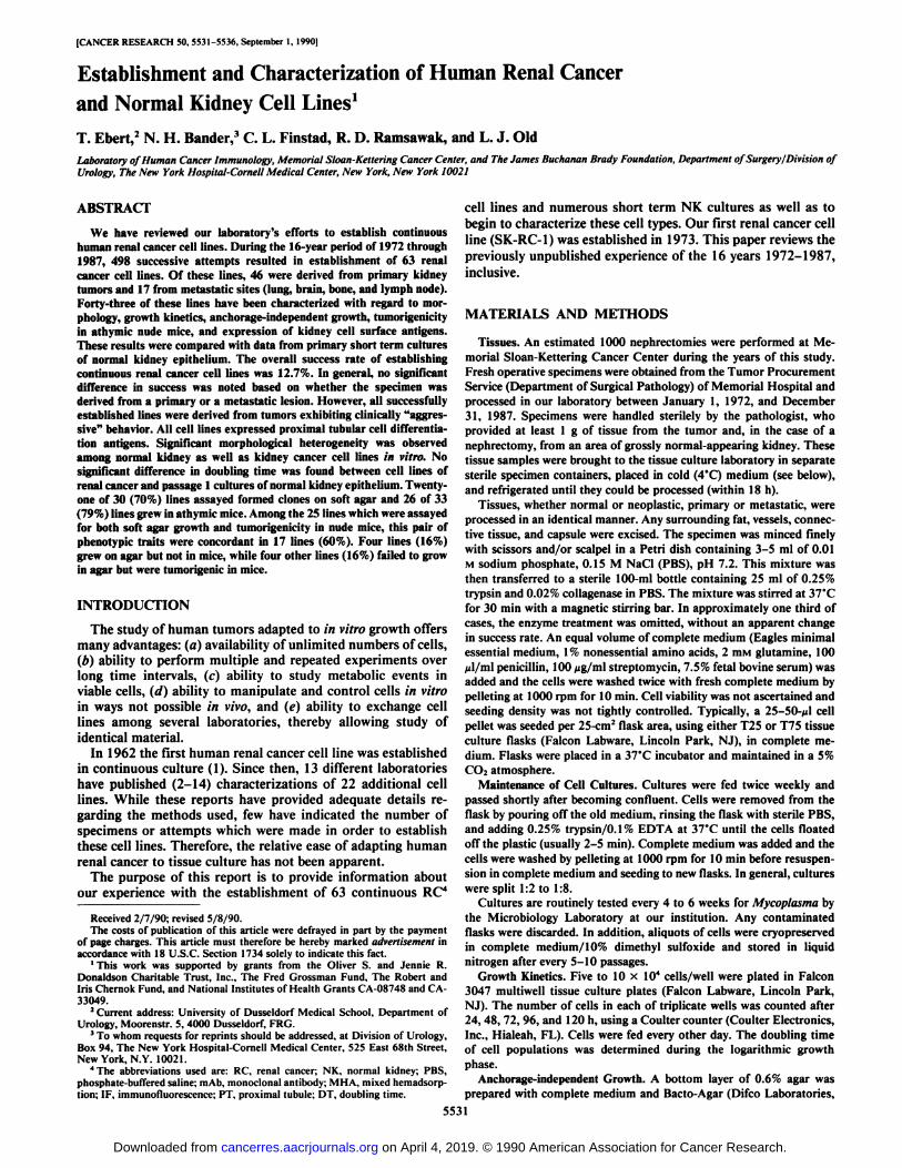
Fig. 1. Photomicrographs of four differentrenal cancer cell lines. In a, SK-RC-7 demonstrates a typical epithelial morphology. In />.SK-RC-6 grows with clefts which separate thecells. In c, SK-RC-28 demonstrates intracel-lular vacuoles, particularly when the cells become confluent. In d, SK-RC-45 has more ofa spindle cell morphology.
5532
on April 4, 2019. © 1990 American Association for Cancer Research. cancerres.aacrjournals.org Downloaded from
NORMAL KIDNEY AND RENAL CANCER IN VITRO
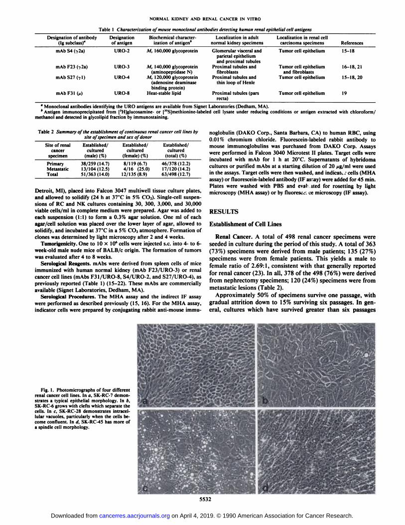
Fig. 2. Photomicrographs of four differentareas of a single, typical, normal kidney epithelial culture, a, small round cells, h. typicalepithelial cells. A "dome" is demonstrated near
the center of the field. These epithelial cellsresemble those seen in Fig. la. c, epithelialcells growing in swirls. </.epithelial cells witha more spindle-like morphology.
appear to have the ability to survive indefinitely in vitro (24).We have arbitrarily defined "established continuous cell lines"
as those cultures which survive 10 passages and 1 year incontinuous culture. These lines are then given an SK-RC designation. Sixty-three continuous RC lines have been establishedin 498 attempts, for an overall success rate of 12.7%. Overall,no significant difference was observed in establishing lines fromprimary [46 of 378 (12.2%)] or from metastatic [17 of 120(14.2%)] lesions.
The 63 lines were derived from 60 patients. In 3 cases, 2separate lines were developed from different sites. Medicalrecords were reviewed for 40 of the 60 patients whose tumorspecimens became established in culture. It was found that, inall cases, these tumor specimens exhibited clinically "aggressive" behavior. Of the 40 patients, 35 either had at the time of
surgery or developed soon thereafter metastatic disease. Theremaining 5 patients (SK-RC-3, -7, -11, -34, and -40) all hadmain renal vein involvement at the time of surgery but did notdevelop metastatic disease during follow-up of at least 4-8years. These findings indicate that a tumor must be biologicallyaggressive if it is to adapt to tissue culture. The site from whichthe specimen is derived (i.e., primary versus metastatic) appearsunimportant.
The sex of the donors was reviewed to determine any impacton the likelihood of adapting tumors to culture. Fifty-one of363 (14%) male and 12 of 135 (8.9%) female-derived tumorsadapted to culture. This difference was not statistically significant.
Morphologically, these renal cancer cell lines take severalforms (Fig. 1). Some are epithelioid with distinct cell borders,others have irregular or poorly defined borders, and othersconsist of spindle cells. Some lines contain cytoplasmic vacuolesor granules. None of the renal cancer lines exhibits contactinhibition.
Normal Kidney. Normal kidney epithelium can be adapted toin vitro growth on virtually every attempt. Unlike the cancerlines, however, these cultures survive only an average of threepassages. These passages can be spread over a period of 2-3months. Alternatively, these cells can be cryopreserved at early
passage (passage 1 or 2), thawed, and reseeded for later use ifdesired. Normal kidney cells adhere and begin dividing rapidlyin culture, in some cases earlier than their neoplastic counterparts. Morphologically, several different cell types are seen ina typical normal kidney culture (Fig. 2). One may note polygonal, round, and ellipsoidal cells. These cell types organize andgrow among each other in different patterns. Contact inhibitionis seen and the cells appear to cease dividing when a confluentmonolayer has formed. Renal epithelial cells can easily bedistinguished from fibroblasts. Fibroblast overgrowth has notbeen a significant problem in our experience.
Antigenic Phenotype
Selection of mAbs. The mAbs utilized in this study, and theantigens they define, have been well characterized with respectto immunochemistry and tissue distribution in vivo and in vitro(Table 1). We and others (22, 25, 26) have shown that antigenicexpression can change as cells adapt to a tissue culture environment. There are four patterns which characterize this shift:antigens which are expressed in vivo can remain expressed invitro (on—on) or may be turned off in vitro (on—off) orantigens not expressed in vivo can be turned on (off—on) orremain unexpressed (off—off). Before antigen expression canbe used as a basis for extrapolating between in vivo and in vitrosettings, one must determine that the particular antigens areexpressed or not expressed in a consistent fashion (i.e., on—on; off—off). The mAbs selected for this study have the characteristic of consistent patterns of reactivity or expression bothin vivo and in vitro. The specificity of these mAbs for renalepithelial antigens in vivo, the derivation of the specimens fromnormal kidney or renal cancers, and the demonstration of aconsistent pattern of expression of these antigens in vivo and invitro offer assurance that the cells growing in vitro are indeedderived from renal epithelia.
Normal Kidney Cells. Antigen expression of more than 50normal kidney cell lines, each derived from a different donor,was studied in detail. Each line displayed an identical patternof antigen expression when assayed at passage 1. That is, threeof four (URO-2, -3, and -4) PT cell markers reacted with at
5533
on April 4, 2019. © 1990 American Association for Cancer Research. cancerres.aacrjournals.org Downloaded from
NORMAL KIDNEY AND RENAL CANCER IN VITRO
least 90% of the cells of each line. A subpopulation of 10-20%of the cells in all NK cultures reacted with the fourth PTantigen (LJRO-8), which is expressed only by cells of the straightportion of the proximal tubule (19).
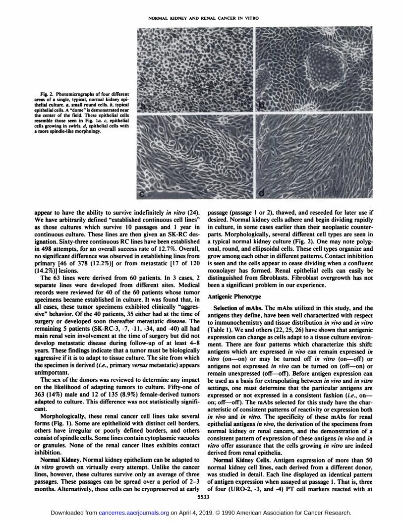
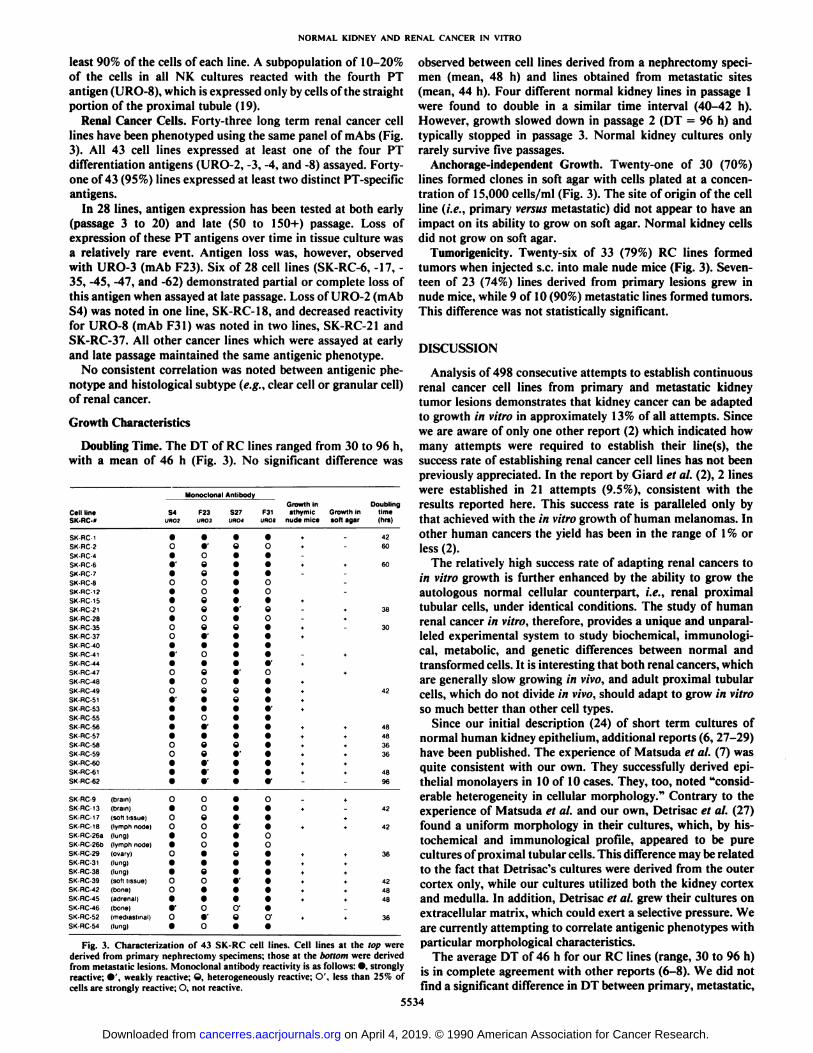
Renal Cancer Cells. Forty-three long term renal cancer celllines have been phenotyped using the same panel of mAbs (Fig.3). All 43 cell lines expressed at least one of the four PTdifferentiation antigens (URO-2, -3, -4, and -8) assayed. Forty-one of 43 (95%) lines expressed at least two distinct PT-specificantigens.
In 28 lines, antigen expression has been tested at both early(passage 3 to 20) and late (50 to 150+) passage. Loss ofexpression of these PT antigens over time in tissue culture wasa relatively rare event. Antigen loss was, however, observedwith URO-3 (mAb F23). Six of 28 cell lines (SK-RC-6, -17, -35, -45, -47, and -62) demonstrated partial or complete loss ofthis antigen when assayed at late passage. Loss of URO-2 (mAbS4) was noted in one line, SK-RC-18, and decreased reactivityfor URO-8 (mAb F31) was noted in two lines, SK-RC-21 andSK-RC-37. All other cancer lines which were assayed at earlyand late passage maintained the same antigenic phenotype.
No consistent correlation was noted between antigenic phenotype and histológica! subtype (e.g., clear cell or granular cell)of renal cancer.
Growth Characteristics
Doubling Time. The DT of RC lines ranged from 30 to 96 h.with a mean of 46 h (Fig. 3). No significant difference was
Cell lineSK-RC--
Monoclonal Antibody
Growth in DoublingS4 F23 S27 F31 athymic Growth in time
uno? UR03 URO4 uftos nude mice soft agar (hrs)
SK-RC-lSK-HC-2SK-RC-4SK-HC-6SK-RC-7SK-RC-8SK-RC-12SK-RC-15SK.RC-21SK-RC-28SK-RC-35SK-RC37SK-RC40SK-RC41SK-RC-44SK-RC-47SK-RC-48SK-RC-49SK-RC-51SK-RC-53SK.RC-55SK-RC-56SK-RC-57SK-RC58SK-RC-59SK-RC-60SK-RC-61SK.RC-62SK-RC-9SKRC-13SK-HC-17SK-RC-18SK
HC-26aSK-RC-26bSK.RC-29SK-RC-31SK-RC-38SK-RC-39SK-RC-42SK-RC-45SK-RC-46SK-RC-52SK-RC-54(brain)(brain)soft
tissue)lymph
node)lung)lymph
node)ovary)lung)lung)soft
tissue)bone)adrenal)bone)mediastmal)(lung],09••00000009••o•oO'•••0000••o•oo••o••o0••o••
«•'Coee000e0ef•O•e0eo•0o*•ee0*••r0oe0oo••e0••O
C•'Õo«1
1)Cr•
Cii1
«42+60*
»60+»
38tt
30•*••+++»
42*»*
*48•i-t-48«»36»»36+
+»*4896,*
42+*
+42t
36+4+
42*48t
48r
+»36I
Fig. 3. Characterization of 43 SK-RC cell lines. Cell lines at the top werederived from primary nephrectomy specimens; those at the bottom were derivedfrom metastatic lesions. Monoclonal antibody reactivity is as follows: •,stronglyreactive: •'.weakly reactive; O. heterogeneously reactive; O', less than 25cc of
cells are strongly reactive: O. not reactive.
observed between cell lines derived from a nephrectomy specimen (mean, 48 h) and lines obtained from metastatic sites(mean, 44 h). Four different normal kidney lines in passage 1were found to double in a similar time interval (40-42 h).However, growth slowed down in passage 2 (DT = 96 h) andtypically stopped in passage 3. Normal kidney cultures onlyrarely survive five passages.
Anchorage-independent Growth. Twenty-one of 30 (70%)lines formed clones in soft agar with cells plated at a concentration of 15,000 cells/ml (Fig. 3). The site of origin of the cellline (i.e., primary versus metastatic) did not appear to have animpact on its ability to grow on soft agar. Normal kidney cellsdid not grow on soft agar.
Tumorigenicity. Twenty-six of 33 (79%) RC lines formedtumors when injected s.c. into male nude mice (Fig. 3). Seventeen of 23 (74%) lines derived from primary lesions grew innude mice, while 9 of 10 (90%) metastatic lines formed tumors.This difference was not statistically significant.
DISCUSSION
Analysis of 498 consecutive attempts to establish continuousrenal cancer cell lines from primary and metastatic kidneytumor lesions demonstrates that kidney cancer can be adaptedto growth in vitro in approximately 13% of all attempts. Sincewe are aware of only one other report (2) which indicated howmany attempts were required to establish their line(s), thesuccess rate of establishing renal cancer cell lines has not beenpreviously appreciated. In the report by Giard et al. (2), 2 lineswere established in 21 attempts (9.5%), consistent with theresults reported here. This success rate is paralleled only bythat achieved with the in vitro growth of human melanomas. Inother human cancers the yield has been in the range of 1% orless (2).
The relatively high success rate of adapting renal cancers toin vitro growth is further enhanced by the ability to grow theautologous normal cellular counterpart, i.e., renal proximaltubular cells, under identical conditions. The study of humanrenal cancer in vitro, therefore, provides a unique and unparalleled experimental system to study biochemical, immunologi-cai, metabolic, and genetic differences between normal andtransformed cells. It is interesting that both renal cancers, whichare generally slow growing in vivo, and adult proximal tubularcells, which do not divide in vivo, should adapt to grow in vitroso much better than other cell types.
Since our initial description (24) of short term cultures ofnormal human kidney epithelium, additional reports (6, 27-29)have been published. The experience of Matsuda et al. (7) wasquite consistent with our own. They successfully derived epithelial monolayers in 10 of 10 cases. They, too, noted "considerable heterogeneity in cellular morphology." Contrary to the
experience of Matsuda et al. and our own, Detrisac et al. (27)found a uniform morphology in their cultures, which, by his-tochemical and immunological profile, appeared to be purecultures of proximal tubular cells. This difference may be relatedto the fact that Detrisac's cultures were derived from the outer
cortex only, while our cultures utilized both the kidney cortexand medulla. In addition, Detrisac et al. grew their cultures onextracellular matrix, which could exert a selective pressure. Weare currently attempting to correlate antigenic phenotypes withparticular morphological characteristics.
The average DT of 46 h for our RC lines (range, 30 to 96 h)is in complete agreement with other reports (6-8). We did notfind a significant difference in DT between primary, metastatic,
5534
on April 4, 2019. © 1990 American Association for Cancer Research. cancerres.aacrjournals.org Downloaded from
NORMAL KIDNEY AND RENAL CANCER IN VITRO
or normal kidney (passage 1) cell lines. However, as alsoreported by Matsuda et al., we observed a prolonged DT inpassage 2 NK cells (96 h). These lines eventually stoppedgrowing completely in passage 3 to 5. Detrisac et al. grew NKcells on an extracellular matrix and by doing so were able toextend the life span of their NK cultures from 3 to 16 passages.
Twenty-one of 30 (70%) RC lines form colonies on soft agar.Anchorage-independent growth is considered a typical featureof transformed cells (30) and has been observed in other RClines as well (6, 7, 11). In a study by Stiles et al. (31), no strongassociation between anchorage-independent growth and tumor-igenicity in athymic nude mice was revealed. This is consistentwith our data for 25 RC lines tested for both growth in softagar and tumorigenicity in nude mice. Results of these assayswere concordant in 17 lines (60%); 4 lines (16%), despiteforming clones on soft agar, did not grow in nude mice; the 4remaining lines (16%) formed tumors in the animals but didnot grow in soft agar. Cell lines which could grow both on agarand in nude mice did not appear to be clinically more aggressivethan those which possessed either trait individually.
Derivation of these cell lines from renal cancer specimensand expression of restricted renal tubular antigens by these celllines confirm that the lines are in fact renal cell carcinomas.The pattern of antigenic expression by these cell lines parallelsour immunohistochemical findings with fresh frozen sectionsof renal cancers. That is, they express proximal tubular differentiation antigens, although not uncommonly they fail to express the full complement of these antigens.
It is interesting that all of the cell lines which could adapt toimmortal growth in vitro were derived from tumors whichexhibited clinically aggressive features (e.g., metastasis or mainrenal vein invasion). Conversely, the finding that only 14.2%of metastatic tumors could be established in vitro implies thatbiological aggressiveness is necessary but not always sufficientfor establishment of a cell line. Further, if biological aggressiveness is a necessary characteristic, it is interesting that thesuccess rate is not significantly different based on whether thespecimen is derived from a metastatic lesion (the sine qua nonof clinical aggressiveness) or a localized primary renal cancer.One explanation for this observation is that all renal cancerprimaries contain biologically aggressive cells. This is, perhaps,not surprising in view of the fact that approximately 50% ofpatients with renal cancer have metastatic disease (25% overt,25% occult) at the time of initial diagnosis. But the converse,that 50% of patients presenting with renal cancer never developmetastasis, even when in some cases the primary tumor isenormous, would have argued that a major proportion of renalcancers are clinically "benign" and do not possess metastatic
potential. This latter position is not consistent with our findingshere.
In summary, this study shows that renal cancer cells cansuccessfully be adapted to in vitro growth. This has provided uswith continuous cultures from primary as well as metastaticlesions from bone, brain, lung, adrenal, and other soft tissuesites. In addition, the study of renal cancer offers the uniqueopportunity to have readily available under the same conditionsthe normal autologous counterpart. This variety of materialallows comparative analysis of genetic, biochemical, and/orimmunological differences between the tumor cell and its normal counterpart, between primary and metastatic lesions, andbetween metastatic lesions of different sites.
ACKNOWLEDGMENTS
tigators include W. F. Whitmore, Jr., M.D., H. Shiku, M.D., R. Ueda,M.D., M. Daly, and D. Morrissey. We also thank the Memorial Sloan-Kettering Cancer Center Department of Pathology and Tumor Procurement Service, without whose cooperation and efforts this workcould not have been accomplished.
REFERENCES
The authors would like to acknowledge the many investigators whocontributed to this work over the 16-year period studied. These inves-
9.
10.
11.
12.
13.
14.
15.
16.
17.
18.
19.
20.
21.
22.
23.
Ishihara, T., Moore, G. E., and Sandberg, A. A. The in vitro chromosomeconstitution of cells from human tumors. Cancer Res., 22: 375-379, 1962.Giard, D. J., Aaronson, S. A., Todaro, G. J., Arnstein, P., Kersey, J. H.,Dosik, H., and Parks, W. P. In vitro cultivation of human tumors: establishment of cell lines derived from a series of solid tumors. J. Nati. Cancer Inst.,57: 1417-1423, 1973.Fogh, J., and Trempe, G. New human tumor cell lines. In: J. Fogh (ed.).Human Tumor Cells In Vitro, p. 115. New York: Plenum Press, 1975.Hoehn, W.. and Schroeder, F. H. Renal cell carcinoma: two new cell linesand a serially transplantable nude mouse tumor (NC 65). Invest. Urol., 16:106-112, 1978.Komatsubara, S. Establishment of a new cell line (NRC-12) derived from ahuman renal cell carcinoma and its characteristics. Jpn. J. Urol., 69: 1535-1542, 1978.Williams, R. D., Elliot, A. Y., Stein, N., and Fraley, E. E. In vitro cultivationof human renal cell cancer. II. Characterization of cell lines. In Vitro, 14:779-786, 1978.Matsuda, M., Osafune, M., Nakano, E., Kotake, T., Sonoda, T., Watanabe,S., Hada. T., Okochi, T., Higashino, K., Yamamura, Y., and Abe, T.Characterization of an established cell line from human renal carcinoma.Cancer Res., 39:4694-4699, 1979.Shrivastav, S., Sharief, Y., Day, J., Feich, C., and Bonar, R. A. Establishmentand characterization of a cell line (SS78) from a human renal cell carcinoma.In Vitro, 17: 1117-1124, 1981.Naito. S., Kanamori, T., Hisano, S., Tanaka. K., Momose, S., and Ramata.N. Human renal cell carcinoma: establishment and characterization of twonew cell lines. J. Urol. 128: 1117-1121, 1982.Sytkowski, A. J., Richie. J. P., and Bicknell, K. A. New human renal cellline established from a patient with erythrocytosis. Cancer Res., 43: 1415-1419, 1983.Grossmann, H. B., Wedemeyer, G., and Ren, L. Human renal carcinoma:characterization of five new cell lines. J. Surg. Oncol.. 28: 237-244, 1985.Kinouchi, T., Kotake, T., Mori, Y., and Abe, T. Human renal cell carcinoma:establishment and characterization of a new cell line (OS-RC-2). In VitroCell Dev. Biol., 21: 195-199, 1985.Bear, A., dayman, R. V.. Eibers, J., Limas, C., Wang, N., Stone, K.,Gebhard, R., Prigge, W., and Palmer, J. Characterization of two human celllines (TK-10, TK-164) of renal cell cancer. Cancer Res., 47: 3856-3862,1987.Hashimura, T., Tubbs, R. R., Connelly, R., Caulfield, M. J., Trindade, C. S.,McMahon, J. T., Gaietti, T. P., Edinger, M., Sandberg, A. A.. Cin, P. D.,Sait, S. J., and Pontes, J. E. Characterization of two cell lines with distinctphenotypes and genotypes established from a patient with renal cell carcinoma. Cancer Res., 49: 7064-7071, 1989.Ueda, R., Ogata. S-I., Morrissey. D. M., Finstad. C. L., Szludlarek. J.,Whitmore, W. F.. Oettgen, H. F., Lloyd, K. O-, and Old, L. J. Cell surfaceantigens of human renal cancer defined by mouse monoclonal antibodies:identification of tissue-specific kidney glycoproteins. Proc. Nati. Acad. Sci.USA. 78: 5122-5126, 1981.Finstad, C. L., Cordon-Cardo, C.. Bander, N. H., Whitmore, W. F., Me-lamed, M. R., and Old, L. J. Specificity analysis of mouse monoclonalantibodies defining cell surface antigens of human renal cancer. Proc. Nati.Acad. Sci. USA, 82: 2955-2959, 1985.Bander, N. H., Cordon-Cardo, C., Finstad, C. L., Whitmore, W. F., Jr.,Vaughan, E. D., Jr., Oettgen, H. F., Melamed, M., and Old, L. J. Inuminohistologie dissection of the human kidney using monoclonal antibodies. J.Urol., m-502-505, 1985.Cordon-Cardo, C., Bander, N. H., Fradet, Y., Finstad. C. L., Whitmore, W.F., Lloyd, K. L., Oettgen, H. F., Melamed, M. R., and Old, L. O. Immu-noanatomic dissection of the human urinary tract by monoclonal antibodies.J. Histochem. Cytochem. 32: 1035-1040, 1984.Bander, N. H., Finstad, C. L., Cordon-Cardo, C., Ramsawak, R. D.,Vaughan. E. D., Jr.. Whitmore, W. F., Jr.. Oettgen, H. F., Melamed, M. R.,and Old, L. J. Analysis of a mouse monoclonal antibody which reacts with aspecific region of the human proximal tubule and subsets renal cell carcinomas. Cancer Res., 49: 6774-6780, 1989.Andy, R. J., Finstad, C. L., Old, L. J., Lloyd, K. O., and Kornfeld, R. Theantigen identified by a mouse monoclonal antibody raised against humanrenal cancer cells is the adenosine deaminase binding protein. J. Biol. Chem.,259:12844-12849.1984.Look. A. T., Ashmun, R. A., Shapiro. L. H., O'Connell, P. J., Gerkis, V.,d'Apice. A. J., Sagawa. K., and Peiper, S. C. Report of the CD 13 (Amino-
peptidase N) Cluster Workshop. In press, 1990.Bander, N. H. Comparison of antigen expression of human renal cancers invivo and in vitro. Cancer (Phila.), 53: 1235-1239, 1984.Paganini-Hill, A., Ross, R. K., and Henderson, B. E. Epidemiology of kidneycancer. In: D. G. Skinner (ed.). Urologica! Cancer, pp. 383-407. New York:Gruñeand Stratton, 1983.
5535
on April 4, 2019. © 1990 American Association for Cancer Research. cancerres.aacrjournals.org Downloaded from
NORMAL KIDNEY AND RENAL CANCER IN VITRO
24. Ueda, R., Shiku, H., Pfreundschuh,M., Takahashi. T., Li, L. T. C., Whit- Tissue culture of human kidney epithelial cells of proximal tubule origin,more, W. F.. Oettgen, H. F.. and Old, L. J. Cell surfaceantigens of human KidneyInt., 25: 383-390, 1984.renalcancerdefinedbyautologoustyping.J. Exp. Med.. ISO:564-579, 1979. 28- Trifillis. A. L., Regec, A. L., and Trump. B. F. Isolation, culture and
25. Rettig, W. J., Real. F. X.. Spengler, B. A., Biedler, J. L., and Old, L. J. characterizationof human renal tubular cells.J. Urol., 133:324-329, 1985.Humanmelanomaproteoglyc™^^ »- ^^Xe A ^j.^;^«-^«*——>«'««•and extr,ns,c signals.Science(Wash^DC). 231: 1281-1284. 1986^ J0 Macpherson,,.. and Montagnier,L. Agarsuspensionculture for the selective
26. Rettig, W. J.. Murty. V. V. V. S.. Mattes, M. J., Chaganti, R. S. K., and assayofce|ls transformedby polyomavirus. Virology,23: 291-294, 1964.Old, L. J. Extracellularmatrix-modulatedexpressionof human cell surface 31 Stiles,C. D.. Desmond,W., Chuman, L. M., Sato, G.. and Saier, M. H., Jr.glycoproteinsA42 and JI43. J. Exp. Med., 164: 1581-1599, 1986. Relationship of cell growth behavior in vitro to tumorigenicityin athymic
27. Detrisac,C. J., Sens, M. A., Julian GarvÃn,A., Spicer,S. S., and Sens, D. A. nude mice.Cancer Res.,36: 3300-3305, 1976.
5536
on April 4, 2019. © 1990 American Association for Cancer Research. cancerres.aacrjournals.org Downloaded from
1990;50:5531-5536. Cancer Res T. Ebert, N. H. Bander, C. L. Finstad, et al. Normal Kidney Cell LinesEstablishment and Characterization of Human Renal Cancer and
Updated version
http://cancerres.aacrjournals.org/content/50/17/5531
Access the most recent version of this article at:
E-mail alerts related to this article or journal.Sign up to receive free email-alerts
Subscriptions
Reprints and
To order reprints of this article or to subscribe to the journal, contact the AACR Publications
Permissions
Rightslink site. Click on "Request Permissions" which will take you to the Copyright Clearance Center's (CCC)
.http://cancerres.aacrjournals.org/content/50/17/5531To request permission to re-use all or part of this article, use this link
on April 4, 2019. © 1990 American Association for Cancer Research. cancerres.aacrjournals.org Downloaded from