Embed Size (px)
Citation preview
M2171 Esophageal Pathology Testing Page 1 of 19
Esophageal Pathology Testing
Policy Number: AHS – M2171 – Esophageal
Pathology Testing
Prior Policy Name and Number, as applicable:
Initial Presentation Date: 07/01/2021
Revision Date: N/A
I. Policy Description
The esophagus is a long tube that serves to connect the mouth to the stomach. Although the
esophagus is primarily a connecting organ, it experiences significant chemical and mechanical trauma.
The esophagus has mechanisms and structures to withstand this damage, but molecular injury is
common (Zhang et al., 2018). Both serological and genetic markers have been suggested to identify,
diagnose, or assess risk in the esophagus.
Eosinophilic esophagitis (EoE) is one such condition, as its nonspecific symptoms (pain, issues
swallowing, vomiting, and so on) may be accompanied by inflammatory markers in the esophagus
(Bonis & Gupta, 2020). Similarly, esophageal cancer is characterized by several nonspecific symptoms,
while a predecessor condition, Barrett esophagus (BE), may have no clinical symptoms at all (Saltzman
& Gibson, 2018; Spechler, 2020).
II. Related Policies
Policy Number Policy Title
AHS-M2109 Molecular Panel Testing of Cancers for Diagnosis, Prognosis, and Identification
of Targeted Therapy
AHS-G2054 Liquid Biopsy
III. Indications and/or Limitations of Coverage
Application of coverage criteria is dependent upon an individual’s benefit coverage at the time of the
request. Medical Policy Statements do not ensure an authorization or payment of services. Please
refer to the plan contract (often referred to as the Evidence of Coverage) for the service(s) referenced
in the Medical Policy Statement. If there is a conflict between the Medical Policy Statement and the
plan contract (i.e., Evidence of Coverage), then the plan contract (i.e., Evidence of Coverage) will be
the controlling document used to make the determination.
Application of coverage criteria is dependent upon an individual’s benefit coverage at the time of the
request. If there is a conflict between this Policy and any relevant, applicable government policy [e.g.
M2171 Esophageal Pathology Testing Page 2 of 19
National Coverage Determinations (NCDs) for Medicare] for a particular member, then the
government policy will be used to make the determination. For the most up-to-date Medicare policies
and coverage, please visit their search website http://www.cms.gov/medicare-
coveragedatabase/overview-and-quick-search.aspx?from2=search1.asp& or the manual website
1. Analysis of PD-L1 expression by immunohistochemistry in esophageal, gastric, or esophagogastric junction cancer tumors MEETS COVERAGE CRITERIA before first-line therapy PD-1 inhibitors, such as pembrolizumab, in patients with locally advanced, recurrent, or metastatic disease (See Notes 1 & 2).
2. Microsatellite instability analysis or MMR analysis MEETS COVERAGE CRITERIA for individuals with locally advanced, recurrent, or metastatic esophageal, gastric, or esophagogastric junction cancer for whom PD-1 inhibitors, such as pembrolizumab, are being considered for therapy (See Notes 1 & 2).
3. Genetic testing of HER2 MEETS COVERAGE CRITERIA for individuals with esophageal, gastric, or esophagogastric junction cancer for whom trastuzumab is being considered for therapy (See Notes 1 & 2).
4. Testing for NTRK gene fusion MEETS COVERAGE CRITERIA for individuals with esophageal, gastric, or esophagogastric junction cancer before first-line or subsequent therapy with larotrectinib or entrectinib (See Notes 1 & 2).
5. Wide-area transepithelial sampling (WATS) for the diagnosis and evaluation of Barrett’s esophagus, low-grade esophageal dysplasia, or high-grade esophageal dysplasia MEETS COVERAGE CRITERIA.
6. The use of genetic testing, including the use of molecular panel tests and gene expression profiling, to assess the risk of eosinophilic esophagitis (EoE) DOES NOT MEET COVERAGE CRITERIA.
7. The use of genetic testing, including the use of molecular panel tests and gene expression profiling, to diagnose or monitor eosinophilic esophagitis (EoE) DOES NOT MEET COVERAGE CRITERIA.
The following does not meet coverage criteria due to a lack of available published scientific literature confirming that the test(s) is/are required and beneficial for the diagnosis and treatment of a patient’s illness.
8. Testing for risk of Barrett’s esophagus and/or esophageal, including esophagogastric junction, cancer using a molecular classifier, such as the BarreGEN test, DOES NOT MEET COVERAGE CRITERIA.
M2171 Esophageal Pathology Testing Page 3 of 19
9. Epigenetic analysis, including but not limited to methylation analysis, of the likelihood for Barrett’s esophagus (such as EsoGuard), esophageal, or esophagogastric junction cancer DOES NOT MEET COVERAGE CRITERIA.
10. The use of the Esophageal String Test (EST) to diagnose, assess, or monitor eosinophilic esophagitis (EoE) DOES NOT MEET COVERAGE CRITERIA.
11. Liquid biopsy—the use of a non-invasive blood, saliva, or other body fluid to test for circulating tumor cells (CTCs), cell-free tumor DNA (cfDNA) fragments, and/or ribonucleoprotein complexes—for genomic profiling of esophageal and esophagogastric junction cancers DOES NOT MEET COVERAGE CRITERIA.
Note 1: For guidance on molecular panel testing of cancers, including esophageal and esophagogastric junction cancers, for targeted therapy, please see AHS-M2109 Molecular Panel Testing of Cancers for Diagnosis, Prognosis, and Identification of Targeted Therapy.
Note 2: For 5 or more gene tests being run on a tumor specimen (i.e. non-liquid biopsy) on the same platform, such as multi-gene panel next generation sequencing, please refer to AHS-R2162 Reimbursement Policy.
IV. Scientific Background
The esophagus is a long tube that connects the mouth to the stomach. Its primary function is to
transport food from the mouth to the stomach. However, this organ is often exposed to difficult
conditions, from abrasive food to the acidic conditions of the stomach. Although mechanisms are in
place to protect against injury (namely the tough squamous cells), it is common to see injury or disease
in the esophagus (Zhang et al., 2018).
Many serological and genetic markers have been proposed as tools to assist in evaluation of
esophageal pathology. Eosinophilic esophagitis (EoE), Barrett’s esophagus (BE), and esophageal
cancer are typically diagnosed with histological analysis from endoscopic biopsy (Bonis & Gupta, 2020;
Saltzman & Gibson, 2018; Spechler, 2020), but biopsies frequently require careful consideration and
resources to perform properly (NCCN, 2019). For these reasons, serum and genetic markers have been
suggested as noninvasive markers for esophageal pathologies.
Eosinophilic Esophagitis (EoE)
Eosinophilic esophagitis (EoE) marked by the presence of eosinophils in the esophagus. Eosinophils
are typically associated with mitigating inflammation but are not normally found in the esophagus.
EoE is represented by a broad set of clinical symptoms, such as difficulty swallowing, chest or
abdominal pain, and feeding dysfunction. Diagnosis is established through endoscopy with biopsies
to confirm eosinophilia. The current diagnostic criteria sets the cutoff for eosinophilia at ≥15
eosinophils per high power field, (60 eosinophils per mm2) although this figure has been heavily
discussed (Bonis & Gupta, 2020; Dellon et al., 2018).
M2171 Esophageal Pathology Testing Page 4 of 19
Laboratory tests have been suggested as a noninvasive adjunct for EoE. Serum IgE will be elevated in
up to 60% of EoE patients, as allergy has a strong association with EoE. Many other markers, such as
eotaxin-3, major basic protein-1, tryptase, chemokines, and serum eosinophil count, have all been
suggested to assist in evaluation of EoE (Bonis & Gupta, 2020; Dellon et al., 2018). Immune system
factors may also contribute to pathology. Since eosinophils are not normally found in the esophagus,
their presence in the esophagus may suggest an underlying issue with the immune system. Various
interleukins, mast cells, and T cells have all been proposed as contributing to pathogenesis, but the
exact pathway and mechanisms are not completely understood (Rothenberg, 2018). Genetic features
have also been used for EoE evaluation. Twin studies and family histories have indicated a role for
genetics in EoE. Several genes have also been identified as potential risk factors, such as CAPN14 (an
interleukin-13 regulator), TSLP (a basophil regulator), and CCL26 (promotes eosinophil movement into
esophagus) (Sherrill & Rothenberg, 2014).
Wen et al. (2013) developed a diagnostic gene expression panel (“EDP”) for EoE. The authors identified
candidate genes using two cohorts of EoE and control patients, then validated these genes with a
separate cohort of 194 patients (91 active EoE, 57 control, 34 ambiguous, 12 reflux). The panel was
found to identify EoE patients at 96% sensitivity and 98% specificity. The authors also noted that the
panel could separate patients in remission from unaffected patients (Wen et al., 2013).
Shoda et al. (2018) used an “EoE Diagnostic Panel” (EDP) to further classify EoE cases by histologic,
endoscopic, and molecular features. The EDP consisted of 95 esophageal transcripts purported to
identify EoE among both unaffected patients and patients with other conditions. 185 biopsies were
studied. The authors identified three clear subtypes of EoE; subtype 1 with a normal-appearing
esophagus and mild molecular changes, subtype 2 with an inflammatory and steroid-responsive
phenotype, and subtype 3 with a “narrow-caliber” esophagus and severe molecular alterations. These
findings were replicated in a 100-biopsy sample (Shoda et al., 2018).
Tests are commercially available for EoE. Noninvasive tests (as an alternative to endoscopy) have been
recently popular. The Esophageal String Test (EST) is one such alternative. The patient swallows a
gelatin-coated capsule with a string wrapped inside. Once the capsule is in the patient’s stomach, the
gelatin dissolves, allowing the capsule to pass through. The string itself is used to collect samples from
the patient’s esophagus and is easily removed from the patient. From there, the sample is analyzed
for several biomarkers (major basic protein-1, eotaxins 2 and 3, and so on) to provide a probability%
(a trademarked “EoEscore”) of esophageal inflammation (Ackerman et al., 2019; EnteroTrack, 2019).
Barrett Esophagus (BE)
Barrett esophagus (BE) is a condition in which the normal squamous tissue lining the esophagus is
replaced by metaplastic columnar epithelium. This new epithelium contains gastric features and is
typically caused by chronic gastroesophageal reflux disease (GERD). This condition predisposes to
esophageal cancer. When noxious substances (gastric acid, bile, et al) are exposed to the squamous
esophageal tissue, the damage is usually repaired through regeneration of these squamous cells. In
BE cases, this damage is repaired not through creation of new squamous cells, but through metaplastic
columnar cells. The exact reason for this is unknown. Although these metaplastic cells are more
resistant to reflux-based damage than the normal squamous cells, these cells frequently show the
oxidative DNA damage that is typical of cancer. Mutations in the p53 tumor suppressor gene appear
M2171 Esophageal Pathology Testing Page 5 of 19
to be the catalyst for cancers, as acquisition of this mutation in conjunction with the replication of the
genome is conducive to carcinogenesis (Spechler, 2020).
Vollmer (2019) performed a review assessing incidence of adenocarcinoma detected during
surveillance of BE. The author identified 55 studies encompassing 61371 total patients. Of the 61371
total patients, 1106 developed adenocarcinoma. Overall, the author found that the model created
from the studies “predicted the per-person probability of developing cancer in 5 years of complete
follow-up is approximately 0.0012”. Variables affecting this probability included mean time of
followup, definition of Barrett metaplasia, and fraction of patients followed up for at least 5 years
(Vollmer, 2019).
Proprietary tests are commercially available for assessment of BE, usually to evaluate risk (BE
progression to cancer, risk of BE itself, and such). For example, BarreGen, offered by Interpace
Diagnostics, uses tumor mutational load (a measure intended to capture total genomic instability of
a sample) to calculate risk of progression. Although many ways can estimate mutational load,
BarreGen tests 10 key genomic loci which are as follows: “1p (CMM1, L-myc), 3p (VHL, HoGG1), 5q
(MCC, APC), 9p (CDKN2A), 10q (PTEN, MXI1), 17p (TP53), 17q (RNF43, NME1), 18q (SMAD4, DCC), 21q
(TFF1, PSEN2) and 22q (NF2)”. These loci encompass integral tumor suppressors and are proposed to
provide an accurate picture of genomic instability (Interpace, 2019; Trindade et al., 2019). Another
test, TissueCypher, also proposes to predict likelihood of progression from BE to esophageal cancer.
The test measures 9 protein biomarkers that represent morphological and cellular changes (p53, p16,
AMACR, CD68, COX2, HER2, K20, HIF1-alpha, CD45RO). These biomarkers are quantified and
converted to a risk score (1-10) and probability of progression (Cernostics, 2019). Finally, a proprietary
imaging system, WATS3D, is commercially available. This imaging system samples from a wider area,
as opposed to only taking focal samples in a traditional biopsy. This technology also provides a
3dimensional image of the sampled area. This technology purports to provide more precise sampling
than the traditional 4-quadrant biopsies, claiming an increased detection rate of BE and other
dysplasias (Diagnostics, 2019).
Esophageal Cancer
Esophageal cancers are largely divided into two groups: squamous cell carcinomas (SCCs) and
adenocarcinomas (EAC). SCCs usually begin in the middle of the esophagus, whereas EACs often
originate near the gastroesophageal junction. Both share several risk factors, such as smoking. Due to
the numerous environmental risk factors for both types of cancer, it is difficult to ascertain the true
impact of genetic factors (Gibson, 2020). These cancers are primarily diagnosed through histologic
examination, usually obtained through endoscopy (Saltzman & Gibson, 2018).
Advancements have been in the molecular characterization of both types of cancer. TP53 mutations
are the most common mutation seen in both types of cancer. Other frequently mutated genes in
adenocarcinoma include ELMO1 and DOCK2 (enhance cell motility), ARID1A, SMARCA4 and ARID2
(chromatin remodelers), and SPG20 (traffics growth factor receptors). BE, as the precursor to
adenocarcinomas, includes certain similarities in genetic mutations but at a less severe rate. Further,
the rate of overlap tended to increase with higher degree of dysplasia (Testa, Castelli, & Pelosi, 2017).
SCC mutations tend to be in genes associated with specific cellular pathways. Genes in ubiquitous
pathways, such as EGFR, NOTCH3, and RB, are frequently mutated in SCC. The molecular profile of
M2171 Esophageal Pathology Testing Page 6 of 19
esophageal SCC tends to align more with other squamous cell cancers (such as head and neck cancers)
rather than EAC (Testa et al., 2017). Numerous gene expression studies have been performed to
further classify molecular subtypes of esophageal cancer (Gonzaga et al., 2017; McLaren et al., 2017;
Visser, Franken, Brosens, Ruurda, & van Hillegersberg, 2017). Gene expression profiles may have
utility in assessing response to treatment, prognosis, or risk assessment.
Li, Qi, Hu, and Wang (2019) investigated potential biomarkers for lymph node metastasis for
esophageal squamous cell carcinoma. 6 studies encompassing 70 patients were included. The authors
identified 9 biomarkers and 4 cellular mechanisms that influence lymph node metastasis. From there,
they identified three biomarkers with broader influence on prognosis of disease, PTEN, STMN1, and
TNFAIP8. The authors suggested that those three biomarkers should be researched further (Li et al.,
2019).
Plum et al. (2019) evaluated HER2 overexpression’s impact on prognosis of esophageal
adenocarcinoma (EAC). 428 EAC patients that underwent a “transthoracic thoraco-abdominal
esophagectomy” were included. The authors identified 44 patients with HER2 positivity (IHC score 3+
or 2+ with gene amplification). This cohort was found to have a better overall survival (OS, 70.1
months vs 24.6 months), along with better histology, absence of lymphatic metastases, and lower
tumor stages. The authors also noted a similarity in results to a large 2012 study (Plum et al., 2019).
Frankell et al. (2019) examined the molecular landscape of esophageal adenocarcinoma (EAC). The
authors assessed 551 genomically characterized EACs. A total of 77 driver genes and “21 non-coding
driver elements” were identified. The authors also found an average of 4.4 driver events per tumor.
A three-way association was found, between hyper-mutation, Wnt signaling, and loss of immune
signaling genes. Finally, the authors also identified “sensitizing events” (events causing a tumor to be
more susceptible to a therapy) to CD4/6 inhibitors in over half of the EAC cases studied (Frankell et
al., 2019).
Validity and Utility
Ackerman et al. (2019) evaluated the ability of the 1-hour Esophageal String Test (EST) to distinguish
between active eosinophilic esophagitis (EoE), inactive eosinophilic esophagitis, and normal esophagi.
134 patients (62 active EoE, 37 inactive EoE, 35 normal) were included. The authors found that eotaxin
3 measured from both EST samples and the control biopsy extracts to be the best marker for
distinguishing active EoE from inactive EoE (by both sensitivity and specificity). Addition of major basic
protein 1 (MBP-1) improved sensitivity by 0.039 (0.652 to 0.693) and specificity by 0.014 (0.261 to
0.275) across all patients (Ackerman et al., 2019).
Hao, Critchley-Thorne, Diehl, and Snyder (2019) performed a cost-effectiveness analysis of an
“adenocarcinoma risk prediction multi-biomarker assay” (TissueCypher’s Barrett’s Esophagus Assay).
A hypothetical cohort of 10000 patients with BE diagnoses (including non-dysplastic intestinal
metaplasia [NBDE], indefinite for dysplasia [IND], and low-grade dysplasia [LGD]) was created. A
Markov decision model was used to compare BE management costs between assay use and the
standard of care (SOC). A surveillance interval of 5 years was used. Low-risk patients were found to
have a 16.6% reduction in endoscopies. High-risk patients were found to have a 58.4% increase in
endoscopic treatments (compared to the SOC arm), leading to a death total of 111 for the assay arm
compared to 204 in the SOC arm (a 45.6% reduction). Overall, the authors calculated the incremental
M2171 Esophageal Pathology Testing Page 7 of 19
cost-effectiveness ratio (ICER) to be $52,483/quality-adjusted life-year (QALY), and they found that
“the probability of the Assay being cost-effective compared to the SOC was 57.3% at the
$100,000/QALY acceptability threshold” (Hao et al., 2019).
Eluri et al. (2018) aimed to validate a genomic panel intended to represent tumor mutational load
(TML). Previously, the authors evaluated a panel of 10 genomic loci from which a TML score was
calculated. This mean TML was found to be significantly higher in 23 BE patients that had progressed
to high-grade dysplasia (HGD) or esophageal adenocarcinoma (EAC) as compared to 46 that had not
progressed. The area under the curve in this prior study was found to be 0.95 at a mutational load
(ML) cutoff of 1 (on a scale of 1-10). In the present study, 159 subjects were included. Cases had
“baseline nondysplastic BE (NDBE) and developed HGD/EAC ≥ 2 years later.” 58 subjects were
progressors and 101 were nonprogressors. The authors identified no difference in mean ML in
preprogression tissue in both cohorts (“ML = 0.73 ± 0.69 vs. ML = 0.74 ± 0.61”). The area under the
curve at the cutoff of ML 1 was only 0.50, and the authors concluded that the “utility of the ML to
stratify BE patients for risk of progression was not confirmed in this study” (Eluri et al., 2018).
Trindade et al. (2019) evaluated tumor mutational load’s (ML) ability to “risk-stratify those that may
progress from non-dysplastic BE to dysplastic disease”. 28 patients were included, and ML levels were
compared between those that progressed to dysplasia and those who had not. 8 total patients
progressed to dysplasia (6 low-grade, 2 high-grade), and 7 of these patients had “some level” of
genomic stability detected (ML ≥.5 on a scale of 1 to 10). 10 of the 20 patients that did not progress
to dysplasia had “no” ML level. The authors also noted that at an ML of ≥1.5, the risk of progression
to high-grade dysplasia was 33%, with a sensitivity of 100% and specificity of 85%. The authors
concluded “that ML may be able to risk-stratify progression to high-grade dysplasia in BE-IND. Larger
studies are needed to confirm these findings” (Trindade et al., 2019).
Moinova et al. (2018) evaluated the ability of two DNA methylation signatures to detect BE.
Methylation signatures of the VIM and CCNA1 loci were evaluated in 173 patients with or without BE.
CCNA1 methylation was found to have an area under the curve of 0.95 for distinguishing BE-related
dysplasia compared to normal esophagi. When the data for VIM methylation was added, the resulting
sensitivity was 95%, and the resulting specificity was 91%. These findings were replicated in a
validation cohort of 86 patients, with the combination of methylation markers detecting BE
metaplasia at 90.3% sensitivity and 91.7% specificity (Moinova et al., 2018).
Critchley-Thorne et al. (2016) validated a pathology panel to predict progression of BE to esophageal
cancer. The authors identified 15 potential biomarkers, which were evaluated in both training and
validation sets. This “classifier” separated patients into three different risk classes: low, intermediate,
and high in the training set of 183. The authors calculated the hazard ratio of intermediate to low risk
at 4.19 and high to low at 14.73. In the validation set (n = 183), the concordance index (an estimation
of area under the curve) of the 15-factor classifier was 0.772, the best of the amounts tested (3, 6, 9,
12, 15, 17). The authors also noted that this classifier provided independent prognostic information
that were outperformed predictions based on other clinicopathological factors, such as segment
length, age, and p53 overexpression (Critchley-Thorne et al., 2016).
Another multicenter study investigated the use of WATS3D with either random or targeted FB in the
detection of esophageal dysplasia (ED). 12,899 patients were enrolled in the study, and WATS3D
detected an additional 213 cases of ED beyond the initial 88 cases identified by FB, representing an
increase of 242%. Regarding screening for BE, WATS increased the overall detection by 153% (from
M2171 Esophageal Pathology Testing Page 8 of 19
13.1% to 33% of the individuals enrolled). The authors noted that the order of testing (e.g. FB or WATS)
did not impact the results. The authors conclude, “In this study, comprised of the largest series of
patients evaluated with WATS, adjunctive use of the technique with targeted and random FB markedly
improved the detection of both ED and BE. These results underscore the shortcomings of FB in
detecting BE-associated neoplasia, which can potentially impact the management and clinical
outcomes of these patients (Smith et al., 2019).”
A study into the cost-effectiveness of WATS3D testing as an adjunct to the standard-of-care forceps
biopsy (FB) used a reference case of a 60-year-old white male with gastroesophageal reflux disease
(GERD) to see the number of screens needed to avert one cancer and one cancer-related death as
well as to calculate the quality-adjusted life years (QALYs) as measured in 2019 U.S. dollars. With this
as a reference case, 320 – 337 individuals would need to be screened using WATS3D to avert one
cancer, and 328 – 367 individuals would be required to avert one death. The additional cost associated
with WATS3D was $1219, but an additional 0.017 QALYs were produced, resulting in an ICER of
$71395/QALY. The authors conclude, “Screening for BE in 60-year-old white male GERD patients is
more cost-effective when WATS3D is used adjunctively to the Seattle protocol than with the Seattle
protocol alone (Singer & Smith, 2020).”
One study compared the use of the WATS3D technology to standard forceps biopsy. 117 individuals
with a history of Barrett’s esophagus with dysplasia had both techniques performed. For the biopsy,
a four-quadrant biopsy quadrant protocol was performed every 1 – 2 cm. Evaluation of the biopsy and
the WATS3D technique was performed by separate pathologists, blinded to each other’s results. “Brush
biopsy [WATS3D] added an additional 16 position cases increasing the yield of dysplasia detection by
42% (95% CI: 20.7 – 72.7). The number needed to test (NNT) to detect one additional case of dysplasia
was 9.4 (95% CI: 6.4 – 17.7).” The authors of the study noted that no statistical difference was evident
between medical centers, the type of forceps used, or between sampling every 1 cm versus every 2
cm. They conclude, “These data suggest that computer-assisted brush biopsy is a useful adjunct to
standard endoscopic surveillance regimens for the identification of dysplasia in Barrett’s esophagus
(Anandasabapathy et al., 2011).”
Another multicenter prospective trial of 4203 patients studied the use of WATS3D as an adjunct to
four-quadrant random forceps biopsy (FB) in detecting Barrett’s esophagus (BE) and esophageal
dysplasia (ED). FB alone detected 594 cases of BE, and the addition of WATS3D detected an additional
493 cases, an increase of 83%. Likewise, WATS3D detected an increase of 88.5% of low-grade dysplasia
(LGD). The authors conclude, “Adjunctive use of WATS to FB significantly improves the detection of
both BE and ED. Sampling effort, an inherent limitation associated with screening and surveillance,
can be improved with WATS allowing better informed decisions to be made about the management
and subsequent treatment of these patients (Gross, Smith, & Kaul, 2018).” These findings support the
earlier study by Johanson and colleagues. In their study of 1266 patients being screened for BE and
ED, they noted an overall increase of 39.8% in the detection of BE when WATS3D (brush biopsy or BB)
was used as an adjunct to FB. They also report that the number of patients needed to test (NNT) to
obtain a positive BE result was 8.7. Interestingly, specifically for patients with gastroesophageal reflux
disease (GERD), the addition of WATS3D resulted in an even higher increase in the detection of BE (by
70.5%) (Johanson, Frakes, & Eisen, 2011).
Another study published in 2018 of a randomized trial at 16 different medical centers (n = 160
patients) compared the order of testing (WATS3D followed by biopsy sampling versus biopsy sampling
M2171 Esophageal Pathology Testing Page 9 of 19
followed by WATS3D) to detect high-grade dysplasia/esophageal adenocarcinoma (HGD/EAC). The
authors also stated secondary aims of determining the amount of additional time required for WATS3D
and the ability of each procedure to separately detect neoplasia. The order of the procedures was not
statistically relevant. The use of WATS3D as an adjunct to biopsy did result in a 14.4% absolute increase
in the number of HGD/EAC cases detected. The authors noted that WATS3D, on average, adds 4.5
minutes to the total procedure time. They conclude, “Results of this multicenter, prospective,
randomized trial demonstrate that the use of WATS in a referral BE population increases the detection
of HGD/EAC (Vennalaganti et al., 2018).”
V. Guidelines and Recommendations
United European Gastroenterology (UEG), The European Society of Pediatric Gastroenterology,
Hepatology and Nutrition (ESPGHAN), the European Academy of Allergy and Clinical Immunology
(EAACI), and the European Society of Eosinophilic Oesophagitis (EUREOS) (Lucendo et al., 2017)
These joint guidelines were published by a task force of 21 physicians and researchers for eosinophilic
esophagitis (EoE). In it, they note that noninvasive biomarkers (inflammatory factors, total IgE,
chemokines, tryptase, et al) are “not accurate” to diagnose or monitor EoE. They remark that absolute
serum eosinophil count fared best in correlating with severity of disease but had a diagnostic accuracy
of 0.754. The guidelines state that histology is necessary for monitoring. The String Test was also
mentioned as having good preliminary results but required further corroboration (Lucendo et al.,
2017).
Updated International Consensus Diagnostic Criteria for Eosinophilic Esophagitis: Proceedings of
the AGREE Conference (Dellon et al., 2018)
These newly published international diagnostic criteria primarily include endoscopic findings.
Although the guidelines emphasize ruling out other diagnoses (in which biomarkers may be useful), it
does not mention any serum or genetic factors for EoE itself (Dellon et al., 2018).
National Comprehensive Cancer Network (NCCN, 2020)
The NCCN notes four syndromes that predispose to an increased risk for esophageal and
esophagogastric junction (EGJ) cancers; tylosis with non-epidermolytic palmoplantar keratoderma
(PPK) with esophageal cancer (including Howel-Evans syndrome), familial Barrett esophagus (FBE),
Bloom Syndrome (BS, BLM gene), and Fanconi Anemia (FA, FANC A-E genes). The RHBDF2 gene has
been associated with tylosis (with non-epidermolytic palmoplantar keratosis) for genetic risk
assessment. Though FBE may be associated with “one or more autosomally inherited dominant
susceptibility alleles,” no gene has been validated. With regards to next-generation sequencing, he
NCCN concludes that “when limited tissue is available for testing, sequential testing of single
biomarkers or use of limited molecular diagnostic panels may quickly exhaust the sample. In these
scenarios, comprehensive genomic profiling via a validated NGS assay performed in a CLIA-approved
laboratory may be used for the identification of HER2 amplification, MSI [microsatellite instability],
and NTRK gene fusions. It should be noted that NGS has several inherent limitations and thus
whenever possible, the use of gold-standard assays (IHC [immunohistochemistry]/FISH [fluorescence
in situ hybridization]/targeted PCR [polymerase chain reaction]) should be performed” (NCCN, 2020).
M2171 Esophageal Pathology Testing Page 10 of 19
Liquid biopsy aids in identifying genetic mutations in solid cancers by looking at circulating tumor DNA
(ctDNA) in blood and can be used in those with advanced disease and cannot undergo clinical biopsies
for disease surveillance and management. Detecting mutations in DNA from esophageal and EGJ
carcinomas “can identify targetable alterations or the evolution of clones with altered treatment
response profiles.” The NCCN has also stated that “a negative result should be interpreted with
caution, as this does not exclude the presence of tumor mutations or amplifications” (NCCN, 2020).
The NCCN notes that “testing for MSI by polymerase chain reaction (PCR) or MMR [mismatch repair]
by IHC should be considered on locally advanced, recurrent, or metastatic esophageal and EGJ cancers
in patients who are candidates for treatment with PD-1 inhibitors.” The NCCN also identifies three
targeted therapeutic agents currently approved by the FDA; trastuzumab, ramucirumab, and
pembrolizumab. Trastuzumab is based on HER2 status and pembrolizumab is based on “testing for
MSI by PCR/MMR by IHC or PD-LA expression by CPS [combined positive score].” Select TRK inhibitors
have also been FDA-approved for NTRK gene fusion-positive tumors.
M2171 Esophageal Pathology Testing Page 11 of 19
Genetic biomarkers such as aneuploidy and loss of p53 heterozygosity have been proposed as useful
for identifying increased risk of progression in BE patients, but the NCCN remarks that these
biomarkers require “further prospective evaluation as predictors of risk for the development of HGD
[high-grade dysplasia] and adenocarcinoma of the esophagus in patients with Barrett esophagus”
(NCCN, 2020).
The NCCN notes that wide-area transepithelial sampling (WATS) has been used to detect esophageal
carcinomas in BE patients. They state, “The use of wide-area transepithelial sampling with
computerassisted 3-dimensional analysis (WATS3D), a relatively new sampling technique combining
an abrasive brush biopsy of the Barrett esophagus mucosa with computer-assisted pathology analysis
to highlight abnormal cells, may help increase the detection of esophageal dysplasia in patients with
Barrett esophagus.” They go on to cite the 2017 study by Vennalaganti and colleagues that shows a
14.4% increase in the number of additional cases of HGD/esophageal adenocarcinoma captured by
using WATS. However, the NCCN remarks that the “utility and accuracy of WATS for detecting
HGD/adenocarcinoma in patients with Barrett esophagus needs to be evaluated in larger phase III
randomized trials” (NCCN, 2020).
American Society for Gastrointestinal Endoscopy (Qumseya et al., 2019)
The ASGE recommends the use of WATS3D as an adjunct to “Seattle protocol biopsy sampling” in
patients with known or suspected BE (conditional recommendation, low quality of evidence). The
society stated that they had downrated the certainty of the recommendation due to possible risk bios,
insistency, and indirectness of the studies that were available at the time of publication since some
of the studies had included LGD (whereas others had not) and many of the studies had been
sponsored by the test’s manufacturer. The society also had noted that, as of the date of publication,
no studies addressing the cost-effectiveness of WATS-3D had been published. (Qumseya et al., 2019)
It should be noted that since the publication of these guidelines the 2020 cost-effectiveness study by
Singer and Smith (2020) has been published.
Society of American Gastrointestinal and Endoscopic Surgeons (SAGES) Technology and Value
Assessment Committee (TVAC) (Docimo, Al-Mansour, & Tsuda, 2020)
The TVAC of SAGES evaluated WATS3D and published their findings and recommendations within the
journal Surgical Endoscopy in 2020. They note that WATS3D is not recommended “as a stand-alone
substitute for cold forcep biopsies.” Within their expert panel recommendation section:
• They state that no significant morbidity or mortality is associated with the testing.
• They also state that “WATS3D increases diagnostic yield by 38 – 150% for Barrett’s
Esophagus, by 40 – 150% for Low Grade Dysplasia; and by 420% for High Grade Dysplasia;
when compared to forceps biopsy alone.”
• WATS3D testing also “has very high inter-observer agreement for the pathological diagnosis of non-dysplastic and dysplastic Barrett’s Esophagus.”
Regarding value, “Increased detection of pre-malignant diseases of the esophagus by the adjunctive
use of WATS3D supports screening and surveillance by the adjunctive use of WATS3D during upper
endoscopy in appropriate patients” (Docimo et al., 2020).
M2171 Esophageal Pathology Testing Page 12 of 19
American Foregut Society (AFS, 2020)
The AFS published a white paper reviewing WATS3D in 2020. After reviewing the literature, they state,
“The American Foregut Society (AFS) Board has concluded that there are sufficient data to support the routine use of WATS3D technology in the diagnosis and ongoing evaluation of Barrett’s esophagus”
(AFS, 2020).
American College of Gastroenterology (ACG, 2016)
The ACG published guidelines on the diagnosis and management of Barrett Esophagus. In it, they state
that no single biomarker (including genetic abnormalities) is “adequate” as a risk stratification tool.
Further, they remark that an entire panel of biomarkers may be required, but no panels were ready
for clinical practice (Shaheen, Falk, Iyer, & Gerson, 2016).
European Society for Medical Oncology (ESMO, 2016)
ESMO does not mention any molecular testing for diagnosis or risk assessment of esophageal cancer.
Testing for HER2 is mentioned for targeted therapy with trastuzumab. The guidelines recommend
following the 2016 ACG guidelines regarding Barrett’s Esophagus screening (Lordick et al., 2016).
Pan-Asian adapted ESMO Clinical Practice Guidelines: a JSMO-ESMO initiative endorsed by CSCO,
KSMO, MOS, SSO and TOS (Muro et al., 2019)
The only biomarker mentioned in these guidelines is HER2; intended “to select patients with
metastatic oesophageal adenocarcinoma for treatment with…trastuzumab”. The guidelines go on to
state that evidence for the role of other biomarkers or agents is “limited” (Muro et al., 2019).
VI. State and Federal Regulations, as applicable
A search for “esophagus” on the FDA website on 11/05/2020 yielded 0 relevant results. A search of
the FDA device database on 11/06/2020 using the query “WATS” or “transepithelial” yielded no
relevant results. Likewise, a search of the CMS database of the active LCD/NCDs for “WATS3D” yielded
no results. Additionally, many labs have developed specific tests that they must validate and perform
in house. These laboratory-developed tests (LDTs) are regulated by the Centers for Medicare and
Medicaid (CMS) as high-complexity tests under the Clinical Laboratory Improvement Amendments of
1988 (CLIA ’88). As an LDT, the U. S. Food and Drug Administration has not approved or cleared this
test; however, FDA clearance or approval is not currently required for clinical use.
VII. Applicable CPT/HCPCS Procedure Codes
Code
Number
Code Description
M2171 Esophageal Pathology Testing Page 13 of 19
81301
Microsatellite instability analysis (eg, hereditary non-polyposis colorectal cancer,
Lynch syndrome) of markers for mismatch repair deficiency (eg, BAT25, BAT26),
includes comparison
of neoplastic and normal tissue, if performed
81479 Unlisted molecular pathology procedure
88104
Cytopathology, fluids, washings or brushings, except cervical or vaginal; smears with
interpretation
88271 Molecular cytogenetics; DNA probe, each (eg, FISH)
88272
Molecular cytogenetics; chromosomal in situ hybridization, analyze 3-5 cells (eg, for
derivatives and markers)
88273
Molecular cytogenetics; chromosomal in situ hybridization, analyze 10-30 cells (eg,
for microdeletions)
88274 Molecular cytogenetics; interphase in situ hybridization, analyze 25-99 cells
88275
Molecular cytogenetics; interphase in
situ hybridization, analyze 100-300 cells
88341
Immunohistochemistry or immunocytochemistry, per specimen; each additional
single antibody stain procedure (List separately in addition to code for primary
procedure)
88342
Immunohistochemistry or
immunocytochemistry, per specimen; initial single antibody stain procedure
M2171 Esophageal Pathology Testing Page 14 of 19
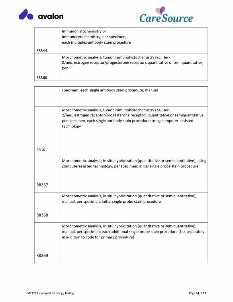
88344
Immunohistochemistry or
immunocytochemistry, per specimen;
each multiplex antibody stain procedure
88360
Morphometric analysis, tumor immunohistochemistry (eg, Her- 2/neu, estrogen receptor/progesterone receptor), quantitative or semiquantitative,
per
specimen, each single antibody stain procedure; manual
88361
Morphometric analysis, tumor immunohistochemistry (eg, Her- 2/neu, estrogen receptor/progesterone receptor), quantitative or semiquantitative,
per specimen, each single antibody stain procedure; using computer-assisted
technology
88367
Morphometric analysis, in situ hybridization (quantitative or semiquantitative), using
computerassisted technology, per specimen; initial single probe stain procedure
88368
Morphometric analysis, in situ hybridization (quantitative or semiquantitative),
manual, per specimen; initial single probe stain procedure
88369
Morphometric analysis, in situ hybridization (quantitative or semiquantitative),
manual, per specimen; each additional single probe stain procedure (List separately
in addition to code for primary procedure)
M2171 Esophageal Pathology Testing Page 15 of 19
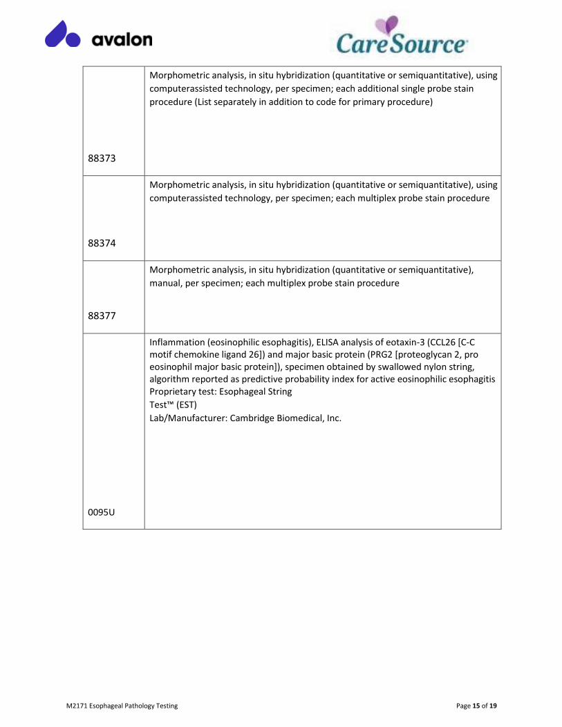
88373
Morphometric analysis, in situ hybridization (quantitative or semiquantitative), using
computerassisted technology, per specimen; each additional single probe stain
procedure (List separately in addition to code for primary procedure)
88374
Morphometric analysis, in situ hybridization (quantitative or semiquantitative), using
computerassisted technology, per specimen; each multiplex probe stain procedure
88377
Morphometric analysis, in situ hybridization (quantitative or semiquantitative),
manual, per specimen; each multiplex probe stain procedure
0095U
Inflammation (eosinophilic esophagitis), ELISA analysis of eotaxin-3 (CCL26 [C-C motif chemokine ligand 26]) and major basic protein (PRG2 [proteoglycan 2, pro eosinophil major basic protein]), specimen obtained by swallowed nylon string, algorithm reported as predictive probability index for active eosinophilic esophagitis Proprietary test: Esophageal String
Test™ (EST)
Lab/Manufacturer: Cambridge Biomedical, Inc.
M2171 Esophageal Pathology Testing Page 16 of 19
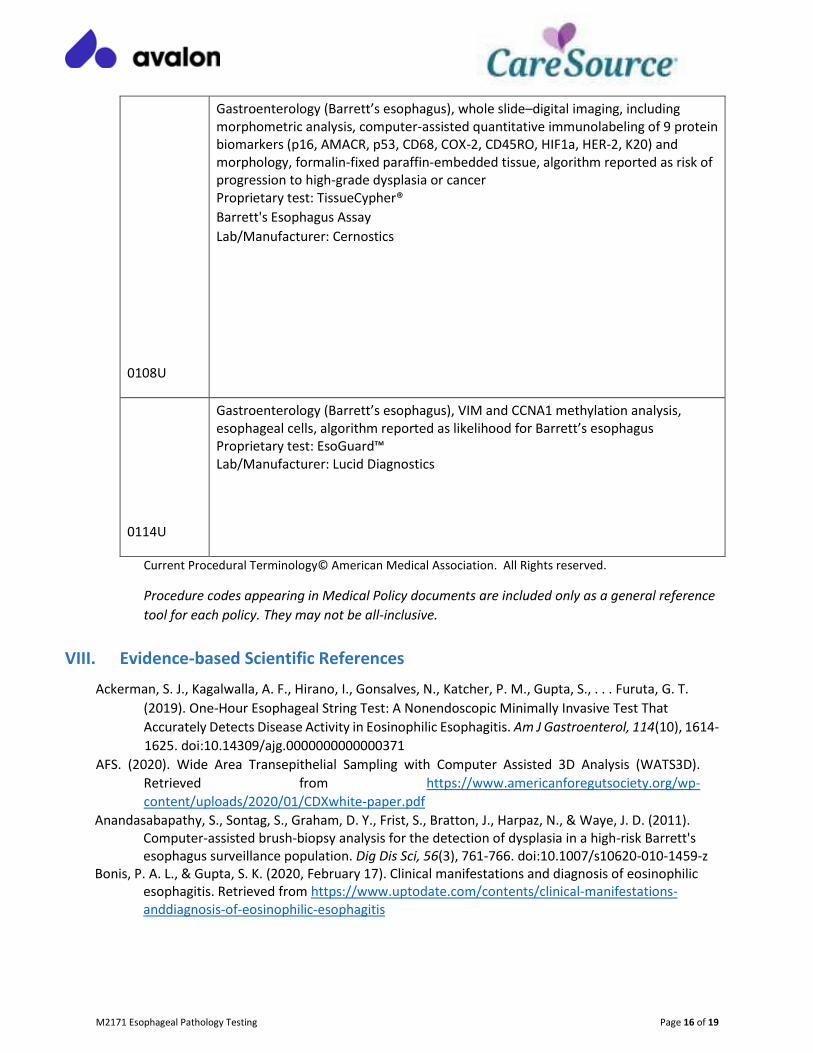
0108U
Gastroenterology (Barrett’s esophagus), whole slide–digital imaging, including morphometric analysis, computer-assisted quantitative immunolabeling of 9 protein biomarkers (p16, AMACR, p53, CD68, COX-2, CD45RO, HIF1a, HER-2, K20) and morphology, formalin-fixed paraffin-embedded tissue, algorithm reported as risk of progression to high-grade dysplasia or cancer Proprietary test: TissueCypher®
Barrett's Esophagus Assay
Lab/Manufacturer: Cernostics
0114U
Gastroenterology (Barrett’s esophagus), VIM and CCNA1 methylation analysis, esophageal cells, algorithm reported as likelihood for Barrett’s esophagus Proprietary test: EsoGuard™ Lab/Manufacturer: Lucid Diagnostics
Current Procedural Terminology© American Medical Association. All Rights reserved.
Procedure codes appearing in Medical Policy documents are included only as a general reference
tool for each policy. They may not be all-inclusive.
VIII. Evidence-based Scientific References
Ackerman, S. J., Kagalwalla, A. F., Hirano, I., Gonsalves, N., Katcher, P. M., Gupta, S., . . . Furuta, G. T.
(2019). One-Hour Esophageal String Test: A Nonendoscopic Minimally Invasive Test That
Accurately Detects Disease Activity in Eosinophilic Esophagitis. Am J Gastroenterol, 114(10), 1614-
1625. doi:10.14309/ajg.0000000000000371
AFS. (2020). Wide Area Transepithelial Sampling with Computer Assisted 3D Analysis (WATS3D).
Retrieved from https://www.americanforegutsociety.org/wp-
content/uploads/2020/01/CDXwhite-paper.pdf
Anandasabapathy, S., Sontag, S., Graham, D. Y., Frist, S., Bratton, J., Harpaz, N., & Waye, J. D. (2011). Computer-assisted brush-biopsy analysis for the detection of dysplasia in a high-risk Barrett's esophagus surveillance population. Dig Dis Sci, 56(3), 761-766. doi:10.1007/s10620-010-1459-z
Bonis, P. A. L., & Gupta, S. K. (2020, February 17). Clinical manifestations and diagnosis of eosinophilic esophagitis. Retrieved from https://www.uptodate.com/contents/clinical-manifestations-anddiagnosis-of-eosinophilic-esophagitis
M2171 Esophageal Pathology Testing Page 17 of 19
Cernostics. (2019). What is the TissueCypher® Barrett’s Esophagus Assay? Retrieved from http://www.cernostics.com/products/
Costa-Barbosa, F. A., Balasubramanian, R., Keefe, K. W., Shaw, N. D., Al-Tassan, N., Plummer, L., . . . Crowley, W. F., Jr. (2013). Prioritizing genetic testing in patients with Kallmann syndrome using clinical phenotypes. J Clin Endocrinol Metab, 98(5), E943-953. doi:10.1210/jc.2012-4116
Critchley-Thorne, R. J., Duits, L. C., Prichard, J. W., Davison, J. M., Jobe, B. A., Campbell, B. B., . . . Falk, G.
W. (2016). A Tissue Systems Pathology Assay for High-Risk Barrett's Esophagus. Cancer
Epidemiol Biomarkers Prev, 25(6), 958-968. doi:10.1158/1055-9965.Epi-15-1164
Dellon, E. S., Liacouras, C. A., Molina-Infante, J., Furuta, G. T., Spergel, J. M., Zevit, N., . . . Bredenoord, A.
J. (2018). Updated International Consensus Diagnostic Criteria for Eosinophilic Esophagitis:
Proceedings of the AGREE Conference. Gastroenterology, 155(4), 1022-1033.e1010.
doi:10.1053/j.gastro.2018.07.009
Diagnostics, C. (2019). BREAKTHROUGHSAMPLING. Retrieved from https://www.cdxdiagnostics.com/wats3d-breakthrough-technology/
Docimo, S., Jr., Al-Mansour, M., & Tsuda, S. (2020). SAGES TAVAC safety and efficacy analysis WATS(3D)
(CDx Diagnostics, Suffern, NY). Surg Endosc. doi:10.1007/s00464-020-07503-w
Eluri, S., Klaver, E., Duits, L. C., Jackson, S. A., Bergman, J. J., & Shaheen, N. J. (2018). Validation of a biomarker panel in Barrett's esophagus to predict progression to esophageal adenocarcinoma. Dis Esophagus, 31(11). doi:10.1093/dote/doy026
EnteroTrack. (2019). The EnteroTracker®. Retrieved from
https://enterotrack.com/enterotrackeroverview
Frankell, A. M., Jammula, S., Li, X., Contino, G., Killcoyne, S., Abbas, S., . . . Fitzgerald, R. C. (2019). The landscape of selection in 551 esophageal adenocarcinomas defines genomic biomarkers for the clinic. Nat Genet, 51(3), 506-516. doi:10.1038/s41588-018-0331-5
Gibson, M. (2020, April 14). Epidemiology and pathobiology of esophageal cancer. Retrieved from
https://www.uptodate.com/contents/epidemiology-and-pathobiology-of-esophageal-cancer
Gonzaga, I. M., Soares Lima, S. C., Nicolau, M. C., Nicolau-Neto, P., da Costa, N. M., de Almeida Simao, T., . . . Ribeiro Pinto, L. F. (2017). TFF1 hypermethylation and decreased expression in esophageal squamous cell carcinoma and histologically normal tumor surrounding esophageal cells. Clin Epigenetics, 9, 130. doi:10.1186/s13148-017-0429-0
Gross, S. A., Smith, M. S., & Kaul, V. (2018). Increased detection of Barrett's esophagus and esophageal dysplasia with adjunctive use of wide-area transepithelial sample with three-dimensional computer-assisted analysis (WATS). United European Gastroenterol J, 6(4), 529-535. doi:10.1177/2050640617746298
Hao, J., Critchley-Thorne, R., Diehl, D. L., & Snyder, S. R. (2019). A Cost-Effectiveness Analysis Of An Adenocarcinoma Risk Prediction Multi-Biomarker Assay For Patients With Barrett's Esophagus. Clinicoecon Outcomes Res, 11, 623-635. doi:10.2147/ceor.S221741
Interpace. (2019). An Innovative Diagnostic Tool for Barrett’s Esophagus Patients. Retrieved from https://barregen.com/
Johanson, J. F., Frakes, J., & Eisen, D. (2011). Computer-assisted analysis of abrasive transepithelial brush biopsies increases the effectiveness of esophageal screening: a multicenter prospective clinical trial by the EndoCDx Collaborative Group. Dig Dis Sci, 56(3), 767-772. doi:10.1007/s10620-0101497-6
Li, J., Qi, Z., Hu, Y. P., & Wang, Y. X. (2019). Possible biomarkers for predicting lymph node metastasis of
esophageal squamous cell carcinoma: a review. J Int Med Res, 47(2), 544-556.
doi:10.1177/0300060518819606
M2171 Esophageal Pathology Testing Page 18 of 19
Lordick, F., Mariette, C., Haustermans, K., Obermannová, R., Arnold, D., & on behalf of the, E. G. C. (2016). Oesophageal cancer: ESMO Clinical Practice Guidelines for diagnosis, treatment and follow-up†. Annals of Oncology, 27(suppl_5), v50-v57. doi:10.1093/annonc/mdw329
Lucendo, A. J., Molina-Infante, J., Arias, A., von Arnim, U., Bredenoord, A. J., Bussmann, C., . . . Attwood, S. E. (2017). Guidelines on eosinophilic esophagitis: evidence-based statements and recommendations for diagnosis and management in children and adults. United European Gastroenterol J, 5(3), 335-358. doi:10.1177/2050640616689525
McLaren, P. J., Barnes, A. P., Terrell, W. Z., Vaccaro, G. M., Wiedrick, J., Hunter, J. G., & Dolan, J. P. (2017). Specific gene expression profiles are associated with a pathologic complete response to neoadjuvant therapy in esophageal adenocarcinoma. Am J Surg, 213(5), 915-920. doi:10.1016/j.amjsurg.2017.03.024
Moinova, H. R., LaFramboise, T., Lutterbaugh, J. D., Chandar, A. K., Dumot, J., Faulx, A., . . . Markowitz, S. D. (2018). Identifying DNA methylation biomarkers for non-endoscopic detection of Barrett's esophagus. Sci Transl Med, 10(424). doi:10.1126/scitranslmed.aao5848
Muro, K., Lordick, F., Tsushima, T., Pentheroudakis, G., Baba, E., Lu, Z., . . . Douillard, J. Y. (2019). PanAsian adapted ESMO Clinical Practice Guidelines for the management of patients with metastatic oesophageal cancer: a JSMO-ESMO initiative endorsed by CSCO, KSMO, MOS, SSO and TOS. Ann Oncol, 30(1), 34-43. doi:10.1093/annonc/mdy498
NCCN. (2019). Esophageal and Esophagogastric Junction Cancers. Retrieved from
https://www.nccn.org/professionals/physician_gls/pdf/esophageal.pdf
NCCN. (2020, August 14). Esophageal and Esophagogastric Junction Cancers. Retrieved from
https://www.nccn.org/professionals/physician_gls/pdf/esophageal.pdf
Plum, P. S., Gebauer, F., Krämer, M., Alakus, H., Berlth, F., Chon, S. H., . . . Loeser, H. (2019). HER2/neu
(ERBB2) expression and gene amplification correlates with better survival in esophageal
adenocarcinoma. BMC Cancer, 19(1), 38. doi:10.1186/s12885-018-5242-4
Qumseya, B., Sultan, S., Bain, P., Jamil, L., Jacobson, B., Anandasabapathy, S., . . . Wani, S. (2019). ASGE guideline on screening and surveillance of Barrett's esophagus. Gastrointestinal Endoscopy, 90(3), 335-359.e332. doi:10.1016/j.gie.2019.05.012
Rothenberg, M. E. (2018, August 6). Eosinophilic esophagitis (EoE): Genetics and immunopathogenesis. Retrieved from https://www.uptodate.com/contents/eosinophilic-esophagitis-eoe-geneticsand-immunopathogenesis
Saltzman, J. R., & Gibson, M. K. (2018, October 22). Clinical manifestations, diagnosis, and staging of esophageal cancer. Retrieved from https://www.uptodate.com/contents/clinicalmanifestations-diagnosis-and-staging-of-esophageal-cancer
Shaheen, N. J., Falk, G. W., Iyer, P. G., & Gerson, L. B. (2016). ACG Clinical Guideline: Diagnosis and
Management of Barrett’s Esophagus. 111(1), 30-50. doi:10.1038/ajg.2015.322
Sherrill, J. D., & Rothenberg, M. E. (2014). Genetic and epigenetic underpinnings of eosinophilic
esophagitis. Gastroenterol Clin North Am, 43(2), 269-280. doi:10.1016/j.gtc.2014.02.003
Shoda, T., Wen, T., Aceves, S. S., Abonia, J. P., Atkins, D., Bonis, P. A., . . . Rothenberg, M. E. (2018). Eosinophilic oesophagitis endotype classification by molecular, clinical, and histopathological analyses: a cross-sectional study. Lancet Gastroenterol Hepatol, 3(7), 477-488. doi:10.1016/s2468-1253(18)30096-7
Singer, M. E., & Smith, M. S. (2020). Wide Area Transepithelial Sampling with Computer-Assisted Analysis
(WATS(3D)) Is Cost-Effective in Barrett's Esophagus Screening. Dig Dis Sci.
doi:10.1007/s10620-020-06412-1
Smith, M. S., Ikonomi, E., Bhuta, R., Iorio, N., Kataria, R. D., Kaul, V., & Gross, S. A. (2019). Wide-area transepithelial sampling with computer-assisted 3-dimensional analysis (WATS) markedly

M2171 Esophageal Pathology Testing Page 19 of 19
improves detection of esophageal dysplasia and Barrett's esophagus: analysis from a prospective multicenter community-based study. Dis Esophagus, 32(3). doi:10.1093/dote/doy099
Spechler, S. (2020, September 8). Barrett's esophagus: Epidemiology, clinical manifestations, and diagnosis. Retrieved from https://www.uptodate.com/contents/barretts-esophagusepidemiology-clinical-manifestations-and-diagnosis
Testa, U., Castelli, G., & Pelosi, E. (2017). Esophageal Cancer: Genomic and Molecular Characterization,
Stem Cell Compartment and Clonal Evolution. Medicines (Basel), 4(3).
doi:10.3390/medicines4030067
Trindade, A. J., McKinley, M. J., Alshelleh, M., Levi, G., Stewart, M., Quinn, K. J., & Thomas, R. M. (2019). Mutational load may predict risk of progression in patients with Barrett’s oesophagus and indefinite for dysplasia: a pilot study. BMJ Open Gastroenterology, 6(1), e000268. doi:10.1136/bmjgast-2018-000268
Vennalaganti, P. R., Kaul, V., Wang, K. K., Falk, G. W., Shaheen, N. J., Infantolino, A., . . . Sharma, P. (2018). Increased detection of Barrett's esophagus-associated neoplasia using wide-area transepithelial sampling: a multicenter, prospective, randomized trial. Gastrointest Endosc, 87(2), 348-355. doi:10.1016/j.gie.2017.07.039
Visser, E., Franken, I. A., Brosens, L. A., Ruurda, J. P., & van Hillegersberg, R. (2017). Prognostic gene expression profiling in esophageal cancer: a systematic review. Oncotarget, 8(3), 5566-5577. doi:10.18632/oncotarget.13328
Vollmer, R. T. (2019). A review of the incidence of adenocarcinoma detected during surveillance for
Barrett's esophagus. Hum Pathol, 84, 150-154. doi:10.1016/j.humpath.2018.09.016
Wen, T., Stucke, E. M., Grotjan, T. M., Kemme, K. A., Abonia, J. P., Putnam, P. E., . . . Rothenberg, M. E.
(2013). Molecular diagnosis of eosinophilic esophagitis by gene expression profiling.
Gastroenterology, 145(6), 1289-1299. doi:10.1053/j.gastro.2013.08.046
Zhang, X., Patil, D., Odze, R. D., Zhao, L., Lisovsky, M., Guindi, M., . . . Appelman, H. D. (2018). The microscopic anatomy of the esophagus including the individual layers, specialized tissues, and unique components and their responses to injury. Ann N Y Acad Sci, 1434(1), 304-318. doi:10.1111/nyas.13705