Embed Size (px)
Citation preview
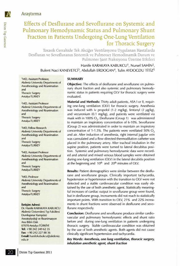
Er! Araştırma
Effects of Desflurane and Sevoflurane on Systemic and Pulmonary Hemodynamic Status and Pulmonary Shunt
Fraction in Patients Undergoing One-Lung Ventilation for Thoracic Surgery
Torasik Cerrahide Tek Akciğer Ventilasyonu Uygulanan Hastalarda Desfluran ve Sevofluranın Sistemik ve Pulmoner Hemodinamik Durum ve
Pulmoner Şant Fraksiyonu Üzerine Etkileri
Hanife KARAKAYA KABUKCU1, Nursel ŞAHİN2, Bülent Naci KANEVETCİ3, Abdullah ERDOĞAN4, Tülin AYDOGDU TİTİZ5
'MD, Assistant Professor, Akdeniz University Departments of Anesthesiology and Reanimation and Thoracic Surgery Antalya-TURKEY
2 M D , Assistant Professor Akdeniz University Departments of Anesthesiology and Reanimation and Thoracic Surgery Antalya-TURKEY
3 MD, Fellow Research Akdeniz University Departments of Anesthesiology and Reanimation and Thoracic Surgery Antalya-TURKEY
4 M D , Assistant Professor Akdeniz University Departments of Anesthesiology and Reanimation and Thoracic Surgery Antalya-TURKEY
5 M D , Professor Akdeniz University Departments of Anesthesiology and Reanimation and Thoracic Surgery Antalya-TURKEY
İletişim Adresi: Dr. Hanife KARAKAYA KABUKCU Akdeniz Üniversitesi Tıp Fakültesi Dumlupınar Kampusu Anesteziyoloji ve Reanimasyon Ana Bilim Dalı 07070-Antalya-TÜRKİYE Tel: +90 242 249 62 35 Fax: + 9 0 242 227 88 36 E-mail: hanifekabukcu@akdeniz. edu.tr
SUMMARY
Objective: The effects of desflurane and sevoflurane on pulmonary shunt fraction and also systemic and pulmonary hemodynamic status in patients requiring OLV for thoracic surgery were evaluated.
Material and Methods: Thirty adult patients, ASA I or II, requiring one-lung ventilation (OLV) for thoracic surgery. Anesthesia was induced with iv propofol (1-2 mg/kg), fentanyl (2 ptg/kg), and vecuronium (0.1 mg/kg), and patients were ventilated via mask with in 100% 0 2 . Desflurane (Group 1) was administered to maintain an inspiratory concentration of 6-10%. Sevoflurane (Group 2) was administrated in order to maintain an inspiratory concentration of 1-1.5%. The patients were ventilated 50% 0 2
and air. After induction of anesthesia, right internal jugular vein was cannulated and a flow-directed thermodilution catheter was placed in the pulmonary artery. After tracheal intubation in the supine position, patients were turned to lateral decubitus position. Systemic and pulmonary hemodynamic data were recorded and arterial and mixed venous blood samples were obtained during one-lung ventilation (OLV) in the lateral decubitis position at the beginning and 10 , h and 20 t h minutes of OLV.
Results: Patient demographics were similar between the desflurane and sevoflurane groups. Clinically important tachycardia, hypotension or hypertension with the transition to OLV were not detected and a stable cardiovascular condition was easily obtained by the use of both anesthetic agent. Statistically meaningful increases of cardiac output in sevoflurane group were found, but in desflurane group, increaments did not reach to statistically important points. With transition to OLV, 2 1 % and 22% increaments in shunt fractions were observed in desflurane and sevoflurane respectively.
Conclusion: Desflurane and sevoflurane produce similar cardiovascular and pulmonary hemodynamic effects and shunt ratio before and during one-lung ventilation in patients undergoing thoracic surgery. Stable cardiovascular condition was obtained by the use of both anesthetic agents. Both agents did not cause clinically significant hypertension and tachycardia.
Key Words: Anesthesia, one lung ventilation, thoracic surgery, inhalation anesthetic agent, shunt fraction
Dirim Tıp Gazetesi 2011
Effects of Desflurane and Sevoflurane on Systemic and Pulmonary Hemodynamic Status and Pulmonary Shunt Fraction in Patients Undergoing One-Lung Ventilation for Thoracic Surgery a
ÖZET
Amaç: Torasik cerrahide tek akciğer ventilas-yonu (TAV) uygulanan hastalarda desfluran ve sevofluranın sistemik ve pulmoner hemodina-mik durum ve pulmoner şant fraksiyonu üzerine etkilerinin değerlendirilmesidir.
Gereç ve Yöntemler: Çalışmaya, torasik cerrahide tek akciğer ventilasyonu (TAV) yapılan 30 erişkin hasta dâhil edildi. Anestezi indüksi-yonunda, intravenöz (IV) propofol (1-2 m.£ fentanil (2 jUg/kg) ve vekuronyum (0.1 m§ uygulanan hastalar %100 0 2 maske ile ventile edildi. Anestezi idamesinde desfluran %6-10 (Grup 1) sevofluran (Grup 2) %1-1.5 inspiratuar konsantrasyonlarda uygulandı. %50 0 2 ve kuru hava ile mekanik ventilasyon yapıldı. Trakeal entübasyondan sonra sağ internal juguler ven kanülü uygulandı ve pulmoner arter kateteri yerleştirildi. Lateral dekübitis pozisyonunda tek akciğer ventilasyonu başlangıcında, 10. ve 20. dakikalarda sistemik ve pulmoner hemodi-nami kaydedilerek, arteriyel ve miks venöz kan örnekleri alındı.
Bulgular: Desfluran ve sevofluran gruplarındaki hastaların demografik özellikleri benzerdir. TAV'a geçişte klinik olarak önemli taşikardi, hipotansiyon veya hipertansiyon bulunmamıştır. Her iki anestezik ajanın kullanımında kardiyo-vasküler stabilite kolay elde edilmiştir. Sevofluran grubunda istatistiksel olarak anlamlı kardiyak "output" artışı bulunmuştur, desfluran grubunda istatistiksel olarak anlamlı bir artış gözlenmemiştir. TAV'a geçişte şant fraksiyonunda artış desfluranda % 2 1 , sevofluranda %22 olarak gözlenmiştir.
Sonuç: Desfluran ve sevofluran torasik cerrahi hastalarında tek akciğer ventilasyonu öncesinde ve sonrasında kardiyovasküler ve pulmoner hemodinamik etkileri ve şant oranları benzerdir. Her iki anestezik ajanın kullanımında kardiyovasküler stabilite kolay elde edilmiştir. Her iki anestezik ajan klinik olarak anlamlı hipertansiyon ve taşikardiye neden olmamıştır. Anahtar Kelimeler: Anestezi, tek akciğer ventilasyonu, torasik cerrahi, inhalasyon anestezik ajanı, şant fraksiyonu
INTRODUCTION
Inhalation anaesthesia is generally favored over iv anaesthesia for surgery requiring one-lung ventilation (OLV) because of its several advantages. These include ease of drug delivery, off
set independent of hepatic or renal function, end-tidal concentration monitoring, broncho-dilatation and the option of gaseous induction (1,2). In contrast, inhalation anaesthetics inhibit the hypoxic pulmonary vasoconstriction (HPV) which is a homeostatic mechanism of pulmonary circulation. HPV maintains the optimal oxygenation of the arterial blood with a mechanism that diverts pulmonary blood flow away from lung regions with low alveolar oxygen tensions towards better ventilated areas of the lung, thus reducing venous admixture (2,3).
Desflurane and sevoflurane are very commonly used volatile anesthetic agents. Their pharmacokinetic properties make them highly attractive for use in thoracic surgery (4). All inhalated anesthetics inhibit HPV to varying degrees and therefore affect intrapulmonary shunting (2). In this study, we compared the effects of desflurane and sevoflurane on pulmonary shunt fraction and on systemic and pulmonary hemodynamic status in patients requiring OLV for thoracic surgery.
MATERIAL AND METHODS
After acquiring the approval from the hospital's ethics committee and informed consent, 30 adult patients, ASA I or II, requiring OLV for thoracic surgery were randomized to receive inhalational anesthesia with desflurane (n= 15, patients 1-15) or sevoflurane (n= 15, patients 16-30). None had a history of obstructive airways disease. Patients demonstrating hemodynamic instability, renal hepatic insufficiency or neurological disease were excluded. Heart disease was evaluated by means of personal medical history, physical status, electrocardiography, and echocardiography.
No preanesthetic medication was administered. After arrival in the operating room, an IV cannula was placed for infusion of 0.9% saline 10 mL.kg 1 and a radial arterial catheter was placed for continuous monitorisation of arterial blood pressure.
Anaesthesia was induced by IV propofol (1-2 mg/kg), fentanyl (2 /Jg/kg), and vecuronium (0.1 mg/kg), and patients were ventilated via a mask with in 100% 0 2 . After induction of anaesthesia, a left-sided double-lumen endobronchial tube (Broncho-Cath, Mallinckrodt Medical, Athlone, Ireland) was placed in all patients and initially positioned by auscultation. The endobronchial tube position was confirmed and adjusted with
Dirim Tıp Gazetesi 2011: yıl: 86 sayı: 1 (20-26)
E Hanife KARAKAYA KABUKCU ve ark.
fiberoptic bronchoscopy. Tracheal and bronchial cuff pressures were measured and kept between 20-40 cm H 2 0 with intermittant measurements and manuel pressure release. Desflurane (Group 1) was administered to maintain an inspiratory concentration of 6-10%. Sevoflurane (Group 2) was administered to maintain an inspiratory concentration of 1-1.5%. The patients were ventilated with 50% 0 2 a n d air. During two-lung ventilation, tidal volumes were adjusted to be 10 mL/kg. As one-lung ventilation began, tidal volumes were readjusted to be 4-5 mL/kg so as to keep the arterial carbondioxide concentrations between 35 and 45 mmHg.
After tracheal intubation, right internal jugular vein was cannulated and a flow-directed ther-modilution catheter (Edwards Lifesciences LLC, Irvine, CA, USA) was placed in the pulmonary artery. In all patients, other monitoring parameters including ECG, pulse oximeter, nasopharyngeal temperature, neuromuscular block, ventilation pressures and volumes, end-tidal carbon-dioxide concentration and urine output were performed. Peak airway pressure, tidal volume, ventilatory rate, both tracheal and bronchial cuff pressures were followed up.
After intubation in the supine position, patients were turned to lateral decubitus position. Systemic and pulmonary hemodynamic datas were recorded and during OLV in the lateral decu-bitis position, arterial and mixed venous blood samples were obtained at the beginning
(0 minutes) and 10 t h and 20 l h minutes during OLV.
In patients with PA catheters; thermodilution cardiac output (CO), stroke volume (SV), systemic and pulmonary vascular resistances (SVR and PVR, respectively), mixed venous blood gases were measured. At the same time, hae-modynamic variables were recorded, including heart rate (HR), mean arterial pressure, mean pulmonary arterial pressures (MPAP), pulmonary arterial occlusion pressures (PCWP) and central venous pressures (CVP). Oxygen consumption (V0 2 ) was determined as the product of cardiac output and the difference between arterial and venous oxygen content. The shunt fraction (Qs/ Qt) was computed using a standard formula Qs/ Q t = (Cc02-Ca02)/(Cc02-Cv02) where Q s = shunt flow, Q t = cardiac output and Cc 0 2 = oxygen content of pulmonary end-capillary, C a 0 2 = arterial oxygen content, Cv 0 2 = mixed venous oxygen content (5).
Dirim Tıp Gazetesi 2011
The two-tailed Student's unpaired t-test compared groups for normally distributed data, which are reported as means ± SD. Comparisons of treatment within each group were made by Paired t-test significance required a value of p < 0.05.
RESULTS
1) Demographic Data
Patient demographics were similar between the desflurane and sevoflurane groups (Table 1).
Table 1. Demographics and preoperative data
Age (year)
Weight (kg)
BSA (m2)
HR (bpm)
MAP (mmHg)
PaO? (mmHg)
PaCO, (mmHg)
Oxygen saturation (%)
Duration of operation (min)
Duration of anaesthesia (min)
Desflurane (n= 15)
47 ± 12
67.8 ± 12
1.76 ± 0 . 1 5
76.6 ± 10
81.9 ± 8.5
93 ± 12
35.5 ± 3.6
97.4 ± 1.2
120 ± 59
171 ± 47
Sevoflurane ( n = 15)
54 ± 13
65.5 ± 13
1.70 ± 0.19
82.3 ± 16
86 ± 18
94 ± 11
36.6 ± 3.9
97.3 ± 0.9
143 ± 62
185 ± 66
BSA= body surface area, HR= heart rate, MAP= mean arterial pressure.
2) Evaluation of Hemodynamical Changes with Transition to One Lung Ventilation According to Baseline (Two-Lung Ventilation) (Table 2)
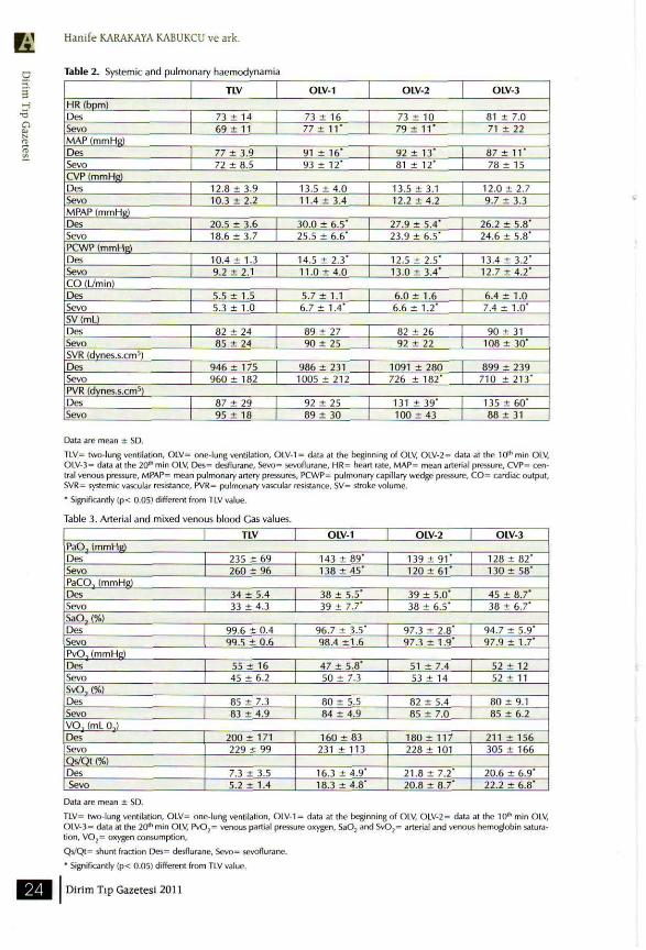
Heart rate: There were no significant differences in desflurane group. In sevoflurane group, statistically meaningful increases were found at the beginning and the 10 t h minute of OLV. They returned back to base-line levels at 20 , h
minute.
Mean arterial pressure: Significant increases were measured at the beginning and during OLV in desflurane group. In group sevoflurane, at the beginning and at the 10 t h minute of OLV, statistically significant increases of mean arterial pressures were observed and at the 20 l h minute of OLV, they returned to base-line levels.
Central venous pressure: CVP did not change at the beginning or during OLV in both groups.
Effects of Desflurane and Sevoflurane on Systemic and Pulmonary Hemodynamic Status and Pulmonary Shunt Fraction in Patients Undergoing One-Lung Ventilation for Thoracic Surgery '&
Mean pulmonary artery pressure: MPAP increased at the beginning and during OLV in both two groups.
Pulmonary capillary wedge pressure: It increased at the beginning and during OLV in desflurane group. In sevoflurane group, it increased after 10 , h minute of OLV.
Cardiac output: In sevoflourane group, statistically meaningful increases were measured. In desflurane group, increases did not reach to statistically important point.
Stroke volume: SV did not change at the beginning or during OLV in desflurane group. In sevoflurane group, it did not change at the beginning and the 10 I h minute of OLV but increased at the 20 , h minute of OLV.
Systemic vascular resistance: It did not change at the beginning and during OLV in desflurane group. In sevoflurane group, it decreased at the 10 t h and 20 , h minutes of OLV.
Pulmonary vascular resistance: In desflurane group, it increased at 10 , h and 20 , h minutes of OLV. It did not change at the beginning or during OLV in sevoflurane group.
Shunt fraction: It increased at the beginning
and during OLV in both group.
3) Evaluation of Changing in the Blood Gases
with Transition to One Lung Ventilation
According to Baseline (Two-Lung Ventilation)
(Table 3-4)
P a 0 2 and Sa0 2 : reductions in Pa0 2 and Sa0 2
occurred during OLV in desflurane and sevoflurane groups.
P a 0 2 : increases in PaC02 occurred during OLV
in desflurane and sevoflurane groups.
P v 0 2 and Sv0 2 . Statistically meaningful changes were not observed in both groups at the beginning and during OLV.
Oxygen consumption: It remained unchanged
during OLV in both groups.
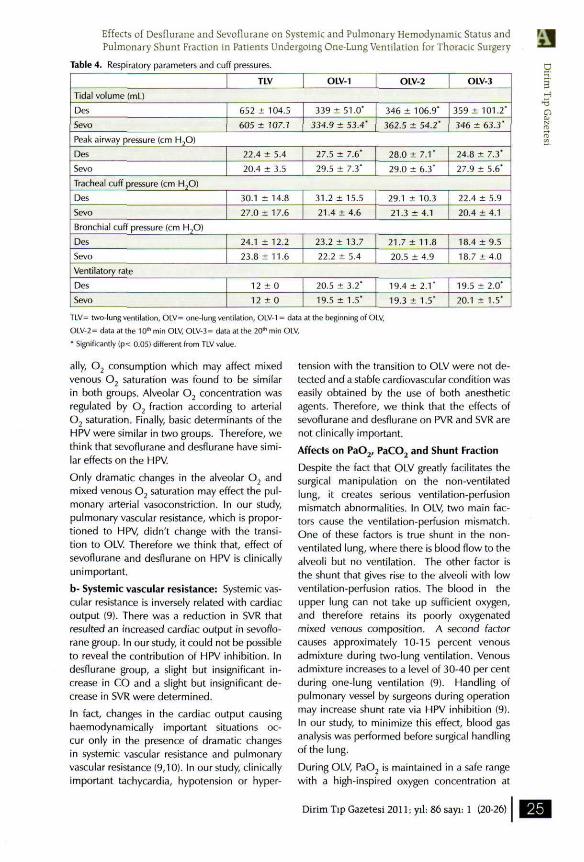
Tidal volume: It reduced in both groups during OLV
Peak airway pressure: It increased in both groups during OLV. Tracheal and bronchial cuff pressures did not change with transition and during OLV.
DISCUSSION
In this study, the effects of desflurane and sevo
flurane on systemic and pulmonary hemodynamic status and pulmonary shunt fraction in patients requiring OLV for thoracic surgery were investigated.
Evaluation of the Heart Rate and Arterial Pressure
In desflurane group, while heart rate did not change with transition to OLV, an increase in blood pressure within normal range, however, was determined. In sevoflurane group, while heart rate increased at beginning and the 10 t h
minute, blood pressure was seen to be increased within normal range. Clinically significant tachycardia or hypertension was not determined. It has been reported that a rapid ascent to desflurane concentration over 1 MAC had caused CNS activation via tracheopulmonary receptors, and resulted in tachycardia and hypertension (6-8). That's why concentration over 1 MAC was never reached in our study. It was also reported that desflurane concentration below 1 MAC did not cause any significant alteration in heart rate (6-8). In our study, although a slight increase in heart rate was seen, it was not clinically significant. We think that, the restrictive effect of fentanyl on sympathetic nervous system, used in induction and maintenance of anaesthesia, may have prevented significant tachycardia and hypertension.
Evaluation of Cardiac Output and its Determinants
Consecutive physiopathological interactions exist among cardiac output, mixed venous 0 2 saturation, and hypoxic pulmonary vasoconstriction (9). A decrease in cardiac output reduces mixed venous 0 2 saturation and results in increases in pulmonary vasoconstriction. An increase in cardiac output, however, increases mixed 0 2 saturation and reduces hypoxic pulmonary vasoconstriction (9). In our study; increase in cardiac output was statistically significant in sevoflurane group. In desflurane group, a slight but insignificant increase was determined.
a- Hypoxic pulmonary vasoconstriction: Hypoxic pulmonary vasoconstriction (HPV) which has been mainly detected by alveolar 0 2 concentration and mixed venous 0 2 saturation is inversely related to the cardiac output (10). In our study, when we had turned to OLV, no significant difference was observed in respect to mixed venous 0 2 saturation and pressure (partial pressure of Ö2) in both groups. Addition-
Dirim Tıp Gazetesi 2011; yıl: 86 sayı: 1 (20-26)
E Hanife KARAKAYA KABUKCU ve ark.
Table 2. Systemic and pulmonary haemodynamia
TLV OLV-1 OLV-2 OLV-3
HR (bpm) Des Sevo
73 ± 14 69 ± 11
73 ± 16 77 ± 1 1 '
73 ± 10 79 ± 11"
81 ± 7.0 71 ± 22
MAP (mmHg) Des Sevo
77 ± 3.9 72 ± 8.5
91 ± 1 6 ' 93 ± 12'
92 ± 13' 81 ± 12'
87 ± 1 1 ' 78 ± 15
CVP (mmHg) Des Sevo
12.8 ± 3.9 10.3 ± 2.2
13.5 ± 4.0 11.4 ± 3.4
13.5 ± 3.1 12.2 ± 4 . 2
12.0 ± 2.7 9.7 ± 3.3
MPAP (mmHg) Des Sevo
20.5 ± 3-6 18.6 ± 3.7
30.0 ± 6.5' 25.5 ± 6.6"
27.9 ± 5.4' 23.9 ± 6.5'
26.2 ± 5.8' 24.6 ± 5.8'
PCWP (mmHg) Des Sevo
10.4 ± 1.3 9.2 ± 2.1
14.5 ± 2.3' 11.0 ± 4.0
12.5 ± 2.5' 13.0 ± 3.4"
13.4 ± 3.2" 12.7 ± 4.2'
CO (l./min) Des Sevo
5.5 ± 1.5 5.3 ± 1.0
5.7 ± 1.1 6.7 ± 1.4'
6.0 ± 1.6 6.6 ± 1.2*
6.4 ± 1.0 7.4 ± 1.0'
SV (mL) Des Sevo
82 ± 24 85 ± 24
89 ± 27 90 ± 25
82 ± 26 92 ± 22
90 ± 31 108 ± 3 0 '
SVR (dynes.s.cm') Des Sevo
946 ± 175 960 ± 1 8 2
986 ± 231 1005 ± 212
1091 ± 280 726 ± 182"
899 ± 239 710 ± 213'
PVR (dynes.s.cm5) Des Sevo
87 ± 29 95 ± 18
92 ± 25 89 ± 30
131 ± 39' 100 ± 4 3
135 ± 6 0 ' 88 ± 31
Data are mean ± SO.
TLV= two-lung ventilation, OLV= one-lung ventilation, OLV-1 = data at the beginning of OLV, OLV-2 = data at the 10,h min OLV, OLV-3 = data at the 20th min OLV, Des= desflurane, Sevo= sevoflurane, HR= heart rate, MAP= mean arterial pressure, CVP= central venous pressure, MPAP= mean pulmonary artery pressures, PCWP= pulmonary capillary wedge pressure, CO= cardiac output, SVR= systemic vascular resistance, PVR= pulmonan/ vascular resistance, SV= stroke volume.
* Significantly (p< 0.05) different from TLV value.
Table 3. Arterial and mixed venous blood Gas values.
TLV OLV-1 OLV-2 OLV-3 PaO, (mmHg) Des Sevo
235 ± 69 260 ± 96
143 ± 89" 138 ± 4 5 '
139 ± 9 1 * 120 ± 6 1 '
128 ± 8 2 ' 130 ± 58*
PaCO, (mmHg) Des Sevo
34 ± 5.4 33 ± 4.3
38 ± 5.5" 39 ± 7.7'
39 + 5.0' 38 ± 6.5'
45 ± 8.7' 38 ± 6.7"
SaO, (%) Des Sevo
99.6 ± 0.4 99.5 ± 0.6
96.7 ± 3.5' 98.4 ± 1 . 6
97.3 ± 2.8' 97.3 ± 1.9"
94.7 ± 5.9" 97.9 ± 1.7*
PvO,(mmHg) Des Sevo
55 ± 16 45 ± 6.2
47 ± 5.8* 50 ± 7.3
51 ± 7.4 53 ± 14
52 ± 12 52 ± 11
SvO, (%) Des Sevo
85 ± 7.3 83 ± 4.9
80 ± 5.5 84 ± 4.9
82 ± 5.4 85 ± 7.0
80 ± 9.1 85 ± 6.2
V O , (mL 0,) Des Sevo Qs/Qt (%) Des Sevo
200 ± 171 229 ± 99
7.3 ± 3.5 5.2 ± 1.4
160 ± 83 231 ± 113
16.3 ± 4 . 9 ' 18.3 ± 4.8'
180 ± 117 228 ± 101
21.8 ± 7.2* 20.8 ± 8.7'
211 ± 156 305 ± 166
20.6 ± 6.9' 22.2 ± 6.8"
Data are mean ± SD.
TLV= two-lung ventilation, OLV= one-lung ventilation, OLV-1 = data at the beginning of OLV, OLV-2 = data at the 10 lh min OLV, OLV-3= data at the 20 l h min OLV, Pv02 = venous partial pressure oxygen, Sa02 and Sv02 = arterial and venous hemoglobin saturation, V 0 2 = oxygen consumption,
Qs/Qt= shunt fraction Des= desflurane, Sevo= sevoflurane.
* Significantly (p< 0.05) different from TLV value.
Dirim Tıp Gazetesi 2011
Effects of Desflurane and Sevoflurane on Systemic and Pulmonary Hemodynamic Status and Pulmonary Shunt Fraction in Patients Undergoing One-Lung Ventilation for Thoracic Surgery
Table 4. Respiratory parameters and cuff pressures.
TLV OLV-1 OLV-2 OLV-3
Tidal volume (mL)
Des
Sevo
652 ± 104.5
605 ± 107.1
339 ± 51.0'
334.9 ± 53.4'
346 ± 106.9"
362.5 ± 54.2'
359 ± 101.2'
346 ± 63.3"
Peak airway pressure (cm H,0)
Des
Sevo
22.4 ± 5.4
20.4 ± 3.5
27.5 ± 7.6"
29.5 ± 7.3" 28.0 ± 7.1'
29.0 ± 6.3'
24.8 ± 7.3'
27.9 ± 5.6'
Tracheal cuff pressure (cm H20)
Des
Sevo
30.1 ± 14.8
27.0 ± 17.6
31.2 ± 15.5
21.4 ± 4.6 29.1 ± 10.3
21.3 ± 4.1
22.4 ± 5.9
20.4 ± 4.1
Bronchial cuff pressure (cm H?0)
Des Sevo
24.1 ± 12.2
23.8 ±11.6
23.2 ± 13.7
22.2 ± 5.4 21.7 ± 11.8
20.5 ± 4.9
18.4 ±9.5
18.7 ±4.0
Ventilatory rate
Des
Sevo
12 ± 0
12 ± 0
20.5 ± 3.2'
19.5 ± 1.5*
19.4 ± 2.1'
19.3 ± 1.5"
19.5 ± 2.0'
20.1 ± 1.5'
TLV= two-lung ventilation, OLV= one-lung ventilation, OLV-1 = data at the beginning of OLV,
OLV-2 = data at the 10* min OLV, OLV-3 = data at the 20* min OLV,
* Significantly (p< 0.05) different from TLV value.
ally, 0 2 consumption which may affect mixed venous 0 2 saturation was found to be similar in both groups. Alveolar 0 2 concentration was regulated by 0 2 fraction according to arterial 0 2 saturation. Finally, basic determinants of the HPV were similar in two groups. Therefore, we think that sevoflurane and desflurane have similar effects on the HPV.
Only dramatic changes in the alveolar 0 2 and mixed venous 0 2 saturation may effect the pulmonary arterial vasoconstriction. In our study, pulmonary vascular resistance, which is proportioned to HPV, didn't change with the transition to OLV. Therefore we think that, effect of sevoflurane and desflurane on HPV is clinically unimportant.
b- Systemic vascular resistance: Systemic vascular resistance is inversely related with cardiac output (9). There was a reduction in SVR that resulted an increased cardiac output in sevoflo-rane group. In our study, it could not be possible to reveal the contribution of HPV inhibition. In desflurane group, a slight but insignificant increase in CO and a slight but insignificant decrease in SVR were determined.
In fact, changes in the cardiac output causing haemodynamically important situations occur only in the presence of dramatic changes in systemic vascular resistance and pulmonary vascular resistance (9,10). In our study, clinically important tachycardia, hypotension or hyper
tension with the transition to OLV were not detected and a stable cardiovascular condition was easily obtained by the use of both anesthetic agents. Therefore, we think that the effects of sevoflurane and desflurane on PVR and SVR are not clinically important.
Affects on P a 0 2 , PaC0 2 and Shunt Fraction
Despite the fact that OLV greatly facilitates the surgical manipulation on the non-ventilated lung, it creates serious ventilation-perfusion mismatch abnormalities. In OLV, two main factors cause the ventilation-perfusion mismatch. One of these factors is true shunt in the non-ventilated lung, where there is blood flow to the alveoli but no ventilation. The other factor is the shunt that gives rise to the alveoli with low ventilation-perfusion ratios. The blood in the upper lung can not take up sufficient oxygen, and therefore retains its poorly oxygenated mixed venous composition. A second factor causes approximately 10-15 percent venous admixture during two-lung ventilation. Venous admixture increases to a level of 30-40 per cent during one-lung ventilation (9). Handling of pulmonary vessel by surgeons during operation may increase shunt rate via HPV inhibition (9). In our study, to minimize this effect, blood gas analysis was performed before surgical handling of the lung.
During OLV, Pa0 2 is maintained in a safe range with a high-inspired oxygen concentration at
Dirim Tıp Gazetesi 2011: yıl: 86 sayı: 1 (20-26)
E 3
Hanife KARAKAYA KABUKCU ve ark.
level of 50% or over. Additionally, we reduced
tidal volume and increased ventilation rate. In
our study, with transition to OLV, a similar re
duction of Pa0 2 in both groups was observed.
There were slight increases in PaC02 values but
they were within normal ranges. Reduction in
0 2 saturation of clinical importance was not de
termined. In patients in whom sufficient Pa0 2
could not be obtained by F i 0 2 level and tidal
volume/ventilation rate regulations, PEEP has
been proposed for restriction of blood flow at
nonventilated lung (11,12). In our study, al
though there were not any patients requiring
this application, during OLV, it is advised to be
aware of all necessary maneuvers to increase
Pa02.
In our study with transition to OLV, 2 1 % and
22% increases in shunt fraction were observed in
desflurane and sevoflurane groups respectively.
In present study, in both groups, adequate arte
rial oxygenation could be maintened with these
shunt rates. Beck et al (13) reported 30% shunt
fraction in OLV with sevoflurane, and Slinger et
al (14) found 39% and 4 1 % shunt fraction with
isoflurane and enflurane respectively in thoracic
surgery. Pagel and et al (15) tested the effects of
desflurane and isoflurane on shunt fraction in
patients undergoing one-lung ventilation. They
found 40% shunt fraction in desflurane group
and 34% shunt fraction in isoflurane group. In
all of these studies, as in our study, increases in
shunt fraction with the onset of OLV were well
tolerated with implement of a high concentrat
ed oxygen inspiration.
CONCLUSION
In present study, it was concluded that, desflu
rane and sevoflurane produce similar cardio
vascular and pulmonary haemodynamic effects
and shunt fraction before and during one-lung
ventilation in patients undergoing thoracic sur
gery. A stable cardiovascular condition was easi
ly obtained by the use of both anesthetic agents.
Both agents did not cause clinically significant
hypertension and tachycardia.
REFERENCES:
1. Pavlin EC, Su |Y. Inhaled anesthetics, cardiopulmonary pharmacology. In: Miller RD editor. Anesthesia.4thed. New York: Churchill Livingstone; 1994. p.125-56.
2. Baden |M, Rice SA. Inhaled anesthetics, metabolism and toxicity. In: Miller RD editor. Anesthesia. 4thed. New York: Churchill Livingstone; 1994. p.157-84.
3. Gothard ), Porter H. Controversies in thoracic anaesthesia. In: Ghosh S, Latimer RD editors. Thoracic Anaesthesia principles and practice. 1sted. Oxford: Butterworth Heinemann; 1999. p.307-28.
4. Dupont J, Tavernier B, Ghosez Y, et al. Recovery after anaesthesia for pulmonary surgery: desflurane, sevoflurane and isoflurane. Br J Anaesthesia 1999;82(3):355-9.
5. Nunn )F. Applied Respiratory Physiology, 4thed. Boston: Butterworths-Heineman; 1993. p.167-76.
6. Ebert T), Muzi M. Sympathetic hyperactivity during desflurane anesthesia in healthy volunteers. Anesthesiology 1993;79131:444-53.
7. Weiskopf RB, Moore MA, Eger El 2nd, et al. Rapid increase in desflurane concentration is associated with greater transient cardiovascular stimulation than rapid increase in isoflurane concentration in humans. Anesthesiology 1994;80151:1035-45.
8. Weiskopf RB, Eger El II, Daniel M, et al. Cardiovascular stimulation induced by rapid increases in humans result from activation of tracheopulmonary and systemic receptors. Anesthesiology 1995;83(6):1173-8.
9. Grossman W. Blood flow measurement: the cardiac output in cardiac catheterization. In: Bairn DS, Grossman W, editors. Grossman's cardiac catheterization, angiography and intervention. 6thed. Philadelphia: Lippincott, Williams&Wilkins; 2000. p.159-78.
10. Eisenkraft JB. Effects of anaesthetics on the pulmonary circulation. Br | Anaesthesia 1990;65111:63-78.
11. Capan LM, Turndorf H, Patel C, et al. Optimization of arterial oxygenation during one-lung anaesthesia. Anesth Analg 1980;59111:847-51.
12. Alfery DD, Benumof JL, Trousdale FR. Improving oxygenation during one-lung ventilation in dogs: the effects of positive end-expiratory pressure and blood flow restriction to the non-ventilated lung. Anesthesiology 1981;55141:381-5.
13. Beck DH, Doepfmer UR, Sinemus Bloch A, Schenk MR, Kox W|. Effects of sevoflurane and propofol on pulmonary shunt fraction during one-lung ventilation for thoracic surgery. Br) Anaesthesia 20O1;86(1):38-43.
14. Slinger R Scott WAC. Arterial oxygenation during one-lung ventilation. Anesthesiology 1995;82141:940-6.
15. Pagel PS, Fu JL, Damask MC, et al. Desflurane and isoflurane produce similar alterations in systemic and pulmonary hemodynamics and arterial oxygenation in patients undergoing one-lung ventilation during thoracotomy.
Anesthesia and Analgesia 1998;87(4):800-7.
Dirim T ı p Gazetesi 2011