Embed Size (px)
Citation preview
Equine Diagnostic Imaging: Evaluation of the Forelimb & Hindlimb &
Back to Basics: Taking Radiography to the Next Level
Myra F. Barrett DVM, MS, DACVR
Introduction:
The subject of diagnostic imaging of the fore and hindlimb is broad and of course
could fill a textbook. The goal of these proceedings is to discuss common areas of
difficulty as well as novel approaches to common imaging exams. These proceedings
will also discuss ways in which to improve basic radiographic interpretation, in part by
comparing to advanced imaging.
Study parameters:
As we learn more about the limitations of diagnostic analgesia in localizing the
lameness to a discrete region, the more we have learned that we need to expand our
included imaging range as well as potentially the number of diagnostic blocks performed.
Lameness localized to the foot is one of the most common causes of lameness, and
therefore one of the most frequently imaged regions. In a patient whose lameness is
markedly improved or abolished with a palmar digital (PDN) nerve block, the source of
the lameness can range from the sole to the distal fetlock region. Imaging of the fetlock
and pastern would be indicated for a horse that responds to an abaxial nerve block, and
imaging of the fetlock and entire metacarpus/metatarsus (MC/MT3) would be indicated
for a horse that improves with a low-four point nerve block. Of course, in horses that
respond partially to one block and partially to another more proximal block, the imaging
area expands. Including intra-articular and intra-thecal analgesia does help further refine
the area to be imaged, although still often includes imaging of areas beyond the synovial
structure of interest. Being aware of the variety of lesions that may occur beyond the
traditionally accepted range of lesions can help decrease the likelihood of missing a
clinically significant imaging abnormality. Obtaining additional radiographs and
performing ultrasound outside the traditional range when no abnormalities are noted
during a typical scan is recommended. For example if a patient’s lameness is improved
with a low four-point nerve block and no abnormalities are noted in the fetlock and distal
cannon region, including the proximal metatarsus/metacarpus is indicated to evaluate for
proximal suspensory ligament disease or other more proximal abnormalities. Likewise,
ideally, MRIs should be evaluated as being acquired so that additional scanning can be
performed if no significant lesions are noted, rather than risking having to repeat the MRI
at a later date to include other anatomic structures.
Diagnostic Imaging of the Forelimb:
Foot:
Similar to many other sources of lameness, lameness localized to the foot is
generally initially evaluated using radiography. Radiography provides good bone detail
and is a useful, readily available and relatively inexpensive screening tool. However, the
more that we learn about pathologic processes in the foot, the more we know that there
are significant limitations to what can be diagnosed with radiographs. Normal
radiographs do not rule out the presence of disease, and similarly, abnormal radiographs
may only show “the tip of the iceberg” in terms of the pathologic processes that are
occurring. Any time advanced imaging of the foot is performed, it is useful to compare
those findings to the radiographs and/or ultrasound findings. Regularly comparing
modalities can markedly improve radiographic and ultrasound interpretation skills. This
may mean repeating the study after the exam to see if additional views can help identify
the pathologic finding identified with advanced imaging.
A standard radiographic study of the foot should include a horizontal DP, lateral,
dorsal 60° palmar (D60P) and skyline view of the navicular bone. Proper positioning is
essential for the skyline view, and malpositioning is one of the most commonly made
mistakes. Poor positioning can easily create the appearance of medullary sclerosis and
loss of corticomedullary distinction. A good way to determine whether a navicular
skyline view is properly positioned is to examine the articulation of the dorsal margin of
the navicular bone and the palmar aspect of the middle phalanx. If this joint space is
clearly defined, then the radiograph is generally correctly positioned. (Figure 1).
Additionally, the foot should be adequately cleaned and prepared to limit debris and gas
artifact. The lateral view is often overlooked for evaluation of the navicular bone. While
less sensitive to changes in the corticomedullary distinction than the skyline view, it is
useful to correlate the findings on the two projections. If there is the appearance of
sclerosis on the skyline view, but excellent corticomedullary distinction on the lateral
view, then there is a greater likelihood that obliquity artifact is affecting the skyline view.
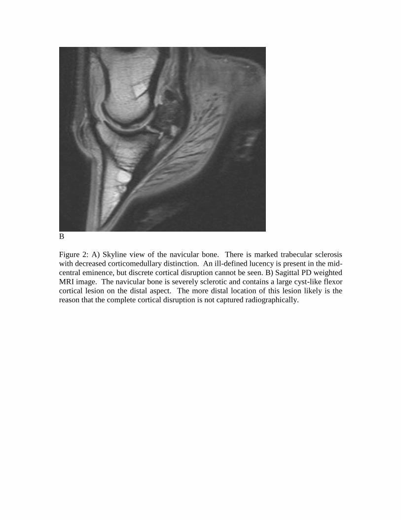
It is important to keep in mind that the flexor cortex of the navicular bone is a
curved structure. This means that it is not possible to be completely tangential to all
components of the cortex with a single skyline image. Knowing this helps explain why a
horse can have a flexor cortex erosion visualized on MRI that is not well visualized
radiographically. However, even when the defect itself is not seen, there should be other
radiographic indicators of disease, particularly medullary sclerosis and a loss of
corticomedullary distinction. (Figure 2) If a flexor cortical erosion is suspected, repeating
the image while varying the angle of the beam slightly can change which portion of the
cortex is highlighted by the radiograph.
While the deep digital flexor tendon and impar ligament cannot be directly
assessed radiographically, there can be osseous changes indicative of disease. Certainly
flexor cortex lysis of the navicular bone is often associated with damage to the deep
flexor tendon. Other osseous changes include focal lucencies on the distal phalanx,
indicative of osseous resorption of the insertion of the impar or deep digital flexor
tendon. These are best appreciated adjacent to the DIP joint space on the D60P view.
Additionally, osseous irregularity and sclerosis of the distal margin of the navicular bone
can accompany enthesopathy of the impar ligament. Distal border fragments of the
navicular bone are vastly underdiagnosed radiographically, but when visualized are often
large enough with sufficient adjacent bone change that concurrent impar ligament
abnormalities are likely.
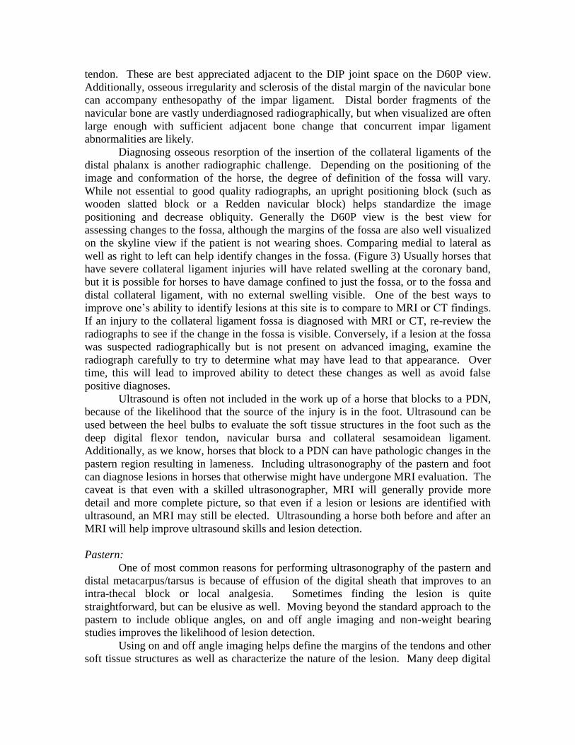
Diagnosing osseous resorption of the insertion of the collateral ligaments of the
distal phalanx is another radiographic challenge. Depending on the positioning of the
image and conformation of the horse, the degree of definition of the fossa will vary.
While not essential to good quality radiographs, an upright positioning block (such as
wooden slatted block or a Redden navicular block) helps standardize the image
positioning and decrease obliquity. Generally the D60P view is the best view for
assessing changes to the fossa, although the margins of the fossa are also well visualized
on the skyline view if the patient is not wearing shoes. Comparing medial to lateral as
well as right to left can help identify changes in the fossa. (Figure 3) Usually horses that
have severe collateral ligament injuries will have related swelling at the coronary band,
but it is possible for horses to have damage confined to just the fossa, or to the fossa and
distal collateral ligament, with no external swelling visible. One of the best ways to
improve one’s ability to identify lesions at this site is to compare to MRI or CT findings.
If an injury to the collateral ligament fossa is diagnosed with MRI or CT, re-review the
radiographs to see if the change in the fossa is visible. Conversely, if a lesion at the fossa
was suspected radiographically but is not present on advanced imaging, examine the
radiograph carefully to try to determine what may have lead to that appearance. Over
time, this will lead to improved ability to detect these changes as well as avoid false
positive diagnoses.
Ultrasound is often not included in the work up of a horse that blocks to a PDN,
because of the likelihood that the source of the injury is in the foot. Ultrasound can be
used between the heel bulbs to evaluate the soft tissue structures in the foot such as the
deep digital flexor tendon, navicular bursa and collateral sesamoidean ligament.
Additionally, as we know, horses that block to a PDN can have pathologic changes in the
pastern region resulting in lameness. Including ultrasonography of the pastern and foot
can diagnose lesions in horses that otherwise might have undergone MRI evaluation. The
caveat is that even with a skilled ultrasonographer, MRI will generally provide more
detail and more complete picture, so that even if a lesion or lesions are identified with
ultrasound, an MRI may still be elected. Ultrasounding a horse both before and after an
MRI will help improve ultrasound skills and lesion detection.
Pastern:
One of most common reasons for performing ultrasonography of the pastern and
distal metacarpus/tarsus is because of effusion of the digital sheath that improves to an
intra-thecal block or local analgesia. Sometimes finding the lesion is quite
straightforward, but can be elusive as well. Moving beyond the standard approach to the
pastern to include oblique angles, on and off angle imaging and non-weight bearing
studies improves the likelihood of lesion detection.
Using on and off angle imaging helps define the margins of the tendons and other
soft tissue structures as well as characterize the nature of the lesion. Many deep digital
flexor tendon injuries in the pastern region are characterized by lobe enlargement and
tearing of the abaxial dorsal margin. The margins of the deep digital flexor tendon
should be evaluated from a palmar medial and palmar lateral approach as well as just
from palmar to increase the likelihood of detecting the lesion. (Figure 4) Additionally,
off angle imaging and non-weight bearing studies improve the ability to outline the
tendon margin and detect changes in shape. Resting the horse’s limb on a farrier sling
during the non-weight bearing exam works well because generally the horse remains
relaxed in this position and it minimizes motion of the limb. (Figure 5). Including off
angle and non-weightbearing studies also improves detection of tearing of the manica
flexoria in the distal metacarpal/tarsal region. Again, any time it is possible, compare the
ultrasound evaluation to advanced imaging findings and/or the results of tenoscopy.
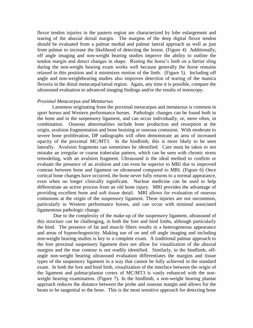
Proximal Metacarpus and Metatarsus
Lameness originating from the proximal metacarpus and metatarsus is common in
sport horses and Western performance horses. Pathologic changes can be found both in
the bone and in the suspensory ligament, and can occur individually, or, more often, in
combination. Osseous abnormalities include bone production and resorption at the
origin, avulsion fragmentation and bone bruising or osseous contusion. With moderate to
severe bone proliferation, DP radiographs will often demonstrate an area of increased
opacity of the proximal MC/MT3. In the hindlimb, this is more likely to be seen
laterally. Avulsion fragments can sometimes be identified. Care must be taken to not
mistake an irregular or coarse trabecular pattern, which can be seen with chronic stress
remodeling, with an avulsion fragment. Ultrasound is the ideal method to confirm or
evaluate the presence of an avulsion and can even be superior to MRI due to improved
contrast between bone and ligament on ultrasound compared to MRI. (Figure 6) Once
cortical bone changes have occurred, the bone never fully returns to a normal appearance,
even when no longer clinically significant. Nuclear medicine can be used to help
differentiate an active process from an old bone injury. MRI provides the advantage of
providing excellent bone and soft tissue detail. MRI allows for evaluation of osseous
contusions at the origin of the suspensory ligament. These injuries are not uncommon,
particularly in Western performance horses, and can occur with minimal associated
ligamentous pathologic change.
Due to the complexity of the make-up of the suspensory ligament, ultrasound of
this structure can be challenging, in both the fore and hind limbs, although particularly
the hind. The presence of fat and muscle fibers results in a heterogeneous appearance
and areas of hypoechogenicity. Making use of on and off angle imaging and including
non-weight bearing studies is key to a complete exam. A traditional palmar approach to
the fore proximal suspensory ligament does not allow for visualization of the abaxial
margins and the true contour is not readily identified. Similarly, in the hindlimb, off-
angle non-weight bearing ultrasound evaluation differentiates the margins and tissue
types of the suspensory ligament in a way that cannot be fully achieved in the standard
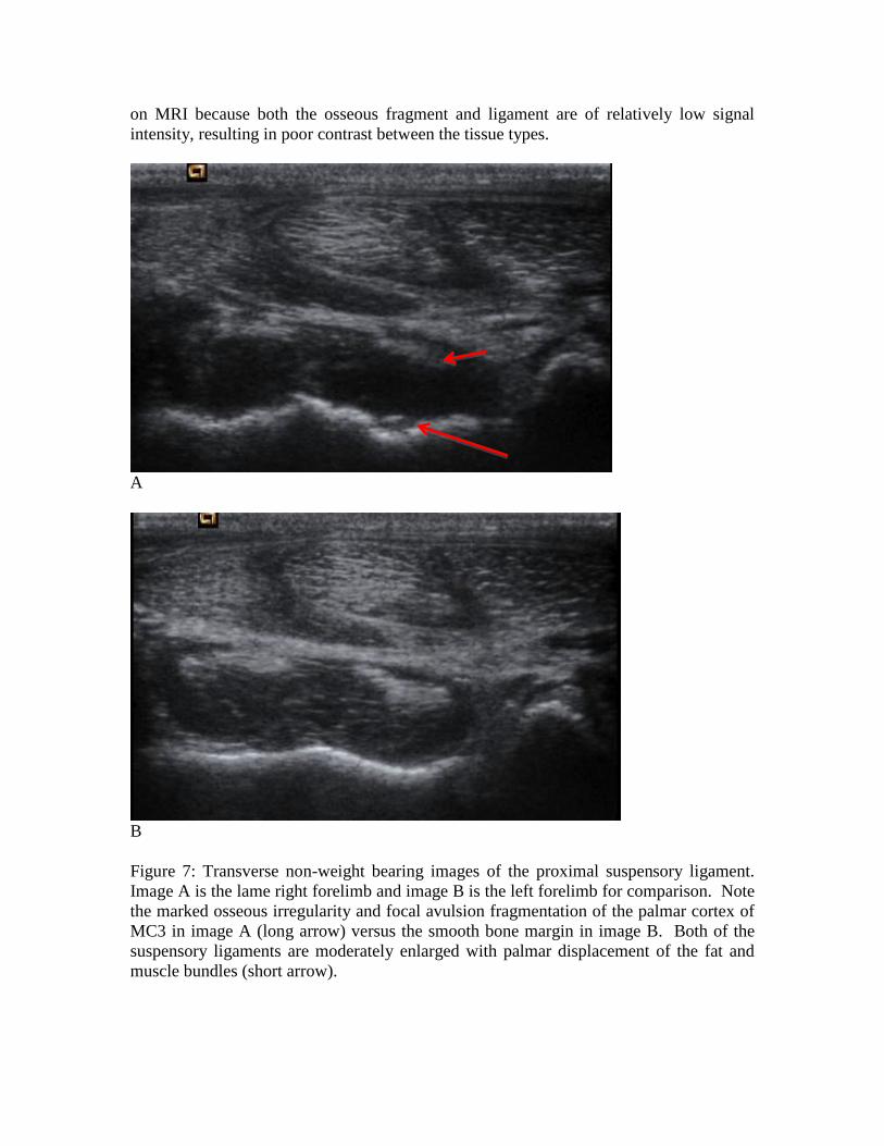
exam. In both the fore and hind limb, visualization of the interface between the origin of
the ligament and palmar/plantar cortex of MC/MT3 is vastly enhanced with the non-
weight bearing examination. (Figure 7). In the hindlimb, a non-weight bearing plantar
approach reduces the distance between the probe and osseous margin and allows for the
beam to be tangential to the bone. This is the most sensitive approach for detecting bone
resorption, proliferation and osseous fragmentation at the origin of the suspensory
ligament. Suspected lesions noted on the long axis approach should be confirmed on
transverse images and vice versa. The appearance of the ligament with off-angle
ultrasound imaging much more closely resembles the appearance on MRI and allows for
better comparison between modalities.
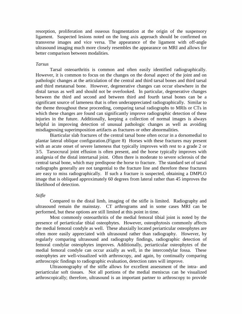
Tarsus
Tarsal osteoarthritis is common and often easily identified radiographically.
However, it is common to focus on the changes on the dorsal aspect of the joint and on
pathologic changes at the articulation of the central and third tarsal bones and third tarsal
and third metatarsal bone. However, degenerative changes can occur elsewhere in the
distal tarsus as well and should not be overlooked. In particular, degenerative changes
between the third and second and between third and fourth tarsal bones can be a
significant source of lameness that is often underappreciated radiographically. Similar to
the theme throughout these proceeding, comparing tarsal radiographs to MRIs or CTs in
which these changes are found can significantly improve radiographic detection of these
injuries in the future. Additionally, keeping a collection of normal images is always
helpful in improving detection of unusual pathologic changes as well as avoiding
misdiagnosing superimposition artifacts as fractures or other abnormalities.
Biarticular slab fractures of the central tarsal bone often occur in a dorsomedial to
plantar lateral oblique configuration.(Figure 8) Horses with these fractures may present
with an acute onset of severe lameness that typically improves with rest to a grade 2 or
3/5. Tarsocrural joint effusion is often present, and the horse typically improves with
analgesia of the distal intertarsal joint. Often there is moderate to severe sclerosis of the
central tarsal bone, which may predispose the horse to fracture. The standard set of tarsal
radiographs generally are not tangential to the fracture line and therefore these fractures
are easy to miss radiographically. If such a fracture is suspected, obtaining a DMPLO
image that is obliqued approximately 60 degrees from lateral rather than 45 improves the
likelihood of detection.
Stifle
Compared to the distal limb, imaging of the stifle is limited. Radiography and
ultrasound remain the mainstay. CT arthrograms and in some cases MRI can be
performed, but these options are still limited at this point in time.
Most commonly osteoarthritis of the medial femoral tibial joint is noted by the
presence of periarticular tibial osteophytes. However, osteophytosis commonly affects
the medial femoral condyle as well. These abaxially located periarticular osteophytes are
often more easily appreciated with ultrasound rather than radiography. However, by
regularly comparing ultrasound and radiography findings, radiographic detection of
femoral condylar osteophytes improves. Additionally, periarticular osteophytes of the
medial femoral condyle can occur axially as well, in the intercondylar fossa. These
osteophytes are well-visualized with arthroscopy, and again, by continually comparing
arthroscopic findings to radiographic evaluation, detection rates will improve.
Ultrasonography of the stifle allows for excellent assessment of the intra- and
periarticular soft tissues. Not all portions of the medial meniscus can be visualized
arthroscopically; therefore, ultrasound is an important partner to arthroscopy to provide
the most complete diagnostic visualization of the joint. In particular, tears of the medial
meniscus located in the mid body may not be seen arthroscopically unless the tearing
radiates to the cranial or caudal horn. It is important to realize that failure to visualize
meniscal damage arthroscopically does not preclude the presence of a tear. Conversely,
visualization of articular cartilage defects without adjacent subchondral damage,
including thin fissures or diffuse fibrillation is poor with ultrasound in comparison to
arthroscopy. This further emphasizes the need for a multi-modality approach to imaging
the stifle.
Summary
In conclusion, the accuracy of diagnostic imaging improves with a dynamic,
“think outside the box” approach. Expanding the normal range of a scan, including
multiple approaches to ultrasound examinations and utilizing multiple radiographic
projections increases the likelihood of lesion detection as well as a more complete
clinical picture. Comparing multiple modalities, including other diagnostic imaging
modalities, surgical results and post mortem findings is one of the best ways to continue
to improve imaging skill.
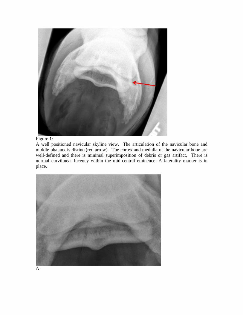
Figure 1:
A well positioned navicular skyline view. The articulation of the navicular bone and
middle phalanx is distinct(red arrow). The cortex and medulla of the navicular bone are
well-defined and there is minimal superimposition of debris or gas artifact. There is
normal curvilinear lucency within the mid-central eminence. A laterality marker is in
place.
A
B
Figure 2: A) Skyline view of the navicular bone. There is marked trabecular sclerosis
with decreased corticomedullary distinction. An ill-defined lucency is present in the mid-
central eminence, but discrete cortical disruption cannot be seen. B) Sagittal PD weighted
MRI image. The navicular bone is severely sclerotic and contains a large cyst-like flexor
cortical lesion on the distal aspect. The more distal location of this lesion likely is the
reason that the complete cortical disruption is not captured radiographically.
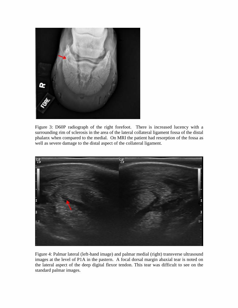
Figure 3: D60P radiograph of the right forefoot. There is increased lucency with a
surrounding rim of sclerosis in the area of the lateral collateral ligament fossa of the distal
phalanx when compared to the medial. On MRI the patient had resorption of the fossa as
well as severe damage to the distal aspect of the collateral ligament.
Figure 4: Palmar lateral (left-hand image) and palmar medial (right) transverse ultrasound
images at the level of P1A in the pastern. A focal dorsal margin abaxial tear is noted on
the lateral aspect of the deep digital flexor tendon. This tear was difficult to see on the
standard palmar images.
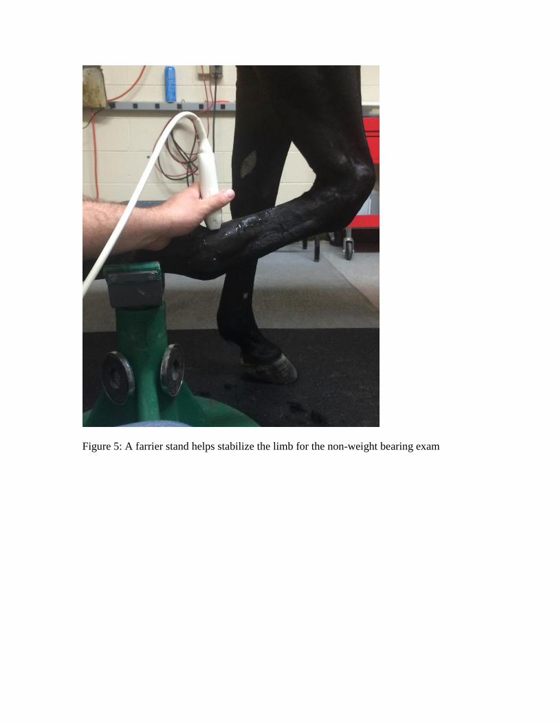
Figure 5: A farrier stand helps stabilize the limb for the non-weight bearing exam
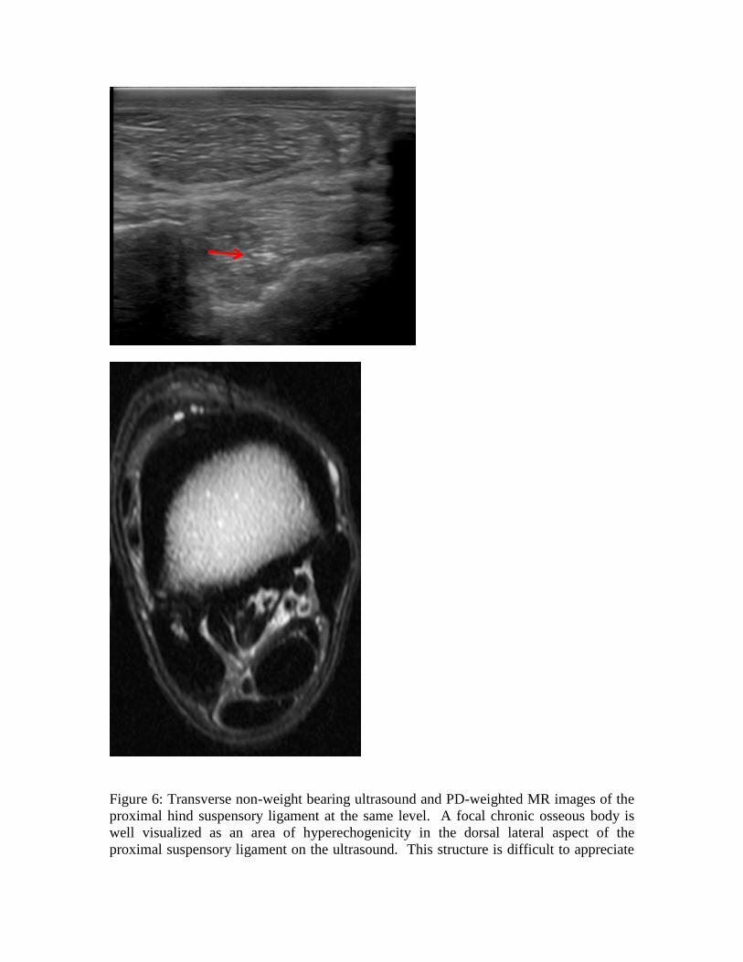
Figure 6: Transverse non-weight bearing ultrasound and PD-weighted MR images of the
proximal hind suspensory ligament at the same level. A focal chronic osseous body is
well visualized as an area of hyperechogenicity in the dorsal lateral aspect of the
proximal suspensory ligament on the ultrasound. This structure is difficult to appreciate
on MRI because both the osseous fragment and ligament are of relatively low signal
intensity, resulting in poor contrast between the tissue types.
A
B
Figure 7: Transverse non-weight bearing images of the proximal suspensory ligament.
Image A is the lame right forelimb and image B is the left forelimb for comparison. Note
the marked osseous irregularity and focal avulsion fragmentation of the palmar cortex of
MC3 in image A (long arrow) versus the smooth bone margin in image B. Both of the
suspensory ligaments are moderately enlarged with palmar displacement of the fat and
muscle bundles (short arrow).
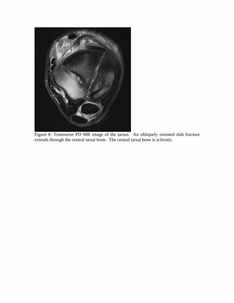
Figure 8: Transverse PD MR image of the tarsus. An obliquely oriented slab fracture
extends through the central tarsal bone. The central tarsal bone is sclerotic.


![Hallux rigidus: Joint preserving alternatives to ...€¦ · “hallux rigidus”[2]. Since then multiple names have been suggested, such as metatarsus primus elevatus, dorsal bunion,](https://img.pdfslide.us/doc/110x75/60689aa250b4be4ec45fd183/hallux-rigidus-joint-preserving-alternatives-to-aoehallux-rigidusa2-since.jpg)