Embed Size (px)
Citation preview
C
El
Wa
b
KLLE
1
ftmcrt
ic
2
5rlfi
1
European Annals of Otorhinolaryngology, Head and Neck diseases 132 (2015) 231–233
Available online at
ScienceDirectwww.sciencedirect.com
ase report
pstein-Barr virus-associated lymphoepithelial carcinoma of thearynx
assim Kermania,∗, Malek Belcadhia, Badreddine Srihab, Mouhamed Abdelkéfia
Service d’ORL, CHU Farhat Hached, avenue Ibn Eljazzar, 4000 Sousse, TunisiaLaboratoire d’anatomie et de cytologie pathologiques, CHU Farhat Hached, avenue Ibn Eljazzar, 4000 Sousse, Tunisia
a r t i c l e i n f o
eywords:ymphoepithelial carcinomaarynxpstein-Barr virus
a b s t r a c t
Introduction: Lymphoepithelial carcinoma is a rare tumour, named after its histological resemblance toundifferentiated nasopharyngeal carcinoma. The pathogenesis of lymphoepithelial carcinoma remainsunknown. This tumour has been described in several organs, but the larynx remains an exceptional site.Case report: The authors report the case of a 73-year-old man who consulted for longstanding dys-phonia and rapidly deteriorating dyspnoea requiring emergency tracheotomy. Endoscopic examinationdemonstrated a tumour of the left hemilarynx with fixed vocal cords. Histological examination andimmunohistochemistry demonstrated lymphoepithelial carcinoma of the larynx. Screening for Epstein-Barr Virus (EBV) by immunohistochemistry and in situ hybridization was positive. Treatment consisting of
neoadjuvant chemotherapy followed by surgical resection and then external beam radiotherapy achievedcure with a follow-up of 18 months since completion of treatment.Discussion: Lymphoepithelial carcinoma of the larynx is rare. Immunohistochemical examination isessential for the positive diagnosis. Epstein-Barr virus-associated lymphoepithelial carcinoma has beenexceptionally reported. The radiosensitivity of this tumour allows conservative first-line treatment.© 2015 Elsevier Masson SAS. All rights reserved.
. Introduction
The most typical form of lymphoepithelial carcinoma is undif-erentiated nasopharyngeal carcinoma [1,2]. More rarely, thisumour can arise in other sites, especially the salivary glands, thy-
us, lung and stomach [1,3], while laryngeal lymphoepithelialarcinoma is extremely rare, as only about thirty cases have beeneported in the literature [1]. It represents 0.2% of all malignantumours of the larynx [1,4,5].
We report a case of Epstein-Barr virus-associated lymphoep-thelial carcinoma of the larynx and discuss the pathogenesis,linical and pathological features and treatment of this tumour.
. Case report
A 73-year-old man, with no notable history, presented with a-month history of dysphonia with recent onset of rapidly dete-
iorating dyspnoea, requiring emergency tracheotomy. Indirectaryngoscopy demonstrated a lesion of the left hemilarynx withxed vocal cords. Neck examination revealed firm, mobile, deep∗ Corresponding author.E-mail address: kermani [email protected] (W. Kermani).
http://dx.doi.org/10.1016/j.anorl.2015.05.004879-7296/© 2015 Elsevier Masson SAS. All rights reserved.
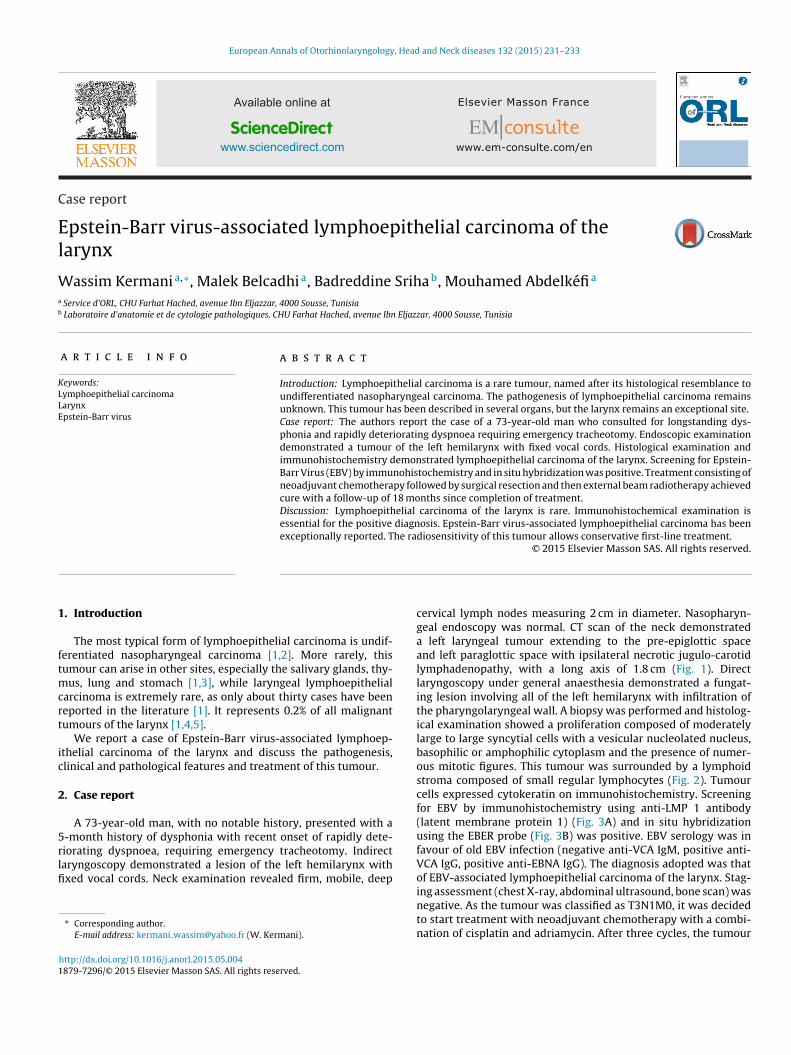
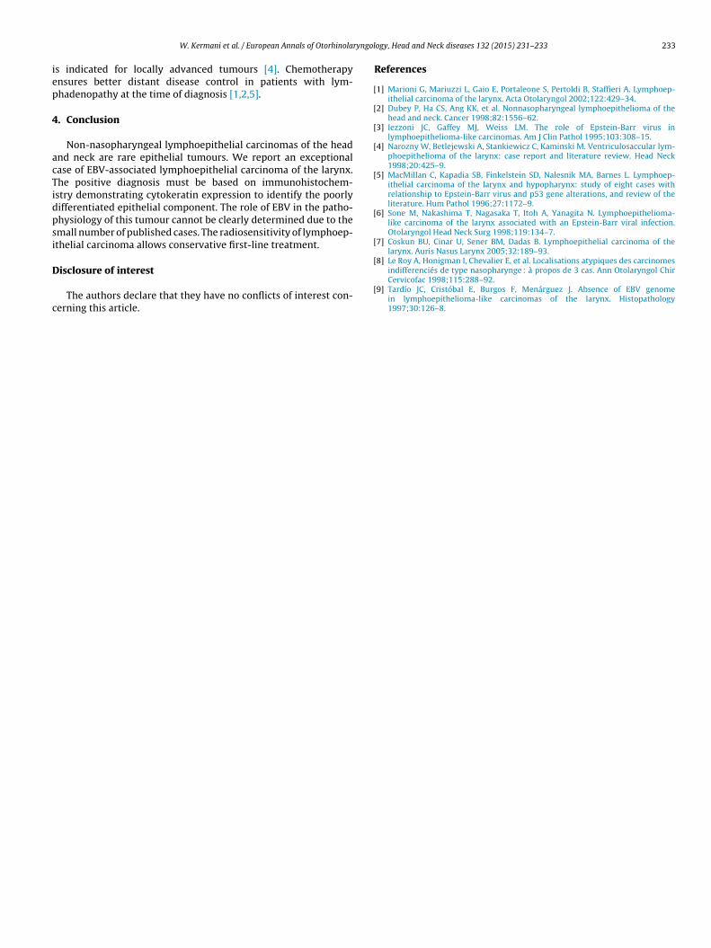
cervical lymph nodes measuring 2 cm in diameter. Nasopharyn-geal endoscopy was normal. CT scan of the neck demonstrateda left laryngeal tumour extending to the pre-epiglottic spaceand left paraglottic space with ipsilateral necrotic jugulo-carotidlymphadenopathy, with a long axis of 1.8 cm (Fig. 1). Directlaryngoscopy under general anaesthesia demonstrated a fungat-ing lesion involving all of the left hemilarynx with infiltration ofthe pharyngolaryngeal wall. A biopsy was performed and histolog-ical examination showed a proliferation composed of moderatelylarge to large syncytial cells with a vesicular nucleolated nucleus,basophilic or amphophilic cytoplasm and the presence of numer-ous mitotic figures. This tumour was surrounded by a lymphoidstroma composed of small regular lymphocytes (Fig. 2). Tumourcells expressed cytokeratin on immunohistochemistry. Screeningfor EBV by immunohistochemistry using anti-LMP 1 antibody(latent membrane protein 1) (Fig. 3A) and in situ hybridizationusing the EBER probe (Fig. 3B) was positive. EBV serology was infavour of old EBV infection (negative anti-VCA IgM, positive anti-VCA IgG, positive anti-EBNA IgG). The diagnosis adopted was thatof EBV-associated lymphoepithelial carcinoma of the larynx. Stag-
ing assessment (chest X-ray, abdominal ultrasound, bone scan) wasnegative. As the tumour was classified as T3N1M0, it was decidedto start treatment with neoadjuvant chemotherapy with a combi-nation of cisplatin and adriamycin. After three cycles, the tumour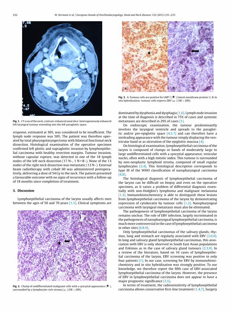
232 W. Kermani et al. / European Annals of Otorhinolaryngology, Head and Neck diseases 132 (2015) 231–233
Fl
rladclwnnbtao
3
b
Fs
ig. 1. CT scan of the neck, contrast-enhanced axial slice: heterogeneously enhancedeft laryngeal tumour extending into the left paraglottic space.
esponse, estimated at 30%, was considered to be insufficient. Theymph node response was 50%. The patient was therefore oper-ted by total pharyngolaryngectomy with bilateral functional neckissection. Histological examination of the operative specimenonfirmed left glottic and supraglottic invasion by lymphoepithe-ial carcinoma with healthy resection margins. Tumour invasion,
ithout capsular rupture, was detected in one of the 18 lymphodes of the left neck dissection (17 N–, 1 N + R–). None of the 13odes of the right neck dissection was metastatic (13 N–). Externaleam radiotherapy with cobalt 60 was administered postopera-ively, delivering a dose of 54 Gy to the neck. The patient presented
favourable outcome with no signs of recurrence with a follow-upf 18 months since completion of treatment.
. Discussion
Lymphoepithelial carcinoma of the larynx usually affects menetween the ages of 50 and 70 years [1,5]. Clinical symptoms are
ig. 2. Clump of undifferentiated malignant cells with a syncytial appearance ( ),urrounded by a lymphocyte-rich stroma ( ) (HE × 200).
Fig. 3. A. Tumour cells are positive for LMP 1 ( ) (latent membrane protein 1). B. Insitu hybridization: tumour cells express EBV ( ) (HE × 200).
dominated by dysphonia and dysphagia [1,6]. Lymph node invasionat the time of diagnosis is described in 75% of cases and systemicmetastases are described in 29% of cases [1].
On endoscopic examination, the tumour predominantlyinvolves the laryngeal ventricle and spreads to the paraglot-tic and/or pre-epiglottic space [4,5,7] and can therefore have amisleading appearance with the tumour simply displacing the ven-tricular band or as ulceration of the epiglottic mucosa [4].
On histological examination, lymphoepithelial carcinoma of thelarynx is composed of clumps or bands of moderately large tolarge undifferentiated cells with a syncytial appearance, vesicularnuclei, often with a high mitotic index. This tumour is surroundedby non-neoplastic lymphoid stroma, composed of small regularlymphocytes [3,4]. This histological description corresponds totype III of the WHO classification of nasopharyngeal carcinoma[4,8].
The histological diagnosis of lymphoepithelial carcinoma ofthe larynx can be difficult on biopsy and even on the operativespecimen, as it raises a problem of differential diagnosis essen-tially with non-Hodgkin’s lymphoma and malignant melanoma[1,3]. Immunohistochemistry is able to distinguish these lesionsfrom lymphoepithelial carcinoma of the larynx by demonstratingexpression of cytokeratin by tumour cells [1,3]. Nasopharyngealcarcinoma with laryngeal metastasis must also be eliminated.
The pathogenesis of lymphoepithelial carcinoma of the larynxremains unclear. The role of EBV infection, largely incriminated inthe pathogenesis of nasopharyngeal lymphoepithelial carcinoma, ismuch more controversial in the case of lymphoepithelial carcinomain other sites [6,8,9].
Only lymphoepithelial carcinomas of the salivary glands, thy-mus, lung and stomach are regularly associated with EBV [3,6,9].In lung and salivary gland lymphoepithelial carcinomas, this asso-ciation with EBV is only observed in South East Asian populationsand Eskimos as in the case of salivary gland tumours [2,3,9]. Ina review of the literature, based on 16 cases of lymphoepithe-lial carcinoma of the larynx, EBV screening was positive in onlyfour patients [1]. In our case, screening for EBV by immunohisto-chemistry and in situ hybridization was strongly positive. To ourknowledge, we therefore report the fifth case of EBV-associatedlymphoepithelial carcinoma of the larynx. However, the presenceof EBV in lymphoepithelial carcinoma does not appear to have a
major prognostic significance [3,5].In terms of treatment, the radiosensitivity of lymphoepithelialcarcinoma allows conservative first-line treatment [1,4,7]. Surgery
ryngol
iep
4
acTidpsi
D
c
[
[
[
[
[
[
[
[
W. Kermani et al. / European Annals of Otorhinola
s indicated for locally advanced tumours [4]. Chemotherapynsures better distant disease control in patients with lym-hadenopathy at the time of diagnosis [1,2,5].
. Conclusion
Non-nasopharyngeal lymphoepithelial carcinomas of the headnd neck are rare epithelial tumours. We report an exceptionalase of EBV-associated lymphoepithelial carcinoma of the larynx.he positive diagnosis must be based on immunohistochem-stry demonstrating cytokeratin expression to identify the poorlyifferentiated epithelial component. The role of EBV in the patho-hysiology of this tumour cannot be clearly determined due to themall number of published cases. The radiosensitivity of lymphoep-thelial carcinoma allows conservative first-line treatment.
isclosure of interest
The authors declare that they have no conflicts of interest con-erning this article.
[
ogy, Head and Neck diseases 132 (2015) 231–233 233
References
1] Marioni G, Mariuzzi L, Gaio E, Portaleone S, Pertoldi B, Staffieri A. Lymphoep-ithelial carcinoma of the larynx. Acta Otolaryngol 2002;122:429–34.
2] Dubey P, Ha CS, Ang KK, et al. Nonnasopharyngeal lymphoepithelioma of thehead and neck. Cancer 1998;82:1556–62.
3] Iezzoni JC, Gaffey MJ, Weiss LM. The role of Epstein-Barr virus inlymphoepithelioma-like carcinomas. Am J Clin Pathol 1995;103:308–15.
4] Narozny W, Betlejewski A, Stankiewicz C, Kaminski M. Ventriculosaccular lym-phoepithelioma of the larynx: case report and literature review. Head Neck1998;20:425–9.
5] MacMillan C, Kapadia SB, Finkelstein SD, Nalesnik MA, Barnes L. Lymphoep-ithelial carcinoma of the larynx and hypopharynx: study of eight cases withrelationship to Epstein-Barr virus and p53 gene alterations, and review of theliterature. Hum Pathol 1996;27:1172–9.
6] Sone M, Nakashima T, Nagasaka T, Itoh A, Yanagita N. Lymphoepithelioma-like carcinoma of the larynx associated with an Epstein-Barr viral infection.Otolaryngol Head Neck Surg 1998;119:134–7.
7] Coskun BU, Cinar U, Sener BM, Dadas B. Lymphoepithelial carcinoma of thelarynx. Auris Nasus Larynx 2005;32:189–93.
8] Le Roy A, Honigman I, Chevalier E, et al. Localisations atypiques des carcinomes
indifferenciés de type nasopharynge : à propos de 3 cas. Ann Otolaryngol ChirCervicofac 1998;115:288–92.9] Tardío JC, Cristóbal E, Burgos F, Menárguez J. Absence of EBV genomein lymphoepithelioma-like carcinomas of the larynx. Histopathology1997;30:126–8.


![Processus al´eatoires et applicationsarXiv:1312.7796v1 [math.HO] 30 Dec 2013 Processus al´eatoires et applications Master 2 Pro de Math´ematiques Universit´e d’Orl´eans Nils](https://img.pdfslide.us/doc/110x75/5fa94c56b56aaf35be33e2b0/processus-aleatoires-et-applications-arxiv13127796v1-mathho-30-dec-2013-processus.jpg)