Embed Size (px)
Citation preview
Journal of Cellular Biochemistry 98:757–769 (2006)
Epigenetic Modifications in OsteogenicDifferentiation and Transformation
David Thomas1,2* and Maya Kansara1
1Ian Potter Foundation Centre for Cancer Genomics and Predictive Medicine,Peter MacCallum Cancer Centre, Victoria 3002, Melbourne, Australia2Department of Medicine, St. Vincent’s Hospital, University of Melbourne, Melbourne, Australia
Abstract Almost all tumors are characterized by both architectural and cellular abnormalities in differentiation.Osteoblast development is relatively well understood, making osteosarcoma a good model for understanding howtumorigenesis perturbs normal differentiation. We argue that there are two key transition points in normal cellulardifferentiation that are the focus of oncogenic events, in both of which epigenetic processes are critical. The first is thetransition from an uncommitted pluripotent precursor (mesenchymal stem cell) to the ‘transit-amplifying compartment’ ofthe osteoblast lineage. This transition, normally exquisitely regulated in space and time, is abnormal in cancer. The secondinvolves termination of lineage expansion, equally tightly regulated under normal circumstances. In cancer, themechanisms that mandate eventual cessation of cell division are almost universally disrupted. This model predicts that keydifferentiation genes in bone, such asRUNX2, act in an oncogenic fashion to initiate entry into a proliferative phase of celldifferentiation, and anti-oncogenically into the post-mitotic state, resulting in ambivalent roles in tumorigenesis.Polycomb genes exemplify epigenetic processes in the stem cell compartment and tumorigenesis, and are implicated inskeletal development in vivo. The epigenetic functions of the retinoblastoma protein, which plays a key role intumorigenesis in bone, is discussed in the context of terminal cell cycle exit. J. Cell. Biochem. 98: 757–769, 2006.� 2006 Wiley-Liss, Inc.
Key words: epignetic; differentiation; cancer; bone; osteoblast
‘‘A tumor is an actively growing tissue,composed of cells derived from one that hasundergone an abnormal type of irreversibledifferentiation; its growth is progressive, due toa persistent delay in maturation of stem cells’’
—[Berenblum, 1962].
Tumors almost universally show abnormal-ities in differentiation, known as anaplasia.Cancer is inherently a metazoan problem, andcan only occur in the context of tissue-specificdifferentiation. Anaplasia occurs at both thecellular and architectural level, and may be duetocell-intrinsicdefects,aberrantinstructivemicro-
environmental cues, or both. Distinguishingcellular from architectural derangement isimportant when integrating experimental datain vitro with in vivo human and animal obser-vations. Perturbations of differentiation pro-grammes in tumor cells can result in abnormalcellular survival, growth, and proliferation, lossof specialised function and acquisition of theability to invade surrounding tissues [Hanahanand Weinberg, 2000]. Functionally, anaplasiaappears to correlate with more aggressivecancer behavior, which suggests that differen-tiation confers a restraint on tumorigenesis.Animal models showed that loss-of-function ofcancer-related genes, whose function is appar-ently not cell-type specific, gives rise to highlyrestricted developmental defects and tumorspectra in vivo. For example, inherited hetero-zygous mutations in the retinoblastoma tumorsuppressor gene (RB) in humans leads to ahighly restricted range of tumors. In the mouse,germline deletion of Rb results in embryoniclethality due to developmental defects inspecific tissues. This implies that the specific
� 2006 Wiley-Liss, Inc.
Grant sponsor: NHMRC Project; Grant number: 350432.
*Correspondence to: David Thomas, Peter MacCallumCancer Centre, St. Andrew’s Place, East Melbourne,Victoria 3002, Australia.E-mail: [email protected]
Received 29 December 2005; Accepted 12 January 2006
DOI 10.1002/jcb.20850
molecular events required for tumorigenesis arefrequently contingent on commitment to speci-fic cell lineages. The molecular basis for loss ofdifferentiation in carcinogenesis is not wellunderstood, and probably varies between tissueand cancer types. Importantly, differentiationtherapy has already been introduced into theclinic, with benefit, giving hope that study of therelationships between differentiation andtumorigenesis will be of use to cancer patients.In this review, we focus on bone developmentand cancer as model systems for gaining in-sights into the relationship between differentia-tion, development, and tumorigenesis.
BONE AND BONE CANCER
Osteosarcoma is the most common bone sar-coma and the third most common malignancy inchildren and adolescents. Approximately 2,500cases are diagnosed per year in the UnitedStates. Osteosarcoma is defined by the presenceof abnormal bone matrix (osteoid). Over 80% ofosteosarcomas are graded histopathologicallyas poorly differentiated [Dahlin, 1957]. Patho-logic classification incorporates degree of differ-entiation in assessing histologic grade, wherethe absence of differentiation heralds a 10–15% decrease in 5-year survival (AJCC stagingmanual, 6th edition). Some aspects of thedifferentiated phenotype are clearly preserved,and the ability of tumor cells to lay down anaberrant matrix is crucial to a diagnosis ofosteosarcoma. Alkaline phosphatase (ALP), anearly and non-specific marker of the osteoblastlineage, is frequently seen in bone tumors. Bycontrast, late markers of osteoblast differentia-tion, such as osteocalcin, are expressed poorly ornot at all in most osteosarcomas [Hopyan et al.,1999]. This suggests that terminal differentia-tion, but not differentiation per se, is antitheti-cal to tumorigenesis. Because the osteoblastdifferentiation program is well understood,osteosarcoma represents an excellent modelfor interrogating the relationships betweendifferentiation and transformation.
NORMAL BONE DEVELOPMENTAND DIFFERENTIATION
Bone development is relatively well under-stood at the cellular and molecular level(reviewed by [Aubin, 1998]). A key feature ofbone development is that differentiation is
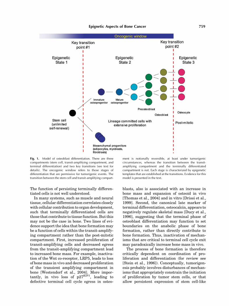
accompanied by a progressive loss of prolifera-tive capacity, as illustrated in Figure 1. Thereare two key steps in cellular differentiation thatappear critical to tumorigenesis. The first keytransition point is the step between a mesench-ymal stem cell and a lineage-restricted progeni-tor cell. The osteoblast lineage begins withmulti-potent mesenchymal stem cells, locatedin the periosteal surfaces and within bonemarrow stroma. Bone has a vast potential forregeneration from pluripotent mesenchymalstem cells [Aubin, 1998]. It is important to notethat the majority of these progenitor cells arethought to be quiescent in the adult skeleton,and quiescence or low proliferative index is ageneral property used in other systems toidentify stem cells (label retention; [Watt,2001]). Stem cells are activated by tightlyregulated and complex signals, whose interac-tions are beginning to be understood. Followinginitial lineage commitment, a phase of lineageexpansion ensues which culminates normally inpermanent cell cycle withdrawal. The initial celldivision is asymmetric, giving rise to anotherstem cell (self-renewal) and a committed osteo-progenitor. Following commitment, the stemcell gives rise to the transit-amplifying com-partment [Watt, 2001]. This phase is associatedwith intensive proliferative activity. The pre-osteoblast is an intermediate stage, whichexpresses both STRO1, ALP, parathyroid hor-mone receptor, and type I collagen, and iscommitted to the osteoblast lineage with exten-sive replicative capacity, but no self-renewalcapacity [Gronthos et al., 1999]. In vitro, the useof agents such as retinoic acid can inducefurther differentiation in the pre-osteoblast.The mature osteoblast expresses ALP, osteo-pontin, bone sialoprotein, and osteocalcin, andlies adjacent to newly synthesized osteoid. Thisstage, which is responsible for the laying downof bone, has limited replicative potential [Steinet al., 1996]. The cumulative effect of therecruitment of stem cells and their expansion,and the functional capacity of mature osteo-blasts, is measured by rates of bone formation invivo. The second key step initiates terminaldifferentiation and permanent cell cycle with-drawal. The terminal stage of the bone lineage isthe post-mitotic osteocyte, often found isolatedwithin bone, presumably embedded withinadvancing osteoid. As an alternate fate, aproportion of cells in the transient amplifyingcompartment may also terminate in apoptosis.
758 Thomas and Kansara
The function of persisting terminally differen-tiated cells is not well understood.
In many systems, such as muscle and neuraltissue, cellular differentiation correlates closelywith cellular contribution to organ development,such that terminally differentiated cells arethose that contribute to tissue function. But thismay not be the case in bone. Two lines of evi-dence support the idea that bone formation maybe a function of cells within the transit-amplify-ing compartment rather than the post-mitoticcompartment. First, increased proliferation oftransit-amplifying cells and decreased egressfrom the transit-amplifying compartment leadto increased bone mass. For example, inactiva-tion of the Wnt co-receptor, LRP5, leads to lossof bone mass in vivo and decreased proliferationof the transient amplifying compartment inbone [Westendorf et al., 2004]. More impor-tantly, in vivo loss of p27KIP1, leading todefective terminal cell cycle egress in osteo-
blasts, also is associated with an increase inbone mass and expansion of osteoid in vivo[Thomas et al., 2004] and in vitro [Drissi et al.,1999]. Second, the canonical late marker ofterminal differentiation, osteocalcin, appears tonegatively regulate skeletal mass [Ducy et al.,1996], suggesting that the terminal phase ofosteoblast differentiation may function to setboundaries on the anabolic phase of boneformation, rather than directly contribute tobone formation. Thus, inactivation of mechan-isms that are critical to terminal cell cycle exitmay paradoxically increase bone mass in vivo.
The process of bone formation is thereforecritically dependent on coordination of pro-liferation and differentiation (for review see[Stein et al., 1996]). Conceptually, tumorigen-esis probably involves disturbances of mechan-isms that appropriately constrain the initiationof proliferation by tumor stem cells, or thatallow persistent expression of stem cell-like
Fig. 1. Model of osteoblast differentiation. There are threecompartments (stem cell, transit-amplifying compartment, andterminal differentiation) and two key transitions (see text fordetails). The oncogenic window refers to those stages ofdifferentiation that are permissive for tumorigenic events. Thetransition between the stem cell and transit-amplifying compart-
ment is notionally reversible, at least under tumorigeniccircumstances, whereas the transition between the transit-amplifying compartment and the terminally differentiatedcompartment is not. Each stage is characterized by epigenetictemplates that are established at the transitions. Evidence for thismodel is presented in the text.
Epignetic Aspects of Bone Cancer 759
features in apparently partially committedcells. The similarities between stem cell proper-ties and those of transformed cells are striking.Both cell types possess unlimited self-renewal,express telomerase, and are undifferentiated asdefined by the absence of lineage-restrictedmarkers (reviewed in [Sharpless and DePinho,2004]). These features raise the possibilitythat mechanisms that maintain ‘stemness’ areimportant to tumorigenesis (disruption of thefirst transition point in Fig. 1). Accumulatingevidence suggests that similar signaling path-ways, including those relevant to bone, con-tribute to the regulation of self-renewal inprogenitor and tumor cells [Reya and Clevers,2005]. There is also good evidence (summarizedbelow) that cancers are characterized by dis-ruption of mechanisms that enforce terminalcell cycle exit (the second key transition pointin Fig. 1).
The notion that both lineage commitment andterminal differentiation are both key stages intumorigenesis has several significant implica-tions. First, it suggests that transcriptionalregulators of differentiation may play ambiva-lent roles in tumorigenesis, since they presideinitially over lineage commitment and expan-sion, but later over terminal aspects of differ-entiation. Second, there are probably both cellintrinsic and extrinsic mechanisms involved inregulating both key transition points, whichmay be perturbed in cancers. In particular, thegrowing recognition that non-cell autonomousmechanisms are critical to tumor formationhas led to speculation about the key role ofepigenetic (mutation-independent) processesin establishing heritable, stem cell-like statesin tumorigenesis. There is also evidence thatepigenetic mechanisms are important at thesecond transition point to the terminally differ-entiated state. These aspects are discussed indetail in this review, with emphasis on bonedevelopment and transformation where data isavailable.
TRANSCRIPTIONAL REGULATION OFOSTEOBLAST DIFFERENTIATION
AND TRANSFORMATION
While the signaling pathways that initiaterecruitment of stem cells towards a specificlineage are still to be fully characterized, thetranscriptional mediators regulating osteoge-nic differentiation have recently been mapped
in some detail [Ducy, 2000]. Critical amongthese is runx2 (CBFA1/Osf2/PEBP2a), a keytranscriptional regulator of osteoblast differen-tiation, belonging to the runt family of trans-cription factors [Ogawa, 1993]. Mice nullizygousfor runx2 exhibit a complete lack of ossification[Komori et al., 1997], while runx2 postnatallyregulates expression of bone-specific genes suchas osteopontin and osteocalcin, and controlsbone matrix deposition [Ducy et al., 1999].Notably, runx2 activity is required for expres-sion of genes, such as osteocalcin, associatedwith terminal differentiation. Runx2 function ismodified dramatically by the co-operating andantagonistic actions of other proteins. Thisis a rapidly expanding field of study. CBFb,a heterodimeric partner, is required for fullactivity, but other co-regulators include TLE2, agroucho family member; MAPK; TGFb throughdirect interactions with SMAD3; and LEF1(reviewed by [Lian et al., 2004]). Transcrip-tional splicing events give rise to functionallydistinct isoforms [Harada et al., 1999]. Down-stream effectors of runx2 function, such asosterix, are critical to bone development[Nakashima et al., 2002]. Taken together, thesedata suggest that the transcriptional activity ofrunx2 depends critically on transcriptionalsplice patterns, co-activators and -repressors,post-translational modifications, and the integ-rity of downstream effectors.
There is good evidence in vivo to suggestthat the runt family appears to function asa tumor suppressor in hematologic cancers[Lund and Van Lohuizen, 2002]. RUNX1(AML1) is mutated in human leukemia, andmice expressing loss-of-function runx1 mutantsare prone to leukemia [Perry et al., 2002].CBFb, the heterodimeric partner of runx pro-teins, is frequently the subject of translocationevents in leukemias (reviewed by [Ito, 2004]).RUNX3 has been reported to be subject togenomic deletion or promoter hypermethyla-tion in gastric cancers [Li et al., 2004]. Theevidence for runx2 is more circumstantial, andis based on in vitro data, suggesting that runx2expression varies with cell cycle status, inhibitsosteoblast proliferation, and promotes terminaldifferentiation [Pratap et al., 2003; Thomaset al., 2004; Galindo et al., 2005]. Furtherevidence comes from data demonstrating thatthe retinoblastoma tumor suppressor protein(pRb) co-operates with runx2 to promote differ-entiation.
760 Thomas and Kansara
The cell cycle regulatory pathway centeredaround pRb is inactivated in almost all humancancers, but individual tumor types seem totarget specific components to achieve this effect(reviewed by [Korenjak and Brehm, 2005]). pRbitself is frequently somatically inactivated inosteosarcomas [Horowitz et al., 1990], whileinherited heterozygous loss of the RB geneconfers a 500-fold greater incidence of osteosar-coma than the general population [Abramsonet al., 1984]. The Rb pocket proteins family playroles in mesenchymal differentiation [Korenjakand Brehm, 2005], and several lines of evidenceimplicate pRb in osteogenesis (reviewed by[Thomas et al., 2003]). pRb co-activates runx2,through direct physical interactions at sites ofactive transcription, and loss of function of pRbattenuates terminal osteoblast differentiationin vitro [Thomas et al., 2001]. Conversely, runx2co-ordinates terminal cell cycle exit throughinduction of the CDK2 inhibitor, p27KIP1, whichin turn is required for normal bone developmentin vitro and in vivo, and is lost in de-differ-entiated human osteosarcomas [Thomas et al.,2004]. More recently, Benevolenskaya et al.[2005] showed that Rbp2 may function as arepressor of runx2-dependent transcription,and that pRb acts to displace Rbp2 fromosteoblast-specific promoters thereby activat-ing differentiation. Interestingly, pRb is likelyto influence osteoblast differentiation throughadditional mechanisms involving chromatinstructure (see below). These data suggest thatrunx2 may function in pathways that hindertumorigenesis.
However, there is considerable debate regar-ding the role of runx2 in cancer [Blyth et al.,2005]. Under some circumstances, runx2 ap-pears to act as an oncogene. Runx2 co-operateswith Myc to cause lymphomas in mice based onproviral insertion sites studies (e.g., [Vaillantet al., 1999]). The same group reported thatectopic expression of runx2 resulted in trans-formation of p53-null fibroblasts [Wotton et al.,2004], and more intriguingly, that runx1 actsas a dominant oncogene in T-cell lymphoma[Wotton et al., 2002]. Amongst other possibleinterpretations, it is intriguing to postulate thatstage-specific signals may cause runx proteinsto act under different circumstances in both anoncogenic and tumor suppressor role. This isconsistent with an oncogenic role for runx2 inthe expansion, and a tumor suppressor role interminal differentiation phases of osteoblast
ontogeny, which might account for the absenceof mutations inRUNX2 itself. Thus, the effect ofRUNX2 expression may depend on the differ-entiation stage in which it functions.
THE ONCOGENIC WINDOW
Several studies have observed that oncogeniceffects depend on the differentiation stage inwhich the oncogene is activated (reviewed by[Weinstein, 2002]). Of particular relevance tothis review, this point was confirmed in anelegant conditional transgenic model of MYC-induced osteosarcomas [Jain et al., 2002]. Briefinactivation of MYC resulted in sustainedregression of the tumors, with differentiationof tumor cells into mature osteocytes. This ob-servation alone is, perhaps, unsurprising. How-ever, Jain et al. [2002] went on to show thatre-activation of MYC did not restore tumori-genic properties, but rather induced apoptosis.These observations were interpreted to suggestthat brief inactivation of MYC ‘appears to causeepigenetic changes in tumor cells that renderthem insensitive to MYC-induced tumorigen-esis’. These studies support the notion thatterminally differentiated cells lie outside an‘oncogenic window,’ and thus are not permissivefor tumorigenesis. The role of epigenetic pro-cesses is discussed in more detail below.
There is some debate as to whether theoncogenic window includes the stem cell com-partment, the transit-amplifying compartment,or both [Huntly and Gilliland, 2005]. Cancercells share the proliferative characteristics ofthe transit-amplifying compartment, and thecapacity for indefinite self-renewal of stemcells. Whether cancers arise as a consequenceof mutational or epigenetic events in transit-amplifying cells that confer immortality; orwhether such events disrupt the precise con-straints on stem cell proliferation prior to truelineage commitment, is open to question. Ithas been argued that cancer-initiating cellsarise in the stem cell compartment, perhaps byepigenetic mechanisms (reviewed by [Feinberget al., 2006]). The cancer stem cell theoryposits that a sub-population of cells within eachcancer possess ‘cancer initiating properties,’analogous to the physiologic ability of stemcells to fully reconstitute organ development(reviewed by [Huntly and Gilliland, 2005]). Thisproperty, along with infinite self-renewal andmulti-potentiality, is the defining characteristic
Epignetic Aspects of Bone Cancer 761
of a true stem cell. Stem cells would appear to liewithin the oncogenic window, and the identifi-cation of the BCR-ABL fusion gene in allhemopoietic lineages in chronic myeloid leuke-mia supports this notion [Huntly and Gilliland,2005].
In osteosarcomas, it has been suggested thata sub-population of multi-potent cancer stemcells without lineage commitment gives rise toaberrantly differentiated osteoblastic progeny[Gibbs et al., 2005]. In this study, a population ofmulti-potent osteosarcoma-derived cells wereidentified which express stem cell markers, aswell as markers of ectoderm and endoderm, aswell as mesodermal gene expression. It is notclear whether the same cells express non-mesodermal genes and mesodermal genes, norwhich population possesses cancer-initiatingproperties [Gibbs et al., 2005]. Clinically, thereare uncommon primary sarcomas of bone thatlack lineage-restricted features, and are desig-nated malignant fibrous histiocytoma of bone[WHO, 2002], while other tumors commonlycontain regions with chondroblastic, osteoblas-tic and fibroblastic differentiation, suggestingthat the cell of origin retains some degree ofmulti-potentiality. These observations suggestthat the cancer-initiating cell has stem cell-likecharacteristics, including multi-potentiality.
However, we favor the notion that osteosar-comas can arise in a transit-amplifying com-partment, perhaps in addition to the stem cellcompartment. Clonal cell lines derived fromprimary osteosarcomas, which presumably ful-fill the criteria for cancer stem cells, expressosteoblastic markers such as ALP in every cell[Thomas and Kansara, unpublished data].This suggests that, at least for a subset ofosteosarcomas, the cancer-initiating cell sharesfeatures of a committed osteoprogenitor. Addi-tionally, it has been argued that a relationshipexists between proliferative rate and the acqui-sition of mutagenic events [Cohen and Ellwein,1991]. This may favor the accumulation ofoncogenic events in the transit-amplifyingcompartment, while stem cells are typicallyquiescent. In support of this concept, osteosar-comas are most frequently observed in adoles-cence, a stage of intensive skeletal growthentailing increased osteoblast activity. Further-more, Paget’s disease of bone, a benign condi-tion characterized by dramatically increasedbone formation and resorption, is also asso-ciated with an increased risk of osteosarcoma.
These data suggest that increased osteoblasticactivity is associated with tumorigenesis. Third,stem cells are up to 100-fold more resistant tomutagenic events than somatic cells, consistentwith the necessity for conserving the geneticcode in cells that give rise to multiple tissues[Cervantes et al., 2002]. This resistance appearsin part due to enhanced apoptotic responses togenotoxic stress and DNA damage [Hong andStambrook, 2004; Saretzki et al., 2004]. Theefficiency of such processes appears inverselyrelated to degree of differentiation. Thus, it maybe that genes that initiate lineage commitmentand expansion, under the influence of genessuch as RUNX2, create conditions favoring theacquisition of tumorigenic events.
NON-CELL AUTONOMOUS REGULATIONOF OSTEOBLAST DIFFERENTIATION
AND TRANSFORMATION
Carcinogenesis may be a process akin todevelopment gone awry (reviewed by [Weaverand Gilbert, 2004]), and non-cell autonomous(microenvironmental) temporospatial cues arecritical to normal development. There is grow-ing recognition of the central importance ofmicroenvironmental context in driving tumorformation, illustrated strikingly by a recentstudy in which nitrosomethylurea (NMU)-treatment of cleared mammary fat pads in vivowas sufficient for the development of epithelialneoplasia, while NMU treatment of epithelialcells alone did not result in tumors [Maffiniet al., 2004]. This experiment implies a domi-nant role for microenvironmental processes inregulating tumor formation, at least in a murinemammary tumor model. In many cases, onco-genic effects of signaling molecules may berelated to physiologic functions in maintenanceor expansion of stem cell compartments. Exam-ples of secreted signaling molecules implicatedin bone development and cancer include theWnts and related proteins, hedgehogs, notch,transforming growth factor-b, parathyroid hor-mone-related protein, and receptor activator ofNFkB ligand. The Wnt pathway illustrates thepotential role of environmental cues in deter-mining the balance between differentiation andproliferation. Wnt signaling is critical to devel-opment, stem cell biology and tumorigenesis[Reya and Clevers, 2005], and is also to bonedevelopment [Westendorf et al., 2004]. Wntsstimulate expansion of the transit-amplifying
762 Thomas and Kansara
compartment. Mice lacking the Wnt co-receptorLrp5 have decreased bone mass and decreasedosteoblast proliferation [Kato et al., 2002]. Inhumans, gain-of-function mutations in LRP5lead to increased bone mass [Boyden et al.,2002], while loss-of-function mutations havebeen the cause of osteoporosis-pseudogliomasyndrome [Gong et al., 2001]. During osteoblastdifferentiation, Wnt signaling contributes toexpansion of the transit-amplifying compart-ment, but is followed by expression of negativeregulators of Wnt signaling. For example, Wntinhibitory factor 1 and SFRP2 are bothexpressed at high levels in association withosteoblast differentiation in vitro, concomitantwith expression of late markers of the osteoblastlineage such as osteocalcin [Vaes et al., 2005].Thus, Wnt signaling may act as one example of amolecular switch integrating proliferation anddifferentiation in the osteoblast lineage, andcould therefore determine the oncogenic ortumor suppressor activity of runx2.
An interesting implication of the importanceof microenvironmental signals in tumor pro-gression is that it predicts a key role forepigenetic processes [Feinberg et al., 2006].Discussed in greater detail below, epigeneticmechanisms are those which result in heritablechanges in gene expression without changes ingene sequence, in contrast to genetic mechan-isms, which are based on sequence alterations.It is notable that, unlike genetic events, epige-netic events are under some circumstancesreversible. The clonogenic effect of microenvir-onmental signals requires a mechanism for‘fixing’ the fate choices induced by those signals,such that all progeny of the recipient progenitorcell express the tumorigenic programme. Itis unlikely that aberrant microenvironmentalsignals act by inducing genetic changes in therecipient tumor progenitor. Epigenetic pattern-ing, however, provides an excellent mechanismfor transmission of accumulated oncogenic sig-nals in a clonal fashion to all daughter cells. Ineffect, the tumorigenic consequences of micro-environmental signals are likely to be the abnor-mal application of epigenetic templates thatmaintain cellular states favoring tumor forma-tion. This interpretation is supported by theNMU-mutagenesis studies described earlier, inwhich microenvironmental signals are criticalto the emergence of the epithelial tumor clone.
There is additional evidence to suggest thatepigenetic events are dominant over mutational
events, based on the reversibility of both lossof differentiation and tumor phenotype. Thisevidence comes from studies of the contributionof cancer-derived genomes to embryonic devel-opment following nuclear transfer [Blellochet al., 2004; Hochedlinger et al., 2004] and blas-tocyst injection [Mintz and Illmensee, 1975].Nuclei derived from a wide range of cancer cellswere able to support normal pre-implantationdevelopment to the blastocyst stage at frequen-cies between 0% and 12% [Hochedlinger et al.,2004]. Nuclei derived from doxycycline-induci-ble rasþ/ink4a�/� melanoma, and fibroblastswere used to generate chimeric mice. Whilethese mice developed cancers with higherpenetrance, shorter latency, and an expandedspectrum compared to the donor mice, the EScells supported differentiation into multiplelineages, including melanocytes, lymphocytes,and fibroblasts. The tumors derived from ES-cell derived chimeras on activated alleles ofRAScarried identical genomic profiles compared tothe donor tumor. Embryonal carcinoma cellnuclei were also used to reconstitute a broadrange of mature neuroepithelium, epithelium,and mesothelial tissues [Blelloch et al., 2004]. Itappears that, just as microenvironmental sig-nals can establish pro-oncogenic epigenetictemplates,underdifferentcircumstancesmicro-environmental signals can revert tumorigenicepigenetic templates. It is difficult to account forsuch observations if mutations are the basis fortumorigenicity and loss of developmental poten-tial in tumor cells.
EPIGENETIC PROCESSES
The eukaryotic genome is vastly more com-plex than that of prokaryotes. In addition tosequence-dependent determinants of functionalspecificity, which include regulatory sequencesand their complementary trans factors, non-sequence dependent (epigenetic) mechanismshave evolved to effectively restrict the availablegenome that is accessible to transactivation.This is logical and elegant in metazoan struc-tures, given that lineage commitment duringdevelopment means that although stem cellsmust carry the entire human genome, cellswithin individual tissues are required to exp-ress only a fraction of all genes. Epigenetic pro-cesses contribute todevelopment, differentiation,aging, carcinogenesis, and autoimmunity (revi-ewed in [Strathdee et al., 2004]). Epigenetic
Epignetic Aspects of Bone Cancer 763
processes may take several forms. One formof epigenetic silencing involves methylation ofthe C5 position of cytosine bases, usually in thecontext of cytosine-phospho-guanine dinucleo-tide pairs, which are often found in clusterscalled CpG islands located at the promoterregions of about 50% of human genes. Methyla-tion of CpG islands causes stable heritabletranscriptional silencing through binding ofmethyl-DNA-specific proteins to affected CpGislands attracts histone-modifying enzymes,which focally establish a silenced chromatinstate. A second form of epigenetic regulation ofgene expression affects chromatin structurethrough covalent modification of histone pro-teins. This field of biology is rapidly evolving,with the recognition that acetylation andmethylation of nucleotides and histones resultsin the establishment of chromatin structuresthat constrain transcriptional competence.Recent data suggest that processes involved inhistone modification may also control DNAmethylation, linking diverse mechanisms ofepigenetic regulation [Vire et al., 2005].
A great deal of evidence supports the idea thatDNA methylation patterns are essential fornormal development, cellular differentiation,and tumorigenesis (reviewed by [Jones andLaird, 1999; Arney and Fisher, 2004]). In bone,reduced CpG methylation has been shown to beassociated with transcriptional activation of thebone-specific rat osteocalcin gene in osteoblasts[Villagra et al., 2002]. While overall methyla-tion is decreased in cancer, �1% of genesare newly silenced by promoter methylation[Costello et al., 2000]. Aberrant de novo methy-lation of CpG islands is found early duringcarcinogenesis. Interestingly, the number ofcancer related genes affected by epigeneticinactivation may exceed the number inacti-vated by mutation (reviewed by [Jones andBaylin, 2002]). Some examples of silencing byDNA promoter methylation include p16INK4A,p73, MLH1, BRCA1, E-cadherin, APC, andVHL. Interestingly, methylation changes incancer cells are not limited to hypermethyla-tion. A small group of genes, including C-JUN,C-MYC, and TCLI, may become hypomethy-lated and reactivated in the course of tumorprogression [Yuille et al., 2001]. Of relevance toosteosarcoma, theRB gene has been shown to besilenced by promoter methylation [Sakai et al.,1991]. Interestingly, many secreted inhibitorsof Wnt signaling, including SFRPs and WIF1,
have been shown to be silenced by promotermethylation in human cancer [Suzuki et al.,2002].
EPIGENETICS AND THE TRANSITION BETWEENSTEM CELL AND TRANSIT-AMPLIFYING
COMPARTMENT
As noted earlier, epigenetic mechanisms havebeen proposed to be critical to disruption of thefirst transition point from stem cell to transit-amplifying compartment [Feinberg et al., 2006].Evidence of the importance of epigenetic pro-cesses in tumorigenesis and development comesfrom recent studies of the polycomb group (PcG)of proteins (reviewed by [Valk-Lingbeek et al.,2004]). PcG proteins play critical roles inassignment of epigenetic states, through his-tone modification. Three PcG complexes havebeen characterized to date. The first, polycombrepressive group 2 (PRC2) initiates silencingthrough methylation of lysine residues onhistones H3 and H1. The second, PRC1, isinvolved in maintenance of stable states of generepression, in part by mechanisms that recog-nize methylated lysines on histone H3. Thethird complex, PRC3, was identified recentlyand targets specific lysine residues (K27 on H3and K26 on H1) via Eed proteins [Kuzmichevet al., 2005]. The developmental importance ofthese complexes is indicated by early embryoniclethality in mice with loss of function of com-ponents of polycomb complexes.
Thepolycomb family ofproteinsappear to spe-cifically regulate the transition from the stemcell compartment to the transit-amplifyingcompartment following lineage commitment.This is exemplified by BMI-1, a key componentof PRC2. Bmi-1-deficient mice manifest defectsin hemopoietic and neuronal development,consistent with a requirement of Bmi-1 inmaintaining the activity of stem cells in thesecompartments [van der Lugt et al., 1994].Moreover, Bmi-1 is in turn regulated by Sonichedgehog, a key morphogen in skeletal devel-opment. Enhancer of zeste (Ezh2), a componentof PRC2, is required for blastocyt developmentand the generation of embryonic stem cell lines[O’Carroll et al., 2001], implying a role in earlyembryonic stem cell function. For example,Ezh2, the PRC1 component Rnf2, and Mph1/Rae28 are highly expressed early embryonicdevelopment [Valk-Lingbeek et al., 2004].These data collectively suggest that polycomb
764 Thomas and Kansara
genes affect epigenetic processes important tomaintenance of the stem cell phenotype.
There is considerable evidence that polycombgenes play key roles in the biology of cancer stemcells. Bmi-1 acts as an oncogene in vivo causingB- or T-cell leukemia [van Lohuizen et al., 1991],and is overexpressed in a variety of humancancers [Vonlanthen et al., 2001]. Bmi-1 mayact in part by stimulating proliferation byrepression of the Ink4a/Arf locus [Jacobs et al.,1999], although the developmental defects inBmi-1 deficient mice are only partially rescuedby loss of the Ink4a/Arf locus. Among other PcGproteins, Ezh2 is overexpressed in prostatecancer, where it plays a role in proliferation[Varambally et al., 2002], although probably notthrough Arf [Bracken et al., 2003]. Cbx7, a novelPcG protein, was identified in a screen toidentify genes that bypass replicative senes-cence [Gil et al., 2004]. Downregulation of Cbx7resulted in increased expression of Ink4a andArf, and is highly expressed in prostate cancers.
Where does all of this fit into bone and bonecancers? Mice deficient in Bmi-1, Mel-18,Rae28, Ring1A, and M33 polycomb genes mani-fest disturbances involving the antero-posterioraspect of the skeleton [Valk-Lingbeek et al.,2004]. Mice lacking both Bmi-1 and M33demonstrated abnormal Hox gene expressionand complex abnormalities of skeletal pattern-ing [Bel et al., 1998]. Array data suggest that theexpression levels of Ezh2 mRNA in bone andbone marrow are amongst the highest in theadult mouse [Su et al., 2004], but there is littledata available specifically interrogating expres-sion of polycomb genes in bone, especially inman. Given the impact of polycomb genes onbone development, there is scant informationregarding the role of polycomb genes in osteo-sarcoma. A recent study suggested that ectopicexpression of Bmi-1 in mesenchymal stem cellsresulted in immortalization, interestingly with-out loss of differentiation [Takeda et al., 2004].A recent study suggested that epigenetic pro-cesses assign stem cell-like or differentiatedproperties in primary cultures of osteosarcomacells [Gibbs et al., 2005].
EPIGENETICS OF THE TRANSITION BETWEENTHE TRANSIT-AMPLIFYING COMPARTMENT
AND TERMINAL DIFFERENTIATION
At the other end of the differentiation pro-gram, epigenetic processes are also at work.
Evidence for this comes from recent dataindicating that pRb has a key role in establish-ment of chromatin structures in senescence, in amanner reminiscent of polycomb functions. Atthe second key transition point, terminal differ-entiation is characterized by irreversible cellcycle withdrawal, a feature shared by senes-cence. Senescence was originally described as aphenomenon observed in long-term culture ofprimary cells, in which the cells entered anirreversible, non-proliferating state after avariable number of population doublings exvivo (the Hayflick limit) [Hayflick and Moor-head, 1961]. Senescence may also function as atumor suppressor response, analogous to apop-tosis. The introduction of oncogenic alleles of rasprovokes a senescent state that depends onintact functioning of the retinoblastoma andp53 pathways [Serrano et al., 1997]. The re-expression of pRb in osteosarcoma cell linesprovoked both aspects of terminal differentia-tion, but also senescence (reviewed by [Thomaset al., 2003]). In addition to evidence of a role inhistone modification at specific promotersmediated by involvement in multi-protein his-tone deacetylating complexes, recent evidencesuggests that pRb may play a role in estab-lishing epigenetic patterning of the genome.Senescence-like states are associated with theestablishment of ‘senescence-associated hete-rochromatic foci,’ which coincides with therecruitment of heterochromatin proteins andpRb to E2F-responsive promoters [Narita et al.,2003]. Interestingly, these foci depend uponintact functioning of the pRb pathway, and arenot seen in reversibly arrested cells in humanfibroblasts. More recently, pRb was shown tocontrol histone methyltransferase activity bydirect interactions with SUV4-20H1 and SUV4-20H2, which methylate lysine 20 on Histone H4[Gonzalo et al., 2005]. The trimethylation ofHistone H4 is associated with pericentric andtelomeric heterochromatin. In cells lacking pRb(alone or in combination with other pocketproteins), there is a decrease in global cytosinemethylation, an increase in Histone H1 and3 acetylation, and a decrease in Histone H4methylation. Gonzalo et al. [2005] postulatethat pRb is involved in maintaining overallchromatin structure and in particular constitu-tive heterochromatin, which leads to genomicinstability and aneuploidy. It is notable thatosteosarcomas are characteristically aneuploidtumors. More controversially, some evidence
Epignetic Aspects of Bone Cancer 765
has been presented that pRb may interact withthe polycomb pathway, either directly [Dahiyaet al., 2001], or by regulation of polycombproteins such as EZH2 [Bracken et al., 2003].
Unlike stem cell recruitment, it is not clearwhether the epigenetic patterning associatedwith senescence/terminal differentiation isregulated by extrinsic signals. Two possibilitiesexist. First, it may be that commitment to anycell lineage involves the automatic acquisitionof ‘mortality.’ One mechanism may be the loss ofexpression of stem cell functions that maintain‘immortality,’ such as the expression of telomer-ase. The telomere ‘clock’ starts counting fromthe moment of lineage commitment, resultingafter a variable number of passages in manda-tory terminal cell cycle exit in terminal differ-entiation [Reddel, 1998]. The second possibilityis that extrinsic signals are either required for,or contribute to, terminal cell cycle exit. If true,contextual signals may be sufficient to initiateterminal differentiation/senescence, an inter-esting subject currently little studied. The‘culture shock’ theory of in vitro senescence isconsistent with this model [Sherr and DePinho,2000]. Induction of senescence in primary cellcultures is, in part, dependent on culture con-ditions, changes in which can result in replica-tive senescence at between 15 and 88 passages[Gospodarowicz et al., 1981]. In vitro, theseconditions include the presence of serum, extra-cellular matrix, and growth factors, but it is notunderstood whether or how these factors mightoperate in vivo to regulate either developmentor tumorigenesis.
CONCLUSIONS
The crucial role of epigenetic events in bothdevelopment and tumorigenesis is clear. Inbone, there is remarkably little data on the roleof epigenetic processes in development ortumorigenesis, despite detailed knowledge oftranscriptional regulation of osteoblast differ-entiation. The ability of tumor nuclei to reca-pitulate normal development suggests thatepigenetic programming may in some cases besufficient for tumorigenesis, and more impor-tantly, be reparable in response to externalsignals. It may be possible to manipulate suchsignals therapeutically. The use of all-transretinoic acid in acute promyelocytic leukemiarepresents proof of principle that exploiting
non-cell autonomous signals to effect changes indifferentiation state is a reasonable strategy.
ACKNOWLEDGMENTS
D.T. is the recipient of an NHMRC CareerDevelopment Award (Regkey number 251752).This work was supported in part by an NHMRCProject Grant (Regkey number 350432). Theauthors thank Izhak Haviv, Patrick Humbert,Andrew Holloway, and David Bowtell for sti-mulating discussions.
REFERENCES
Abramson DH, Ellsworth RM, Kitchin FD, Tung G. 1984.Second nonocular tumors in retinoblastoma survivors.Are they radiation-induced? Ophthalmology 91:1351–1355.
Arney KL, Fisher AG. 2004. Epigenetic aspects of differ-entiation. J Cell Sci 117:4355–4363.
Aubin JE. 1998. Advances in the osteoblast lineage.Biochem Cell Biol 76:899–910.
Bel S, Core N, Djabali M, Kieboom K, Van der Lugt N,Alkema MJ, Van Lohuizen M. 1998. Genetic interactionsand dosage effects of polycomb group genes in mice.Development 125:3543–3551.
Benevolenskaya EV, Murray HL, Branton P, Young RA,Kaelin WG, Jr. 2005. Binding of pRB to the PHD proteinRBP2 promotes cellular differentiation. Mol Cell 18:623–635.
Berenblum I. 1962. General pathology, 2nd edition.Philadelphia: Saunders.
Blelloch RH, Hochedlinger K, Yamada Y, Brennan C, KimM, Mintz B, Chin L, Jaenisch R. 2004. Nuclear cloning ofembryonal carcinoma cells. Proc Natl Acad Sci USA 101:13985–13990.
Blyth K, Cameron ER, Neil JC. 2005. The RUNX genes:Gain or loss of function in cancer. Nat Rev Cancer 5:376–387.
Boyden LJ, Mao J, Belsky J, Mitzer L, Farhi A, Minick MA,Dianquing W, Insogna K, Lifton RP. 2002. High bonedensity due to a mutation in LDL-receptor-relatedprotein 5. N Engl J Med 346:1513–1521.
Bracken AP, Pasini D, Capra M, Prosperini E, Colli E,Helin K. 2003. EZH2 is downstream of the pRB-E2Fpathway, essential for proliferation and amplified incancer. EMBO J 22:5323–5335.
Cervantes RB, Stringer JR, Shao C, Tischfield JA,Stambrook PJ. 2002. Embryonic stem cells and somaticcells differ in mutation frequency and type. Proc NatlAcad Sci USA 99:3586–3590.
Cohen SM, Ellwein LB. 1991. Genetic errors, cell prolifera-tion, and carcinogenesis. Cancer Res 51:6493–6505.
Costello JF, Fruhwald MC, Smiraglia DJ, Rush LJ,Robertson GP, Gao X, Wright FA, Feramisco JD,Peltomaki P, Lang JC, Schuller DE, Yu L, BloomfieldCD, Caligiuri MA, Yates A, Nishikawa R, Su Huang H,Petrelli NJ, Zhang X, O’Dorisio MS, Held WA, CaveneeWK, Plass C. 2000. Aberrant CpG-island methylationhas non-random and tumour-type-specific patterns. [seecomment]. Nat Genet 24:132–138.
766 Thomas and Kansara
Dahiya A, Wong S, Gonzalo S, Gavin M, Dean DC. 2001.Linking the Rb and polycomb pathways. Mol Cell 8:557–569.
Dahlin DC. 1957. Bone tumors. Charles C. Thomas:Springfield.
Drissi H, Hushka D, Aslam F, Nguyen Q, Buffone E, Koff A,van Wijnen A, Lian JB, Stein JL, Stein GS. 1999. Thecell cycle regulator p27kip1 contributes to growth anddifferentiation of osteoblasts. Cancer Res 59:3705–3711.
Ducy P. 2000. Cbfa1: A molecular switch in osteoblastbiology. Dev Dyn 219:461–471.
Ducy P, Desbois C, Boyce B, Pinero G, Story B, Dunstan C,Smith E, Bonadio J, Goldstein S, Gundberg C, Bradley A,Karsenty G. 1996. Increased bone formation in osteocal-cin-deficient mice. Nature 382:448–452.
Ducy P, Starbuck M, Priemel M, Shen J, Pinero G, GeoffroyV, Amling M, Karsenty G. 1999. A Cbfa1-dependentgenetic pathway controls bone formation beyond embryo-nic development. Genes Dev 13:1025–1036.
Feinberg AP, Ohlsson R, Henikoff S. 2006. The epigeneticprogenitor origin of human cancer. Nat Rev Genet 7:21–33.
Galindo M, Pratap J, Young DW, Hovhannisyan H, Im HJ,Choi JY, Lian JB, Stein JL, Stein GS, van Wijnen AJ.2005. The bone-specific expression of Runx2 oscillatesduring the cell cycle to support a G1-related antiproli-ferative function in osteoblasts. J Biol Chem 280:20274–20285.
Gibbs CP, Kukekov VG, Reith JD, Tchigrinova O, SuslovON, Scott EW, Ghivizzani SC, Ignatova TN, SteindlerDA. 2005. Stem-like cells in bone sarcomas: Implicationsfor tumorigenesis. Neoplasia 7:967–976.
Gil J, Bernard D, Martinez D, Beach D. 2004. PolycombCBX7 has a unifying role in cellular lifespan. Nat CellBiol 6:67–72.
Gong Y, Slee RB, Fukai N, Rawadi G, Roman-Roman S,Reginato AM, Wang H, Cundy T, Glorieux FH, Lev D,Zacharin M, Oexle K, Marcelino J, Suwairi W, Heeger S,Sabatakos G, Apte S, Adkins WN, Allgrove J, Arslan-Kirchner M, Batch JA, Beighton P, Black GC, Boles RG,Boon LM, Borrone C, Brunner HG, Carle GF, Dallapic-cola B, De Paepe A, Floege B, Halfhide ML, Hall B,Hennekam RC, Hirose T, Jans A, Juppner H, Kim CA,Keppler-Noreuil K, Kohlschuetter A, LaCombe D,Lambert M, Lemyre E, Letteboer T, Peltonen L, RamesarRS, Romanengo M, Somer H, Steichen-Gersdorf E,Steinmann B, Sullivan B, Superti-Furga A, Swoboda W,van den Boogaard MJ, Van Hul W, Vikkula M, VotrubaM, Zabel B, Garcia T, Baron R, Olsen BR, Warman ML.2001. LDL receptor-related protein 5 (LRP5) affects boneaccrual and eye development. Cell 107:513–523.
Gonzalo S, Garcia-Cao M, Fraga MF, Schotta G, Peters AH,Cotter SE, Eguia R, Dean DC, Esteller M, Jenuwein T,Blasco MA. 2005. Role of the RB1 family in stabilizinghistone methylation at constitutive heterochromatin.Nat Cell Biol 7:420–428.
Gospodarowicz D, Hirabayashi K, Giguere L, Tauber JP.1981. Factors controlling the proliferative rate, final celldensity, and life span of bovine vascular smooth musclecells in culture. J Cell Biol 89:568–578.
Gronthos S, Zannettino ACW, Graves SE, Ohta S, Hay SJ,Simmons PJ. 1999. Differential cell surface expressionof the STRO-1 and alkaline phosphatase antigens on
discrete developmental stages of primary cultures ofhuman bone cells. J Bone Miner Res 14:4755.
Hanahan D, Weinberg RA. 2000. The hallmarks of cancer.Cell 100:57–70.
Harada H, Tagashira S, Fujiwara M, Ogawa S, KatsumataT, Yamaguchi A, Komori T, Nakatsuka M. 1999. Cbfa1isoforms exert functional differences in osteoblast differ-entiation. J Biol Chem 274:6972–6978.
Hayflick L, Moorhead PS. 1961. The serial cultivation ofhuman diploid cell strains. Exp Cell Res 25:585–621.
Hochedlinger K, Blelloch R, Brennan C, Yamada Y, Kim M,Chin L, Jaenisch R. 2004. Reprogramming of a melanomagenome by nuclear transplantation. Genes Dev 18:1875–1885.
Hong Y, Stambrook PJ. 2004. Restoration of an absent G1arrest and protection from apoptosis in embryonic stemcells after ionizing radiation. Proc Natl Acad Sci USA101:14443–14448.
Hopyan S, Gokgoz N, Bell RS, Andrulis IL, Alman BA,Wunder JS. 1999. Expression of osteocalcin and itstranscriptional regulators core-binding factor alpha 1and MSX2 in osteoid-forming tumours. J Orthop Res 17:633–638.
Horowitz JM, Park SH, Bogenmann E, Cheng JC, YandellDW, Kaye FJ, Minna JD, Dryja TP, Weinberg RA. 1990.Frequent inactivation of the retinoblastoma anti-onco-gene is restricted to a subset of human tumor cells. ProcNatl Acad Sci USA 87:2775–2779.
Huntly BJ, Gilliland DG. 2005. Leukaemia stem cells andthe evolution of cancer-stem-cell research. Nat RevCancer 5:311–321.
Ito Y. 2004. Oncogenic potential of the RUNX gene family:‘Overview’. Oncogene 23:4198–4208.
Jacobs JJ, Kieboom K, Marino S, DePinho RA, vanLohuizen M. 1999. The oncogene and polycomb-groupgene bmi-1 regulates cell proliferation and senescencethrough the ink4a locus. Nature 397:164–168.
Jain M, Arvanitis C, Chu K, Dewey W, Leonhardt E, TrinhM, Sundberg CD, Bishop JM, Felsher DW. 2002.Sustained loss of a neoplastic phenotype by briefinactivation of MYC. Science 297:102–104.
Jones PA, Baylin SB. 2002. The fundamental role ofepigenetic events in cancer. Nat Rev Genet 3:415–428.
Jones PA, Laird PW. 1999. Cancer epigentics comes of age.Nat Genet 21:163–167.
Kato M, Patel MS, Levasseur R, Lobov I, Chang BH, GlassDA II, Hartmann C, Li L, Hwang TH, Brayton CF, LangRA, Karsenty G, Chan L. 2002. Cbfa1-independentdecrease in osteoblast proliferation, osteopenia, and per-sistent embryonic eye vascularization in mice deficient inLrp5, a Wnt coreceptor. J Cell Biol 157:303–314.
Komori T, Yagi H, Nomura S, Yamaguchi A, Sasaki K,Deguchi K, Shimizu Y, Bronson RT, Gao YH, Inada M,Sato M, Okamoto R, Kitamura Y, Yoshiki S, Kishimoto T.1997. Targeted disruption of Cbfa1 results in a completelack of bone formation owing to maturational arrest ofosteoblasts. Cell 89:755–764.
Korenjak M, Brehm A. 2005. E2F-Rb complexes regulatingtranscription of genes important for differentiation anddevelopment. Curr Opin Genet Dev 15:520–527.
Kuzmichev A, Margueron R, Vaquero A, Preissner TS,Scher M, Kirmizis A, Ouyang X, Brockdorff N, Abate-Shen C, Farnham P, Reinberg D. 2005. Composition andhistone substrates of polycomb repressive group com-
Epignetic Aspects of Bone Cancer 767
plexes change during cellular differentiation. Proc NatlAcad Sci USA 102:1859–1864.
Li QL, Kim HR, Kim WJ, Choi JK, Lee YH, Kim HM, Li LS,Kim H, Chang J, Ito Y, Youl Lee K, Bae SC. 2004.Transcriptional silencing of the RUNX3 gene by CpGhypermethylation is associated with lung cancer. Bio-chem Biophys Res Commun 314:223–228.
Lian JB, Javed A, Zaidi SK, Lengner C, Montecino M, vanWijnen AJ, Stein JL, Stein GS. 2004. Regulatory controlsfor osteoblast growth and differentiation: Role of Runx/Cbfa/AML factors. Crit Rev Eukaryot Gene Expr 14:1–41.
Lund AH, Van Lohuizen M. 2002. RUNX: A trilogy ofcancer genes. Cancer Cell 1:213–215.
Maffini MV, Soto AM, Calabro JM, Ucci AA, SonnenscheinC. 2004. The stroma as a crucial target in rat mammarygland carcinogenesis. J Cell Sci 117:1495–1502.
Mintz B, Illmensee K. 1975. Normal genetically mosaicmice produced from malignant teratocarcinoma cells.Proc Natl Acad Sci USA 72:3585–3589.
Nakashima K, Zhou X, Kunkel G, Zhang Z, Deng JM,Behringer RR, de Crombrugghe B. 2002. The novel zincfinger-containing transcription factor osterix is requiredfor osteoblast differentiation and bone formation. Cell108:17–29.
Narita M, Nunez S, Heard E, Narita M, Lin AW, HearnSA, Spector DL, Hannon GJ, Lowe SW. 2003. Rb-mediated heterochromatin formation and silencing ofE2F target genes during cellular senescence. Cell 113:703–716.
O’Carroll D, Erhardt S, Pagani M, Barton SC, Surani MA,Jenuwein T. 2001. The polycomb-group gene Ezh2 isrequired for early mouse development. Mol Cell Biol 21:4330–4336.
Ogawa E. 1993. PEBP2/PEA2 represents a family oftranscription factors homologous to the products of theDrosophila runt gene and the human AML1 gene. ProcNatl Acad Sci USA 90:6859–6863.
Perry C, Eldor A, Soreq H. 2002. Runx1/AML1 in leukemia:Disrupted association with diverse protein partners.Leuk Res 26:221–228.
Pratap J, Galindo M, Zaidi SK, Vradii D, Bhat BM,Robinson JA, Choi JY, Komori T, Stein JL, Lian JB,Stein GS, van Wijnen AJ. 2003. Cell growth regulatoryrole of Runx2 during proliferative expansion of preosteo-blasts. Cancer Res 63:5357–5362.
Reddel RR. 1998. A reassessment of the telomere hypoth-esis of senescence. Bioessays 20:977–984.
Reya T, Clevers H. 2005. Wnt signalling in stem cells andcancer. Nature 434:843–850.
Sakai T, Toguchida J, Ohtani N, Yandell DW, RapaportJM, Dryja TP. 1991. Allele-specific hypermethylation ofthe retinoblastoma tumor-suppressor gene. Am J HumGenet 48:880–888.
Saretzki G, Armstrong L, Leake A, Lako M, von Zglinicki T.2004. Stress defense in murine embryonic stem cells issuperior to that of various differentiated murine cells.Stem Cells 22:962–971.
Serrano M, Lin AW, McCurrach ME, Beach D, Lowe SW.1997. Oncogenic ras provokes premature cell senescenceassociated with accumulation of p53 and p16INK4a. Cell88:593–602.
Sharpless NE, DePinho RA. 2004. Telomeres, stem cells,senescence, and cancer. J Clin Invest 113:160–168.
Sherr CJ, DePinho RA. 2000. Cellular senescence: Mitoticclock or culture shock? Cell 102:407–410.
Stein GS, Lian JB, Stein JL, Van Wijnen AJ, Montecino M.1996. Transcriptional control of osteoblast growth anddifferentiation. Physiol Rev 76:593–629.
Strathdee G, Sim A, Brown R. 2004. Control of geneexpression by CpG island methylation in normal cells.Biochem Soc Trans 32:913–915.
Su AI, Wiltshire T, Batalov S, Lapp H, Ching KA, Block D,Zhang J, Soden R, Hayakawa M, Kreiman G, Cooke MP,Walker JR, Hogenesch JB. 2004. A gene atlas of themouse and human protein-encoding transcriptomes. ProcNatl Acad Sci USA 101:6062–6067.
Suzuki H, Gabrielson E, Chen W, Anbazhagan R, vanEngeland M, Weijenberg MP, Herman JG, Baylin SB.2002. A genomic screen for genes upregulated by de-methylation and histone deacetylase inhibition in humancolorectal cancer. Nat Genet 31:141–149.
Takeda Y, Mori T, Imabayashi H, Kiyono T, Gojo S, MiyoshiS, Hida N, Ita M, Segawa K, Ogawa S, Sakamoto M,Nakamura S, Umezawa A. 2004. Can the life span ofhuman marrow stromal cells be prolonged by bmi-1, E6,E7, and/or telomerase without affecting cardiomyogenicdifferentiation? J Gene Med 6:833–845.
Thomas DM, Carty SA, Piscopo DM, Lee JS, Wang WF,Forrester WC, Hinds PW. 2001. The retinoblastomaprotein acts as a transcriptional coactivator required forosteogenic differentiation. Mol Cell 8:303–316.
Thomas DM, Yang HS, Alexander K, Hinds PW. 2003.Role of the retinoblastoma protein in differentiation andsenescence. Cancer Biol Ther 2:124–130.
Thomas DM, Johnson SA, Sims NA, Trivett MK, Slavin JL,Rubin BP, Waring P, McArthur GA, Walkley CR, Hollo-way AJ, Diyagama D, Grim JE, Clurman BE, BowtellDD, Lee JS, Gutierrez GM, Piscopo DM, Carty SA, HindsPW. 2004. Terminal osteoblast differentiation, mediatedby runx2 and p27KIP1, is disrupted in osteosarcoma.J Cell Biol 167:925–934.
Vaes BL, Dechering KJ, van Someren EP, Hendriks JM,van de Ven CJ, Feijen A, Mummery CL, Reinders MJ,Olijve W, van Zoelen EJ, Steegenga WT. 2005. Micro-array analysis reveals expression regulation of Wntantagonists in differentiating osteoblasts. Bone 36:803–811.
Vaillant F, Blyth K, Terry A, Bell M, Cameron ER, Neil J,Stewart M. 1999. A full-length Cbfa1 gene productperturbs T-cell development and promotes lymphoma-genesis in synergy with myc. Oncogene 18:7124–7134.
Valk-Lingbeek ME, Bruggeman SW, van Lohuizen M.2004. Stem cells and cancer: The polycomb connection.Cell 118:409–418.
van der Lugt NM, Domen J, Linders K, van Roon M,Robanus-Maandag E, te Riele H, van der Valk M,Deschamps J, Sofroniew M, van Lohuizen M, et al.1994. Posterior transformation, neurological abnormal-ities, and severe hematopoietic defects in mice with atargeted deletion of the bmi-1 proto-oncogene. Genes Dev8:757–869.
van Lohuizen M, Verbeek S, Scheijen B, Wientjens E, vander Gulden H, Berns A. 1991. Identification of cooperat-ing oncogenes in E mu-myc transgenic mice by provirustagging. Cell 65:737–752.
Varambally S, Dhanasekaran SM, Zhou M, Barrette TR,Kumar-Sinha C, Sanda MG, Ghosh D, Pienta KJ, Sewalt
768 Thomas and Kansara
RG, Otte AP, Rubin MA, Chinnaiyan AM. 2002. Thepolycomb group protein EZH2 is involved in progressionof prostate cancer. Nature 419:624–629.
Villagra A, Gutierrez J, Paredes R, Sierra J, Puchi M,Imschenetzky M, Wijnen Av A, Lian J, Stein G, Stein J,Montecino M. 2002. Reduced CpG methylation is asso-ciated with transcriptional activation of the bone-specificrat osteocalcin gene in osteoblasts. J Cell Biochem 85:112–122.
Vire E, Brenner C, Deplus R, Blanchon L, Fraga M, DidelotC, Morey L, van Eynde A, Bernard D, Vanderwinden J-M, Bollen M, Esteller M, Di Croce L, de Launoit Y, FuksF. 2005. The polycomb group protein EZH2 directlycontrols DNA methylation. Nature (Advanced onlinepublication, Dec 14 2005; doi 10.1038/nature 04431).
Vonlanthen S, Heighway J, Altermatt HJ, Gugger M,Kappeler A, Borner MM, van Lohuizen M, Betticher DC.2001. The bmi-1 oncoprotein is differentially expressed innon-small cell lung cancer and correlates with INK4A-ARF locus expression. Br J Cancer 84:1372–1376.
Watt FM. 2001. Stem cell fate and patterning in mamma-lian epidermis. Curr Opin Genet Dev 11:410–417.
Weaver VM, Gilbert P. 2004. Watch thy neighbor: Cancer isa communal affair. J Cell Sci 117:1287–1290.
Weinstein IB. 2002. Cancer. Addiction to oncogenes—theAchilles heal of cancer. Science 297:63–64.
Westendorf JJ, Kahler RA, Schroeder TM. 2004. Wntsignaling in osteoblasts and bone diseases. Gene 341:19–39.
WHO. 2002. Pathology and genetics of tumours of softtissue and bone. Lyons: IARC Press.
Wotton S, Stewart M, Blyth K, Vaillant F, Kilbey A, NeilJC, Cameron ER. 2002. Proviral insertion indicates adominant oncogenic role for Runx1/AML-1 in T-celllymphoma. Cancer Res 62:7181–7185.
Wotton SF, Blyth K, Kilbey A, Jenkins A, Terry A,Bernardin-Fried F, Friedman AD, Baxter EW, Neil JC,Cameron ER. 2004. RUNX1 transformation of primaryembryonic fibroblasts is revealed in the absence of p53.Oncogene 23:5476–5486.
Yuille MR, Condie A, Stone EM, Wilsher J, Bradshaw PS,Brooks L, Catovsky D. 2001. TCL1 is activated bychromosomal rearrangement or by hypomethylation.Genes Chromosomes Cancer 30:336–341.
Epignetic Aspects of Bone Cancer 769