Embed Size (px)
Citation preview
GLOBAL WATER PATHOGEN PROJECTPART THREE. SPECIFIC EXCRETED PATHOGENS: ENVIRONMENTAL ANDEPIDEMIOLOGY ASPECTS
THE LIVER FLUKES:CLONORCHIS SINENSIS,OPISTHORCHIS SPP, ANDMETORCHIS SPP.
K. Darwin MurrellUniversity of CopenhagenCopenhagen, Denmark
Edoardo PozioIstituto Superiore di SanitàRome, Italy
Copyright:
This publication is available in Open Access under the Attribution-ShareAlike 3.0 IGO (CC-BY-SA 3.0 IGO)license (http://creativecommons.org/licenses/by-sa/3.0/igo). By using the content of this publication, the usersa c c e p t t o b e b o u n d b y t h e t e r m s o f u s e o f t h e U N E S C O O p e n A c c e s s R e p o s i t o r y(ht tp : / /www.unesco.org/openaccess / terms-use-ccbysa-en) .
Disclaimer:The designations employed and the presentation of material throughout this publication do not imply theexpression of any opinion whatsoever on the part of UNESCO concerning the legal status of any country,territory, city or area or of its authorities, or concerning the delimitation of its frontiers or boundaries. Theideas and opinions expressed in this publication are those of the authors; they are not necessarily those ofUNESCO and do not commit the Organization.
Citation:Murell, K.D., Pozio, E. 2017. The Liver Flukes: Clonorchis sinensis, Opisthorchis spp, and Metorchis spp. In:J . B . R o s e a n d B . J i m é n e z - C i s n e r o s , ( e d s ) G l o b a l W a t e r P a t h o g e n sP r o j e c t . h t t p : / / w w w . w a t e r p a t h o g e n s . o r g ( R o b e r t s o n , L ( e d s ) P a r t4 Helminths) http://www.waterpathogens.org/book/liver-flukes Michigan State University, E. Lansing, MI,UNESCO.Acknowledgements: K.R.L. Young, Project Design editor; Website Design (http://www.agroknow.com)
Published: January 15, 2015, 3:45 pm, Updated: July 27, 2017, 10:36 am
The Liver Flukes: Clonorchis sinensis, Opisthorchis spp, and Metorchis spp.
3
Summary
The liver and intestinal fish-borne zoonotic trematodes(flukes) are important parasites of humans and animals andare estimated to infect more than 18 million people,especially in Asia. The diseases caused by fish-borne liverflukes, clonorchiasis, opisthorchiasis and methorchiasis,can be severe. Infection with high worm burdens has highimpact on health status in endemic areas; a recentestimation of the effect of liver flukes on morbidity yieldedDALY value of 275,370. Because fish are a major source ofprotein and an important export commodity in westernSiberia and South East Asia these diseases are of botheconomic and public health concern.
Clonorchis sinensis is endemic in southern China, Koreaand northern Vietnam, whereas O. viverrini is endemic inthe Lower Mekong Basin, including Thailand, Lao People'sDemocratic Republic), Cambodia and south and centralVietnam. Opisthorchis felineus has been documented in atleast 12 countries of the European Union, Belarus, Ukraine,and in Western Siberia (Russia). Methorchis species arewide spread, and reported from North America, Eurasia,and East Asia; however, information on human infections isvery limited. The infective stage for humans, as well foranimals, is the larval metacercaria stage present in fish thatmatures to the adult stage in the hepatobiliary system ofhumans and other fish-eating mammals. A significantfeature of the epidemiology of these parasites is their widedefinitive host range, which includes not only domesticanimals but also sylvatic mammals such as rodents andcarnivores. The adult flukes can survive for up to ten yearsin the host, producing around 200 eggs per day. Thisresults in considerable contamination of the environment.Water becomes contaminated with fluke eggs fromindiscriminate deposition of infected human and animalexcreta, which, if ingested by appropriate snail hosts, arethe source of the infective metacercariae found in fish.While those fecal egg sources associated with householdfish ponds can be addressed by sanitation approaches, thecommon infection of wild fish from the sylvatic cycle of liverflukes is not amenable to sanitation interventions.
Further, the snail intermediate host species are diverse andabundant in water bodies. These features make control ofthese zoonotic parasites difficult and focuses prevention onhuman food behaviors, and mass drug treatment ofcommunities. Procedures to limit contamination of ponds,lakes, and rivers, with human and animal feces containingliver fluke eggs are limited, but methods focusing on theeducation of consumers, farmers, and fishermen will bediscussed.
The Liver Flukes: Clonorchissinensis, Opisthorchis spp, andMetorchis spp.1.0 Epidemiology of the Disease and
Pathogens
Trematode parasites of the genera Clonorchis,Opisthorchis and Metorchis, commonly referred to as liverflukes, are transmitted to humans and other mammals bythe ingestion of fish infected with their larval stages whichultimately come from snails infected due to excreta andpolluted waters (Chai et al., 2005; Mordvinov et al., 2012;Petney et al., 2013). These zoonotic helminths are of publichealth concern because of the serious pathology they caninduce in the liver and bile ducts (Sithithaworn et al.,2007a; Pakharukova and Mordvinov, 2016). According tothe Food and Agriculture Organization and the WorldHealth Organization (FAO/WHO, 2014), they rank 8th
overall in global health importance among 24 food-borneparasites. Because their life cycles require intermediatehosts that are aquatic (snails and fish) infected due toexcretion of the eggs of this parasite from feces of infectedhumans and other mammals, they may, especially whenassociated with aquaculture systems, be a consideration inthe design of sanitation systems for human and animalexcreta.
1.1 Global Burden of Disease
1.1.1 Global distribution
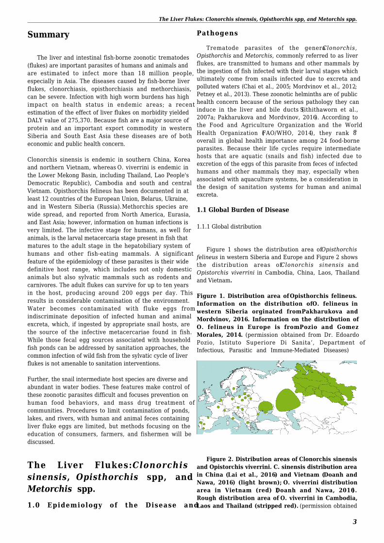
Figure 1 shows the distribution area of Opisthorchisfelineus in western Siberia and Europe and Figure 2 showsthe distribution areas of Clonorchis sinensis andOpistorchis viverrini in Cambodia, China, Laos, Thailandand Vietnam.
Figure 1. Distribution area of Opisthorchis felineus.Information on the distribution of O. felineus inwestern Siberia orginated from Pakharukova andMordvinov, 2016. Information on the distribution ofO. felineus in Europe is from Pozio and GomezMorales, 2014. (permission obtained from Dr. EdoardoPozio, Istituto Superiore Di Sanita’, Department ofInfectious, Parasitic and Immune-Mediated Diseases)
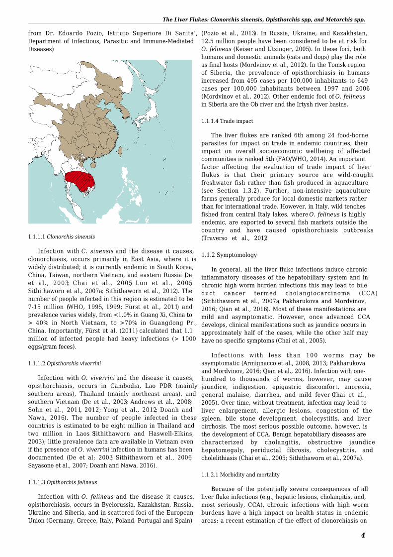
Figure 2. Distribution areas of Clonorchis sinensisand Opistorchis viverrini. C. sinensis distribution areain China (Lai et al., 2016) and Vietnam (Doanh andNawa, 2016) (light brown); O. viverrini distributionarea in Vietnam (red) (Doanh and Nawa, 2016).Rough distribution area of O. viverrini in Cambodia,Laos and Thailand (stripped red). (permission obtained
The Liver Flukes: Clonorchis sinensis, Opisthorchis spp, and Metorchis spp.
4
from Dr. Edoardo Pozio, Istituto Superiore Di Sanita’,Department of Infectious, Parasitic and Immune-MediatedDiseases)
1.1.1.1 Clonorchis sinensis
Infection with C. sinensis and the disease it causes,clonorchiasis, occurs primarily in East Asia, where it iswidely distributed; it is currently endemic in South Korea,China, Taiwan, northern Vietnam, and eastern Russia (Deet al. , 2003; Chai et al. , 2005; Lun et al. , 2005;Sithithaworn et al., 2007a; Sithithaworn et al., 2012). Thenumber of people infected in this region is estimated to be7-15 million (WHO, 1995, 1999; Fürst et al., 2011) andprevalence varies widely, from <1.0% in Guang Xi, China to> 40% in North Vietnam, to >70% in Guangdong Pr.,China. Importantly, Fürst et al. (2011) calculated that 1.1million of infected people had heavy infections (> 1000eggs/gram feces).
1.1.1.2 Opisthorchis viverrini
Infection with O. viverrini and the disease it causes,opisthorchiasis, occurs in Cambodia, Lao PDR (mainlysouthern areas), Thailand (mainly northeast areas), andsouthern Vietnam (De et al., 2003; Andrews et al., 2008;Sohn et al., 2011, 2012; Yong et al., 2012; Doanh andNawa, 2016). The number of people infected in thesecountries is estimated to be eight million in Thailand andtwo million in Laos (Sithithaworn and Haswell-Elkins,2003); little prevalence data are available in Vietnam evenif the presence of O. viverrini infection in humans has beendocumented (De et al; 2003; Sithithaworn et al., 2006;Sayasone et al., 2007; Doanh and Nawa, 2016).
1.1.1.3 Opithorchis felineus
Infection with O. felineus and the disease it causes,opisthorchiasis, occurs in Byelorussia, Kazakhstan, Russia,Ukraine and Siberia, and in scattered foci of the EuropeanUnion (Germany, Greece, Italy, Poland, Portugal and Spain)
(Pozio et al., 2013). In Russia, Ukraine, and Kazakhstan,12.5 million people have been considered to be at risk forO. felineus (Keiser and Utzinger, 2005). In these foci, bothhumans and domestic animals (cats and dogs) play the roleas final hosts (Mordvinov et al., 2012). In the Tomsk regionof Siberia, the prevalence of opisthorchiasis in humansincreased from 495 cases per 100,000 inhabitants to 649cases per 100,000 inhabitants between 1997 and 2006(Mordvinov et al., 2012). Other endemic foci of O. felineusin Siberia are the Ob river and the Irtysh river basins.
1.1.1.4 Trade impact
The liver flukes are ranked 6th among 24 food-borneparasites for impact on trade in endemic countries; theirimpact on overall socioeconomic wellbeing of affectedcommunities is ranked 5th (FAO/WHO, 2014). An importantfactor affecting the evaluation of trade impact of liverflukes is that their primary source are wild-caughtfreshwater fish rather than fish produced in aquaculture(see Section 1.3.2). Further, non-intensive aquaculturefarms generally produce for local domestic markets ratherthan for international trade. However, in Italy, wild tenchesfished from central Italy lakes, where O. felineus is highlyendemic, are exported to several fish markets outside thecountry and have caused opisthorchiasis outbreaks(Traverso et al., 2012).
1.1.2 Symptomology
In general, all the liver fluke infections induce chronicinflammatory diseases of the hepatobiliary system and inchronic high worm burden infections this may lead to bileduct cancer termed cholangiocarcinoma (CCA)(Sithithaworn et al., 2007a; Pakharukova and Mordvinov,2016; Qian et al., 2016). Most of these manifestations aremild and asymptomatic. However, once advanced CCAdevelops, clinical manifestations such as jaundice occurs inapproximately half of the cases, while the other half mayhave no specific symptoms (Chai et al., 2005).
Infections with less than 100 worms may beasymptomatic (Armignacco et al., 2008, 2013; Pakharukovaand Mordvinov, 2016; Qian et al., 2016). Infection with one-hundred to thousands of worms, however, may causejaundice, indigestion, epigastric discomfort, anorexia,general malaise, diarrhea, and mild fever (Chai et al.,2005). Over time, without treatment, infection may lead toliver enlargement, allergic lesions, congestion of thespleen, bile stone development, cholecystitis, and livercirrhosis. The most serious possible outcome, however, isthe development of CCA. Benign hepatobiliary diseases arecharacterized by cholangitis, obstructive jaundicehepatomegaly, periductal fibrosis, cholecystitis, andcholelithiasis (Chai et al., 2005; Sithithaworn et al., 2007a).
1.1.2.1 Morbidity and mortality
Because of the potentially severe consequences of allliver fluke infections (e.g., hepatic lesions, cholangitis, and,most seriously, CCA), chronic infections with high wormburdens have a high impact on health status in endemicareas; a recent estimation of the effect of clonorchiasis on
The Liver Flukes: Clonorchis sinensis, Opisthorchis spp, and Metorchis spp.
5
morbidity yielded DALY value of 275,370 (Fürst et al.,2011), a relatively high impact for a helminthic disease. Inhighly endemic foci of O. felineus in Western Siberia, CCAwas detected in 77% of patients with opisthorchiasis,versus 34.2% of patients without opisthorchiasis(Pakharukova and Mordvinov, 2016).
1.2 Taxonomic Classification of the Agents
The fishborne liver flukes of public health importancebelong to the trematode family Opisthorchiidae (Scholtz,
2008). The most prevalent and important species areClonorchis sinensis, Opisthorchis viverrini, and O. felineus,members of the subfamily Opisthorchiinae. These speciesare similar in morphology, life cycles, and modes oftransmission, which often causes difficulties in specificdiagnosis. Their geographic distributions are basicallyallopatric, however. Other species of the Opisthorchisgenus have been reported from humans only rarely and willnot be considered further in this chapter.
1.2.1 Physical description of the agents
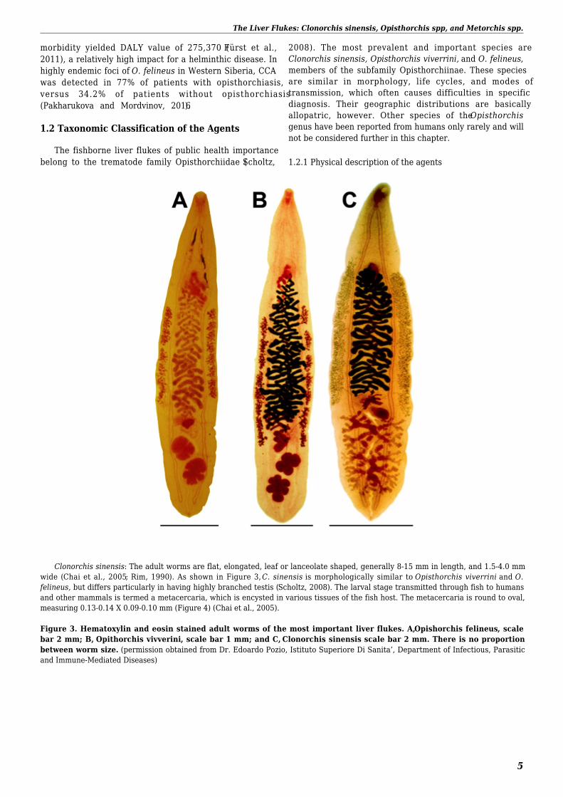
Clonorchis sinensis: The adult worms are flat, elongated, leaf or lanceolate shaped, generally 8-15 mm in length, and 1.5-4.0 mmwide (Chai et al., 2005; Rim, 1990). As shown in Figure 3, C. sinensis is morphologically similar to Opisthorchis viverrini and O.felineus, but differs particularly in having highly branched testis (Scholtz, 2008). The larval stage transmitted through fish to humansand other mammals is termed a metacercaria, which is encysted in various tissues of the fish host. The metacercaria is round to oval,measuring 0.13-0.14 X 0.09-0.10 mm (Figure 4) (Chai et al., 2005).
Figure 3. Hematoxylin and eosin stained adult worms of the most important liver flukes. A, Opishorchis felineus, scalebar 2 mm; B, Opithorchis vivverini, scale bar 1 mm; and C, Clonorchis sinensis scale bar 2 mm. There is no proportionbetween worm size. (permission obtained from Dr. Edoardo Pozio, Istituto Superiore Di Sanita’, Department of Infectious, Parasiticand Immune-Mediated Diseases)
The Liver Flukes: Clonorchis sinensis, Opisthorchis spp, and Metorchis spp.
6
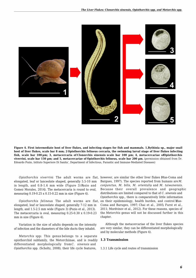
Figure 4. First intermediate host of liver flukes, and infecting stages for fish and mammals. 1, Bythinia sp., major snailhost of liver flukes, scale bar 8 mm; 2 Opisthorchis felineus cercaria, the swimming larval stage of liver flukes infectingfish, scale bar 100 µm; 3, metacercaria of Clonorchis sinensis scale bar 100 µm; 4, metacercariae of Opisthorchisviverrini, scale bar 150 µm; and 5, metacercariae of Opisthorchis felineus, scale bar 200 µm. (permission obtained from Dr.Edoardo Pozio, Istituto Superiore Di Sanita’, Department of Infectious, Parasitic and Immune-Mediated Diseases)
Opisthorchis viverrini: The adult worms are flat,elongated, leaf or lanceolate shaped, generally 5.5-10 mmin length, and 0.8-1.6 mm wide (Figure 3) (Pozio andGomez Morales, 2014). The metacercaria is round to oval,measuring 0.19-0.25 x 0.15-0.22 mm in size (Figure 4).
Opisthorchis felineus: The adult worms are flat,elongated, leaf or lanceolate shaped, generally 7-12 mm inlength, and 1.5-2.5 mm wide (Figure 3) (Pozio et al., 2013).The metacercaria is oval, measuring 0.25-0.30 x 0.19-0.23mm in size (Figure 4).
Variation in the size of adults depends on the intensityof infection and the diameters of the bile ducts they inhabit.
Metorchis spp: This genus belongs to a separateopisthorchid subfamily, the Metorchiinae, and is readilydifferentiated morphologically from C. sinensis andOpisthorchis spp. (Scholtz, 2008); their life cycle features,
however, are similar the other liver flukes (Mas-Coma andBargues, 1997). The species reported from humans are M.conjunctus, M. bilis, M. orientalis, and M. taiwanensis.Because their overall prevalence and geographicdistributions are limited compared to that of C. sinensis andOpisthorchis spp., there is comparatively little informationon their epidemiology, health burden, and control (Mas-Coma and Baruges, 1997; Chai et al., 2005; Furst et al.,2011; Mordvinov et al., 2012). For these reasons, species ofthe Metorchis genus will not be discussed further in thischapter.
Although the metacercariae of the liver flukes speciesare very similar, they can be differentiated morphologicallyand by molecular methods (Figure 4).
1.3 Transmission
1.3.1 Life cycle and routes of transmission
The Liver Flukes: Clonorchis sinensis, Opisthorchis spp, and Metorchis spp.
7
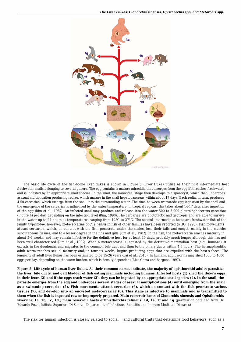
The basic life cycle of the fish-borne liver flukes is shown in Figure 5. Liver flukes utilize as their first intermediate hostfreshwater snails belonging to several genera. The egg contains a mature miracidia that emerges from the egg if it reaches freshwaterand is ingested by an appropriate snail species. In the snail, the miracidial stage then develops to a sporocyst, which then undergoesasexual multiplication producing rediae, which mature in the snail hepatopancreas within about 17 days. Each redia, in turn, produces4-50 cercariae, which emerge from the snail into the surrounding water. The time between trematode egg ingestion by the snail andthe emergence of the cercariae is influenced by the water temperature; in tropical regions, this takes about 14-17 days after ingestionof the egg (Rim et al., 1982). An infected snail may produce and release into the water 500 to 5,000 pleurolophocercus cercariae(Figure 4) per day, depending on the infection level (Rim, 1990). The cercariae are phototactic and geotropic and are able to survivein the water up to 24 hours at temperatures ranging from 12°C to 27°C. The second intermediate hosts are freshwater fish of thefamily Cyprinidae; however, metacercariae of C. sinensis in fish of other families have been reported (WHO, 1995). Fish movementsattract cercariae, which, on contact with the fish, penetrate under the scales, lose their tails and encyst, mainly in the muscles,subcutaneous tissues, and to a lesser degree in the fins and gills (Rim et al., 1982). In the fish, the metacercaria reaches maturity inabout 5-6 weeks, and may remain infective for the definitive host for at least 30 days, probably much longer although this has notbeen well characterized (Rim et al., 1982). When a metacercaria is ingested by the definitive mammalian host (e.g., humans), itexcysts in the duodenum and migrates to the common bile duct and then to the biliary ducts within 4-7 hours. The hermaphroditicadult worm reaches sexual maturity and, in four-six weeks, begins producing eggs that are expelled with the host’s feces. Thelongevity of adult liver flukes has been estimated to be 15-26 years (Lai et al., 2016). In humans, adult worms may shed 1000 to 4000eggs per day, depending on the worm burden, which is density-dependent (Mas-Coma and Bargues, 1997).
Figure 5. Life cycle of human liver flukes. As their common names indicate, the majority of opisthorchid adults parasitizethe liver, bile ducts, and gall bladder of fish eating mammals including humans. Infected hosts (1) shed the fluke's eggsin their feces (2) and if the eggs reach water (3), they can be ingested by an appropriate snail species (4). In the snail, theparasite emerges from the egg and undergoes several stages of asexual multiplications (4) until emerging from the snailas a swimming cercariae (5). Fish movements attract cercariae (6), which on contact with the fish penetrate varioustissues (7), and develop into an encysted metacercariae (8). This stage is infective to mammals and is transmitted tothem when the fish is ingested raw or improperly prepared. Main reservoir hosts of Clonorchis sinensis and Opisthorchisviverrini: 1a, 1b, 1c, 1d,; main reservoir hosts of Opisthorchis felineus: 1d, 1e, 1f and 1g. (permission obtained from Dr.Edoardo Pozio, Istituto Superiore Di Sanita’, Department of Infectious, Parasitic and Immune-Mediated Diseases)
The risk for human infection is closely related to social and cultural traits that determine food behaviors, such as a
The Liver Flukes: Clonorchis sinensis, Opisthorchis spp, and Metorchis spp.
8
fondness for raw or inadequately prepared fish (i.e.,cooked, frozen, or pickled). The consumption of raw orundercooked fish is widely practiced, particularly inlocalities near lakes, reservoirs, streams and ponds wherefresh fish are readily available (WHO, 1995). For example,in China, raw fish is commonly served after dipping brieflyin boiling soup and immediately eaten, or in hot ricecongee. In Thailand, a major source of infection with O.viverrini is consumption of raw or inadequately cooked,frozen, salted, or smoked fish in a dish called Koi-pla. In
Italy, large O. felineus human outbreaks occurred from2007 to 2011 from the consumption of marinated tenchfillets at restaurants or during social events (Pozio et al.,2013). A strong risk factor for C. sinensis infection,especially for males, is the consumption of raw fish at socialgatherings where alcoholic drinks are served (Chai et al.,2005). Studies have shown that liver fluke metacercariae infish tissue are moderately tolerant to low levels of heat,freezing, and pickling (Table 1).
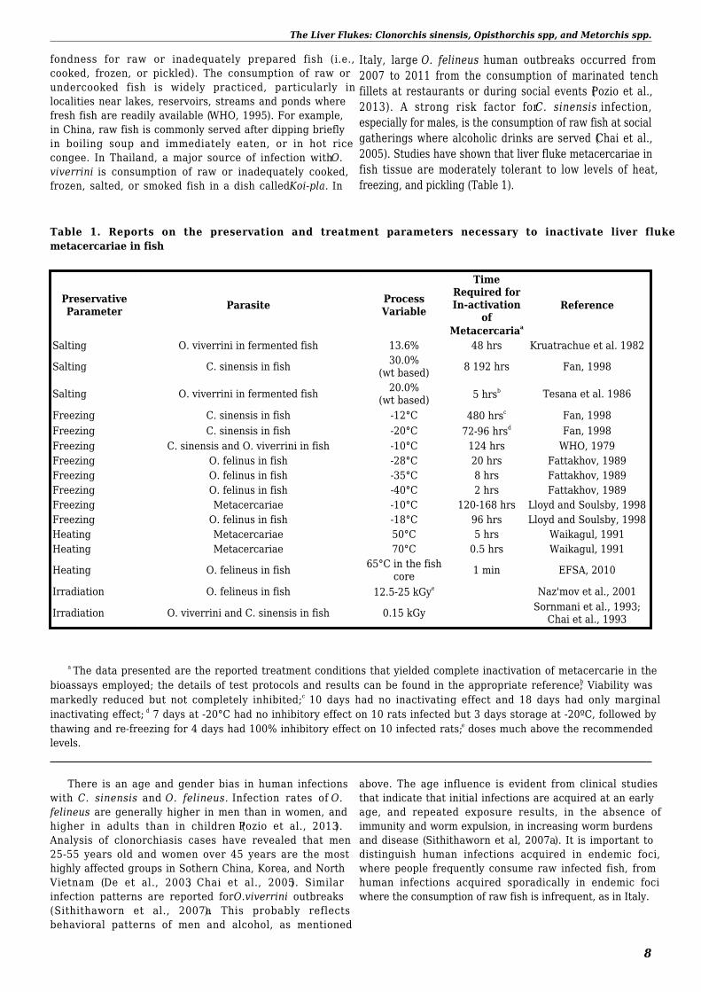
Table 1. Reports on the preservation and treatment parameters necessary to inactivate liver flukemetacercariae in fish
PreservativeParameter Parasite Process
Variable
TimeRequired forIn-activation
ofMetacercariaa
Reference
Salting O. viverrini in fermented fish 13.6% 48 hrs Kruatrachue et al. 1982
Salting C. sinensis in fish 30.0%(wt based) 8 192 hrs Fan, 1998
Salting O. viverrini in fermented fish 20.0%(wt based) 5 hrsb Tesana et al. 1986
Freezing C. sinensis in fish -12°C 480 hrsc Fan, 1998Freezing C. sinensis in fish -20°C 72-96 hrsd Fan, 1998Freezing C. sinensis and O. viverrini in fish -10°C 124 hrs WHO, 1979Freezing O. felinus in fish -28°C 20 hrs Fattakhov, 1989Freezing O. felinus in fish -35°C 8 hrs Fattakhov, 1989Freezing O. felinus in fish -40°C 2 hrs Fattakhov, 1989Freezing Metacercariae -10°C 120-168 hrs Lloyd and Soulsby, 1998Freezing O. felinus in fish -18°C 96 hrs Lloyd and Soulsby, 1998Heating Metacercariae 50°C 5 hrs Waikagul, 1991Heating Metacercariae 70°C 0.5 hrs Waikagul, 1991
Heating O. felineus in fish 65°C in the fishcore 1 min EFSA, 2010
Irradiation O. felineus in fish 12.5-25 kGye Naz'mov et al., 2001
Irradiation O. viverrini and C. sinensis in fish 0.15 kGy Sornmani et al., 1993;Chai et al., 1993
a The data presented are the reported treatment conditions that yielded complete inactivation of metacercarie in thebioassays employed; the details of test protocols and results can be found in the appropriate reference; b Viability wasmarkedly reduced but not completely inhibited; c 10 days had no inactivating effect and 18 days had only marginalinactivating effect; d 7 days at -20°C had no inhibitory effect on 10 rats infected but 3 days storage at -20ºC, followed bythawing and re-freezing for 4 days had 100% inhibitory effect on 10 infected rats; e doses much above the recommendedlevels.
There is an age and gender bias in human infectionswith C. sinensis and O. felineus. Infection rates of O.felineus are generally higher in men than in women, andhigher in adults than in children (Pozio et al., 2013).Analysis of clonorchiasis cases have revealed that men25-55 years old and women over 45 years are the mosthighly affected groups in Sothern China, Korea, and NorthVietnam (De et al., 2003; Chai et al., 2005). Similarinfection patterns are reported for O.viverrini outbreaks(Sithithaworn et al., 2007a). This probably reflectsbehavioral patterns of men and alcohol, as mentioned
above. The age influence is evident from clinical studiesthat indicate that initial infections are acquired at an earlyage, and repeated exposure results, in the absence ofimmunity and worm expulsion, in increasing worm burdensand disease (Sithithaworn et al, 2007a). It is important todistinguish human infections acquired in endemic foci,where people frequently consume raw infected fish, fromhuman infections acquired sporadically in endemic fociwhere the consumption of raw fish is infrequent, as in Italy.
The Liver Flukes: Clonorchis sinensis, Opisthorchis spp, and Metorchis spp.
9
1.3.2 Epidemiological role of the intermediate and reservoirhosts
The liver fluke C. sinensis utilizes, as first intermediatehost, snail species including Alocinma longicornis, Bithyniaspp., Melanoides tuberculatus, Parafossalurus spp., andThiara (WHO, 1995; Sithithaworn et al., 2007a; Hung et al.,2013). The major snail hosts for O. viverrini and O. felineusbelong to the genus Bithynia. The prevalence of larvalstages in snails is always quite low and does not oftenexceed 1% in endemic foci (De Liberato et al., 2011).
The second intermediate hosts are mainly fish of thefamily Cyprinidae. Metacercariae of C. sinensis have beenrecovered f rom f i sh be longing to the generaAcanthogobius, Abbottina, Carassius, Cirrhinus,Crassiodes, Cultrichthys, Cyprinus, Ctenopharyngodon,Erythroculter, Gnathopogon, Hemibarbus, Hemiculter,Hypomesus, Hypophthalmichthys, Ischikauia, Opsariicthys,Oreochromis, Parabramis, Pseudogobio, Pseudorabora,Pungtungia, Rhodeus, Sarcocheilichthys, Toxobramus,Xenocypris, and Zacco (WHO, 1995; Hung et al., 2013).Metacercariae of O. viverrini were detected in fish of thegenera Carassius, Channa, Cyclocheilicthys, Hampala,Esomus, Osteochilus, Puntioplites, and Puntius (WHO,1995). Metacercariae of O. felineus were detected in fish ofthe genera Alburnus, Abramis, Aspius, Blicca, Carassius,Chondrostoma, Cobitis, Cyprinus, Gobio, Leucaspius,Leuciscus, Phoxinus, Rutilus, Scardinius, and Tinca(Erhardt et al., 1962; Pozio et al., 2013; Pakharukova andMordvinov, 2016). There are reports of shrimp foundinfected with metacercariae that morphologically wereidentified as C. sinensis (Chen et al., 2010), but follow upstudies to verify their identity do not appear to have beenconducted. Because shrimp are intermediate hosts for othertrematode species, this report must be provisional.
The large number of fish species reported infected withliver fluke metacerariae (see above) implies that theseparasites have low host specificity (WHO, 1995; Chai et al.,2005). ) It is therefore important to be aware that inlocations where these parasites are found, infections mayoccur in several fish species, and the relative infection ratesmay fluctuate independently, an important considerationfor epidemiological studies. Importantly, wild fish fromclean fresh water sources, such as rivers and reservoirs,are usually preferred for preparing raw fish dishes.
All fish eating mammals, including humans, arepotential final hosts of liver flukes. Hosts for C. sinensis andO. viverrini, besides humans, include feral and domesticcats and dogs, and pigs and rats (Rattus); these hostspecies play the crucial role of reservoir host (Mas-Comaand Bargues, 1997; Lun et al., 2005; Lan-Anh et al., 2009;Petney et al., 2013). However, C. sinensis has also beenreported from sylvatic animals such as martins, civet cats,badgers, monkeys, weasels, muskrats, foxes and rice rats(Mas-Coma and Bargues, 1997; Hung et al., 2013; Petney etal., 2013). The role of sylvatic reservoir hosts is veryimportant in the epidemiology of O. felineus, which has aneven wider spectrum of final hosts; it has been reportedfrom domestic (e.g., cats, dogs, pigs), synanthropic (e.g.,muskrats, rats) and 28 wild animals (e.g., otters, polecats,
polar and red foxes, sable, seals, wild boar, wolverines)(Mordvinov et al., 2012; Pozio et al., 2013; Chai et al.,2005).
The role of a sylvatic cycle in the epidemiology of C.sinensis and O. viverrini has not been well studied. A rolefor wild animal reservoirs is suggested from surveys of fishinfections in endemic areas, which frequently demonstratethat the prevalence of liver fluke metacercariae is oftenhigher in wild fish from reservoirs, canals, streams and ricefields than in fish from farm ponds (Mas-Coma andBargues, 1997; Chai et al., 2005; Phan et al., 2010; Li et al.,2013).
Cats, dogs and pigs are considered to be the mostimportant reservoir hosts in the domestic habitat, becauseof their wide distribution and large populations (WHO,1995). Although it is a common assumption that farmhouseholds, including humans, dogs, pigs, and cats, playessential roles in liver fluke epidemiology, the greatestinfection risk factor for domestic cats and dogs is thecommon practice of allowing such animals to roam andscavenge freely in the communities (Lan-Anh et al., 2009;Aunpromma et al . , 2012; Petney et al . , 2013).Epidemiological studies on the role of cultured and wild-caught fish in liver fluke transmission have demonstratedthat sylvatic hosts, both fish and mammals, can sustain thelife cycle and risk for humans in the absence of a domesticcycle (Li et al., 2013; Clausen et al., 2015).
The probable explanation for the higher infection ratesof C. sinenesis and O. viverinni in wild caught fish mayberelated to both biotic and abiotic factors associated withthe different aquatic habitats of vector snails, especiallyBythinia spp. Recent research in Thailand and Vietnam onthe ecology and distribution in various aquatic habitats of amajor snail host in the genus Bithynia, revealed a greaterabundance in rice fields, streams, and small canals than inlakes and in farm ponds (Brockelman et al., 1986; Ngern-klun et al., 2006; Petney et al., 2012; Doanh and Nawa,2016). Further, investigations on the abiotic factorsaffecting the abundance of Bithynia siamemsisgonoiomphalos, the major snail host for O. vivverini,revealed the importance of water depth and temperature,level of dissolved oxygen, pH, and salinity (Nithiuthai et al.,2002). These conditions may not be met in farm ponds thatare generally stagnant, warm, and with low oxygen levels.
1.4 Population and Individual Control Measures
1.4.1 Treatment options
Prevention and control of human liver fluke infectionsmust begin with an effective education effort directed atenabling consumers to understand the risks associated witheating raw or undercooked fish, regardless of source.Currently, the major strategies for community preventionand control encompasses fecal examination and treatmentof individual cases with praziquantel (25 mg/kg three timesdaily for 2-3 days), and environmental sanitation bybuilding and use of household latrines (Chai et al., 2005;Sithithaworn et al., 2007a). Mass chemotherapy withpraziquantel (40 mg in a single dose) is recommended by
The Liver Flukes: Clonorchis sinensis, Opisthorchis spp, and Metorchis spp.
10
WHO (1995), but the sustainability of control by thisapproach is uncertain (Chai et al., 2005; Clausen et al.,2015). O. felineus infection in humans can be treated withpraziquantel (25 mg/kg orally 3×/day for 1–2 days) oralbendazole (10 mg/kg/day orally in 2 doses for 7–14 days).The treatment with praziquantel is, as a rule, effective,whereas treatment with albendazole can fail and theindividual has to be treated again with another drug(Armignacco et al., 2008, 2013). After the secondtreatment, eggs are generally not detected in fecalsamples; however, in very few cases, eggs have beendetected after a period from 5 to 6 months up to 2 yearsdespite treatment of the patients with albendazole (Pozio etal., 2013). Although the effectiveness of treatment can bedetermined based on the search for eggs in stool samples,very few eggs are produced in cases of unsuccessfultreatment; for this reason, ELISA could be used, althoughthe antibody levels decrease very slowly to the cut-off value(Pozio et al., 2013). Vaccines to prevent liver fluke infectionin humans have not been developed.
1.4.2 Hygiene measures
Emphasis on hygienic measures applicable tohouseholds and restaurants may be the most fruitfulapproach to control in endemic areas, at least for theimmediate future. Treatment of fish to inactivate themetacercariae by heating to temperatures of 70 °C orabove are effective (WHO, 1995). Table 1 lists parametersfor inactivation by salting and freezing, pickling, andirradiation that can be effective (WHO, 1995). Other foodpreparation methods for household use that may inactivatemetacercariae, such as microwaving, smoking, fermenting,and marinating, need further investigation before guidanceon their use can be formulated. O. felineus metacercariae,for example, can survive in smoked fish causing humaninfections (Yossepowitch et al., 2004), nor does marinatingdoes not kill O. felineus metacercariae present in tenchmuscles (Armignacco et al., 2008, 2013; Traverso et al.,2012).
Education of food handlers on the risk from fish inendemic areas and on the need to keep preparationcounters and utensils clean between individual fishpreparations is highly recommended. It should be notedthat many home freezers can reach temperatures of -6°C or-12°C, , but domestic freezers that can operate at atemperature of -18°C or below are more effective; infectedfish need to be frozen in all parts of the products for longerthan 24 hours to ensure that all parasites are inactivated.
Investigations in Thailand have demonstrated that lowdose irradiation of freshwater fish can prevent infectivity ofO. viverrini metacercariae when such fish are prepared inlocal dishes made from raw or semi processed fish(Bhaibulaya, 1985). Experimental studies should beextended in order to evaluate the usefulness of this controlmeasure in food processing (EFSA, 2010).
2.0 Environmental Occurrence andPersistence of Larval Liver Fluke Stages
2.1. Detection Methods
Detection is made most often by microscopy howevertrained parasitologists are needed to make theidentification. Eggs containing the miracidia of liver flukescan be detected in the feces by using standard fecal exammethods (e.g., Kato-Katz technic). Fecal deposits picked upfrom the ground adjacent to fish ponds, rice paddies,reservoirs and streams, and from latrines and pig pens, canbe collected and tested. This approach has been successfulin identifying reservoir hosts with aquaculture activitiesand assessing their role in the epidemiology of fishborneflukes (Lan-Anh et al., 2009).
Possible ongoing transmission may be detected byexamining snails collected from fishponds and localwaterbodies and examining them for the presence ofopithorchid sporocysts and/or rediae, and for cercariae.This can be done either by crushing the snail and viewingthe remnants under a stereomicroscope or by allowing thesnails to shed cercariae directly into water-filled containers.However, because snails may be infected with othertrematodes with pleurolophocercous type cercaria (Figure4), especially heterophyid intestinal flukes, a specificidentification of liver fluke cercariae may not be possible.
Examination of local fish can also provide an indicationof liver fluke transmission in the area. Fish tissue can beexamined directly by microscope for metacercariae (Figure4) or, preferably, after pepsin digestion to free anymetacercariae present before examination (WHO, 1995;Thu et al., 2007; De Liberato et al., 2011).
Use of molecular methods such as PCR, can makedetection more reliable (Jeon et al., 2012). Distinguishingliver fluke eggs from the intestinal heterophyid fluke eggscan also be difficult (Ditrich et al., 1992), and molecularmethods applicable to egg identification have also beendeveloped (Sato et al., 2009; Sanpool et al., 2012;Armignacco et al., 2008).
2.2 Environmental Contamination with Eggs
There are no data on the occurrence of liver fluke eggsin sewage or various types of polluted waters. The numberof eggs per gram of human feces can be used to roughlycalculate potential egg concentration in sewage. In a studycarried out in a focus of clonorchiasis of South Korea, themean concentration of eggs was reported to be 2.8 x 10³
per gram of human feces, with a range of 12 to 6.6x10⁴ pergram (Kim et al., 2011). On average, humans excreteapproximately 100 grams of feces per person per day andin highly endemic foci of clonorchiasis the prevalence ofinfection can reach 70%. Eggs could sediment out into thesolids, such as sludge or carried along in wet sewage, and ifthese waste materials are emptied into water bodies (ricefields, ponds, streams, canals, or rivers) that containsuitable snail hosts and fish, the liver fluke life cycle couldbe sustained.
2.3 Survival (Persistence) of Eggs in theEnvironment
The Liver Flukes: Clonorchis sinensis, Opisthorchis spp, and Metorchis spp.
11
Because of a lack of a desiccation-resistant protectivecoating, trematode eggs and cercariae are extremelyfragile.. Limited studies under laboratory conditions haveyielded some information on their survival. Faust and Khaw(1927) and Komiya (1966) reported that C. sinensis eggssurvived in isotonic solution at 2°C - 4°C for up to 3months, and at 26 °C for up to 1 month. In fresh night soil,the survival time was 2 days at 25 °C; survival decreasedwith age of the night soil. Drodzdov (1962) observedsurvival of O. felineus eggs for 160 days in river water heldat 0 °C - 5 °C. Field research on egg persistence in soil andwater under natural environmental conditions is needed.Similarly, studies on the survival of eggs in sewage, sludge,surface water, wastewater and irrigation water areinsufficient to draw any conclusions.
3.0 Reducing Environmental Contaminationwith Liver Fluke Eggs, Snail IntermediateHosts, and Cercariae by Sanitation Measures
3.1 Education and Community-based Actions
Successful control of these parasitic trematodesrequires reducing the probability of transmission.Modifying or breaking the transmission cycle can occur atany stage of the parasite’s life cycle, but both snail and fishinfections have proven difficult to control in naturalhabitats (Sithithaworn et al., 2007a). Therefore, mostcontrol programs aim at reducing and interruptingtransmission at the reservoir host (including humans) level.The approaches to accomplishing this differ between thethree liver fluke species. For examples, wild mammals playa very important role in maintaining O. felineus endemicity(Pozio et al., 2013). In the case of C. sinensis and O.viverrini, uncontrolled free-roaming cats and dogs areimportant in sustaining their life cycles; the importance ofwild mammal reservoirs in the epidemiology of these flukeshas not been adequately investigated but may besignificant. Because of these non-human hosts, and theimportance of wild-caught fish intermediate hosts, the mostimportant control intervention is the education ofconsumers in endemic areas. Education of the relevantcommunities on the epidemiology and health consequencesof liver flukes is a key component of any control programfor all three species (WHO, 1995; Jongsuksuntigul andImsomboon, 1997, 2003). Health education both at thevillage and school levels is of major significance, becauseinfection is often unapparent and CCA only develops aftermany years (Sithithaworn et al., 2007a). Education shouldbe aimed at explaining the pathology of the disease and inparticular, its association with CCA, and how the parasite istransmitted by various final and intermediate hosts. It isparticularly important to stress the role that eating raw orpartially cooked fish plays in disease transmission (WHO,1995; Jongsuksuntigul and Imsomboon, 2003).
An example of the impact of an education program isthe experience in Thailand. Education on the risks ofinfection led to the frequency of consumption of raw, orpartially cooked or fermented fish decreasing from 14% in1990 to 7% in 1994 (Jongsuksuntigul and Imsomboon,1997). In addition to education programs, which are aimed
at reducing the risk of infection, anthelminthic treatment isstill required to reduce the output of eggs from infectedindividuals into the environment. Because of the lack ofacquired protective immunity in human, dog and catinfections, reinfection of people in endemic areas is likely ifthey continue to eat raw fish (Sornmani et al., 1984;Upatham et al., 1988; Clausen et al., 2015).
3.2 Control of Snails
There are many methods employed or proposed tocontrol snails, including chemical control (molluscicides),physical control (Hoffman, 1970; Khamboonraung et al.,1997), ecological control (Wang et al. 2007), and biologicalcontrol (Hung et al., 2014). Although these methods areapplicable to aquaculture ponds, control of snails inmarshes, rivers, reservoirs and rice paddies is impracticaland may be environmentally harmful. Many molluscicides,such as copper sulfate (Reddy et al., 2004), endosulfan(Otludil et al., 2004), and bayluscide (Dai et al., 2010), havebeen used but they have toxic effects on fish, water plants,and small organisms, and in modern aquaculture systemsthey are either prohibited or discouraged. Biobasedmolluscicides, including plant components, may have lesstoxic effects on humans, aquatic animals, and plants, andresearch on them should be encouraged, especially theircost-effectiveness. Studies on biological control withpredatory fish (on snails) such as black carp have recentlyshown promise, but this approach is still in the early phaseof research and development (Hung et al., 2013).
3.3 Sanitation Technologies
There are no data available on removal of the eggs bywastewater treatment. Their small size (average 27 X 15μm) may account for their ability to escape removal fromwater by standard filtration systems or sedimentation. Alsothere are very few recommendations on standard sanitationmeasures for the control of human liver flukes, the lack ofwhich may be attributed to the unique epidemiology ofhuman infections and the ecology of the liver flukes. Asdiscussed in Section 1.3.2, the liver flukes have a widerange of both domestic and sylvatic reservoir hosts, andsurveys of snails and fish for liver fluke metacercariaeconsistently reveal a major cycling of liver flukes throughwild, non-cultured fish residing in lakes, reservoirs,streams, rivers, marshes and rice fields (Sithithaworn et al.,2007b; Thu et al., 2007; Li et al., 2013). In contrast, surveysof fish produced in aquaculture ponds generally reveal lowprevalence of C. sinensis, and O. viverrini (Chi et al., 2008;Thien et al., 2007; Li et al., 2013; Chen et al., 2010;Pitaksakulrat et al., 2013), even when the humanprevalence of C. sinensis, for example, in a community ishigh (Dung et al., 2007). Further, the human prevalence ofliver flukes in aquaculture systems may be overestimatedbecause of diagnostic confusion between their eggs withthose of fishborne intestinal flukes (Heterophyidae) (Ditrichet al., 1992; De et al., 2003; Chi et al., 2008; Thien et al.,2007).
The major snail vectors of liver flukes (e.g., Bythiniaspp., Parafossarulus spp.), however, are not common in fishponds, preferring instead moving water associated with
The Liver Flukes: Clonorchis sinensis, Opisthorchis spp, and Metorchis spp.
12
lakes, reservoirs, streams and canals (Petney et al., 2012).
In contrast, the major snail host for the heterophyids,Melanoidies tuberculata, is very common in aquacultureponds (Dung et al 2010; Madsen and Hung 2014). Becausemost people in endemic areas consume both wild caughtand cultured fish, it is difficult to determine the actualsource of the infected fish. Importantly, wild fish are readilyavailable in local markets and they are often preferred forraw fish dishes because they are obtained from water thatis cleaner and less polluted than that normally found infarm ponds. The importance of wild caught fish in thetransmission of liver flukes underscores the difficulty ofapplying standard sanitation approaches for prevention andcontrol of liver flukes.
3.4 Role for Sanitation Interventions inAquaculture
When there is evidence that significant transmissionfrom cultured fish is an important source of humaninfections, there are interventions that can be implementedin aquaculture systems to control fish-borne liver andintestinal flukes. These interventions, developed inextensive field trials (Khambooraung et al., 1997; Clausenet al., 2015), are designed to control fish infections byeliminating egg contamination of the ponds and reducingthe snail populations. They require a strong program offarmer education and improvements in managementpractices and pond infrastructure, as follows:
Education:1.
Before initiating the program’s changes to pondinfrastructure and management, the farmers shouldreceive training on the basics of the biology andepidemiology of fishborne zoonotic trematodes andthe health benefits to themselves and their familiesfor the prescriptive prevention and controlinterventions.Household members must be encouraged to avoid
eating raw fish and to prevent consumption of rawor dead fish by farm animals, including dogs, catsand pigs.
Interventions to prevent egg and host fecal2.contamination of the pond environment require:
Modification of pond embankments to preventsurface water run-off from entering the pond byinstalling a cement barrier at least 10-15 cm abovethe bank top.Installation of fencing to exclude reservoir hosts,especially cats and dogs, from the immediate pondenvironment.Prevent discharge into the pond of all waste fromlatrines and livestock pens.
Interventions to prevent and control snails in the3.fish pond require that:
Before restocking the ponds with juvenile fishbetween the harvests, the pond should be drainedand dried completely for at least 5 days. The top 3-5cm of bottom mud should be removed to a site notadjacent to the pond.All vegetation in ponds should be removed, and aliner (plastic or cement) applied to the banks of theponds.Aquatic vegetation must be removed at least 3 mfrom the water intake portal (the inlet for pondwater replenishment) and all in-coming waterfiltered through a 5 mm mesh screen beforeentering the pond.
Additional public health actions for endemic4.communities. Educate households:
on the risk from inadequately prepared fish food;to avoid contaminating water bodies with humanand animal waste to the extent possible;on the signs and symptoms of liver fluke infection,and to seek medical treatment when infection issuspected.
The Liver Flukes: Clonorchis sinensis, Opisthorchis spp, and Metorchis spp.
13
ReferencesAndrews, RH, Sithithaworn, P and Petney, TN (2008). Opisthorchis viverrini: an underestimated parasite in world health.Trends Parasitol. 24, pp. 497–501.
Armignacco, O, Caterini, L, Marucci, G, Ferri, F, Bernardini, G, G Raponi, N et al. (2008). Human illnesses caused byOpisthorchis felineus flukes, Italy. Emerg Infect Dis. 14, pp. 1902–1905.
Armignacco, O, Ferri, F, Gomez-Morales, MA, Caterini, L and Pozio, E (2013). Cryptic and asymptomatic Opisthorchisfelineus infections. Am J Trop Med Hyg. 88, pp. 364–366.
Aunpromma, S, Tangkawattana, P, Papirom, P, Kanjampa, P, Tesana, S, Sripa, B et al. (2012). High prevalence ofOpisthorchis viverrini infection in reservoir hosts in four districts of Khon Kaen Province, an opisthorchiasis endemic areaof Thailand. Parasitol Int. 61, pp. 60–64.
Bhaibulaya, M (1985). Human infection with Bertiella studeri in Thailand. Southeast Asian J Trop Med Public Health. 16,pp. 505–507.
Brockelman, WY, Upatham, ES, Viyanant, V, Ardsungnoen, S and Chantanawat, R (1986). Field studies on the transmissionof the human liver fluke, Opisthorchis viverrini, in northeast Thailand: population changes of the snail intermediate host.Int J Parasitol. 16, pp. 545–552.
Chai, JY, Huh, S,, Kook, J, Jung, KC, Park, EC et al. (1993). An epidemiological study of metagonimiasis along the upperreaches of the Namhan River. Korean J Parasitol. 31, pp. 99–108.
Chai, JY, Murrell, KD and Lymbery, AJ (2005). Fish-borne parasitic zoonoses: status and issues. Int J Parasitol. 35, pp.1233–1254.
Chen, D, Chen, J, Huang, J, Chen, X, Feng, D, Liang, B et al. (2010). Epidemiological investigation of Clonorchis sinensisinfection in freshwater fishes in the Pearl River Delta. Parasitol Res. 107, pp. 835–839.
Chi, T, Dalsgaard, A, Turnbull, J, Tuan, P and Murrell, KD (2008). Prevalence of zoonotic trematodes in fish from aVietnamese fish-farming community. J Parasitol. 94, pp. 423–428.
Clausen, JH, Madsen, H, Van, PT, Dalsgaard, A and Murrell, KD (2015). Integrated parasite management: path tosustainable control of fishborne trematodes in aquaculture. Trends Parasitol. 31, pp. 8–15.
Coles, GC,, Wang, W and Liang, YS (2010). Toxicity of a novel suspension concentrate of niclosamide against Biomphalariaglabrata. Trans R Soc Trop Med Hyg. 104, pp. 304–306.
De Liberato, C, Scaramozzino, P, Brozzi, A, Lorenzetti, R, Di Cave, D, Martini, E et al. (2011). Investigation on Opisthorchisfelineus occurrence and life cycle in Italy. Vet Parasitol. 177, pp. 67–71.
De, NV, Murrell, KD, Cong, LD, Cam, P, Chau, LV, Toan, N et al. (2003). The food-borne trematode zoonoses of Vietnam.Southeast J Trop Med Pub Health. Southeast Asian J Trop Med Public Health. 34, pp. 12–34.
Ditrich, O, Nasincová, V, Scholz, T and Giboda, M (1992). Larval stages of medically important flukes (Trematoda) fromVientiane province, Laos. Part II. Cercariae. Ann Parasitol Hum Comp. 67, pp. 75–81.
Doanha, PN and Nawab, Y (2016). Clonorchis sinensis and Opisthorchis spp. in Vietnam: current status and prospects.Trans R Soc Trop Med Hyg. 110, pp. 13–20.
Drozdov, VN (1962). Survival of Opisthorchis felineus eggs (Rivolta, 1884) in various environmental conditions. MedParazitol (Mosk). 31, pp. 323–336.
Dung, BT, Madsen, H and DT, T (2010). Distribution of freshwater snails in family-based VAC ponds and associated
The Liver Flukes: Clonorchis sinensis, Opisthorchis spp, and Metorchis spp.
14
waterbodies with special reference to intermediate hosts of fish-borne zoonotic trematodes in Nam Dinh Province,Vietnam. Acta Trop. 116, pp. 15–23.
Dung, D, Van De, N, Waikagul, J, Dalsgaard, A, Chai, JY, Sohn, WM et al. (2007). Fishborne zoonotic intestinal trematodes,Vietnam. Emerg Infect Dis. 13, pp. 1828–1833.
EFSA (2010). Scientific Opinion on risk assessment of parasites in fishery products. Atlantic. 8,
Erhardt, A, Germer, WD and Hörning, B (1962). Die Opisthorchiasis, hervorgerufen durch den KatzenleberegelOpisthorchis felineus (Riv.). Parasitologische Schriftenreihe. Veb Gustav Fischer Verlag, Jena. 15,
Fan, PC (1998). Viability of metacercariae of Clonorchis sinensis in frozen or salted freshwater fish. International Journalfor Parasitology. 28, pp. 603–605.
Fattakhov, RG (1989). Low- temperature regimes for decontamination of fish of the larvae Opisthorchis (in Russian).Medicine Parazitology (Mosk). 5, pp. 63–65.
Faust, EC and Khaw, OK (1927). Studies on Clonorchis sinensis (Cobbold).. Am J Hyg Monographic Series. 8,
Fürst, T, Keiser, J and Utzinger, J (2011). Global burden of human food-borne trematodiasis: a systematic review andmetaanalysis. Lancet Infect Dis. 12, pp. 210–221.
Hoffman, GL (1970). Control methods for snail-borne zoonoses. J Wildl Dis. 6, pp. 262–265.
Hung, NM, Madsen, H and Fried, B (2013). Global status of fish-borne zoonotic trematodiasis in humans. Acta Parasitol.58, pp. 231–258.
Jeon, HK, Lee, D, Park, H, Min, DY, Rim, HJ, Zhang, H et al. (2012). Human infections with liver and minute intestinalflukes in Guangxi, China: analysis by DNA sequencing, ultrasonography, and immunoaffinity chromatography. Korean JParasitol. 50, pp. 391–394.
Jongsuksuntigul, P and Imsomboon, T (2003). Opisthorchiasis control in Thailand. Acta Trop. 88, pp. 229–232.
Jongsuksuntigul, P and Imsomboon, T (1997). The impact of a decade long opisthorchiasis control program in northeasternThailand. Social Sciences and Medicine. 28, pp. 551–557.
Keiser, J and Utzinger, J (2005). Emerging foodborne trematodiasis. Emerg Infect Dis. 11, pp. 1507–1514.
Khamboonraung, C, Keawvichit, R, Wongworapat, K, Suwanrangsi, S, Hongpromyart, M, Sukhawat, K et al.(1997). Application of hazard analysis critical control point (HACCP) as a possible control measure for Opisthorchisviverrini infection in cultured carp (Puntius gonionotus). Southeast Asian J Trop Med Public Health. 28, pp. 65–72.
Kim, JH, Choi, MH, Bae, YM, Oh, JK, Lim, MK and Hong, ST (2011). Correlation between discharged worms and fecal eggcounts in human clonorchiasis. PLoS Negl Trop Dis. 5, pp. e1339.
Komiya, Y (1966). Clonorchis and clonorchiasis. Adv Parasitol. 4, pp. 53–106.
Kruatrachue, M, Chitramvong, YP, Upatham, ES, Vichari, S and Viyanant, V (1982). Effects of physico-chemical factors onthe infection of hamsters by metacercariae of Opisthorchis viverrini. Southeast Asian J Trop Med Public Health. 13, pp.614–617.
Lai, DH, Hong, XK, Su, BX, Liang, C, Hide, G, Zhang, X et al. (2016). Current status of Clonorchis sinensis andclonorchiasis in China. Trans R Soc Trop Med Hyg. 110, pp. 21–27.
Lan-Anh, NT, Phuong, NT, Murrell, KD, Johansen, MV, Dalsgaard, A, Thu, LT et al. (2009). Animal reservoir hosts and fish-borne zoonotic trematode infections on fish farms, Vietnam. Emerg Infect Dis. 15, pp. 540–546.
The Liver Flukes: Clonorchis sinensis, Opisthorchis spp, and Metorchis spp.
15
Li, K, Clausen, JH, Murrell, KD, Liu, L and Dalsgaard, A (2013). Risks for fishborne zoonotic trematodes in Tilapiaproduction systems in Guangdong Province, China. Vet Parasitol. 198, pp. 223–229.
Lloyd, S and Soulsby, L (1998). Other cestode infections. Zoonoses: biology, clinical practice and public health control. (,Soulsby, L and Simpson, DIH, ed.).Oxford, Oxford University Presspp. 653–656.
Lun, ZR, Gasser, RB, Lai, DH, Li, AX, Zhu, XQ, Yu, XB et al. (2005). Clonorchiasis: a key foodborne zoonosis in China.Lancet Infect Dis. 5, pp. 31–41.
Madsen, H and Hung, NM (2014). An overview of freshwater snails in Asia with main focus on Vietnam. Acta Trop. 140, pp.105–117.
Mas-Coma, S and Bargues, MD (1997). Human liver flukes: A review. Res Rev Parasitol. 57, pp. 145–218.
Mordvinov, VA, Yurlova, NI, Ogorodova, LM and Katokhin, AV (2012). Opisthorchis felineus and Metorchis bilis are themain agents of liver fluke infection of humans in Russia. Parasitol Int. 61, pp. 25–31.
Naz'mov, VP, Fedorov, KP, Serbin, VI and Auslender, VL (2001). Decontamination of freshly caught fish infected withOpisthorchis metacercariae by fast electron irradiation. Med Parazitol (Mosk). 2, pp. 26-27.
Ngern-klun, R, Sukantason, K, Tesna, S, Snpakdee, D and Irvine, K (2006). Field investigations of Bithynia funiculata,intermediate host of Opisthorchis viverrini in northern Thailand. Southeast Asian J Trop Med Public Health. 37, pp.662–672.
Nithiuthai, S, Wiwanitkit, V, Suwansaksri, J and Chaengphukeaw, P (2002). A survey of trematode cercariae in Bithyniagoniomphalos in northeast Thailand. Southeast Asian J Trop Med Public Health. 33, pp. 106–109.
Food and Agriculture Organization of the United Nations World Health Organization (2014). Multicriteria-based rankingfor risk management of food-borne parasites. Microbiological Risk Assessment Series. Rome. 23, pp. 302.
Otludil, B, Cengiz, EI, Yildirim, MZ, Unver, O and Unlü, E (2004). The effects of endosulfan on the great ramshorn snailPlanorbarius corneus (Gastropoda, Pulmonata): a histopathological study. Chemosphere. 56, pp. 707–716.
Pakharukova, MY and Mordinov, VA (2016). The liver fluke Opisthorchis felineus: biology, epidemiology and carcinogenicpotential. Trans R Soc Trop Med Hyg. 110, pp. 28–36.
Petney, T, Sithithaworn, P, Andrews, R, Kiatsopit, N, Tesana, S, Grundy-Warr, C et al. (2012). The ecology of the Bithyniafirst intermediate hosts of Opisthorchis viverrini. Parasitol Int. 61, pp. 38–45.
Petney, TN, Andrews, RH, Saijuntha, W, Wenz-Mucke, A and Sithaworn, P (2013). The zoonotic fishborne liver flukesClonorchis sinensis, Opithorchis felineus, and O. viverrini. Int J Parasitol. 43, pp. 1032–1046.
Phan, VT, Ersbøll, AK, Bui, TQ, Nguyen, HT, Murrell, D and Dalsgaard, A (2010). Fish-borne zoonotic trematodes incultured and wild-caught freshwater fish from the Red River Delta, Vietnam. Vector Borne Zoonotic Dis. 10, pp. 861–866.
Pitaksakulrat, O, Sithithaworn, P, Laoprom, N, Laha, T, Petney, TN and Andrews, RH (2013). A cross-sectional study on thepotential transmission of the carcinogenic liver fluke Opisthorchis viverrini and other fishborne zoonotic trematodes byaquaculture fish. Foodborne Pathol Dis. 10, pp. 35–41.
Pozio, E, Armignacco, O, Ferri, F and Morales, MAGomez (2013). Opisthorchis felineus, an emerging infection in Italy andits implication for the European Union. Acta Trop. 126, pp. 54–62.
Pozio, E and Morales, MAGomez (2014). Clonorchiasis and Opisthorchiasis. Helminth Infections and their Impact on GlobalPublic Health. (Bruschi, F, ed.)., Springer-Verlag Wienpp. 123–152.
Qian, MB, Utzinger, J, Keiser, J and Zhou, XN (2016). Clonorchiasis. Lancet2. 387, pp. 800–810.
Reddy, A, Ponder, EL and Fried, B (2004). . Effects of copper sulfate toxicity on cercariae and metacercariae of
The Liver Flukes: Clonorchis sinensis, Opisthorchis spp, and Metorchis spp.
16
Echinostoma caproni and Echinostoma trivolvis and on the survival of Biomphalaria glabrata snails. J Parasitol. 90, pp.1332–1337.
Rim, HJ, Lee, YM, Lee, JS and Joo, KH (1982). Therapeutic field trial with praziquantel (Biltricide(R)) in a rural populationinfected with Clonorchis sinensis. Kisaengchunghak Chapchi. 20, pp. 1–8.
Rim, HJ. (1990). Clonorchiasis in Korea. Kisaengchunghak Chapchi. 28S, pp. 63–78.
Sanpool, O, Intapan, PM, Thanchomnang, T, Janwan, P, Lulitanond, V, Doanh, PN et al. (2012). Rapid detection anddifferentiation of Clonorchis sinensis and Opisthorchis viverrini eggs in human fecal samples using a duplex real-timefluorescence resonance energy transfer PCR and melting curve analysis. Parasitol Res. 111, pp. 89–96.
Sato, M, Thaenkham, U, Dekumyoy, P and Waikagul, J (2009). Discrimination of O. viverrini, C. sinensis, H. pumilio and H.taichui using nuclear DNA-based PCR targeting ribosomal DNA ITS regions. Acta Trop. 109, pp. 81–83.
Sayasone, S, Odermatt, P, Phoumindr, N, Vongsaravane, X, Sensombath, V, Phetsouvanh, R et al. (2007). Epidemiology ofOpisthorchis viverrini in a rural district of southern Lao PDR. Trans R Soc Trop Med Hyg. 101, pp. 40–47.
Scholtz, T (2008). Family Opisthorchiidae. Keys to the Trematoda, Vol.3. (Bray, RA, Gibson, A and Jones, DI, ed.).London,CAB Internat and Nat Hist Museumpp. 9–49.
Sithithaworn, P, Andrews, RH, Nguyen, VD, Wongsaroj, T, Sinuon, M, Odermatt, P et al. (2012). The current status ofopisthorchiasis and clonorchiasis in the Mekong Basin. Parasitol Int. 61, pp. 10–16.
Sithithaworn, P and Haswell-Elkins, M (2003). Epidemiology of Opisthorchis viverrini. Acta Trop. 88, pp. 187–194.
Sithithaworn, P, Nuchjungreed, C, Srisawangwong, T, Ando, K, Petney, TN, Chilton, NB et al. (2007). Genetic variation inOpisthorchis viverrini (Trematoda: Opisthorchiidae) from northeast Thailand and Laos PDR based on random amplifiedpolymorphic DNA analyses. Parasitol Res. 100, pp. 613–617.
Sithithaworn, P, Sukavat, K, Vannachone, B, Sophonphong, K, Ben-Embarek, P, Petney, T et al. (2006). Epidemiology offood-borne trematodes and other parasite infections in a fishing community on the Nam Ngum reservoir, Lao PDR.Southeast Asian J Trop Med Public Health. 37, pp. 1083–1090.
Sithithaworn, P, Yongvanit, P, Tesna, S and Pairojkul, C (2007). Liver flukes. Foodborne Parasitic Zoonoses. Fish and Plant-borne Parasites. (Murrell, KD and Fried, B, ed.)., Springerpp. 3–52.
Sohn, WM, Shin, EH, Yong, TS, Eom, KS, Jeong, HG, Sinuon, M et al. (2011). Adult Opisthorchis viverrini flukes in humans,Takeo, Cambodia. Emerg Infect Dis. 17, pp. 1302–1304.
Sohn, WM, Yong, TS, Eom, KS, Pyo, KH, Lee, MY, Lim, H et al. (2012). Prevalence of Opisthorchis viverrini infection inhumans and fish in Kratie Province, Cambodia. Acta Trop. 124, pp. 215–220.
Sornmani, S, Impand, P and Bundising, C (1993). Irradiation of fish to control the infectivity of the liver fluke Opisthorchisviverrini. Use of Irradiation to Control Infectivity of Food-borne Parasites. Vienna, International Atomic Energy Agencypp.115–127.
Sornmani, S, Vivatanasesth, P, Impand, P, Phatihatakorn, W, Sitabutra, P and Schelp, FP (1984). Infection and re-infectionrates of opisthorchiasis in the water resource development area of Nam Pong project, Khon Kaen Province, northeastThailand. Ann Trop Med Parasitol. 78, pp. 649–656.
Tesana, S, Kaewkes, S and Phinlaor, S (1986). Infectivity and survivorship of Opisthorchis viverrini metacercariae infermented fish. Journal of Parasitology of the Tropical Medical Association Thailand. 9, pp. 21–30.
Thien, PC, Dalsgaard, A, Thanh, BN, Olsen, A and Murrell, KD (2007). Prevalence of fishborne zoonotic parasites inimportant cultured fish species in the Mekong Delta, Vietnam. Parasitol Res. 101, pp. 1277–1284.
Thu, ND, Dalsgaard, A, Loan, LT and Murrell, KD (2007). Survey for zoonotic liver and intestinal trematode metacercariae
The Liver Flukes: Clonorchis sinensis, Opisthorchis spp, and Metorchis spp.
17
in cultured and wild fish in An Giang Province, Vietnam. Korean J Parasitol. 45, pp. 45–54.
Traverso, A, Repetto, E, Magnani, S, Meloni, T, Natrella, M, Marchisio, P et al. (2012). A large outbreak of Opisthorchisfelineus in Italy suggests that opisthorchiasis develops as a febrile eosinophilic syndrome with cholestasis rather than ahepatitis-like syndrome. Eur J Clin Microbiol Infect Dis. 31, pp. 1089–1093.
Upatham, ES, Viyanant, V, Brockelman, WY, Kurathong, S, Lee, P and Kraengraeng, R (1988). Rate of re-infection byOpisthorchis viverrini in an endemic northeast Thai community after chemotherapy. Int J Parasitol. 18, pp. 643–649.
WHO (1999). Food safety issues associated with products from aquaculture. Geneva, WHO Tecg Report Serpp. 9–12,45–49.
WHO (1995). Control of foodborne trematode infections. Geneva, WHO Tech Report Serpp. 849.
WHO (1979). Parasitic zoonoses. Report of a WHO Expert Committee with Participation of FAO. Geneva, World HealthOrganization. WHO Technical Report Series, No. 637
Waikagul, J (1991). Intestinal fluke infections in Southeast Asia. Southeast Asia J Trop Med Public Health. 22, pp. 158–162.
Wang, QP, Chen, XG and Lun, ZR (2007). Invasive freshwater snail, China. Emerg Infect Dis. 13, pp. 1119–1120.
Yong, TS, Shin, EH, Chai, JY, Sohn, WM, Eom, KS, Lee, DM et al. (2012). High prevalence of Opisthorchis viverriniinfection in a riparian population in Takeo Province, Cambodia. Korean J Parasitol. 50, pp. 173–176.
Yossepowitch, O, Gotesman, T, Assous, M, Marva, E, Zimlichman, R and Dan, M (2004). Opisthorchiasis from imported rawfish. Emerg Infect Dis. 10, pp. 2122–2126.