Embed Size (px)
Citation preview

11 January 1969
Current Practice
MEDICINE IN THE TROPICS
Splenomegaly in the Tropics
P. D. MARSDEN,* M.D., M.R.C.P.; P. J. S. HAMILTON,t M.B., D.T.M.&H., D.P.H.
Brit. med. J., 1969, 1, 99-102
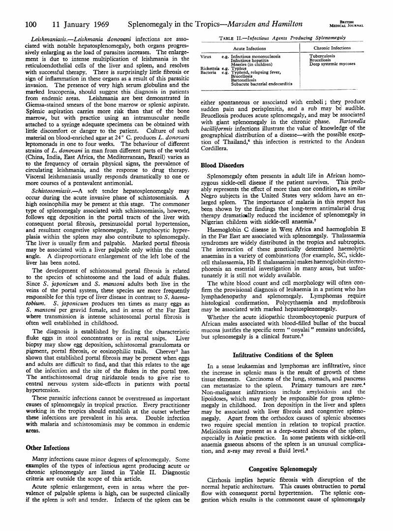
A palpable spleen, which may even be visible, is a common
physical finding in the tropics and sub-tropics (Fig. 1). Forexample, in a cross-sectional survey half the medical inpatientsat Mulago Hospital, Kampala, Uganda, had clinical spleno-megaly, and Pryor' has recorded 62% of a village population inNew Guinea with splenomegaly to the level of the umbilicus.Many specific causes of splenomegaly are now known, but there
FIG. 1.-Visible splenomegaly associated withscars of therapeutic lacerations by local practi-
tioners.
still remains a large group of patients in the tropics with grosssplenomegaly of unknown aetiology. This condition hasrecently" been termed " Tropical Splenomegaly Syndrome."In order to discuss this group in the true context of spleno-megaly it is important to consider first the known aetiologicalagents, and to remember that all causes of splenomegaly inEurope and North America can also occur in the tropics.The spleen is an important site for the production of lympho-
cytes and plasma cells. It is also rich in phagocytic cells. Itremoves defective red cells from the circulation, and the so-
called pitting function of the spleen relates to its capacity toremove intracellular granules, leaving the cells intact. In health
* Department of Clinical Tropical Medicine, London School of Hygieneand Tropical Medicine.
t Department of Medical Statistics and Epidemiology, London School ofHygiene and Tropical Medicine.
the bloodflow through the spleen is mainly rapid and intra-sinusoidal. In splenomegaly there is usually a second slowercirculation through the pulp cords.3 The spleen becomescongested following any rise in pressure within the splenicvein. Occasional intrasplenic erythropoiesis as in myelofibrosissuggests that the spleen retains its capacity to produce red cells.Hypersplenism is a term applied to a depression of one or more
blood elements (red cells, white cells, platelets) associated withsplenomegaly.
Detailed lists of the causes of splenomegaly can be found instandard texts.4 Only certain groups of conditions associatedwith splenomegaly which are important and prevalent in thetropics will be discussed here (see Table I).
TABLE I.-A Guide to Common Causes of Splenomegaly in the Tropics
(1) Parasitic infectionsMalariaLeishmaniasisSchistosomiasis
(2) Other infectionsViralRickettsialBacterialFungal
(3) Blood disorders(4) Infiltrative conditions of the spleen(5) Congestive splenomegaly(6) Tropical splenomegaly syndrome
(a) With hepatic sinusoidal lymphocytosis(b) Without hepatic sinusoidal lymphocytosis
Parasitic Infections
Malaria.-The incidence of splenomegaly in the populationwhere malaria is endemic is related to the intensity of malariatransmission in that area. In adults " spleen rates " tend tobe lower in areas of stable malaria than in areas where unstableseasonal malaria occurs. In a variety of experimental animalsthe remarkable splenic activation following plasmodial infec-tion can be easily demonstrated, although the degree of responsevaries among individuals within a population. In children inmalarious areas spleen rates are higher than in adults, and theparasites are easier to find in the peripheral blood. Asimmunity develops parasitaemia decreases, but a high level ofimmunity to malaria is associated with the persistence of smallnumbers of parasites in the circulation. Dramatic reactivationof such latent infection may be seen after splenectomy. Inacute malaria, however, a palpable spleen may not be present.The careful and repeated examinations of thick and thin filmsfor malaria parasites stained with Giemsa or Leishman stainremains one of the most important laboratory procedures In
tropical practice.The spleen enlarges in malaria as the result of several patho-
logical processes-namely, reticulo-endothelial and lymphoidhyperplasia probably reflecting the immune response, pigmentdeposition, and varying degrees of sinusoidal dilatation.
All degrees of splenomegaly, often associated with hepato-megaly, are encountered in malaria.
BRuisTIMDICAL JOURNAL 99
Splenomegaly in the Tropics-Marsden and Hamilton
Leishmaniasis.-Leishmania donovani infections are asso-
ciated with notable hepatosplenomegaly, both organs progres-
sively enlarging as the load of parasites increases. The enlarge-ment is due to intense multiplication of leishmania in thereticuloendothelial cells of the liver and spleen, and resolveswith successful therapy. There is surprisingly little fibrosis or
sign of inflammation in these organs as a result of this parasiticinvasion. The presence of very high serum globulins and themarked leucopenia, should suggest this diagnosis in patientsfrom endemic areas. Leishmania are best demonstrated in
Giemsa-stained smears of the bone marrow or splenic aspirate.Splenic aspiration carries more risk than that of the bonemarrow, but with practice using an intramuscular needleattached to a syringe adequate specimens can be obtained withlittle discomfort or danger to the patient. Culture of suchmaterial on blood-enriched agar at 24° C. produces L. donovanileptomonads in one to four weeks. The behaviour of differentstrains of L. donovani in man from different parts of the world(China, India, East Africa, the Mediterranean, Brazil) varies as
to the frequency of certain physical signs, the prevalence ofcirculating leishmania, and the response to drug therapy.Visceral leishmaniasis usually responds dramatically to one or
more courses of a pentavalent antimonial.Schistosomiasis.-A soft tender hepatosplenomegaly may
occur during the acute invasive phase of schistosomiasis. Ahigh eosinophilia may be present at this stage. The commoner
type of splenomegaly associated with schistosomiasis, however,follows egg deposition in the portal tracts of the liver withconsequent portal fibrosis, presinusoidal portal hypertension,and resultant congestive splenomegaly. Lymphocytic hyper-plasia within the spleen may also contribute to splenomegaly.The liver is usually firm and palpable. Marked portal fibrosismay be associated with a liver palpable only within the costalangle. A disproportionate enlargement of the left lobe of theliver has been noted.The development of schistosomal portal fibrosis is related
to the species of schistosome and the load of adult flukes.Since S. japonicum and S. mansoni adults both live in theveins of the portal system, these species are more frequentlyresponsible for this type of liver disease in contrast to S. haema-tobium. S. japonicum produces ten times as many eggs as
S. mansoni per gravid female, and in areas of the Far Eastwhere transmission is intense schistosomal portal fibrosis isoften well established in childhood.The diagnosis is established by finding the characteristic
fluke eggs in stool concentrates or in rectal snips. Liverbiopsy may show egg deposition, schistosomal granulomata or
pigment, portal fibrosis, or eosinophilic trails. Cheever5 hasshown that established portal fibrosis may be present when eggs
and adults are difficult to find, and that this relates to the age
of the infection and the site of the flukes in the portal tree.
The antischistosomal drug niridazole tends to give rise to
central nervous system side-effects in patients with portalhypertension.These parasitic infections cannot be overstressed as important
causes of splenomegaly in tropical practice. Every practitionerworking in the tropics should establish at the outset whetherthese infections are prevalent in his area. Double infectionwith malaria and schistosomiasis may be common in endemicareas.
Other Infections
Many infections cause minor degrees of 8plenomegaly. Someexamples of the types of infectious agent producing acute or
chronic splenomegaly are listed in Table II. Diagnosticcriteria are outside the scope of this article.
Acute splenic enlargement, even in areas where the pre-
valence of palpable spleens is high, can be suspected clinicallyif the spleen is soft and tender. Infarcts of the spleen can be
BRITISHMEDICAL JOURNAL
TABLE II.-Infectious Agents Producing Splenomegaly
Acute Infections Chronic Infections
Virus e.g. Infectious mononucleosis TuberculosisInfectious hepatitisMeasles (in children)
Rickettsia e.g. TyphusBacteria e.g. Typhoid, relapsing fever,
BrucellosisBartonellosisSubacute bacterial endocarditis
Brucellosis
Deep systemic mycoses
either spontaneous or associated with emboli; they producesudden pain and perisplenitis, and a rub may be audible.Brucellosis produces acute splenomegaly, and may be associatedwith giant splenomegaly in the chronic phase. Bartonellabacilliformis infections illustrate the value of knowledge of thegeographical distribution of a disease-with the possible excep-tion of Thailand,6 this infection is restricted to the AndeanCordillera.
Blood Disorders
Splenomegaly often presents in adult life in African homo-zygous sickle-cell disease if the patient survives. This prob-ably represents the effect of more than one condition, as similarNegro subjects in the United States very seldom have an en-
larged spleen. The importance of malaria in this respect hasbeen shown by the findings that long-term antimalarial drugtherapy dramatically reduced the incidence of splenomegaly inNigerian children with sickle-cell anaemia.7
Haemoglobin C disease in West Africa and haemoglobin Ein the Far East are associated with splenomegaly. Thalassaemiasyndromes are widely distributed in the tropics and subtropics.The interaction of these genetically determined haemolyticanaemias in a variety of combinations (for example, SC, sickle-cell thalassaemia, Hb E thalassaemia) makes haemoglobin electro-phoresis an essential investigation in many areas, but unfor-tunately it is still not widely available.The white blood count and cell morphology will often con-
firm the provisional diagnosis of leukaemia in a patient who haslymphadenopathy and splenomegaly. Lymphomas requirehistological confirmation. Polycythaemia and myelofibrosismay be associated with marked hepatosplenomegaly.Whether the acute idiopathic thrombocytopenic purpura of
African males associated with blood-filled bullae of the buccalmucosa justifies the specific term " onyalai " remains undecided,but splenomegaly is a clinical feature.8
Infiltrative Conditions of the Spleen
In a sense leukaemias and lymphomas are infiltrative, sincethe increase in splenic mass is the result of growth of thesetissue elements. Carcinoma of the lung, stomach, and pancreascan metastasize to the spleen. Primary tumours are rare.4
Non-malignant infiltrations include amyloidosis and thelipoidoses, which may rarely be responsible for gross spleno-megaly in childhood. Iron deposition in the liver and spleenmay be associated with liver fibrosis and congestive spleno-megaly. Apart from the orthodox causes of splenic abscessestwo require special mention in relation to tropical practice.Melioidosis may present as a deep-seated abscess of the spleen,especially in Asiatic practice. In some patients with sickle-cellanaemia gaseous abscess of the spleen is an unusual complica-tion, and x-ray may reveal a fluid level.9
Congestive SplenomegalyCirrhosis implies hepatic fibrosis with disruption of the
normal hepatic architecture. This causes obstruction to portalflow with consequent portal hypertension. The splenic con-
gestion which results is the commonest cause of splenomegaly
100 11 January 1969

In some parts of the tropics-for example, Algeria, Hong Kong,and Calcutta. In a book10 with the misleading title of"Tropical Splenomegaly," Basu and Aikat discuss this syn-
drome in Calcutta and resurrect the old concept of Banti thatsplenomegaly may precede liver fibrosis and influence thedevelopment of cirrhosis. The evidence brought forwardto support this hypothesis is weak, and we prefer the conceptof Primary liver disease.
Boyer et al.1" from Calcutta suggest that congestive spleno-megaly may arise in some patients as the result of portal hyper-tension due to non-cirrhotic portal fibrosis associated withintrahepatic portal vein sclerosis-a process which can bedemonstrated histologically in wedge liver biopsies. Sincesuch portal fibrosis is common in many parts of the tropicsthis mechanism may operate in many cases of the tropicalsplenomegaly syndrome. Sherlock'2 has recently reviewed theliterature relating to hepatolienal fibrosis without cirrhosis.
Extrahepatic portal obstruction due to portal vein throm-bosis and cavernous malformation of the portal vein is anotherimportant cause of portal hypertension and congestive spleno-megaly. Umbilical sepsis in childhood is common in thetropics, and may result in more frequent extrahepatic portalobstructions in later life.
Clinical stigmata of hepatic insufficiency or hepatic failuresuggest cirrhosis. Liver biopsy may settle the question ofwhether or not cirrhotic or non-cirrhotic portal fibrosis ispresent. A bruit may be present in the splenic and hepaticareas in patients with portal hypertension. A barium swallowmay show oesophageal varices. Direct measurement of portalpressure and splenovenography are required to establish thedegree of portal hypertension and whether an extrahepatic blockis present. Techniques for measuring liver and splenic floware seldom available in tropical practice.
Tropical Splenomegaly Syndrome
This is really a diagnosis by exclusion, and has been variouslytermed idiopathic splenomegaly, tropical splenomegaly, "BigSpleen Disease," cryptogenetic splenomegaly, Bengal spleno-megaly, splenomegalie algerienne, and African macroglobulin-aemia. Clinically patients complain of a dragging pain in theabdomen, intermittent fevers, and debility with marked spleno-megaly present for many years, and hepatomegaly particularlyof the left lobe. There is seldom any peripheral lymphadeno-pathy.
Present evidence suggests it is helpful to classify these cases
as follows:
(a) Association with Hepatic Sinusoidal Lymphocytosis
A curious liver biopsy change, hepatic sinusoidal lympho-cytosis (H.S.L.) (see Fig. 2) has been observed in many suchpatients in Algeria, India, Aden, New Guinea, and many partsof equatorial Africa. The hepatic sinusoidal infiltrate is mainlylymphocytic, though other chronic inflammatory cells are seen.In both liver and spleen there are varying degrees of sinusoidaland reticuloendothelial hyperplasia, but no malaria pigment.Marked erythrophagocytosis has been described in the spleen."3The association of marked splenomegaly with this liver changehas been called " Big Spleen Disease,"'4 although lesser degreesof H.S.L. do occur without clinical splenomegaly."1 The causeof this cellular infiltration of the liver sinusoids is unknown.In Algeria and Calcutta, where satisfactory malaria control hasbeen established, this syndrome has almost disappeared. Long-term antimalarial therapy results in a reduction of splenicsize'6 and disappearances of hepatic sinusoidal lymphocytosis.'7An increase of serum globulins in the gammaglobulin frac-
tion is a feature of this syndrome. This is due to an increasein the IgG, and especially the IgM (macroglobulins)
BRMSHMEDICAL JOURNAL 101
fractions.'8 In Kampala and New Guinea patients withsplenomegaly and hepatic sinusoidal lymphocytosis have a highmalarial fluorescent antibody titre.'5 19 It has been suggestedthat this condition represents an abnormal immune responseto malaria.
FIG. 2.-Hepatic sinusoidal lymphocytosis. There is alsosinusoidal dilatation but no malaria pigment. The basic archi-tecture is preserved and and parenchymal cells are normaL
(X 175 approx.)
In adult patients with this condition in Kampala persistentblood film examinations showed a higher incidence ofP. malariae parasitaemia.20 This finding was not confirmed inNew Guinea.'5The absence of malaria pigment in the liver and spleen of
these patients has been cited as evidence against the conceptthat malaria might be playing a part. The length of timemalaria pigment persists in the tissues of man is not known,but it may be of doubtful value in longstanding infections as ahistological marker in indicating malaria infection.21 Not allpatients with clinical malaria have pigment demonstrable onliver biopsy.22As in other patients with marked splenomegaly certain com-
plications are common. A feeling of weight in the abdomenassociated with the enlarged organ, which has been presentfor many years, is a frequent symptom. The co-existentanaemia is the compound result of an increased plasma volume,a reduction of red cell survival time, and trapping of largevolumes of red cells in the splenic pool.23 24
Portal hypertension is commonly associated with this condi-tion, and occurs in the absence of obvious intra- or extra-hepatic obstruction. It may be related to increased splenicflow.23
(b) Without Hepatic Sinusoidal Lymphocytosis
Even in malarious areas tropical splenomegaly syndromeoccurs without hepatic sinusoidal lymphocytosis, and thp aetio-
logy is quite unknown. Whether non-cirrhotic portal fibrosisis responsible as suggested by Boyer and his colleagues'1requires investigation. It is likely that the mechanism of theanaemia found in these cases is similar to that described above.
Treatment
The therapeutic approach to the tropical splenomegaly syn-drome associated with hepatic sinusoidal lymphocytosis hasvaried from centre to centre. Antimalarial chemotherapy has beenused.'6 26 At Ibadan with a daily dose of 100 mg. proguanila slow but progressive diminution in the size of the spleen overa matter of months was noted associated with an improvementin anaemia and general health. In such patients antimalarialchemoprophylaxis under medical surveillance should probablybe continued for as long as they reside in a malarious area.
11 January 1969 Splenomegaly in the Tropics-Marsden and Hamilton

102 11 January 1969 Splenomegalyin the Tropics-Marsden and Hamilton MEDICAL JOURNAL
More information on the success of this therapeutic approachis needed.There is usually an element of haemolysis in the anaemia in
these cases, and folic-acid deficiency is a possibility whichshould be corrected. Exchange transfusion may be requiredin cases of acute haemolysis such as may occur in pregnancy.26
In tropical splenomegaly syndrome simple splenectomy hasbeen attended by a low initial mortality,27 and markedlyImproves the co-existent anaemia.26 28 The susceptibility ofsplenectomized individuals to malaria and other infectionsmakes this a risky procedure of doubtful long-term value. Itis worth noting that the only three Babesia infections of man(a protozoon ill adapted to man) have all occurred inindividuals who previously had had a splenectomy.
ConclusionThe classification used is an oversimplification. For
Instance, among the conditions that have not been consideredhydatid disease or veno-occlusive disease of the liver may causesplenomegaly in a tropical area. In contrast to London teach-ing hospital practice collagen diseases and rheumatoid arthritisdo not deserve consideration as important causes of spleno-megaly in the tropics, although they are seen. Such conditionsare swamped by the welter of other infections. Several pro-cesses may be going on in the spleen at the same time andcontributing to its increase in size.Even in centres where every diagnostic facility is available
the aetiology of marked splenomegaly may remain unknown.In the tropics, where such a situation is encountered far morefrequently, further research is needed.
REFERENCES
' Pryor, D. S., Quart. 7. Med., 1967, 36, 321.' Brit. med. 7., 1967, 4, 614.' Richards, J. D. M., and Toghill, P. J., 7. Path. Bact., 1967, 93, 653.' Blaustein, A. U., The Spleen, 1963. New York.I Cheever, A. W., Amer. 7. trop. Med. Hyg., 1968, 17, 38.' Whitaker, J. A., Fort, E., Weinman, D., Tamasatit, P., and Panas-
Ampol, K., Nature (Lond.), 1966, 212, 855.
'Hendrickse, R. G., In Symposium on Abnormal Haemoglobins inAfrica, ed. J. H. P. Jonxis, 1965, p. 445. Oxford.
"Wilkinson, J., E. Afr. Med. 7., 1953, 30, 243.Pieters, G., and Lambotte, C., in Symposium on Abnormal Haemo-globins in Africa, ed. J. H. P. Jonxis, 1965, p. 285. Oxford.
Basu, A. K., and Aikat, B. K., Tropical Splenomegaly, 1963. London.Boyer, J. L., et al., Ann. intern. Med., 1967, 66, 41.
12 Sherlock, S., Postgrad. med. 7., 1968, 44, 109.Pitney, W. R., Pryor, D. S., and Tait Smith, A., 7. Path. Bact., 1968,95, 417.
Hamilton, P. J. S., Gebbie, D. A. M., Hutt, M. S. R., Lothe, F., andWilks, N. E., Brit. med. 7., 1966, 2, 548.
15Marsden, P. D., Connor, D. H., Voller, A., Kelly, A., Schofield,F. D., and Hutt, M. S. R., Bull. Wld Hith Org., 1967, 36, 901.
16 Watson-Williams, E. J., Allan, N. C., and Fleming, A. F., Brit. med.7., 1967, 4, 416.
1 Lowenthal, M. N., and Hutt, M. S. R., E. Afr. med. 7., 1968, 43,100.
Charmot, C., and Vargues, R., Sem. H6p. Paris, 1963, 39, 1421.9 Gebbie, D. A. M., Hamilton, P. J. S., Hutt, M. S. R., Marsden, P. D.,
Voller, A., and Wilks, N. E., Lancet, 1964, 2, 392.Marsden, P. D., Hutt, M. S. R., Wilks, N. E., Voller, A., Blackman,
U., Shah, K. K., Connor, D. H., Hamilton, P. J. S., Banwell, J. G.,and Lunn, H. F., Brit. med. 7., 1965, 1, 89.
Hutt, M. S. R., personal communication, 1968.22 Srichaikul, T., Amer. 7. trop. Med. Hyg., 1959, 8, 110.29 Richmond, J., Donaldson, G. W. K., Williams, R., Hamilton, P. J. S.,
and Hutt, M. S. R., Brit. 7. Haemat., 1967, 13, 348.24 Pryor, D. S., Quart. 7. Med., 1967, 36, 337.25 Williams, R., Parsonson, A., Somers, K., and Hamilton, P. J. S.,
Lancet, 1966, 1, 329.26 Hamilton, P. J. S., Richmond, J., Donaldson, G. W. K., Williams, R.,
Hutt, M. S. R., and Lugumba, V., Brit. med. 7., 1967, 3, 823.27 Paliwoda, T., and Hut, M. S. R., E. Afr. med. 7., 1968, 45, 1.28 Pryor, D. S., Brit. med. 7., 1967, 3, 825.
B.M.J. PublicationsThe following are available from the Publishing Manager, B.M.A.House, Tavistock Square, London W.C.1. The prices includepostage.
The New General Practice ... Price 16s.B.M.7. Cumulative Index, 1967 Price 30s.
(15s. to B.M.A. members)Porphyria-a Royal Malady ... ... Price 13s. 6d.Is There an Alternative? ... ... Price 7s. 6d.Treatment of Common Skin Diseases Price lOs.Charles Hastings and Worcester ... Price 3s. 6d.
ANY QUESTIONS ?We publish below a selection of questions and answers of general interest.
New Influenza VaccineQ.-Soine of the partners in this practice,
and their staff, have had the usual injectionof influenza vaccine this year. Should wealso have an injection of the new vaccineagainst Asiatic virus?
A.-Though they were vaccinated earlier thisyear, the doctors and their staff should receivea dose of the new vaccine prepared from anA2 strain antigenically similar to that isolatedin Hong Kong at the onset of the epidemic.The vaccine used in the early autumz hadbeen prepared from influenza virus strainsthen currently endemic in this country andagainst which most adults already haddemonstrable antibodies. The vaccineboosted antibody titres against these strains,but was less effective in stimulating antibodyproduction against the Hong Kong strain.Only a few individuals have demonstrableantibodies against the Hong Kong strain, and,though the vaccine given in early autumnwould have produced a weak antibody re-sponse in some of the recipients, a dose ofthe new Hong Kong strain vaccine wouldgreatly increase the titres of these antibodies.
Risk of Spina Bifida
Q.-A girl has a half-brother (theirmother is the same) with spina bifida. Whatis the risk of the girl having children withspina bifida ?
A.-No empirical risk figures are availablefor this situation, but any children of thepatient will be third-degree relatives of theboy with spina bifida. The risk here is prob-ably less than 1%.1
REFERENCE
l Carter, C. O., David, P. A., and Laurence,K. M., 7. med. Genet., 1968, 5, 81.
Lithium and Foetal Abnormalities
Q.-Is there any risk that lithium car-bonate may cause foetal abnormalities whentaken in pregnancy ?
A.-A systematic study, with present-dayanimal breeding techniques, to answer thisquestion, has not yet been carried out. Never-
theless, it is known that lithium ions whenadded in sufficient concentration to thenutrient media of developing sea urchins andchick embryos may produce developmentalabnormalities.1 2 In mammals, such as miceand rats, experiments neither implicate norclear lithium definitely.3 4 For this reasonvery careful consideration should be givenbefore lithium carbonate is administered topregnant women, particularly in the firsttrimester. Obviously, if a patient is knowno become severely psychotic in the absenceof lithium, the possible harmful effects onthe foetus resulting from this disturbedmental state and the possible physical con-comitants must be balanced against theunknown risks of the drug. In fact, thewriter has had one such case, and Schou'has reported six others when lithium wasgiven throughout pregnancy without anycomplications and the delivery of normalbabies.
REFERENCES
Schou, M., Pharmacol. Rev., 1957, 9, 17.2 Schou, M., 7. psychiat. Res., 1968, 6, 67.3 Bass, A. D., Yntema, C. L., Hammond, W. S.,
and Frazer, M. L., 7. Pharmacol. exp. Ther.,1951, 101, 362.
Trautner, E. M., Penncuik, P. R., Morris,R. J. H., Gershon, S., and Shankly, K. H.,Aust. 7. exp. Biol. med. Sci., 1958, 36, 305.