Embed Size (px)
Citation preview

IRISA ZĪLE
EPIDEMIOLOGICAL ASPECTS
OF CONGENITAL ANOMALIES AND
ASSOCIATED RISK FACTORS
IN LATVIA
Summary of Doctoral Thesis
Speciality – Public Health and Epidemiology
Riga, 2013

2
Doctoral thesis were elaborated in Rīga Stradiņš University (RSU),
Public Health and Epidemiology Department
Scientific supervisors of the thesis:
Dr. med., Associate Professor Anita Villeruša,
Rīga Stradiņš University
Official reviewers:
Dr. med., Professor Ģirts Briģis (Rīga Stradiņš University)
Professor Juris Krūmiņš (University of Latvia)
Professor Apolinaras Zaborskis (Lithuanian University
of Health Sciences)
Presentation of the doctoral thesis will take place on April 8, 2013 at
17.00 on the open meeting of the Promotion Council of Theoretical
Medicine disciplines of Rīga Stradiņš University (RSU) in Riga, at
Dzirciema Street 16, in the Hippocrate auditorium.
The doctoral thesis is available at the library and home page
(www.rsu.lv) of Rīga Stradiņš University.
Elaboration of the thesis was supported by ESF program “Support for Doctoral
Students in Obtaining the Scientific Degree in Rīga Stradiņš University”.
The head of the Promotion Council of Medicine disciplines
Dr. habil. med., Professor Jānis Vētra
Secretary of Promotional Council:
Dr. habil. med., Professor Līga Aberberga-Augškalne

3
CONTENT
1. Introduction ................................................................................................ 5
Aim of the doctoral thesis .............................................................................. 8
Tasks of the doctoral thesis, hypothesis .......................................................... 8
Scientific novelty of the thesis ........................................................................ 9
Practical significance of this doctoral thesis ................................................... 10
Structure of doctoral thesis ............................................................................. 11
2. Material and methods ................................................................................ 12
2.1. Data selection .................................................................................. 12
2.2. Definitions and calculations of prevalence and mortality ................. 15
2.3. Statistical analysis............................................................................. 16
3. Results ....................................................................................................... 18
3.1. Live births prevalence of congenital anomalies and changes
from year 2000 to 2010 ................................................................................... 18
3.2. Maternal age and congenital anomalies of newborns ....................... 19
3.3. Mortality from congenital anomalies and changes
from year 2000 to 2010 ................................................................................... 20
3.4. Associations between congenital anomalies and factors characterising
mother and newborn and antenatal care ......................................................... 22
3.5. Results of multivariate analysis in relation to odds for newborn
congenital anomalies ...................................................................................... 26
3.6. Associations between total congenital anomalies and different
maternal diseases ............................................................................................ 28
3.7. Associations between different maternal diseases and particular
diagnosis groups of congenital anomalies ...................................................... 29
3.8. Evaluation of congenital anomaly prenatal diagnostics basing on data
of the CCUH patient medical histories ........................................................... 31
3.8.1. Coverage of live-born congenital anomalies in relation to data on
patients treated in hospital ............................................................................. 32
3.9. Coverage of live-born congenital anomaly registration at birth
institutions in relation to congenital anomaly register data ............................ 34

4
4. Discussion .................................................................................................. 35
5. Conclusions ................................................................................................ 40
Confirmation of hypotheses of the doctoral thesis ......................................... 42
6. Practical recommendations ........................................................................ 43
References ..................................................................................................... 45
List of publications and abstracts .................................................................. 48

5
INTRODUCTION
Congenital malformations in newborns are one of the major public
health problems in Latvia, as well as around the world. They have a leading
role in perinatal and infant morbidity and mortality (1-5). The global
prevalence is around 2 - 3% of all live births (5-9), nevertheless some studies
suggest a prevalence of 3 - 5% (4, 10-12). Congenital anomalies, malformation
and pathologies are defined as physical (functional and/or structural) defects at
birth may cause by genetic issues, environmental factors, the intrauterine
(uterus) environment, chromosomal abnormality and/or factors of other origin
that may be diagnosed during prenatal period, childbirth or postnatal period
(2-4, 8). Congenital anomalies have been a cause for 24.2% of infant deaths in
Latvia during 2011 (13). According to World Health Organization Health for
All mortality database (HFA-MDB), the mortality rate for infants under 1 year
has increased by 20 % from year 2008 to 2009, 187/100 000 to 221/100 000
respectively. Moreover, infant mortality data of Latvia exceeds the European
average indicators for 20 - 40% (152.15/100 000 to 149.25/100 000,
respectively). Nevertheless in 2010, the number of infant deaths caused by
congenital anomalies has diminished – 121.08/100 000 (14).
Child health largely depends on availability of mother and child health
care system, which also includes reproductive and perinatal health care (1, 3).
In 2008 Latvia government established improvement in mother and child health
care system as top priority for the Ministry of Health, and endorsed this policy
by the Medical Treatment Law. New and improved procedures and
arrangements to visit directly specialists to undergo preventive and routine
medical examination for pregnant women. This amendment help in diagnosing
different maternal disease in time, thus directly improving maternal health,
reducing birth complications, positively effecting new-born’s health and the
health of future babies in Latvia (15).

6
In Latvia there has been a slight decrease in percentage of women
receiving timely antenatal care (until 12th week of the pregnancy). In 2005
around 90% of pregnant women received care but in 2010 it was around 87%
(16). Smoking, use of alcohol, narcotics or other addictive substances during
pregnancy has a negative effect on the health of the women and on foetal
development. Public Health statistics indicates that approximately 10% of
labouring women in Latvia have smoked during pregnancy, 0.5% have
consumed alcohol, whereas 0.1% have used narcotics (16, 17).
Improvement of mother and child health as well as reduction of infant
mortality is one of many goals of the new public health policy “Public Health
Guidelines for 2011 – 2017” in 2011 (18), the action plan “Plan for Improving
Mother and Child Heath for 2012 – 2014” developed by the Ministry of Health
in Latvia (19) to achieve these goals. The Action Plan provides changes related
to the prenatal diagnostics and requires additional medical examinations for
pregnant women, including ultrasound screening and necessary tests to
facilitate timely diagnosing of congenital anomalies (19, 20).
World Health Organisation database “Health for All” (HFA - DB) shows
that from 1990 until the beginning of 2000 there has been a rise in the
prevalence of congenital anomalies among live both in European region as a
whole and in European Union. While from 2000 till 2010, the prevalence for
congenital anomalies have dropped both in European region and in European
union (21). Indicators characterising the prevalence of congenital anomalies are
influenced by many factors, for example the national registration system,
termination of pregnancy due to congenital anomalies, prenatal diagnostics,
occurrence of risk factors, as well as availability and the quality of health care
services (3, 8, 22-24).
To decrease the infant mortality rate, disabilities and complications in
birth due to congenital anomalies, primary prevention and improving mother
and child health more knowledge is needed. As congenital anomalies are one of

7
the main causes of infant death and poor health for the children with congenital
anomalies, epidemiological research of congenital anomalies is significant for
the field of reproductive and perinatal health (4, 30). The field of congenital
anomalies among newborn has not been sufficiently studied in Latvia, thus the
need for population-based studies of congenital anomalies has been stressed. A
literature survey shows earlier clinical research on particular diagnosis of
congenital anomalies in Latvia (25-29) but no epidemiological population
based studies.
Health Surveys conducted in Latvia are detailed research of more
clinical character on sub-groups of specific congenital anomalies or diagnosis
in paediatric surgery, dentistry and/or genetics and they do not fully
characterise the prevalence of total major congenital anomalies. In Latvia,
within the framework of the state research programme “Scientific Research of
Main Malformations Imperilling Latvia’s Population Survival and Life Quality
by Multidisciplinary Research Consortium” Project No 6 “Development of
Algorithms Based on Modern Technologies for Diagnostics and Treatment of
Congenital Malformations of Children for Reduction of Patient Mortality,
Survival Prolongation and Improvement of Life Quality”, the mortality due to
congenital anomalies and survival were analysed (31).
Results of these studies indicate both the problems and to-do antenatal
foetuses as to minimize births with congenital abnormalities.
This Doctoral thesis aims at finding out latest trends in the live birth
prevalence of congenital anomalies and mortality due to congenital anomalies
in Latvia, reflecting comparisons between cases and control correlations
between frequency indicators and various factors influencing perinatal period,
evaluation of congenital anomaly registration in the country, as well as identify
problems in existing registration systems. Results of this all study will indicate
the problems and the measure to be taken to reduce the risk to be born with
congenital anomalies.

8
Aim of the doctoral thesis: The overall aim of this doctoral thesis work
is to gain epidemiological knowledge about the congenital anomalies in new
born. Further to investigate the prevalence of the congenital anomalies of live
births and mortality due to congenital anomalies among newborn and infants in
Latvia, associated antenatal care, mother and newborn health risk factors, as
well as assessment of coverage of birth defects registration in the Latvia.
Tasks of the doctoral thesis:
1. To calculate the prevalence of congenital anomalies of live birth, from
2000 till 2010, including break down by diagnose group.
2. To study the association between prevalence of congenital anomalies
of live birth and maternal age.
3. To study the influence of newborn congenital anomalies on perinatal
and infant mortality from 2000 till 2010.
4. To investigate the effect of health status of a mother, complications
during pregnancy and childbearing and other perinatal factors on congenital
anomalies of live births.
5. To investigate coverage of prenatal diagnostics and conformity of
postnatal diagnosis with the patient medical history data of the Children's
Clinical University Hospital, Riga (CCUH).
6. To assess problems and completeness of congenital anomaly
registration in Latvia and to develop suggestions for the improvement of
congenital anomaly information registration system.
7. To develop suggestions for early identification of pregnant women in
risk zone for giving birth to a child with congenital anomalies and identification
of newborn in risk zone for congenital anomalies.
Hypotheses of doctoral thesis:
• Changes in prevalence of congenital anomalies are influenced by rise
of the mean age of a mother.

9
• Changes in mortality due to congenital anomalies are affected by
general perinatal and infant mortality trends.
• Mothers giving birth to child with congenital anomalies behave more
risky towards antenatal care and their own health.
• Congenital anomaly prevalence is influenced by prenatal diagnostics
and completeness of registration system in the country.
Scientific novelty of the thesis
To our knowledge this doctoral thesis is the first kind including
nationwide cohort study performed to estimate the prevalence of congenital
anomaly among all live birth on such a long time period (2000 – 2010),
covering all newborn in Latvia including a comparison group without
congenital anomalies. Estimating the prevalence and mortality trends and risk
factors for congenital anomalies.
Retrospective and cross-sectional cohort analyses were performed by
obtaining data from nationwide, population based registers that were inter-
linked as follows: Medical Birth Register (MBR) that include basic
information on prevalence of congenital anomalies at birth, the National
Causes of Death Register (NCDR) that provides information on all deaths and
main cause of death. With an aim to give insight into the congenital
surveillance system in country and problems related with the registration of
congenital anomalies, new born and infants up till 1 year old treated at
Children's Clinical University Hospital, Riga (CCUH) during 2003-2008 for
congenital anomalies and data of the Congenital Anomaly Register (CAR)
were linked with MBR.
Live birth prevalence of congenital anomalies is analysed by major
congenital anomaly diagnosis groups, it is very important to diagnose these
congenital pathologies timely. The timely (prenatal) diagnose of a life threating
congenital anomaly is also very helpful in deciding termination of pregnancy

10
by a pregnant women. As well as to ensure obstetrical and neonatal care
appropriate to the condition of a foetus and, if necessary, emergency surgical
treatment is also available. In MBR the diagnoses are classified according to
The International Statistical Classification of Diseases and Related Health
Problems, Tenth Revision (ICD-10). These diagnosis codes were
correspondingly re-grouped, adapting the methodology of the European
network of population-based registries for the epidemiologic surveillance of
congenital anomalies (European Surveillance of Congenital Anomalies -
EUROCAT). That allowed a wider comparison of Latvian national average
prevalence of congenital anomalies with EUROCAT average.
Prevalence of congenital anomalies while controlling for risk factors by
using multiple logistic regression. This method allowed assessing associations
among different risk factors characterising health of a newborn and mother and
health-related behaviour with occurrence of congenital anomalies regardless of
other risk factors. Differences in the occurrence of risk factors were also
studied by making comparisons with the control group. This control group was
selected from all healthy newborn not diagnosed with any congenital anomaly
or any other health disorder at birth and who were born during the same
reference period.
Practical significance of the doctoral thesis
In this doctoral thesis we identified the associations between antenatal
factors, maternal and newborn health and prevalence of congenital anomalies at
birth. Characterisation of epidemiological situation in Latvia related to
congenital anomalies gaves insight into the main problems in the field of
diagnostics of congenital anomalies, their registration system and identification
and evaluation of potential risk factors. The identification and strength of such
risk factors can in turn be used for primary prevention plan.

11
The information gained from this thesis work can be used in clinical
applications by primary health care specialists, gynaecologists, obstetricians
and neonatologists. For example, the recommendations on risk factors can be
used in a health literacy program for pregnant women in the beginning of their
pregnancies. The Epidemiological analyses provides a complete description of
the prevalence of congenital anomalies, as well as shows that there are
significant differences in the prevalence of various preventable risk factors in
cohort of newborn with congenital anomalies, in comparison to healthy
children.
The gained knowledge is useful and important for a variety of primary
prevention programmes for improvement of perinatal, sexual and reproductive
health of Latvian population.
The findings from this doctoral thesis shows presents that the prevalence
of congenital anomalies are significantly affected by weaknesses in the existing
anomalies in the country accounting systems. In order to provide a complete
and high-quality registration system, it is necessary to ensure the inter linking
medical birth register with hospital records and Congenital Anomaly Register
(CAR). Quality and coverage of the registration can also be improved by
training midwifes, gynaecologists and other medical professionals responsible
for registering births.
Structure of doctoral thesis
The doctoral thesis is written in Latvian. It consists of 8 sections:
introduction, literature review, materials and methods, results, discussion,
conclusions, practical recommendations and references. The work consists of
118 pages, including 12 tables and 22 figures. Bibliography consists of 172
references. Doctoral thesis has 7 annexes. There are 6 publications included in
this doctoral thesis.

12
2. MATERIALS AND METHODS
2.1. Data selection
In order to achieve the main aim for this doctoral thesis and to test the
hypotheses data from nationwide population-based registers were used. We
obtained data from the Medical Birth Register (MBR), National Causes of
Death Register (NCDR), Congenital Anomaly Register (CAR), as well as
information from the patient medical histories of the Children's Clinical
University Hospital (CCUH) on infants hospitalization due to congenital
anomalies. Cross-sectional and retrospective case-control study design was
used. This research has been approved by the Riga Stradiņ’š University Ethics
Committee.
Main data source MBR was used to estimate the prevalence of
congenital anomaly and it was also used to estimate of the association and
strength of potential risk factors and congenital anomalies, from 2000 - 2010.
The MBR is based on information provided on cards issued to newborns by
maternity units across the country. Congenital anomalies included in the
analyses were diagnosed by a neonatal doctor using ultrasound examinations
and genetic investigation among other methods. These investigations were
performed during the time spent in maternity unit after delivery.
According to World Health Organisation’s (WHO) definition congenital
anomalies, malformation and pathologies are defined as physical (functional
and/or structural) defects at birth (2, 3). The diagnosis of congenital anomalies
was analysed according to The International Statistical Classification of
Diseases and Related Health Problems, Tenth Revision (ICD-10): Q00-Q99 -
congenital malformations, deformations and chromosomal abnormalities (32).
The data analysis included all the live births with congenital anomalies
during the time period (2000 – 2010). The total number of such live births was
7451, of which 4927 were having major congenital anomalies, see table 2.1.

13
2.1. Table
Number of live births with congenital anomalies and the proportion of all
live births in Latvian (2000-2010)
Year Live births
with CA (n)
%, of live
births
Of them with
major CA (n)
%, of live
births
Control
group – live
births without
CA (n)
2000 548 2.7 414 2.0 13773
2001 707 3.6 493 2.5 12951
2002 723 3.6 464 2.3 13428
2003 797 3.8 516 2.5 14250
2004 767 3.8 414 2.0 13780
2005 761 3.5 457 2.1 15072
2006 718 3.2 492 2.2 16074
2007 747 3.2 481 2.1 16458
2008 633 2.6 436 1.8 17516
2009 584 2.7 413 1.9 13941
2010 466 2.4 347 1.8 11765
kopā 7451 3.5 4927 2.1 159008
Notes: CA – congenital anomalies; n – number of newborn
Congenital anomalies were defined as lethal if the defects cause
stillbirth or infant death or pregnancies are terminated after the prenatal
diagnosis. They were defined as severe if the defects without medical
intervention caused disability or death defects together (8, 33, 34).
Prevalence for the whole time period was calculated for whole all Q
diagnosis group (ICD-10; Q00–Q99), as well as prevalence for all major
congenital anomalies and prevalence of live births by maternal age group.
These results were compared with the average prevalence of congenital
anomalies in Europe regarding to EUROCAT data. Pathology diagnose codes

14
for the analysis of the results were re-grouped, adapting the methodology of the
EUROCAT (European surveillance of congenital anomalies) (35): nervous
system (Q00, Q01, Q02, Q03, Q04, Q05, Q06, Q07); eye (Q10.0, Q10.4,
Q10.6-Q10.7, Q11-Q15); ear, face and neck (Q16, Q17.8, Q18.3, Q18.8);
congenital heart defects (Q20-Q26); respiratory system (Q30-Q34); oro-facial
clefts (Q35-Q37); digestive system (Q38–Q39, Q40.2-Q40.9, Q41-Q45,
Q79.0); abdominal wall defects (Q79.2, Q79.3, Q79.5); urinary system (Q60-
Q64, Q79.4); genital (Q50-Q52, Q54-56); limb (Q65.0-Q65.2, Q65.8-Q65.9,
Q66.0, Q68.1-Q68.2, Q68.8, Q69-Q74); musculo-skeletal system (Q75.0-75.1,
Q75.4-Q75.9, Q76.1-Q76.4, Q76.6-Q76.9, Q77, Q78, Q79.6-Q79.9);
chromosomal (Q90-Q92, Q93, Q96-99); other congenital anomalies/syndromes
(Q27, Q28, Q80-Q85, Q89).
Our Studies cover calculation of prevalence of congenital anomalies,
mortality indicators, investigation of associations between odds for congenital
anomalies and various factors: maternal age and factors characterising health
status of a mother (clinical history, complications during pregnancy and
childbirth, abortion history), factors characterising antenatal care (timeliness
and having antenatal care); harmful lifestyle of parents (smoking, use of
alcohol and psychoactive substances); characteristics of a newborn (gender,
birth weight and gestational week).
Number of deaths caused by congenital anomalies was calculated by
using the NCDR register. With the aim to find more about prenatal diagnostics
of congenital anomalies and registration of diagnoses for live births, the data
for a group of hospitalized infants was also analysed. The Medical record of
patients with congenital anomalies from CCUH the leading inpatient medical
treatment institution in Latvia, providing treatment to newborn and infants with
congenital anomalies, was used to achieve this aim.
The first-time hospitalization medical history of 1788 (1605 of them
with major congenital anomalies) infant patients with congenital anomalies
15
(2003-2008) were selected, these infants were one year old at time of
hospitalization. The diagnosis at discharge time of this first-time hospitalization
was used as the base of analyses.
There were 212 medical histories having records on infant congenital
anomaly diagnosed prenatally (mentioned cases were major congenital
anomalies), and the data were used for the assessment of diagnostics. Out of the
selected 212 histories only 86 had reference to the pregnancy week during
which pathology was diagnosed in the ultrasound examination.
In order to assess the completeness of the registration of congenital
anomalies at birth (MBR), useful data were only on 629 patients treated by the
CCUH, in these cases it was possible data linkage with MBR.
To characterise situation regarding registration of congenital anomalies
more in details and conformity of diagnosis in the MBR, additionally also data
of the CAR were used (2000 – 2010). Inter-comparison of diagnoses was based
on 587 records from the CAR, which were available linkage with MBR. Also
within the framework of this analysis, diagnoses of the congenital anomalies
were grouped in compliance with the EUROCAT major congenital anomaly
sub-groups.
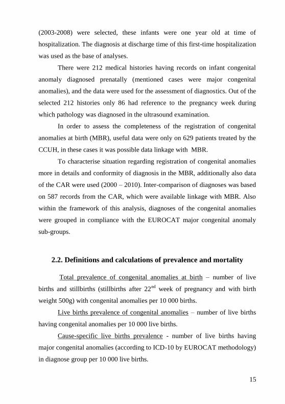
2.2. Definitions and calculations of prevalence and mortality
Total prevalence of congenital anomalies at birth – number of live
births and stillbirths (stillbirths after 22nd
week of pregnancy and with birth
weight 500g) with congenital anomalies per 10 000 births.
Live births prevalence of congenital anomalies – number of live births
having congenital anomalies per 10 000 live births.
Cause-specific live births prevalence - number of live births having
major congenital anomalies (according to ICD-10 by EUROCAT methodology)
in diagnose group per 10 000 live births.

16
Stillbirths with congenital anomalies – number of stillbirths (stillbirths
after 22nd
week of pregnancy and with birth weight 500g) having congenital
anomalies per 10 000 live births and stillbirths.
Neonatal mortality due to congenital anomalies – number of deaths
during neonatal period (0-27 days) due to congenital anomalies per 1000 live
births.
Postneonatal mortality due to congenital anomalies – number of deaths
during postneonatal period (28 days after births up to 1 year) due to congenital
anomalies per 1000 live births.
Perinatal mortality due to congenital anomalies – number of deaths
during perinatal period (stillbirths + deaths during first 6 days after birth) due to
congenital anomalies per 1000 live births and stillbirths.
Infant mortality due to congenital anomalies – number of live-born
deaths during the first year of life due to congenital anomalies per 1000 live
births.
Infant death proportion – number of infant deaths caused by congenital
anomalies during a certain period of time/total number of infant deaths during
this time period *100.
2.3. Statistical analysis
Data necessary for the study were entered and coded in SPSS for
Windows 19.0 statistical data processing programme. According to the study
objectives, new variables of the categories were developed basing on factors
included in the analysis. Total prevalence (incl. live and stillbirths) and live
birth prevalence of congenital anomalies and trends in perinatal and infant
mortality during the time period from 2000 till 2010 were assessed using linear
regression. The χ² test was used to compare the subgroups for data analysis in

17
2x2 tables. The higher χ2, the higher difference between observed
characteristics within groups.
The association between specific risk factors and congenital anomalies
among newborn were expressed with odds ratio (OR) (36, 37). OR measure the
odds of congenital anomalies of newborns on comparison of factors with
newborns in the reference group (healthy newborns). If the OR is greater than
1, the odds for congenital anomaly related to specific factor is greater than in
reference group, but if the OR is less than 1, the odds of newborn congenital
anomalies in case of this factor are lower, as compared to the reference group.
Nonadjusted odds ratio was used to calculate probability of congenital
anomalies for newborn in relation to various determining factors, e.g., mother
diseases in history, harmful lifestyle factors of parents etc. Moreover, OR
calculations were made to analyse association of the maternal age on diagnoses
of particular congenital anomalies. Multiple logistic regression was used in
cases, when there are several independent variables that can affect the
dependant (e.g., maternal age which is related with higher risk of certain
congenital anomalies) and it is necessary to match these variables with each
other. Odds ratio (OR) was obtained using multiple logistic regression which
shows how the increases or decreases the occurrence. The reference value for
regression was used healthy newborns (without congenital diagnoses at birth or
other diseases, as perinatal period conditions).
Mutual comparison of congenital anomaly coverage and correspondence
of diagnoses within the data analysis (MBR with CCUH data, as well as MBR
with CAR) was carried out with the help of calculations made basing on similar
methodology in another research (38). Coincidence of diagnoses or positive
registered cases is equal with number of newborn correctly diagnosed with
congenital anomaly in MBR divided by the total number of newborn having
congenital anomalies that might have been inter-identified in compared
databases (MBR with CCUH; MBR with CAR). Coverage of congenital
18
anomaly cases or completeness is equal with number of newborn with correctly
diagnosed or unspecified congenital anomaly in MBR divided by the total
number of newborn having congenital anomalies that might have been inter-
identified in compared databases (MBR with CCUH; MBR with CAR).
Significance level of 0.05 was selected for all statistical tests. The confidence
interval (CI) for estimated results was set to 95%.
3. RESULTS
3.1. Live births prevalence of congenital anomalies and changes
from year 2000 to 2010
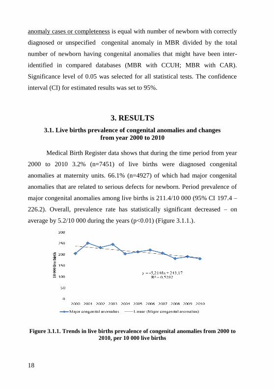
Medical Birth Register data shows that during the time period from year
2000 to 2010 3.2% (n=7451) of live births were diagnosed congenital
anomalies at maternity units. 66.1% (n=4927) of which had major congenital
anomalies that are related to serious defects for newborn. Period prevalence of
major congenital anomalies among live births is 211.4/10 000 (95% CI 197.4 –
226.2). Overall, prevalence rate has statistically significant decreased – on
average by 5.2/10 000 during the years (p<0.01) (Figure 3.1.1.).
Figure 3.1.1. Trends in live births prevalence of congenital anomalies from 2000 to
2010, per 10 000 live births

19
The most common anomalies in the structure of major congenital
anomalies are congenital heart defects (34.2%), limb anomalies (19.5%) and
urinary system abnormalities (13.1%). Also the highest prevalence rate among
live births from 2000 to 2010 are related with congenital heart defects, limb and
urinary system anomalies. Period live births prevalence of congenital heart
defects (Q20 - Q26) is 72.3/10 000 (95% CI 63.8 - 81.2). The prevalence of
congenital heart defects has slightly decreased - on average by 2.5 cases, but
not statistically significant (p>0.05). Total period prevalence of various limb
defects of live births is 41.2/10 000 (95% CI 34.9 – 48.2), the prevalence rate
has decreased statistically significantl (p<0.001) on average by 2.8 cases/10
000 per year. Total period prevalence of congenital urinary system anomalies
(Q60 - Q64, Q79.4) is 27.6/10 000 (95% CI 25.5 – 29.7). The prevalence rate
of this congenital anomaly subgroup in 10-year period has increased.
Prevalence of live births congenital urinary system anomalies in 2010 is
30.3/10 000; 95% CI 23.0 – 39.2) and it has increased on average 1.5 times
compared to 2000 (19.3/10 000; 95% CI 13.7 – 26.4), but the increasing trend
by 0.3/10 000 per year not statistically significant (p>0.05). During the period
from 2000 to 2010 the live births prevalence of nervous system congenital
anomalies had increased on average by 0.4/10 000 (p,0.05) per year.
3.2. Maternal age and congenital anomalies of newborns
The average maternal age of a newborn with congenital anomalies (2000
– 2010) is 27.7 years (SD 5.8), whereas in control group (healthy newborns) -
26.9 years (SD 5.7). The difference is statistically significant - 0.74 years (95%
CI 0.69 – 0.77). There is high correlation (r=0.8; p<0.01) between maternal age
and prevalence rate of congenital anomalies at birth.
Total live birth prevalence of congenital anomalies is 118.1 per 10 000
live births (95% CI 103.7 – 134.0) for mothers up to 19 years of age, 214.5/10

20
000 (95% CI 106.5 – 297.4) for mothers 20-34 years of age and 261.0/10 000
(95% CI 242.1 – 275.0) – among mother aged 35 years and older. These study
results showed an age-related risk for abdominal wall defects, oro-facial clefts
and chromosomal anomalies.
Younger mothers (aged under 19) are more likely to have children with
cleft lip and cleft palate (OR=1.8), abdominal wall defects (OR=2.0) and
chromosomal anomalies (OR=2.4), as compared to mothers aged 20 – 34.
While older mothers (35 years and over) has 5 times greater odds for
chromosomal anomalies of newborn (OR=5.3) and lower odds (OR=0.77) for
heart defects in comparison with live births to mothers in age group 20 – 34.
3.3. Mortality from congenital anomalies and changes
from year 2000 to 2010
Mortality analysis of congenital anomalies includes all Q group
diagnose codes according to ICD-10 (Q00 – Q99). During the time period from
2000 to 2010 shows that 15.1% (n=368) of all perinatal deaths are related to
congenital anomalies, according to the NCDR. Period perinatal mortality due to
congenital anomalies is 1.6 (95% CI 1.4 – 1.8) per 1000 live births and
stillbirths. Statistically significant decreasing trend during the analyzed time
period was observed in both: perinatal mortality associated with congenital
anomalies - 0.2/1000 (p<0.01) and in total perinatal mortality – 0.5/1000 live
births during a year (p<0.001) (Figure 3.3.1.).

21
Figure 3.3.1. Trends in total perinatal mortality and perinatal mortality due to
congenital anomalies from 2000 to 2010, per 1000 live births and stillbirths
In the congenital anomaly structure of perinatal mortality the highest
proportion due to congenital anomalies recorded in the early neonatal period -
25.0% (n=225) with average early neonatal mortality 1.0/1000 live births and
stillbirths (95% CI 0.8 – 1.1), respectively - 9.3% (n=143) of stillbirths and
stillbirth rate due to congenital anomalies – 0.6 (95% CI 0.5 – 0.7) per 1000
live births and stillbirths.
Congenital anomalies are causing an average 28.4% (n=566) of all
infant deaths (2000 – 2010). In 2010 infant death proportion due to congenital
anomalies decreased by 9.2 percent points in comparison with 2000.
The decreasing trend of total infant mortality during the analyzed time
period is higher than of mortality associated with congenital anomalies (Figure
3.3.2.).

22
Figure 3.3.2. Trends in total infant mortality and infant mortality due to
congenital anomalies from 2000 to 2010, per 1000 live births
Average infant mortality due to congenital anomalies during the time
period is 2.4 per 1000 live birth. There is a statistically significant (p<0.01)
decreasing trend by 0.2/1000 per year. The decrease trend also found for total
infant mortality rate - 0.5 cases per year (p<0.001). During time period (2000 –
2010) slight decrease (p<0.05) was observed also in the structure of perinatal
period deaths due to congenital anomalies - 0.8% (95% CI -1.6; -0.2) and in
neonatal period deaths – 0.9% (95% CI -1.7; -0.1). Proportion of death
associated with congenital anomalies in postneonatal period does not change
significantly.
3.4. Associations between congenital anomalies and factors
characterising mother and newborn and antenatal care
Within the framework of analysis, various mother and perinatal factors
were characterised basing on the total data, including all newborn having
congenital anomalies (Q00-Q99), characterising major and minor congenital
defects (n=7451). Differences in the occurrence of risk factors was performed
with the help of control group that was formed by healthy newborn, which

23
during the researched time period (2000 – 2010) were not diagnosed with any
kind of pathology at birth (n=159008).
Newborn factors
Newborn having congenital anomalies more frequently are born from
multiple pregnancies (χ2=42.5; p<0.001), respectively 2.2% cases (n=162) over
1.3% (n=2050) in control group (see Table 3.4.1.).
3.4.1. Table
Shares in groups of characteristics and odds ratio (ORnonadjusted) for
newborn having congenital anomalies in relation to newborn factors
Characteristics CA yes1 CA no
2 χ
2 OR OR
95%CI
Sex
boys 54.2 49.1 74.5*** 1.2*** 1.2; 1.3
girls 45.8 50.9 1.0
Multiple 2.2 1.3 42.5*** 1.7*** 1.5; 2.0
Gestational week ≤36 6.6 0.3 4972.4*** 24.0*** 21.1; 27.3
Birth weight ≤2499 g 6.4 0.6 2834.1*** 11.4*** 10.2; 12.7
Notes: reference category: healthy newborn (OR=1) 1 CA yes – case group (newborn having congenital anomalies); 2CA no – control group
(healthy newborn, not having congenital anomalies); *** p<0.001
It was observed that boys have congenital anomalies 1.2 times more
commonly than girls. Newborn with congenital anomalies more often are born
in preterm birth (OR=24.0), as compared to the newborn having low birth
weight (≤2499) (OR=11.4).
Lifestyle factors
Regardless the fact that harmful lifestyle factors of parents medically are
poorly documented in medical records, analysis of the data showed that there
are statistically significant differences between the groups. Proportion of
smokers among mothers giving birth to children having congenital anomalies is

24
slightly higher (χ2=6.3; p<0.01), correspondingly 10.1% (n=753), while in
control group 9.2% (n=14703). 0.6% (n=45) of women giving birth in the case
group and 0.2% (n=336) of the control group have used alcohol (χ2=48.1;
p<0.001). While consumption of psychoactive substances was recorded only in
small number of cases, such records had 0.3% (n=20) of unhealthy children
mothers and 0.05% (n=75) in control group, (χ2=61.1; p<0.001).
In relation to the harmful health factors of father, statistically significant
differences were observed in the proportions of smokers – 42.9% (n=3197) in
the group of newborn with a congenital anomalies and 37.9% (n=60228) in
control group (χ2=76.4; p<0.001). Control group had slightly more notes on the
father use of alcohol in medical documentation (χ2=12.2; p<0.001) – 4.1%
(n=6529) over 3.3% (n=245), respectively (see Table 3.4.2.).
3.4.2. Table
Shares of characteristics and odds ratio (ORnonadjusted) for newborn having
congenital anomalies in relation to lifestyle factors
Characteristics CA yes1 CA no
2 χ
2 OR OR 95%CI
Mothers
used of alcohol 0.6 0.2 48.1*** 2.9*** 2.1; 3.9
smoking 10.1 9.2 6.3** 1.1 1.0; 1.2
used of psychoactive
substances
0.3 0.05 61.1*** 5.7*** 3.5; 9.4
Father
used of alcohol 3.3 4.1 12.2*** 0.8*** 0.7; 0.9
smoking 42.9 37.9 76.4*** 1.2*** 1.1; 1.3
Used of psychoactive
substances
0.1 0.04 2.6 1.9 0.9; 4.6
Notes: reference category: healthy newborn (OR=1) 1 CA yes – case group (newborn having congenital anomalies); 2CA no – control group
(healthy newborn, not having congenital anomalies); *** p<0.001

25
Analysis of the nonadjusted odds ratio showed that harmful health
factors noticeably increase the odds for congenital anomalies among newborn.
Mothers having newborn with congenital anomalies 2.9 times more often have
consumed alcohol during the pregnancy (OR=2.9) and on average 5 times more
they have used psychoactive substances (OR=5.7). In respect to the harmful
health factors of father: 1.2 times more fathers of newborn having congenital
anomalies have records on smoking (OR=1.2), if compared to the group of
healthy newborn.
Factors characterising antenatal care and health of a mother
Mothers having newborn with congenital defects on average 1.3 times
often (χ2=16.7; p<0.001) have not received antenatal care, i.e., 3.1% (n=232) in
case group and 2.4% (n=3769) in control group. Also delayed antenatal care
was observed 1.3 times more (χ2=31.9; p<0.001) for mothers having newborn
with congenital anomalies (see Table 3.4.3.).
3.4.3. Table
Shares in groups of characteristics and odds ratio (ORnonadjusted) for
newborn having congenital anomalies in relation to factors characterising
antenatal care and health of a mother
Characteristics CA yes1 CA no
2 χ
2 OR OR 95%CI
not received antenatal care 3.1 2.4 16.7*** 1.3*** 1.2; 1.5
delayed antenatal care 8.9 7.2 31.9*** 1.3*** 1.2; 1.4
mother diseases in history 33.9 24.4 229.7*** 1.6*** 1.5; 1.7
pregnancy complications 39.1 38.5 0.95 1.0 0.9; 1.5
delivery complications 44.4 43.1 4.2 1.1 1.0; 1.1
medical abortions in
history
1.8 1.5 5.5* 1.2* 1.1; 1.5
Notes: reference category: healthy newborn (OR=1) 1 CA yes – case group (newborn having congenital anomalies); 2CA no – control group
(healthy newborn, not having congenital anomalies); *** p<0.001

26
Nonadjusted odds ratios show that, in comparison with the mothers aged
under 19, the possibility for newborn congenital anomalies increases along with
the age of a mother - from 1.9 (95% CI 1.7 – 2.1) to mothers aged 20-34 to 2.1
(95% CI 1.9 – 2.4) to mothers aged 35 and over.
Mothers having newborn with congenital defects 1.6 times more often
had history on various diseases and 1.2 times more medical abortions, as
compared to the group of healthy newborn. Whereas odds ratios in relation to
the complications during pregnancy and childbirth did not differ statistically
significantly between the both groups.
3.5. Results of multivariate analysis in relation to odds for newborn
congenital anomalies
With an aim to find out relations among various mother, antenatal and
perinatal factors with odds for newborn congenital anomalies, a multivariate
logistic regression analysis was performed, mutually matching the
characteristics included in the analysis. Analysis was based on multistage
multiple logistic regression, forming separate models.
Initially model included also age of a mother, diseases of a mother
before pregnancy and during it, as well as pregnancy and delivery
complications. Statistical significance of odds ratio remains only for age of a
mother (ORadjusted=1.16) and illnesses of a mother (ORadjusted=1.59), thus further
analysis engaged only those factors. Close correlation was observed between
newborn gestation age and weight at birth, therefore model included only
weight at birth as a factor, such choice is substantiated also by comparison of
characteristics prevalence indicators with the share of preterm deliveries (see
Table 3.5.1.).

27
3.5.1. Table
Odds ratio (OR) for congenital anomalies among newborn over control
group in relation to factors characterising antenatal care, health of a
mother and newborn
Factors
ORadjusted (95%CI)
ORnoadj 1stmodel 2
ndmodel 3
rdmodel 4
thmodel
Maternal age ≥35
years
1.21***
(1.12;
1.28)
1.15***
(1.12; 1.23
1.14***
(1.12;1.23)
1.15***
(1.16;
1.24)
1.15***
(1.16;
1.24)
Not received
antenatal care
1.32***
(1.16;
1.51)
1.17
(0.98;
1.40)
1.14
(0.96;1.37)
1.15
(0.96;
1.37)
1.02
(0.85;
1.23)
Not received early
antenatal care till
12th
gestational week
1.27***
(1.17;
1.37)
1.19**
(1.19;
1.36)
1.19**
(1.17;
1.32)
1.16**
(1.14;
1.29)
1.11**
(1.10;
1.24)
Mother diseases in
history
1.59***
(1.51;
1.68)
1.61**
(1.53;
1.70)
1.57***
(1.52;
1.70)
1.57***
(1.49;
1.66)
1.56***
(1.48;
1.65)
Medical abortions in
history
1.23*
(1.14;
1.47)
1.27***
(1.11;
1.53)
1.27**
(1.12;
1.53)
1.27***
(1.15;
1.53)
1.27**
(1.15;
1.53)
Mother consumed
achocol
2.87***
(2.10;
3.92)
-
2.24***
(1.59;
3.17)
2.36***
(2.21;
6.66)
1.85***
(1.28;
2.68)
Mother use
psychoactive
substances
5.7***
(3.48;
9.35)
-
3.99***
(2.30;
6.93)
3.83***
(2.21;
6.66)
3,61***
(1,97;
6,26)
Father smoked
1.23***
(1.18;
1.29)
- -
1.25***
(1.19;
1.31)
1.23***
(1.17;
1.30)
Low birth weight
(≤2499g)
11.36***
(10.15;
12.71)
- - -
10.49***
(9.21;
11.77)
Notes: reference category covers mothers aged ≤35, have received antenatal care, has
early registered for antenatal care (until 12th pregnancy week), no illnesses in history,
no medical abortions in history, no harmful life factors of mother and father, weight at
birth ≥2500g.
*p<0,05; **p<0,01; ***p<0,001

28
Gradual supplementation of regression model with various factors did
not show notable changes in the OR values. Statistically significant correlations
are observed also in total multivariate model after the adjustment of factors in
relation to the age of a mother, early antenatal care, mother diseases and
medical abortions in history, harmful lifestyle factors of parents and newborn
birth weight. The higher odds for congenital anomalies was recorded for low
birth weight; still this influence is slightly reducing after adjustment with other
characteristics included in the model (ORadjusted=10.49), use of psychoactive
substances (ORadjusted=3.61) and alcohol (ORadjusted=1.85) during pregnancy, as
well as diseases in medical history (ORadjusted=1.56). OR is not statistically
significant for antenatal care (was or was not received).
3.6. Associations between total congenital anomalies and different
maternal diseases
Mothers having newborn with congenital anomalies in history have
more records on chronic or acute diseases, if compared to the group of healthy
newborn, correspondingly – 33.9% (n=2173) over 24.4% (n=38827),
(χ2=299.73; p<0.001). Analysis by the disease group covered study on the
relation of congenital anomalies with syphilis, other sexually transmitted
diseases (including Chlamydia, gonococcus infection, etc. unspecified
infections within this diagnose group). Separate investigation included also
correlation of the diabetes mellitus with prevalence of congenital anomalies
among newborn, researching types I and II, and gestational diabetes, as well as
genitourinary tract infections during pregnancy (including kidney, genital etc.
unspecified infections within this diagnose group). Less significant odds was
observed of other diseases complicating pregnancy and delivery period

29
(according to the ICD-10 codes: O99), e.g., influence of diseases of endocrine,
respiratory, circulatory, digestive system etc.
It was discovered that for mothers with diabetes mellitus odds for
newborn having congenital anomalies is on average 6 times higher (OR=6.3;
p<0.001) than one of mothers not having mentioned disease. Pregnancy
diabetes shows 3 times lower relation to congenital anomalies (OR=2.3;
p<0.001) than diabetes of types I and II.
Sexually transmitted diseases rise the odds for congenital anomalies 2
times (OR=2.0; p<0.001), while in the case of syphilis odds ratio increases
noticeably – on average 4 times (OR=8.7; p<0.001). As regards diseases of
genitourinary tract, no notable differences between the groups (mothers having
newborn with congenital anomalies and mothers having healthy children) were
observed (OR=1.2; p<0.05). Odds ratio in relation to various mother diseases
complicating pregnancy and delivery period were less significant (OR=1.4;
p<0.001).
After adjustment by the age of the mother, odds ratio in relation to the
disease diagnoses remained at the previous level, except gestational diabetes
mellitus – in such cases OR diminished slightly (OR=2.2; p<0.001), showing
closer correlation between the age of a mother and odds for newborn congenital
anomalies.
3.7. Associations between different maternal diseases and particular
diagnosis groups of congenital anomalies
To make separate estimate of the mother diseases as a potential risk
factor for development of major congenital anomalies, data analysis was based
on mother disease as independent characteristics and particular anomaly sub-
group – as a dependent one.

30
Closer relation to separate maternal diseases was observed for seven
sub-groups of congenital anomaly diagnosis. Mothers having record on
sexually transmitted disease (STD) during pregnancy had 3 times higher odds
for newborn to have anomalies of nervous system (OR=3.2; p<0.001), genitals
(OR=3.3; p<0.001), chromosomes (OR=3.7; p<0.001), while slightly lower
odds ratio was observed for heart defects (OR=2.9; p<0.001) (see Tables
3.7.1.).
3.7.1. Table
Odds ratio (OR) for newborn congenital anomalies in relation to
different maternal diseases in history
Congenital
anomalies STD
Diabetes
mellitus
Gestational
diabetes
mellitus
Genitourinary
tract
infections
Other
diseases1
Nervous system
3.2***
(1.6;
6.6)
NS NS NS NS
Eye NS NS NS 3.8*
(2.1; 10.5) NS
Heart defects
2.9***
(2.1;
3.7)
4.8**
(1.5; 8.3)
2.6**
(1.3; 5.3) NS
1.6***
(1.4; 1.8)
Cleft lip and cleft
palate NS NS NS NS
1.5*
(1.2; 2.2)
Urinary system NS NS NS NS 1.4*
(1.2; 1.8)
Genital
3.3***
(1.9;
5.9)
NS NS NS
Chromosomal
3.7***
(1.8;
7.58)
NS NS NS 1.8**
(1.2;2.6)
Notes: reference category – dont have nav noteiktās slimības anamnēzē 1Other diseases (ICD-10; O99); NS – not statistically significant;
*p<0,05; **p<0,01; ***p<0,001

31
Whereas pregnant women suffering from diabetes mellitus have higher
odds ratio for newborn with heart defects (OR=4.8; p<0.01). In the case of
gestational diabetes mellitus this odds reduces slightly, still it is statistically
significant (OR=2.6; p<0.01). Genitourinary tract infections in mother history
determine higher odds for eye anomalies among newborn (OR=3.8; p<0.05).
Also other diseases, although slightly, increase odds for congenital defects
among newborn from 1.4 to 1.8. Higher possibility for congenital anomalies
was observed in following groups: congenital heart defects, cleft lip and cleft
palate, urinary system and chromosome anomalies.
3.8. Evaluation of congenital anomaly prenatal diagnostics basing
on data of the CCUH patient medical histories
Analysis of the hospital’s medical histories indicates that only in 11.9%
(n=212) of all studied CCUH patient medical histories that have records on
treatment of congenital anomalies (n=1788) (on time period 2003 – 2008) there
were note that congenital pathology was diagnosed during prenatal ultrasound
examinations. Mentioned notes mainly concerned major congenital anomalies.
Evaluation of the cases diagnosed during ultrasound examinations in
particular diagnose group shows that most often antenatal diagnoses concerned
urinary system anomalies (renal dysplasia, congenital hydronephrosis etc.) –
27.6% (n=107) (95% CI 23.4 – 32.3) of all hospitalised newborn having urinary
system pathologies (n=387) (see Figure 3.8.1.).

32
Figure 3.8.1. Diagnostics of most common major congenital anomalies, %
(2003 – 2008)
Only in 40.6% (95% CI 34.2 – 47.3) (n=86) of all antenatally diagnosed
anomaly cases, on which there was a record in medical document (n=212),
there was a note on gestational week, during which ultrasound examination
showed pathology. It varied from 21st to 30
th gestational week.
3.8.1. Coverage of live-born congenital anomalies in relation to data
on patients treated in hospital
Unfortunately only for 1/3 (n=629) of the CCUH hospitalised newborn
it was possible to identify the cases and compare them with the MBR.
Diagnose coincidence or number of properly registered cases in the
MBR, if compared to the hospital data, comprised 34.8% (95% CI 31.2 – 38.6),
in compliance with the formula used in the calculation methodology
(219*100/(219+41+369), i.e., (219*100 / 219 (diagnosis of congenital anomaly
coincides in MBR and CCUH) + 41 (diagnoses of congenital anomaly differ) +
369 (there was no diagnosis on congenital anomaly in MBR if compared to
CCUH data)). Whereas for 38.5% diagnosis was not given in the maternity

33
units and for 6.5% it initially was other congenital pathology or for 20.2%
condition of perinatal period (see Figure 3.8.1.1.).
Figure 3.8.1.1. Coverage of live-born congenital anomalies in birth
institutions as compared to CCUH data, during the time period
from 2003 till 2008, %
Evaluation of the possible completeness of congenital anomalies cases
in the Medical Birth Register in comparison with the hospital data shows that it
was observed in 41.3% (95% CI 37.6 – 45.2), in compliance with the formula
used in the calculations ((219+41)*100/219+41+369), i.e., (219 (diagnosis of
congenital anomaly coincide in both databases) + 41 (not specified diagnosis of
congenital anomaly in MBR as compared to the CCUH)*100 / 219 (diagnosis
of congenital anomaly coincide in both databases) + 41 (diagnosis of congenital
anomaly differs) + 369 (there was no diagnosis on congenital anomaly in MBR
if compared to CCUH data)). However, when assessing the results, it should be
taken into account that CCUH hospitalised cases covered the age under 1 year,
thus part of congenital anomalies regards later age that is not reflected in the
MBR.

34
3.9. Coverage of live-born congenital anomaly registration at birth
institutions in relation to congenital anomaly register data
Analysis included 587 cases (2000 – 2010) on which information
allowing identifying the person was available, and thus it was possible to link
the registration systems.
Coincidence of diagnosis or number of properly registered cases in the
MBR accounted for 53.8% (95% CI 49.8 – 57.8), in compliance with the
formula used in the methodology of calculation (316*100/(316+88+183), i.e.
(316*100/316 (diagnosis of congenital anomaly coincides in MBR and CAR) +
88 (diagnosis of congenital anomaly differs in MBR if compared to CAR) +
183 (there was no diagnosis of congenital anomaly in MBR, if compared to
CAR)) (see Figure 3.9.1.).
Figure 3.9.1. Coverage of live-born congenital anomalies at birth
institutions in comparison with the Congenital Anomaly Register (2000 – 2010), %
Evaluation of the completeness of registered cases in MBR, as compared
to CAR, shows that it comprises 68.2% (95% CI 65.0 – 72.4), in compliance
with the formula used in the methodology of the calculation
((316+88)*100/(316+88+183)), i.e., (316 (diagnosis of congenital anomaly
coincides in both databases) + 88 (MBR has unspecified diagnosis of
congenital anomaly)*100 / 316 (diagnosis of congenital anomaly coincides in

35
both databases) + 88 (diagnosis of congenital anomaly differs) + 183 (there was
no diagnosis of congenital anomaly in MBR, if compared to CAR).
The highest share of diagnosis coincidence and more complete
registration in birth institutions is related to visual congenital defects, such as
cleft lip and cleft palate (61.0%; 95% CI 50.2-70.8) and limb anomalies
(76.6%; 95% CI 62.8-86.4). Congenital heart defects (37.5%; 95% CI 26.7-
49.7) had the lowest share in diagnosis coverage in the MBR
4. DISCUSSION
Doctoral thesis allowed ascertaining that in Latvia there are problems in
registration of congenital anomalies, e.g., there are several mutually unlinked
databases, there is no united and complete registration system of congenital
anomalies, and that, in turn, interferes epidemiological researches. Therefore,
with an aim to get more detailed information of the prevalence of anomalies,
within the framework of the study and within the boundaries of possibilities,
several systems registering diagnosed cases of congenital anomalies and deaths
caused by them, were merged and analysed. Also epidemiological researched
conducted in other countries have faced similar problems in relation to the
calculations of prevalence data. Similar approach to data analysis is used in
finding the completeness of registration and prevalence of pathologies (38-42).
Data of congenital anomaly monitoring system show that in Europe the
average period (2000 – 2004) prevalence for live-born comprises 199.3/10 000
(8), whereas period (2000 – 2010) prevalence in Latvia analysed within the
framework of the doctoral thesis is slightly higher – 211.4 per 10 000 live
births. Evaluation of the five-year period (2000 – 2004) shows that in Latvia
prevalence of major congenital anomalies among newborn, if compared to
EUROCAT average prevalence, is even higher - 227.5/10 000. Comparatively
larger occurrence may be explained with more frequent prevalence of

36
congenital anomalies among newborn and with hyper-diagnostics of cases in
birth institutions, as, after the data are sent to the Medical Birth Register, the
diagnoses are not supplemented or changed. However analysis of the study
results indicates that in separate diagnosis groups pathologies are not diagnosed
in maternity wards, but later, therefore in the Medical Birth Register there is no
information on cases diagnosed during postneonatal period.
Regardless the fact that mean age of a mother is increasing (43), the
prevalence of newborn congenital anomalies in Latvia during the time period
since 2000 has reduced statistically significantly - on average by 5.2 cases
annually per 10 000 live births. This finding to some extent conflicts with the
discoveries of scientific literature and researches saying that older women have
higher risk for having child with congenital anomalies. It has an explanation,
because mean age of a mother still falls within the age group under 30 years.
As it was proved by the data analysis, females at this age are more careful
towards the course of pregnancy: apply for antenatal care earlier, undergo
examinations more carefully and pay attention to the treatment and control of
diseases.
Prevalence of congenital anomalies among newborn in respect to the age
of a mother indicated influence of the age in separate diagnose groups. Younger
mothers (aged under 19) are more likely to have children with cleft lip and cleft
palate (OR=1.8), abdomen wall defects (OR=2.0) and chromosomal anomalies
(OR=2.4), as compared to mothers aged 20 – 34. Whereas, if mother is aged
over 35, newborn 5 times more often (OR=5.3) are diagnosed with congenital
chromosomal anomalies than children born to mothers aged between 20 and 34.
These data meet the findings of other similar researches.
As congenital anomalies is the second most common mortality cause
during perinatal and infant age, mortality reduction largely depends on the
factors related to this phenomenon. One of the factors considered to be of a
great significance is improvement of pathology diagnostics and selection of

37
most suitable antenatal care (3, 8). Antenatal care is related to favourable
outcome of the delivery, diminishing the number of children born pre-term, as
well as low weight at birth, thus influencing also infant mortality rate (3, 8, 46).
Results of the research indicated that unsuitable antenatal care (mother of a
newborn has not applied for antenatal care or did it with delay) has close
relation with higher early neonatal mortality (OR=3).
During the time period from 2000 till 2010, perinatal and infant
mortality in Latvia both due to congenital anomalies and totally has reduced
statistically significantly.
Doctoral thesis covered also evaluation of congenital anomaly potential
risk factors. In scientific literature there are researches studying and comparing
prevalence of various antenatal and neonatal factors in breakdown by groups –
newborn having congenital anomalies and not having pathologies (30, 47).
Retrospective analysis of factors characterising antenatal care and age of a
mother as well as perinatal factors of newborn in relation to congenital
anomalies in Latvia during 11-year period was based on control group –
healthy newborn not diagnosed with congenital anomalies or any other
perinatal pathologies in maternity units. Multivariate regression model used in
the doctoral thesis showed relation with factors covered by the analysis.
Nevertheless measured odds ratio in relation with antenatal care and factors
characterising health of a mother indicated little differences between the two
groups (OR= 1.2 - 1.8), they are statistically significant. It means that analysed
risk factor slightly less, but still, increase the odds for newborn congenital
anomalies. The closet relation was observed with the birth weight of a newborn
(OR=10.4). Such associations is described also in other studies, when
comparing newborn with and without congenital anomalies, emphasizing that
perinatal mortality increases 7 times (48), while in other research the risk of
neonatal mortality is up to 53.1 times higher in a group of newborn having
normal weight at birth (≥2500 g), as compared to newborn not having

38
pathologies, as well as 21.3 times higher risk of neonatal mortality is for pre-
term born having congenital anomalies, that, of course, is related not only to
prematurity, but also to the type of congenital anomaly (47).
Smoking, alcohol and addictive substances has a negative influence on
health of the pregnant, development of a foetus that often is a cause of
spontaneous abortion, foetal organ system disorders, premature births,
stillbirths and infant mortality during their first week of life (49, 50).
Regardless the number of cases having note on harmful life factors of parents
registered in the medical documentation is small, data analysis showed some
associations. After adjustment the factors (age of a mother, diseases in history,
newborn’s weight at birth, early antenatal care), higher odds for newborn
congenital anomalies was observed with mother’s use of alcohol (OR=1.85)
and psychoactive substances (OR=3.61). Results of the study show that, during
the monitoring of pregnancy and after delivery, particular attention should be
devoted to cases having a note on mother’s use of alcohol and psychoactive
substances, different diseases in mother history and insufficient weight of a
foetus. These factors may serve as indicators for the necessity to examine
newborn carefully for potential congenital pathology.
One of the pre-conditions for reduction of prevalence of congenital
anomalies among live-born is possibly faster diagnosing of disorders in
pregnancy and foetal development. Main problem faced in Latvia is rather late
discovery of pathologies. As it can be concluded from patient medical records
in this study, pathology (anomalies of nervous system, abdomen wall defects
etc.) on average is diagnosed during the 28th
week of a pregnancy, depending
on pathology diagnose group the time varied between 21st and 30
th week of a
pregnancy. It should be taken into account that in Latvia abortions because of
medical indications are performed until the 24th
week of a pregnancy. Thus
delayed prenatal diagnostics influences also total prevalence indicators, as well
as indicates that it is necessary to improve quality of prenatal diagnostics, as

39
other studies show that early diagnosed neural tube, abdomen defects,
chromosome anomalies more often are related to termination of pregnancy.
Data show that rates of prenatally diagnosis cases on average in Europe,
according to EUROCAT, are rather high, e.g., in relation to gastroschisis –
95%, spina bifida – 81%, Down's syndrome – 72% (51).
Iner-linkage of databases (within the boundaries of possibilities) and
analysing coincidence of diagnoses, it turned out that on average in 34.8% -
53.8% diagnoses coincide. Analysis of information on infants having
congenital anomalies treated in hospital (in relation to MBR data) showed that
in 20% of the cases delivery units had recorded only perinatal period health
disorders and in 38% of the cases pathology was not diagnosed at birth; that, of
course, is related to short period spent in maternity unit after birth (that most
often comprises less than 4 days) and therefore to limited opportunity to
observe the newborn and conduct more detailed examination. Likewise there is
a part of congenital anomalies, which manifest themselves at a later age.
In research carried out in Australia, when comparing Congenial
Anomaly Register information with the genetic centre and children hospital
patient data, 54% of cases indicated coincidence of diagnoses (41). Assessment
of congenital anomaly completeness in MBR and linking with other databases
led to the conclusion that on average 41.3% - 68.2% of congenital anomalies
are discovered in delivery units. Comparison of congenital anomaly coverage in
breakdown by diagnosis groups shows that most often maternity units register
visual congenital defects, whereas the lowest coverage is related to congenital
heart defects and chromosome anomalies. Also in other similar research on
registration coverage it was found out that most frequently data of the newborn
register and birth certificate indicate or have note on visual congenital defects
that are more visible (42).
Data on congenital anomalies from Latvian Medical Birth Register has
not been widely used before. But this study shows that MBR data may be

40
useful for epidemiological studies on congenital anomalies in Latvia, as well as
by using all live birth cohort-based control group for mutual comparison of
potential risk factors. In future, significant factor for using register data (incl.
MBR) in research that will ensure and increase epidemiological significance of
accumulated information will lay in possibilities to link data with other
registration systems in the country. It will improve opportunity for more
complete analysis of newborn and infant health in relation to various perinatal
factors.
5. CONCLUSIONS
1. During the time period from year 2000 till 2010, prevalence of major
congenital anomalies among newborn in Latvia statistically significantly has
reduced annually by 5.2/10 000. Breakdown by separate diagnose group shows
statistically significant slight reduction only in total prevalence of limb defects
– 2.8/10 000 and small rise in congenital anomalies of nervous system – 0.4/10
000.
2. Total congenital anomaly period prevalence (2000 – 2010) among
newborn rises along with the age of a mother. For children having congenital
anomalies mean age of a mother (27.7 years) is higher than one for children not
having pathologies (26.9 years), the difference is statistically significant– 0.74
years.
3. Mothers aged under 19 have higher odds of newborn for cleft lip and
cleft palate (OR=1.8); abdomen wall defects (OR=2.0) and chromosome
anomalies (OR=2.4), as compared to mothers aged 20 - 34. In comparison with
mothers aged 20 – 34, mothers aged over 35 are more likely to have newborn
with chromosome anomalies (OR=5.3).
4. During the time period from 2000 till 2010, statistically significant
reduction can be observed in both perinatal (of 0.2 cases annually per 1000 live

41
births and stillbirths) and infant (of 0.2/1000 live births) mortality caused by
congenital anomalies, along with decrease in total perinatal (of 0.5/1000 live
births and stillbirths) and infant (of 0.5 cases annually per 1000 live births)
mortality.
5. Results of the multivariate analysis indicate higher odds ratio (OR)
for newborn congenital anomalies in relation to following factors characterising
health of a mother and newborn: age of a mother, late antenatal care, mother
diseases (syphilis, other sexually transmitted diseases, diabetes mellitus,
gestational diabetes, genitourinary tract infections), medical abortions in
history, mother’s use of alcohol and psychoactive substances, low newborn’s
weight at birth.
6. Delivery institutions most often and more completely diagnose visual
congenital defects of newborn: cleft lip and cleft palate, limb defects, while the
most incompletely – congenital heart defects and chromosome anomalies.
7. Prenatal diagnostics of congenital anomalies is delayed - on average
during 28th
week of a pregnancy.
8. Congenital anomaly total prevalence registration data are influenced
by: quality of medical documentation records, incl. specification of approved
final diagnosis in registers, as well as late-diagnosed cases of congenital
anomalies among infants under 1 year and lack of comprehensive information
on termination of pregnancy due to foetus congenital anomalies.
9. Analysis of congenital anomaly prevalence indicators is obstructed by
independent work and activities of national databases managed by various
institutions and impossible linking of the information available in these
databases. Possibilities for inter-linkage would increase the usefulness of
accumulated and stored information in research and practical medicine.

42
Confirmation of hypotheses of the doctoral thesis
First hypothesis that changes in prevalence of congenital anomalies are
influenced by rise in mean age of a mother was partially confirmed. Live birth
prevalence rates were significantly higher among mothers aged over 35 years.
In the same time, there is a reduction in prevalence of congenital anomalies,
regardless the rise in mean age of women giving birth.
The second hypothesis that changes in mortality caused by congenital
anomalies are influenced by total perinatal and infant mortality trends was
partially confirmed. As during the analysed time period perinatal and infant
mortality caused by congenital anomalies diminished, also total perinatal and
infant mortality dropped. Moreover slight decrease was observed in the
structure of congenital anomaly death causes during perinatal and postneonatal
period. Still total mortality reduction is faster than mortality caused by
congenital anomalies that is rather stable in annual dynamics.
The third hypothesis that mothers having newborn with congenital
anomalies act more risky towards antenatal care and own health was proved, as
results of the multivariate analysis shows that prevalence of congenital
anomalies in association with maternal late antenatal care, mother’s diseases in
history and mother’s use of alcohol and psychoactive substances.
The fourth hypothesis that prevalence indicators are influenced by
prenatal diagnostics and completeness of national registration system was
confirmed, because analysis data showed that on average 41.3% - 68.2% of
congenital anomalies are diagnosed at maternity units.

43
6. PRACTICAL RECOMMENDATIONS
1. Problems encountered in calculations of congenital anomaly
prevalence show necessity to improve national system of congenital anomaly
registration:
• by ensuring interlink or reversible exchange of information between
the Medical Birth Register and hospitals on confirmation or specification of
congenital anomaly diagnosis after discharge from maternity unit (enhancing
completion of medical documentation “Supplementary ticket of newborn card”
in health care institutions and sending it to the Medical Birth Register);
• by improving linkage between the Medical Birth Register and
Congenital Anomaly Register or data exchange on cases of congenital
anomalies;
• by developing the Medical Birth Register and accession to the
EUROCAT database, with an aim to ensure comprehensive statistical data on
Latvia, as well as internationally comparable data and make registration of
infant congenital anomaly cases diagnosed during the postneonatal period.
2. Primary health care professionals (general practitioners, nurses) in
their preventive work should give information and pay attention to women at
reproductive age, education on the harmful lifestyle factors, treatment of
chronic and acute diseases, influence of antenatal care on pregnancy outcome,
with an aim to improve female knowledge and skills in situations covering
issues related to the pregnancy planning.
3. Obstetrical specialists (gynaecologists, midwifes) in situations, when
during pregnancy risk factors as mother diseases (diabetes mellitus, sexually
transmitted diseases, genitourinary diseases), spontaneous, medical abortions in
history, smoking, use of psychoactive substances, insufficient rise in foetus
weight are discovered, have to evaluate the results of sonography more

44
carefully and think about the necessity for additional examination, to eliminate
congenital pathologies of a foetus.
4. Neonatologists, pediatricians, and family doctors should devote
particular attention in cases, when newborn in birth institutions are diagnosed
with conditions of perinatal period that may serve as nonspecific indicators for
higher congenital anomaly risk.
5. Within the framework of the public health promotion and education
programs on sexual and reproductive health, attention should be directed not
only to factors related to contraception, family planning, infertility, but also
issues on avoidable risk factors for congenital anomalies and prevention
options should be emphasized.

45
REFERENCES
1. The world health report 2005: Make every mother and child count // World Health
Organization, 2005. – Pp. – 2, 6, 17, 19. 80, 106, 153.
2. Christianson A., P. Howson C.P., Modell B. Global report on birth defect // March
of dimes New York, 2006. - Pp. 2 - 9, 14 – 17, 22
3. World Health Organization. Birth defects. Report by Secretariat // Sixty-third
Word health assambly Provisional agenda item 11.7. A63/10, 1 April 2010. - Pp. 1-7.
4. Adams M.M., Alexander G.R., Kirby R.S., Wingate M.S. Infant morbidity. Birth
defects // Perinatal epidemiology for public health practice, 2009. - Pp. 137-142.
5. Lowdermilk D.L., Perry S.E., Cashion K. et al. Newborn complications.
Hemolytic disorders and congenital anomalies // Maternity and women’s health care. –
10th edition, 2011. – 873.
6. Boyd P. A., Armstrong B., Dolk H. et. al. Congenital anomaly surveillance in
England - ascertainment deficiencies in the national system // British Medical Journal,
2005; 1: 1-5.
7. Patel Z.M., Adhia R.A. Birth defects surveillance study // Indian J Pediatr, 2005;
72 (6): 489-491.
8. Special Report: The Status of Health in the European Union: Congenital
Malformations. EUROCAT Central Registry. June 2009:4 // http://www.eurocat-
network.eu/content/Special-Report-Con-Malfs.pdf (sk.15.042012.)
9. Munim S., Nadeem S., Khuwaja N.A. The accurancy of ultrasound in the
diagnosis of congenital abnormalities // J Pak Med Assoc, 2006; 56 (1): 16-18.
10. Tomatir A.G., Demirhan H.,Sorkun H.C. et. al. Major congenital anomalies: a
five-year retrospective regional study in Turkey // Genetics and Molecular Researc,
2009; 8 (1): 19-27.
11. Savitz D.A., Hertz-Picciotto I., Poole C. et. al. Epidemiologic Measures of the
Course and Outcome of Pregnancy // Epidemiol Rev, 2002; 24: 91–101.
12. Society for reproductive, endocrinology and infertility// http://www.ivf1.com/age-
birth-defects/ (sk.20.02.2012.)
13. Statistikas dati par 2011. gadu. Mirstība. Slimību profilakses un kontroles centrs //
http://www.spkc.gov.lv/veselibas-aprupes-statistika/ (sk. 01.08.2012.)
14. European Health for All Database (HFA-MDB). World Health Organization
Regional Office for Europe. Updated: January 2012 // http://data.euro.who.int/hfamdb/
(sk.05.02.2012.).
15. 08.05.2008. likums "Grozījumi Ārstniecības likumā", spēkā ar 11.06.2008.
16. Latvijas veselības aprūpes statistikas gadagrāmata 2010. / Veselības Ekonomikas
centrs – 12. izdevums – Rīga, 2011. – 1, 183., 186., 187. lpp.
17. Skrule J., Štāle M., Upmale S. Mātes un bērna veselība perinatālajā periodā //
Sabiedrības veselības aģentūra 2009. – 19.-20. lpp.
18. MK Rīkojums Nr. 504. Sabiedrības veselības pamatnostādnes 2011. – 2017.
gadam. 05.10.2011.

46
19. MK Rīkojums Nr. 269. Mātes un bērna veselības uzlabošanas plāns 2012.-2014.
gadam. 19.06.2012.
20. MK noteikumi Nr. 574 Grozījumi Ministru kabineta 2006. gada 25. jūlija
noteikumos Nr. 611. Dzemdību palīdzības nodrošināšanas kārtība. 21.08.2012.
21. European health for all databases (HFA-DB). World Health Organization Regional
Office for Europe. Updated: January 212. // http://data.euro.who.int/hfadb/
(sk.10.05.2012.)
22. DH/DETR/Environment Agency Joint Research Programme on the Possible
Health Effects of Landfill Sites. A Study of the geographical variation in overall rates of
congenital abnormalities and the rates of specific abnormalities // Project LSHTM/00/2,
2003. – Pp. - 1-6.
23. Vrijheid M., Dolk H., Stone D. et. al. Socioeconomic inequalities in risk of
congenital anomaly // Arch Dis Child, 2000; 82: 349-352.
24. Dolk H. EUROCAT: 25 years of European surveillance of congenital anomalies //
Arch Dis Child Fetal Neonatal Ed, 2005; 90: 355-357.
25. Lubaua I. Ehonokardiogrāfisks koronārās asinsrites henodinamisks un anatomisks
vērtējums bērniem ar iedzimtām sirdskaitēm. RSU, Rīga 2006. – 66., 105. lpp.
26. Mozgis Dz.. Klīniskie, morfoloģiskie un biomehāniskie iedzimto anorektālo
anomāliju pētījumi. RSU, Rīga 2004.
27. Ābola Z. Barības vada atrēzija – klīniskie un morfofunkcionālie pētījumi. RSU,
Rīga 2007.
28. Lāce B., Barkāne B., Akota I. The most common genetic syndromes and
associated anomalies in Latvian patients with cleft lip with or without palate //
Stomatologija, 2006; 8 (2): 57-60.
29. Nagle E.. Teibe U. Balode I. Craniofacial morphology in parents of cleft children
and healthy individuals // Stomatologija, 2006; 8 (2): 53-6.
30. Werler M.M. Birth defects // Louis G.B., Platt R. Reproductive and perinatal
epidemiology, 2011. – Pp. – 184-206.
31. Pētersons A., Ābola Z., Villeruša A. u.c. Uz modernām tehnoloģijām balstītu
iedzimtu patoloģiju diagnostikas un ārstēšanas algoritmu izstrāde bērniem. // Autoru
kolektīvs Valda Pīrāga redakcijā. Latvijas iedzīvotāju dzīvildzi un dzīves kvalitāti
apdraudošās slimības, Rīga 2009. – 77. - 93. lpp.
32. Starptautiskā statistiskā slimību un veselības problēmu klasifikācija.
Desmitā (10.) redakcijas (SSK-10). 1. Sējums // Pasaules Veselības
organizācija, 1992; 7. nodaļa.
33. Wilson G.N. Birth defects and syndromology // Pediatric genetics: short
course, 2000. – Pp. 4., 5., 254 – 260.
34. Czeizel AE. Birth defects are preventable // Int J Med Sci, 2005; 2: 91-92.
35. Coding of EUROCAT Subgroups of Congenital anomalies //
http://www.eurocat-
network.eu/aboutus/datacollection/guidelinesforregistration/malformationcodin
gguides (sk. 12.04.2012.).

47
36. Peacok J.L., Peacok P.J. Oxford handbook of medical statistics, 2011. –
162. – 165., 268. – 269., 406. – 411.
37. Vittinghoff E., Glidden D.V., Shiboski S.C., McCulloch C.E. Regression
methods in biostaistics, 2005. – Pp. – 44. – 53.
38. Larsen H., Nielsen L.G., Bendsen J. et al. Predictive value and
completeness of the registration of congenital abnormalities in three Danish
population -based registries // Scand J Public Health, 2003; 31 (12): 13 - 16.
39. Gissler M., Louhiala P., Hemminki E. Nordic Medical Birth Registers in
epidemiological research // Eur J Epidemiol, 1997; 13: 169 – 175.
40. Irgens L.M. The Medical Birth Registry of Norway. Epidemiological
research and surveillance throughout 30 years // Acta Obstet Gynaecol Scand,
2000; 79: 435 - 439.
41. Riley M., Phyland S., Halliday J. Validation study of the Victorian Birth
Defects Register // J Paediatr Child Health, 2004; 40: 544 – 548.
42. Honein M.A., Paulozzi L.J. Birth defects surveillance: assesing the „Gold
standard” // Am J of Publich Health, 1999; 89 (9): 1238 – 1240.
43. LR Centrālā statistikas pārvalde. Statistikas datu bāzes //
http://data.csb.gov.lv/DATABASE/Iedz/Iedzīvotāji%20B%20Dzimstība/Vēl%
20nepārrēķināti%20dati%20pēc%202011.gada%20tautas%20skaitīšanas%20re
zultātiem/Vēl%20nepārrēķināti%20dati%20pēc%202011.gada%20tautas%20sk
aitīšanas%20rezultātiem.asp (sk.10.04.2012.)
44. Hollier L.M., Leveno K.J., Kelly M.A. et al. Maternal age and
malformations in singleton births // Obstet Gynecol, 2000; 96: 701 – 706.
45. Loane M., Dolk H., Morris J.K. Maternal age–specific risk of non-
chromosomal anomalies // BJOG, 2009; 116: 1111 – 1119.
46. Smith L.K., Budd J.L.S., Field D.J., Draper E.S. Socioeconomic
inequalities in outcome of pregnancy and neonatal mortality associated with
congenital anomalies: population based study // BMJ, 2011; 343: 1 – 9.
47. Malcoe L.H., Shaw G.M., Lammer E.J.,Herman A.A. The effect of
congenital anomalies on mortality risk in white and black infants // Am J of
Publich Health, 1999; 89 (6): 887-892.
48. Costa C.M.S., Gama S.G.N., Leal M.C. Congenital malformations in Rio
de janeiro, Brazil: prevalence and associated factors // Cadernos de Sasde
Psblica, 2006; 22 (11): 2423-2431.
49. El-Mohandes A., Herman A.A., El-Khorazaty M.N. et. al. Prenatal Care
Reduces the Impact of Illicit Drug use on Perinatal Outcomes // Journal of
Perinatology, 2003; 23: 354 – 360.
50. Wolfe E.L., Davis T., Guydish J., Delucchi K.L. Mortality Risk
Associated with Perinatal Drug and Alcohol Use in California // J Perinatol,
2005; 25 (2): 93 – 100.

48
51. EURO – PERISTAT project, with SCPE, EUROCAT, EURONEOSTAT.
European Perinatal health report, 2008. – Pp. – 51. – 53., 168 – 177.
List of publications and abstracts
Publications
1. A.Villeruša, I. Zīle, L. Lagūna, A. Pētersons. Iedzimto patoloģiju
izplatība Latvijā // RSU zinātniskie raksti 2008, Rīga, 2009, lpp.
2. Villeruša A., Zīle I., Pētersons A. Registration of congenital anomalies
in Latvia // Collection of Scientific papers 2009. Research articles in
medicine & pharmacy, Rīga, RSU, 2010, 103. - 106. lpp.
3. I. Zīle, A.Villeruša, A. Pētersons. Iedzimto anomāliju izplatības
raksturojums jaundzimušajiem pēc mātes dzīvesvietas // RSU
zinātniskie raksti 2009, Rīga, 423. - 434. lpp.
4. I. Zīle, A. Buza, A. Villeruša. Mirstības analīze jaundzimušajiem ar
iedzimtām anomālijām // Statistikas zinātnisko pētījumu rezultāti 2010,
Rīga, Centrālā statistikas pārvalde, 303.-311.lpp.
5. I. Zīle. A. Gederte, A. Villeruša. Kopējā perinatālā mirstība un
perinatālā mirstība no iedzimtām anomālijām Latvijā 2000. - 2009. gadā
// RSU zinātniskie raksti 2011 (1.sējums), Rīga, 208. – 216. lpp.
6. Ebela I., Zile I., Zakis A., Folkmanis V., Rumba-Rozenfelde I. Mortality
of children under five and prevalence of newborn congenital anomalies
in relation to macroeconomic and socio-economic factors in Latvia //
Medicina (Kaunas), 2011; 47 (12): 667 - 674.
Abstracts
1. I. Zīle, A.Villeruša. Iedzimto hromosomu anomāliju izplatība
jaundzimušajiem saistībā ar mātes vecumu // RSU Zinātniskā konference, tēzes,
Rīga, 2009, 164. lpp.

49
2. I. Zīle, A.Villeruša. Iedzimto anomāliju izplatību ietekmējošie faktori
jaundzimušo vidū Latvijā // RSU Zinātniskā konference, tēzes, Rīga 2010, 64.
lpp.
3. I. Zīle, A.Villeruša. Asinsrites un balsta un kustību sistēmas iedzimtās
kroplības ietekmējošie faktori jaundzimušajiem // RSU Zinātniskā konference,
tēzes, Rīga, 2010, 121. lpp.
4. I. Zīle, A.Villeruša. Zīdaiņu ar iedzimtām anomālijāmmirstības
struktūra 2000.-2007. gadā pēc nāves cēloņa pamatdiagnozes // RSU Zinātniskā
konference, tēzes, Rīga, 2011, 267. lpp.
5. I. Zīle, A.Villeruša. Iedzimto anomāliju izplatība jaundzimušajiem
(2000.-2010.) // RSU Zinātniskā konference, tēzes, Rīga, 2012, 197. lpp.
6. Villerusa A., Laguna L. Zile I. Prevalence of congenital anomalies in
Latvia in 2000 – 2005 // European Journal of Public health 2008.
7. Zile I., Villerusa A. A survival analysis of newborns with congenital
anomalies in Latvia from 2000 to 2007 // European Journal of Public health
2010; 20 (1): 240.
8. Zile I., Villerusa A. Correspondence between the diagnoses at birth
and death of infants with the most common congenital anomalies in Latvia //
Journal of Inherited metabolic disease 2011; 34 (1): 11.
9. Zile I., Villerusa A. Epidemiological analysis of prevalence and
mortality rate of newborns’ Down syndrome in Latvia // NordicEpi 2011,
Abstract Book. – Pp. - 30.
10. Zile I., Villerusa A. Analysis of newborn mortality due to congenital
anomalies associated with perinatal period risk factors // Journal of
epidemiology & Community health, 2011, 65 (1): 471 - 472.
11. Zile I., Villerusa A. Epidemiological analysis of prevalence of
newborns neural tube defects in Latvia // ISMCK 2011, Abstract book, Tomus
66, 1 (1): 213.

50
12. Zile I., Villerusa A. Analysis of newborns' neonatal deaths due to
congenital anomalies correlation with antenatal and neonatal characteristics in
Latvia // European Journal of Public health 2011, 21 (1): 211.
13. Zīle I., Villeruša A. Infant mortality due to congenital anomalies in
Latvia in relation with macroeconomic indicators // Nordstat, 2012. – Pp. – 77.
14. Zīle I., Kviļūna D., Villeruša A. Prevalence of surgical congenital
anomalies of newborns in Latvia (2000 – 2010) // Final programme and
abstracts book, RSU, Riga, 2012. – Pp. - 53.
15. Zīle I., Villeruša A. Congenital anomalies of newborns and
associated maternal risk factors in Latvia // Abstracts — 45th Annual SER
Meeting. Minneapolis, MN, June 27(eve)-30, 2012 – 230S.
16. Villeruša A., Zīle I. Congenital anomalies of newborns related by
maternal age and associated antenatal risk factors in Latvia // 9th European
IUHPE Health promotion Conference. Abstract Compendium, 2012.
17. Zīle I., Villeruša A. The association between major birth defects of
newborns and maternal age in Latvia // European Journal of Public health 2012,
22 (2): 236.

51
ACKNOWLEDGEMENTS
Author of the doctoral thesis expresses the deepest gratitude to:
the supervisor - professor Anita Villeruša - for advises and encouraging
during whole doctoral studies and progress of the doctoral thesis;
collective of the Riga Stradiņš University Department of Public Health
and Epidemiology for friendly support and advises;
work colleagues of the Centre for Disease Prevention and Control for
responsiveness and empathy during the progress of work and Centre for the
opportunity to use the statistical data for research;
Children's Clinical University Hospital in Riga (int. al. archive) for the
opportunity to use the statistical data for research;
and thanks to the family for the support and understanding during the
creative process of the study.
Research was conducted in the Riga Stradiņš University Department of
Public Health and Epidemiology with the support from ESF National
Programme “Support for doctoral programs and postdoctoral research” project
“Support for doctoral programs and postdoctoral research in medical sciences”
and ESF project “Support for doctoral study programs and research towards a
degree in Riga Stradiņš University”.


![9887basem aldeek some%final epidemiological aspects about cancer in king saudi[1]1 [autosaved]](https://img.pdfslide.us/doc/110x75/558438bad8b42a77068b4c31/9887basem-aldeek-somefinal-epidemiological-aspects-about-cancer-in-king-saudi11-autosaved.jpg)