Embed Size (px)
Citation preview
ENZYMATIC CLEAVAGE OF RNA BY RNA
Nobel Lecture, December 8, 1989
S I D N E Y A L T M A N
Department of Biology, Yale University, New Haven, CT 06520, USA
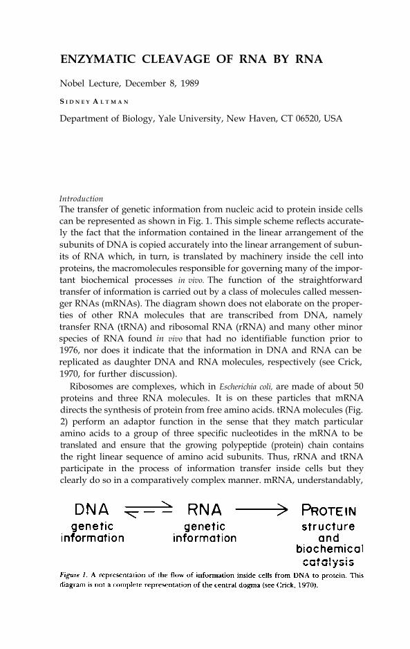
IntroductionThe transfer of genetic information from nucleic acid to protein inside cellscan be represented as shown in Fig. 1. This simple scheme reflects accurate-ly the fact that the information contained in the linear arrangement of thesubunits of DNA is copied accurately into the linear arrangement of subun-its of RNA which, in turn, is translated by machinery inside the cell intoproteins, the macromolecules responsible for governing many of the impor-tant biochemical processes in vivo. The function of the straightforwardtransfer of information is carried out by a class of molecules called messen-ger RNAs (mRNAs). The diagram shown does not elaborate on the proper-ties of other RNA molecules that are transcribed from DNA, namelytransfer RNA (tRNA) and ribosomal RNA (rRNA) and many other minorspecies of RNA found in vivo that had no identifiable function prior to1976, nor does it indicate that the information in DNA and RNA can bereplicated as daughter DNA and RNA molecules, respectively (see Crick,1970, for further discussion).
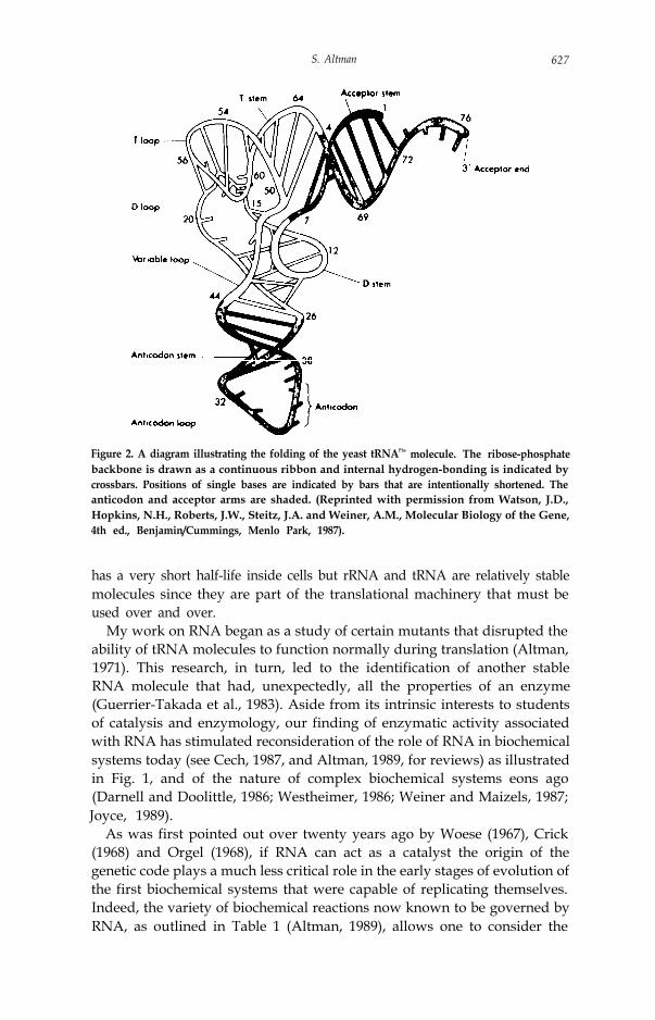
Ribosomes are complexes, which in Escherichia coli, are made of about 50proteins and three RNA molecules. It is on these particles that mRNAdirects the synthesis of protein from free amino acids. tRNA molecules (Fig.2) perform an adaptor function in the sense that they match particularamino acids to a group of three specific nucleotides in the mRNA to betranslated and ensure that the growing polypeptide (protein) chain containsthe right linear sequence of amino acid subunits. Thus, rRNA and tRNAparticipate in the process of information transfer inside cells but theyclearly do so in a comparatively complex manner. mRNA, understandably,
S. Altman 627
Figure 2. A diagram illustrating the folding of the yeast tRNAPhe molecule. The ribose-phosphatebackbone is drawn as a continuous ribbon and internal hydrogen-bonding is indicated bycrossbars. Positions of single bases are indicated by bars that are intentionally shortened. Theanticodon and acceptor arms are shaded. (Reprinted with permission from Watson, J.D.,Hopkins, N.H., Roberts, J.W., Steitz, J.A. and Weiner, A.M., Molecular Biology of the Gene,4th ed., Benjamin/Cummings, Menlo Park, 1987).
has a very short half-life inside cells but rRNA and tRNA are relatively stablemolecules since they are part of the translational machinery that must beused over and over.
My work on RNA began as a study of certain mutants that disrupted theability of tRNA molecules to function normally during translation (Altman,1971). This research, in turn, led to the identification of another stableRNA molecule that had, unexpectedly, all the properties of an enzyme(Guerrier-Takada et al., 1983). Aside from its intrinsic interests to studentsof catalysis and enzymology, our finding of enzymatic activity associatedwith RNA has stimulated reconsideration of the role of RNA in biochemicalsystems today (see Cech, 1987, and Altman, 1989, for reviews) as illustratedin Fig. 1, and of the nature of complex biochemical systems eons ago(Darnell and Doolittle, 1986; Westheimer, 1986; Weiner and Maizels, 1987;Joyce, 1989).
As was first pointed out over twenty years ago by Woese (1967), Crick(1968) and Orgel (1968), if RNA can act as a catalyst the origin of thegenetic code plays a much less critical role in the early stages of evolution ofthe first biochemical systems that were capable of replicating themselves.Indeed, the variety of biochemical reactions now known to be governed byRNA, as outlined in Table 1 (Altman, 1989), allows one to consider the
628 Chemistry 1989
Table 1. Some Properties of Catalytic RNAs
RNA End Groups” Cofactorb Mechanism1. Group I introns 5'-P, 3’-OH Yes Transesterification2. Group II introns 5’-P, 3’-OH No Transesteritication3. Ml RNA 5’-P, 3’-OH No Hydrolysis4. Viroid/satellite 5’-OH, 2’,3’-cyclic No Transesterification
phosphate5. Lead ion/tRNA 5’-OH, 2’3’-cyclic NO Similar to RNAse A
phosphatea The end groups are those produced during the initial cleavage step of self-splicingreactions or during the usual cleavage reactions of other RNA species.b This column refers to the use of a nucleotide cofactor.
possibility that a large number of diverse enzymatic reactions took place inthe absence of protein. To add further substance to these ideas about life onearth over a billion years ago, it is important to understand exactly howcatalytic RNA, as we know it, works and what role it plays in vivo today. Thisdiscussion deals primarily with the catalytic RNA subunit of the enzymeribonuclease P from Escherichia coli.
A BRIEF ACCOUNT OF STUDIES OF RIBONUCLEASE P
Finding the substrateIn October, 1969 I arrived at the MRC laboratory of Molecular Biology inCambridge, England ostensibly to study the three-dimensional structure oftRNA through the use of physical-chemical methods. On my arrival, SydneyBrenner and Francis Crick informed me that the crystal structure of yeasttRNAPhe had recently been solved (Rim et al., 1974; Robertus et al., 1974)and that there was no further need to engage in the studies originallyoutlined for me. I was further instructed to get settled, to think about a newproblem for a week or two, and then to return for another discussion.Although some of my colleagues remember me as being upset by thatconversation with Brenner and Crick, the feeling must have passed quicklybecause I only recall being presented with a marvelous opportunity tofollow my own ideas.
I proposed to make acridine-induced mutants of tRNATyr from E. coli todetermine if altering spatial relationships in tRNA, by deleting or adding anucleotide to its sequence, would drastically alter the function of themolecule. Since Brenner and John D. Smith and their colleagues (Abelsonet al., 1970; Russell et al., 1970; Smith et al., 1971) had just completed aclassic series of studies of base-substitution mutants of tRNATyr, they werenot overly excited by the prospect of someone simply producing moremutants. Nevertheless, Brenner and Crick did not prevent me from pushingahead and John Smith, in time, provided valuable advice about the geneticsof the system in use in the laboratory.
The mutants I made lacked the usual function of suppressor tRNAs and
S. Altman 629
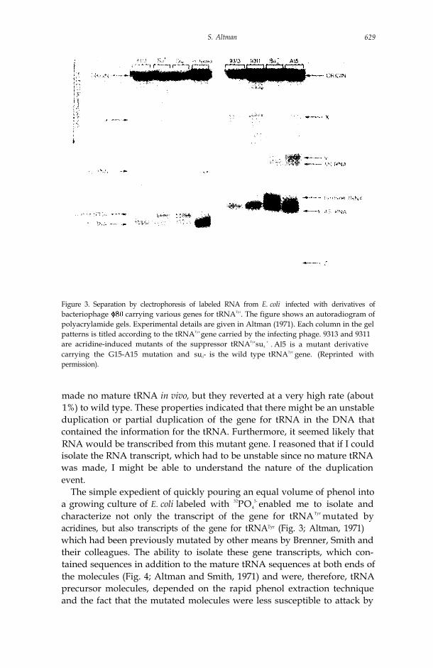
Figure 3. Separation by clectrophoresis of labeled RNA from E. coli infected with derivatives ofbacteriophage carrying various genes for tRNATyr. The figure shows an autoradiogram ofpolyacrylamide gels. Experimental details are given in Altman (1971). Each column in the gelpatterns is titled according to the tRNATyr gene carried by the infecting phage. 9313 and 9311are acridine-induced mutants of the suppressor tRNATyrsu3
+ . Al5 is a mutant derivativecarrying the G15-A15 mutation and su0- is the wild type tRNATyr gene. (Reprinted withpermission).
made no mature tRNA in vivo, but they reverted at a very high rate (about1%) to wild type. These properties indicated that there might be an unstableduplication or partial duplication of the gene for tRNA in the DNA thatcontained the information for the tRNA. Furthermore, it seemed likely thatRNA would be transcribed from this mutant gene. I reasoned that if I couldisolate the RNA transcript, which had to be unstable since no mature tRNAwas made, I might be able to understand the nature of the duplicationevent.
The simple expedient of quickly pouring an equal volume of phenol intoa growing culture of E. coli labeled with 32PO4
3- enabled me to isolate andcharacterize not only the transcript of the gene for tRNATyr mutated byacridines, but also transcripts of the gene for tRNATyr (Fig. 3; Altman, 1971)which had been previously mutated by other means by Brenner, Smith andtheir colleagues. The ability to isolate these gene transcripts, which con-tained sequences in addition to the mature tRNA sequences at both ends ofthe molecules (Fig. 4; Altman and Smith, 1971) and were, therefore, tRNAprecursor molecules, depended on the rapid phenol extraction techniqueand the fact that the mutated molecules were less susceptible to attack by
630 Chemistry 1989
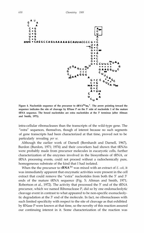
Figure 4. Nucleotide sequence of the precursor to The arrow pointing toward thesequence indicates the site of cleavage by RNase P on the 5’ side of nucleotide 1 of the maturetRNA sequence. The boxed nucleotides are extra nucleotides at the 3’ terminus (after Altmanand Smith, 1971).
intra-cellular ribonucleases than the transcripts of the wild-type gene. The“extra” sequences, themselves, though of interest because no such segmentsof gene transcripts had been characterized at that time, proved not to beparticularly revealing per se.
Although the earlier work of Darnell (Bernhardt and Darnell, 1967),Burdon (Burdon, 1971; 1974) and their coworkers had shown that tRNAswere probably made from precursor molecules in eucaryotic cells, furthercharacterization of the enzymes involved in the biosynthesis of tRNA, ortRNA processing events, could not proceed without a radiochemically pure,homogeneous substrate of the kind that I had isolated.
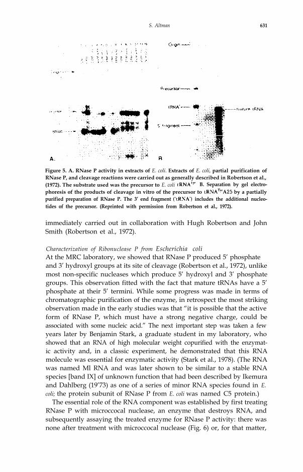
When the the precursor to was mixed with an extract of E. coli, itwas immediately apparent that enzymatic activities were present in the cellextract that could remove the “extra” nucleotides from both the 5’ and 3’ends of the mature tRNA sequence (Fig. 5; Altman and Smith, 1971;Robertson et al., 1972). The activity that processed the 5’ end of the tRNAprecursor, which we named Ribonuclease P, did so by one endonucleolyticcleavage event in contrast to what appeared to be non-specific exonucleoly-tic degradation at the 3’ end of the molecule. In fact, no ribonucleases withsuch limited specificity with respect to the site of cleavage as that exhibitedby RNase P were known at that time, so the novelty of this reaction assuredour continuing interest in it. Some characterization of the reaction was
S. Altman 631
Figure 5. A. RNase P activity in extracts of E. coli. Extracts of E. coli, partial purification ofRNase P, and cleavage reactions were carried out as generally described in Robertson et al.,(1972). The substrate used was the precursor to E. coli B. Separation by gel electro-phoresis of the products of cleavage in vitro of the precursor to by a partiallypurified preparation of RNase P. The 3’ end fragment includes the additional nucleo-tides of the precursor. (Reprinted with permission from Robertson et al., 1972).
immediately carried out in collaboration with Hugh Robertson and JohnSmith (Robertson et al., 1972).
Characterization of Ribonuclease P from Escherichia coliAt the MRC laboratory, we showed that RNase P produced 5’ phosphateand 3’ hydroxyl groups at its site of cleavage (Robertson et al., 1972), unlikemost non-specific nucleases which produce 5’ hydroxyl and 3’ phosphategroups. This observation fitted with the fact that mature tRNAs have a 5’phosphate at their 5’ termini. While some progress was made in terms ofchromatographic purification of the enzyme, in retrospect the most strikingobservation made in the early studies was that “it is possible that the activeform of RNase P, which must have a strong negative charge, could beassociated with some nucleic acid.” The next important step was taken a fewyears later by Benjamin Stark, a graduate student in my laboratory, whoshowed that an RNA of high molecular weight copurified with the enzymat-ic activity and, in a classic experiment, he demonstrated that this RNAmolecule was essential for enzymatic activity (Stark et al., 1978). (The RNAwas named Ml RNA and was later shown to be similar to a stable RNAspecies [band IX] of unknown function that had been described by Ikemuraand Dahlberg (19’73) as one of a series of minor RNA species found in E.coli; the protein subunit of RNase P from E. coli was named C5 protein.)
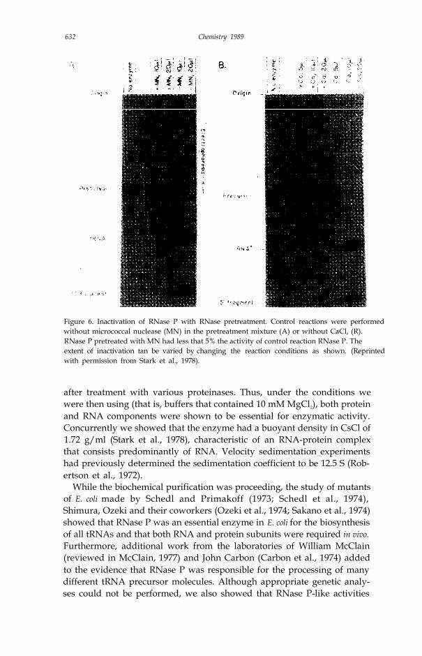
The essential role of the RNA component was established by first treatingRNase P with microccocal nuclease, an enzyme that destroys RNA, andsubsequently assaying the treated enzyme for RNase P activity: there wasnone after treatment with microccocal nuclease (Fig. 6) or, for that matter,
632 Chemistry 1989
Figure 6. Inactivation of RNase P with RNase pretreatment. Control reactions were performedwithout micrococcal nuclease (MN) in the pretreatment mixture (A) or without CaCl, (R).RNase P pretreated with MN had less that 5% the activity of control reaction RNase P. Theextent of inactivation tan be varied by changing the reaction conditions as shown. (Reprintedwith permission from Stark et al., 1978).
after treatment with various proteinases. Thus, under the conditions wewere then using (that is, buffers that contained 10 mM MgCl2), both proteinand RNA components were shown to be essential for enzymatic activity.Concurrently we showed that the enzyme had a buoyant density in CsCl of1.72 g/ml (Stark et al., 1978), characteristic of an RNA-protein complexthat consists predominantly of RNA. Velocity sedimentation experimentshad previously determined the sedimentation coefficient to be 12.5 S (Rob-ertson et al., 1972).
While the biochemical purification was proceeding, the study of mutantsof E. coli made by Schedl and Primakoff (1973; Schedl et al., 1974),Shimura, Ozeki and their coworkers (Ozeki et al., 1974; Sakano et al., 1974)showed that RNase P was an essential enzyme in E. coli for the biosynthesisof all tRNAs and that both RNA and protein subunits were required in vivo.Furthermore, additional work from the laboratories of William McClain(reviewed in McClain, 1977) and John Carbon (Carbon et al., 1974) addedto the evidence that RNase P was responsible for the processing of manydifferent tRNA precursor molecules. Although appropriate genetic analy-ses could not be performed, we also showed that RNase P-like activities
S. Altman 633
existed in the extracts of cells from many other organisms, including hu-mans (Altman and Robertson, 1973; Garber et al., 1978). These earlystudies showed that RNase P was capable of cleaving many different tRNAprecursor molecules and that there was no identifiable similarity in terms ofnucleotide sequence around the sites of cleavage. The manner in which theenzyme recognized its sites of cleavage in different substrates with suchselectivity seemed worthy of study, and recognition of some feature of thestructure in solution, common to all tRNA precursor molecules, was sus-pected.
When Stark’s experiments were published we did not have the temerity tosuggest, nor did we suspect, that the RNA component alone of RNase Pcould be responsible for its catalytic activity. The fact that an enzyme had anessential RNA subunit, in itself, seemed heretical enough. Shortly there-after, however, when Ryszard Kole demonstrated that the enzyme consistedof an RNA (Ml RNA) and a protein subunit (C5 protein; Mr ~ 14,000),which were not covalently linked and which could be separated into inactivesubunits and then reconstituted to form an active enzyme (Kole and Alt-man, 1979), the similarities in chemical composition and properties ofassembly of this system to that of the ribosome were sufficiently striking thatwe could not escape thinking about the possibility that the RNA, at the veryleast, participated in the formation of the active site of the enzyme. Indeed,making the comparison with ribosomes proved to be important in overcom-ing some resistance to the idea that an enzyme could have an RNA subunit.From a purely chemical point of view, there was no reason why RNA couldnot participate in formation of the active site (Kole and Altman, 1981) oreven in catalysis itself.
The advent of recombinant DNA technology and powerful systems forthe transcription in vitro of isolated pieces of DNA enabled us to character-ize in some detail the RNA subunit of RNase P (377 nucleotides in length;Reed et al., 1982) and to prepare large quantities for biochemical experi-ments. Concurrent progress in our purification of the protein subunitprepared us for a series of experiments, conducted in collaboration withNorman Pace’s group, in which we made hybrid enzymes with subunitsfrom E. coli (prepared in our laboratory) and from B. subtilis (prepared inPace’s laboratory). As an offshoot of these experiments, Cecilia Guerrier-Takada in my laboratory was testing reconstituted enzymes under ionicconditions optimal for the activity of the holoenzyme from B. subtilis anddifferent from the ones we had previously usually employed. She found, incontrol experiments, that the RNA subunit from E. coli, exhibited catalyticactivity of its own in buffers that contained 60 mM MgCl2 (An example ofsuch reactions is shown in Fig. 7; in fact the catalytic activity of Ml RNA isevident when the concentration of Mg2+ is greater than 20 mM; Guerrier-Takada et al., 1983). The protein subunit of the enzyme increased the bya factor of 10-20 but had little effect on the Km. These observations werepossible because of the purity of our preparations of Ml RNA and the useof a natural substrate, the precursor to tRNATyr from E. coli.
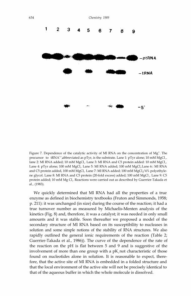
634 Chemistry 1989
Figure 7. Dependence of the catalytic activity of Ml RNA on the concentration of Mg2+. Theprecursor to tRNATyr,abbreviated as pTyr, is the substrate. Lane 1: pTyr alone; 10 mM MgCl 2.Iane 2: Ml RNA added; 10 mM MgCl2. Lane 3: Ml RNA and C5 protein added: 10 mM MgCl 2.Lane 4: pTyr alone; 100 mM MgCl2. Lane 5: Ml RNA added; 100 mM MgCl2.Lane 6:: Ml RNAand C5 protein added, 100 mM MgCl2. Lane 7: Ml RNA added; 100 mM MgCl2/4% polyethyle-ne glycol. Lane 8: Ml RNA and C5 protein (20-fold excess) added; 100 mM MgCl 2. Lane 9: C5protein added; 10 mM Mg Cl2. Reactions were carried out as described by Guerrier-Takada etal., (1983).
We quickly determined that Ml RNA had all the properties of a trueenzyme as defined in biochemistry textbooks (Fruton and Simmonds, 1958;p. 211): it was unchanged (in size) during the course of the reaction; it had atrue turnover number as measured by Michaelis-Menten analysis of thekinetics (Fig. 8) and, therefore, it was a catalyst; it was needed in only smallamounts and it was stable. Soon thereafter we proposed a model of thesecondary structure of Ml RNA based on its susceptibility to nucleases insolution and some simple notions of the stability of RNA structures. We alsorapidly outlined the general ionic requirements of the reaction (Table 2;Guerrier-Takada et al., 1986)). The curve of the dependence of the rate ofthe reaction on the pH is flat between 5 and 9 and is suggestive of theinvolvement of more than one group with a pKa not characteristic of thosefound on nucleotides alone in solution. It is reasonable to expect, there-fore, that the active site of Ml RNA is embedded in a folded structure andthat the local environment of the active site will not be precisely identical tothat of the aqueous buffer in which the whole molecule is dissolved.
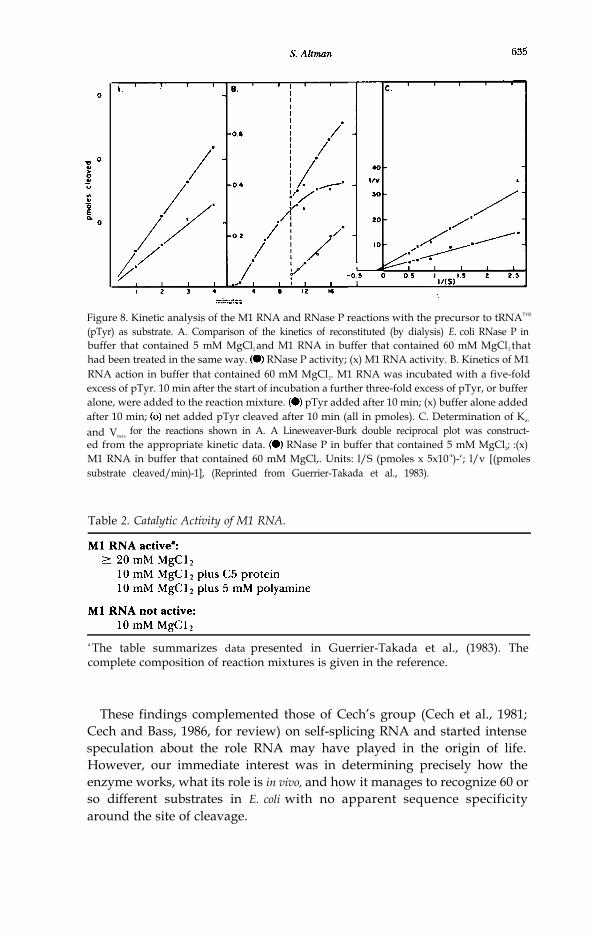
Figure 8. Kinetic analysis of the M1 RNA and RNase P reactions with the precursor to tRNATYR
(pTyr) as substrate. A. Comparison of the kinetics of reconstituted (by dialysis) E. coli RNase P inbuffer that contained 5 mM MgCl2 and M1 RNA in buffer that contained 60 mM MgCl2 thathad been treated in the same way. RNase P activity; (x) M1 RNA activity. B. Kinetics of M1RNA action in buffer that contained 60 mM MgCl2. M1 RNA was incubated with a five-foldexcess of pTyr. 10 min after the start of incubation a further three-fold excess of pTyr, or bufferalone, were added to the reaction mixture. pTyr added after 10 min; (x) buffer alone addedafter 10 min; net added pTyr cleaved after 10 min (all in pmoles). C. Determination of Km
and Vmaxfor the reactions shown in A. A Lineweaver-Burk double reciprocal plot was construct-
ed from the appropriate kinetic data. RNase P in buffer that contained 5 mM MgCl2; :(x)M1 RNA in buffer that contained 60 mM MgCl,. Units: l/S (pmoles x 5x10⁻⁴)-‘; l/v [(pmolessubstrate cleaved/min)-1], (Reprinted from Guerrier-Takada et al., 1983).
Table 2. Catalytic Activity of M1 RNA.
a The table summarizes data presented in Guerrier-Takada et al., (1983). Thecomplete composition of reaction mixtures is given in the reference.
These findings complemented those of Cech’s group (Cech et al., 1981;Cech and Bass, 1986, for review) on self-splicing RNA and started intensespeculation about the role RNA may have played in the origin of life.However, our immediate interest was in determining precisely how theenzyme works, what its role is in vivo, and how it manages to recognize 60 orso different substrates in E. coli with no apparent sequence specificityaround the site of cleavage.

636 Chemistry 1989
RECENT WORK
StructureThe original model of the secondary structure of M1 RNA has been exten-sively refined by phylogenetic analysis (Fig. 9) carried out primarily by Paceand coworkers (James et al., 1988). However, this analysis has not yetyielded a satisfactory correlation between the phenotypes of mutants (Lu-melsky and Altman, 1988) and features of the secondary structure of M1RNA or its analogs from other bacteria (see below). It does provide the basisfor hypotheses about the regions of M1 RNA that are essential for function(Waugh et al., 1989), as indicated by evolutionary conservation, and it
Figure 9. A model for the secondary structure of M1 RNA based on extensive phylogeneticanalysis of the nucleotide sequence of the RNA subunit of RNase P from several eubacteria.(Reprinted with permission from James et al., 1988).

S. Altmn 637
highlights the necessity of determining the three-dimensional features ofthe structure. To this end, additional phylogenetic comparisons, utilizingthe data concerning the homolog of M1 RNA from several eucaryoticspecies (Miller and Martin, 1983; Krupp et al., 1986; Lee and Engelke,1989), and crystallographic studies are in progress. One observation ofcontinuing interest from these studies is that the evolutionary clock for boththe RNA and protein subunits of RNase P seems to be a very fast one incomparison with that for rRNAs (Lawrence et al., 1987; Gold, 1988).Although the function of RNase P, as judged by the antigenic properties ofthe protein (Gold et al., 1988; Mamula et al., 1989), its ability to cleavevarious substrates and to reconstitute active enzyme with subunits fromdifferent organisms (Guerrier-Takada et al., 1983; Gold and Altman, 1986;Lawrence et al., 1987) has been highly conserved, the nucleotide sequencesof the genes for the subunits of the enzyme have drifted extremely rapidly(Gold, 1988; Bartkiewicz et al., 1989).
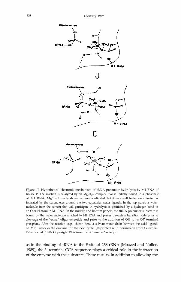
MechanismThe detailed mechanism of the reaction catalyzed by RNase P is not knownbut two proposals have been made. In one case (Guerrier-Takada et al.,1986), a variation of the SN2 in-line displacement mechanism has beensuggested in which a complex between one Mg ion and six water moleculesfacilitates the nucleophilic action of a water molecule in solution (Fig. 10).Investigations of the rRNA self-splicing reaction in Tetrahymenu in TomCech’s laboratory indicate that the original proposal for the mechanism ofthe RNase P reaction may also be relevant in the self-splicing reaction(Cech, 1987; McSwiggen and Cech, 1989). In the other proposal for themechanism of the RNase P reaction (Reich et al., 1988), the nucleophile isderived from groups on the surface of the enzyme and the role of themagnesium ion is not as clearly specified. Attempts are underway in ourlaboratory to test the first model, by the insertion of a phosphothioate bondat the cleavage site and analysis of the stereochemistry of the cleavedproduct.
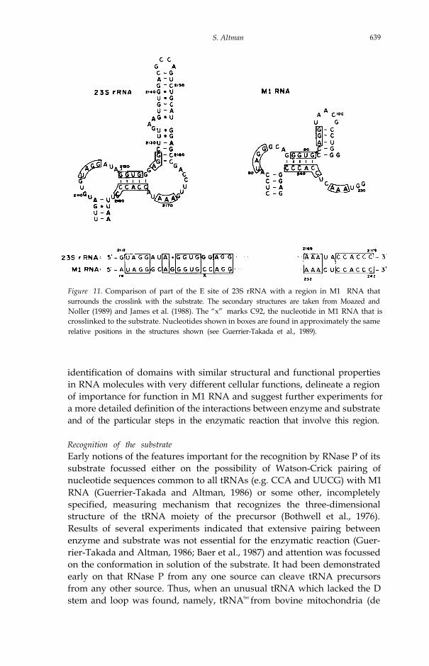
While many aspects of structure-function relationships and clues to themechanism of the reaction may be revealed if the crystal structure of theenzyme becomes available, the determination of a crystal structure mayprove to be elusive. We have, therefore, embarked on an attempt to identifyregions of Ml RNA that are critical for the reaction by cross-linking thesubstrate to the enzyme by irradiation with ultraviolet light. Such experi-ments have revealed that a cross-link is formed between a nucleotide closeto the site of cleavage in the substrate (C -3) and residue C92 in M1 RNA(Guerrier-Takada et al., 1989). If C92 is deleted from M1 RNA, the kineticsof the enzymatic reaction, and its site of cleavage with particular substrates,are significantly altered. Furthermore, the region of secondary structurearound C92 in M1 RNA resembles that of the tRNA E site in 23S rRNA(Fig. 11). Additional studies have shown that this site is important in thebinding of the aminoacyl stem of a tRNA precursor to the enzyme and that,
638 Chemistry 1989
Figure 10. Hypothetical electronic mechanism of tRNA precursor hydrolysis by M1 RNA ofRNase P. The reaction is catalyzed by an Mg-H2O complex that is initially bound to a phosphateof M1 RNA. Mg2+ is formally shown as hexacoordinated, but it may well be tetracoordinated asindicated by the parentheses around the two equatorial water ligands. In the top panel, a watermolecule from the solvent that will participate in hydrolysis is positioned by a hydrogen bond toan O or N atom in M1 RNA. In the middle and bottom panels, the tRNA precursor substrate isbound by the water molecule attached to M1 RNA and passes through a transition state prior tocleavage of the “extra” oligonucleotide and prior to the addition of OH to its O5’ terminalphosphate. After the reaction steps shown here, a solvent water chain between the axial ligandsof Mg2+ recocks the enzyme for the next cycle. (Reprinted with permission from Guerrier-Takada et al., 1986. Copyright 1986 American Chemical Society).
as in the binding of tRNA to the E site of 23S rRNA (Moazed and Noller,1989), the 3’ terminal CCA sequence plays a critical role in the interactionof the enzyme with the substrate. These results, in addition to allowing the
S. Altman 639
Figure 11. Comparison of part of the E site of 23S rRNA with a region in M1 RNA thatsurrounds the crosslink with the substrate. The secondary structures are taken from Moazed andNoller (1989) and James et al. (1988). The “x” marks C92, the nucleotide in M1 RNA that iscrosslinked to the substrate. Nucleotides shown in boxes are found in approximately the samerelative positions in the structures shown (see Guerrier-Takada et al., 1989).
identification of domains with similar structural and functional propertiesin RNA molecules with very different cellular functions, delineate a regionof importance for function in M1 RNA and suggest further experiments fora more detailed definition of the interactions between enzyme and substrateand of the particular steps in the enzymatic reaction that involve this region.
Recognition of the substrateEarly notions of the features important for the recognition by RNase P of itssubstrate focussed either on the possibility of Watson-Crick pairing ofnucleotide sequences common to all tRNAs (e.g. CCA and UUCG) with M1RNA (Guerrier-Takada and Altman, 1986) or some other, incompletelyspecified, measuring mechanism that recognizes the three-dimensionalstructure of the tRNA moiety of the precursor (Bothwell et al., 1976).Results of several experiments indicated that extensive pairing betweenenzyme and substrate was not essential for the enzymatic reaction (Guer-rier-Takada and Altman, 1986; Baer et al., 1987) and attention was focussedon the conformation in solution of the substrate. It had been demonstratedearly on that RNase P from any one source can cleave tRNA precursorsfrom any other source. Thus, when an unusual tRNA which lacked the Dstem and loop was found, namely, tRNASer from bovine mitochondria (de
640 Chemistry 1989
Bruijn and Klug, 1983), we examined whether M1 RNA could cleave ananalog of a precursor to that tRNA.
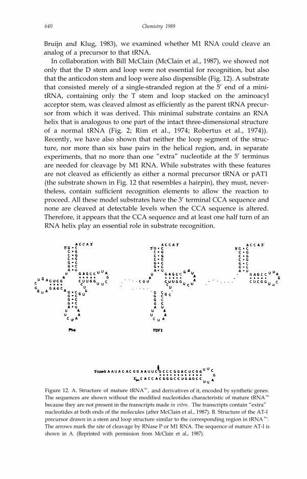
In collaboration with Bill McClain (McClain et al., 1987), we showed notonly that the D stem and loop were not essential for recognition, but alsothat the anticodon stem and loop were also dispensible (Fig. 12). A substratethat consisted merely of a single-stranded region at the 5’ end of a mini-tRNA, containing only the T stem and loop stacked on the aminoacylacceptor stem, was cleaved almost as efficiently as the parent tRNA precur-sor from which it was derived. This minimal substrate contains an RNAhelix that is analogous to one part of the intact three-dimensional structureof a normal tRNA (Fig. 2; Rim et al., 1974; Robertus et al., 1974)).Recently, we have also shown that neither the loop segment of the struc-ture, nor more than six base pairs in the helical region, and, in separateexperiments, that no more than one “extra” nucleotide at the 5’ terminusare needed for cleavage by M1 RNA. While substrates with these featuresare not cleaved as efficiently as either a normal precursor tRNA or pAT1(the substrate shown in Fig. 12 that resembles a hairpin), they must, never-theless, contain sufficient recognition elements to allow the reaction toproceed. All these model substrates have the 3’ terminal CCA sequence andnone are cleaved at detectable levels when the CCA sequence is altered.Therefore, it appears that the CCA sequence and at least one half turn of anRNA helix play an essential role in substrate recognition.
Figure 12. A. Structure of mature tRNAPhe, and derivatives of it, encoded by synthetic genes.The sequences are shown without the modified nucleotides characteristic of mature tRNA Phe
because they are not present in the transcripts made in vitro. The transcripts contain “extra”nucleotides at both ends of the molecules (after McClain et al., 1987). B. Structure of the AT-lprecursor drawn in a stem and loop structure similar to the corresponding region in tRNA Phe.The arrows mark the site of cleavage by RNase P or M1 RNA. The sequence of mature AT-l isshown in A. (Reprinted with permission from McClain et al., 1987).
641S. Altman
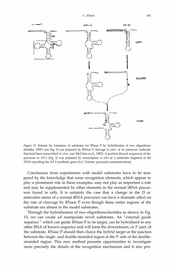
Figure 13. Scheme for formation of substrates for RNase P by hybridization of two oligoribonu-rleotides. TDF-I (see Fig. 3) was prepared by RNAse P cleavage in vitro of its precursor moleculethat had been transcribed in vitro (see McClain et al., 1987). A portion (boxed sequence) of theprecursor to AT-1 (Fig. 3) was prepared by transcription in vitro of a restriction fragment of theDNA encoding the AT-I synthetic gene (A.C. Forster, personal communication).
Conclusions from experiments with model substrates have to be tem-pered by the knowledge that some recognition elements, which appear toplay a prominent role in these examples, may not play as important a roleand may be supplemented by other elements in the normal tRNA precur-sors found in cells. It is certainly the case that a change in the D oranticodon stems of a normal tRNA precursor can have a dramatic effect onthe rate of cleavage by RNase P even though these entire regions of thesubstrate are absent in the model substrates.
Through the hybridization of two oligoribonucleotides as shown in Fig.13, we can create ad manipulate novel substrates. An “external guidesequence ” which can guide RNase P to its target, can be hybridized to anyother RNA of known sequence and will form the downstream, or 3’ part, ofthe substrate. RNase P should then cleave the hybrid target at the junctionbetween the single- and double-stranded region at the 5’ side of the double-stranded region. This new method presents opportunities to investigatemore precisely the details of the recognition mechanism and it also pro-

642 Chemistry 1989
Figure 14. Targeting of RNA for cleavage by RNase P. An external guide sequence (EGS) isshown by the shorter line ending in NCCA. N is most frequently found to be A in tRNAmolecules. The region of the EGS shown as hydrogen-bonded is designed to be complementaryto a region of known sequence in the RNA to be targeted.
vides, in principle, a means to inactivate any mRNA of known sequence invivo. Aside from the problem of the expression of the external guidesequence in vivo, the method does have the advantage that RNase P isalready present in cells of all types. Providing that the hybrid can bedesigned to be compatible with the cleavage-site specificity of the enzyme inthe particular host organism of choice, the target RNA should be inactivat-ed.
In this example of the use of one oligoribonucleotide to target anotherRNA that is to be cleaved by RNase P, substrate recognition by the enzymeresembles, in a formal sense, selection of the site of cleavage by some of theother known RNA catalysts. Group I introns and the satellite and similarRNAs use guide sequences (Cech, 1987; Altman, 1989, for reviews) in theselection of cleavage sites or to form structures in which a cleavage sitebecomes defined. In virtually all other respects, these reactions are quitedistinct from that carried out by RNase P.
The Past, Present and Future of RNase PThe discovery of RNA catalysis has led to new hypotheses about the originof the earliest self-replicating biochemical systems from which the questionof the origin of the genetic code can be excluded. Models of these earlysystems rely entirely on RNA as the genetic material and as the source ofcatalytic activity (Fig. 15; Darnell and Doolittle, 1986; Weiner and Maizels,1987; Joyce, 1989). All this speculation clearly presupposes that what we seein present-day systems reflects, in some manner the properties of RNA over
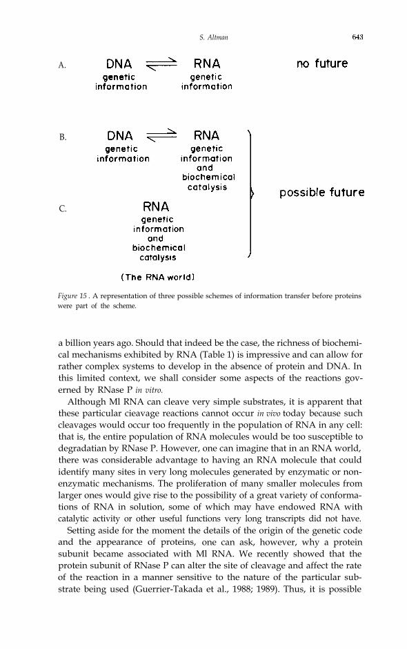
S. Altman
A.
B.
C.
Figure 15 . A representation of three possible schemes of information transfer before proteinswere part of the scheme.
a billion years ago. Should that indeed be the case, the richness of biochemi-cal mechanisms exhibited by RNA (Table 1) is impressive and can allow forrather complex systems to develop in the absence of protein and DNA. Inthis limited context, we shall consider some aspects of the reactions gov-erned by RNase P in vitro.
Although Ml RNA can cleave very simple substrates, it is apparent thatthese particular cieavage reactions cannot occur in vivo today because suchcleavages would occur too frequently in the population of RNA in any cell:that is, the entire population of RNA molecules would be too susceptible todegradatian by RNase P. However, one can imagine that in an RNA world,there was considerable advantage to having an RNA molecule that couldidentify many sites in very long molecules generated by enzymatic or non-enzymatic mechanisms. The proliferation of many smaller molecules fromlarger ones would give rise to the possibility of a great variety of conforma-tions of RNA in solution, some of which may have endowed RNA withcatalytic activity or other useful functions very long transcripts did not have.
Setting aside for the moment the details of the origin of the genetic codeand the appearance of proteins, one can ask, however, why a proteinsubunit became associated with Ml RNA. We recently showed that theprotein subunit of RNase P can alter the site of cleavage and affect the rateof the reaction in a manner sensitive to the nature of the particular sub-strate being used (Guerrier-Takada et al., 1988; 1989). Thus, it is possible
644 Chemistry 1989
that proteins may have fine-tuned the site specificity of RNA enzymes byenhancing the rates of reaction at particular sites and with particularsubstrates. What we see today as the “normal” cleavage sites of RNAenzymes may have been selected for over the eons, in conjunction with theappearance of protein cofactors, as physiological conditions changed dur-ing evolutionary time. The “unselected” reactions, for example those withvery small hairpin substrates, became in consequence second- or lower-order reactions and are no longer relevant to events in vivo.
Finally, why do RNA enzymes only cleave phosphodiester bonds? Thereare three answers that come readily to mind. First and most trivially, RNAenzymes may cleave other classes of bonds and we just have not yet madethe critical observations or found the right reaction conditions (the last partof the answer is a generic response to questions about the lack of success inperforming certain reactions in vitro). Second, it is possible that, in the RNAworld, perhaps RNA molecules could only cleave phosphodiester bonds: itwas a primitive world and no other reactions were governed by enzymes.Once proteins appeared on the scene there was no further need to diversifyRNA enzymes. Lastly, and most important, the chemistry of RNA enzymes,when sufficiently well-understood, may indicate to us that there is a compel-ling reason why RNA molecules cleave only phosphoester bonds. Thevalidity, or lack of it, of this last answer can be tested by direct experimenta-tion, and therein lies the work of the next several years.
ACKNOWLEDGEMENTS
My indebtedness to so many people makes it impractical to list them allhere. Nevertheless, I wish to express my gratitude to my parents, my family,my teachers (especially Leonard Lerman, Mathew Meselson, Sydney Bren-ner and Lee Grodzins), my professional colleagues and collaborators (espe-cially, Hugh Robertson and Bill McClain) and my students and coworkers inmy laboratory (especially Cecilia Guerrier-Takada, Ben Stark, Ryszard Kole,Robin Reed and Madeline Baer). They have all tolerated my bouts ofobsessiveness and have shared the moments of discovery and pleasure. Thetaxpayers of the United States, through the agencies of the National Insti-tutes of Health and the National Science Foundation, have generouslysupported my work.
S. Altman 645
REFERENCES
Abelson, J. N., Gefter, M. L., Barnett, L., Landy, A. and Russell, R. L. (1970) J. Mol.Biol. 47 15-28.
Attman, S. (1971) Nature New Biology 229 19-21.Attman, S. (1989) Adv. Enzymol., ed A. Meister (J. Wiley, NY) Vol. 62, pp. 1-36.Attman, S. and J. D. Smith (1971) Nature New Biology 233 35-39.Attman, S. and Robertson, H. D. (1973) Molec. Cell. Biochem. 1 83-93.Baer, M. F., Reilly, R. M., McCorkle, G. M., Hai, T-Y, Altman, S. and RajBhandary,
U. I.. (1989) J. Biol. Chem. 26.3 2344-2351.Bartkiewicz, M., Gold, H. and Altman, S. (1989) Genes and Development 3 488-
499.Bernhardt, D. and Darnell, J. E. (1967) J. Mol. Biol. 42 43-56Bothwell, A. I.. M., Stark, B. C. and Altman, S. (1976) Proc. Nat. Acad. Sci. USA 73
1912-1916.Burdon, R. H. (1971) Prog. Nucl. Acids Res. Mol. Biol. 11 33 - 79.Burdon, R. H. (1974) Brookhaven Symp. Biol. 26 138-153.Carbon, J., Chang, S. and Kirk, L. L. (1974) Brookhaven Symp. Biol. 26 26-36.Cech, T. R. (1987) Science 236 1532-1539.Cech, T. R. and Bass, B.L. (1986) Annu. Rev. Biochem. 55 599-629.Cech, T. R., Zaug, A. J. and Grabowski, P. J. (1981) Cell 27 487-496.Chang, D.D. and Clayton, D.A. (1987) Science 235 1178-1184.Crick, F. (1968) J. Mol. Biol. 38 367-379.Crick, F. (1970) Nature 277 561-563.Darnell, J. E. and Doolittle, W. F. (1986) Proc. Natl. Acad. Sci. USA 83 1271-1275.de Bruijn, M. H. L. and Klug, A. (1983) EMBO J. 2 1309-1321.Fruton, J., and Simmonds, S. (1958) General Biochemistry, 2nd ed. (New York: J.
Wiley and Sons).Garber, R. I.., Siddiqui, M. A. Q. and Altman, S. (1978) Proc. Nat. Acad. Sci. USA.
75 635-639.(Gilbert, W. (1986) Nature 319 618.Gold, H. A. (1988) Ph.D. Thesis, Yale University, New Haven, CT USA.Gold, H. A. and Altman, S. (1986) Cell 44 243-249.Gold, H. A., Craft, J., Hardin, J. A., Bartkiewicz, M. and Altman, S. (1988) Proc.
Nat. Acad. Sci. USA 85 5483-5487.Guerrier--Takada, C. and Altman, S. (1984) Biochemistry 23 6327-6334.Guerrier-Takada, C. and Altman, S. (1986) Cell 45 l77-183.Guerrier-Takada, C. Gardiner, K., Marsh, T., Pace, N., and Altman, S. (1983) Cell
35 849-857.Guerrier-Takada, C., Haydock, K., Allen, L. and Altman, S. (1986) Biochemistry 25
1509-1515.Guerrier-Takada, C., Knap, A. K., Lumetsky, N. and Altman, S. (1989) Science, 246
1578-1584.Guerrier-Takada, C., van Betkum, A., Pleij, C. W. A. and Altman, S. (1988) Cell 53
267-272 .Guthrie, C., Seidman, J. G., Altman, S., Barrett, B. G., Smith, J. D. and McClain, W.
H. (1973) Nature New Biol. 246 6-11.tkemura, T. and Dahlberg, J. E. (1973) J. Biol. Chem. 248 5024-5032.James, B., Olsen, G. J., Lin, J. and Pace, N. (1988) Cell 52 19-26.Joyce, G. F. (1989) Nature 338 217-223.Kim, S. H., Suddath, F. L., Quigtey, G. J., McPherson, A., Sussman, J. L., Wang, A.
H. J., Seeman, N. C. and Rich, A. (1974) Science 185 435-440.Kote, R. and Altman, S. (1979) Proc. Natl. Acad. Sci. USA 76 3795-3799.Kole, R. and Altman, S. (1981) Biochem. 20 1902-1906.
646 Chemistry 1989
Krupp, G., Cherayil, B., Frendeway, D., Nishikawa, S. and Soll. D. (1986) EMBO J. 51697-1703.
Lawrence, N. P., Richman, A., Amini, R. and Altman, S. (1987) Proc. Natl. Acad.Sci. USA 84 6825 - 6829.
Lee, J-Y., and Engelke, D. R. (1989) Molec. Cell. Biol. 9 2536-2543.Lumelsky, N. and Altman, S. (1988) J. Mol. Biol. 202 443-454.Mamula, M.J., Baer, M., Craft, J. and Altman, S. (1989) Proc. Nat. Acad. Sci. USA,
86 8717-8721.McClain, W. H. (1977) Accts. Chem. Res. 10 418-425.McClain, W. H., Guerrier-Takada, C. and Altman, S. (1987) Science 238 527-530.McSwiggen, J. A. and Cech, T. R. (1989) Science 244 679-683.Miller, D. L. and Martin, N. C. (1983) Cell 34 91 1-917.Moazed, D. and Noller, H. F. (1989) Cell 57 585-597.Orgel, L. (1968) J. Mol. Biol. 38 381-393.Ozeki, H., Sakano, H., Yamada, S., Ikemura, T. and Shimura, Y. (1974) Brookhaven
Symp. Biol. 26 89-105.Reed, R., Baer, M., Guerrier-Takada, C., Donis-Keller, H. and Altman, S. (1982)
Cell 30 627-636.Reich, C. I., Olsen, G. J., Pace, B. and Pace, N. R. (1988) Science 239 178-181.Robertson, H. D. (1986) Nature 322 16-17.Robertson, H. D., Altman, S. and Smith, J. D. (1972) J. Biol. Chem. 247 5243-
5251.Robertus, J. D., Ladner, J. E., Finch, J. T., Rhodes, D., Brown, R. S., Clark, B. F. C.
and Klug, A. (1974) Nature 250 546-55 1.Russell, R. L., Abelson, J. N., Landy, A., Gefter, M. L., Brenner, S. and Smith, J. D.
(1970) J. Mol. Biol. 47 1-13.Sakano, H., Yamada, S., Ikemura, T., Shimura, Y. and Ozeki, H. (1974) Nucleic
Acids Res. 1 355-371.Schedl, P., and Primakoff, P. (1973) Proc. Natl. Acad. Sci. USA 70 2091-2095.Schedl, P., Primakoff, P. and Roberts, J. (1974) Brookhaven Symp. Biol. 26 53-76.Smith, J. D., Barnett, L., Brenner, S. and Russell, R.L. (1971) J. Mol. Biol. 54 1-14.Stark, B. C., Kole, R., Bowman, E. J. and Altman, S. (1978) Proc. Natl. Acad. Sci.
USA 75 3717-3721.Waugh, D. S., Green, C. J. and Pace, N. R. (1989) Science 244 1569-1571.Weiner, A. M. and Maizels, N. (1987) Proc. Nat. Acad. Sci. USA 84 7383-7387.Westheimer, F. H. (1986) Nature 319 534-535.Woese, C. R. (1967) The Origins of the Genetic Code (Harper and Row, New York)


![Education Strategies for Identifying RNA Splicing ...vorgogoz/articles/D.pachea... · this with other steps in RNA processing, such as capping, cleavage, and polyadenylation [6]](https://img.pdfslide.us/doc/110x75/5f0253b47e708231d403b8cd/education-strategies-for-identifying-rna-splicing-vorgogozarticlesdpachea.jpg)