Embed Size (px)
Citation preview
1
A new type of signal peptidase cleavage site identified in an RNA virus polyprotein
Ioana Bintintan and Gregor Meyers*
From the Institut für Immunologie, Friedrich-Loeffler-Institut, D-72001 Tübingen
Running Title: Processing of pestivirus Erns
E1
Address correspondence to Gregor Meyers, Institut für Immunologie, Friedrich-Loeffler-Institut, Paul-
Ehrlich-Str. 28, D-72076 Tübingen, Germany; Tel. +49 7071-9670; Fax. +49 7071-967303; E-mail:
Pestiviruses, a group of enveloped positive
strand RNA viruses belonging to the family
Flaviviridae, express their genes via a
polyprotein that is subsequently processed
by proteases. The structural protein region
contains typical signal peptidase cleavage
sites. Only the site at the carboxyterminus of
the glycoprotein Erns
is different since it does
not contain a hydrophobic transmembrane
region but an amphipathic helix functioning
as the Erns
membrane anchor. Despite the
absence of a hydrophobic region the site
between the carboxyterminus of Erns
and
E1, the protein located downstream in the
polyprotein, is cleaved by signal peptidase
as demonstrated by mutagenesis and
inhibitor studies. Thus, Erns
E1 is processed
at a novel type of signal peptidase cleavage
site showing a different membrane topology.
Prevention of glycosylation or introduction
of mutations into the carboxyterminal
region of Erns
severely impair processing
presumably by preventing proper
membrane interaction or disturbing a
conformation critical for the protein to be
accepted as a substrate by signal peptidase.
INTRODUCTION
Classical swine fever virus (CSFV) belongs
to the genus Pestivirus that also includes the
animal viruses bovine viral diarrhea virus
(BVDV) and border disease virus of sheep.
The genus Pestivirus is part of the family
Flaviviridae which also comprises the genera
Flavivirus and Hepacivirus (1).
Pestiviruses are positive strand RNA-
viruses with a single stranded genome of ~12.3
kb length that contains a single open reading
frame coding for a polyprotein of about 4000
amino acids (2). The polyprotein is co- and
posttranslationally processed by cellular and
viral proteases into at least 12 mature proteins
(3-12), arranged in the polyprotein in the order
NH2-Npro
, C, Erns
, E1, E2, p7, NS2, NS3,
NS4A, NS4B, NS5A, and NS5B-COOH. C,
Erns
, E1 and E2 are part of the virion (13,14),
with C forming the capsid, and Erns
, E1 and E2
representing glycoproteins associated with the
virus envelope. Erns
and E2 elicit neutralizing
antibody responses that can lead to protective
immunity (15-18).
The pestivirus Erns
protein is a highly
unusual protein. It forms a disulfide-linked
dimer of ~90 kDa, about half of which is due
to glycosylation (9,13,19). The protein displays
homology to the RNases of the T2/S
superfamily (20-22). In different test systems it
was shown that Erns
indeed has RNase activity,
a feature that is unique among viral
glycoproteins (22-25). The protein is essential
for virus growth (21), but the RNase activity is
dispensable. Viruses, in which the RNase
activity has been knocked out, are clinically
attenuated (26,27). A role for Erns
in immune
evasion has been proposed (28-31).
http://www.jbc.org/cgi/doi/10.1074/jbc.M109.083394The latest version is at JBC Papers in Press. Published on January 21, 2010 as Manuscript M109.083394
Copyright 2010 by The American Society for Biochemistry and Molecular Biology, Inc.
by guest on February 11, 2018http://w
ww
.jbc.org/D
ownloaded from
2
Erns
can be found in virus free cell culture
fluid of infected cells, and in the blood of
infected animals (9,29,32). It lacks a
hydrophobic region that could serve as a
transmembrane anchor and there is also no
consensus sequence for GPI anchor addition. It
is nevertheless bound to the virion and
associated with intracellular membranes
(9,13,14,32,33). Erns
membrane binding is
achieved by the carboxyterminal region
forming an amphipathic helix that is inserted in
plane into the membrane (32-34).
The topology of the Erns
membrane anchor
raises interesting questions concerning the
protease that cleaves the polyprotein at the Erns
C-terminus. Processing of the region of the
polyprotein encompassing the viral structural
proteins and the first two nonstructural proteins
p7 and NS2 is attributed to cellular proteases.
The processing sites at the aminotermini of
Erns
, E2, p7 and NS2 comply to the demands
for signal peptidase cleavage sites, namely a
positively charged N-terminal (n-) region and a
central hydrophobic (h-) region, followed by a
more hydrophilic part (c-region) containing the
cleavage site with small and uncharged
residues at positions -3 and -1 as suggested by
von Heijne (35,36). The results of sequence
and mutagenesis analyses clearly support the
idea of SPase cleavage at these sites in the
viral polyprotein [(5,6,9) and 2]. In contrast,
the site separating Erns
and E1 does not comply
with the above described features of a SPase
cleavage site. The -3/-1 residues at the Erns
/E1
cleavage site are in agreement with a sequence
processed by SPase, but a hydrophobic region
upstream of the cleavage site is missing. This
fact raises the question, whether SPase cleaves
this site as proposed in some reports or which
other protease is responsible for processing the
polyprotein here.
In the work presented here we analyzed the
cleavage at the Erns
carboxyterminus with
different approaches including mutagenesis
and inhibitor studies. For the first time, we
provide clear evidence that processing between
Erns
and E1 is executed by SPase which reveals
the existence of a new type of SPase cleavage
site not described so far.
EXPERIMENTAL PROCEDURES
Cells and viruses. BHK-21 cells (kindly
provided by T. Rümenapf) were grown in
Dulbecco’s modified Eagle’s medium
supplemented with 10% fetal calf serum and
nonessential amino acids. The modified
vaccinia virus strain Ankara containing the
phage T7 RNA polymerase (MVA-T7) (37)
was kindly provided by B. Moss (National
Institutes of Health, Bethesda, Md.).
Construction of recombinant plasmids.
Restriction and subcloning were done
according to standard procedures (38). Unless
stated otherwise, all restriction and modifying
enzymes were purchased from New England
Biolabs (Frankfurt, Germany) and Fermentas
GmbH (Sankt Leon-Roth, Germany). Plasmid
pCITE 2a(+) was obtained from AGS
(Heidelberg, Germany). It contains a T7 RNA
polymerase promotor followed by a
picornavirus IRES. The synthetic DNA
oligonucleotides were purchased from
Metabion (München, Germany).
From the infectious cDNA clone of the CSFV
strain Alfort/Tübingen (39) coding sequences
were amplified with the pairs of primers NcoI
(neu) / E1-3SXbaR, NcoI (neu) / E0-3SXbaI,
E05SIII / E1-3SXbaR, E05SIII / E0-3SXbaI
and inserted into the NcoI/XbaI sites of pCITE
2a(+) yielding the constructs Npro
-E1, Npro
-Erns
,
SSeqErns
-E1 and SSeqErns
, respectively.
To obtain construct SSeqE1-E2, the cDNA of
E1E2 was amplified from plasmid p578 (39).
In addition, the coding sequence of the Erns
signal peptide (SSeq) [nucleotides 1120 to
1173 of the full length cDNA clone pA/CSFV
(39)], was also incorporated in the SSeqErns
-E1
and SSeqErns
constructs. This sequence was
amplified by PCR with the primers IB72, IB73
and E05SIII as follows. First, a PCR fragment
by guest on February 11, 2018http://w
ww
.jbc.org/D
ownloaded from
3
was obtained from plasmid p578 with the
primers IB72 and HPS38.2. This PCR product
was then amplified with IB73 and HPS 38.2
and used for the generation of a further PCR
fragment by amplification with E05SIII and E2
d(-). This PCR fragment was restricted with
NcoI and XbaI and inserted into the NcoI/XbaI
sites of pCITE 2a(+).
The construct mellSSeqErns
-E1 containing the
signal peptide of mellitin (mellSSeq) was
generated in a similar way as described above.
The coding sequence of the mellitin signal
peptide was obtained from Qiagen (Hilden,
Germany) and amplified in three steps by PCR
as described before. First, a PCR fragment was
amplified from plasmid SSeqErns
-E1 with the
primers IB131 and pCITErev. This PCR
product was amplified with IB132 and
pCITErev to obtain the second PCR fragment
that contains part of the mellSSeq-coding
sequence. The rest of the coding sequence was
introduced by the third PCR with the second
PCR fragment as template and the primers
IB133 and E1-3SXbaR. The final product was
cut with NcoI and XbaI and inserted into
pCITE 2a(+), cut with NcoI/XbaI.
The constructs ppLSSeqErns
-E1 and
ppLSSeqErns
-E1* are also based on SSeqErns
-
E1, with V5 tag and preprolactin signal peptide
preceding Erns
E1. The sequence coding for the
preprolactin signal peptide coding sequence
(ppLSSeq) was amplified from a preprolactin
vector (kindly provided by Heiner Niemann)
by PCR with the primers IB97 and IB98 for
ppLSSeqErns
-E1 or with IB97 and IB100 for
ppLSSeqErns
-E1*. In order to introduce the
sequence coding for a V5 tag, the resulting
PCR fragments were used as templates in a
further PCR with primers IB94 and IB98 for
ppLSSeqErns
-E1 or IB94 and IB100 for
ppLSSeqErns
-E1*. The resulting PCR products
were cut with NcoI and EcoRI. The primers
IB99 and E1-3SXbaR were used to amplify the
Erns
E1-coding fragment from the SSeqErns
-E1
plasmid. After restriction with EcoRI and
XbaI, the resulting fragment was ligated
together with the previously obtained fragment
coding for V5-ppLSSeq and with pCITE 2a(+),
cut with NcoI/XbaI.
QuikChange mutagenesis (Stratagene,
Heidelberg, Germany) was employed
according to the suppliers instructions to
introduce substitutions, insertions or deletions.
The constructs SSeqErns
-E1 and Npro
-Erns
served as templates for all mutagenesis
approaches for characterisation of the Erns
/E1
cleavage site.
The cloned PCR products were all verified by
nucleotide sequencing with the BigDye
Terminator Cycle Sequencing Kit (PE Applied
Biosystems, Weiterstadt, Germany). Sequence
analysis and alignments were done with
Genetics Computer Group software (40).
Further details of the cloning procedures and
the sequences of the primers used for cloning
and mutagenesis are available on request.
Transient expression of proteins. BHK-21
cells were infected with vaccinia virus MVA-
T7, subsequently transfected with the desired
cDNA construct using SuperFect (Qiagen) and
labeled with 35
S amino acids as described
earlier (32,33). Where mentioned, tunicamycin
(Boehringer Mannheim, Germany) or BFA
(Calbiochem, Bad Soden, Germany) were
added to the medium during starving and
labeling periods to a concentration of 10 µg/ml
(tunicamycin) or 1 µg/ml (BFA).
Preparation and fractionation of cell
extracts. Crude extracts of transiently
transfected cells were prepared under
denaturing conditions as described before (32).
Alternatively, transfected BHK-21cells were
harvested via fractionation essentially as
described (32,33,41). Briefly, the tissue culture
fluid of infected cells containing the secreted
proteins was removed as the first fraction. The
cells were harvested by scraping them into 1.5
ml of PBS and then passaged 10 times through
a 27-gauge needle. Nuclei and cell debris
obtained by low-speed centrifugation (700 x g,
3 min) were collected in the pellet, and the
supernatant of this centrifugation step was used
by guest on February 11, 2018http://w
ww
.jbc.org/D
ownloaded from
4
to recover the membrane fraction by high-
speed centrifugation (107,000 x g; rotor
TLA100.3; Beckmann TL100 centrifuge, for
25 min). The water-soluble proteins were
found in the supernatant of this centrifugation
step. All samples were resuspended in 1x RIP
buffer (20mM Tris, 100mM NaCl, 1mM
EDTA, 1% Triton X-100, 0.1% DOC, 0.1%
SDS, 1 mg/l BSA, pH 7.6), denatured by the
addition of 1% SDS and heated to 95°C before
they were chilled on ice. After sonication, the
samples were brought to the final
concentration of 0.3% SDS by the addition of
1x RIP buffer and were used further in
immunoprecipitation experiments.
Immunoprecipitation and quantification of
Erns
. Immunoprecipitation of proteins was
carried out as described (32) with 5 µl rabbit
Erns
antiserum or 100 µl mab 24/16 (anti-Erns
).
After incubation of samples with
Staphylococcus aureus (42), the bound
immune complexes were eluted in sample
buffer by heating to 95°C. The S. aureus
bacteria were removed by centrifugation, and
the supernatants were analyzed by 10% SDS-
PAGE. Where specified in the text, the
precipitated proteins were treated before
electrophoresis with 1 µl Endo H or PNGase F
(New England Biolabs) for 1 h at 37 °C as
suggested by supplier.
The gels were exposed to PhsphorImaging
plates and scanned with a Fujifilm Bas 1500
scanner (Raytest, Straubenhardt, Germany).
Signal intensities were determined using TINA
2.0 software (Raytest). The amount of Erns
cleaved or uncleaved contained in all four
steps of fractionation were taken as 100 %.
The overwhelming amount of the viral
glycoproteins analyzed here was found in the
membrane fractions obtained after the low-
speed or high-speed centrifugation. The
soluble fraction contained only minimal
amounts of viral proteins (data not shown).
Therefore, only the results obtained for the
tissue culture fluid and the membrane
containing fractions are shown in the present
report.
Cell-free translation. A coupled
transcription/translation system (TNT® T7
Coupled Reticulocyte Lysate System,
Promega, Heidelberg, Germany) supplemented
with canine microsomal membranes (Promega)
was used to study Erns
E1 cleavage in vitro. The
assays were performed as suggested by
supplier in a final volume of 12,5 µl. Proteins
were labeled with 35
S-methionine. In some
cases inhibitors were added at the final
concentration indicated in the legend to Fig. 4.
MeOSuc-Ala-Ala-Pro-Val Chloromethyl
ketone (Signal Peptidase Inhibitor, SP-I) was
obtained from Sigma (Taufkirchen, Germany).
The proteases inhibitors: DAPT (for gamma-
Secretase), Leupeptin (for Serine/Cysteine
Proteases) and 1, 10-Phenanthroline (for Site-2
protease, S2P) were purchased from Sigma and
the Signal Peptide Peptidase inhibitor (Z-LL)2-
ketone from Calbiochem. The glycosylation
acceptor peptide Ac-NYT-NH2 was kindly
provided by Mark O. Lively.
For in vitro translation of preprolactin, the
RNA was generated by in vitro transcription
using SP6 polymerase (NEB), basically as
described before (43). The SP-I for the
inhibition of the preprolactin cleavage was
used at a concentration of 2.5 mM. For all the
other assays, SP-I was added to a final
concentration of 1.5 mM.
Translation was carried out at 30°C for 1h or
for 1h 30 min. Thereafter, the samples were
transferred on ice and provided with antibodies
for immunoprecipitation. Where indicated
Proteinase K was added after completion of
translation to destroy proteins that had not
been translocated. For control purposes, Triton
X-100 was added in some cases. The protease
protection assays were conducted as described
(32).
RESULTS
Erns
E1 is cleaved in the ER - The protease
processing the Erns
E1 site in the pestivirus
by guest on February 11, 2018http://w
ww
.jbc.org/D
ownloaded from
5
polyprotein is not known, mainly because the
amino acid sequence at the cleavage site does
not fit into the schemes of the possible
processing enzymes. As a first step towards
identification of the responsible protease we
wanted to determine where in the cell the
processing occurs. Erns
is preceded by a signal
peptide responsible for translocation of this
protein and the following E1 into the ER.
Processing of this fusion protein could either
occur in the ER or in another compartment of
the secretory pathway through which the
protein is transported on its way out of the cell.
Brefeldin A (BFA) can be used to prevent
protein transport from the ER to the Golgi
resulting in accumulation of translocated
proteins in the ER (44-48). Cells transiently
expressing the region of the pestivirus
polyprotein encompassing Npro
/C/Erns
/E1
(construct Npro
-E1, Fig. 1A) were treated with
BFA. Immunoprecipitation with an antiserum
against Erns
showed that in addition to the
Erns
E1 precursor also the processed Erns
protein
was present (Fig. 1B, lane 1). Due to the
altered glycosylation in consequence of the
BFA treatment, the bands migrated slightly
different than those in the control without BFA
(Fig. 1B, lanes 1 and 2). To support this
hypothesis, the precipitated proteins were
treated with endoglycosidase H (Endo H) to
remove the non complex carbohydrates from
the proteins. After this treatment, the products
obtained in the presence of BFA were not
distinguishable from the control (Fig. 1B, lanes
3 and 4). Cleavage of Erns
E1 in the presence of
BFA indicates that the processing occurs in the
ER since BFA blocks ER to Golgi transport
(44,46-48). Since, however, BFA treatment
also leads to redistribution of Golgi proteins
into the ER (45), involvement of Golgi
components cannot be excluded by this assay.
The results obtained with the Endo H assay
localize Erns
E1 processing to the ER or
cisGolgi compartment.
Processing of the Erns
E1 precursor can also be
observed after in vitro translation (Fig. 1C).
Cleavage of the fusion protein is dependent on
the presence of microsomal membranes (Fig.
1C, lanes 1 and 4, respectively) indicating that
translocation of the protein into the
membranous compartment is essential which
in consequence indicates that a protease within
a membranous compartment cleaves the
protein. In fact, Erns
E1 is translocated in this
assay into ER-derived vesicles before
processing. This point is proven by a protease
protection approach showing that the
unglycosylated Erns
E1 precursor (Erns
E1-CH in
Fig. 1C) was destroyed by Proteinase K
whereas the bands representing glycosylated
Erns
E1 and fully processed Erns
were protected
unless the membranes were destroyed with
Triton-X 100 (compare lanes 1, 2 and 3 in Fig.
1C). Since transport of proteins to vesicles
originating from other membrane
compartments is not known for microsomes it
can be concluded that processing of the Erns
E1
precursor takes place in the ER.
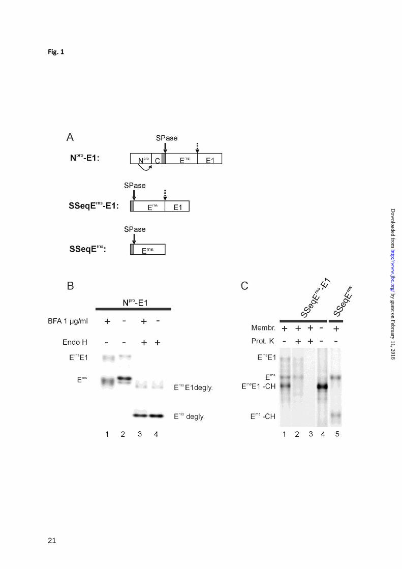
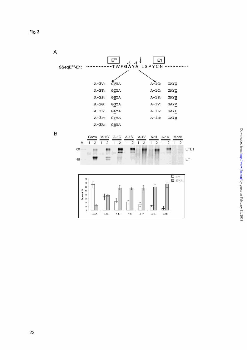
Erns
E1 cleavage can be blocked by mutations at
the cleavage site – Experimental support for
the hypothesis that a protein is cleaved by
SPase can be obtained by mutagenesis analysis
for the -1 and -3 positions of the cleavage site.
As a general rule these positions have to be
occupied by small and uncharged amino acids
in an SPase cleavage site. The presence of
large and/or charged residues at -1 or -3 blocks
SPase processing (35).
The -1 and -3 positions of the Erns
/E1 cleavage
site are occupied by alanine (C-terminal
sequence of Erns
‘...GAYA’). A variety of
mutants were established in which the alanine
residues were replaced by other amino acids
(Fig. 2A). The effects of these mutations were
analysed in transient expression studies by
quantification of the uncleaved versus cleaved
Erns
E1. As would be expected for a SPase
cleavage site the exchange of R, F or L for A
resulted in almost complete prevention of
Erns
E1 processing (Fig. 2B and C).
Interestingly, also constructs with G, C or S at
position -1 (Fig. 2B) and V, T, S, G at position
-3 (Fig. 2C) showed a severe reduction of
cleavage efficiency. This finding was not in
by guest on February 11, 2018http://w
ww
.jbc.org/D
ownloaded from
6
agreement with the described requirements for
a SPase cleavage site.
For control purposes we wanted to test the
same changes in the context of a typical SPase
cleavage site and therefore introduced the
mutations at the carboxyterminal end of the
signal peptide preceding Erns
in the viral
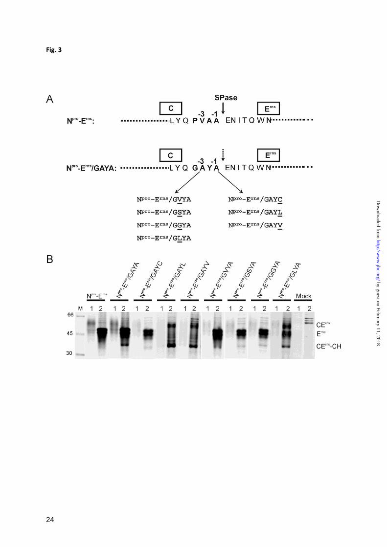
polyprotein (Fig. 3). We first changed the
carboxyterminus of the wt signal peptide, that
ends with the residues PVAA, into GAYA
(construct Npro
-Erns
/GAYA) to display the
sequence found at the Erns
/E1 site (Fig. 3A).
Afterwards, several exemplary exchanges of
the above described mutagenesis scheme were
introduced into the latter sequence. In transient
expression studies results were obtained that
would be expected for a typical SPase cleavage
site with L at -1 or -3 and V at -1 blocking
processing but V, G or S at -3 and C at -1
having basically no influence on processing
efficiency (Fig. 3B and C). A direct
comparison of the effects of different
mutations introduced at the aminoterminal or
carboxyterminal cleavage sites of Erns
makes
these differences obvious (Fig. 3C, Tab. 1).
Taken together, the results of the mutagenesis
studies as summarized in Tab. 1 cannot answer
the question, whether the Erns
/E1 site is cleaved
by SPase, but it has to be stressed that all
changes known to impair SP cleavage also
block the Erns
E1 processing. The finding that
also exchanges expected to be neutral in a
typical SPase cleavage site reduce the
processing efficiency severely might indicate
that additional requirements have to be
fulfilled to allow SPase activity in this special
case.
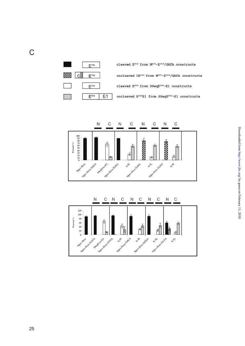
The influence of protease inhibitors on Erns
E1
cleavage – Experiments with specific
inhibitors can help to identify the protease
active on a given substrate. Therefore,
construct SSeqErns
-E1 was translated in a cell
free system in the presence of different known
protease inhibitors. Among the huge number of
different substances blocking proteases we
selected inhibitors specific for intramembrane-
cleaving proteases or signal peptidase to cover
the likely candidates that could be responsible
for Erns
E1 cleavage. Cell free translation of
construct SSeqErns
-E1 and immuno-
precipitation with anti-Erns
monoclonal
antibody 24/16 (mab 24/16) resulted in
detection of an unglycosylated precursor
product of ca. 43 kDa (Fig. 4A, lane 7). In the
presence of microsomal membranes and
absence of inhibitor two more bands were
detected that represent the glycosylated
precursor Erns
E1 of about 66 kDa and the
cleaved glycosylated Erns
protein of ca. 48 kDa.
Addition of inhibitors against Signal Peptide
Peptidase, gamma-Secretase, S2P-
Metalloprotease or of Leupeptin as
Serine/Cysteine protease inhibitor did slightly
influence the ratio between the different
products but not principally change the
outcome (Fig. 4A, lanes 2, 3, 4, 5). In contrast,
the signal peptidase inhibitor MeOSuc-Ala-
Ala-Pro-Val Chloromethyl-Ketone (SP-I) (49-
52) was able to prevent processing so that the
band representing processed Erns
was missing
(Fig. 4A, lane 1).
As controls for the SPase inhibition by SP-I its
influence on processing of two proteins
containing typical SPase cleavage sites was
tested. SSeqE1-E2 encodes a typical signal
peptide followed by CSFV E1 and E2. For this
construct, the detection of the unprocessed
glycosylated E1E2 precursor of about 70 kDa
(E1E2 in Fig. 4B) after immunoprecipitation
with an anti E2 monoclonal antibody proves
blocking of SPase processing. This result was
obtained when SSeqE1-E2 was translated in
the presence of SP-I (Fig. 4B, lane 1).
However, the precursor was absent, when
translation was done in the presence of
membranes but absence of inhibitor (Fig. 4B,
lane 2). Since free glycosylated E2 has about
the same size as unglycosylated E1E2, the
product of the processing is not readily
detected but becomes visible after removal of
N-linked carbohydrates with PNGaseF (Fig.
4B, lane 3).
As a second control construct ppL86AA was
used that encodes the aminoterminal 86 amino
acids of preprolactin. SPase processing leads to
release of the corresponding prolactin
by guest on February 11, 2018http://w
ww
.jbc.org/D
ownloaded from
7
fragment. Again, this fragment was almost not
detected in the presence of SP-I (Fig. 4C).
Taken together, the inhibitor studies strongly
support the conclusion that processing of
Erns
E1 is executed by SPase.
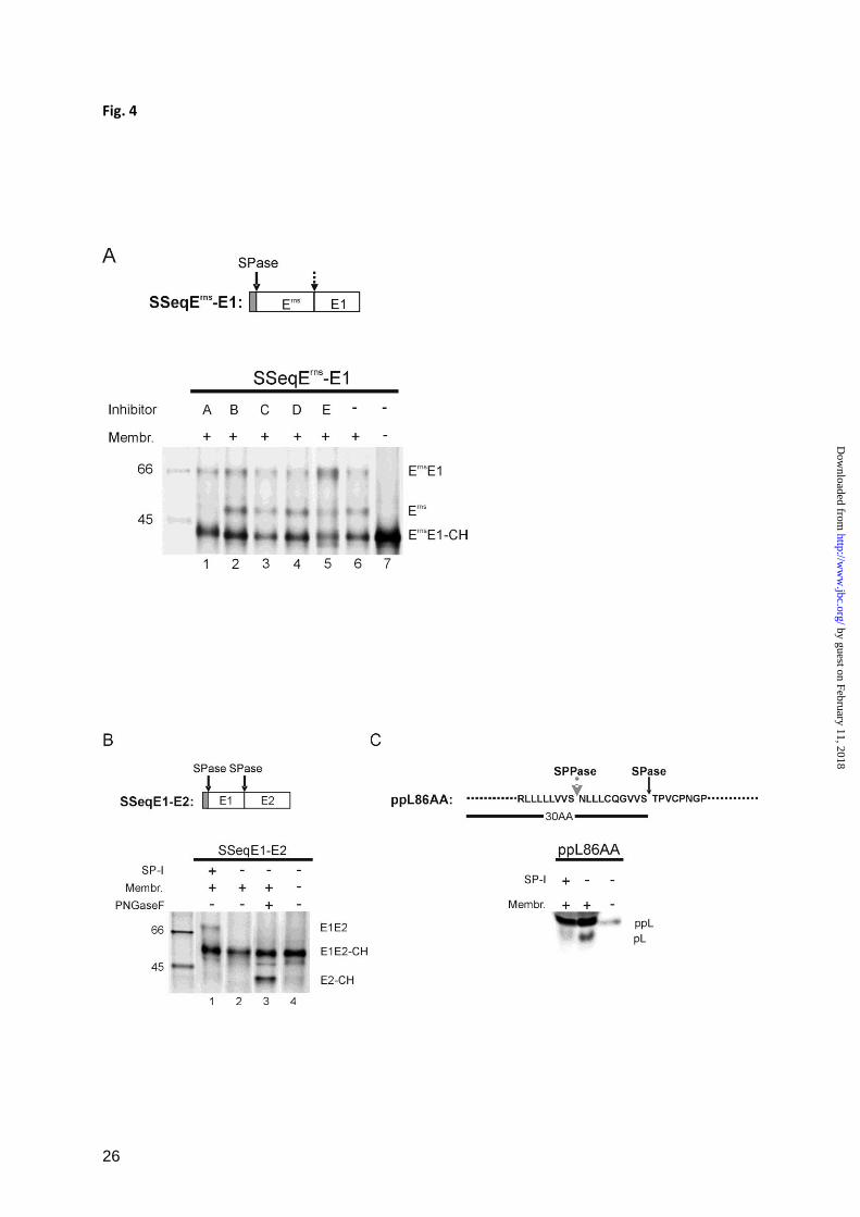
The type of signal peptide and its removal are
not crucial for Erns
E1 processing – The internal
signal peptide responsible for translocation of
Erns
into the ER might have special features
important for Erns
E1 processing. More
importantly, processing of a protein at more
than one site can occur in a hierarchic order
where one cleavage has to precede another
processing step. This principle is usually seen
with SPase and SPPase cleavages of signal
peptides with SPPase activity being dependent
on the completion of the SPase cleavage (53-
55). Similarly, hierarchical cleavage patterns
are quite common for processing of viral
polyproteins. It therefore could not be
excluded that the observed repression of Erns
E1
processing by SP-I represents an indirect effect
resulting from prevention of SP removal from
the Erns
aminoterminus. We therefore analyzed
the influence of different signal peptides and of
abrogation of signal peptide processing on
Erns
E1 cleavage.
When the original signal peptide of Erns
in
construct SSeqErns
-E1 was replaced by a
preprolactin translocation signal fused with an
aminoterminal V5 tag (construct ppLSSeqErns
-
E1) or by an insect signal peptide from the
mellitin gene (construct mellSSeqErns
-E1)
translocation, glycosylation and processing of
the pestivirus glycoprotein precursor occurred
in the same way as in the wt construct (Fig.
5A, lanes 2, 5 and 8). Also the inhibition of the
cleavage by SP-I was obvious by the absence
of the processed Erns
band ( Fig. 5A, lanes 1, 4
and 7).
To test, whether blocking of signal peptide
cleavage prevents processing of the Erns
/E1
site, the von Heijne motif of the preprolactin
signal peptide in construct ppLSSeqErns
-E1
was mutated (construct ppLSSeqErns
-E1*,
serine at position -1 of the SPase cleavage site
replaced by proline). After in vitro translation,
the products were immunoprecipitated with
mab 24/16 against Erns
or with an anti V5 mab.
Fully processed Erns
could be precipitated with
mab 24/16 after translation of this construct in
the absence of the SP-I as for the wt construct
(Fig. 5B, lanes 9 and 3, respectively).
Importantly, the processed Erns
as well as
glycosylated Erns
E1 precursor were also
detected with the anti V5 mab (Fig. 5B, lane
10). This finding proved that removal of the
signal peptide was inhibited as desired. Once
again, addition of SP-I prevented release of
Erns
, regardless whether the signal peptide
mutation was present or not (Fig. 5B, lanes 7
or 1, respectively).
In summary, these data show that neither the
nature of the signal peptide nor its processing
have a significant influence on processing at
the Erns
/E1 site.
Erns
E1 cleavage is dependent on glycosylation
– A typical signal peptide representing a
substrate for SPase is composed of a basic
sequence followed by a hydrophobic
transmembrane region and a carboxyterminal
polar region containing the von Heijne motif at
positions -3 and -1. The data described above
show that the Erns
/E1site is processed by SPase
even though only the carboxyterminal polar
region is present whereas the two preceding
parts of a typical cleavage site are replaced by
an amphipathic helix. The mutation analysis
showed that for Erns
E1 cleavage the
requirements with regard to the -1/-3 positions
are more stringent than for regular signal
peptide processing. These findings could
indicate that protein conformation might have
a major influence on the processing.
Because of its high content of carbohydrates,
folding of Erns
is most likely heavily influenced
by glycosylation. We therefore analyzed
whether prevention of N-glycosylation has an
impact on Erns
E1 processing. Constructs Npro
-
Erns
and SSeqErns
-E1 were translated in vitro in
the presence or absence of Ac-NYT-NH2, a
tripeptide substrate of glycosyltransferase that
can competitively block glycosylation of other
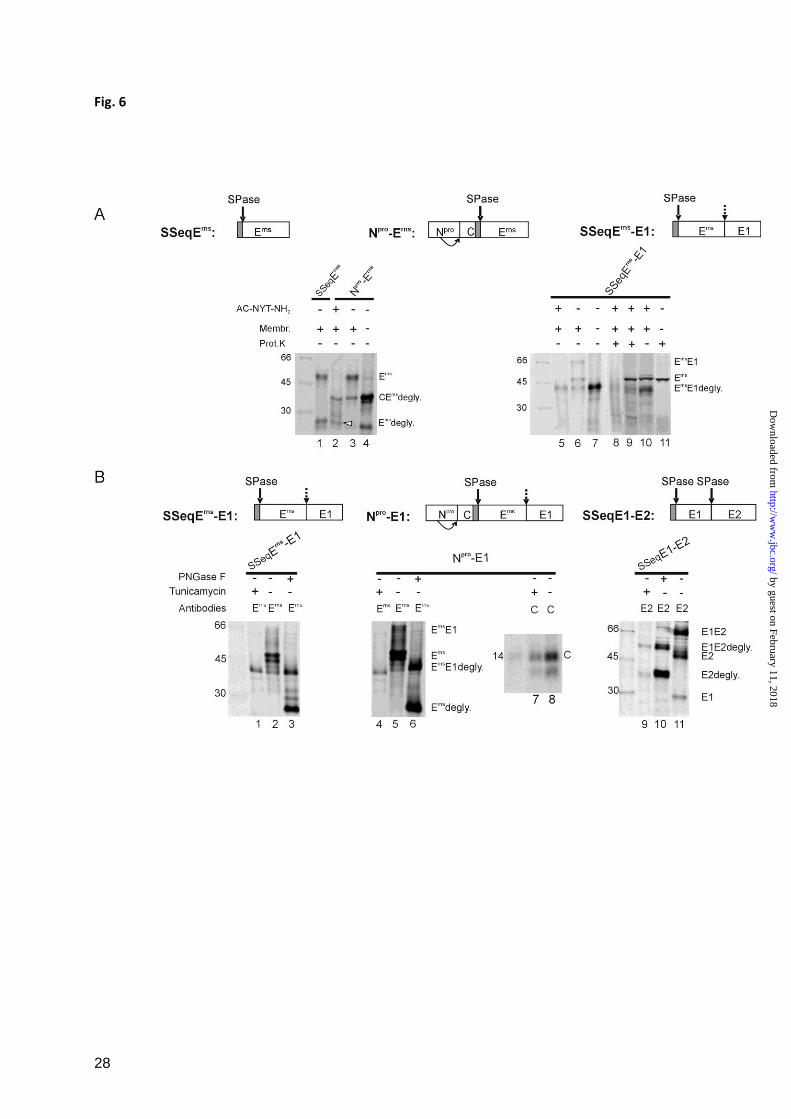
substrates (Fig. 6A) (48,56). To provide a size
by guest on February 11, 2018http://w
ww
.jbc.org/D
ownloaded from
8
marker for unglycosylated and glycosylated
Erns
, construct SSeqErns
was translated without
addition of the inhibitor (Fig. 6A, lane 1).
After translation, Erns
or fusion proteins
containing Erns
were precipitated with mab
24/16. For Npro
-Erns
, processed Erns
was clearly
detected as unglycosylated protein band of ca.
26 kDa in the presence of the competitive
inhibitor (marked with a white arrowhead in
Fig.6A, lane 2). This finding proves that
despite the inhibition of glycosylation SPase
processing of the internal signal peptide
connecting capsid protein C and Erns
occurs. In
contrast, cleaved unglycosylated Erns
was not
detected after translation of SSeqErns
-E1 in the
presence of the inhibitor (Fig. 6, lane 5). To
prove that the inhibitor did not prevent
translocation of the proteins, proteinase
protection assays were conducted.
Unglycosylated Erns
E1 was precipitated with
the Erns
specific antibody when SSeqErns
-E1
was translated in the presence of membranes,
and treated with Proteinase K (Fig. 6A, lane 8).
Since this band was somewhat hidden in a
smear, a further control was conducted without
immunoprecipitation. In this experiment the
unprocessed Erns
E1 precursor was detected
very clearly, regardless, whether the sample
was treated with Proteinase K or not (Fig. 6A,
lanes 9 and 10, respectively), but was not
present, when proteinase treatment was done
with a sample translated in the absence of
membranes (Fig. 6A, lane 11).
As a second approach, different CSFV
expression constructs were transiently
expressed in BHK-21 cells in the presence of
the N-glycosylation inhibitor tunicamycin. In
addition to SSeqErns
-E1, constructs Npro
-E1 and
SSeqE1-E2 were used, which contain the
polyprotein region covering Npro
/C/SP/Erns
/E1
or SP/E1/E2, respectively (Fig. 6B). Proteins
expressed from the transiently transfected
cDNA constructs were labeled with radioactive
amino acids in situ and subsequently
precipitated with mab 24/16 for Erns
, A18 for
E2 or rabbit antiserum D1* for C. Before SDS-
PAGE, part of the precipitates were treated
with PNGaseF to remove the N-linked
carbohydrates.
Fully processed Erns
without carbohydrates has
a size of about 26 kDa. A corresponding band
was detected for SSeqErns
-E1 or Npro
-E1 when
the constructs were expressed in the absence of
tunicamycin and the precipitated proteins
treated with PNGaseF (Fig. 6B, lanes 3 and 6).
In contrast, only the 43 kDa band of
nonglycosylated Erns
E1 was detected, when
expression was done in the presence of
tunicamycin (Fig. 6B, lanes 1 and 4). Thus,
processing of the Erns
/E1 site was inhibited by
tunicamycin treatment. However, this effect
was specific for this individual processing site,
since the cleavage at the typical SPase sites at
the aminoterminus of Erns
or at the E1/E2
border occurred also when glycosylation was
prevented. This fact could be concluded from
the detection of the C protein (Fig. 6B, lane 7)
or processed deglycosylated E2 (Fig. 6B, lane
9) when expression was done unter
tunicamycin treatment. Tunicamycin is a
widely used substance that inhibits N-
glycosylation but not translocation or SPase
activity. Accordingly, processing of the C/Erns
and the E1/E2 sites was observed whereas
processing of the Erns
E1 precursor was blocked
in the presence of the drug.
Taken together, the results show that
processing of Erns
E1 can only occur, when the
protein is glycosylated, a finding which
strongly supports the conclusion that the
overall conformation of the protein is
especially important for processing of Erns
E1.
The integrity of the Erns
membrane anchor is
crucial for Erns
E1 processing – The prevention
of N-glycosylation has certainly a major effect
on the structure of a protein containing 9
potential sites for N-glycosylation. To
investigate whether also less invasive
alterations of the Erns
structure can influence
Erns
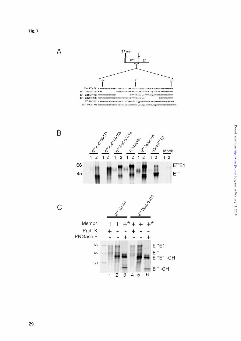
E1 processing further mutants were tested.
The C-terminal region of Erns
is of special
interest because it represents the membrane
anchor of the protein which interacts with the
lipid bilayer via an amphipathic helix thereby
by guest on February 11, 2018http://w
ww
.jbc.org/D
ownloaded from
9
possibly presenting the cleavage site to SPase.
Three different deletion mutants were
established on the basis of SSeqErns
-E1. Each
mutant protein contained a deletion of 14
amino acids of the Erns
carboxyterminal region
located upstream (Erns
-Del 158-171), at the
aminoterminal border (Erns
-Del 172-185) or in
the center (Erns
-Del 200-213) of the region
mapped as membrane anchor (Fig. 7A). After
transient expression of the latter two
constructs, a severe reduction of Erns
E1
processing was observed in comparison with
the wt construct. For Erns
-Del 172-185,
processing seemed to be completely blocked
since no Erns
could be detected. In contrast
construct Erns
-Del 158-171 showed about wt
processing efficiency (Fig. 7B). Similar results
were obtained for two further mutants with 14
amino acid deletions located further upstream
of position 158 (data not shown).
A more subtle effect on the arrangement of the
Erns
amphipathic helix can be achieved by
insertion of single alanine residues into the
helix resulting in a ca. 100 degree twist around
the helix axis. This alteration provokes a
significant disturbance of the amphipathic
character of the helix without affecting the
overall helical structure. Insertion of alanine at
position 191 of the Erns
sequence was shown
before to reduce the efficiency of membrane
binding from about 87% (wt) to ca. 30% (32).
When this mutation was introduced into
SSeqErns
-E1 the transient expression analysis
revealed a severe reduction of Erns
E1
processing (Fig. 7B, construct Erns
-Ala191).
The amphipathic character of the helix can in
part be restored when 3 or 4 alanine residues
are inserted. Indeed, a higher processing
efficiency was observed for construct Erns
-
3xAla191 compared to Erns
-Ala191. However,
the restoring effect was rather low, which fits
with the results obtained in analysis of
membrane association of equivalent mutants at
position 191 (32).
Secretion of the cleaved Erns
of mutants Erns
-
Del 200-213, Erns
-3xAla191 and Erns
-Ala191
was more efficient than for the wt or the Erns
-
Del 158-171 protein, but less impressive than
after expression of the mutated Erns
alone (32),
due to the overall much lower amount of
cleaved Erns
compared to the yield after
expression of Erns
alone. As expected, the
Erns
E1 fusion protein is not secreted at all since
the transmembrane region at the
carboxyterminus of E1 ensures efficient
membrane binding of the fusion protein. To
prove that the introduced mutations do not
impair protein translocation, proteinase
protection assays were conducted for
constructs Erns
-Ala191 and Erns
-Del200-213.
These assays showed that both the
glycosylated Erns
E1 precursor and the fully
processed Erns
protein were protected from the
Proteinase K whereas the unglycosylated
precursor (Erns
E1-CH) was destroyed (Fig. 7C,
lanes 1 and 2 or 4 and 5). The fact that the
formerly mentioned bands represent
glycosylated proteins and thus must have been
translocated was also proven by
deglycosylation with PNGaseF of transiently
expressed proteins precipitated from
transfected BHK-21 cells (Fig. 7C, lanes 3 and
6). Moreover, the PNGaseF treated samples
demonstrate again the poor processing of the
two mutant constructs, since the Erns
-CH band
is much less prominent than the precursor band
Erns
E1-CH.
Importantly, all the different mutations with
alterations affecting membrane interaction of
the Erns
C-terminal helix showed significantly
reduced processing of the Erns
E1 precursor
whereas changes outside of the helix had no
significant effect.
DISCUSSION
Posttranslational modification of viral envelop
proteins is usually done by the host cellular
system that is normally responsible for
trimming and maturation of cellular (surface)
proteins. These modifications include removal
of signal peptides by SPase and SPPase
cleavage, protein glycosylation, acylation,
by guest on February 11, 2018http://w
ww
.jbc.org/D
ownloaded from
10
phosphorylation and post ER maturation
cleavage by e.g. Golgi proteases. These
processes are essential to obtain functional
viral envelope proteins.
Positive strand RNA viruses express their
proteins via polyproteins that are subsequently
processed into the mature products. The
enveloped members of this virus group employ
host sytems also for cleavage of the
polyprotein region giving rise to the viral
structural proteins. To enable such a
processing scheme, the structural polyprotein
adopts a multi membrane spanning topology,
in which individual proteins are separated by
transmembrane regions, often organized in a
head to tail arrangement of stop transfer
sequence and signal peptide. These structures
are then cleaved at typical SPase cleavage sites
in the signal peptide moiety. Upon completion
of the processing process, the transmembrane
regions form the membrane anchor of the
aminoterminal cleavage product. The same
concept is also found in pestiviruses with
SPase cleavage occurring at typical SPase
cleavage sites at the aminotermini of
glycoproteins Erns
and E2 and the non-
structural proteins p7 and NS2. The only site
that does not conform to SPase cleavage site
requirements is the one between Erns
and E1.
This site has long been known to be special
since processing of Erns
E1 is delayed, so that
this Erns
E1 can always be detected as a
precursor in infected cells (9). It could be
hypothesized that the Erns
E1 fusion protein has
a biological function in the virus life cycle, but
the fact that viruses with an artificial deletion
of the Erns
-coding sequence can be efficiently
complemented with Erns
in trans argues against
this hypothesis (21,57,58). On the other hand,
it has recently been shown that processing of
Erns
E1 is crucial for pestivirus viability (59).
Since the Erns
/E1 processing site lacks a
hydrophobic h-region which is regarded as a
crucial part of a SPase cleavage site it was
speculated whether the delayed processing at
this site was due to processing in a downstream
compartment of the secretory pathway (9,59)
or resulted from inefficient SPase cleavage in
consequence of the unusual structure. As
reported here the Erns
E1 precursor is cleaved in
the ER. Moreover, the data show that this site
is cleaved by SPase in spite of its unusual
features. This conclusion is based on the facts
that processing can be blocked by a SPase
inhibitor that was used before in a variety of
analyses (49-52) and by typical exchanges of
the amino acids at the -1 or -3 positions of the
cleavage site. However, the mutagenesis
analyses indicated again that the Erns
/E1
cleavage site is unusual. Introduction of
cysteine at position -1 considerably impaired
Erns
E1 processing whereas the same exchange
in a regular SPase cleavage site had basically
no effect. Similarly, a glycine at position -3 in
the context of the Erns
/E1site had almost the
same inhibitory effect as a leucine in -3
whereas glycine at -3 was neutral when tested
in the context of typical SPase cleavage site.
These results make it obvious that sequence
requirements exceeding those known for a
typical SPase cleavage site have to be fulfilled
in order to allow processing of Erns
E1. This
hypothesis in supported by the results obtained
with mutants with alterations introduced rather
far upstream of the Erns
/E1 cleavage site. Two
deletion mutants and a variant with a single
alanine insertion 36 residues upstream of the
cleavage site showed considerably reduced or
even no processing of the precursor. In part,
these results can simply be explained by the
fact that mutations disturbing the
carboxyterminal amphipathic helix lead to
reduced membrane binding of the Erns
carboxyterminus thereby impairing also the
contact of the cleavage site with SPase. This is
true for the alanine insertion at position 191
(32) and has certainly to be taken into account
for the two deletions introduced into the
carboxyterminal region. However, the
argument of reduced membrane binding can
hardly explain the results obtained with the
point mutations at positions -3 and -1.
Moreover, it has also to be questioned why the
inhibition of protein glycosylation was able to
prevent Erns
E1 processing since this
manipulation does not directly affect the C-
by guest on February 11, 2018http://w
ww
.jbc.org/D
ownloaded from
11
terminus of the protein. Membrane binding of
a unglycosylated artificial fusion protein
containing the amphipathic helix of Erns
has
been demonstrated (33). It therefore seems
justified to speculate that at least in the latter
cases structural constraints prevent proper
interaction between enzyme and substrate, so
that it cannot be cleaved.
When signal peptides, the regular substrates of
SPase, were compared, a high degree of
variation was detected that concerned both the
length and sequences of the n-, h- and c-
regions (35,60). Thus, one could conclude that
SPase is highly flexible with regard to its
substrate as long as the general scheme and the
-1, -3 rule are obeyed. It even can be
questioned whether most of the typical features
of a signal peptide are actually important for its
acceptance as a substrate by SPase or whether
these features are only necessary for the
initiation of translocation. In an oversimplified
view, one could regard only the c-region as the
sequence interacting with the protease with the
-1, -3 residues being responsible for substrate
recognition. However, such relaxed conditions
would result in a huge number of SPase
cleavage sites and thus contradict the fact that
in reality SPase shows a high degree of
substrate specificity (60,61). It is therefore
obvious, that in addition to the c-region further
parameters are necessary to define whether a
certain sequence represents an SPase substrate.
One of these parameters could be the context
of substrate and protease with the translocon.
However, it has been shown that purified
SPase can execute a specific posttranslational
cleavage of a substrate in vitro (62-66). In such
assay mixtures, regular interaction of substrate
and SPase with the translocon is highly
unlikely. Similarly, the substrate investigated
here is most likely not associated like a
standard signal peptide with the sec61
complex. The Erns
/E1 cleavage site has to be
inserted into the membrane after complete
translocation of Erns
and E1 since a fully
glycosylated Erns
E1 precursor can be detected
within infected cells. Both glycosylation and
generation of the E1 carboxyterminus by SPase
cleavage of the E1/E2 site can only be
executed after completion of E1 translocation.
The Erns
E1 precursor is subsequently
processed as shown in pulse-chase experiments
(9). The membrane interaction of the Erns
amphipathic helix results in an in plane
configuration of the Erns
/E1 cleavage site
region instead of the regular transmembrane
situation with the signal peptide still
interacting with the translocon. The different
topology of the Erns
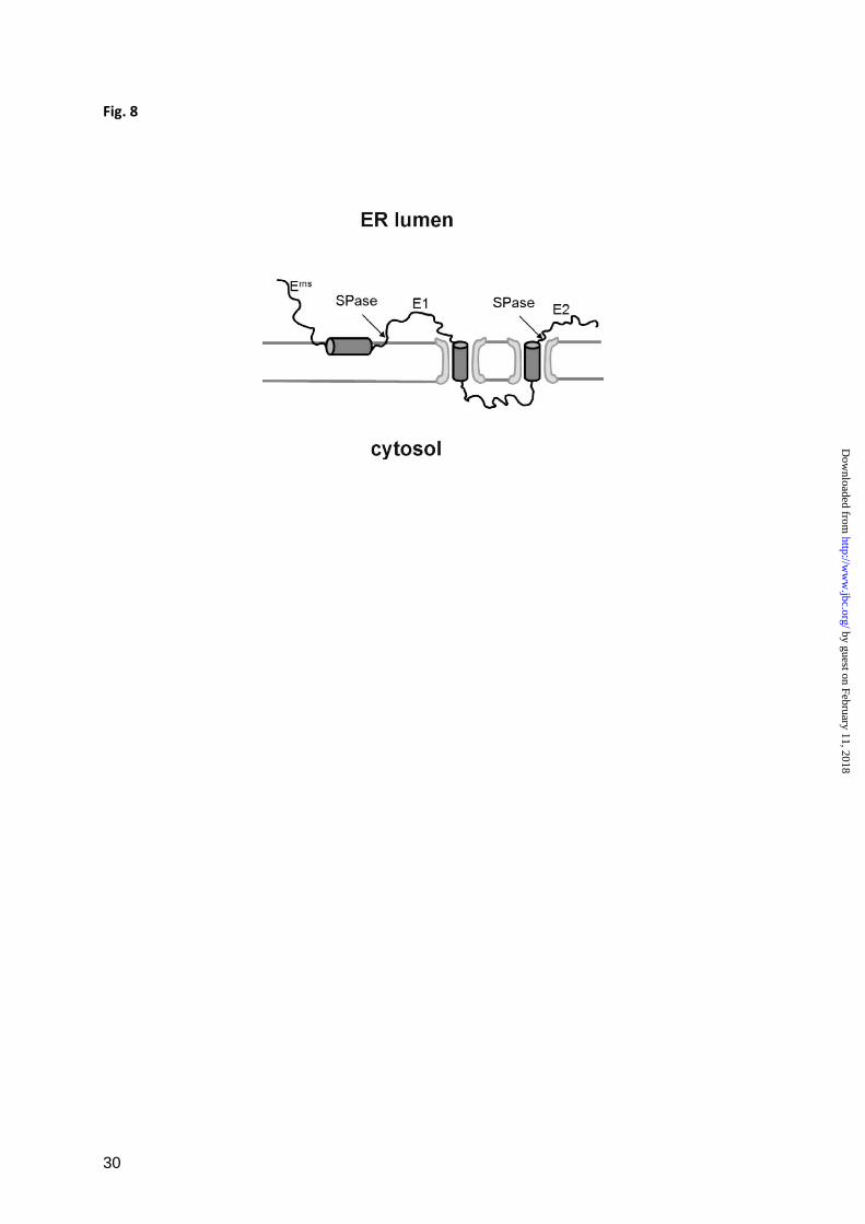
E1 site is demonstrated in
Fig. 8, where the processing of the pestivirus
polyprotein region containing Erns
, E1 and the
aminoterminal part of E2 is illustrated. The
carboxyterminus of E1 contains two
transmembrane regions, a stop transfer
sequence and a signal sequence responsible for
translocation of E2. The signal sequence is
processed by SPase in a typical transmembrane
topology whereas the Erns
/E1 site is preceded
by the in plane amphipathic helix.
It is known for signal peptides that the position
of the cleavage site relative to the membrane
surface is important for substrate recognition
by SPase since artificial lengthening of the h-
region of a signal peptide prevents SP cleavage
(67). Moreover, it has been reported that the
typical alpha-helical conformation of the h-
region must not extend into the c-terminal part
of the c-region, to ensure that the -1,-3 residues
are located in a region of extended
conformation (66,68,69). Thus, a parameter for
substrate recognition should be the correct
presentation of a suitable cleavage site in a
given ‘cleavage space’ close to the membrane
surface. For Erns
E1, this hypothesis means that
the amphipathic helix has to be inserted into
the membrane in a way that the cleavage site is
presented and orientated correctly in this
‘cleavage space’. This requirement can explain
why alteration of the ectodomain structure by
prevention of glycosylation, changes affecting
the amphipathic helix itself or even subtle
influences like point mutations can affect
cleavage efficiency of this substrate since all
these alterations can disturb the conformation
of the complex composed of the C-terminal
amphipathic helix and the membrane. Even a
by guest on February 11, 2018http://w
ww
.jbc.org/D
ownloaded from
12
subtle change of the conformation of this
complex can affect the positioning of the
cleavage site with respect to the enzyme.
These ideas indicate that a variety of demands
have to be fulfilled to achieve the cleavage of a
substrate like the Erns
E1 precursor. To our
present knowledge the Erns
/E1 site represents
the first SPase cleavage site in which the
membrane traversing h-region is missing and is
replaced by an amphipathic helix embedded in
plane into the membrane. However, in the past,
putative virus specific biological features often
turned out to be also used by the cells
themselves, representing alternatives in the
standard repertoire of cell biology which are
designed to serve special demands.
Pestivirus Erns
represents a very fascinating
viral protein. It is an essential component of
the virus particle, engaged in the infection
process, but it is also involved in repression of
the host’s immune response to a pestivirus
infection (26-31). The immune repressive
function of Erns
is connected with its enzymatic
activity, namely its ability to hydrolyze RNA.
So far, the target of the RNase is not known.
Different theories have been put forward and
in all of them the fact, that Erns
is secreted from
the infected cell, plays a central role. Massive
loss of the protein from the cell harbouring
replicating virus would be deleterious since it
would interfere with the formation of
infectious virus progeny. Thus, an equilibrium
between retention and secretion has to be
established that retains sufficient amounts of
the protein within the infected cell. It is
tempting to speculate that the unusual way by
which this protein is bound to the membrane is
one of the features playing a role in
establishment of this equilibrium. This unusual
membrane anchor has imposed another
problem on the virus, namely to assure
processing of the unusual structure at the
Erns
/E1 border. As shown in this report,
evolution has led to the point that this site is
cleaved by SPase despite the absence of the
hydrophobic h-region and, more importantly, a
totally different membrane topology of the
substrate. It can therefore be hypothesized that
the interaction of this substrate with the
enzyme is more sensible to mutations and
structural changes than the standard substrate.
More information on the structure of the Erns
C-terminus in the context of a membrane
environment is urgently needed to fully
understand this interesting substrate/enzyme
interaction. Most likely, the results of these
future analyses will also shed some light onto
the still quite unclear basis of SPase cleavage
specificity in general and will probably provide
yet further interesting open questions on the
fascinating Erns
protein.
ACKNOWLEDGEMENTS
The authors thank Maren Ziegler, Petra Wulle and Janett Wieseler for excellent technical assistance.
We are very grateful to Mark O. Lively for helpful discussions in the early phase of the project and for
providing the glycosylation acceptor Ac-NYT-NH2. The authors also wish to thank Robert Stark and
Heinz-Jürgen Thiel for providing the rabbit anti Erns
serum. This study was supported by grant
Me1367/4 from the Deutsche Forschungsgemeinschaft.
FOOTNOTES 1 Abbreviations:
BFA: Brefeldin A
BHK-21 cells: Baby hamster kidney cells
CSFV. Classical swine fever virus (formerly known as hog cholera virus)
EndoH: Endoglycosidase H
IRES: internal ribosomal entry site
kDa: kilodalton
by guest on February 11, 2018http://w
ww
.jbc.org/D
ownloaded from
13
Mab: Monoclonal antibody
PNGaseF: Peptide N-glycosidase F
Prot. K: Proteinase K
SP: Signal peptide
SPase: Signal peptidase
SPPase: Signal peptide peptidase
SP-I: Signal peptidase inhibitor
Wt: wildtype
2 Bintintan and Meyers, unpublished results
Reference List
1. Heinz, F. X., Collett, M. S., Purcell, R. H., Gould, E. A., Howard, C. R., Houghton, M., Moormann, J. M., Rice, C. M., and Thiel, H.-J. (2000) Family Flaviviridae. In M.H.V., R. v., Fauquet, C. M., Bishop, D. H. L., Carstens, E. B., Estes, M. K., Lemon, S. M., Maniloff, J., Mayo, M. A., McGeoch, D. J., Pringle, C. R., and Wickner, R. B., editors. Virus Taxonomy. Seventh Report of the International Committee on Taxonomy of Viruses, Academic Press, San Diego, USA
2. Lindenbach, B. D., Thiel, H.-J., and Rice , C. M. (2007) Flaviviridae: The Viruses and Their Replication. In Knipe, D. M. and Howley, P. M., editors. Fields Virology, Lippincott - Raven Publishers, Philadelphia, New York
3. Collett, M. S., Larson, R., Belzer, S., and Retzel, E. (1988) Virology 165, 200-208
4. Collett, M. S., Moennig, V., and Horzinek, M. C. (1989) J Gen Virol 70, 253-266
5. Elbers, K., Tautz, N., Becher, P., Rümenapf, T., and Thiel, H.-J. (1996) J Virol 70, 4131-4135
6. Harada, T., Tautz, N., and Thiel, H. J. (2000) J Virol 74, 9498-506
7. Heimann, M., Roman-Sosa, G., Martoglio, B., Thiel, H. J., and Rümenapf, T. (2006) J. Virol. 80, 1915-1921
8. Lackner, T., Muller, A., Konig, M., Thiel, H. J., and Tautz, N. (2005) J. Virol. 79, 9746-9755
9. Rümenapf, T., Unger, G., Strauss, J. H., and Thiel, H.-J. (1993) J Virol 67, 3288-3295
10. Tautz, N., Elbers, K., Stoll, D., Meyers, G., and Thiel, H.-J. (1997) J Virol 71, 5415-22
11. Wiskerchen, M., Belzer, S. K., and Collett, M. S. (1991) J Virol 65, 4508-4514
12. Xu, J., Mendez, E., Caron, P. R., Lin, C., Murcko, M. A., Collett, M. S., and Rice, C. M. (1997) J Virol 71, 5312-5322
13. Thiel, H.-J., Stark, R., Weiland, E., Rümenapf, T., and Meyers, G. (1991) J Virol 65, 4705-4712
by guest on February 11, 2018http://w
ww
.jbc.org/D
ownloaded from
14
14. Weiland, F., Weiland, E., Unger, G., Saalmüller, A., and Thiel, H. J. (1999) J. Gen. Virol. 80 ( Pt 5), 1157-1165
15. Bolin, S. R., Moennig, V., Kelso Gourley, N. E., and Ridpath, J. (1988) Arch Virol 99, 117-123
16. Donis, R. O., Corapi, W., and Dubovi, E. J. (1988) J Gen Virol 69, 77-86
17. Weiland, E., Stark, R., Haas, B., Rümenapf, T., Meyers, G., and Thiel, H.-J. (1990) J Virol 64, 3563-3569
18. Weiland, E., Ahl, R., Stark, R., Weiland, F., and Thiel, H.-J. (1992) J Virol 66, 3677-3682
19. Konig, M., Lengsfeld, T., Pauly, T., Stark, R., and Thiel, H. J. (1995) J. Virol. 69, 6479-6486
20. Horiuchi, H., Yanai, K., Takagi, M., Yano, K., Wakabayashi, E., Sanda, A., Mine, S., Ohgi, K., and Irie, M. (1988) J. Biochem. (Tokyo) 103, 408-418
21. Hulst, M. M. and Moormann, R. J. (2001) Methods Enzymol. 342, 431-440
22. Schneider, R., Unger, G., Stark, R., Schneider-Scherzer, E., and Thiel, H.-J. (1993) Science 261, 1169-1171
23. Hausmann, Y., Roman-Sosa, G., Thiel, H. J., and Rümenapf, T. (2004) J. Virol. 78, 5507-5512
24. Hulst, M. M., Himes, G., Newbigin, E., and Moormann, R. J. M. (1994) Virology 200, 558-565
25. Windisch, J. M., Schneider, R., Stark, R., Weiland, E., Meyers, G., and Thiel, H.-J. (1996) J Virol 70, 352-358
26. Meyer, C., Von Freyburg, M., Elbers, K., and Meyers, G. (2002) J. Virol. 76, 8494-8503
27. Meyers, G., Saalmüller, A., and Büttner, M. (1999) J Virol 73, 10224-35
28. Iqbal, M., Poole, E., Goodbourn, S., and McCauley, J. W. (2004) J. Virol. 78, 136-145
29. Magkouras, I., Mätzener, P., Rumenapf, T., Peterhans, E., and Schweizer, M. (2008) J. Gen. Virol. 89, 2501-2506
30. Mätzener, P., Magkouras, I., Rumenapf, T., Peterhans, E., and Schweizer, M. (2009) Virus Res. 140, 15-23
31. Meyers, G., Ege, A., Fetzer, C., von, F. M., Elbers, K., Carr, V., Prentice, H., Charleston, B., and Schürmann, E. M. (2007) J. Virol. 0, 02372-06
32. Tews, B. A. and Meyers, G. (2007) J. Biol. Chem. 282, 32730-32741
33. Fetzer, C., Tews, B. A., and Meyers, G. (2005) J. Virol. 79, 11901-11913
34. Langedijk, J. P. (2002) J. Biol Chem 277, 5308-14
35. von Heijne G. (1990) J. Membr. Biol. 115, 195-201
36. von Heijne G. (1986) Nucleic Acids Res. 14, 4683-4690
37. Wyatt, L. S., Moss, B., and Rozenblatt, S. (1995) Virology 210, 202-205
by guest on February 11, 2018http://w
ww
.jbc.org/D
ownloaded from
15
38. Sambrook, J. and Russell, D. W. (2001) Molecular cloning: a laboratory manual, Cold Spring Harbor Laboratory, Cold Spring Harbor, N.Y.
39. Meyers, G., Thiel, H.-J., and Rümenapf, T. (1996) J Virol 70, 1588-1595
40. Devereux, J., Haeberli, P., and Smithies, O. A. (1984) Nucleic Acids Res 12, 387-395
41. Shmulevitz, M. and Duncan, R. (2000) EMBO J. 19, 902-912
42. Kessler, S. W. (1981) Use of protein A bearing staphylococci for the immunoprecipitation and isolation of antigens from cells. In Langone, J. J. and van Vunakis, H., editors. Methods in Enzymology, Academic Press, New York
43. Meyers, G., Tautz, N., Becher, P., Thiel, H.-J., and Kümmerer, B. (1996) J Virol 70, 8606-8613
44. Fujiwara, T., Oda, K., Yokota, S., Takatsuki, A., and Ikehara, Y. (1988) J. Biol. Chem. 263, 18545-18552
45. Lippincott-Schwartz, J., Yuan, L. C., Bonifacino, J. S., and Klausner, R. D. (1989) Cell 56, 801-813
46. Misumi, Y., Misumi, Y., Miki, K., Takatsuki, A., Tamura, G., and Ikehara, Y. (1986) J. Biol. Chem. 261, 11398-11403
47. Oda, K., Hirose, S., Takami, N., Misumi, Y., Takatsuki, A., and Ikehara, Y. (1987) FEBS Lett. 214, 135-138
48. Wieland, F. T., Gleason, M. L., Serafini, T. A., and Rothman, J. E. (1987) Cell 50, 289-300
49. Casanova, C. L., Xue, G., Taracha, E. L., and Dobbelaere, D. A. (2006) Mol. Biochem. Parasitol. 149, 144-154
50. Nilsson, I., Johnson, A. E., and von Heijne, G. (2002) FEBS Lett. 516, 106-108
51. Nilsson, I., Johnson, A. E., and von Heijne, G. (2003) J. Biol. Chem. 278, 29389-29393
52. van Geest M., Nilsson, I., von Heijne, G., and Lolkema, J. S. (1999) J. Biol. Chem. 274, 2816-2823
53. Martoglio, B. (2003) Biochem. Soc. Trans. 31, 1243-1247
54. Weihofen, A., Lemberg, M. K., Ploegh, H. L., Bogyo, M., and Martoglio, B. (2000) J. Biol. Chem. 275, 30951-30956
55. Weihofen, A., Binns, K., Lemberg, M. K., Ashman, K., and Martoglio, B. (2002) Science 296, 2215-2218
56. Lau, J. T., Welply, J. K., Shenbagamurthi, P., Naider, F., and Lennarz, W. J. (1983) J. Biol. Chem. 258, 15255-15260
57. Reimann, I., Semmler, I., and Beer, M. (2007) Virology 366, 377-386
58. Widjojoatmodjo, M. N., van Gennip, H. G., Bouma, A., van Rijn, P. A., and Moormann, R. J. (2000) J. Virol. 74, 2973-2980
by guest on February 11, 2018http://w
ww
.jbc.org/D
ownloaded from
16
59. Wegelt, A., Reimann, I., Zemke, J., and Beer, M. (2009) J. Gen. Virol. 90, 2462-2467
60. von Heijne G. (1985) J. Mol. Biol. 184, 99-105
61. von Heijne G. (1984) J. Mol. Biol. 173, 243-251
62. Jackson, R. C. and Blobel, G. (1977) Proc. Natl. Acad. Sci. U. S. A 74, 5598-5602
63. Jackson, R. C. and Blobel, G. (1980) Ann. N. Y. Acad. Sci. 343, 391-404
64. Lively, M. O. (1989) Curr. Opin. Cell Biol. 1, 1188-1193
65. Lively, M. O., Newsome, A. L., and Nusier, M. (1994) Methods Enzymol. 244, 301-314
66. Paetzel, M., Karla, A., Strynadka, N. C., and Dalbey, R. E. (2002) Chem. Rev. 102, 4549-4580
67. Nilsson, I., Whitley, P., and von Heijne, G. (1994) J. Cell Biol. 126, 1127-1132.
68. Karla, A., Lively, M. O., Paetzel, M., and Dalbey, R. (2005) J. Biol. Chem. 280, 6731-6741
69. von Heijne G. (1998) Nature 396, 111, 113
70. Choo, K. H. (2008) BMC Bioinformatics 9 Suppl12, 15
71. Seidah, N. G., Mowla, S. J., Hamelin, J., Mamarbachi, A. M., Benjannet, S., Toure, B. B., Basak, A., Munzer, J. S., Marcinkiewicz, J., Zhong, M., Barale, J. C., Lazure, C., Murphy, R. A., Chretien, M., and Marcinkiewicz, M. (1999) Proc. Natl. Acad. Sci. U. S. A 96, 1321-1326
by guest on February 11, 2018http://w
ww
.jbc.org/D
ownloaded from
17
Signal Peptides:
"-1": Ala, Gly, Cys, Ser, Thr
"-3": Ala, Val, Ser, Cys, Thr
Site-directed mutagenesis at the
Erns
/E1 site:
"-1": Ala >> Gly >>> Cys > Ser > Leu > Val > Arg
"-3": Ala > Val >> Thr > Ser >>> Gly > Leu > Phe > Arg
TABLE 1
Amino acids at positions -1 and -3 in signal peptides cleaved by SPase and in the Erns
/E1 cleavage site.
The upper part presents the residues found in standard SPase cleavage sites with the incidence
decreasing from left to right (35,60,70). Below the residues tested in the context of the Erns
/E1
cleavage site are listed with the cleavage efficiency decreasing from left to right. The intensity of the
decrease is indicated: >: decrease; >>: strong decrease and >>>: very strong decrease of processing
efficiency.
by guest on February 11, 2018http://w
ww
.jbc.org/D
ownloaded from
18
FIGURE LEGENDS
FIGURE 1
Erns
E1 is cleaved in the ER. A, shows a schematic drawing of the cDNA constructs used in the
expression studies presented in B and C. The names of the constructs are given on the left side. Bars
show the expressed regions of the viral polyprotein with the designations of the mature viral proteins.
Signal peptides are shown as gray bars. A bent arrow indicates autocatalytic cleavage of Npro
at its
carboxyterminus. A solid arrow points at known SPase cleavage sites, whereas the site investigated
here is marked with a broken arrow. B, SDS-PAGE with products precipitated with a rabbit serum
against Erns
from cells transiently expressing the Npro
-E1 construct in the absence or presence of
Brefeldin A (BFA) as indicated on top of the gel. The precipitated proteins were loaded directly onto
the gel or pretreated with endoglycosidase H (Endo H) as indicated above the gel. C, SDS-PAGE with
products precipitated with the Erns
specific mab 24/16 after in vitro translation of the constructs
specified on top. Translation was done in the presence or absence of canine microsomal membranes as
indicated. To prove translocation of proteins into membrane vesicles, aliquots of the translation
products obtained after translation in the presence of membranes were treated with Proteinase K (lane
2) or Proteinase K and Triton X-100 (lane 3) as described before (32).
The names of the different products are indicated on the left and right sides of gels in B and C. The
term ‘degly.’ indicates the removal of carbohydrate side chains by deglycosylation, whereas ‘-CH’
means that glycosylation did not occur because of absence of membranes or inefficient translocation.
FIGURE 2
Erns
E1 processing efficiency of constructs with mutations at positions -1 and -3 of the cleavage site. A,
presents the mutations introduced into construct SSeqErns
-E1. The sequence around the Erns
/E1
cleavage site (broken arrow) is given and positions -1 and -3 are indicated. Below, the names of the
different constructs with the indicated mutations at -3 or -1 are highlighted. B and C show the results
obtained after expression of the mutations at positions -1 (B) or -3 (C). On top, a SDS-PAGE is shown
with the proteins precipitated with the rabbit Erns
serum from cell culture fluid (lanes numbered 1) or
the membrane fractions of cells (lanes numbered 2) expressing the constructs given above or no
plasmid (Mock). The left lane marked ‘M’ contains a protein size marker and the size of the visible
bands in kDa is given on the left. On the right, the names of the precipitated proteins are given. Below,
a diagram shows the quantification of the results with the intracellular amounts of cleaved Erns
(white
bars) and uncleaved Erns
E1 precursor (gray bars) given here as percentage of the total recovered Erns
protein. Mean values of at least 3 independent experiments are given and the standard deviation is
indicated. Please note that the construct designated ‘GAYA’ represents the wt.
FIGURE 3
Processing efficiency of constructs with mutations at positions -1 and -3 in a standard signal peptide
cleavage site. A presents the original sequence context in the internal signal peptide located between
capsid protein C and Erns
in the CSFV polyprotein (construct Npro
-Erns
, upper part) and the sequence of
the mutant Npro
-Erns
/GAYA with the C-terminal part of the signal peptide replaced by the GAYA motif
by guest on February 11, 2018http://w
ww
.jbc.org/D
ownloaded from
19
found at the Erns
C-terminus. Below, the names of the different constructs with the indicated mutations
at -3 or -1 of Npro
-Erns
/GAYA are given. B shows the results obtained after transient expression,
immunprecipitation and SDS PAGE of the mutated proteins. C, diagrams showing the processing
efficiency determined for the different mutants presented in (B) in comparison with the results
obtained for the equivalent mutations in the context of the Erns
C-terminus demonstrated in Fig. 2. On
top, the meaning of the different types of bars is given. Below, the results for mutations affecting
positions -1 (upper diagram) or -3 (lower diagram) are shown. Each section of the diagrams presents
one type of mutation with its effect on processing of the standard SPase cleavage site at the N-
terminus of Erns
(N) or on processing at the Erns
C-terminus (C). See also legend to Fig. 2.
FIGURE 4
Influence of different protease inhibitors on Erns
E1 processing. A, SDS-PAGE with products of in vitro
translation experiments with construct SSeqErns
-E1 (shown on top) in the presence of the protease
inhibitors specified by letters above the gel (A: Signal peptidase inhibitor (SP-I): MeOSuc-Ala-Ala-
Pro-Val Chloromethyl Ketone used at 1.5 mM-2.5mM; B: Signal Peptid Peptidase inhibitor: (ZLL)2-
Ketone, used at 10 µM; C:gamma-Secretase inhibitor: DAPT, used at 100 µM; D:Serine/Cysteine
Protease inhibitor: Leupeptin, used at 25 µM; E:S2P-Metalloprotease inhibitor: 1,10-Phenanthroline,
used at 5 mM). Please note that the concentration at which 1,10 Phenanthroline was used in this study
was shown to inhibit also the site 1 intramembrane protease (S1P) (71).Translation of the RNA in the
absence of inhibitor or in the absence of inhibitor and microsomal membranes, as specified by the
code on top of the gel, served as controls. Lane 5 showing the products obtained in the presence of
inhibitor E was exposed 5 times longer than the other lanes. See also legend to Fig. 1.
Effects of the SPase inhibitor (SP-I) on processing of construct SSeqE1-E2 (B) or a preprolactin
construct (C) were also determined. For the preprolactin construct which includes the first 86 amino
acids of the precursor protein, the signal peptide of 30 amino acids with the SPase and the signal
peptide peptidase cleavage sites are specified. Protein size marker bands are shown with the molecular
weights given in kDa.
FIGURE 5
Importance of the type of signal peptide and of signal peptide cleavage on Erns
E1 processing. A, the
upper part shows schemes of the analyzed constructs in which Erns
E1 was combined with different
signal peptides. Below, SDS-PAGE with proteins precipitated with mab 24/16 after in vitro translation
of the constructs indicated on above the gels. Translation was done in the presence or absence of
microsomal membranes or SP-I as indicated. (B) On top constructs pplSSeqErns
-E1 and variant
pplSSeqErns
-E1* with the cleavage site SP/Erns
blocked by mutation, are shown. Below, SDS-PAGE
with products of in vitro translation precipitated with mab 24/16 against Erns
or an anti V5 mab as
specified above the gel. See also legend to Fig. 1.
FIGURE 6
Erns
E1 processing under prevention of glycosylation. A shows the results obtained after in vitro
translation. On top, the constructs used in the studies are presented schematically. Below, SDS-PAGE
gels are shown with products precipitated with mab 24/16 after in vitro translation of the indicated
by guest on February 11, 2018http://w
ww
.jbc.org/D
ownloaded from
20
products in the presence or absence of the N-glycosylation acceptor (competitive inhibitor) AC-NYT-
NH2 (lanes 1 to 8). Lanes 8 to 11 show the results of Proteinase K protection assays with lanes 9 to 11
containing products of in vitro translation without immunoprecipitation. Presence or absence of
Proteinase K is indicated on top of the gel. Lane 11 shows a control established by translation in the
absence of membranes and inhibitor followed by Proteinase K treatment. White arrow marks
unglycosylated Erns
cleaved by SPase from the Npro
/C/Erns
precursor. B, results of transient expression
assays conducted with the constructs shown on top. The gels show the proteins precipitated with the
given antibodies (Erns
: mAb 24/16 against Erns
, C: rabbit serum against C protein; E2: mAb A18
against E2) from the extracts of cells transfected with the indicated constructs. The transfected cells
were treated with tunicamycin as indicated above of the gels. Some of the precipitated proteins were
deglycosylated with PNGase F (lanes 3, 6 and 10). See also legend to Fig. 1 and 2. Please note that E1
is visible in lane 11 due to coprecipitation with E2 because of formation of a disulfide linked
heterodimer in infected cells (13).
FIGURE 7
Effect of short deletions or alanine insertions in the Erns
C-terminal region on Erns
/E1 processing. A,
construct SSeqErns
-E1 is shown with the carboxyterminal region of Erns
presented as a blow up with
the sequence given in the 1 letter code (residue number in the mature Erns
given on top). The deletions
and the position of the inserted alanine residues (underlined) are demonstrated with the names of the
corresponding expression constructs specified on the left. In analogy to the bovine viral diarrhea virus
Erns
, the amphipathic helix should start around position 180 as defined by an alanine insertion scanning
approach (32). B, SDS-PAGE with the proteins precipitated with mab 24/16 from tissue culture fluid
(lanes 1) and cell extracts (lanes 2) of cells transfected with the indicated constructs. C, shows controls
that were either done with Proteinase K protection assays using products of in vitro translation (lanes
1,2,4 and 5) or proteins precipitated from transfected cells (similar as in B) that were deglycosylated
with PNGaseF. * indicates that the membranes were not added as in the in vitro translation assays but
were present because of expression in cells. See also legend to Fig. 1 and 2.
FIGURE 8
Schematic presentation of the membrane topology and processing scheme of the pestivirus polyprotein
region encompassing Erns
, E1 and the aminoterminal part of E2 (not drawn to scale). Transmembrane
regions and the Erns
C-terminal amphipathic helix are shown as cylinders whereas the rest of the
proteins are indicated by black lines. The two gray horizontal lines represent the ER- or cytosol-facing
surfaces of the ER-membrane. The translocon is indicated as a light gray structure. SPase cleavage
sites are marked by arrows and ‘SPase’. The signal sequence upstream of Erns
responsible for Erns
translocation is not shown. Please note that the transmembrane region upstream of the E2 protein is a
regular signal sequence with a typical transmembrane topology that stands in marked contrast to the in
plane configuration of the Erns
amphipathic helix preceding the Erns
/E1 SPase cleavage site.
by guest on February 11, 2018http://w
ww
.jbc.org/D
ownloaded from
Ioana Bintintan and Gregor MeyersA new type of signal peptidase cleavage site identified in an RNA virus polyprotein
published online January 21, 2010J. Biol. Chem.
10.1074/jbc.M109.083394Access the most updated version of this article at doi:
Alerts:
When a correction for this article is posted•
When this article is cited•
to choose from all of JBC's e-mail alertsClick here
by guest on February 11, 2018http://w
ww
.jbc.org/D
ownloaded from


![Education Strategies for Identifying RNA Splicing ...vorgogoz/articles/D.pachea... · this with other steps in RNA processing, such as capping, cleavage, and polyadenylation [6]](https://img.pdfslide.us/doc/110x75/5f0253b47e708231d403b8cd/education-strategies-for-identifying-rna-splicing-vorgogozarticlesdpachea.jpg)