Embed Size (px)
Citation preview
7/26/2019 Enzymatic Cell Wall Degradation of Chlorella Vulgaris and Other Microalgae for Biofuels Production 2012
http://slidepdf.com/reader/full/enzymatic-cell-wall-degradation-of-chlorella-vulgaris-and-other-microalgae 1/15
O R I G I N A L A R T I C L E
Enzymatic cell wall degradation of Chlorella vulgarisand other microalgae for biofuels production
Henri G. Gerken • Bryon Donohoe • Eric P. Knoshaug
Received: 13 August 2012 / Accepted: 4 September 2012
Springer-Verlag Berlin Heidelberg (outside the USA) 2012
Abstract Cell walls of microalgae consist of a polysac-
charide and glycoprotein matrix providing the cells with aformidable defense against its environment. We character-
ized enzymes that can digest the cell wall and weaken this
defense for the purpose of protoplasting or lipid extraction. A
growth inhibition screen demonstrated that chitinase, lyso-
zyme, pectinase, sulfatase, b-glucuronidase, and lamina-
rinase had the broadest effect across the various Chlorella
strains tested and also inhibited Nannochloropsis and Nan-
nochloris strains. Chlorella is typically most sensitive to
chitinases and lysozymes, both enzymes that degrade poly-
mers containing N -acetylglucosamine. Using a fluorescent
DNA stain, we developed rapid methodology to quantify
changes in permeability in response to enzyme digestion and
found that treatment with lysozyme in conjunction with
other enzymes has a drastic effect on cell permeability.
Transmission electron microscopy of enzymatically treated
Chlorella vulgaris indicates that lysozyme degrades the
outer surface of the cell wall and removes hair-like fibers
protruding from the surface, which differs from the activity
of chitinase. This action on the outer surface of the cell
causes visible protuberances on the cell surface and pre-sumably leads to the increased settling rate when cells are
treated with lysozyme. We demonstrate radical ultrastruc-
tural changes to the cell wall in response to treatment with
various enzyme combinations which, in some cases, causes a
greater than twofold increase in the thickness of the cell wall.
The enzymes characterized in this study should prove useful
in the engineering and extraction of oils from microalgae.
Keywords Growth inhibition Permeability Lysozyme
Nitrogen depletion
Abbreviations
NREL National renewable energy laboratory
TFA Triflouroacetic acid
TLS Trilaminar structure
TEM Transmission electron microscopy
CCAP Culture collection of algae and protozoa
NCMA Formerly CCMP, Provasoli-Guillard National
Center for marine algae and microbiota
ATCC American type culture collection
UTEX Culture collection of algae at the University of
Texas at Austin
ASP NREL Aquatic species program
mBBM Modified bold’s basal medium
ASW Artificial seawater
PBS Phosphate buffered saline
SEM Scanning electron microscopy
Introduction
As algae are increasingly being considered as a potential
feedstock to meet a portion of our liquid transportation fuels
Electronic supplementary material The online version of thisarticle (doi:10.1007/s00425-012-1765-0 ) contains supplementarymaterial, which is available to authorized users.
H. G. Gerken E. P. Knoshaug (&)
National Renewable Energy Laboratory, National Bioenergy
Center, 15013 Denver West Parkway, Golden, CO 80401, USA
e-mail: [email protected]
H. G. Gerken
Laboratory for Algae Research and Biotechnology,
Arizona State University, Mesa, AZ, USA
B. Donohoe
National Renewable Energy Laboratory, Biosciences Center,
Golden, CO, USA
1 3
Planta
DOI 10.1007/s00425-012-1765-0
7/26/2019 Enzymatic Cell Wall Degradation of Chlorella Vulgaris and Other Microalgae for Biofuels Production 2012
http://slidepdf.com/reader/full/enzymatic-cell-wall-degradation-of-chlorella-vulgaris-and-other-microalgae 2/15
demand, many hurdles to the development of an algal
biomass to liquid transportation fuel process have been
indentified (Scott et al. 2010; Wijffels and Barbosa 2010;
Knoshaug and Darzins 2011). A major hurdle that has
generated much interest from the industrial and research
communities is the extraction of the internal lipid bodies
from algal cells. Typically, lipid extraction methods use
solvents such as toluene, hexane, butanol, ethanol, metha-nol or ionic liquids to extract lipids from intact, chemically
treated or mechanically ruptured cells (Pienkos and Darzins
2009; Shen et al. 2009; Brennan and Owende 2010; Lee
et al. 2010; Siddiquee and Rohani 2011; Kim et al. 2012).
Due to the chemically complex and structurally robust
nature of algal cell walls, many current extraction processes
require large chemical or energy loads. Technoeconomic
analysis suggests that for an algae-to-biofuel process to be
profitable, the post-extraction algal biomass must also be
utilized as either materials recycled back into the process or
as a feedstock for further conversion to saleable co-product
(Davis et al. 2011). Enzymatic degradation of the algal cellwalls prior to lipid extraction has the potential to facilitate
both lipid extraction and post-extraction use of the algal
biomass. Weakened cell walls could reduce solvent and
energy inputs needed for lipid extraction by either liberating
cell wall mono or polysaccharides or by improving acces-
sibility of cell wall polymers to microorganisms, enzymes
or reagents involved in downstream algal biomass pro-
cessing such as anaerobic digestion, fermentation or direct
conversion. At the National Renewable Energy Laboratory
(NREL), Chlorella vulgaris UTEX395 has been established
as our model green alga, because it grows rapidly, can attain
a high lipid content ([45 % of cell dry weight), has large
scale cultivation history, and has reports of successful
genetic transformation (Guarnieri et al. 2012). The tran-
scriptome and proteome of this organism have recently been
revealed under nitrogen-replete and -deplete conditions
(Guarnieri et al. 2011), and a genome sequencing project is
also underway.
Microalgal cell walls are complex and poorly understood
(Popper and Tuohy 2010). The Chlorella intraspecies var-
iation in cell walls as well as variations observed in a single
strain grown under different conditions can be dramatic,
and thus it is difficult to predict which of the compounds
noted below will be present in any one strain. While some
Chlorella species have only a single microfibrillar layer,
others have two layers with the microfibrillar layer proximal
to the cytoplasmic membrane and a mono or trilaminar
outer layer (Yamada and Sakaguchi 1982). The cell walls of
C. vulgaris and other green microalgae are known to have
rigid wall components embedded within a more plastic
polymeric matrix. This matrix is defined as the fraction that
is hydrolyzable in 1 N NaOH or 2 M triflouroacetic acid
(TFA) and contains uronic acids, rhamnose, arabinose,
fucose, xylose, mannose, galactose, and glucose. The rigid,
TFA-resistant cell wall is either glucosamine or a glucose-
mannose polymer (Takeda 1991). The predominant amino
sugar found in the rigid cell wall of Chlorella Pbi is
N -acetylglucosamine present as a chitin-like glycan. Chitin
(poly-b-1,4-D- N -acetylglucosamine), a polymer commonly
found in shellfish, insects, fungi, worms, and mushrooms
which can be deacetylated, either partially or fully, to pro-duce chitosan (poly-b-1,4-D-glucosamine). Strong acid
(6 N HCl) was needed to hydrolyze the rigid cell wall of a
Chlorella sp., which removed the acetyl group from
N -acetylglucosamine leaving glucosamine while hydrolysis
with strong sulfuric acid failed to release detectable neutral
sugars suggesting that this strain of Chlorella does not have
cellulose in the rigid cell wall. Digestion of the rigid wall
with chitinase released N -acetylglucosamine yet complete
digestion required a chitosanase, which indicates that glu-
cosamine polymers are also present in the rigid wall of
Chlorella Pbi (Kapaun and Reisser 1995). Based on these
previous studies, the genus Chlorella was in-part defined ascontaining glucosamine as the common dominant cell wall
polymer (Huss et al. 1999). The neutral cell wall sugars of
symbiotic Chlorella strains contain decreasing amounts
glucose, rhamnose, arabinose, mannose, xylose, fucose, and
galactose, respectively (Kapaun et al. 1992). Other than the
neutral sugars (*25 %), the cell wall contained 15–20 %
uronic acids, 10–15 % glucosamine, and 6–10 % protein.
This cell wall composition was reported to be similar to the
glucosamine containing cell wall composition of the non-
symbiotic algae C. vulgaris, Chlorella sorokiniana, and
Chlorella kessleri; however, the authors were unable to
determine the composition of 30–44 % of the cell wall
(Kapaun et al. 1992).
Unusual and previously undescribed polymers have also
been isolated from C. vulgaris cell walls. Two rhamnose
containing polymers and a b-D-galactan polymer were
isolated (Ogawa et al. 1998, 1999, 2001), and a number of
unidentified sugars that in some cases can be present in
substantial amounts (Burczyk et al. 1995). Other sugars
include 2-O-methyl-L-rhamnose and 3-O-methyl-L-rham-
nose, identified as residues in an acidic polysaccharide of
C. vulgaris (Ogawa et al. 1997). The cell walls of C. vul-
garis are readily stained by Ruthenium, Red which typi-
cally adheres to pectin-like substances or acidic sugars
(Takeda 1991). The presence of pectin has also been
demonstrated in C. vulgaris and has been shown to increase
on extended exposure to alkaline pH of 9.5 (Malis-Arad
et al. 1980).
Algal cell walls are highly persistent in sediments rich in
organic matter preserved in terrestrial and marine envi-
ronments. Algaenan is postulated as being the reason for
the persistance of algal cell walls (Derenne et al. 1992;
Gelin et al. 1999). Initially described as plant lignin-like
Planta
1 3
7/26/2019 Enzymatic Cell Wall Degradation of Chlorella Vulgaris and Other Microalgae for Biofuels Production 2012
http://slidepdf.com/reader/full/enzymatic-cell-wall-degradation-of-chlorella-vulgaris-and-other-microalgae 3/15
and also likened to the sporopollenin of spores and pollen
grains, algaenan has been suggested to be responsible for
the resistance of algae to bacterial degradation (Gunnison
and Alexander 1975). Typically, algaenan, an aliphatic,
non-hydrolyzable polymer is located in a thin (10–20 nm)
outer wall layer with a trilaminar structure (TLS). Two
electron-dense layers surround a less dense layer, which
survives drastic chemical treatment typically involvingmultiple extractions with KOH, strong acids or trifluoro-
acetic acid (Gelin et al. 1999). The main building blocks of
algaenan are described as C30–34 mono- or di- unsaturated
x-hydroxy fatty acids which, through a combination of
ester and ether linkages, create a very strong and recalci-
trant structure similar to cutin (Blokker et al. 1998) and
lacking in carotenoid moieties (Derenne et al. 1992). C.
vulgaris lacks a typical TLS cell wall structure (Corre et al.
1996) and was found to be more sensitive to detergents
than a species, Chlorella emersonii, having a TLS outer
cell wall structure. However, as the cells aged, they
become more resistant to detergents, particularly C. vul-
garis (Corre et al. 1996). Algaenan from Botryococcus
braunii race A has been suggested to change in composi-
tion, mainly in the reduction of terminal methyl groups,
depending on growth stage and conditions (Simpson et al.
2003).
Enzymatic digestion of algal cell walls has been previ-
ously studied as a methodology to make proto or sphero-
plasts of algal cells usually as a step in cell preparation for
genetic transformation. Naked protoplasts, termed sphero-
plasts, of Chlorella saccharophila and Chlorella ellipsoi-
dea were made using an enzyme mix containing cellulase,
hemicellulase, and pectinase activities (Braun and Aach
1975). However, these green algae, which are presumed
not to have an outer layer or sporopollenin (now known as
algaenan), required up to 90 h to display any effect from
the enzymes. The cells became very sensitive to physical
pressures, bursting simply from the pressure of a cover slip
placed on a microscope slide (Braun and Aach 1975).
Similarly, protoplasts were made from C. vulgaris K-73122
using acromopeptidase, cellulase, chitosanase, gluczyme,
and uskizyme; however, only *88 % of the cells were
considered osmotically labile (Honjoh et al. 2003). Pro-
toplasts were also made in C. ellipsoidea, C. vulgaris, and
C. saccharophila using three different enzyme mixes
containing cellulase, macerozyme, pectinase, cellulysin,
and driselase, although 7 of the 12 strains tested showed no
protoplast formation with any of the formulations (Yamada
and Sakaguchi 1982).
Bacterial degradation was examined by incubating C.
emersonii (TLS outer wall) or C. vulgaris (non-TLS outer
wall) with two types of aerobic heterotrophic bacteria.
After 4 months, there was no bacterial attachment to or
penetration into the algal cells, indicating the effects of
degradation must be due to extracellular enzymes produced
by the bacteria (Afi et al. 1996). In addition, similar
changes to the internal contents of the algal cells indicate
that the TLS layer did not protect against the diffusion of
enzymes into algal cells possessing this cell wall structure
even though the cell wall architecture remained un-degra-
ded. Similarly, as Chlamydomonas reinhardtii or Selena-
strum capricornutum cells aged and become nitrogen orphosphate limited, they became resistant to digestion by
grazers suggesting the layers in the cell wall became
thicker and more protective to enzymatic digestion in the
gut (Van Donk et al. 1997). Snail gut enzymes (glusulase)
have been observed to digest the inner, less electron-dense
cell wall layer, but did not affect the TLS layer in Chlorella
fusca cell wall fragments. When glusulase and a wide
variety of other enzymes, including lysozyme, were tried
on intact cells, there was no effect on the cell walls as
judged by transmission electron microscopy (TEM) imag-
ing (Atkinson et al. 1972). C. fusca contains a TLS outer
layer enclosing all the other cell wall layers and isimpervious to enzymatic attack or penetration by the
enzymes to attack other internal layers. Interestingly, dur-
ing cell reproduction, the inner non-TLS component was
degraded by internal enzymes (Atkinson et al. 1972) and
enzymes with the activities of b-D-fucosidase, b-D-man-
nosidase, and b-D-glucosidase were identified that play
roles in the disintegration of the mother cell wall (spo-
rangium) to release autospores (Walter and Aach 1987).
These enzymes released oligomers from the inner wall to
loosen the cell wall rather than completely degrade it while
not impacting the outer TLS wall. Similarly, subtilase-like
serine proteases perform this function in C. reinhardtii and
Volvox carteri (Fukada et al. 2006; Kubo et al. 2009).
In the work presented here, we sought to define the
extent of degradation that a variety of enzymatic activities
have on the cell walls of C. vulgaris. As the cell wall
provides the main protective barrier between the algae and
its surroundings, a detailed characterization of enzymes
that act on the wall could enable the rational design of an
enzyme cocktail that would allow efficient degradation of
the wall. Removal of the wall could allow more efficient
transformation technologies to be developed, conversion of
these cell wall sugars into a more usable substrate for
downstream processes, and potentially ease lipid extraction
from these organisms.
Materials and methods
Strains, enzymes, and growth conditions
We acquired the following strains from established algae
collections [Culture Collection of Algae and Protozoa
Planta
1 3
7/26/2019 Enzymatic Cell Wall Degradation of Chlorella Vulgaris and Other Microalgae for Biofuels Production 2012
http://slidepdf.com/reader/full/enzymatic-cell-wall-degradation-of-chlorella-vulgaris-and-other-microalgae 4/15
(CCAP), Provasoli-Guillard National Center for Marine
Algae and Microbiota (NCMA), formerly CCMP], Amer-
ican Type Culture Collection (ATCC) and the Culture
Collection of Algae at The University of Texas at Austin
(UTEX), C. emersonii CCAP 211/11N, Chlorella varia-
bilis NC64A (ATCC 50258), C. vulgaris UTEX 26, 30,
259, 265, 395, 396, 1803, 1809, 1811, 2714, and CCAP
211/11B, Phaeodactylum tricornutum CCMP 632, and S.capricornutum UTEX 1648. We also used the following
strains from the NREL Aquatic Species Program (ASP)
collection Ankistrodesmus falcatus ANKIS1, Chlorella
sp. CHLOR1, Franceia sp. FRANC1, Nannochloris sp.
NANNO2, Nannochloropsis sp. NANNP2, and Oocystis
pusilla OOCYS1 (Barclay et al. 1987; Sheehan et al. 1998).
Achromopeptidase, alginate lyase, a-amylase (960 FAU/
mL), chitinase, chitosanase (25.9 U/mL), chondroitinase
ABC (5 U/mL), drieselase, b-galactosidase (1,000 U/mL),
b-glucosidase, hemicellulase, heparinase II (10 U/mL),
hesperidinase, hyaluronidase, kitalase, laminarinase, lyti-
case, lysing enzymes from Aspergillus sp., lysozyme,mutanolysin (10 U/mL), pectinase (3,000 U/mL), pectoly-
ase, phospholipase A1 (10 kU/mL), phospholipase A2,
proteinase K, sulphatase (3.37 mg/mL), and trypsin were
purchased from Sigma. b-glucuronidase (140 U/mL) was
purchased from Roche. Neuraminidase (50 kU/mL), b- N -
acetylhexosaminidase (5 kU/mL), b- N -acetylglucosamini-
dase (4 kU/mL), a-1-6 mannosidase (32 kU/mL), a-2-3
neuraminidase (50 kU/mL), 1-2,3 mannosidase (40 kU/
mL), and endo-a- N -acetylgalactosaminidase (40,000 kU/
mL) were purchased from New England Biolabs. Glusulase
was purchased from NEN (10,000 U sulfatase and
90,000 U b-glucuronidase/mL). Zymolyase (10 mg/mL)
was purchased from ZymoResearch. Macerozyme was
purchased from RPI corp. Cellulase was purchased from
Onozuka. Stock concentrations of enzymes were 20 mg/
mL unless otherwise noted.
Chlorella and Selenastrum were grown in a modified
Bold’s Basal medium (mBBM) (3 mM NaNO3, 170 lM
CaCl22 H2O, 304 lM MgSO47H2O, 431 lM K 2HPO4,
1.3 mM KH2PO4, 428 lM NaCl, 12 lM Na2EDTA, 2.2 lM
FeCl36H2O, 1.2 lM MnCl24H2O, 220 nM ZnSO47H2O,
50 nM CoCl26H2O, 99 nM Na2MoO42H2O, 6.4 lM
CuSO45H2O, 184 lM H3BO3) (UTEX media recipe for
Modified Bold 3N medium). C. variabilis NC64A was
grown on mBBM containing Proteose Peptone [0.1 % (w/v)]
and sucrose [0.5 % (w/v)] (Van Etten et al. 1991). Nan-
nochloris, Nannochloropsis, and Oocystis were grown in
SERI Type I media (3 mM NaNO3, 20.2 mM MgCl26H2O,
2.6 mM KCl, 2.2 mM NaHCO3, 36.2 mM NaCl, 12.4 mM
CaSO41/2H2O, 3 nM thiamine-HCl, 8.2 nM biotin, 0.8 nM
cobalamin B12, 0.6 mM KH2PO4, 81 uM Na2EDTA,
5.4 uM FeCl36H2O, 553 uM H3BO3, 22 uM MnCl24H2O,
2.2 uM ZnCl2, 0.55 uM CoCl26H2O) (Barclay et al. 1987).
Ankistrodesmus was grown in SERI Type II media (3 mM
NaNO3, 0.252 mM CaCl2, 9.6 mM MgCl26H2O, 18.8 mM
Na2SO4, 6.3 mM KCl, 14.4 mM NaHCO3, 2.2 mM
Na2CO3, 25.9 mM NaCl, 3 nM thiamine-HCl, 8.2 nM bio-
tin, 0.8 nM cobalamin B12, 0.6 mM KH2PO4, 81 uM
Na2EDTA, 5.4 uM FeCl36H2O, 553 uM H3BO3, 22 uM
MnCl24H2O, 2.2 uM ZnCl2, 0.55 uM CoCl26H2O)
(Barclay et al. 1987). Franceia was grown in artificial sea-water (ASW) medium at 10 % of full strength (47.8 mM
NaCl, 3 mM MgSO47H2O, 2.6 mM MgCl26H2O, 1 mM
CaCl2, 1 mM KCl, 3 mM NaNO3, 2.4 mM NaHCO3, 1 mM
Na2SiO39 H2O, 97 uM H3BO3, 37 uM NaH2PO4, 78 uM
Na2EDTA, 12 uM FeC6H5O7, 40 nM CuSO45H2O, 77 nM
ZnSO47H2O,42 nM CoCl26H2O,25 nMNa2MoO42H2O,
1.1 uM MnSO4H2O, 5 mM HEPES, 300 nM thiamine-
HCl, 2 nM biotin, 0.4 nM cobalamin B12) modified from
Brown (1982). Phaeodactylum was grown in a defined ver-
sion of f/2 medium (445 mM NaCl, 32.5 mM MgCl2,
18.3 mM MgSO4, 8.8 mM CaSO41/2H2O, 9.4 mM KCl,
1.4 mM NaBr, 883 uM NaNO3, 36.2 uM NaH2PO4H2O,300 nM thiamine-HCl, 2 nM biotin, 0.4 nM cobalamin B12,
11.7 uM Na2EDTA, 11.7 uM FeCl36H2O, 910 nM
MnCl24H2O, 40 nM CuSO45H2O, 77 nM ZnSO47H2O,
42 nM CoCl26H2O, 25 nM Na2MoO42H2O) based on
(Glover 1977). Plate media contained 15 g/L agar. Cultures
were grown in shake flasks at 25 C with 16 h light and 8 h
dark cycles with cool white fluorescent illumination
(200 lmol photons /m2 s) in an incubator supplemented
with a 5 % CO2 /air mixture.
Enzyme inhibition assay
To assay growth inhibition due to enzymatic activity,
200 lL of algal cells normalized to an OD750 of 1.0 was
mixed with 4 mL of media containing 7.5 g/L agar, which
was at 42 C and poured on a petri plate containing 15 g/L
agar. Once hardened, 10 lL of enzyme stock was then
spotted on the top agar and plates were incubated in the
light at 23 C for 5 days. As a negative control for enzy-
matic activity, enzymes were heat denatured at 100 C for
10 min and spotted on plates.
18S DNA sequencing
Genomic DNA for the 18S ribosomal subunit was ampli-
fied by colony PCR using the 360FE: 50-CGGAGARGG
MGCMTGAGA-30 forward and 1391RE: 50-GGGCGGT
GTGTACAARGRG-3 0 reverse primers (Dawson and Pace
2002). Briefly, cells were boiled for 10 min in 2X Phusion
GC buffer. After boiling, primers (0.5 uM final concen-
tration), dNTPs (0.2 mM final concentration), Phusion
DNA polymerase (Finnzymes, 0.01 U final concentration),
and water were added to the reaction mixture such that the
Planta
1 3
7/26/2019 Enzymatic Cell Wall Degradation of Chlorella Vulgaris and Other Microalgae for Biofuels Production 2012
http://slidepdf.com/reader/full/enzymatic-cell-wall-degradation-of-chlorella-vulgaris-and-other-microalgae 5/15
final concentration of Phusion GC buffer was 1X. Ther-
mocycler conditions were as follows: initial denaturation
98 C for 30 s, followed by 29 cycles of 98 C for 10 s,
56 C for 30 s, 72 C for 2 min, with a final extension of
72 C for 10 min. Amplified products were electrophore-
sed on a 0.8 % agarose gel, purified using a QIAquick Gel
Extraction Kit (Qiagen), and sequenced using the above
primers.
Flow cytometry
Cell permeability assays were performed on the Amnis
ImagestreamX using SYTOX Green DNA staining dye
(Invitrogen S7020). To 1 mL of 1 OD750 standardized
actively growing algal culture, 25 lL of enzyme stock was
added. The cells were tumbled overnight at room temper-
ature. After 20 h, 1 lL of SYTOX Green (200 uM) was
added, incubated for 2 min, and loaded on the Image-
streamX where 20,000 cells were imaged. Samples were
excited using a 488 nm laser and 660–740 nm (chloro-phyll) and 480–560 nm (Sytox) emission data as well as
bright field image data were collected. Populations were
gated for in-focus cells and analyzed for permeability.
Transmission electron microscopy
Algal cells were digested by tumbling overnight at room
temperature having 25 lL of enzyme stock per mL of algal
cells. After which, 150 mM mannitol was added to cell
suspension and cells were centrifuged at 3,0009g for
2 min. Cells were frozen with a Leica EM PACT2 high
pressure freezing system and fixed in 2 % OsO4 with 0.1 %
uranyl acetate. Samples were rinsed with bone dry acetone
and subjected to a freeze substitution using a Leica AFS
Freeze Substitution system (4 days at -80 C, 1 day at
-20 C, 1 day at 4 C, 2 h at room temperature, and rinsed
three times in dry acetone) followed by embedding in
EPON812 at 60 C for 48 h. Thin sections of *60 nm
thickness were obtained with a Diatome diamond knife on
a Leica EM UTC ultramicrotome (Leica, Wetzlar, Ger-
many), placed on 0.5 % Formvar coated copper slot grids,
and stained with 2 % uranyl acetate and Reynolds lead
citrate for 6 min each. Stained sections were then visual-
ized using an FEI Tecnai G2 20 Twin 200 kV LaB6 TEM
(FEI, Hillsboro, OR) controlled by Serial EM program
(http://bio3d.colorado.edu/SerialEM/index.html).
Scanning electron microscopy
Algal cells were digested by tumbling overnight at room
temperature having 25 lL of enzyme stock per mL of
algal cells. Cells were centrifuged at 2,0009g for 1 min.
Primary fixation involved resuspension of cells in 2.5 %
glutaraldehyde in 0.2 M phosphate buffered saline (PBS) at
30 C. Cells were then rinsed in 0.2 M PBS at 30 C fol-
lowed by secondary fixation in 1 % osmium tetroxide in
0.2 M PBS at 30 C. Cells were again rinsed in 0.2 M PBS
at 30 C followed by a final water rinse. Samples were then
freeze-dried, sputter coated with 5 nM iridium, and imaged
using a FEI Quanta FEG 400 scanning electron microscope
(SEM) at an accelerating voltage of 30 kV. Images wereobtained at 25,0009 magnification.
Statistical analysis
All experiments were performed in biological triplicate to
ensure reproducibility. Experimental results were obtained
as mean value ± SD and reported error bars on the figures.
Electron microscopy figures were assembled from repre-
sentative micrographs that display the characteristic
features observed in each condition. In total [1,100
micrographs, each often capturing multiple cells, were
individually analyzed between 25 and 79 micrographs foreach enzyme treatment condition.
Results
Enzymes inhibit algal growth
To begin this exploration of enzyme activity against algal
cell walls, we developed a reasonable throughput growth
inhibition assay on agar plates, where the ability to inhibit
growth was compared to heat denatured enzyme. Growth
inhibition suggests the enzyme is either degrading the cell
wall during construction or the enzyme interferes with
precursor generation prior to assembly into the cell wall.
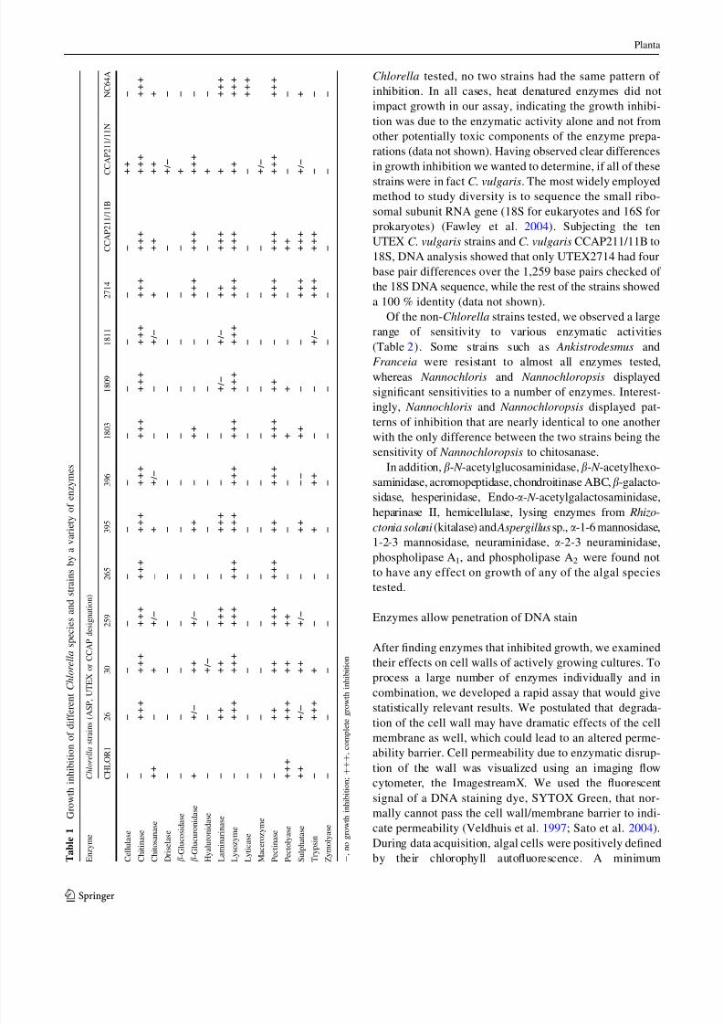
While no individual enzymes affected growth on all strains
tested, chitinase, lysozyme, and pectinase had the broadest
effect on the Chlorella strains (Table 1). In addition,
chitosanase, b-glucuronidase, laminarinase, sulfatase, and
trypsin inhibited growth of the majority of Chlorella
strains. C. vulgaris UTEX395 was also inhibited by
mutanolysin and proteinase K. Alginate lyase showed
peculiar results in that a halo of increased growth typically
would form around the spotted area. We interpreted this to
be an effect of the release of mono-saccharides from the
agar substrate near the area, where the enzyme was spotted.
As such, we could not conclude that alginate lyase inhib-
ited growth using a plate-based assay for most strains,
though Chlorella sp. CHLORI and C. emersonii CCAP211/
11N had obvious zones of inhibition (data not shown).
Of the Chlorella strains we tested, only C. emersonii
CCAP211/11N showed sensitivity to cellulase and had
only very minor inhibition by drieselase and macerozyme,
which both contain a cellulase. Of the 14 strains of
Planta
1 3
7/26/2019 Enzymatic Cell Wall Degradation of Chlorella Vulgaris and Other Microalgae for Biofuels Production 2012
http://slidepdf.com/reader/full/enzymatic-cell-wall-degradation-of-chlorella-vulgaris-and-other-microalgae 6/15
Chlorella tested, no two strains had the same pattern of
inhibition. In all cases, heat denatured enzymes did not
impact growth in our assay, indicating the growth inhibi-
tion was due to the enzymatic activity alone and not from
other potentially toxic components of the enzyme prepa-
rations (data not shown). Having observed clear differences
in growth inhibition we wanted to determine, if all of these
strains were in fact C. vulgaris. The most widely employedmethod to study diversity is to sequence the small ribo-
somal subunit RNA gene (18S for eukaryotes and 16S for
prokaryotes) (Fawley et al. 2004). Subjecting the ten
UTEX C. vulgaris strains and C. vulgaris CCAP211/11B to
18S, DNA analysis showed that only UTEX2714 had four
base pair differences over the 1,259 base pairs checked of
the 18S DNA sequence, while the rest of the strains showed
a 100 % identity (data not shown).
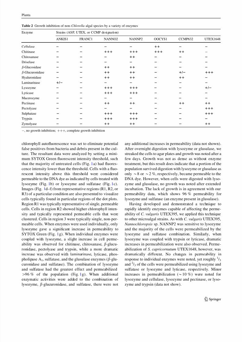
Of the non-Chlorella strains tested, we observed a large
range of sensitivity to various enzymatic activities
(Table 2). Some strains such as Ankistrodesmus and
Franceia were resistant to almost all enzymes tested,whereas Nannochloris and Nannochloropsis displayed
significant sensitivities to a number of enzymes. Interest-
ingly, Nannochloris and Nannochloropsis displayed pat-
terns of inhibition that are nearly identical to one another
with the only difference between the two strains being the
sensitivity of Nannochloropsis to chitosanase.
In addition, b- N -acetylglucosaminidase, b- N -acetylhexo-
saminidase, acromopeptidase, chondroitinase ABC,b-galacto-
sidase, hesperinidase, Endo-a- N -acetylgalactosaminidase,
heparinase II, hemicellulase, lysing enzymes from Rhizo-
ctonia solani (kitalase) and Aspergillus sp., a-1-6 mannosidase,
1-2-3 mannosidase, neuraminidase, a-2-3 neuraminidase,
phospholipase A1, and phospholipase A2 were found not
to have any effect on growth of any of the algal species
tested.
Enzymes allow penetration of DNA stain
After finding enzymes that inhibited growth, we examined
their effects on cell walls of actively growing cultures. To
process a large number of enzymes individually and in
combination, we developed a rapid assay that would give
statistically relevant results. We postulated that degrada-
tion of the cell wall may have dramatic effects of the cell
membrane as well, which could lead to an altered perme-
ability barrier. Cell permeability due to enzymatic disrup-
tion of the wall was visualized using an imaging flow
cytometer, the ImagestreamX. We used the fluorescent
signal of a DNA staining dye, SYTOX Green, that nor-
mally cannot pass the cell wall/membrane barrier to indi-
cate permeability (Veldhuis et al. 1997; Sato et al. 2004).
During data acquisition, algal cells were positively defined
by their chlorophyll autofluorescence. A minimum T a b l e 1
G r o w t h i n h i b i t i o n o f d i f f e r e n t C h l o r e l l a s p e c i e s a n d s t r a i n s b y a v a
r i e t y o f e n z y m e s
E n z y m e
C h l o r e l l a s t r a i n s ( A S P , U T E X o r C C A P d e s i g n a t i o n )
C H L O R 1
2 6
3 0
2 5 9
2 6 5
3 9 5
3 9 6
1 8 0 3
1 8 0 9
1 8 1 1
2 7 1 4
C C A P 2 1 1 / 1 1 B
C C A P 2 1 1 / 1 1 N
N C 6 4 A
C e l l u l a s e
2
2
2
2
2
2
2
2
2
2
2
2
1 1
2
C h i t i n a s e
2
1
1 1
1 1 1
1 1 1
1 1 1
1 1 1
1 1 1
1 1 1
1 1 1
1 1 1
1 1 1
1 1 1
1 1 1
1 1 1
C h i t o s a n a s e
1 1
2
1
1 / 2
-
1
1 / 2
2
2
1 / 2
1
1 1
1 1
1
D r i s e l a s e
2
2
2
2
2
2
2
2
2
2
2
2
1 / 2
2
b - G l u c o s i d a s e
2
2
2
2
2
2
2
2
2
2
2
2
1
2
b - G l u c u r o n i d a s e
1
1
/ 2
1 1
1 / 2
2
1 1
2
1 1
2
2
1 1 1
1 1 1
1 1 1
2
H y a l u r o n i d a s e
2
2
1 / 2
2
2
2
2
2
2
2
2
2
1
2
L a m i n a r i n a s e
2
1
1
1 1
1 1 1
2
1 1 1
2
2
1 / 2
1 / 2
1 1
1 1 1
1
1 1 1
L y s o z y m e
2
1
1 1
1 1 1
1 1 1
1 1 1
1 1 1
1 1 1
1 1 1
1 1 1
1 1 1
1 1 1
1 1 1
1 1
1 1 1
L y t i c a s e
2
2
2
2
2
2
2
2
2
2
2
2
2
1 1 1
M a c e r o z y m e
2
2
2
2
2
2
2
2
2
2
2
2
1 / 2
P e c t i n a s e
2
1
1
1 1
1 1 1
1 1 1
1 1
1 1 1
1 1 1
1 1
2
1 1 1
1 1 1
1 1 1
1 1 1
P e c t o l y a s e
1 1 1
1
1 1
1 1
1 1
2
2
2
1
1
2
2
1 1
2
2
S u l p h a t a s e
1 1
1
/ 2
1 1
1 / 2
2
1 1
2 2
1 1
2
2
1 1 1
1 1 1
1 / 2
1
T r y p s i n
2
1
1 1
1
2
2
1
1 1
2
2
1 / 2
1 1 1
1 1 1
2
2
Z y m o l y a s e
2
2
2
2
2
2
2
2
2
2
2
2
2
2
- , n o g r o w t h i n h i b i t i o n ; ? ? ? , c o m p l e t e g r o w t h i n h i b i t i o n
Planta
1 3
7/26/2019 Enzymatic Cell Wall Degradation of Chlorella Vulgaris and Other Microalgae for Biofuels Production 2012
http://slidepdf.com/reader/full/enzymatic-cell-wall-degradation-of-chlorella-vulgaris-and-other-microalgae 7/15
chlorophyll autofluorescence was set to eliminate potential
false positives from bacteria and debris present in the cul-
ture. The resultant data were analyzed by setting a mini-
mum SYTOX Green fluorescent intensity threshold, such
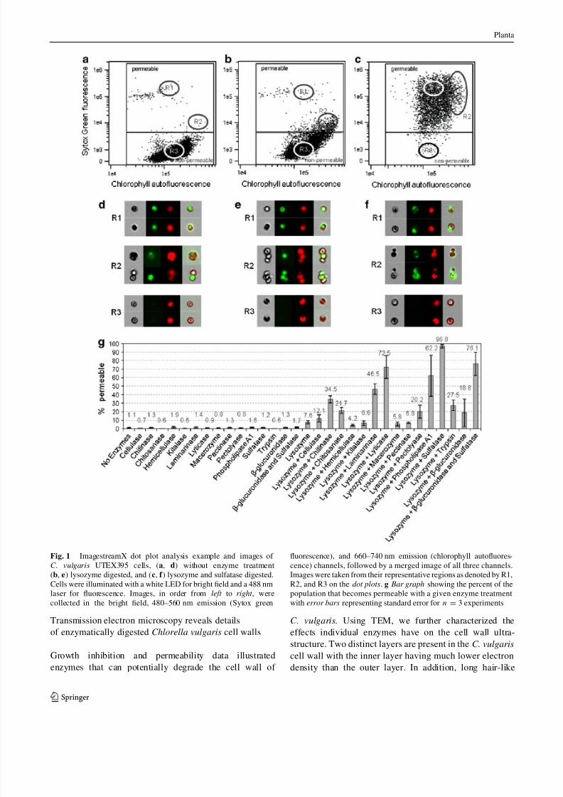
that the majority of untreated cells (Fig. 1a) had fluores-
cence intensity lower than the threshold. Cells with a fluo-
rescent intensity above this threshold were considered
permeable to the DNA dye as indicated by cells treated withlysozyme (Fig. 1b) or lysozyme and sulfatase (Fig. 1c).
Images (Fig. 1d–f) from representative regions (R1, R2, or
R3) of a particular condition are also presented to visualize
cells typically found in particular regions of the dot plots.
Region R1 was typically representative of single, permeable
cells. Cells in region R2 showed higher chlorophyll inten-
sity and typically represented permeable cells that were
clustered. Cells in region 3 were typically single, non-per-
meable cells. When enzymes were used individually, only
lysozyme gave a significant increase in permeability to
SYTOX Green (Fig. 1g). When individual enzymes were
coupled with lysozyme, a slight increase in cell perme-ability was observed for chitinase, chitosanase, b-glucu-
ronidase, pectolyase and trypsin, while a more dramatic
increase was observed with laminarinase, lyticase, phos-
pholipase A1, sulfatase, and the glusulase enzymes (b-glu-
curonidase and sulfatase). The combination of lysozyme
and sulfatase had the greatest effect and permeabilized
[96 % of the population (Fig. 1g). When additional
enzymatic activities were added to the combination of
lysozyme, b-glucuronidase, and sulfatase, there were not
any additional increases in permeability (data not shown).
After overnight digestion with lysozyme or glusulase, we
streaked the cells to agar plates and growth was noted after a
few days. Growth was not as dense as without enzyme
treatment, but this result does indicate that a portion of the
population survived digestion with lysozyme or glusulase as
only *8 or *2 %, respectively, became permeable to the
DNA dye. However, when cells were digested with lyso-zyme and glusulase, no growth was noted after extended
incubation. The lack of growth is in agreement with our
permeability data, which shows 96 % permeability for
lysozyme and sulfatase (an enzyme present in glusulase).
Having developed and demonstrated a technique to
rapidly identify enzymes capable of affecting the perme-
ability of C. vulgaris UTEX395, we applied this technique
to other microalgal strains. As with C. vulgaris UTEX395,
Nannochloropsis sp. NANNP2 was sensitive to lysozyme
and the majority of the cells were permeabilized by the
lysozyme and sulfatase combination. Similarly, when
lysozyme was coupled with trypsin or lyticase, dramaticincreases in permeabilization were also observed. Perme-
abilization of S. capricornutum UTEX1648, however, was
dramatically different. No changes in permeability in
response to individual enzymes were noted, yet roughly 1 / 3and 2 / 3 of the cells were permeabilized using lysozyme and
sulfatase or lysozyme and lyticase, respectively. Minor
increases in permeabilization (*10 %) were noted for
lysozyme and cellulase, lysozyme and pectinase, or lyso-
zyme and trypsin (data not show).
Table 2 Growth inhibition of non-Chlorella algal species by a variety of enzymes
Enzyme Strains (ASP, UTEX, or CCMP designation)
ANKIS1 FRANC1 NANNO2 NANNP2 OOCYS1 CCMP632 UTEX1648
Cellulase 2 2 2 2 11 2 2
Chitinase 2 2 111 111 111 11 -
Chitosanase 2 2 2 11 2 2 2
Driselase 2 2 2 2 2 2 2
b-Glucosidase 2 2 11 11 2 2 2
b-Glucuronidase 2 2 11 11 2 1 / 2 111
Hyaluronidase 2 2 11 11 2 11 2
Laminarinase 1 / 2 2 2 2 2 2 2
Lysozyme 2 2 111 111 2 2 1 / 2
Lyticase 2 2 111 111 2 2 2
Macerozyme 2 2 2 2 2 2 2
Pectinase 2 2 11 11 2 11 11
Pectolyase 2 2 2 2 2 2 111
Sulphatase 2 2 111 111 2 2 111
Trypsin 2 2 111 111 2 2 2
Zymolyase 2 2 11 11 2 2 11
-, no growth inhibition; ???, complete growth inhibition
Planta
1 3
7/26/2019 Enzymatic Cell Wall Degradation of Chlorella Vulgaris and Other Microalgae for Biofuels Production 2012
http://slidepdf.com/reader/full/enzymatic-cell-wall-degradation-of-chlorella-vulgaris-and-other-microalgae 8/15
Transmission electron microscopy reveals details
of enzymatically digested Chlorella vulgaris cell walls
Growth inhibition and permeability data illustrated
enzymes that can potentially degrade the cell wall of
C. vulgaris. Using TEM, we further characterized the
effects individual enzymes have on the cell wall ultra-
structure. Two distinct layers are present in the C. vulgaris
cell wall with the inner layer having much lower electron
density than the outer layer. In addition, long hair-like
Fig. 1 ImagestreamX dot plot analysis example and images of C. vulgaris UTEX395 cells, (a, d) without enzyme treatment
(b, e) lysozyme digested, and (c, f ) lysozyme and sulfatase digested.
Cells were illuminated with a white LED for bright field and a 488 nm
laser for fluorescence. Images, in order from left to right , were
collected in the bright field, 480–560 nm emission (Sytox green
fluorescence), and 660–740 nm emission (chlorophyll autofluores-cence) channels, followed by a merged image of all three channels.
Images were taken from their representative regions as denoted by R1,
R2, and R3 on the dot plots. g Bar graph showing the percent of the
population that becomes permeable with a given enzyme treatment
with error bars representing standard error for n = 3 experiments
Planta
1 3

7/26/2019 Enzymatic Cell Wall Degradation of Chlorella Vulgaris and Other Microalgae for Biofuels Production 2012
http://slidepdf.com/reader/full/enzymatic-cell-wall-degradation-of-chlorella-vulgaris-and-other-microalgae 9/15
fibers protrude from the outer layer (Fig. 2a, c). In agree-
ment with the growth inhibition and permeability data
displayed in Table 1 and Fig. 1, we found that lysozyme
had a dramatic effect on the cell wall of C. vulgaris.
Lysozyme, an enzyme that typically acts on bacterial
peptidoglycan, a polymer of b-1,4 linked N -acetylgluco-
samine and N -acetylmuramic acid, seems to swell the outer
surface of the electron-dense layer and lower the density of
the outermost portion such that there are now three distinct
layers. Lysozyme also removes the hair-like fibers that
protrude from the surface of the cell (Figs. 2b and 3a).
Graves et al. (1999) demonstrated that these hair-like fibers
on the surface of C. variabilis NC64A are hyaluron, a
linear polysaccharide chain composed of alternating b-1,4-
glucuronic acid and b-1,3- N -acetylglucosamine groups,
and may play a role in preventing superinfection of the
PBCV-1 virus. Curiously, chitinase, which degrades poly-
b-1,4-D- N -acetylglucosamine, seemed to give a different
pattern of digestion to the C. vulgaris wall, not affecting
the hair-like fibers, but causing a general decrease in
electron density of the outer wall similar to lysozyme
(Fig. 2d). Chitosanase (Fig. 2e) caused a minor thinning of
the electron-dense outer region of the cell wall, while
laminarinase (Fig. 2f) caused a slight increase in density of
the inner wall. Trypsin digestion caused a general decrease
in density of the outer wall in a fashion similar to that of
chitinase and an increase in cell wall thickness (Fig. 2g).
While sulfatase, b-glucuronidase, pectinase, and cellulase
caused no systematic changes to the ultrastructure of the C.
vulgaris cell wall (Fig. 2h–k), we were surprised to see
quite dramatic changes in the wall in response to phos-
pholipase A1. Phospholipase A1 digestion removed the
hair-like fibers and caused a general widening of the outer
wall resulting in visible stratification of layers in the outer
wall (Fig. 2l).
Increased permeability when certain enzymes are cou-
pled with lysozyme, and the ability of lysozyme to degrade
the outer surface of the C. vulgaris cell led us to hypoth-
esize that lysozyme is opening up the cell wall for further
degradation by enzymes that cannot normally cross the
outer wall of C. vulgaris to find substrates buried within the
wall. When lysozyme was coupled with chitinase, we saw a
dramatic change in the cell wall ultrastructure. The cell
wall more than doubled in width and the electron-dense
outer wall was thinned to a greater degree than lysozyme
alone (Fig. 3b). When lysozyme was added in conjunction
with some enzymes such as cellulase, pectinase or phos-
pholipase A1, we noticed that the outer wall no longer
maintained a smooth interaction with the inner wall. The
outer wall seems to ‘‘push in’’ and displace the membrane
toward the center of the cell (Fig. 3c, d, g). When coupled
with lysozyme, chitosanase, trypsin, and b-glucuronidase
showed no drastic effect over that of lysozyme alone
(Fig. 3e, h, i), whereas sulfatase and b-glucuronidase with
Fig. 2 Transmission electron micrographs of C. vulgaris UTEX395
digested with a no enzyme, b lysozyme, c no enzyme, d chitinase,
e chitosanase, f laminarinase, g trypsin, h sulfatase, i b-glucuronidase,
j pectinase, k cellulase, l phospholipase A1. Scale bar 200 nm (a, b),
100 nm (c–l)
Planta
1 3
7/26/2019 Enzymatic Cell Wall Degradation of Chlorella Vulgaris and Other Microalgae for Biofuels Production 2012
http://slidepdf.com/reader/full/enzymatic-cell-wall-degradation-of-chlorella-vulgaris-and-other-microalgae 10/15
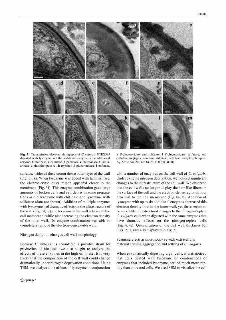
sulfatase widened the electron-dense outer layer of the wall
(Fig. 3 j, k). When lysozyme was added with laminarinase,
the electron-dense outer region appeared closer to the
membrane (Fig. 3f). This enzyme combination gave large
amounts of broken cells and cell debris in some prepara-
tions as did lysozyme with chitinase and lysozyme with
sulfatase (data not shown). Addition of multiple enzymes
with lysozyme had dramatic effects on the ultrastructure of
the wall (Fig. 3l, m) and location of the wall relative to the
cell membrane, while also increasing the electron density
of the inner wall. No enzyme combination was able to
completely remove the electron-dense outer wall.
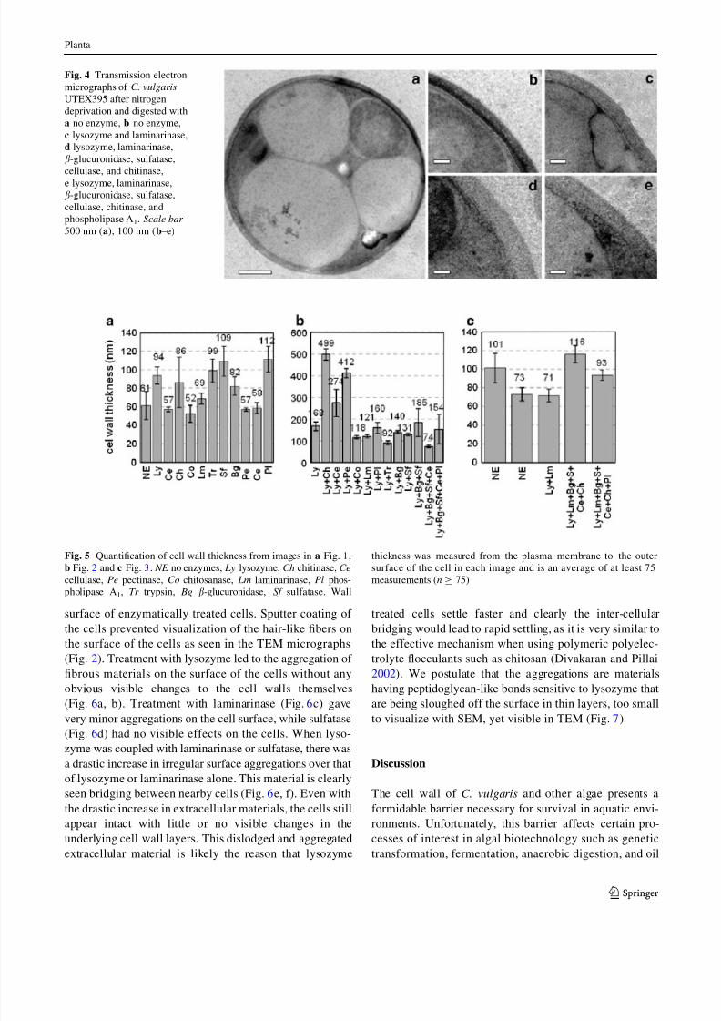
Nitrogen depletion changes cell wall morphology
Because C. vulgaris is considered a possible strain for
production of biodiesel, we also sought to analyze the
effects of these enzymes in the high oil phase. It is very
likely that the composition of the cell wall could change
dramatically under nitrogen deprivation conditions. Using
TEM, we analyzed the effects of lysozyme in conjunction
with a number of enzymes on the cell wall of C. vulgaris.
Under extreme nitrogen deprivation, we noticed significant
changes to the ultrastructure of the cell wall. We observed
that the cell walls no longer display the hair-like fibers on
the surface of the cell and the electron-dense region is now
proximal to the cell membrane (Fig. 4a, b). Addition of
lysozyme with up to six additional enzymes decreased this
electron density now in the inner wall, yet there seems to
be very little ultrastructural changes to the nitrogen-deplete
C. vulgaris cells when digested with the same enzymes that
have dramatic effects on the nitrogen-replete cells
(Fig. 4c–e). Quantification of the cell wall thickness forFigs. 2, 3, and 4 is displayed in Fig. 5.
Scanning electron microscopy reveals extracellular
material causing aggregation and settling of C. vulgaris
When enzymatically digesting algal cells, it was noticed
that cells treated with lysozyme or combinations of
enzymes that included lysozyme, settled much more rap-
idly than untreated cells. We used SEM to visualize the cell
Fig. 3 Transmission electron micrographs of C. vulgaris UTEX395
digested with lysozyme and the additional enzyme, a no additional
enzyme, b chitinase, c cellulase, d pectinase, e chitosanase, f lamin-
arinase, g phospholipase A1, h trypsin, i b-glucuronidase, j sulfatase,
k b-glucuronidase and sulfatase, l b-glucuronidase, sulfatase, and
cellulase, m b-glucuronidase, sulfatase, cellulase, and phospholipase
A1. Scale bar 200 nm (a–c), 100 nm (d–m)
Planta
1 3
7/26/2019 Enzymatic Cell Wall Degradation of Chlorella Vulgaris and Other Microalgae for Biofuels Production 2012
http://slidepdf.com/reader/full/enzymatic-cell-wall-degradation-of-chlorella-vulgaris-and-other-microalgae 11/15
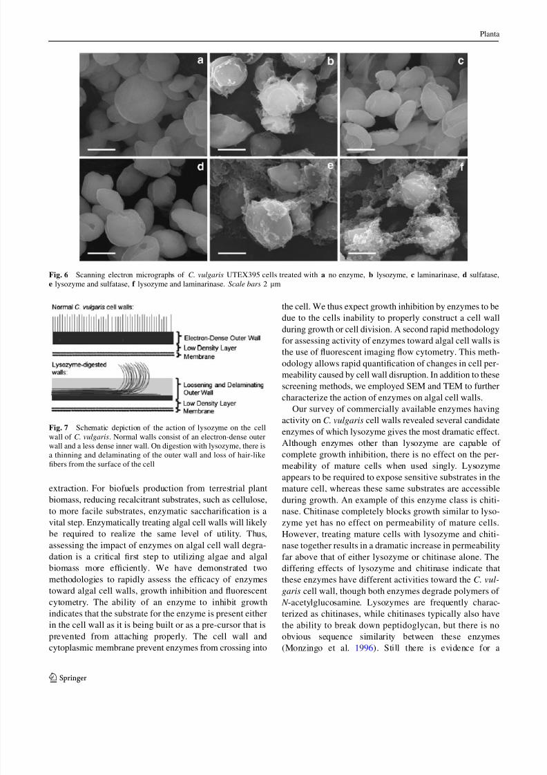
surface of enzymatically treated cells. Sputter coating of
the cells prevented visualization of the hair-like fibers on
the surface of the cells as seen in the TEM micrographs
(Fig. 2). Treatment with lysozyme led to the aggregation of
fibrous materials on the surface of the cells without any
obvious visible changes to the cell walls themselves
(Fig. 6a, b). Treatment with laminarinase (Fig. 6c) gave
very minor aggregations on the cell surface, while sulfatase(Fig. 6d) had no visible effects on the cells. When lyso-
zyme was coupled with laminarinase or sulfatase, there was
a drastic increase in irregular surface aggregations over that
of lysozyme or laminarinase alone. This material is clearly
seen bridging between nearby cells (Fig. 6e, f). Even with
the drastic increase in extracellular materials, the cells still
appear intact with little or no visible changes in the
underlying cell wall layers. This dislodged and aggregated
extracellular material is likely the reason that lysozyme
treated cells settle faster and clearly the inter-cellular
bridging would lead to rapid settling, as it is very similar to
the effective mechanism when using polymeric polyelec-
trolyte flocculants such as chitosan (Divakaran and Pillai
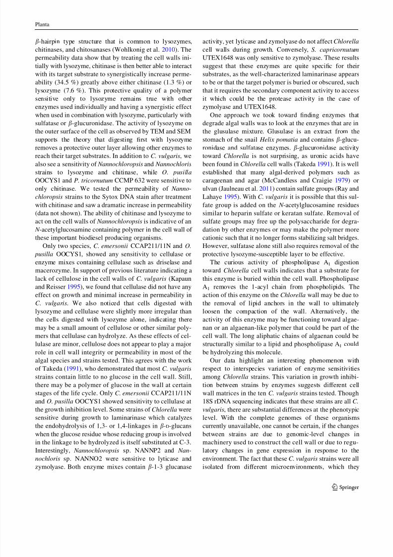
2002). We postulate that the aggregations are materials
having peptidoglycan-like bonds sensitive to lysozyme that
are being sloughed off the surface in thin layers, too small
to visualize with SEM, yet visible in TEM (Fig. 7).
Discussion
The cell wall of C. vulgaris and other algae presents a
formidable barrier necessary for survival in aquatic envi-
ronments. Unfortunately, this barrier affects certain pro-
cesses of interest in algal biotechnology such as genetic
transformation, fermentation, anaerobic digestion, and oil
Fig. 4 Transmission electron
micrographs of C. vulgaris
UTEX395 after nitrogen
deprivation and digested with
a no enzyme, b no enzyme,
c lysozyme and laminarinase,
d lysozyme, laminarinase,
b-glucuronidase, sulfatase,
cellulase, and chitinase,
e lysozyme, laminarinase,
b-glucuronidase, sulfatase,
cellulase, chitinase, and
phospholipase A1. Scale bar
500 nm (a), 100 nm (b–e)
Fig. 5 Quantification of cell wall thickness from images in a Fig. 1,
b Fig. 2 and c Fig. 3. NE no enzymes, Ly lysozyme, Ch chitinase, Ce
cellulase, Pe pectinase, Co chitosanase, Lm laminarinase, Pl phos-
pholipase A1, Tr trypsin, Bg b-glucuronidase, Sf sulfatase. Wall
thickness was measured from the plasma membrane to the outer
surface of the cell in each image and is an average of at least 75
measurements (n C 75)
Planta
1 3
7/26/2019 Enzymatic Cell Wall Degradation of Chlorella Vulgaris and Other Microalgae for Biofuels Production 2012
http://slidepdf.com/reader/full/enzymatic-cell-wall-degradation-of-chlorella-vulgaris-and-other-microalgae 12/15
extraction. For biofuels production from terrestrial plant
biomass, reducing recalcitrant substrates, such as cellulose,
to more facile substrates, enzymatic saccharification is a
vital step. Enzymatically treating algal cell walls will likely
be required to realize the same level of utility. Thus,
assessing the impact of enzymes on algal cell wall degra-dation is a critical first step to utilizing algae and algal
biomass more efficiently. We have demonstrated two
methodologies to rapidly assess the efficacy of enzymes
toward algal cell walls, growth inhibition and fluorescent
cytometry. The ability of an enzyme to inhibit growth
indicates that the substrate for the enzyme is present either
in the cell wall as it is being built or as a pre-cursor that is
prevented from attaching properly. The cell wall and
cytoplasmic membrane prevent enzymes from crossing into
the cell. We thus expect growth inhibition by enzymes to be
due to the cells inability to properly construct a cell wall
during growth or cell division. A second rapid methodology
for assessing activity of enzymes toward algal cell walls is
the use of fluorescent imaging flow cytometry. This meth-
odology allows rapid quantification of changes in cell per-
meability caused by cell wall disruption. In addition to these
screening methods, we employed SEM and TEM to further
characterize the action of enzymes on algal cell walls.
Our survey of commercially available enzymes havingactivity on C. vulgaris cell walls revealed several candidate
enzymes of which lysozyme gives the most dramatic effect.
Although enzymes other than lysozyme are capable of
complete growth inhibition, there is no effect on the per-
meability of mature cells when used singly. Lysozyme
appears to be required to expose sensitive substrates in the
mature cell, whereas these same substrates are accessible
during growth. An example of this enzyme class is chiti-
nase. Chitinase completely blocks growth similar to lyso-
zyme yet has no effect on permeability of mature cells.
However, treating mature cells with lysozyme and chiti-
nase together results in a dramatic increase in permeabilityfar above that of either lysozyme or chitinase alone. The
differing effects of lysozyme and chitinase indicate that
these enzymes have different activities toward the C. vul-
garis cell wall, though both enzymes degrade polymers of
N -acetylglucosamine. Lysozymes are frequently charac-
terized as chitinases, while chitinases typically also have
the ability to break down peptidoglycan, but there is no
obvious sequence similarity between these enzymes
(Monzingo et al. 1996). Still there is evidence for a
Fig. 6 Scanning electron micrographs of C. vulgaris UTEX395 cells treated with a no enzyme, b lysozyme, c laminarinase, d sulfatase,
e lysozyme and sulfatase, f lysozyme and laminarinase. Scale bars 2 lm
Fig. 7 Schematic depiction of the action of lysozyme on the cell
wall of C. vulgaris. Normal walls consist of an electron-dense outer
wall and a less dense inner wall. On digestion with lysozyme, there is
a thinning and delaminating of the outer wall and loss of hair-like
fibers from the surface of the cell
Planta
1 3
7/26/2019 Enzymatic Cell Wall Degradation of Chlorella Vulgaris and Other Microalgae for Biofuels Production 2012
http://slidepdf.com/reader/full/enzymatic-cell-wall-degradation-of-chlorella-vulgaris-and-other-microalgae 13/15
b-hairpin type structure that is common to lysozymes,
chitinases, and chitosanases (Wohlkonig et al. 2010). The
permeability data show that by treating the cell walls ini-
tially with lysozyme, chitinase is then better able to interact
with its target substrate to synergistically increase perme-
ability (34.5 %) greatly above either chitinase (1.3 %) or
lysozyme (7.6 %). This protective quality of a polymer
sensitive only to lysozyme remains true with otherenzymes used individually and having a synergistic effect
when used in combination with lysozyme, particularly with
sulfatase or b-glucuronidase. The activity of lysozyme on
the outer surface of the cell as observed by TEM and SEM
supports the theory that digesting first with lysozyme
removes a protective outer layer allowing other enzymes to
reach their target substrates. In addition to C. vulgaris, we
also see a sensitivity of Nannochloropsis and Nannochloris
strains to lysozyme and chitinase, while O. pusilla
OOCYS1 and P. tricornutum CCMP 632 were sensitive to
only chitinase. We tested the permeability of Nanno-
chloropsis strains to the Sytox DNA stain after treatmentwith chitinase and saw a dramatic increase in permeability
(data not shown). The ability of chitinase and lysozyme to
act on the cell walls of Nannochloropsis is indicative of an
N -acetylglucosamine containing polymer in the cell wall of
these important biodiesel producing organisms.
Only two species, C. emersonii CCAP211/11N and O.
pusilla OOCYS1, showed any sensitivity to cellulase or
enzyme mixes containing cellulase such as driselase and
macerozyme. In support of previous literature indicating a
lack of cellulose in the cell walls of C. vulgaris (Kapaun
and Reisser 1995), we found that cellulase did not have any
effect on growth and minimal increase in permeability in
C. vulgaris. We also noticed that cells digested with
lysozyme and cellulase were slightly more irregular than
the cells digested with lysozyme alone, indicating there
may be a small amount of cellulose or other similar poly-
mers that cellulase can hydrolyze. As these effects of cel-
lulase are minor, cellulose does not appear to play a major
role in cell wall integrity or permeability in most of the
algal species and strains tested. This agrees with the work
of Takeda (1991), who demonstrated that most C. vulgaris
strains contain little to no glucose in the cell wall. Still,
there may be a polymer of glucose in the wall at certain
stages of the life cycle. Only C. emersonii CCAP211/11N
and O. pusilla OOCYS1 showed sensitivity to cellulase at
the growth inhibition level. Some strains of Chlorella were
sensitive during growth to laminarinase which catalyzes
the endohydrolysis of 1,3- or 1,4-linkages in b-D-glucans
when the glucose residue whose reducing group is involved
in the linkage to be hydrolyzed is itself substituted at C-3.
Interestingly, Nannochloropsis sp. NANNP2 and Nan-
nochloris sp. NANNO2 were sensitive to lyticase and
zymolyase. Both enzyme mixes contain b-1-3 glucanase
activity, yet lyticase and zymolyase do not affect Chlorella
cell walls during growth. Conversely, S. capricornutum
UTEX1648 was only sensitive to zymolyase. These results
suggest that these enzymes are quite specific for their
substrates, as the well-characterized laminarinase appears
to be or that the target polymer is buried or obscured, such
that it requires the secondary component activity to access
it which could be the protease activity in the case of zymolyase and UTEX1648.
One approach we took toward finding enzymes that
degrade algal walls was to look at the enzymes that are in
the glusulase mixture. Glusulase is an extract from the
stomach of the snail Helix pomatia and contains b-glucu-
ronidase and sulfatase enzymes. b-glucuronidase activity
toward Chlorella is not surprising, as uronic acids have
been found in Chlorella cell walls (Takeda 1991). It is well
established that many algal-derived polymers such as
carageenan and agar (McCandless and Craigie 1979) or
ulvan (Jaulneau et al. 2011) contain sulfate groups (Ray and
Lahaye 1995). With C. vulgaris it is possible that this sul-fate group is added on the N -acetylglucosamine residues
similar to heparin sulfate or keratan sulfate. Removal of
sulfate groups may free up the polysaccharide for degra-
dation by other enzymes or may make the polymer more
cationic such that it no longer forms stabilizing salt bridges.
However, sulfatase alone still also requires removal of the
protective lysozyme-susceptible layer to be effective.
The curious activity of phospholipase A1 digestion
toward Chlorella cell walls indicates that a substrate for
this enzyme is buried within the cell wall. Phospholipase
A1 removes the 1-acyl chain from phospholipids. The
action of this enzyme on the Chlorella wall may be due to
the removal of lipid anchors in the wall to ultimately
loosen the compaction of the wall. Alternatively, the
activity of this enzyme may be functioning toward algae-
nan or an algaenan-like polymer that could be part of the
cell wall. The long aliphatic chains of algaenan could be
structurally similar to a lipid and phospholipase A1 could
be hydrolyzing this molecule.
Our data highlight an interesting phenomenon with
respect to interspecies variation of enzyme sensitivities
among Chlorella strains. This variation in growth inhibi-
tion between strains by enzymes suggests different cell
wall matrices in the ten C. vulgaris strains tested. Though
18S rDNA sequencing indicates that these strains are all C.
vulgaris, there are substantial differences at the phenotypic
level. With the complete genomes of these organisms
currently unavailable, one cannot be certain, if the changes
between strains are due to genomic-level changes in
machinery used to construct the cell wall or due to regu-
latory changes in gene expression in response to the
environment. The fact that these C. vulgaris strains were all
isolated from different microenvironments, which they
Planta
1 3
7/26/2019 Enzymatic Cell Wall Degradation of Chlorella Vulgaris and Other Microalgae for Biofuels Production 2012
http://slidepdf.com/reader/full/enzymatic-cell-wall-degradation-of-chlorella-vulgaris-and-other-microalgae 14/15
have presumably adapted to, could lead to differential gene
expression patterns when all strains are put into similar
conditions. In addition, within a single strain of algae, there
appears to be extreme responses to minuscule changes in
the environment. We noted some high variability in the cell
wall permeability study data which may be indicative of
subtle differences in cell wall composition and architecture
due to minute differences in growth conditions duringreplicate experiments. Previous studies suggested that algal
cell walls change dramatically as the cell ages, such that an
older culture subjected to the same enzymatic treatment is
more resistant (Van Donk et al. 1997). The range of
enzymatic activities showing high degrees of variability in
their impact on cell wall permeability between our repli-
cate experiments suggests that no one single, easily iden-
tifiable component is changing, but rather wholesale
changes in cell wall composition may be occurring.
The changes in cell walls in response to the environment
are highlighted with the TEM images of cell walls after
nitrogen depletion (Fig. 4). Changes in ultrastructure fornitrogen-deplete cells may be due to the cells scavenging
the amino sugars from the wall to use as a source of
nitrogen. This would help explain the extended period of
growth for C. vulgaris when cells are transferred to nitro-
gen-free medium (Guarnieri et al. 2011). The cell walls of
nitrogen-deplete cells look similar to lysozyme digested
cells (Figs. 3a, 4a) and lack exterior hairs. The cells could
be resorbing this layer, which contains N -acetylglucosa-
mine as a source of nitrogen under nitrogen deprivation
conditions. Loss of hair-like fibers supports this hypothesis
since hyaluron contains N -acetylglucosamine and has been
postulated to be the major component making up these
hairs (Graves et al. 1999).
In conclusion, we have found that mature Chlorella cell
walls are protected by a durable polymer of unknown
composition that is sensitive to lysozyme treatment.
Lysozyme alone does not have a huge impact in permea-
bilizing mature cells, yet by removing this protective layer,
other substrates become available to a wide range of dif-
fering enzymatic activities. This layer must be established
late in the growth or maturing stage of individual cells as
growing cells are readily susceptible to a variety of
enzymes causing slow or a complete block in growth. Our
results show that in methodologies or processes using intact
algal cells or residual algal biomass, enzymatic treatment
can have large impacts of the permeability of the algal cell
walls and may be useful in optimization. Certainly lyso-
zyme and certain other enzymes can play a role in the
facilitated utilization of algae or algal biomass.
Acknowledgments The authors would like to thank Jonathan
Meuser for help with 18S RNA gene sequencing, Ben Smith for help
with quantification of settling, Todd Vinzant and the Biomass Surface
Characterization Laboratory at NREL for help in SEM image
acquisition, and Philip Pienkos for technical discussions and manu-
script review. This project was funded by NREL’s Laboratory
Directed Research and Development program.
References
Afi L, Metzger P, Largeau C, Connan J, Berkaloff C, Rousseau B(1996) Bacterial degradation of green microalgae: incubation of
Chlorella emersonii and Chlorella vulgaris with Pseudomonas
oleovorans and Flavobacterium aquatile. Org Geochem 25:
117–130
Atkinson AW, Gunning BESJ, John PCL (1972) Sporopollenin in the
cell wall of Chlorella and other algae: ultrastructure, chemistry,
and incorporation of 14
C-acetate, studied in synchronous
cultures. Planta 107:1–32
Barclay W, Johansen J, Chelf P, Nagle N, Roessler PG, Lemke P
(1987) Microalgae culture collection 1986–1987. SERI/SP-232-
3079 www.osti.gov/bridge/product.biblio.jsp?osti_id=6953341
Blokker P, Schouten S, Van Den Ende H, De Leeuw JW, Sinninghe
Damaste JS (1998) Cell wall-specific w-hydroxy fatty acids in
some freshwater green microalgae. Phytochemistry 49:691–695
Braun E, Aach HG (1975) Enzymatic degradation of the cell wall of Chlorella. Planta 126:181–185
Brennan L, Owende P (2010) Biofuels from microalgae—a review of
technologies for production, processing, and extractions of
biofuels and co-products. Renew Sustain Energy Rev 14:
557–577
Brown LM (1982) Production of axenic cultures of algae by an
osmotic method. Phycologia 21:408–410
Burczyk J, Terminska-Pabis K, Smietana B (1995) Cell wall neutral
sugar composition of Chlorococcalean algae forming and not
forming acetolysis resistant biopolymer. Phytochemistry 38:
837–841
Corre G, Templier J, Largeau C, Rousseau B, Berkaloff C (1996)
Influence of cell wall composition on the resistance of two
Chlorella species (Chlorophyta) to detergents. J Phycol 32:584–
590Davis R, Aden A, Pienkos PT (2011) Techno-economic analysis of
autotrophic microalgae for fuel production. Appl Energy 88:
3524–3531
Dawson SC, Pace NR (2002) Novel kingdom-level eukaryotic
diversity in anoxic environments. PNAS 99:8324–8329
Derenne S, Largeau C, Berkaloff C, Rousseau B, Wilhelm C, Hatcher
PG (1992) Non-hydrolysable macromolecular constituents from
outer walls of Chlorella fusca and Nanochlorum eucaryotum.
Phytochemistry 31:1923–1929
Divakaran R, Pillai VNS (2002) Flocculation of algae using chitosan.
J Appl Phycol 14:419–422
Fawley MW, Fawley KP, Buchheim MA (2004) Molecular diversity
among communities of freshwater microchlorophytes. Microb
Ecol 48(4):489–499. doi:10.1007/s00248-004-0214-4
Fukada K, Inoue T, Shiraishi H (2006) A posttranslationally regulatedprotease, VheA, is involved in the liberation of juveniles from
parental spheroids in Volvox carteri. Plant Cell 18:2554–2566
Gelin F, Volkman JK, Largeau C, Derenne S, Sinninghe Damaste JS,
De Leeuw JW (1999) Distribution of aliphatic, nonhydrolyzable
biopolymers in marine microalgae. Org Geochem 30:147–159
Glover H (1977) Effects of iron deficiency on Isochrysis galbana
(Chrysophyceae) and Phaeodactylum tricornutum (Bacillario-
phyceae). J Phycol 13:208–212
Graves MV, Burbank DE, Roth R, Heuser J, DeAngelis PL, VanEtten
JL (1999) Hyaluronan synthesis in virus PBCV-1 infected
Chlorella-like green algae. Virology 257:15–23
Planta
1 3
7/26/2019 Enzymatic Cell Wall Degradation of Chlorella Vulgaris and Other Microalgae for Biofuels Production 2012
http://slidepdf.com/reader/full/enzymatic-cell-wall-degradation-of-chlorella-vulgaris-and-other-microalgae 15/15
Guarnieri MT, Nag A, Smolinski SL, Darzins A, Seibert M, Pienkos
PT (2011) Examination of triacylglycerol biosynthetic pathways
via de novo transcriptomic and proteomic analyses in an
unsequenced microalga. PLoS ONE 6:e25851
Guarnieri MT, Laurens LM, Knoshaug EP, Chou YC, Donohoe BS,
Pienkos PT (2012) Complex systems engineering: a case study
for an unsequenced microalga. In: Patnaik R (ed) Engineering
complex phenotypes in industrial strains, Wiley, New York
Gunnison D, Alexander M (1975) Basis for the resistance of several
algae to microbial decompostion. Appl Microbiol 29:729–738
Honjoh K, Suga K, Shinohara F, Maruyama I, Miyamoto T, Hatano S,
Iio M (2003) Preparation of protoplasts from Chlorella vulgaris
K-73122 and cell wall regeneration of protoplasts from C.
vulgaris K-73122 and C-27. J Fac Agric 47:257–266
Huss VAR, Frank C, Hartmann EC, Hirmer M, Kloboucek A, Seidel
BM, Wenzeler P, Kessler E (1999) Biochemical taxonomy and
molecular phylogeny of the genus Chlorella sensu late (Chlo-
rophyta). J Phycol 35:587–598
Jaulneau V, Lafitte C, Corio-Costet M-F, Stadnik MJ, Salamagne S,
Briand X, Esquerre-Tugaye M-T, Dumas B (2011) An Ulva
armoricana extract protects plants against three powdery mildew
pathogens. Eur J Plant Pathol 131:393–401
Kapaun E, Reisser W (1995) A chitin-like glycan in the cell wall of a
Chlorella sp. (Chlorococcales, Chlorophyceae). Planta 197:577–
582
Kapaun E, Loos E, Reisser W (1992) Cell wall composition of virus-
sensitive symbiotic Chlorella species. Phytochemistry 31:3101–
3104
Kim YH, Choi YK, Park J, Lee S, Yang YH, Kim HJ, Park TJ, Kim
YH, Lee SH (2012) Ionic liquid-mediated extraction of lipids
from algal biomass. Bioresour Technol 109:312–315
Knoshaug EP, Darzins A (2011) Algal biofuels: the process. Chem
Eng Progr 107:37–47
Kubo T, Kaida S, Abe J, Saito T, Fukuzawa H, Matsuda Y (2009) The
Chlamydomonas hatching enzyme, sporangin, is expressed in
specific phases of the cell cycle and is localized to the flagella of
daughter cells within the sporangial cell wall. Plant Cell Physiol
50(3):572–583. doi:10.1093/pcp/pcp016
Lee JY, Yoo C, Jun SY, Ahn CY, Oh HM (2010) Comparison of
several methods for effective lipid extraction from microalgae.
Bioresour Technol 101:S75–S77
Malis-Arad S, Friedlander M, Ben-Arie R, Richmond AE (1980)
Alkalinity-induced aggregationin Chlorella vulgaris I.Changesin
cell volume and cell-wall structure. Plant Cell Physiol 21:27–35
McCandless EL, Craigie JS (1979) Sulfated polysaccharides in red
and brown algae. Ann Rev Plant Physiol 30:41–53
Monzingo AF, Marcotte EM, Hart PJ, Robertas JD (1996) Chitinases,
chitosanases, and lysozymes can be divided into procaryotic and
eucaryotic families sharing a conserved core. Nat Struct Mol
Biol 3(2):133–140
Ogawa K, Yamaura M, Maruyama I (1997) Isolation and identifica-
tion of 2-O-methyl-L-rhamnose and 3-O-methyl-L-rhamnose as
constituents of an acidic polysaccharide of Chlorella vulgaris.
Biosci Biotechnol Biochem 61:539–540Ogawa K, Yamaura M, Ikeda Y, Kondo S (1998) New aldobiuronic
acid, 3-O-a-D-glucopyranuronosyl-L-rhamnopyranose, from an
acidic polysaccharide of Chlorella vulgaris. Biosci Biotechnol
Biochem 62:2030–2031
Ogawa K, Ikeda Y, Kondo S (1999) A new trisaccharide,
a-D-glucopyranuronosyl-(1–3)-a-L-rhamnopyranosyl-(1–2)-a-L-
rhamnopyranose from Chlorella vulgaris. Carbohydr Res 321:
128–131
Ogawa K, Arai M, Naganawa H, Ikeda Y, Kondo S (2001) A new
b-D-galactan having 3-O-methyl-D-galactose from Chlorella
vulgaris. J Appl Glycosci 48:325–330
Pienkos PT, Darzins A (2009) The promise and challenges of
microalgal-derived biofuels. Biofuels Bioprod Bioref 3:431–440
Popper ZA, Tuohy MG (2010) Beyond the green: understanding the
evolutionary puzzle of plant and algal cell walls. Plant Physiol
153:373–383
Ray B, Lahaye M (1995) Cell-wall polysaccharides from the marine
green alga Ulva ‘‘rigida’’ (Ulvales, Chlorophyta). Extraction and
chemical composition. Carbohydr Res 274:251–261
Sato M, Murata Y, Mizusawa M, Iwahashi H, Oka S (2004) A simple
and rapid dual-fluorescence viability assay for microalgae.
Microbiol Cult Coll 20:53–59
Scott SA, Davey MP, Dennis JS, Horst I, Howe CJ, Lea-Smith DJ,
Smith AG (2010) Biodiesel from algae: challenges and pros-
pects. Curr Opin Biotechnol 21:277–286
Sheehan JT, Dunahay J, Benemann JR, Roessler PG (1998) A look
back at the US department of energy’s aquatic species
program—biodiesel from algae NREL/TP-580-24190 www.nrel.
gov/biomass/pdfs/24190.pdf
Shen Y, Pei Z, Yuan W, Mao E (2009) Effect of nitrogen and
extraction method on algae lipid yield. Int J Agric Biol Eng
2:51–57
Siddiquee MN, Rohani S (2011) Lipid extraction and biodiesel
production from municipal sewage sludges: a review. Renew
Sustain Energy Rev 15:1067–1072
Simpson AJ, Zang X, Kramer R, Hatcher PG (2003) New insights on
the structure of algaenan from Botryococcus braunii race A and
its hexane insoluble botryals based on multidimensional NMR
spectroscopy and electrospray-mass spectrometry techniques.
Phytochemistry 62:783–796
Takeda H (1991) Sugar composition of the cell wall and the
taxonomy of Chlorella (Chlorophyceae). J Phycol 27:224–232
Van Donk E, Lurling M, Hessen DO, Lokhorst GM (1997) Altered
cell wall morphology in nutrient-deficient phytoplankton and its
impact on grazers. Limnol Oceanogr 42:357–364
Van Etten JL, Lane LC, Meints RH (1991) Viruses and virus-like
particles of eukaryotic algae. Microbiol Rev 55:586–620
Veldhuis MJW, Cucci TL, Sieracki ME (1997) Cellular DNA content
of marine phytoplankton using two new fluorochromes: taxo-
nomic and ecological implications. J Phycol 33:527–541
Walter JK, Aach HG (1987) Isolation and characterization of the
enzymes involved in disintegration of the cell wall of Chlorella
fusca. Physiol Plantarum 70:485–490
Wijffels RH, Barbosa MJ (2010) An outlook on microalgal biofuels.
Science 329:796–799
Wohlkonig A, Huet J, Looze Y, Wintjens R (2010) Structural
relationships in the lysozyme superfamily: significant evidence
for glycoside hydrolase signature motifs. PLoS ONE 5:e15388.doi:10.1371/journal.pone.0015388
Yamada T, Sakaguchi K (1982) Comparative studies on Chlorella
cell walls: induction of protoplast formation. Arch Microbiol
132:10–13
Planta
1 3

![Earth Science & Climatic Change...Algae Green algae Chlorella vulgaris is grown near Petrich in South-West Bulgaria [21]. For the test dry extract of Chlorella vulgaris produced by](https://img.pdfslide.us/doc/110x75/5e93d3fcb8548c28da3c9f6b/earth-science-climatic-change-algae-green-algae-chlorella-vulgaris-is.jpg)


![Comparative Study of Catalyst for Biodiesel Synthesis from ... · Comparative Study of Catalyst for Biodiesel Synthesis from Microalgae Chlorella vulgaris Swati Sonawane1, ... [12]](https://img.pdfslide.us/doc/110x75/5e5947bfe8092e68554a33ac/comparative-study-of-catalyst-for-biodiesel-synthesis-from-comparative-study.jpg)