Embed Size (px)
Citation preview

Envoplakin, a Novel Precursor of the Comified Envelope That Has Homology to Desmoplaldn Christ iana Ruhrberg,* M.A. Nasser Hajibagheri,* Marcia Simon, ~ Thomas P. Dooley,II and Fiona M. Watt*
*Keratinocyte Laboratory and CElectron Microscopy Unit, Imperial Cancer Research Fund, London WC2A 3PX, United Kingdom; ~Department of Oral Biology and Pathology, School of Dental Medicine, Department of Dermatology, School of Medicine, Health Sciences Center, State University of New York at Stony Brook, Stony Brook, New York 11794-8702; and IIDepartment of Molecular Pharmacology, Southem Research Institute, Birmingham, Alabama 35205
Abstract. The cornified envelope is a layer of trans- glutaminase cross-linked protein that is deposited under the plasma membrane of keratinocytes in the outermost layers of the epidermis. We present the se- quence of one of the cornified envelope precursors, a protein with an apparent molecular mass of 210 kD. The 210-kD protein is translated from a 6.5-kb mRNA that is transcribed from a single copy gene. The mRNA was upregulated during suspension-induced terminal differentiation of cultured human keratinocytes. Like other envelope precursors, the 210-kD protein became insoluble in SDS and [3-mercaptoethanol on activation of transglutaminases in cultured keratinocytes. The protein was expressed in keratinizing and nonkeratiniz- ing stratified squamous epithelia, but not in simple epi-
thelia or nonepithelial cells. Immunofluorescence stain- ing showed that in epidermal keratinocytes, both in vivo and in culture, the protein was upregulated during terminal differentiation and partially colocalized with desmosomal proteins. Immunogold EM confirmed the colocalization of the 210-kD protein and desmoplakin at desmosomes and on keratin filaments throughout the differentiated layers of the epidermis. Sequence analysis showed that the 210-kD protein is homologous to the keratin-binding proteins desmoplakin, bullous pemphigoid antigen 1, and plectin. These data suggest that the 210-kD protein may link the cornified enve- lope to desmosomes and keratin filaments. We propose that the 210-kD protein be named "envoplakin."
T hE cornified envelope is a layer of insoluble protein, ~15 nm thick, that is deposited under the plasma membrane of keratinocytes in the outermost layers
of the epidermis (reviewed by Reichert et al., 1993; Simon, 1994). The cornified envelope provides a protective bar- rier between the environment and the living layers of the skin, and is believed to play an important role in maintain- ing the structural integrity of the epidermis. The envelope is made of several precursor proteins that are cross-linked by e-(~/-glutamyl) lysine bonds in a calcium-dependent re- action that is catalyzed by epidermal transglutaminases. In lamellar ichthyosis, an autosomal recessive disorder of the skin, reduced activity of the membrane-bound, kerati- nocyte-specific transglutaminase (TGK) 1 results in severe perturbation of epidermal differentiation and function (Huber et al., 1995).
Please address all correspondence to Dr. F. Watt, Keratinocyte Labora- tory, Imperial Cancer Research Fund, 44 Lincoln's Inn Fields, London WC2A 3PX, United Kingdom. Tel.: 44 171 269 3528; Fax: 44 171 269 3078.
1. Abbreviations used in this paper: BPAG1, bullous pemphigoid antigen 1; DPI, desmoplakin I; FSG/PBS, fish skin gelatin in PBS; TGK, keratin- ocyte transglutaminase.
Current models propose that in the first step of cornified envelope assembly TGK catalyzes the cross-linking of in- volucrin at the plasma membrane, and that other, less abundant, envelope precursors such as cornifin, elafin, and the small proline-rich proteins are added subsequently (Eckert et al., 1993; Steinert and Marekov, 1995). The cy- toplasmic surface of the envelope is believed to be com- posed of loricrin (Steinert and Marekov, 1995). All enve- lope precursors that have been characterized so far are soluble cytoplasmic proteins, with the exception of loric- rin, which is a component of insoluble cytoplasmic aggre- gates (keratohyalin granules). It is not clear whether the cellular localization of TGK is sufficient to direct the as- sembly of the envelope to the inner face of the plasma membrane, or whether specific membrane-associated pre- cursors are required for anchorage. A further unanswered question is how keratin filaments and desmosomes are linked to the cornified envelope (Haftek et al., 1991; Ming et al., 1994; Steinert and Marekov, 1995).
In 1984, Simon and Green identified two membrane- associated proteins with apparent molecular masses of 195 and 210 kD that become incorporated into the cornified envelope on transglutaminase activation. Antibodies to the two proteins were absorbed by isolated cornified enve-
© The Rockefeller University Press, 0021-95251961081715115 $2.00 The Journal of Cell Biology, Volume 134, Number 3, August 1996 715-729 715
on March 5, 2007
ww
w.jcb.org
Dow
nloaded from
lopes. Both proteins are expressed by epidermal kerati- nocytes, but not by dermal fibroblasts, and are upregu- lated during keratinocyte terminal differentiation. Simon and Green proposed that the two proteins might anchor other envelope proteins to the plasma membrane.
We now report the sequencing of overlapping eDNA clones encoding the 210-kD cornified envelope precursor. The predicted structure of the 210-kD protein, its homol- ogy with other known proteins, and its expression pattern strongly suggest that it is associated with the plasma mem- brane and may link keratin filaments and desmosomes to the cornified envelope.
Materials and Methods
Screening of cDNA Libraries and cDNA Sequencing A mouse polyclonal antiserum (M) raised against the 210-kD protein (Si- mon and Green, 1984) was used to screen a random-primed keratinocyte hgtll expression library, as described previously (Hudson et al., 1992), and a eDNA clone (p210-1) containing a 1-kbp insert was isolated. A probe (P1) derived from this clone was used to screen an oligo-dT-primed plasmid library (kindly provided by P. Jones, Imperial Cancer Research Fund, London) and a second random-primed kgtll library (a gift from R. Buxton, National Institute for Medical Research, London), and two eDNA clones were isolated, p210-21 from the plasmid library and p210- 141 from the kgtll library. A further eDNA clone, p210-23, was isolated from the second hgtll library using a probe (P141) containing the 5' end of clone p210-141. For screening of libraries with DNA probes, eDNA fragments were radiolabeled by random priming (Sambrook et al., 1989). Hybridizations were performed at 65°C for 16 h in a hybridization buffer containing 0.5 M sodium phosphate buffer, pH 7.2, 1 mM EDTA, pH 8.0, 1% BSA, 7% SDS, and 100 i~g/ml denatured and fragmented herring sperm DNA (Boehringer Mannheim, Lewes, UK). Washing was per- formed at 65°C in 0.1 × SSC (1 × SSC = 150 mM NaCI, 15 mM sodium ci- trate) and 0.1% SDS. The inserts of the hgtll clones were subcloned into pBluescript II KS(+/-) (Stratagene Ltd., Cambridge, UK) for sequencing. The eDNA clones were sequenced by the dideoxy chain termination method using the Sequenase II kit (Amersham International pie., Bucks, UK) and oligonucleotides synthesized by Oligonucleotide Synthesis Ser- vices, ICRF.
Southern and Northern Blot Analyses Genomic DNA was isolated from cultured human foreskin keratinocytes, digested to completion with restriction enzymes (New England Biolabs, Hitchin, UK), electrophoresed in a 1% agarose gel, and transferred to Hy- bondN membrane (Amersham International plc.), according to the manu- facturer's instructions. A DNA molecular weight standard (1-kbp ladder; GIBCO BRL, Paisley, UK) was electrophorased in the same gel. Hybrid- ization was performed at 65°C for 16 h with a DNA probe (radiolabeled as described above) in a hybridization buffer containing 0.9 M NaCI, 50 mM sodium phosphate buffer, pH 7.2, 5 mM EDTA, pH 8.0, 0.1% BSA, 0.1% polyvinylpyrrolidine, 0.1% Ficoll (Pharmacia, St. Albans, UK), and 100 p.g/ml denatured and fragmented herring sperm DNA (Boehringer Mann- heim). Washing was performed as described above.
Total RNA was isolated from cultured human keratinocytes by extrac- tion with guanidine thiocyanate (Sambrook et al., 1989). Poly(A) ÷ RNA was purified from total RNA using oligo(dT)-cellulose spin columns (Pharmacia). 2 Ixg of poly(A) + RNA per lane and RNA molecular weight standards (RNA ladder; GIBCO BRL) were separated on 1% formalde- hyde gels, as described (Sambrook et al., 1989). To facilitate the transfer of large RNA species, RNA was partially hydrolyzed by soaking the gel for 20 rain in 50 mM NaOH, then neutralizing the gel for 30 min in 20 × SSC. The RNA was transferred to an HybondN membrane, as recom- mended by the manufacturer. Hybridizations were performed at 42°C for 16 h with probes, labeled as described above, in a hybridization buffer containing 50% formamide, 0.9 M NaCI, 50 mM sodium phosphate buffer, pH 7.2, 5 mM EDTA, pH 8.0, 0.1% BSA, 0.1% polyvinylpyrrolidine, 0.1% Fieoll (Pharmaeia), and 100 p.g/ml denatured and fragmented her- ring sperm DNA (Boehringer Mannheim). Washing was performed as de-
scribed above. The involucrin probe was derived from plasmid pI-2 (Eck- ert and Green, 1986).
Computational Analysis of the Predicted Amino Acid Sequence Secondary structure predictions were performed using the algorithms of Gamier et al. (1978) or Chou and Fasman (1978), as implemented by MacVector 3.5 (International Biotechnologles Inc., Cambridge, UK). Coiled coil analyses were performed with the program MacStripe (Knight and Kendriek-Jones, 1993), based on the algorithm described by Lupas et al. (1991). The predicted protein sequence was examined for potential transmembrane domains using MacVector 3.5, based on the method of Kyte and Doolittle (1982), and Top Pred II (Claros and yon Heijne, 1994), based on the method of Argos and Rao (1986). The SwissProt and PIR protein databases were searched with the program BLAST at the Na- tional Center for Biotechnology Information (Bethesda, MD). Dotplot homology comparisons were performed using the programs COMPARE and DOTPLOT in the UWGCG (University of Wisconsin Genetics Com- puter Group) software suite.
Cell Culture Normal human keratinocytes from newborn foreskins (strains kk, kp, and kq, passages 4--7) were cultured in the presence of mitomycin C-treated 3T3 feeder cells in a 3 + 1 mixture of DME and Ham's F12 medium sup- plemented with 10% FCS, 1.8 x 10 -4 M adenine, 5 p.g/mi insulin, 0.5 p,g/ ml hydrocortisone, 10 -1° M cholera toxin, and 10 ng/ml epidermal growth factor, as previously described (Rheinwald and Green, 1975; Watt, 1994a). Keratinocytes were seeded at a concentration of 2 x 10⁵/75-crn 2 flask, 105/ 25 cm2-flask, or 2 × 10⁴/35-mm dish.
Induction of Terminal Differentiation To induce terminal differentiation, keratinocytes were disaggregated in trypsin/EDTA, washed, and resuspended at a final concentration of 5 × 10⁵ cells/ml in growth medium containing 1.45% methylcellulose (Aldrich Chemical Co. Ltd., Milwaukee, WI), as described previously (Green, 1977; Watt, 1994b). The cell suspensions were cultured in bacterial culture plastic dishes coated with 0.4% polyHEMA (Aldrich Chemical Co. Ltd.) for 24 h and harvested by a fivefold dilution in ice-cold PBS followed by centrifugation.
Antibodies A peptide corresponding to the COOH-terminal 14-amino acid residues of the protein encoded by the open reading frame of the isolated eDNA contig was synthesized and conjugated to keyhole limpit hemocyanin (Pierce Chemical Co., Rockford, IL) by Peptide Synthesis Services, ICRF. The conjugated peptide was injected into a rabbit to raise the antiserum CR-1. The polycional rabbit DPIflI antiserum used for immunoblotting was a kind gift from A. Magee (NIMR, London) (Arnemarm et al., 1993). The mouse monoclonal DPI/II antibody used for immunofluorescence and immunogold EM was purchased from ICN (Thame, UK). The pan- desmoglein mouse mAb DG3.10 was purchased from Cymbus Bioscience Ltd. (Chilworth, UK). Involuerin was detected with the polyclonal rabbit antiserum DH1 (Dover and Watt, 1987). FITC-conjugated goat anti-rab- bit IgG was obtained from Vector Laboratories (Peterborough, UK). FITC-conjugated goat anti-mouse IgG and rhodamine-eonjugated goat anti-rabbit IgG were purchased from Tago Inc. (Burlingame, CA). Goat anti-mouse IgG conjugated to 5-nm gold was purchased from Bio Cell In- ternational (Cardiff, UK). HRP-conjugated sheep anti-mouse or donkey anti-rabbit IgGs were purchased from Amersham International pie.
Immunoblotting Confluent keratinocyte cultures were washed in ice-cold PBS and lysed for 10 rain in Laemmli SDS-PAGE sample buffer (Laemmli, 1970) con- taining 10% 13-mercaptoethanol and 10 mM EDTA, but no bromophenol blue. Cell extracts were boiled for 5 rain and then centrifuged at top speed in an Eppendorf microfuge for 5 min at 4°C to remove insoluble material. The protein content was determined using the Bin Rad Dc assay system (Bin Rad). Extracts were stored at -70°C until further use. An equal vol- ume of Laemmli SDS-PAGE sample buffer containing 10% 13-mercapto- ethanol and bromophenol blue was added, and the samples were heat de-
The Journal of Cell Biology, Volume 134, 1996 716
on March 5, 2007
ww
w.jcb.org
Dow
nloaded from

natured by boiling for 3 min. Samples and molecular weight standards (high range prestained protein molecular weight markers; GIBCO BRL) were resolved on a 6% polyacrylamide gel using the buffer system of Laemmli (1970) and transferred to nitrocellulose membrane (Schleicher & Schuell Inc., Dassel, Germany) in a transfer buffer containing 50 mM Tris, 380 mM glycine, 0.1% SDS, and 20% methanol (Towbin et al., 1979) at 30 V overnight. The membranes were stored dry at room temperature until use. For some experiments, samples were immunoprecipitated be- fore blotting.
To inhibit nonspecific antibody binding to nitrocellulose, membranes were incubated for 1 h at room temperature in PBS containing 5% recon- stituted skim milk powder (Marvel, Cadbury, UK) and 0.05% Tween 20. Each membrane was incubated for 1 h at room temperature with the pri- mary antibody diluted in PBS containing 2.5% reconstituted skim milk powder and 0.05% Tween 20. After extensive washing in PBS containing 0.05% Tween 20, the membrane was incubated for 1 h at room tempera- ture with an HRP-conjugated secondary antibody diluted in PBS contain- ing 2.5% reconstituted skim milk powder and 0.05% Tween 20. The mem- brane was washed as before and then in PBS alone. The blot was developed using the ECL kit (Amersham International pie.) and exposed to Kodak X-Omat film (Eastman Kodak Co., Rochester, NY).
Immunoprecipitations Protein was extracted from confluent keratinocyte cultures in ice-cold CSK buffer (50 mM NaC1, 300 mM sucrose, 10 mM Pipes, pH 6.8, 3 mM MgC12, and 0.5% Triton X-100; Fey et al., 1984) containing 250 mM (NH4)2SO4, 2 mM PMSF, and I p,g/ml leupeptin. Samples were precleared with protein A-Sepharose beads (Pharmacia) for 1 h on a rotating wheel at 4°C. The beads were pelleted, and the supernatants transferred to a clean tube. The supernatants were immnnoprecipitated for 2 h with 10 ILl antiserum, followed by incubation with protein A-Sepharose beads for I h on a rotating wheel at 4°C to collect the immune complexes. The protein A-Sepharose beads were washed in ice-cold CSK buffer five times and then boiled in Laemmli sample buffer containing 10% 13-mercaptoetha- nol. The released proteins were separated on a 6% SDS-PAGE gel and transferrred to nitrocellulose, as described above.
Induction of Cross-linking by Transglutaminases Confluent keratinocyte cultures were washed twice in serum-free growth medium and incubated for 5 h in the same medium at 37°C in the presence of 0.04% Triton X-100 to induce formation of cornified envelopes (Rice and Green, 1979). To inhibit the activation of transglutaminases, control cultures were incubated for 30 min at 37°C in the presence of 20 mM cys- teamine, pH 7.5, before the addition of Triton X-100. For each incubation condition, protein was extracted from the cultures with equal volumes of Laemmli sample buffer containing 10% [3-mercaptoethanol, but no bro- mophenol blue. Cell extracts were boiled for 10 min and then centrifuged at top speed in an Eppendorf microfuge for 5 min at 4°C to remove insolu- ble material. Equal volumes of extracts were processed for immunoblot- ting as described above.
Immunof luorescence
Human tissue from various body sites was obtained at biopsy or autopsy, embedded into OCT compound (Miles Inc., Stoke Poges, UK), and frozen in isopentane cooled in liquid nitrogen. Frozen tissue sections (6 wm)
were air dried and incubated in PBS containing 0.2% fish skin gelatin for 30 min, followed by incubation for 1 h at room temperature in PBS con- taming 0.2% fish skin gelatin and a 100-fold dilution of the antiserum CR-1 or the preimmune serum of rabbit CR-1. The sections were washed four times in PBS containing 0.2% fish skin gelatin and then incubated with FITC-conjugated goat anti-rabbit IgG for 1 h at room temperature, fol- lowed by three washes in PBS and one in distilled water. For double label- ing, sections were first incubated with the rabbit antiserum CR-1, followed by the mouse monoclonal DPFII antibody, and then a mixture of rhodamine-conjugated anti-rabbit IgG and an FITC-conjugated anti- mouse IgG. The sections were mounted in Gelvatol (Monsanto, St. Louis, MO) and examined using an Axiophot microscope (Carl Zeiss Ltd., Oberkochen, Germany).
Keratinocyte colonies grown on coverslips in 35-mm dishes were washed three times in PBS and permeabilized with 0.1% Triton X-100 or with CSK immunoprecipitation buffer for 4 rain at room temperature. Af- ter three additional washes in PBS, cells were stained with the antiserum CR-1, as described above. Double labeling was performed as described above with the rabbit antiserum CR-1 and the mouse monoclonal pan- desmoglein antibody DG3.10, or with CR-1 and a mouse monoclonal DPI/II antibody.
Immunogold EM A piece of neonatal human foreskin was subjected to cryofixation using the HPM 010 high pressure freezing equipment (Leica) as described by Studer et al. (1989). The frozen sample was freeze-substituted in a Leica CS auto freeze substitution apparatus (Leica, Milton Keynes, UK), first in anhydrous acetone at -70°C for 72 h, and then by warming to -35°C for 48 h. The samples were washed with anhydrous acetone and embedded in Lowicryl K4M resin at -35°C, followed by UV polymerization at -350C for 48 h. Thin sections were prepared with a diamond knife on a Reichert Ultracut E microtome (Leica) and mounted on carbon-coated grids.
After a 10-rain incubation on a drop of PBS, the grids were blocked in 0.05% fish skin gelatin in PBS (FSG/PBS) for 30 min and then incubated with a 1:50 dilution of the antiserum CR-1 or the preimmune serum in FSGfPBS overnight at 4°C. The sections were washed three times with FSG/PBS and then incubated with a 1:35 dilution of protein A conjugated to 10 nm gold (Cell Biology Department, Utrecht, The Netherlands) in FSG/ PBS for 30 rain. After three washes each in PBS and distilled water, the sections were air-dried, contrasted with saturated aqueous uranyl acetate (12 min) and lead citrate (6 min), and examined with a Jeol 1200 electron microscope (JEOL UK Ltd., Herts, UK). For double labeling the sections were first labeled with CR-1, followed by protein A conjugated to 10 nm gold, and then with the mouse monoclonal DPI/II antibody described above, followed by a 1:35 dilution of goat anti-mouse IgG conjugated to5 nm gold.
Results
Isolation of Overlapping cDNA Clones Encoding the 210-kD Cornified Envelope Precursor A random-primed keratinocyte kgtll cDNA expression li- brary was screened with a previously described mouse poly- clonal antiserum (M) raised against the 210-kD cornified envelope precursor described by Simon and Green (1984).
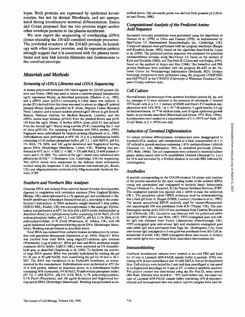
Figure 1. I so la ted e D N A c lones r e p r e s e n t i n g t h e m R N A for t he 210-kD p ro t e in and par t ia l re- s t r ic t ion m a p of t he c o m p o s i t e c D N A . C lone n a m e s are s h o w n on t he r ight . T h e pos i t ions o f the pu ta t ive s tar t c o d o n ( A T G ) a n d s top c o d o n ( T G A ) a re indicated. T h e c D N A f r a g m e n t s u sed as p robes a re s h o w n be low the res t r ic t ion m a p (P23, nuc leo t ides 1-1082; P141, nuc leo t ides 5 7 5 - 1976; P1, nuc leo t ides 1775-2864; P21, nuc le - o t ides 2582-6153); P0.5, nuc leo t ides 3205-3709.
Ruhrberg et al. Cornified Envelope Protein with Homology to Desmoplakin 717
on March 5, 2007
ww
w.jcb.org
Dow
nloaded from

1
91 1
181 28
271 58
361 88
451 118
541 148
631 178
721 208
811 238
901 268
991 298
1081 328
1171 358
1261 388
1351 418
1441 448
1531 478
1621 508
1711 538
1801 568
1891 598
1981 628
2071 658
GC TGAC CAG CC AGTGAGGACG CC CG CTGC C TCC CA CC TGCC C TCC TGCC GTCT~GCCAGCC AAGC C CAGCC T~AGC CAGCA C ~"i"GC C T
TTAC GAC C AT~AAGGGGCTGAGC AAAGGC TC CCAGC~C~TCC C~ CA ~GGG C TCCCC f~C C AAC~TC C C C C AAA~ ~ C C
M F K G L S K G S Q G K G S P K G S P A K G S P K G S>
~QAGCAGGCACAGC CGGGCTGC CACC CAGGAG CTGGC C CTTCTCATC TCC CGCATGCAAGCCAA CGCCGA CCAGGTGGAGCGGGACATCC P S R H S R A A T Q E L A L L I S R M Q A N A D Q V E R D I>
I ~. NN
TGGAGACGCAGAAGAGGCTGCAGC AGGAC C GGC TGAAC AGTGAGCAGAGC CAGGCC CTGCAGCACCAGCAGGAGACGGGC CGCAGC C TGA L E T Q K R L Q Q D R L N S E Q S Q A L Q H Q Q E T G R S L>
AGGAGG CTGAGGTGCTGC TC AAGGAC C TC TTC CTGGACGTC, GACAAGGCCCGGCGGCTCAAGCACC CGCAGGC TGAGGAGA%~fGAGAAGG K E A E V L L K D L F L D V D K A R R n K H P Q A E E I E K>
AC ATCAAGCAGC TG CA CGAG CGGG TGACC C AGGAGTGTGCGGAG TACCGTGC C C TGTACGAGAAGATGGTGC TGC C CC C CGACGTGGGAC D I K Q L H E R V T Q E C A E Y R A 5 Y E K M V L P P D V G>
NN <'1
CCAGGGTCGACTGGGCACGCGTC-CTC~GAGCAGAAACAGAAC~CAGGTCTGCGCAGGCCAGTACGGGCCGGG~ATGGCGGAGC~AG~C P R V D W A R V L E Q K Q K Q V C A G Q Y G P G M A E L E Q>
AGATCGCCGAGCACAACATC CTGCAGAAGGAGATCGACGCCTATGGGCAGCAGCTGCGGAGCCTCGTGGGGCCGGATGCAGCCACCATCC Q I A E H N I L Q K E I D A Y G Q Q L R S L V G P D A A T I>
GGAGC CAATA CC GAGA C C TA C TGAAGC-CGG CGTC GTGGCGCGGGCAGAGC CTGGGCAG C C TG TA CACGCACCTC CAGGGC TGCACGCGGC R S Q Y R D L L K A A S W R G Q S [. G S L Y T H L Q G C T R>
Z <-I I "~ Y AGCTGAGCGCCCTGGC TGAGCAGCAGCGCCGCATCCTGCAGCAGGACTGGAGCGAC CTCATGGCCGAC CC TG~CGTGCGGCGGGAGT Q L S A L A E Q Q R R I L Q Q D W S D L M A D P A G V R R E>
A C ~ A C ~ G C A ~ A C ~ C ~ C A ~ A G ~ G ~ C C A ~ A C ~ C ~ A ~ ~ C ~ Y E H F K Q H E L L S Q E Q S V N Q L E D D G E R M V E L R >
ACCCCGC~TGC~CCCA~CA~CCCACCA~CCC~GA~AG~AG~CT~C~CC~TATC~CA~AGACCC H P A V G P I Q A H Q E A L K M E W Q N F L N L C I C Q E T >
A G C ~ A G C A ~ A C T A C C ~ C ~ C A ~ C C ~ C T C A G ~ C A ~ C C C ~ C G ~ C ~ C ~ C T T G G A ~ Q L Q H V E D Y R R F Q E E A D S V S Q T L A K L N S N L D >
CC~GTACAGCCC~ACCTGGGG~CCCCC~CCCCCACA~C~~CAGA~AAAAAC~C~CCGT~ A K Y S P A P G G P P G A P T E L L Q Q L E A E E K R L A V >
CCGAGAGGGCCACTGGGGACCTGCAGCGGCGAAGCCGGGATGTGGCCCCTCTGCCACAGCGAAGAAACCCCC CTCAGCAGCC CC ~GCACG T E R A T G D L Q R R S R D V A P L P Q R R N P P Q Q P L H>
Y<~
TC-GACAGC ATCTGCGAC TGGGA CTCAGGAGAAGTGCAGCTC-C TGCAGGGTGAGCGGTATAAGCTGGTAGATAACAC TGAA C CGCACGC C T V D S I C D W D S G E V Q L L Q G E R Y K L V D N T E P H A>
~G~CA~CC~AGACC~GCG~G~CCGCC~C~C~A~CCA~ACCA~CCC~C~CCA~ W V V Q G P G G E T K R R P A A C F C I P A P D P D A V A R >
CC~CC~C~CC~AGAGC~A~CC~AG~TTGGCCA~GTCC~AGCC~C~CAG~GTCTCT~ A S R L A S E L Q A L K Q K L A T V Q S R L K A S A V E S L >
GGC C CAGC CAGCAGGC TC CATC TGGC TCAGAC CTGGCCAA CC CACAGGCC CAGAAGCTC C ~ACACAGATGA CC CGGC TGGATGGAGAC C R P S Q Q A P S G S D L A N P Q A Q K L L T Q M T R L D G D>
X <-I I~. W
TGGGAC AGATAGAGAC, GCAGGTGC TC.GC C TGGGCGC GGGC C CCGCTGAGC CGCC CC AC AC CC TTGGAGGACTTC-GAGGGC CGCATC CACA L G Q I E R Q V L A W A R A P L S R P T P 5 E D L E G R I H>
GC CATGAGGGCACAOC CCAGCGCC T~CAGAGCCTC, GGAACGGAGAAGGAGAC AGCC CAGAAGGAGTGCGAGGCG~'r~ TGTC CACGCC-GC S H E G T A Q R L Q S L G T E K E T A Q K E C E A F L S T R>
W~-I
CCGTGGGCCCCGC TGCCC TGCAGCTGCC CGTAGC CC TCAACAGC GTGAAGAACAAGTTCAGTGACGTGCAGG~'~CTGTGCAGCC TC TACG P V G P A A L Q L P V A L N S V K N K F S D V Q v L C S L Y>
I ~. V
GGGAGAAAGC CAAGGC TGCC CTGGATCTGGAGCGGCAGATCCAGGA TGCGGACAGGGTCATC CGAGGC TTCGAGGC CA C C C TGGT~CAGG G E K A K A A L D L E R Q I Q D A D R V I R G F E A T L V Q>
AGGC CCCCATCC CTGCTGAACCGGGGGCTCTGCAGGAGAGGGTCAGCGAG~AGCGCCAGCGGAGGGAGC TGCTGGAACAGCAGAC CT E A P I P A E P G A L Q E R V S E L Q R Q R R E L L E Q Q T>
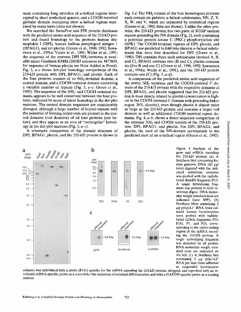
Figure 2. Nucleotide sequence of the composite cDNA encod- ing the 210-kD protein and pre- dicted amino acid sequence. Nu- meration of the cDNA sequence begins with the first nucleotide of the putative translation initia- tion codon (ATG, bold), which is preceded by an in-frame stop codon (TGA, doubly under- lined) that is also present in ge- nomic DNA (data not shown). Numeration of the deduced amino acid sequence begins with the first in-frame methionine of the long open reading frame (M, bold). A long direct repeat close to the beginning of the long open reading frame (underlined) precedes the region encoding the NN domain and encodes the amino acid sequence KGSP four times. The boundaries of the NH2-terminal domains (NN, Z, Y, X, W, and V), the central rod domain, and the COOH-termi- nal C domain are indicated by arrows (nomenclature of do- mains according to Green et al., 1992). The long open reading frame terminates in a stop codon (indicated by an asterisk), which is followed by a consensus poly- adenylation signal (AATAAA) in the 3' untranslated region (doubly underlined). The se- quence data are available from GenBank/EMBL/DDBJ under accession number U53786.
A probe (P1) derived from the isolated c D N A clone, p210-1, was used to isolate two overlapping c D N A clones, p210-141 and p210-21 (see Fig. 1). The clone p210-21 con- tained the polyadenylated 3' end of the cDNA. A probe (P141) derived from p210-141 was used to isolate a further c D N A clone, p210-23, which contained the putative trans- lation start codon in a Kozak consensus sequence for translation initiation (Kozak, 1991), preceded by an in- frame stop codon (Fig. 2).
A map of the isolated c D N A clones and a partial restric- tion map of the composite c D N A are shown in Fig. 1. The c D N A sequence and the amino acid sequence encoded by the long open reading frame are shown in Fig. 2. The encoded protein has a theoretical relative molecular mass of 231 kD, which is in good agreement with the apparent molecular mass of 210 kD in SDS-PAGE. The composite c D N A contains 6,457 bp, corresponding to the size of the 6.5-kb m R N A detected by Northern blot analysis (see below).
The Journal of Cell Biology, Volume 134, 1996 718
on March 5, 2007
ww
w.jcb.org
Dow
nloaded from

...continued
2161 GCGTGCTGC GGC TACAC CG CG CG CTGAAGGC CTCGGAGCAC GC ATGCGC T~CCCTGCAGAACAAC TTCC AGGAGTTC TGCC AAGACC TGC 688 C V L R L H R A L K A S E H A C A A L Q N N F Q E F C Q D L>
2251 C TC GC CAGCAGCG CCAGGTGC GAGC CC TC AC CGAC CGCTAC CACGCC GTAGGGGA CCAG CTGGAC CTGCGGGAGAAGGTGGTGCAGGATG 718 P R Q Q R Q V R A L T D R Y H A V G D Q L D L R E K V V Q D>
2341 CCGCCCTCACC TACCAGCAGTTCAAGAAC TGCAAGGATAACCTGAGC TC CTGGCTGGAGCACCTGCCCCGCAGCCAGGTGCGGCCCAGCG 748 A A L T Y Q Q F K N C K D N L S S W L E H L P R S Q V R P S>
2431 A CGGC C C CAGC CAGATC GC C TAC AAGC ~/GCAGG CG CAGAAGAGGC TGAC GC AGGAGATC CAGAGC CGAGAG CGGGACAGGGCC ACAG CA T 778 D G P S Q I A Y K L Q A Q K R L T Q E I Q S R E R D R A T A>
2521 CCCAC CTCTCCCAGGCC CTGCAGGCAGCGCTCCAGGACTATGAGCTCCAGGCAGACACCTACCGCTGCTCT'FI~AGCC CACCCTGGCAG 808 S H L S Q A L Q A A L Q D Y E L Q A D T Y R C S L E P T L A>
2611 TGTCAGC C C C C AAGAGAC C C C GAGTGGCTCC C C TGCAAGAGAGCATC CAAGCC CAGGAGAAGAAC CTTG CAAAGGCC TATAC TGAGGTTG 838 V S A P K R P R V A P L Q E S I Q A Q E K N L A K A Y T E V>
2701 CAG CAGC AC AGCAGC AGC TGC TC CAGC AGCTGGAGTT~GCTAGAAAAATGC TGGAGAAGAAGGAGCTCAGTGAGGAC ATCCGAAGGAC C C 868 A A A Q Q Q L L Q Q L E F A R K M L E K K E L S E D I R R T>
2791 A TGATGC AAAGCAGGC~C TC CGAGAGC C C TGC CC AAGCAGGGAGAGAG TCAGAGGC C C TGAAGGCC CAGC TGGAAGAGGAGAGGAAGC GGG 898 H D A K Q G S E S P A Q A G R E S E A L K A Q L E E E R K R>
V ,c- I I-~ rod domain
2881 TGGC C C GGGTGCAGCATGAGC TGGAGGCGCAGAGGAGC CAA CTGC TGCAGCTGAGGAC C CAGC GGC C CTTGGAGAGGC TGGAGGAGAAGG 928 V A R V Q H E L E A Q R S Q L L Q L R T Q R P L E R L E E K>
2971 AAGTGGTAGAGq'r CTAC CGGGAC CC CC AGCTGGAGGGCAGC CTG TCCAGGG TGAAGGC C CAGGTGGAGGAGGAGGGCAAGCGGCGGGC TG 958 E V V E F Y R D P Q L E G S L S R V K A Q V E E E G K R R A>
3061 GC C TGCAGG CAGACC TC-GAAGTGGCAG CC CAGAAGGTCGTGCAGC TGGAAAGCAAGAGGAAGAC CATGCAGCC TCATCTGC TGAC CAAGG 988 G L Q A D L E V A A Q K V V Q L E S K R K T M Q P H L L T K~
3151 AGGTC AC CC AGGTGGAGAGGGAC C C CGGC CTGGACAGCC AGGCGG CC CAGC TCAGGATC CAGATC CAGC AGC TC CGC GGGGAGGATGCCG 1018 E V T Q V E R D P G L D S Q A A Q L R I Q I Q Q L R G E D A~
3241 TCATC ~ GGCC CGGC TGGAAGCM3 C~GAAGGAGCTAC TC~3C C C TTGAGAAGAC~3GA~TGGACGTCxAAC~=AGAAGGTCG TGGTGAAAG 1048 V I S A R L E G L K K E L L A L E K R E V D V K E K V V V K>
3331 AGGTAGTCAAGGTGGAGAAGAATCTGGAAATGG TC AAGGCAGC C CAGGC TC TGAGGC TGCAGA ~GAGGAGGATG C TGC GC GGAGGAAGC 1078 E V V K V E K N L E M V K A A Q A L R L Q M E E D A A R R K>
3421 AGGCGGAGGAGGC TG TGGC CAAG C TAC AGC-C TCGCATCGAAGAC C TGGAGCGGGC TATCAG CTCGGTGGAG C C CAAGGTCATCGTGAAGG 1108 Q A E E A V A K L Q A R I E D L E R A I S S V E P K V I V K>
3511 AGG TGAAGAAC, G~]GAG CAGGAC C CAC~ CTC C TC CAGGAGTC CTCC AGGC TC.AGGAGC CTCC TCGAGGAGGAGAGGAC CAAG~CGCGA 1138 E V K K V E Q D P G L L Q E S S R L R S L L E E E R T K N A>
3601 C GC TGGC CAGGGAGC TGAGCGAC C TGC ACAG CAAGTACAGCGTGGTGGAGAAGCAGAGG CC CAAAGTGCAGC TC CAGGAGCGCGTC CACG 1168 T L A R E L S D L H S K Y S V V E K Q R P K V Q L Q E R V H>
3691 AGATC TTC C AGGTGGATC C GGAGAC AGAG CAGGAGATCACTCGGC TC AAGGCCAAGC TG CAGGAGATGG CGGGCAAGAGGAGC GGTG ~G 1198 E I F Q V D P E T E Q E I T R L K A K L Q E M A G K R S G V~
3781 AGAAGGAGGTGGAGAAG C TGC TGC C CGAC C TGGAGGTCC TC42GGGC C CAGAAGC C CACGGTGGAG TACAAGGAGGTGAC CCAGGAGG TGG 1228 E K E V E K L L P D L E V L R A Q K P T V E Y K E V T Q E V~
3871 TGAGG CA TG AGAGGAGC C C CGAGGTGC TGCG TGAGATTGAC CGC C TGAAGG C TCAGC TCAACGAGC TCGTC AA CAGC CACGGGCG CTC C C 1258 V R H E R S P E V L R E I D R L K A Q L N E L V N S H G R S>
3961 AGGAG CAGC TCATCC GC CTGC AGGGTGAGCG CGACGAGTGGAGGC GCGACGGGGC CAAGGTGGAGAC CAAGAC GGTGAG CAAGGAGGTC, G 1288 Q E Q L I R L Q G E R D E W R R D G A K V E T K T V S K E V>
4051 TGC GC CACGAGAAGGAC C C GGTGC TGGAGAAAGAAGC AGAGTGGC TC CGCCAGGAGGTGCGGGAGGC GGCC CAGAAGAGGCGGGC CGCGG 1318 V R H E K D P V L E K E A E W L R Q E V R E A A Q K R R A A>
4141 AGGAC GC GG TGTACGAGC TGC AGAGCAAGCGC C TGCTGC %~GAGAGGAGGAAGCC CGAGGAGAAGGTGGTGGTGCAGGAGG TGGTGGTC A 1348 E D A V Y E L Q S K R L L L E R R K P E E K V V V Q E V V V,
Analysis of the Predicted Amino Acid Sequence of the 210-kD Protein
The deduced amino acid sequence of the 210-kD protein (Fig. 2) contains 36% hydrophobic residues (alanine, leu- cine, isoleucine, phenylalanine, tryptophan, and valine) and 32% charged residues (aspartate, glutamate, lysine, and arginine). A hydrophilicity plot based on the method of Kyte and Doolittle (1982) revealed that the protein was
generally very hydrophilic and did not contain any regions with a high probability of forming a transmembrane do- main (data not shown). The absence of potential trans- membrane domains was confirmed using the algorithm of Argos and Rao (1986). Secondary structure analyses, based on the methods of Chou and Fasman (1978) or Gamier et al. (1978), predicted an NH2-terminal domain characterized by the presence of short a-helical segments, a central do-
Ruhrberg et al. Cornified Envelope Protein with Homology to Desmoplakin 719
on March 5, 2007
ww
w.jcb.org
Dow
nloaded from

• .. conC inued
4231 C C CAGAAGGAC CCGAAGCTGCGCGAGGAGCACAGC CC, GC TGAGCGGGAGCC TGK~AT~AGGAGGTC, GGCCGGCGGCGC CAGC TAGAGC ~-rG 1378 T Q K D P K L R E E H S R L S G S L D E E V G R R R Q L E L>
4321 AGG TGCAC-CAGC TGC GGGC CGGCGTGGAGGAGCAGGAGGGC CPGC TCAGCTTC CAGGAGGA CCGCAGCAAGAAGC TC-GC CGTGGAGAGGG 1408 E V Q Q L R A G V E E Q E G L L S F Q E D R S K K L A V E R>
4411 AGC TGCGGCAG CTGA CC TIV, AGGATCCAGGAGC TC GAGAAGCGGC C TCC CACGGTTGCAGGAGAAGATCATCA TGGAGGAAGTGGTCAAG 1438 E L R Q L T 5 R I Q E L E K R P P T V A G E D H H G G S G Q>
4501 C TGGAGAAGGA CC CGGAC C TGGAGAAGTC CACGGAAC, CC C TGCGTGGGACC TC, GACCAGGAGAAG AC CCAGGTAA CCGA~ ~ 1468 A G E G P G P G E V H G S P A W D L D Q E K T Q v T E L N R>
4591 AGTGCAAGAAC CTGCAGGTCCAGATTGACGTCC TC CAGAAAGC CAAATCGCAGGAGAAGACCA TC TACAAGGAAGTGATCCCGGTGCAGA 1498 E C K N L Q V Q I D V L Q K A K S Q E K T I Y K E V I R V Q>
4681 AGGAC CGCG TC CTGGAAGA ~AGCGGGCC CG CO TG TC, GGAGATGCTCAACAGGGAGCGCACGGCC CGGCAGGCCC GGGAGGAGGAGG CAC 1528 K D R V L E D E R A R V W E M L N R E R T A R Q A R E E E A>
4771 GGC GC CTGCGGGAGCGCATTGAC CGGGCC GAGACGCTGGGGAGAACC TC, GTCC CGGGAGGAGTCCGAGCTG CAGAGGC4: CCGGGACCAGG 1558 R R L R E R I D R A E T L G R T W S R E E S E L Q R A R D Q>
4861 C CGAC CAGGAGTG TC, GG CGGC ~CAGCAGGAGCTGCGGGCTCTGGAGAGGCAGAAGCAGCAGCAGACAC TGCAGC TGCAGGAGGAGTCGA 1588 A D Q E C G R L Q Q E L R A L E R Q K Q Q Q T L Q L Q E E S>
4951 AGC TGCTCAGC CAGAAGACGGAGAGCGAGCGACAGAAGGCGGC CCAG~CAGGAGCTCTCGCGGC TGGAGGCGGC CATC C TCCGCG 1618 K L L S Q K T E S E R Q K A A Q R G Q E L S R L E A A I L R>
5041 AGAAGGAC CAGATCTACGAGAAGGAGCGGAC GC TC CGGGAC CTCCACGCCAAGGTGAGCCGGGAGGAGC TCAGCCAGGAGACC CAGACGC 1648 E K D Q I Y E K E R T L R D L H A K V S R E E L S Q E T Q T>
5131 GAGAGAC CAAC CTTTCC AC CAAGATCTCC ATCC TOGAAC CCGAGA~GGACATGTC C C CATACGAGGC CTACAAGAGGGGCA TCA 1678 R E T N L S T K I S I L E P E T G K D M S P Y E A Y K R G I>
5221 TCGACAGGGGC CAGTAC TTGCAGC TGCAGGAGC TCGAGTGTGACTGGGAGGAGGTCA CCAC CTCGGGGC C C TGTGGGGAGGAGTC TGTGC 1708 I D R G Q Y 5 Q L Q E L E C D W E E V T T S G P C G E E S V>
5311 TCC TGGACCGCAAGAGCGGGAAGCAGTAC TC CA TCGAGGCC GC CC TC CGCTGC CGGCGCATCTCTAAGGAGGAGTAC CA TC TGTACAAGG 1738 L L D R K S G K Q Y S I E A A L R C R R I S K E E Y H L Y K>
5401 A ~ CACC TG CC CA TC TC CGAG TI'TGCGCTGCTTGTAGCTGGGGAGAC CAAGCCAAGCTCCTCACTCTC CATCGGC TC TATC ATCTCCA 1768 D G H L P I S E F A L L V A G E T K P S S S L S I G S I I S>
5491 AGTCC CCGCTCGC CTC C C CGGCC CC CCAGAGCACCAGTTTC TTCTCTCC CAGCTTCTCTCTCGGGC TCGGTGATGACAGCTTC CCTATCG 1798 K S P L A S P A P Q S T S F F S P S F S L G L G D D S F P I>
5581 C CGGGATC TATGACACAACCACAGACAACAAGTGCAGCATCAAGACGGCCGTGGCCAAGAACATGCTGGAC CC CATCAC~GGCAGAAGC 1828 A G I Y D T T T D N K C S I K T A V A K N M L D P I T G Q K>
l~C
5671 TAC TGGAGG CC CAGGCGGC CACAGGGGGC ATCG TGGA CC TGCTCAC, C CGTG~TAC TC TGTGCACAAGGCGATGGAGAGGGGCC TGA 1858 L L E A Q A A T G G I V D L L S R E R Y S V H K A M E R G L>
5761 TCGAGAACACC TC CACACAGAGGCTGC TTAACG CC CA~CTTCACCGGCATCGAGGAC C CCGTCACCAAGAAGAGGC TC TCGG~/GG 1888 I E N T S T Q R L L N A Q K A F T G I E D P V T K K R L S V>
5851 GCGAGGC CGTC CAGAAGGGCTGGATGC C C CGGGAGAGCGTGCTCC CACACC TGCAGGTG CAGCAC CTGAC C~TCATCGACC CCA 1918 G E A V Q K G W M P R E S V L P H L Q V Q H L T G G L I D P>
5941 AGAGGACAGGC CGCATC C C CATC CAGC AGGC CC TCCTCTCCGGGATGATCAGTGAAGAGCTGGCC CAGC TC CTGCAGGACGAGTCCA~ T 1948 K R T G R I P I Q Q A L L S G M I S E E L A Q L L Q D E S S>
6031 ACGAGAAGGATTTGA CAGACC CCATCTC CAAGGAACGGC TGAGC TACAAGGAGGC CATGGGCCGCTGCC~GAC C C CC ~ C 1978 Y E K D L T D P I S K E R L S Y K E A M G R C R K D P L S G>
6121 TGC TGCTCC TGCCAGCGGCAC TGGAGGGGTACCGC TGCTAC CGCTCCGC CTCCCCCACCGTCCCGCGCTCC CTTC~CACGGG CC AA 2008 L L L L P A A L E G Y R C Y R S A S P T V P R S L R t
C~
6211 GGAGC CAGTGGGGAAGTGC GTGTGTTGGGCCAGGTAGGATA CGTACACCTCTI~C CTCAGAGCAGCC TCATCC CAGGCAGTGGGTCTTC C
6301 C TC TGTC CAAC CA CTG Fr F~AIT~-~CTAACGAGGTGA TGGGCTCC CTCC CC TAAC CTTGGAGC CTGATCCATC CC CAGAC C AGGAC
6391 AGCAGCCACTCAGTTCTTCCTCCACCTCCACCCAGTGATCCCAATAAACGAATTCTGTCTCCCCGTG - ~|y(A) tail
Figure 2.
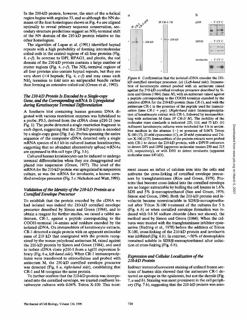
Figure 3. Homology of the 210-kD protein to DPI, BPAG1, and plectin. Dot matrix homology comparisons of the 210-kD protein with DPI (a), BPAG1 (b), and plectin (c) were performed with the software COMPARE and DOTPLOT; a dot was placed when 13 amino acids showed identity within a window of 20 amino acids. Secondary structure predictions (see text and Fig. 4) suggest that the domain structure of the 210-kD protein is similar to that of DPI, BPAG1, and plectin (d). The designation of the NH2-terminal domains NN, Z,
The Journal of Cell Biology, Volume 134, 1996 720
on March 5, 2007
ww
w.jcb.org
Dow
nloaded from

a b
A B C B C
I 1,000 2.000 ~ i 0 1,000 2,000 ] ~ J . . . . l . . . . . . . . . . . . . . , , l . . . . 2.000 C I I -i .; : , . . ~ i . ~ l ~ 2,000 C • . . . . . . ":: , ~ '~ . , t~ ; [ ....... . . . . - . . : , . . :;..
: ~ ; " , r -~ ' '~ ~ ~ " + ' / ' . .... r t " " "~" :':':":' : : , . . . . , . . . ~ " ~ . . . . . : ' " ~ ' " ' " ' " " ~ • . ~ 7 " . ' : " ; ' . ' : , , " . " . • . . . . , o . , . :'~',,i...n " . | . . . l l . . ¢ . , : : , "
i.. N-term. I rod I C-term.-~l I,- N.4emm. I rod I C-term~
DPI (28'71 amino i d a reddms) BPAG1 (:IMIO Imlno acid rmidms)
| | !
i ! !
B B B B B C
1 , 0 0 0 2,000 3,000 4,000 . . . . t . . . . I , , , j i , , , , i
• "" "" : " " "" ' i : • . . . . : . • : , . ' . " . ~ . •
• ~. ' . "- . . : . [ Y q d K . ~ , . . ~"¢.._~.'. " " " ' - - ' - - ~ . . . - . • . . , ' ÷ L . . : , . o.,.,;~.~.... • : . . . • . ~ . . . ......-..;-..'~.;-.. i - ~ ; : , . ,7 ° . . . •
, . . . . o . . . . e le 4 - . ~a . . • # - . . . - .
. . . . , . . . . . . .~ ,~ . ~ • .
• . . - . :.. ~ . . : . , . . . . "~ 's '" . r % " " • s . . . . -;'-. . • ~ / . , . , , . . . . . - ; . ' . . . - .
t . ° " , ' . ' , - , ' ~ . o . ' ~ , . " ,- . . . . I I , I ' I l l i I . . . . i . . . . i i
I- N4erm. I rod I C,lerm.
pleotin (4140 mmino arid residues)
2,000
1,000
L 0
I
D P I
(C, SRS~
B P A G I
p l e c t l n
(GSRX)4
p 2 1 0
Y , X , W , a n d V , a n d t h e C O O H - t e r m i n a l r e p e a t d o m a i n s A , B , a n d C f o r D P I , B P A G 1 , a n d p l e c t i n is a c c o r d i n g t o G r e e n e t a l . ( 1 9 9 2 ) ;
f o r d o m a i n b o u n d a r i e s o f t h e 2 1 0 - k D p r o t e i n , s e e F ig . 2. T h e r o d d o m a i n s a r e r e p r e s e n t e d b y f i l l ed r e c t a n g l e s . ( G S R S ) 6 a n d ( G S R X ) 4
r e f e r t o r e p e a t m o t i f s a t t h e C O O H t e r m i n u s o f D P I a n d p l e c t i n , r e s p e c t i v e l y .
Ruhrberg et al. Cornified Envelope Protein with Homology to Desmoplakin 721
on March 5, 2007
ww
w.jcb.org
Dow
nloaded from

Figure 4. Direct sequence comparison and coiled coil analysis of the 210-kD pro- tein, DPI, BPAG1, and plec- tin. Sequence comparison of the NH2 termini (a) and the COOH termini (b) of the four homologues (domain boundaries of DPI, BPAG1, and plectin accord- ing to Green et al., 1992). Amino acid residues in the sequence alignments are numbered according to their position in the protein se- quence; stop codons are indi- cated with asterisks. (c-f) Histograms of the probability of forming a coiled coil (y axis), as predicted by the Lu- pas algorithm, versus the po- sition in the amino acid se- quence (x axis) for the 210- kD protein (c), plectin (d), DPI (e), and BPAG1 (f). Each division on the x axis scale corresponds to 100 amino acid residues. Re- gions with a value of P > 0.9 for more than 28 consecutive amino acid residues are pre- dicted to adopt a coiled coil conformation.
The Journal of Cell Biology, Volume 134, 1996 722
on March 5, 2007
ww
w.jcb.org
Dow
nloaded from
main containing long stretches of a-helical regions inter- rupted by short nonhelical spacers, and a COOH-terminal globular domain containing short a-helical regions sepa- rated by many turns (data not shown).
We searched the SwissProt and PIR protein databases with the predicted amino acid sequence of the 210-kD pro- tein and found homology to the proteins human des- moplakin ! (DPI), human bullous pemphigoid antigen 1 (BPAG1), and rat plectin (Green et al., 1990, 1992; Sawa- mura et al., 1991a; Virata et al., 1991; Wiche et al., 1991; the sequence of the extreme DPI NH2-terminus is avail- able under GenBank/EMBL/DDBJ accession no. M77830; for sequence of human plectin see Note Added in Proof). Fig. 3, a-c shows dot-plot homology comparisons of the 210-kD protein with DPI, BPAG1, and plectin. Each of the four proteins consists of an NH2-terminal domain, a central domain, and a COOH-terminal domain containing a variable number of repeats (Fig. 3, a-c; Green et al., 1992). The sequence of the NH2- and COOH-terminal do- mains appears to be well conserved between the four pro- teins, indicated by areas of linear homology in the dot-plot matrices. The central domain sequences are considerably diverged, although a large number of heptad repeats with the potential of forming coiled coils are present in the cen- tral domains (rod domains) of all four proteins (see be- low), and they appear as an area of "rectangular" homol- ogy in the dot-plot matrices (Fig. 3, a-c).
A schematic comparison of the domain structure of DPI, BPAG1, plectin, and the 210-kD protein is shown in
Fig. 3 d. The NH2 termini of the four homologous proteins each contain six putative oL-helical subdomains, NN, Z, Y, X, W, and V, which are separated by nonhelical regions (Green et al., 1992; data not shown). Unlike the other pro- teins, the 210-kD protein has two pairs of KGSP tandem repeats preceding the NN domain (Fig. 2), each containing a potential protein kinase C (PKC) phophorylation site (SPK). The COOH-terminal repeats of DPI, plectin, and BPAG1 are predicted to fold into discrete a-helical subdo- mains that were first described for DPI (Green et al., 1990). DPI contains three such subdomains (termed A, B, and C), BPAG1 contains two (B and C), plectin contains six (five B and one C) (Green et al., 1990, 1992; Sawamura et al., 1991a; Wiche et al., 1991), and the 210-kD protein contains one (C) (Fig. 3, a-d).
A comparison of the predicted amino acid sequences of the entire NH2 terminus and the COOH-terminal C do- main of the 210-kD protein with the respective domains of DPI, BPAG1, and plectin suggested that the 210-kD pro- tein is most closely related to plectin (conservation is high- est in the COOH-terminal C domain with preceding linker regon: 36% identity), even though plectin is almost twice as large as the 210-kD protein and contains a larger rod domain as well as additional COOH-terminal repeat do- mains. Fig. 4, a-b, shows a direct sequence comparison of the extreme NH2 and COOH termini of the 210-kD pro- tein, DPI, BPAG1, and plectin. For DPI, BPAG1, and plectin, the start of the NN-domain corresponds to the predicted.start of an a-helical region (Green et al., 1992).
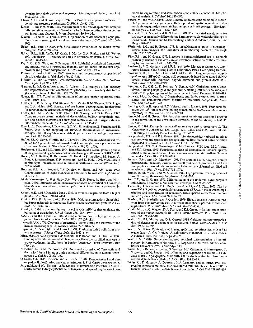
Figure 5. Analysis of the gene and mRNA encoding the 210-kD protein. (a) A Southern blot containing hu- man genomic DNA (10 p.g/ lane) digested with the indi- cated restriction enzymes was probed with the radiola- beled BamHI fragment P0.5. A single hybridizing frag- ment was present in each re- striction digest. DNA molec- ular weight standard sizes are indicated (lane MW). (b) Northern blots containing 2 p~g poly(A) + RNA from cul- tured human keratinocytes were probed with radiola- beled cDNA fragments P23, P141, P1, and P21, corre- sponding to the entire coding region of the mRNA encod- ing the 210-kD protein. A single hybridizing fragment was detected by all probes. RNA molecular weight stan- dard sizes are indicated on the left. (c) A Northern blot containing 2 /.tg poly(A) + RNA per lane from adherent or suspended keratinocyte
cultures was hybridized with a probe (P141) specific for the mRNA encoding the 210-kD protein, stripped, and reprobed with an in- volucrin mRNA-specific probe as a control for the induction of terminal differentiation and with a GAPDH-specific probe as a loading control.
Ruhrberg et al. Cornified Envelope Protein with Homology to Desmoplakin 723
on March 5, 2007
ww
w.jcb.org
Dow
nloaded from
In the 210-kD protein, however, the start of the a-helical region begins with arginine 33, and so although the NN do- mains of the four homologues shown in Fig. 4 a are aligned optimally to reveal primary sequence conservation, sec- ondary structure predictions suggest an NH2-terminal shift of the NN domain of the 210-kD protein relative to the other homologues.
The algorithm of Lupas et al. (1991) identified heptad repeats with a high probability of forming intermolecular coiled coils in the central regions of all four proteins (Fig. 4, c-f). In contrast to DPI, BPAG1, and plectin, the rod domain of the 210-kD protein contains a large number of stutter regions (Fig. 4, c-f). The NH2-terminal regions of all four proteins also contain heptad repeats, but they are very short (<4 heptads; Fig. 4, c-f) and may cause each NH2 terminus to fold into an antiparallel bundle rather than forming an extensive coiled coil (Green et al., 1992).
The 210-kD Protein Is Encoded by a Single-copy Gene, and the Corresponding mRNA Is Upregulated during Keratinocyte Terminal Differentiation
A Southern blot containing human genomic DNA di- gested with various restriction enzymes was hybridized to a probe, P0.5, derived from the eDNA clone p210-21 (see Fig. 1). The probe detected a single restriction fragment in each digest, suggesting that the 210-kD protein is encoded by a single-copy gene (Fig. 5 a). Probes spanning the entire sequence of the composite eDNA detected only a single mRNA species of 6.5 kb in cultured human keratinocytes, suggesting that no abundant alternatively spliced mRNAs are expressed in this cell type (Fig. 5 b).
Cultured human keratinocytes can be induced to undergo terminal differentiation when they are disaggregated and placed into suspension (Green, 1977). The level of the mRNA for the 210-kD protein was upregulated in suspension culture, as was the mRNA for involucrin, a known corn- ified envelope precursor (Fig. 5 c; Nicholson and Watt, 1991).
Validation of the Identity of the 210-kD Protein as a Cornified Envelope Precursor
To establish that the protein encoded by the cDNA we had isolated was indeed the 210-kD cornified envelope precursor described by Simon and Green (1984), and to obtain a reagent for further studies, we raised a rabbit an- tiserum, CR-1, against a peptide corresponding to the COOH-terminal 14-amino acid residues encoded by the isolated eDNA. On immunoblots of keratinocyte extracts, CR-1 detected a single protein with an apparent molecular mass of 210 kD that comigrated with the protein recog- nized by the mouse polyclonal antiserum M, raised against the 210-kD protein by Simon and Green (1984), and used to isolate cDNA clone p210-1 from a kgt l l expression li- brary (Fig. 6 a, left-hand side). When CR-1 immunoprecip- itates were transferred to nitrocellulose and probed with antiserum M, the 210-kD cornified envelope precursor was detected (Fig. 6 a, right-hand side), establishing that CR-1 and M recognize the same protein.
To further confirm that the 210-kD protein was incorpo- rated into the cornified envelope, we treated confluent ke- ratinocyte cultures with 0.04% Triton X-100. This treat-
Figure 6. Confirmation that the isolated eDNA encodes the 210- kD corrtified envelope precursor. (a) (Left-hand side): Immuno- blot of keratinocyte extract probed with an antiserum raised against the 210-kD cornified envelope precursor described by Si- mon and Green (1984) (lane M), with an antiserum raised against a peptide corresponding to the COOH terminus encoded by the putative eDNA for the 210-kD protein (lane CR-1), and with the antiserum CR-1 in the presence of the peptide used for immuni- zation (lane CR-1 + pep). (Right-hand side): Immunoprecipita- tion of keratinocyte extract with CR-1, followed by immunoblot- ting with antiserum M (lane IP CR-1/M). The mobility of the molecular mass standards is indicated: 220, 110, and 75 kD. (b) Adherent keratinocyte cultures were incubated for 5 h in serum- free medium in the absence ( - ) or presence of 0.04% Triton X-100 (T), 20 mM cysteamine (C), or 20 mM cysteamine and Tri- ton X-100 (CT). Immunoblots of the protein extracts were probed with CR-1 to detect the 210-kD protein, with a DPI/II antiserum to detect DPI and DPI! (apparent molecular masses 250 and 215 kD, respectively), or with DH1 to detect involucrin (apparent molecular mass 140 kD).
ment causes an influx of calcium ions into the cells and activates the cross-linking of cornified envelope precur- sors by transglutaminases (Rice and Green, 1979). Pro- teins that become cross-linked into the cornified envelope are no longer extractable by boiling the cell lysates in 1.6% SDS and 5% ~-mercaptoethanol (Sun and Green, 1976; Simon and Green, 1984). Both the 210-kD protein and in- volucrin became nonextractable in SDS/13-mercaptoetha- nol after Triton X-100 treatment of the cultures for 5 h (Fig. 6 b) or when cornified envelope formation was in- duced with 0.8 M sodium chloride (data not shown), the method used by Simon and Green (1984). When the cul- tures were treated with the transglutaminase inhibitor cyste- amine (Siefring et al., 1978) before the addition of Triton X-100, cross-linking of the 210-kD protein and involucrin was inhibited (Fig. 6 b). In contrast, ~50% of desmoplakin remained soluble in SDS/13-mercaptoethanol after induc- tion of cross-linking (Fig. 6 b).
Expression and Cellular Localization of the 210-kD Protein
Indirect immunofluorescent staining of unfixed frozen sec- tions of human skin showed that the antiserum CR-1 de- tected an epitope in the epidermis, but not the dermis (Fig. 7, a and b). Staining was most prominent in the cell periph- ery (Fig. 7 b), suggesting that the 210-kD protein was asso-
The Journal of Cell Biology, Volume 134, 1996 724
on March 5, 2007
ww
w.jcb.org
Dow
nloaded from

Figure 7. The tissue distribution and cellular localization of the 210-kD protein, as determined by indirect immunofluorescence with the CR-1 antiserum. (a-c) Epidermis. (a) Preimmune serum from rabbit CR-1, (b and c) double labeling with CR-1 (b) and an antibody to DPIflI (c), (d) cervical mucosa labeled with CR-1; (e-g) cultured keratinocytes; (e) single labeling with CR-1; (f and g) double labeling with CR-1 (f), and an antibody to DPIflI (g). Bars: (a--c) 50 Ixm; (d and e) 100 Ixm; (fand g) 20 I~m. Arrows in b and c show position of the dermal/epidermal boundary.
Ruhrberg et al. Cornified Envelope Protein with Homology to Desmoplakin 725
on March 5, 2007
ww
w.jcb.org
Dow
nloaded from

ciated with the plasma membrane. Nonspecific labeling of the stratum corneum with preimmune serum was variable and precluded any conclusions regarding CR-1 labeling of this layer. CR-1 staining was strongest in the upper spinous and granular layers (Fig. 7 b). In contrast, an antibody spe- cific for DPI/II showed strong labeling of the basal layer, as well as the suprabasal layers (Fig. 7, b vs. c).
To examine the distribution of the 210-kD protein, we stained frozen sections of various tissues with CR-1 (Fig. 7, b and d; data not shown). All stratified squamous epithelia examined (epidermis from neonatal foreskin and adult breast, keratinized and nonkeratinized oral mucosa, oeso- phageal, and cervical mucosa) were positively stained. CR-1 did not stain the simple epithelium of endocervical glands. The antiserum also failed to stain fibroblasts or en- dothelial cells in the dermis and did not stain brain tissue. These results are consistent with the earlier conclusion that the 210-kD protein is keratinocyte-specific (Simon and Green, 1984).
When stratified colonies of cultured foreskin kerati- nocytes were permeabilized and stained with CR-1, stain- ing was concentrated at the plasma membrane in the first suprabasal layers, but became more uniform in the upper- most layers (Fig. 7 e), confirming that the 210-kD protein is upregulated during differentiation of epidermal kerati- nocytes. At higher magnification, the staining appeared punctate, reminiscent of desmosomal junctions (Fig. 7 f, see Watt et al., 1984). Double-label immunofluorescence with CR-1 and antibodies specific for desmogleins (data not shown) or DPI/II (Fig. 7, f and g) showed colocaliza- tion of the 210-kD protein with these desmosomal pro- teins in differentiating cells.
The 210-kD protein could not be detected with the CR-1 antiserum in cultured keratinocytes or epidermis treated with aldehyde fixatives. We could, however, carry out im- munogold EM when epidermal sections were prepared by high pressure freezing and freeze substitution. In these sections, there was very little labeling with the preimmune serum of any of the cell layers, including the stratum cor- neum (Fig. 8 d). CR-1 showed extensive colocalization with a DPI/II antibody in the satellite region of desmosomes (Fig. 8, a and b) and along keratin filaments throughout the cytoplasm of differentiated keratinocytes (Fig. 8 c). We saw no evidence of specific labeling of keratohyalin granules with either CR-1 or anti-DPI/II (Fig. 8 c). The CR-1 antibody labeled cornified cells strongly, and the la- beling was not confined to the cell periphery (Fig. 8 e).
Discussion In this study, we describe the cloning and sequencing of overlapping cDNA clones that encode the membrane- associated 210-kD cornified envelope precursor first de- scribed by Simon and Green (1984). The authenticity of the isolated eDNA was established with an antiserum (CR-1) raised against a peptide corresponding to the pre-
dieted COOH-terminal 14-amino acid residues of the pro- tein encoded by the isolated cDNA. On immunoblots, the CR-1 antiserum detected a single protein with an apparent molecular mass of 210 kD, which was also recognized by an antiserum raised against the 210-kD cornified envelope precursor by Simon and Green (1984). Furthermore, the protein detected by the CR-1 antiserum was cross-linked into the cornified envelope on transglutaminase activation in cultured keratinocytes, as reported by Simon and Green (1984).
The 210-kD protein appears to be keratinocyte-specific, but, as observed for involucrin (Banks-Schlegel and Green, 1981), its expression is not restricted to keratinizing strati- fied squamous epithelia. Expression of the 210-kD protein increased during terminal differentiation of epidermal ke- ratinocytes both in vivo and in culture. When cultured epi- dermal keratinocytes were disaggregated and placed in suspension to induce terminal differentiation, the mRNA for the 210-kD protein was upregulated, suggesting that the increase in 210-kD protein during epidermal differen- tiation is at least partly caused by increased transcription or increased mRNA stability.
The 210-kD cornified envelope precursor is homologous to the intermediate filament-associated proteins desmo- plakin I (DPI), bullous pemphigoid antigen 1 (BPAG1), and plectin. Sequence analyses predict that the four ho- mologous proteins have a similar domain structure with an NH2-terminal globular domain, a central rod domain, and a COOH-terminal globular domain (Green et al., 1990; Sawamura et al., 1991a; Virata et al., 1992; Wiche et al., 1991). The NH2 termini of BPAG1, plectin, and DPI are predicted to each contain a bundle of antiparallel a-helices (NN, Z, Y, X, W, and V; Green et al., 1992), which are also conserved in the 210-kD protein. The COOH termini of the four homologues contain a variable number of tandem repeats that are predicted to be organized into discrete subdomains that consist of et helices separated by t3 turns, and which were first described for DPI (Green et al., 1990). DPI contains three such subdomains, BPAG1 two, plectin six (Green et al., 1990; Sawamura et al., 1991a; Vi- rata et al., 1992; Wiche et al., 1991), and the 210-kD pro- tein one. The C-domain, with its preceding linker region, is the only COOH-terminal subdomain conserved in all four proteins. A comparison of the predicted amino acid se- quences of the NH2- and COOH-terminal domains of the 210-kD protein with the respective domains of human DPI, human BPAG1, and rat plectin shows that the pri- mary sequence of the 210-kD protein is most closely re- lated to plectin, even though plectin is almost twice as large as the 210-kD protein, with a larger rod and addi- tional COOH-terminal repeats.
Although the central domain sequences of the four pro- teins are highly diverged, they all have a large number of heptad repeats with the potential of forming coiled coils with a dimerisation partner. There is direct evidence for homodimerization of DPI (O'Keefe et al., 1989), and ro-
Figure 8. Localization of the 210-kD protein in epidermis, as determined by immunogold EM with the CR-1 antiserum. (a-c) Double labeling with CR-1 (10 nm gold) and an antibody to DPI/II (5 nm gold); some of the areas showing colocalization of CR-1 (large arrows) and DPI/II (small arrows) are indicated by arrows. (d) Labeling with the preimmune serum of rabbit CR-1. (e) Single labeling with CR-1. The upper cell is a comified cell. Bar, 200 nm.
Ruhrberg et al. Cornified Envelope Protein with Homology to Desmoplakin 727
on March 5, 2007
ww
w.jcb.org
Dow
nloaded from

tary shadowing suggests that the same is true for plectin (Foisner and Wiche, 1987). In contrast to DPI, BPAG1, and plectin, the rod domain of the 210-kD protein contains a large number of stutter regions, and this may have implica- tions for its structure and its interaction with other proteins.
A single mRNA species of 6.5 kb encoding the 210-kD protein can be detected on Northern blots of human kera- tinocytes. In contrast, two alternatively spliced desmo- plakin mRNAs, encoding DPI and DPII, are transcribed from the desmoplakin gene, and DPII is believed to con- tain an abbreviated rod domain (Virata et al., 1992). BPAG1 expression is confined to basal keratinocytes (Sawamura et al., 1991b; Tamai et al., 1993), but alterna- tive splice products containing BPAG1 exons, BPFG in the pancreatic carcinoma cell line FG (Hopkinson and Jones, 1994) and dystonin in the brain (Brown et al., 1995), have been described.
Immunofluorescence analysis suggested that the 210-kD protein is associated with the plasma membrane and shows partial colocalization with desmosomal proteins. Immunogold EM showed colocalization of the 210-kD protein and DPI/II at desmosomes and along keratin fila- ments in differentiating keratinocytes. At the EM level, the antiserum to the 210-kD protein labeleled cornified cells strongly, but labeling was not concentrated at the cell periphery; it is possible that the epitope becomes inacces- sible in mature envelopes (Steinert and Marekov, 1995; Ishida-Yamamoto et al., 1996).
The extensive association of DPI/II and the 210-kD pro- tein with keratin filaments has not been observed previ- ously by immunofluorescence or immunogold EM. Epi- thelial cells are known to contain two different pools of DPI/II, one that cannot be extracted by nonionic deter- gents and one that is easily extracted with low detergent concentrations (Duden and Franke, 1988; Pasdar and Nel- son, 1988a,b). Our evidence suggests that there are also two pools of the 210-kD protein: Triton X-100-soluble protein is readily immunoprecipitated by the CR-1 antise- rum (Fig. 6 a), yet immunofluorescence staining of Triton X-100-extracted keratinocytes shows that they also con- tain insoluble 210-kD protein (Fig. 7, e and f). We suggest that the detergent-soluble pools of DPI/II and the 210-kD protein may be weakly associated with keratin filaments, and that this association is not preserved by conventional chemical fixation and permeabilization procedures. How- ever, it is preserved by the high pressure freezing/freeze substitution technique that we used for our EM analysis.
There is evidence that desmosomal components (Haftek et al., 1991; Steinert and Marekov, 1995) and keratin fila- ments (Haftek et al., 1991; Ming et al., 1994; see also Fig. 5 b) become incorporated into the cornified envelope. We speculate that the 210-kD protein may anchor them to the envelope. In addition to our EM evidence, the sequence homology between the 210-kD protein, plectin, DPI/II, and BPAG1 strongly suggests such a role. Plectin is found in a wide variety of different cell types and is thought to play a role in the cross-linking of intermediate filaments to each other, as well as to microtubules and microfilaments, and may anchor these networks to the cell membrane (re- viewed by Foisner and Wiche, 1991). Association of plec- tin with vimentin and keratins is dependent on its COOH- terminal domain (Wiche et al., 1993). DPI and DPII are
the most abundant constituents of desmosomes (reviewed by Schwarz et al., 1990), and they anchor keratin filaments to desmosomes: the DPI/II COOH terminus associates with the keratin network when overexpressed in cultured cells (Stappenbeck et al., 1992, 1993), and it binds a subset of keratins in vitro (Kouklis et al., 1995). BPAG1 is in- volved in anchoring the keratin network to hemidesmo- somes in keratinocytes (Guo et al., 1995, and references therein).
We have not determined whether the 210-kD protein is a direct substrate for transglutaminases, or whether it be- comes cross-linked into the envelope in association with other envelope precursors, but it is interesting that the NH2-terminal domain, but not the rod or the COOH-ter- minal domain of the 210-kD protein, contains a larger pro- portion of glutamine residues (11.6%) than DPI/II (8.1%), plectin (8.1%), or BPAG1 (7%). Like its homologues, the 210-kD protein lacks a transmembrane domain, but the NH2-terminal domain may mediate association with des- mosomes, as observed for DPI/II (Stappenbeck et al., 1992), and may be in close proximity to membrane-bound transglutaminase (TGK) (Chakravarty and Rice, 1989). It is conceivable that its relatively high NH2-terminal gluta- mine content could make the 210-kD protein a better sub- strate for cross-linking by transglutaminases than DPI/II. Alternatively, or additionally, free carboxyl groups of NH2-terminal glutamine residues that are not cross-linked by transglutaminase could serve as attachment sites for hy- droxyceramides, the major component of the lipid mono- layer that replaces the plasma membrane in fully differen- tiated corneocytes (Reichert et al., 1993).
In conclusion, the 210-kD protein is a cornified enve- lope precursor that is likely to link the envelope to both keratin filaments and desmosomes. Because the 210-kD protein is an envelope precursor that belongs to the des- moplakin family and because of deficits in our classical ed- ucation, we propose that the 210-kD protein be named "envoplakin."
We thank everyone who provided us with reagents; Michael P. Mitchell and Paul S. Freemont for advice on protein sequence analysis; Kenneth J. Blight and Stephen Gschmeissner for their help with EM; George Elia for preparation of frozen sections; and Stella Keeble and R. Dawn Ober- moeller for their contributions to the project.
Received for publication 22 December 1995 and in revised form 26 April 1996.
Note Added in Pro@ The human plectin gene has now been sequenced. See Liu, C.-G., C. Maercker, M.J. Castafion, R. Hauptman, and G. Wiche. 1996. Proc. Natl. Acad. Sci. USA. 93:4278-4283.
References
Argos, P., and J.K. Rao. 1986. Prediction of protein structure. Methods Enzy- tool. 130:185-207.
Arnemann, J., K.H. Sullivan, A.I. Magee, LA. King, and R.S. Buxton. 1993. Stratification-related expression of isoforms of the desmosomal cadherins in human epidermis. J. Cell Sci. 104:741-750.
Banks-Schlcgel, S., and H. Green. 1981. Involucrin synthesis and tissue assem- bly by keratinocytes in natural and cultured human epithelia. J. Cell Biol. 90: 732-737.
Brown, A., G. Bernier, M. Mathieu, J. Rossant, and R. Kothary. 1995. The mouse dystonia musculorum gene is a neural isoform of bullous pemphigoid antigen 1. Nature Genetics. 10:301-306.
Chakravarty, R., and R,H. Rice. 1989. Acylation of keratinocyte transglutami- nase by palmitic and myristic acids in the membrane anchorage region. J. Biol. Chem. 264:625-629.
Chou, P.Y., and G.D. Fasman. 1987. Prediction of the secondary structure of
The Journal of Cell Biology, Volume 134, 1996 728
on March 5, 2007
ww
w.jcb.org
Dow
nloaded from
proteins from their amino acid sequence. Adv. EnzymoL Relat. Areas MoL BioL 47:45-148.
Claros, M.G., and G. yon Heijne. 1994. TopPred II: an improved software for membrane structure predictions. CABIOS. 10:685-686.
Dover, R., and F.M. Watt. 1987. Measurement of the rate of epidermal terminal differentiation: expression of involucrin by S-phase keratinocytes in culture and in psoriatic plaques. Z Invest. Dermatol. 89:349-352.
Duden, R., and W.W. Franke. 1988. Organization of desmosomal plaque pro- teins in cells growing at low calcium concentrations. J Cell. BioL 107:1049- 1063.
Eckert, R.L., and H. Green. 1986. Structure and evolution of the human involu- crin gene. Cell 46:583-589.
Eckert, R.L., M.B. Yaffe, J.F. Crish, S. Murthy, E.A. Rorke, and J.F. Welter. 1993. Involucrin - - structure and role in envelope assembly. J. Invest. Der- matoL 100:613-617.
Fey, E.G., K.M. Wan, and S. Penman. 1984. Epithelial cytoskeletal framework and nuclear matrix-intermediate filament scaffold: three-dimensional orga- nization and protein composition. Z Cell Biol. 98:1973-1984.
Foisner, R., and G. Wiche. 1987. Structure and hydrodynamic properties of plectin molecules. Z Mot. BioL 198:515-531.
Foisner, R., and G. Wiche. 1991. Intermediate filament-associated proteins. Curt. Opin. Cell BioL 3:75-81.
Garnier, J., D.J. Osguthorpe, and B. Robson. 1978. Analysis of the accuracy and implications of simple methods for predicting the secondary structure of globular proteins. J. Mol. Biol. 120:97-120.
Green, H. 1977. Terminal differentiation of cultured human epidermal cells. Cell 11:405--416.
Green, K.J., D. A. Parry, P.M. Steinert, M.L. Virata, R.M. Wagner, B.D. Angst, and L.A. Nilles. 1990. Structure of the human desmoplakins. Implications for function in the desmosomal plaque. J. BioL Chem. 265:2603-2612.
Green, K.J., M.L. Virata, G.W. Elgart, J.R. Stanley, and D.A. Parry. 1992. Comparative structural analysis of desmoplakin, bullous pemphigoid anti- gen and plectin: members of a new gene family involved in organization of intermediate filaments. Int. J. BioL MacromoL 14:145-153.
Guo, L., L. Degenstein, J. Dowling, Q.C. Yu, R. Wollmann, B. Perman, and E. Fuchs. 1995. Gene targeting of BPAGI: abnormalities in mechanical strength and cell migration in stratified epithelia and ueurologic degenera- tion. Cell. 81:233-243.
Haftek, M., G. Serre, V. Mils, and J. Thivolet. 1991. Immunocytochemical evi- dence for a possible role of cross-linked keratinocyte envelopes in stratum corneum cohesion. J. Histochem, Cytochem. 39:1531-1538.
Hopkinson, S.B., and J.C. Jones. 1994. Identification of a second protein product of the gene encoding a human epidermal autoantigen. Biochem. Z 300:851-857.
Huber, M., I. Rettler, K. Bernasconi, E. Frenk, S.P. Lavrijsen, M. Ponec, A. Bon, S. Lautenschlager, D.F. Schorderet, and D. Hohl. 1995. Mutations of keratinocyte transglutaminase in lamellar ichthyosis. Science (Wash. DC). 267:525-528.
Hudson, D.L., K.L. Weiland, T.P. Dooley, M. Simon, and F.M. Watt. 1992. Characterisation of eight monoclonal antibodies to ivolucrin. Hybridoma. 11:367-379.
Ishida-Yamamoto, A., R.A. Eady, F.M. Watt, D.R. Roop, D. Hohl, and H. li- zuka. 1996. Immunoelectron microscopic analysis of cornified cell envelope formation in normal and psoriatic epidermis. J. Histochem. Cytochem. 44: 167-175.
Knight, A.E., and J. Kendrick-Jones. 1993. A myosin-like protein from a higher plant. J. MoL BioL 1991:148-154.
Kouklis, P.D., E. Hutton, and E. Fuchs. 1994. Making a connection: direct bind- ing between keratin intermediate filaments and desmosomal proteins. J. Cell Biol. 127:1049-1060.
Kozak, M. 1991. Structural features in eukaryofic mRNAs that modulate the initiation of translation. J. BioL Chem. 266:19867-19870.
Kyte, J., and R.F. Doolittle. 1982. A simple method for displaying the hydro- pathic character of a protein. J. Mol. BioL 157:105-132.
Laemmli, U.K. 1970. Cleavage of structural proteins during the assembly of the head of bacteriophage T4. Nature (Lond.). 227:680-685.
Lupas, A., M. Van Dyke, and J. Stock. 1991. Predicting coiled coils from pro- tein sequences. Science (Wash. DC). 252:1162-1164.
Ming, M.E., H.A. Daryanani, L.P. Roberts, H.P. Baden, and J.C. Kvedar. 1994. Binding of keratin intermediate filaments (K10) to the cornified envelope in mouse epidermis: implications for barrier function. J. Invest. Dermatol. i03: 780-784.
Nicholsou, L.J., and F.M. Watt. 1991. Decreased expression of fibronectin and the alpha 5 beta 1 integrin during terminal differentiation of human kerati- nocytes. J. Cell Sci. 98:225-232.
O'Keefe, E.J., H.P. Erickson, and V. Bennett. 1989. Desmoplakin I and des- moplakin II. Purification and characterization. J. BioL Chem. 264:8310-8318.
Pasdar, M., and W.J. Nelson. 1988a. Kinetics of desmosome assembly in Madin- Darby canine kidney epithelial cells: temporal and spatial regulation of des-
moplakin organization and stabilization upon cell-cell contact. II. Morpho- logical analysis. Z Cell BioL 106:687-695.
Pasdar, M., and W.J. Nelson. 1988b. Kinetics of desmosome assembly in Madin- Darby canine kidney epithelial cells: temporal and spatial regulation of des- moplakin organization and stabilization upon cell-cell contact. I. Biochemi- cal analysis. J. Cell BioL 106:677--685.
Reichert, U., S. Michel, and R. Schmidt. 1993. The cornified envelope: a key structure of terminally differentiating keratinocytes. In Molecular Biology of the Skin. M. Darmon and M. Blumenberg, editors. Academic Press, Inc., San Diego. 107-150.
Rheinwald, J.G., and H. Green. 1975. Serial cultivation of strains of human epi- dermal keratinocytes: the formation of keratinizing colonies from single cells. Cell 6:331-343.
Rice, R.H., and H. Green. 1979. Presence in human epidermal cells of a soluble protein precursor of the cross-linked envelope: activation of the cross-link- ing by calcium ions. Cell, 18:6814594.
Sambrook, J., T. Maniatis, and E.F. Fritsch. 1989. Molecular Cloning: A Labo- ratory Manual. Cold Spring Harbor Laboratory Press, Cold Spring Harbor.
Sawamura, D., K. Li, M.L. Chu, and J. Uitto. 1991a. Human bullous pemphi- goid antigen (BPAG1). Amino acid sequences deduced from cloned cDNAs predict biologically important peptide segments and protein domains. J. BioL Chem. 266:17784-17790.
Sawamura, D., K.H. Li, K. Nomura, Y. Sugita, A.M. Christiano, and J. Uitto. 1991b. Bullous pemphigoid antigen: cDNA cloning, cellular expression, and evidence for polymorphism of the human gene. J. Invest. DermatoL 96:908-915.
Schwarz, M.A., K. Owaribe, J. Kartenbeck, and W.W. Franke. 1990. Desmo- somes and hemidesmosomes: constitutive molecular components. Annu. Rev. Cell BioL 6:461-491.
Siefring, G.J., A,B. Apostol, P.T. Velasco, and L. Lorand. 1978. Enzymatic ba- sis for the Ca2+-induced cross-linking of membrane proteins in intact human erythrocytes. Biochemistry. 17:2598-2604.
Simon, M., and H. Green. 1984. Participation of membrane-associated proteins in the formation of the cross-linked envelope of the keratinocyte. Cell. 36: 827-834.
Simon, M. 1994. The epidermal cornified envelope and its precursors. In The Keratinocyte Handbook. I.M. Leigh, E.B. Lane, and F.M. Watt, editors. Cambridge University Press, Cambridge. 275-292.
Stappenbeck, T.S., and K.J. Green. 1992, The desmoplakin carboxyl terminus coaligns with and specifically disrupts intermediate filament networks when expressed in cultured cells. Z Cell BioL 116:1197-1209.
Stappenbeck, T.S., E.A. Bornslaeger, C.M. Corcoran, H.H. Luu, M.L. Virata, and K.J. Green. 1993. Functional analysis of desmoplakin domains: specifi- cation of the interaction with keratin versus vimentin intermediate filament networks. J. Cell BioL 123:691-705.
Steinert, P.M., and L.N. Marekov. 1995. The proteins elafin, filaggrin, keratin intermediate filaments, loricrin, and small proline-rich proteins 1 and 2 are isodipeptide cross-linked components of the human epidermal cornified cell envelope. J. Biol. Chem. 270:17702-17711.
Studer, D., M. Michel, and M. Mueller. 1989. High pressure freezing comes of age. Scanning Microscopy Supplement. 3:253-269.
Sun, T.T., and H. Green. 1976. Differentiation of the epidermal keratinocyte in cell culture: formation of the cornified envelope. Cell 9:511-521.
Tamai, K., D. Sawamura, H.C. Do, Y. Tamai, K. Li, and J. Uitto. 1993. The hu- man 230-kD bullous pemphigoid antigen gene (BPAG1). Exon-intron orga- nization and identification of regulatory tissue specific elements in the pro- moter region. Z Clin. Invest, 92:814-822.
Towbin, H., T. Staehelin, and J. Gordon. 1979. Electrophoretic transfer of pro- teins from polyacrylamide gels to nitrocellulose sheets: procedure and some applications. Proc. Natl. Acad. Sci. USA. 76:4350-4354.
Virata, M.L, R.M. Wagner, D.A. Parry, and K.J. Green. 1992. Molecular struc- ture of the human desmoplakin I and II amino terminus. Proc. Natl. Acad. Sci. USA. 89:544-548.
Watt, F.M., D.L. Mattey, and D.R. Garrod. 1984. Calcium-induced reorganiza- tion of desmosomal components in cultured human keratinocytes. J. Cell Biol. 99:2211-2215.
Watt, F.M. 1994a. Cultivation of human epidermal keratinocytes with a 3T3 feeder layer. In Cell Biology: A Laboratory Handbook. J.E. Celis, editor. Academic Press, Inc., San Diego. 83-89.
Watt, F.M. 1994b. Suspension-induced terminal differentiation of kerati- nocytes. In Kerafinocyte Methods. I. L. Leigh, and F. M. Watt, editors. Cam- bridge University Press, Cambridge. 113.
Wiche, G., B. Becker, K. Luber, G. Weitzer, M.J. Castanon, R. Hauptmann, C. Stratowa, and M. Stewart. 1991. Cloning and sequencing of rat plectin indi- cates a 466-kD polypeptide chain with a three-domain structure based on a central alpha-helical coiled coil. J. Cell BioL 114:83-99.
Wiche, G., D. Gromov, A. Donovan, M.J. Castanon, and E. Fuchs. 1993. Ex- pression of plectin mutant cDNA in cultured cells indicates a role of COOH- terminal domain in intermediate filament association. J. Cell BioL 121:6074519.
Ruhrberg et al. Cornified Envelope Protein with Homology to Desmoplakin 729
on March 5, 2007
ww
w.jcb.org
Dow
nloaded from