Embed Size (px)
Citation preview
Entry of Listeria monocytogenes in MammalianEpithelial Cells: An Updated View
Javier Pizarro-Cerda1,2,3, Andreas Kuhbacher1,2,3, and Pascale Cossart1,2,3
1Institut Pasteur, Unite des Interactions Bacteries Cellules, Paris F-75015, France2INSERM, U604, Paris F-75015, France3INRA, USC2020, Paris F-75015, France
Correspondence: [email protected]; [email protected]
Listeria monocytogenes is a bacterial pathogen that promotes its internalization into hostepithelial cells. Interaction between the bacterial surface molecules InlA and InlB and theircellular receptors E-cadherin and Met, respectively, triggers the recruitment of endocyticeffectors, the subversion of the phosphoinositide metabolism, and the remodeling of theactin cytoskeleton that lead to bacterial engulfment. Additional bacterial surface and secret-ed virulence factors also contribute to entry, albeit to a lesser extent. Here we review theincreasing number of signaling effectors that are reported as being subverted byL. monocytogenes during invasion of cultured cell lines. We also update the current knowl-edge of the early steps of in vivo cellular infection, which, as shown recently, challengesprevious concepts generated from in vitro data.
Listeria monocytogenes is a saprophytic gram-positive bacterium responsible for human
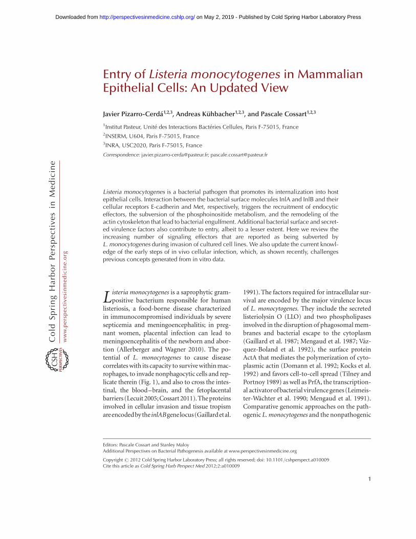
listeriosis, a food-borne disease characterizedin immunocompromised individuals by severesepticemia and meningoencephalitis; in preg-nant women, placental infection can lead tomeningoencephalitis of the newborn and abor-tion (Allerberger and Wagner 2010). The po-tential of L. monocytogenes to cause diseasecorrelates with its capacity to survive within mac-rophages, to invade nonphagocytic cells and rep-licate therein (Fig. 1), and also to cross the intes-tinal, the blood–brain, and the fetoplacentalbarriers (Lecuit 2005;Cossart 2011). The proteinsinvolved in cellular invasion and tissue tropismareencodedbythe inlABgenelocus(Gaillardetal.
1991). The factors required for intracellular sur-vival are encoded by the major virulence locusof L. monocytogenes. They include the secretedlisteriolysin O (LLO) and two phospholipasesinvolved in the disruption of phagosomal mem-branes and bacterial escape to the cytoplasm(Gaillard et al. 1987; Mengaud et al. 1987; Vaz-quez-Boland et al. 1992), the surface proteinActA that mediates the polymerization of cyto-plasmic actin (Domann et al. 1992; Kocks et al.1992) and favors cell-to-cell spread (Tilney andPortnoy 1989) as well as PrfA, the transcription-al activatorof bacterial virulence genes (Leimeis-ter-Wachter et al. 1990; Mengaud et al. 1991).Comparative genomic approaches on the path-ogenic L. monocytogenes and the nonpathogenic
Editors: Pascale Cossart and Stanley Maloy
Additional Perspectives on Bacterial Pathogenesis available at www.perspectivesinmedicine.org
Copyright # 2012 Cold Spring Harbor Laboratory Press; all rights reserved; doi: 10.1101/cshperspect.a010009
Cite this article as Cold Spring Harb Perspect Med 2012;2:a010009
1
ww
w.p
ersp
ecti
vesi
nm
edic
ine.
org
on May 2, 2019 - Published by Cold Spring Harbor Laboratory Press http://perspectivesinmedicine.cshlp.org/Downloaded from
Listeria innocua has led in recent years to theidentification of many additional bacterial fac-tors required for infection (Camejo et al. 2011;Cossart 2011). In this article, we will summa-rize ourcurrent understanding of bacterial effec-tors and cellular signaling cascades that are sub-verted by L. monocytogenes in order to invademammalian epithelial cells.
InlA AND InlB: TWO MAJOR VIRULENCEFACTORS INVOLVED IN CELL INVASION
The two major invasion proteins of L. monocy-togenes, InlA and InlB, belong to the so-calledinternalin family. Indeed, L. monocytogenes ge-nome encodes 27 proteins now known as in-ternalins, based on the presence of tandemlyarranged leucine-rich repeats (LRRs) in theiramino-terminal domain, which in several caseshave been shown to be involved in protein–pro-tein interactions with host-cell ligands (Marinoet al. 2000; Cabanes et al. 2002; Bierne et al.2007). Internalins are classified in three differ-ent groups according to their association to theL. monocytogenes surface through their carboxy-
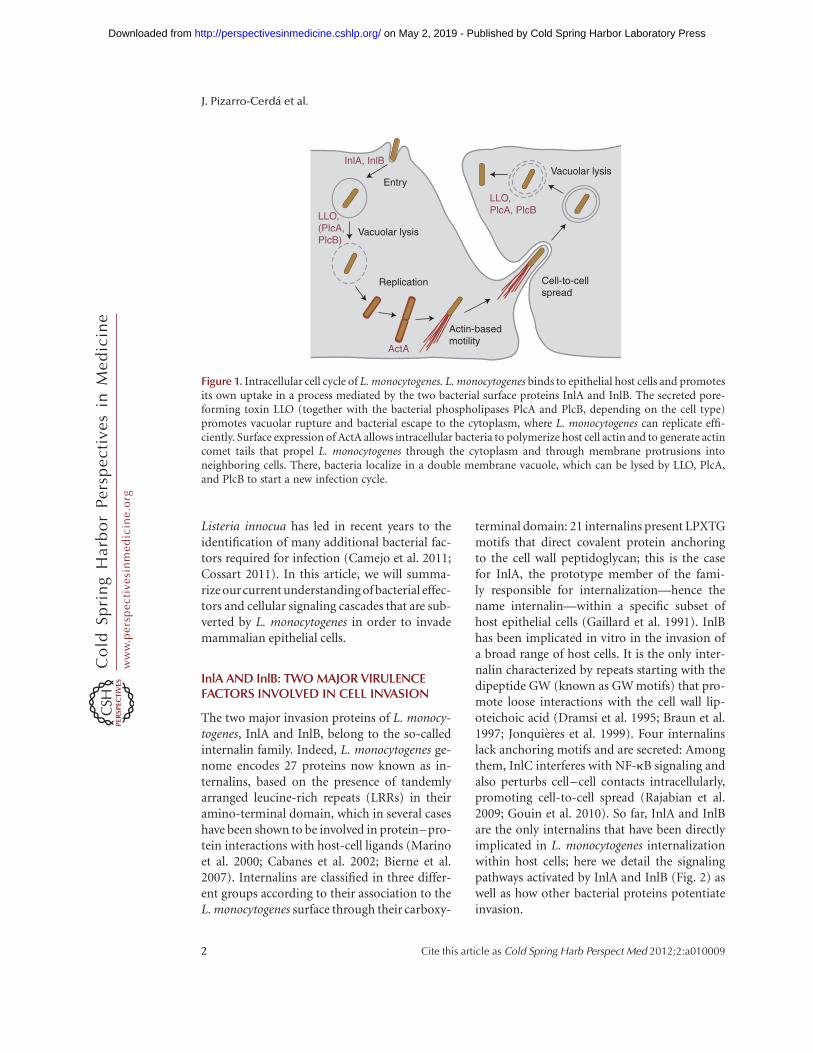
terminal domain: 21 internalins present LPXTGmotifs that direct covalent protein anchoringto the cell wall peptidoglycan; this is the casefor InlA, the prototype member of the fami-ly responsible for internalization—hence thename internalin—within a specific subset ofhost epithelial cells (Gaillard et al. 1991). InlBhas been implicated in vitro in the invasion ofa broad range of host cells. It is the only inter-nalin characterized by repeats starting with thedipeptide GW (known as GW motifs) that pro-mote loose interactions with the cell wall lip-oteichoic acid (Dramsi et al. 1995; Braun et al.1997; Jonquieres et al. 1999). Four internalinslack anchoring motifs and are secreted: Amongthem, InlC interferes with NF-kB signaling andalso perturbs cell–cell contacts intracellularly,promoting cell-to-cell spread (Rajabian et al.2009; Gouin et al. 2010). So far, InlA and InlBare the only internalins that have been directlyimplicated in L. monocytogenes internalizationwithin host cells; here we detail the signalingpathways activated by InlA and InlB (Fig. 2) aswell as how other bacterial proteins potentiateinvasion.
Vacuolar lysis
Cell-to-cellspread
Actin-basedmotility
Replication
Vacuolar lysis
Entry
LLO,PlcA, PlcB
LLO,(PlcA,PlcB)
InlA, InlB
ActA
Figure 1. Intracellular cell cycle of L. monocytogenes. L. monocytogenes binds to epithelial host cells and promotesits own uptake in a process mediated by the two bacterial surface proteins InlA and InlB. The secreted pore-forming toxin LLO (together with the bacterial phospholipases PlcA and PlcB, depending on the cell type)promotes vacuolar rupture and bacterial escape to the cytoplasm, where L. monocytogenes can replicate effi-ciently. Surface expression of ActA allows intracellular bacteria to polymerize host cell actin and to generate actincomet tails that propel L. monocytogenes through the cytoplasm and through membrane protrusions intoneighboring cells. There, bacteria localize in a double membrane vacuole, which can be lysed by LLO, PlcA,and PlcB to start a new infection cycle.
J. Pizarro-Cerda et al.
2 Cite this article as Cold Spring Harb Perspect Med 2012;2:a010009
ww
w.p
ersp
ecti
vesi
nm
edic
ine.
org
on May 2, 2019 - Published by Cold Spring Harbor Laboratory Press http://perspectivesinmedicine.cshlp.org/Downloaded from
Listeria
Listeria
Clustering
Arp2/3
Caveolin Caveolin
Src
InlA
Lipid raft
Hakai
p120
Arp2/3
Src
The InlB pathway
The InlA pathwayA
Actin
α-Catenin
α-Catenin
p120
β-Catenin
β-Catenin
E-cadherin
Hip1R
MyoVI
Clathrin
Dab2
Listeria
Listeria
Ub
Ub
Ub
Ub
P
P
P
P
Ub
Ub
Ub
P
P
P
P
P
P
P
Listeria
Listeria
ListeriaListeria
InIB InIB
GAGs
Met
Cbl
gC1q-RMet
Soluble InIB
Arp2/3
Actin
Cholesterol
Clathrin
Hip1R
Dab2
MyoVI
Arp2/3
WAVE/N-WASP
LIM-K
Rac/Cdc42Gab1
PI3K
CofilinCofilin
Cortactin
Dynamin
Actin
Hip1R
MyoVI
Clathrin
Dab2
Lipid raft
PIP3
B
Cortactin
Dynamin
Figure 2. Signaling cascades activated via the InlA- and InlB-invasion pathways. Interaction of InlA and/or InlBwith their respective host-cell surface receptors E-cadherin and Met induces ubiquitination of the receptors bythe ubiquitin ligases Hakai in the case of E-cadherin or Cbl in the case of Met and subsequent recruitment of theclathrin endocytosis machinery (Dab2, clathrin, dynamin, Hip1R, MyoVI), which provides an initial platformfor actin cytoskeleton polymerization. (See following page for legend.)
Listeria monocytogenes Entry in Mammalian Epithelial Cells
Cite this article as Cold Spring Harb Perspect Med 2012;2:a010009 3
ww
w.p
ersp
ecti
vesi
nm
edic
ine.
org
on May 2, 2019 - Published by Cold Spring Harbor Laboratory Press http://perspectivesinmedicine.cshlp.org/Downloaded from
InlA Interacts with the Adhesion MoleculeE-Cadherin to Promote Invasion in SpecificCellular Subpopulations
E-cadherin is an adhesion molecule involved inthe formation of adherens junctions at the intes-tinal barrier, the blood–brain barrier, and theplacenta, which has been identified as the cellu-lar receptor for InlA (Mengaud et al. 1996). Inmammalian tissues, polarized epithelial cells arecharacterized by the presence of several adhesionstructures including tight and adherens junc-tions, which confer to a given organ imperme-ability to the external environment. Homophilicinteractions between the extracellular domainsof E-cadherin molecules in adjacent epithelialcells are required for the initial formation ofadherens junctions; these structures are furtherstabilized by dynamic interactions of the E-cadherin cytoplasmic domain with elements ofthe clathrin-mediated endocytosis machineryand the actin cytoskeleton. As discussed below,L. monocytogenes subverts these cellular mecha-nisms to promote bacterial internalization. In-teraction between the InlA LRRs and E-cadherinis sufficient to promote bacterial adhesion andentry into host cells: Indeed, LRRs expression onthe surface of the noninvasive species L. innocuaor on latex beads allows particle internalizationin Caco-2 cells (Lecuit et al. 1997).
Remarkably, the InlA/E-cadherin interac-tion is species specific: A proline at position 16of human or guinea pig E-cadherin is critical forits interaction with InlA, whereas a glutamic acidat the same position in mouse or rat E-cadherinprecludes InlA recognition. Elegant studies haveshown that expression of a murine E-cadherindisplaying a proline instead of a glutamic acid atposition 16 allows recognition of this moleculeby InlA in vitro and in vivo (Lecuit et al. 1999,2001). The crystal structure of the InlA LRRs in
association with the first extracellular domain ofthe human E-cadherin has illustrated the molec-ular basis of this specificity: The proline 16 isaccommodated in a hydrophobic pocket createdby a deletion of one amino acid in the InlALRR6, which shortens this repeat to 21 residues(instead of 22 observed in all other repeats); abulky glutamic acid at this position does not fitthe InlA LRRs hydrophobic pocket and preventsE-cadherin binding (Schubert et al. 2002). Ow-ing to the specific distribution of E-cadherin incertain tissues, InlA plays a critical role in vivo inthe bacterial traversal of the intestinal barrierand the tropism for the placenta (see below).Moreover, owing to its species-specific interac-tion with E-cadherin, InlA appears as the mostimportant effector determining the susceptibil-ity of a host to a L. monocytogenes infection.
InlB Interacts with the Hepatocyte GrowthFactor Receptor Met to PromoteInternalization in a Wide Variety ofMammalian Cells
InlB, which is encoded by the second gene ofthe inlAB locus (Gaillard et al. 1991), allows L.monocytogenes internalization in a broad rangeof cell lines including HeLa, HEp-2, HepG2,TIB73, and Vero cells (Dramsi et al. 1995; Ling-nau et al. 1995; Braun et al. 1998, 1999; Greif-fenberg et al. 1998). Several host-cell moleculeshave been identified as potential receptors forInlB; among them, the hepatocyte growth factor(HGF) receptor Met plays the most critical rolefor bacterial internalization (Shen et al. 2000).Met is a ubiquitous receptor tyrosine kinase thatcontrols cell migration and growth during em-bryogenesis, as well as invasion and metastasis incancercells (Trusolino and Comoglio 2002). Themature protein is a disulphide-linked hetero-dimer formed by a short extracellular a chain
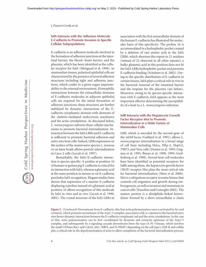
Figure 2. (Continued) Downstream from E-cadherin, this first actin polymerization wave is activated by Src andcortactin, which promote recruitment of the Arp2/3 complex; association of b/a-catenins to the bacterial entrysites favors dynamic interactions between the E-cadherin cytoplasmic tail and the actin cytoskeleton. In the caseof Met, actin polymerization can be first coordinated by dynamin and cortactin upstream of the Arp2/3complex, and subsequently by a signaling cascade downstream from the type IA PI 3-kinase, which involvesthe small GTPases Rac1 and Cdc42, abi1, WAVE, and N-WASP (depending on the cell type); LIM-K and cofilinplay a critical role in the depolymerization of actin to allow completion of the bacterial internalization process.
J. Pizarro-Cerda et al.
4 Cite this article as Cold Spring Harb Perspect Med 2012;2:a010009
ww
w.p
ersp
ecti
vesi
nm
edic
ine.
org
on May 2, 2019 - Published by Cold Spring Harbor Laboratory Press http://perspectivesinmedicine.cshlp.org/Downloaded from
and a longer b chain that comprises an ectodo-main, a transmembrane domain, and acytoplas-mic tail where a kinase domain and phosphor-ylatable motifs are located (Birchmeier et al.2003). InlB functionally mimics HGF, the natu-ral Met ligand, through the binding of its LRRs(Braun et al. 1999; Marino et al. 1999; Niemannet al. 2007). As in the case of the InlA/E-cadherininteraction, InlB interaction with Met is speciesspecific: InlB interacts with human and mouseMet, but does not recognize the guinea-pig orrabbit receptor (Khelef et al. 2006). In suscepti-ble species, InlB is reported to dimerize on bind-ing to Met (Ferraris et al. 2010) leading to thereceptordimerization, the autophosphorylationof the protein-docking motifs, and the recruit-ment of protein effectors that induce signals tothe actin cytoskeleton and the endocytosis ma-chinery (see below). CD44v6, a member of theCD44 family of type I transmembrane glycopro-teins, has been reported to potentiate Met acti-vation by HGF and also by InlB (Orian-Rous-seau et al. 2002; Ponta et al. 2003) but conflictingresults exist concerning its implication duringL. monocytogenes invasion (Jung et al. 2009;Dortet et al. 2010). The “B repeat” of InlB, oftenreferred to as a spacer domain between the LRRsand GWmodules, has also been reported to par-ticipate in the activation of Met and to endowInlB with stronger biological activities thanthose elicited by HGF (i.e., activation of a Ras-MAP kinase pathway) (Copp et al. 2003). How-ever, structural studies indicate that it does notinteract with Met, leading to the proposal thatthe B repeat may interact with a nonidentifiedcellular receptor (Ebbes et al. 2011).
Although the amino-terminal LLR domainof InlB is critical for activating Met, the threecarboxy-terminal GW modules are required tostabilize the InlB/Met interaction. As men-tioned previously, the GW modules were origi-nally described as mediating loose interactionsbetween InlB and the lipoteichoic acids of theL. monocytogenes cell wall (Braun et al. 1997;Jonquieres et al. 1999) and interestingly, solubleheparin can detach InlB from the bacterial cellwall (Jonquieres et al. 2001). Heparin and gly-cosaminoglycans (GAGs), which are highlycharged extracellular matrix polysaccharides,
favor the surface clustering of growth factorsincluding HGF, potentiating their activity bystabilizing their interaction with their receptors(Zhang 2010). Purified InlB was shown to bindto purified GAGs through its GW domains andinvasion of Chinese hamster ovary (CHO) cellsis potentiated 10-fold when GAGs are producedat the cell surface (Jonquieres et al. 2001). Theseresults suggest that the GW modules of InlBpresent the dual function of attaching InlB tothe bacterial cell wall and of enhancing L. mono-cytogenes entry triggered by the LRRs domain,reinforcing the similarities between the mecha-nisms of cell activation exerted by InlB and HGF(Jonquieres et al. 2001; Banerjee et al. 2004).The GW domains of InlB have been reportedto bind another ubiquitous cellular molecule,the receptor of the globular part of the comple-ment component C1q (gC1q-R) (Braun et al.2000; Marino et al. 2002). Intriguingly, gC1q-Ris a glycosylphosphatidylinositol (GPI)-an-chored protein that does not present a cytoplas-mic domain, suggesting that signaling on inter-action with InlB is transduced through anothercofactor. It has been reported that interactionbetween gC1q-R and InlB GW domains antag-onizes rather than enhances InlB signaling (Ma-rino et al. 2002; Banerjee et al. 2004). Therefore,the precise role of gC1q-R in L. monocytogenesinfection remains to be clarified.
InlB expression by L. innocua does not in-duce bacterial adhesion to Vero cells (Jonquiereset al. 1999) suggesting that InlB does not func-tion as an adhesin but instead as a potent sig-naling invasin that mediates L. monocytogenesinternalization through interactions with mul-tiple host-cell ligands. The potential synergy ofthe InlA- and the InlB-invasion pathways hasnot been fully explored yet but it has been re-cently found that InlB plays, together with InlA,a critical role in the colonization of the placentain vivo (Disson et al. 2008) (see below).
The Clathrin-Mediated EndocytosisMachinery Is Involved in the EarlySteps of Entry
The clathrin-mediated endocytosis machineryis involved in the cell invasion by L. monocyto-
Listeria monocytogenes Entry in Mammalian Epithelial Cells
Cite this article as Cold Spring Harb Perspect Med 2012;2:a010009 5
ww
w.p
ersp
ecti
vesi
nm
edic
ine.
org
on May 2, 2019 - Published by Cold Spring Harbor Laboratory Press http://perspectivesinmedicine.cshlp.org/Downloaded from
genes (Veiga and Cossart 2005; Veiga et al. 2007;Bonazzi et al. 2011). For both the InlA andthe InlB pathways, the initial events in the in-ternalization process are the posttranslationalmodifications of the two main Listeria receptorsE-cadherin and Met: Indeed, E-cadherin isphosphorylated by Src and then ubiquitinatedby Hakai (Bonazzi et al. 2008), whereas Metautophosphorylation leads to the recruitmentof Cbl, which in turn ubiquitinates Met (Veigaand Cossart 2005). Recent studies have shownthat the clathrin adaptor Dab2 is recruitedto the L. monocytogenes receptors followed bythe clathrin heavy and light chains; interestingly,the clathrin heavy chain is phosphorylated andthis event is critical for the bacterial inter-nalization process (Bonazzi et al. 2011). Theprotein Hip1R, which interacts with the clathrinlight chain and also with the actin cytoskeleton,is then recruited; in addition, myosin VI canbind Hip1R and interact with actin filaments:Its capacity to move toward the minus end ofthe actin filaments fits well with a function inpulling the bacteria inside the cell (Bonazzi et al.2011). Small interfering RNA (siRNA) experi-ments have shown that other members of theCME machinery play a critical role, in particu-lar, dynamin (Veiga and Cossart 2005), which inturn can recruit cortactin, an activator of Arp2/3, a main actin nucleator; a first wave of actinpolymerization could thus be triggered by cor-tactin. Structural studies reveal that clathrin-coated pits are present at the L. monocytogenesentry foci and are probably the sites of earlyactin rearrangements (Bonazzi et al. 2011). In-terestingly, in JEG-3 epithelial cells, clathrin-in-dependent mechanisms involving caveolin alsomediate bacterial internalization downstreamfrom the InlA-invasion pathway (Bonazzi et al.2008).
Cytoskeletal and Membrane Rearrangementsduring Invasion Are Regulated byActin-Binding Proteins, Small GTPases,PI 3-Kinase, and Phosphoinositides
As shown by numerous studies, following thefirst wave of actin polymerization describedabove or concomitantly, a second wave of actin
rearrangements takes place during the L. mono-cytogenes invasion process, involving a PI 3-ki-nase, small GTPases of the Rho family, and sev-eral actin-binding proteins. The use of the PI3-kinase inhibitors wortmannin and LY294002showed in early studies that type IA PI 3-kinaseactivity is necessary for L. monocytogenes in-fection of Caco-2 and Vero cells via the InlA-and InlB-invasion pathways, respectively (Ire-ton et al. 1996). In Caco-2 cells, however, noincrease in PI(3,4)P2 or PI(3,4,5)P3 levels wasdetected on interaction with L. monocytogenes,because in these cells PI3 kinase is constitutivelyand highly expressed. On the contrary, 10- and20-fold increases on PI(3,4)P2 or PI(3,4,5)P3
levels are detected on infection of Vero cellswith L. monocytogenes via the InlB-invasionpathway (Ireton et al. 1996). In these cells, ac-tivation of type IA PI 3-kinase is promotedby the protein adaptors Gab1, Cbl, and Shc,which are phosphorylated and recruited to theMet cytoplasmic tail, translocating with themthe p85 subunit to the L. monocytogenes inva-sion foci (Ireton et al. 1999; Shen et al. 2000).Gab1 can also be translocated to the plasmamembrane through its pleckstrin homologydomain, which binds PI(3,4,5)P3 (Basar et al.2005) and indirectly boosts type IA PI 3-kinaseactivity via recruitment of the adaptor proteinCrkII (Sun et al. 2005; Dokainish et al. 2007).PI(3,4,5)P3 produced by the type IA PI 3-ki-nase plays a critical role in the recruitmentof molecules that modulate actin cytoskeletondynamics (see below) to the plasma membraneand a recent study has shown that the 50-phos-phatase OCRL restricts bacterial entry by pre-cisely reducing the levels of PI(4,5)P2 andPI(3,4,5)P3 at L. monocytogenes entry foci (Kuh-bacher et al. 2012).
Actin polymerization following the InlB/Met interaction differs among cell lines: InVero cells, plasma membrane lipid rafts promotethe clustering of PI(3,4,5)P3 and favor the trans-location of Rac1 to the plasma membrane (Se-veau 2004; Seveau et al. 2007); together with theWiskott-Aldrich syndrome protein (WASP)-re-lated protein complex WAVE2, Rac1 promotesactin polymerization by the Arp2/3 complex(Bierne 2001; Bierne et al. 2005). In HeLa
J. Pizarro-Cerda et al.
6 Cite this article as Cold Spring Harb Perspect Med 2012;2:a010009
ww
w.p
ersp
ecti
vesi
nm
edic
ine.
org
on May 2, 2019 - Published by Cold Spring Harbor Laboratory Press http://perspectivesinmedicine.cshlp.org/Downloaded from
cells, Rac1 and Cdc42 are required for Arp2/3activation downstream from PI(3,4,5)P3, to-gether with N-WASP, WAVE1, and WAVE2 (Bi-erne et al. 2005); in this cell line, PI(3,4,5)P3
is also recognized by ARAP2, a protein thatfunctions as a GAP for the small GTPase Arf6;ARAP2 promotes actin rearrangements duringL. monocytogenes invasion in part by antagoniz-ing the activity of Arf6 (Gavicherla et al. 2010).In murine fibroblasts, Cdc42 participates in theactivation of Rac1 via PI 3-kinase-dependentand -independent mechanisms (Bosse et al.2007). A recent systems-biology study has iden-tified nine genes encoding known upstream reg-ulators or downstream effectors of the type IAPI 3-kinase required for invasion of target cellsby L. monocytogenes including the small GTPaseRab5 and several regulators of Arf or Rac1small GTPases (Jiwani et al. 2011). PI4P, a phos-phoinositide usually present at the Golgi andproduced by type II PI 4-kinases, is detectedat bacterial entry sites and is critical for inva-sion in LoVo and HeLa cells. PI4P does notseem to function as a PI(3,4,5)P3 precursorbut rather acts as an independent effector fos-tering bacterial invasion downstream from thetetraspanin CD81 (Pizarro-Cerda et al. 2007;Tham et al. 2010).
Concerning the InlA/E-cadherin invasionpathway, inhibition of Src kinase with the phar-macological agent PP1 in L2071hEcad cellsshowed that phosphorylation of cortactin bySrc contributes to the recruitment of the Arp2/3 complex to bacterial invasion foci and initi-ates actin polymerization (Sousa et al. 2007).Among the small GTPases of the Rho familyknown to control actin dynamics, only Rac1(but not Cdc42 or RhoA) appeared to be re-quired for L. monocytogenes entry in Caco-2cells (Sousa et al. 2007). The interactions be-tween E-cadherin and the actin cytoskeletonare highly complex (Yamada et al. 2005). Inboth adherens junction formation and internal-ization of Listeria, they involve a and b catenins(Lecuit et al. 2000). A two-hybrid screen usinga-catenin as bait identified the autosomal re-cessive hypercholesterolemia GTPase-activatingprotein 10 (ARHGAP10) as a molecule requiredfor a-catenin recruitment and actin cytoskele-
ton rearrangements during L. monocytogenesinvasion (Sousa et al. 2005a). However, ARH-GAP10 promotes GTP hydrolysis by Cdc42and RhoA but not Rac1; because Cdc42 andRhoA are not involved in InlA mediated en-try, ARHGAP10 may function as an adaptorprotein rather than GAP favoring interactionsbetween a-catenin and still nonidentified mo-lecular partners during bacterial internaliza-tion. The unconventional Myosin VIIa has alsobeen identified as a binding partner of the trans-membrane molecule vezatin and E-cadherinin the InlA-invasion pathway and could par-ticipate, together with actin, in membrane rear-rangements required for bacterial entry (Sousaet al. 2004).
Elongation of actin filaments and depoly-merization of actin are tightly controlled eventsduring L. monocytogenes internalization. Se-questration of the actin-binding proteins Ena/VASP to the mitochondria (by the expressionof mitochondrial targeting motifs) as well assiRNA inactivation of VASP inhibit the entry ofbacteria or InlB-coated beads in Vero cells, dem-onstrating a role for VASP in entry (Bierne et al.2005). In addition, overexpression of the LIMkinase, which phosphorylates and inactivatesthe actin depolymerizing factor cofilin, inducesthe accumulation of actin filaments beneathbacteria in Vero and REF-52 cells, preventinginternalization and showing that actin depoly-merization is critical during the late stages ofthe entry process (Bierne 2001). Interestingly,phosphorylated cofilin also drives phosphati-dylcholine cleavage by host-cell phospholipaseD1 (PLD1) and perturbation of the cellular lev-els of PLD1 blocks L. monocytogenes entry inVero and HepG2 cells (Han et al. 2011).
Among the many other cellular actin inter-acting partners, septins represent a group ofsmall GTPases known to polymerize into oli-gomeric protein complexes and filaments(Surka et al. 2002) that regulate cytokinesis, ves-icle transport, and intracellular compartmen-talization in a wide range of organisms (Beiteset al. 2005; Estey et al. 2011). A member of theseptin family, SEPT9, was initially identified asbeing recruited together with actin to the entrysite of InlB-coated beads in LoVo cells (Pizarro-
Listeria monocytogenes Entry in Mammalian Epithelial Cells
Cite this article as Cold Spring Harb Perspect Med 2012;2:a010009 7
ww
w.p
ersp
ecti
vesi
nm
edic
ine.
org
on May 2, 2019 - Published by Cold Spring Harbor Laboratory Press http://perspectivesinmedicine.cshlp.org/Downloaded from
Cerda et al. 2002). Yeast two-hybrid screens andimmunoprecipitation experiments revealed theinteraction of SEPT9 with SEPT2, SEPT7, andSEPT11 in resting or InlB-stimulated HeLacells (Mostowy et al. 2009a,b) and atomic forcemicroscopy has shown that SEPT2 and SEPT9contribute to the anchorage of Met to the ac-tin cytoskeleton (Mostowy et al. 2011). Inac-tivation of septins by siRNA highlighted differ-ent contributions of SEPT2 and SEPT11 toL. monocytogenes invasion: SEPT2 knockdownseverely modifies the actin cytoskeleton and in-hibits bacterial entry (Mostowy et al. 2009b),whereas SEPT11 knockdown increases host-cell infection without modifying the actin cyto-skeleton or affecting the Met signaling path-way, suggesting that SEPT11 rather restricts theInlB-mediated cell invasion (Mostowy and Cos-sart 2009).
In summary, the multiple interactions be-tween the clathrin-mediated endocytosis ma-chinery, the various phosphoinositides, andthe actin cytoskeleton during L. monocytogenescellular invasion highlight a very complex pro-cess whose dynamics vary with the nature of theinfected cell. Genome-wide siRNA screens inprogress should help in deciphering the orches-tration of the molecular events that are key tocellular infection, and will address, in particular,whether internal membrane components areshuttled to the plasma membrane during entry.
Role of InlA and InlB in the Traversal of theHost Barriers In Vivo
Different lines of evidence show a critical con-tribution of InlA during L. monocytogenes in-fection in vivo. Indeed, epidemiological studies(Olier et al. 2003; Jacquet et al. 2004) show thatstrains in which InlA is truncated and secretedare frequently found in asymptomatic carriersor in food products, whereas full-length InlA ispresent far more frequently in clinical strains,suggesting that expression of an intact InlAmolecule correlates with full bacterial virulence.When E-cadherin was identified as the receptorfor InlA (Mengaud et al. 1996), a role for thisinternalin in the traversal of the intestinal bar-rier was proposed because E-cadherin is an ad-
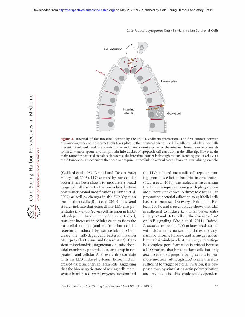
hesion molecule localized at the basolateral sur-face of several cell types of the small intestinevilli, including enterocytes (absorptive cells)and goblet (mucus-secreting) cells. Studies inguinea pigs, which express an E-cadherin rec-ognized by InlA, as well as in transgenic miceexpressing the human E-cadherin under thecontrol of the iFABP intestinal promotor, haveconfirmed that the presence of InlA is requiredfor the traversal of the small intestine barrier invivo through enterocyte invasion (Lecuit et al.2001). Of note, in enterocytes E-cadherin ismainly present at adherens junctions wheremolecules are hidden below tight junctionsand therefore are not normally exposed to thelumen of the intestine (Sousa et al. 2005b). Us-ing rabbit ligated ileal loops and Madin-Darbycanine kidney (MCDK) cells as a polarizedepithelium model, it has been proposed thatL. monocytogenes does not disrupt the entero-cyte tight junctions to gain access to E-cadherin,but instead interacts with exposed E-cadherinat sites where apoptotic cells are expelled anddetached from the epithelium by extrusion,suggesting that L. monocytogenes exploits thedynamic nature of epithelial renewal and junc-tional remodeling in small intestinal villi tobreach this barrier (Fig. 3) (Pentecost et al.2006). Of note, infection of wild-type mice in-tragastrically with a L. monocytogenes strain ex-pressing a modified InlA that binds murine E-cadherin suggested that InlB also participates inbacterial internalization at small intestine villi;experiments in MDCK cells suggest that InlBdoes not act as an adhesin but rather acceleratesbacterial internalization by promoting endocy-tosis of junctional components (Pentecost et al.2010). It has thus been proposed that InlA pro-vides specificity of adhesion to exposed E-cad-herin at the tip of small intestine villi and InlBactivates Met to accelerate junction endocytosisand bacterial invasion of enterocytes.
A recent study in iFABP transgenic mice hasidentified several discrete locations where E-cad-herin is luminally accessible, including extrud-ing enterocytes at the tip of villi as describedabove, but also villus epithelial folds and partic-ularly junctions between mucus-secreting gob-let cells, which are recognized as the preferred
J. Pizarro-Cerda et al.
8 Cite this article as Cold Spring Harb Perspect Med 2012;2:a010009
ww
w.p
ersp
ecti
vesi
nm
edic
ine.
org
on May 2, 2019 - Published by Cold Spring Harbor Laboratory Press http://perspectivesinmedicine.cshlp.org/Downloaded from
sites for L. monocytogenes translocation at thesmall intestine (Nikitas et al. 2011). The InlA-dependent bacterial translocation through gob-let cells is extremely rapid and surprisingly LLOand ActA independent; pharmacological andmicroscopical analyses reveal that L. monocyto-genes does not escape from its internalizationvacuole in goblet cells and rather takes advantageof a transcytosis mechanism that allows fast tra-versal of the small intestinal barrier (Fig. 3) (Ni-kitas et al. 2011). Although the small intestine is amajor site for L. monocytogenes invasion, exper-iments in gerbils, which are natural hosts forL. monocytogenes, and in humanized transgenicmice that express E-cadherin in all physiologi-cal tissues, show that bacterial InlA-dependenttranslocation can also be observed at the cae-cum and the colon of the large intestine (Dissonet al. 2008). Studies in rat ligated ileal loopsfrom the lower part of the small intestine sug-gest furthermore that Peyer’s patches, which arelymphoid nodules lined by antigen-samplingmicrofold (M) cells, may represent L. monocy-togenes InlA- and InlB-independent invasionsites at the intestinal level (Pron et al. 1998;Corr et al. 2006).
As mentioned above, epidemiological dataindicate that full-length InlA is predominant inclinically isolated L. monocytogenes strains, andthis is particularly striking in pregnancy-related
cases, in which 100% of isolated strains displayan intact InlA molecule (Jacquet et al. 2004). Inthe placenta, E-cadherin is particularly enrichedat the continuous multinucleated syncytiotro-phoblast layer, which contributes to the barrierfunction of this organ, and studies in placentalexplants from human fetoplacental listeriosiscases as well as in primary cell cultures andplacental cell lines indicate that interaction be-tween InlA and the syncytiotrophoblast E-cad-herin is critical for the traversal of the fetopla-cental barrier (Lecuit et al. 2004). During thefirst trimester of pregnancy, the syncytiotropho-blast overlies a cytotrophoblast layer, which alsoparticipates in the placental barrier, and analy-ses of first trimester human placental culturessuggest that cytotrophoblasts can also be tar-geted by L. monocytogenes via InlA/E-cadherininteractions or cell-to-cell spread from infectedphagocytes (Robbins et al. 2010; Zeldovich et al.2011). The conjugated action of InlA and InlBto fetoplacental listeriosis was originally debat-ed: Although it was shown in human matureplacental explants that InlB participated togeth-er with InlA to syncytiotrophoblast invasion(Lecuit et al. 2004), in the guinea-pig modelno role was shown for InlA despite its permis-siveness to the InlA pathway (Bakardjiev et al.2006), and in the mouse model no role wasobserved for InlB despite its permissiveness to
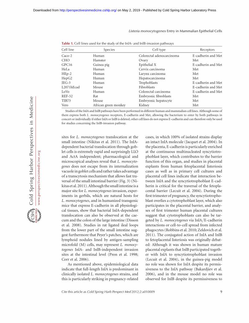
Table 1. Cell lines used for the study of the InlA- and InlB-invasion pathways
Cell line Species Cell type Receptors
Caco-2 Human Colorectal adenocarcinoma E-cadherin and MetCHO Hamster Ovary MetGPC16 Guinea pig Epithelial X E-cadherin and MetHeLa Human Cervix carcinoma MetHEp-2 Human Larynx carcinoma MetHepG2 Human Hepatocarcinoma MetJEG-3 Human Trophoblasts E-cadherin and MetL2071hEcad Mouse Fibroblasts E-cadherin and MetLoVo Human Colorectal carcinoma E-cadherin and MetREF-52 Rat Embryonic fibroblasts MetTIB73 Mouse Embryonic hepatocyte MetVero African green monkey Kidney Met
Studies of the InlA and InlB pathways have been performed in different human and mammalian cell lines. Although some of
them express both L. monocytogenes receptors, E-cadherin and Met, allowing the bacterium to enter by both pathways in
concert or individually if either InlA or InlB is deleted, other cell lines do not express E-cadherin and can therefore only be used
for studies concerning the InlB-invasion pathway.
Listeria monocytogenes Entry in Mammalian Epithelial Cells
Cite this article as Cold Spring Harb Perspect Med 2012;2:a010009 9
ww
w.p
ersp
ecti
vesi
nm
edic
ine.
org
on May 2, 2019 - Published by Cold Spring Harbor Laboratory Press http://perspectivesinmedicine.cshlp.org/Downloaded from
the InlB pathway (Le Monnier et al. 2007). Thedifferent organization of the placenta in theseanimal models could partially account for dif-ferences reported in each of these studies. How-ever, these apparently contradictory observa-tions were subsequently explained when it wasfound in gerbils and knock-in mice ubiquitous-ly expressing humanized E-cadherin, that pla-cental infection by L. monocytogenes takes placeonly when both InlA- and InlB-invasion path-ways are functional as in humans (Disson et al.2008). It is not the case in the guinea pig inwhich Met is not a receptor for InlB (Khelefet al. 2006), and it is also not the case in themouse in which E-cadherin is not a receptorfor InlA (Lecuit et al. 1999).
Modulation of InlA- and InlB-DependentInvasion Pathways by Other Internalins
To date, among the 27 known L. monocytogenesinternalins, InlA and InlB are the major viru-lence factors implicated in cell invasion. Severalstudies suggest that other members of the inter-nalin family may modulate L. monocytogenes in-vasion (Chatterjee et al. 2006) by behaving asadhesins, by indirectly affecting the surface ex-posure of InlA and/or InlB, or by directly inter-acting with putative novel cellular receptors. Forexample, expression of the LPXTG-anchoredInlJ by L. innocua leads to bacterial adhesion(but no invasion) to human JEG-3 and HT29polarized epithelial cells (Sabet et al. 2008), butits potential cell ligand has not been identifiedyet. The gene cluster inlGHE is required for fullinvasion of Caco-2 cells by the L. monocytogenesEGDe strain and it has been hypothesized thatits expression may indirectly modulate the bac-terial cell wall organization, therefore affectingInlA presentation at the L. monocytogenes sur-face (Bergmann et al. 2002).
A particular case has been reported for InlF:Under standard cell culture conditions, no rolehas been observed for this LPXTG internalinduring invasion of Caco-2 and HepG2 cells invitro or during Balb/c mice infection in vivo(Dramsi et al. 1997). However, in humanHeLa and HEp-2 cells, pharmacological inhibi-tion of the RhoA/Rho kinase signaling pathway
using the drug Y27632 increases bacterial ad-hesion in an InlF-dependent manner (Kirchnerand Higgins 2008). Moreover, in murine TIB75hepatocytes and L2 fibroblasts, Y27632 treat-ment promotes InlF-dependent cell invasionby L. monocytogenes or by L. innocua-expressingInlF (but not InlA or InlB); Balb/c mice expo-sure to the HA-1077 drug, which also inhibitsRho kinases, increases the InlF-dependent vir-ulence of L. monocytogenes in vivo (Kirchnerand Higgins 2008). The mechanisms by whichthe RhoA/Rho kinase signaling pathway affectsinfection are unknown: Because this cascadeenhances the assembly of actin-myosin struc-tures, it was hypothesized that perturbation ofactin-myosin interactions may lead to the cellsurface exposure of a nonidentified InlF host-cell receptor (Carragher et al. 2006).
For several internalins including InlI, norole has been attributed to date (Sabet et al.2005). Interestingly, although InlC2 and InlDin the strain EGD do not participate in the in-vasion of Caco-2 or S180 cells in vitro and donot contribute to bacterial virulence in BALB/cmice in vivo (Dramsi et al. 1997), these inter-nalins are the major targets of the rabbit humor-al response to L. monocytogenes, together withInlA (Yu et al. 2007). For other members of theinternalin family, different roles in bacterial in-tracellular stages have been identified, as is thecase for InlC, which perturbs NF-kB signaling(Gouin et al. 2010) and cell-to-cell spread (Ra-jabian et al. 2009), or InlK, which allows escapeof autophagy (Dortet et al. 2011). There are 14remaining internalins that have not been char-acterized yet.
ROLE OF NONINTERNALIN MOLECULES INBACTERIAL ADHESION AND INVASION
Several molecules not belonging to the inter-nalin family have been reported to contributeto the L. monocytogenes adhesion and invasionof host cells using invitro systems. This is the caseof LLO, a major virulence factor belonging tothe family of cholesterol-dependent cytotoxinsfirst identified as a molecule allowing phagoso-mal maturation arrest and phagosomal lysis thatleads to bacterial escape to the cytoplasmic space
J. Pizarro-Cerda et al.
10 Cite this article as Cold Spring Harb Perspect Med 2012;2:a010009
ww
w.p
ersp
ecti
vesi
nm
edic
ine.
org
on May 2, 2019 - Published by Cold Spring Harbor Laboratory Press http://perspectivesinmedicine.cshlp.org/Downloaded from
(Gaillard et al. 1987; Dramsi and Cossart 2002;Henry et al. 2006). LLO secreted by extracellularbacteria has been shown to modulate a broadrange of cellular activities including histoneposttranscripional modifications (Hamon et al.2007) as well as changes in the SUMOylationprofile of host cells (Ribet et al. 2010) and severalstudies indicate that extracellular LLO also po-tentiates L. monocytogenes cell invasion in InlA/InlB-dependentand -independent ways. Indeed,transient increases in cellular calcium from theextracellular milieu (and not from intracellularreservoirs) induced by extracellular LLO in-crease the InlB-dependent bacterial invasionof HEp-2 cells (Dramsi and Cossart 2003). Tran-sient mitochondrial fragmentation, mitochon-drial membrane potential loss, and drop in res-piration and cellular ATP levels also correlatewith the LLO-induced calcium fluxes and in-creased bacterial entry in HeLa cells, suggestingthat the bioenergetic state of resting cells repre-sents a barrier to L. monocytogenes invasion and
the LLO-induced metabolic cell reprogramm-ing promotes efficient bacterial internalization(Stavru et al. 2011); the molecular mechanismsthat link this reprogramming with phagocytosisare currently unknown. A direct role for LLO inpromoting bacterial adhesion to epithelial cellshas been proposed (Krawczyk-Balska and Bie-lecki 2005), and a recent study shows that LLOis sufficient to induce L. monocytogenes entryin HepG2 and HeLa cells in the absence of InAor InlB signaling (Vadia et al. 2011). Indeed,L. innocua-expressing LLO or latex beads coatedwith LLO are internalized in a cholesterol-, dy-namin-, tyrosine kinase-, and actin-dependentbut clathrin-independent manner; interesting-ly, complete pore formation is critical becausea LLO variant that binds to host cells but onlyassembles into a prepore complex fails to pro-mote invasion. Although LLO seems thereforesufficient to trigger bacterial invasion, it is pro-posed that, by stimulating actin polymerizationand endocytosis, this cholesterol-dependent
Cell extrusion
Enterocytes
Goblet cell
E-cadherin
Intestinalvillus tip
Figure 3. Traversal of the intestinal barrier by the InlA-E-cadherin interaction. The first contact betweenL. monocytogenes and host target cells takes place at the intestinal barrier level. E-cadherin, which is normallypresent at the basolateral face of enterocytes and therefore not exposed to the intestinal lumen, can be accessibleto the L. monocytogenes invasion protein InlA at sites of apoptotic cell extrusion at the villus tip. However, themain route for bacterial translocation across the intestinal barrier is through mucus-secreting goblet cells via arapid transcytosis mechanism that does not require intracellular bacterial escape from its internalizing vacuole.
Listeria monocytogenes Entry in Mammalian Epithelial Cells
Cite this article as Cold Spring Harb Perspect Med 2012;2:a010009 11
ww
w.p
ersp
ecti
vesi
nm
edic
ine.
org
on May 2, 2019 - Published by Cold Spring Harbor Laboratory Press http://perspectivesinmedicine.cshlp.org/Downloaded from
cytotoxin cooperates with InlA and InlB to po-tentiate efficient bacterial infection in a broadrange of host cells (Vadia et al. 2011).
ActA is another major L. monocytogenes vir-ulence factor that has been referred to play a rolein bacterial invasion of host cells. ActA was firstdescribed as a protein promoting actin polymer-ization in the intracellularenvironment and bac-terial cell-to-cell spread (Domann et al. 1992;Kocks et al. 1992). Heterologous expression ofActA in the nonpathogenic species L. innocuaallows bacterial internalization in epithelial po-larized and nonpolarized cells (Caco-2, MCDK,HeLa, and Vero) but not in COS-1 fibroblasts,suggesting that ActA is sufficient to directly ac-tivate an invasion pathway specific for epithelialcells (Suarez et al. 2001). The amino-terminalregion of ActA is similar to the domain of thePlasmodium falciparum circumsporozoite pro-tein involved in heparate sulfate recognition andhepatocyte binding (Pancake et al. 1992; Coppiet al. 2007) and it has been shown that the pres-ence of heparan sulfate at the surface of CHOcells is required for the entry of L. monocytogenesinan ActA-dependentmanner(Alvarez-Domin-guez et al. 1997). The in vivo conditions inwhich the ActA invasion-associated functionwould be critical for virulence remain to be char-acterized.
Several cell membrane enzymes involved inthe modification of the L. monocytogenes cellwallarchitecture have been also shown to affect bac-terial adhesion and/or invasion invitro as well asvirulence in vivo. For example, the lipoteichoicacid modifiers GtcA (Promadej et al. 1999) andDltA (Abachin et al. 2002), the lipoprotein LpeA(Reglier-Poupet et al. 2003), the prolipoproteindiacylglyceryl transferase Lgt (Machata et al.2008), and the protein MprF involved in themodification of the phospholipid lysylphospha-tidylglycerol (Thedieck et al. 2006) play roles inL. monocytogenes adhesion or invasion in celllines by probably modulating the electronegativ-ityof the bacterial surface charge and/or alteringthe surface expression of adhesins/invasins. Aparticular class of cell membrane enzymes isthe autolysins, which are involved in the diges-tionof the cellwall peptidoglycan:Amongthe sixputative autolysins encoded by the L. monocyto-
genes genome, three of them (Ami, Auto, andIspC) characterized by the presence of carboxy-terminal GW repeats have been implicated inbacterial attachment to host cells or invasion.Ami, a N-acetylmuramoyl-L-alanine amidase(Braun et al. 1997) is required for adhesion butnot for invasion of host cells in an DinlA, DinlB,or DinlAB background only (Milohanic et al.2000), and interestingly, the eight GW repeatsof Ami are sufficient to promote L. monocyto-genes adhesion to host cells (Milohanic et al.2001). Auto, the only L. monocytogenes autolysinabsent fromthe genome ofL. innocua, is requiredfor invasion (not adhesion) to several mamma-lian cell lines (Cabanes et al. 2004), whereas IspCis required for bacterial adhesion and/or inva-sion in a cell line-dependent manner (Wang andLin 2008). Intriguingly, the Dami, Dauto, andDispC strains do not display major morpholog-ical abnormalities (ActA surface expression isonly affected in the DispC mutant) suggestingthat these molecules probably play a minor roleas autolysins in vitro; their importance is high-lighted by the reduced virulence of the mutantstrains in the mouse model invivo. The potentialcellular ligands for these autolysins or the pre-cise mechanisms governing their implicationin adhesion and invasion have not been identi-fied to date.
Other bacterial molecules involved in L.monocytogenes adhesion and/or invasion includethe LPXTG protein Vip (Cabanes et al. 2005), theadhesins Lap (Jagadeesan et al. 2011), and LapB(Reisetal.2010)aswellasthefibronectin-bindingprotein A (FbpA) required also as an escort pro-tein for LLO and InlB (Dramsi et al. 2004).
CONCLUDING REMARKS
L. monocytogenes is a fascinating bacterial path-ogen able to survive in a saprophytic environ-ment and to induce disease in mammalianhosts. Adhesion to host cells and invasion ofthe intracellular space are critical steps for thetraversal of host barriers leading to organ infec-tion and bacterial spread in the organism; notsurprisingly, many bacterial effectors have beenshown to participate in every stage of the infec-tious process. We are starting to understand the
J. Pizarro-Cerda et al.
12 Cite this article as Cold Spring Harb Perspect Med 2012;2:a010009
ww
w.p
ersp
ecti
vesi
nm
edic
ine.
org
on May 2, 2019 - Published by Cold Spring Harbor Laboratory Press http://perspectivesinmedicine.cshlp.org/Downloaded from
global principles that govern the regulation ofthe expression of these virulence factors. Thechallenge for future studies is to understandhow the combinatorial function of the bacterialand cellular factors specifically contributes tothe pathophysiology of listeriosis in vivo.
ACKNOWLEDGMENTS
Research in P. Cossart’s laboratory is supportedby the Pasteur Institute, ERC advanced grant233348, the Institut National de la Sante et dela Recherche Medicale, the Institut National dela Recherche Agronomique, the Agence Natio-nale de la Recherche, the Louis-Jeantet Founda-tion, and the Fondation Le Roch Les Mousque-taires. A.K. is a recipient of a scholarship fromthe Pasteur-Paris University International Doc-toral Program/Institut Carnot Maladies Infec-tieuses.
REFERENCES
Abachin E, Poyart C, Pellegrini E, Milohanic E, Fiedler F,Berche P, Trieu-Cuot P. 2002. Formation of D-alanyl-li-poteichoic acid is required for adhesion and virulence ofListeria monocytogenes. Mol Microbiol 43: 1–14.
Allerberger F, Wagner M. 2010. Listeriosis: A resurgent food-borne infection. Clin Microbiol Infect 16: 16–23.
Alvarez-Dominguez C, Carrasco-Marın E, Leyva-Cobian F.1993. Role of complement component C1q in phagocy-tosis of Listeria monocytogenes by murine macrophage-like cell lines. Infect Immun 61: 3664–3672.
Alvarez-Dominguez C, Vazquez-Boland JA, Carrasco-Ma-rın E, Lopez-Mato P, Leyva-Cobian F. 1997. Host cellheparan sulfate proteoglycans mediate attachment andentry of Listeria monocytogenes, and the listerial surfaceprotein ActA is involved in heparan sulfate receptor rec-ognition. Infect Immun 65: 78–88.
Bakardjiev AI, Theriot JA, Portnoy DA. 2006. Listeria mono-cytogenes traffics from maternal organs to the placentaand back. PLoS Pathog 2: e66.
Banerjee M, Copp J, Vuga D, Marino M, Chapman T, VanDer Geer P, Ghosh P. 2004. GW domains of the Listeriamonocytogenes invasion protein InlB are required for po-tentiation of Met activation. Mol Microbiol 52: 257–271.
Basar T, Shen Y, Ireton K. 2005. Redundant roles for Metdocking site tyrosines and the Gab1 pleckstrin homologydomain in InlB-mediated entry of Listeria monocyto-genes. Infect Immun 73: 2061–2074.
Beites CL, Campbell KA, Trimble WS. 2005. The septinSept5/CDCrel-1 competes with a-SNAP for binding tothe SNARE complex. Biochem J 385: 347–353.
Bergmann B, Raffelsbauer D, Kuhn M, Goetz M, Hom S,Goebel W. 2002. InlA- but not InlB-mediated internali-
zation of Listeria monocytogenes by non-phagocyticmammalian cells needs the support of other internalins.Mol Microbiol 43: 557–570.
Bierne H. 2001. A role for cofilin and LIM kinase in Listeria-induced phagocytosis. J Cell Biol 155: 101–112.
Bierne H, Miki H, Innocenti M, Scita G, Gertler FB,Takenawa T, Cossart P. 2005. WASP-related proteins,Abi1 and Ena/VASP are required for Listeria invasioninduced by the Met receptor. J Cell Sci 118: 1537–1547.
Bierne H, Sabet C, Personnic N, Cossart P. 2007. Internalins:A complex family of leucine-rich repeat-containing pro-teins in Listeria monocytogenes. Microbes Infect 9: 1156–1166.
Birchmeier C, Birchmeier W, Gherardi E, Vande Woude GF.2003. Met, metastasis, motility and more. Nat Rev MolCell Biol 4: 915–925.
Bonazzi M, Veiga E, Pizarro-Cerda J, Cossart P. 2008. Suc-cessive post-translational modifications of E-cadherinare required for InlA-mediated internalization of Listeriamonocytogenes. Cell Microbiol 10: 2208–2222.
Bonazzi M, Vasudevan L, Mallet A, Sachse M, Sartori A,Prevost MC, Roberts A, Taner SB, Wilbur JD, BrodskyFM, et al. 2011. Clathrin phosphorylation is requiredfor actin recruitment at sites of bacterial adhesion andinternalization. J Cell Biol 195: 525–536.
Bosse T, Ehinger J, Czuchra A, Benesch S, Steffen A, Wu X,Schloen K, Niemann HH, Scita G, Stradal TEB, et al.2007. Cdc42 and phosphoinositide 3-kinase drive Rac-mediated actin polymerization downstream of c-Met indistinct and common pathways. Mol Cell Biol 27: 6615–6628.
Braun L, Dramsi S, Dehoux P, Bierne H, Lindahl G,Cossart P. 1997. InlB: An invasion protein of Listeriamonocytogenes with a novel type of surface association.Mol Microbiol 25: 285–294.
Braun L, Ohayon H, Cossart P. 1998. The InIB protein ofListeria monocytogenes is sufficient to promote entry intomammalian cells. Mol Microbiol 27: 1077–1087.
Braun L, Nato F, Payrastre B, Mazie JC, Cossart P. 1999. The213-amino-acid leucine-rich repeat region of the Listeriamonocytogenes InlB protein is sufficient for entry intomammalian cells, stimulation of PI 3-kinase and mem-brane ruffling. Mol Microbiol 34: 10–23.
Braun L, Ghebrehiwet B, Cossart P. 2000. gC1q-R/p32, aC1q-binding protein, is a receptor for the InlB invasionprotein of Listeria monocytogenes. EMBO J 19: 1458–1466.
Cabanes D, Dehoux P, Dussurget O, Frangeul L, Cossart P.2002. Surface proteins and the pathogenic potential ofListeria monocytogenes. Trends Microbiol 10: 238–245.
Cabanes D, Sousa S, Cebria A, Lecuit M, Garcıa-DelPortillo F, Cossart P. 2005. Gp96 is a receptor for a novelListeria monocytogenes virulence factor, Vip, a surfaceprotein. EMBO J 24: 2827–2838.
Camejo A, Carvalho F, Reis O, Leitao E, Sousa S, Cabanes D.2011. The arsenal of virulence factors deployed by Listeriamonocytogenes to promote its cell infection cycle. Viru-lence 2: 379–394.
Carragher NO, Walker SM, Scott Carragher LA, Harris F,Sawyer TK, Brunton VG, Ozanne BW, Frame MC.2006. Calpain 2 and Src dependence distinguishes
Listeria monocytogenes Entry in Mammalian Epithelial Cells
Cite this article as Cold Spring Harb Perspect Med 2012;2:a010009 13
ww
w.p
ersp
ecti
vesi
nm
edic
ine.
org
on May 2, 2019 - Published by Cold Spring Harbor Laboratory Press http://perspectivesinmedicine.cshlp.org/Downloaded from
mesenchymal and amoeboid modes of tumer cell inva-sion: A link to integrin function. Oncogene 25: 5726–5740.
Chatterjee SS, Otten S, Hain T, Lingnau A, Carl UD,Wehland J, Domann E, Chakraborty T. 2006. Invasivenessis a variable and heterogeneous phenotype in Listeriamonocytogenes serotype strains. Int J Med Microbiol 296:277–286.
Copp J, Marino M, Banerjee M, Ghosh P, Van Der Geer P.2003. Multiple regions of internalin B contribute to itsability to turn on the Ras-mitogen-activated protein ki-nase pathway. J Biol Chem 278: 7783–7789.
Coppi A, Tewari R, Bishop JR, Bennett BL, Lawrence R,Esko JD, Billker O, Sinnis P. 2007. Heparan sulfate pro-teoglycans provide a signal to plasmodium sporozoites tostop migrating and productively invade host cells. CellHost Microbe 2: 316–327.
Corr S, Hill C, Gahan CGM. 2006. An in vitro cell-culturemodel demonstrates internalin- and hemolysin-indepen-dent translocation of Listeria monocytogenes across Mcells. Microb Pathogenesis 41: 241–250.
Cossart P. 2011. Illuminating the landscape of host-patho-gen interactions with the bacterium Listeria monocyto-genes. Proc Natl Acad Sci 108: 19848–19491.
Cossart P, Toledo-Arana A. 2008. Listeria monocytogenes, aunique model in infection biology: An overview. Mi-crobes Infect 10: 1041–1050.
Disson O, Grayo S, Huillet E, Nikitas G, Langa-Vives F,Dussurget O, Ragon M, Le Monnier A, Babinet C,Cossart P, et al. 2008. Conjugated action of two species-specific invasion proteins for fetoplacental listeriosis. Na-ture 455: 1114–1118.
Dokainish H, Gavicherla B, Shen Y, Ireton K. 2007. Thecarboxyl-terminal SH3 domain of the mammalian adap-tor CrkII promotes internalization of Listeria monocyto-genes through activation of host phosphoinositide 3-ki-nase. Cell Microbiol 9: 2497–2516.
Domann E, Wehland J, Rohde M, Pistor S, Hartl M,Goebel W, Leimeister-Wachter M, Wuenscher M,Chakraborty T. 1992. A novel bacterial virulence genein Listeria monocytogenes required for host cell microfil-ament interaction with homology to the proline-rich re-gion of vinculin. EMBO J 11: 1981–1990.
Dortet L, Veiga E, Bonazzi M, Cossart P. 2010. CD44-Inde-pendent activation of the Met signaling pathway by HGFand InlB. Microbes Infect 12: 919–927.
Dortet L, Mostowy S, Louaka AS, Gouin E, Nahori M-A,Wiemer EAC, Dussurget O, Cossart P. 2011. Recruitmentof the major vault protein by InlK: A Listeria monocyto-genes strategy to avoid autophagy. PLoS Pathog 7:e1002168.
Dramsi S, Cossart P. 2002. Listeriolysin O: A genuine cyto-lysin optimized for an intracellular parasite. J Cell Biol156: 943–946.
Dramsi S, Cossart P. 2003. Listeriolysin O-mediated calciuminflux potentiates entry of Listeria monocytogenes into thehuman Hep-2 epithelial cell line. Infect Immun 71:3614–3618.
Dramsi S, Biswas I, Maguin E, Braun L, Mastroeni P,Cossart P. 1995. Entry of Listeria monocytogenes into he-patocytes requires expression of inIB, a surface protein
of the internalin multigene family. Mol Microbiol 16:251–261.
Dramsi S, Dehoux P, Lebrun M, Goossens PL, Cossart P.1997. Identification of four new members of the inter-nalin multigene family of Listeria monocytogenes EGD.Infect Immun 65: 1615–1625.
Dramsi S, Bourdichon F, Cabanes D, Lecuit M, Fsihi H,Cossart P. 2004. FbpA, a novel multifunctional Listeriamonocytogenes virulence factor. Mol Microbiol 53: 639–649.
Drevets DA, Bronze MS. 2008. Listeria monocytogenes: Epi-demiology, human disease, and mechanisms of brain in-vasion. FEMS Immunol Med Mic 53: 151–165.
Ebbes M, Bleymuller WM, Cernescu M, Nolker R, Brut-schy B, Niemann HH. 2011. Fold and function of theInlB B-repeat. J Biol Chem 286: 15496–15506.
Estey MP, Kim MS, Trimble WS. 2011. Septins. Curr Biol 21:R384–R387.
Ferraris DM, Gherardi E, Di Y, Heinz DW, Niemann HH.2010. Ligand-mediated dimerization of the Met receptortyrosine kinase by the bacterial invasion protein InlB. JMol Biol 395: 522–532.
Gaillard JL, Berche P, Mounier J, Richard S, Sansonetti P.1987. In vitro model of penetration and intracellulargrowth of Listeria monocytogenes in the human entero-cyte-like cell line Caco-2. Infect Immun 55: 2822–2829.
Gaillard JL, Berche P, Frehel C, Gouin E, Cossart P. 1991.Entry of L. monocytogenes into cells is mediated by inter-nalin, a repeat protein reminiscent of surface antigensfrom gram-positive cocci. Cell 65: 1127–1141.
Gaillard JL, Jaubert F, Berche P. 1996. The inlAB locus me-diates the entry of Listeria monocytogenes into hepato-cytes in vivo. J Exp Med 183: 359–369.
Gavicherla B, Ritchey L, Gianfelice A, Kolokoltsov AA,Davey RA, Ireton K. 2010. Critical role for the hostGTPase-activating protein ARAP2 in InlB-mediated en-try of Listeria monocytogenes. Infect Immun 78: 4532–4541.
Gouin E, Adib-Conquy M, Balestrino D, Nahori M-A,Villiers V, Colland F, Dramsi S, Dussurget O, Cossart P.2010. The Listeria monocytogenes InlC protein interfereswith innate immune responses by targeting the IkB ki-nase subunit IKKa. Proc Natl Acad Sci 107: 17333–17338.
Gregory SH, Sagnimeni AJ, Wing EJ. 1996. Expression of theinlAB operon by Listeria monocytogenes is not requiredfor entry into hepatic cells in vivo. Infect Immun 64:3983–3986.
Greiffenberg L, Goebel W, Kim KS, Weiglein I, Bubert A,Engelbrecht F, Stins M, Kuhn M. 1998. Interaction ofListeria monocytogenes with human brain microvascularendothelial cells: InlB-dependent invasion, long-term in-tracellular growth, and spread from macrophages to en-dothelial cells. Infect Immun 66: 5260–5267.
Hamon MA, Batsche E, Regnault B, Tham TN, Seveau S,Muchardt C, Cossart P. 2007. Histone modifications in-duced by a family of bacterial toxins. Proc Natl Acad Sci104: 13467–13472.
Han X, Yu R, Ji L, Zhen D, Tao S, Li S, Sun Y,Huang L, Feng Z, Li X, et al. 2011. InlB-Mediated Liste-ria monocytogenes internalization requires a balanced
J. Pizarro-Cerda et al.
14 Cite this article as Cold Spring Harb Perspect Med 2012;2:a010009
ww
w.p
ersp
ecti
vesi
nm
edic
ine.
org
on May 2, 2019 - Published by Cold Spring Harbor Laboratory Press http://perspectivesinmedicine.cshlp.org/Downloaded from
phospholipase D activity maintained through phospho-cofilin. Mol Microbiol 81: 860–880.
Henry R, Shaughnessy L, Loessner MJ, Alberti-Segui C,Higgins DE, Swanson JA. 2006. Cytolysin-dependent de-lay of vacuole maturation in macrophages infected withListeria monocytogenes. Cell Microbiol 8: 107–119.
Ireton K, Payrastre B, Chap H, Ogawa W, Sakaue H,Kasuga M, Cossart P. 1996. A role for phosphoinositide3-kinase in bacterial invasion. Science 274: 780–782.
Ireton K, Payrastre B, Cossart P. 1999. The Listeria monocy-togenes protein InlB is an agonist of mammalian phos-phoinositide 3-kinase. J Biol Chem 274: 17025–17032.
Jacquet C, Doumith M, Gordon JI, Martin PMV, Cossart P,Lecuit M. 2004. A molecular marker for evaluating thepathogenic potential of foodborne Listeria monocyto-genes. J Infect Dis 189: 2094–2100.
Jagadeesan B, Fleishman Littlejohn AE, Amalaradjou MAR,Singh AK, Mishra KK, La D, Kihara D, Bhunia AK. 2011.N-terminal Gly(224)-Gly(411) domain in Listeria adhe-sion protein interacts with host receptor Hsp60. PLoSONE 6: e20694.
Jiwani S, Wang Y, Dowd GC, Gianfelice A, Pichestapong P,Gavicherla B, Vanbennekom N, Ireton K. 2011. Identifi-cation of components of the host type 1A phosphoinosi-tide 3-kinase pathway that promote internalization ofListeria monocytogenes. Infect Immun 80: 1252–1266.
Jonquieres R, Bierne H, Mengaud J, Cossart P. 1998. TheinlA gene of Listeria monocytogenes LO28 harbors a non-sense mutation resulting in release of internalin. InfectImmun 66: 3420–3422.
Jonquieres R, Bierne H, Fiedler F, Gounon P, Cossart P. 1999.Interaction between the protein InlB of Listeria monocy-togenes and lipoteichoic acid: A novel mechanism of pro-tein association at the surface of gram-positive bacteria.Mol Microbiol 34: 902–914.
Jonquieres R, Pizarro-Cerda J, Cossart P. 2001. Synergy be-tween the N- and C-terminal domains of InlB for effi-cient invasion of non-phagocytic cells by Listeria mono-cytogenes. Mol Microbiol 42: 955–965.
Jung C, Matzke A, Niemann HH, Schwerk C, Tenenbaum T,Orian-Rousseau V. 2009. Involvement of CD44v6 inInlB-dependent Listeria invasion. Mol Microbiol 72:1196–1207.
Khelef N, Lecuit M, Bierne H, Cossart P. 2006. Species spe-cificity of the Listeria monocytogenes InlB protein. CellMicrobiol 8: 457–470.
Kirchner M, Higgins DE. 2008. Inhibition of ROCK activityallows InlF-mediated invasion and increased virulence ofListeria monocytogenes. Mol Microbiol 68: 749–767.
Kocks C, Gouin E, Tabouret M, Berche P, Ohayon H,Cossart P. 1992. L. monocytogenes-induced actin assemblyrequires the actA gene product, a surface protein. Cell 68:521–531.
Krawczyk-Balska A, Bielecki J. 2005. Listeria monocytogeneslisteriolysin O and phosphatidylinositol-specific phos-pholipase C affect adherence to epithelial cells. CanJ Microbiol 51: 745–751.
Kuhbacher A, Dambournet D, Echard A, Cossart P, Pizarro-Cerda J. 2012. Phosphatidylinositol 5-phosphatase ocu-locerebrorenal syndrome of Lowe protein (OCRL) con-
trols actin dynamics during early steps of Listeria mono-cytogenes infection. J Biol Chem 287: 13128–13136.
Lecuit M. 2005. Understanding how Listeria monocytogenestargets and crosses host barriers. Clin Microbiol Infect 11:430–436.
Lecuit M, Ohayon H, Braun L, Mengaud J, Cossart P. 1997.Internalin of Listeria monocytogenes with an intact leu-cine-rich repeat region is sufficient to promote internal-ization. Infect Immun 65: 5309–5319.
Lecuit M, Dramsi S, Gottardi C, Fedor-Chaiken M, Gum-biner B, Cossart P. 1999. A single amino acid in E-cad-herin responsible for host specificity towards the humanpathogen Listeria monocytogenes. EMBO J 18: 3956–3963.
Lecuit M, Hurme R, Pizarro-Cerda J, Ohayon H, Geiger B,Cossart P. 2000. A role for a-and b-catenins in bacterialuptake. Proc Natl Acad Sci 97: 10008–10013.
Lecuit M, Vandormael-Pournin S, Lefort J, Huerre M,Gounon P, Dupuy C, Babinet C, Cossart P. 2001. A trans-genic model for listeriosis: Role of internalin in crossingthe intestinal barrier. Science 292: 1722–1725.
Lecuit M, Nelson DM, Smith SD, Khun H, Huerre M,Vacher-Lavenu M-C, Gordon JI, Cossart P. 2004. Target-ing and crossing of the human maternofetal barrier byListeria monocytogenes: Role of internalin interactionwith trophoblast E-cadherin. Proc Natl Acad Sci 101:6152–6157.
Leimeister-Wachter M, Haffner C, Domann E, Goebel W,Chakraborty T. 1990. Identification of a gene that posi-tively regulates expression of listeriolysin, the major vir-ulence factor of Listeria monocytogenes. Proc Natl Acad Sci87: 8336–8340.
Le Monnier A, Autret N, Join-Lambert OF, Jaubert F,Charbit A, Berche P, Kayal S. 2007. ActA is required forcrossing of the fetoplacental barrier by Listeria monocy-togenes. Infect Immun 75: 950–957.
Linden SK, Bierne H, Sabet C, Png CW, Florin TH,McGuckin MA, Cossart P. 2008. Listeria monocytogenesinternalins bind to the human intestinal mucin MUC2.Arch Microbiol 190: 101–104.
Lingnau A, Domann E, Hudel M, Bock M, Nichterlein T,Wehland J, Chakraborty T. 1995. Expression of the Lis-teria monocytogenes EGD inlA and inlB genes, whoseproducts mediate bacterial entry into tissue culture celllines, by PrfA-dependent and -independent mechanisms.Infect Immun 63: 3896–3903.
Machata S, Tchatalbachev S, Mohamed W, Jansch L, Hain T,Chakraborty T. 2008. Lipoproteins of Listeria monocyto-genes are critical for virulence and TLR2-mediated im-mune activation. J Immunol 181: 2028–2035.
Madarame H, Seuberlich T, Abril C, Zurbriggen A,Vandevelde M, Oevermann A. 2011. The distribution ofE-cadherin expression in listeric rhombencephalitis ofruminants indicates its involvement in Listeria monocy-togenes neuroinvasion. Neuropath Appl Neuro 37: 753–767.
Marino M, Braun L, Cossart P, Ghosh P. 1999. Structure ofthe lnlB leucine-rich repeats, a domain that triggers hostcell invasion by the bacterial pathogen L. monocytogenes.Mol Cell 4: 1063–1072.
Listeria monocytogenes Entry in Mammalian Epithelial Cells
Cite this article as Cold Spring Harb Perspect Med 2012;2:a010009 15
ww
w.p
ersp
ecti
vesi
nm
edic
ine.
org
on May 2, 2019 - Published by Cold Spring Harbor Laboratory Press http://perspectivesinmedicine.cshlp.org/Downloaded from
Marino M, Braun L, Cossart P, Ghosh P. 2000. A frameworkfor interpreting the leucine-rich repeats of the Listeriainternalins. Proc Natl Acad Sci 97: 8784–8788.
Marino M, Banerjee M, Jonquieres R, Cossart P, Ghosh P.2002. GWdomains of the Listeria monocytogenes invasionprotein InlB are SH3-like and mediate binding to hostligands. EMBO J 21: 5623–5634.
Mengaud J, Chenevert J, Geoffroy C, Gaillard JL, Cossart P.1987. Identification of the structural gene encoding theSH-activated hemolysin of Listeria monocytogenes: Lister-iolysin O is homologous to streptolysin O and pneumo-lysin. Infect Immun 55: 3225–3227.
Mengaud J, Dramsi S, Gouin E, Vazquez-Boland JA,Milon G, Cossart P. 1991. Pleiotropic control of Listeriamonocytogenes virulence factors by a gene that is auto-regulated. Mol Microbiol 5: 2273–2283.
Mengaud J, Ohayon H, Gounon P, Mege R-M, Cossart P.1996. E-cadherin is the receptor for internalin, a surfaceprotein required for entry of L. monocytogenes into epi-thelial cells. Cell 84: 923–932.
Milohanic E, Pron B, Berche P, Gaillard JL. 2000. Identifi-cation of new loci involved in adhesion of Listeria mono-cytogenes to eukaryotic cells. European Listeria GenomeConsortium. Microbiology 146: 731–739.
Milohanic E, Jonquieres R, Cossart P, Berche P, Gaillard JL.2001. The autolysin Ami contributes to the adhesion ofListeria monocytogenes to eukaryotic cells via its cell wallanchor. Mol Microbiol 39: 1212–1224.
Mostowy S, Cossart P. 2009. From pathogenesis to cell biol-ogy and back. Cell Host Microbe 5: 510–513.
Mostowy S, Danckaert A, Tham TN, Machu C, Guadag-nini S, Pizarro-Cerda J, Cossart P. 2009a. Septin 11 re-stricts InlB-mediated invasion by Listeria. J Biol Chem284: 11613–11621.
Mostowy S, Tham TN, Danckaert A, Guadagnini S, Bois-son-Dupuis S, Pizarro-Cerda J, Cossart P. 2009b. Septinsregulate bacterial entry into host cells. PLoS ONE 4:e4196.
Mostowy S, Janel S, Forestier C, Roduit C, Kasas S, Pizarro-Cerda J, Cossart P, Lafont F. 2011. A role for septins in theinteraction between the Listeria monocytogenes invasionprotein InlB and the Met receptor. Biophys J 100: 1949–1959.
Niemann HH, Jager V, Butler PJG, van den Heuvel J,Schmidt S, Ferraris D, Gherardi E, Heinz DW. 2007.Structure of the human receptor tyrosine kinase met incomplex with the Listeria invasion protein InlB. Cell 130:235–246.
Nikitas G, Deschamps C, Disson O, Niault T, Cossart P,Lecuit M. 2011. Transcytosis of Listeria monocytogenesacross the intestinal barrier upon specific targeting ofgoblet cell accessible E-cadherin. J Exp Med 208: 2263–2277.
Olier M, Pierre F, Rousseaux S, Lemaıtre J-P, Rousset A,Piveteau P, Guzzo J. 2003. Expression of truncated Inter-nalin A is involved in impaired internalization of someListeria monocytogenes isolates carried asymptomaticallyby humans. Infect Immun 71: 1217–1224.
Orian-Rousseau V, Chen L, Sleeman JP, Herrlich P, Ponta H.2002. CD44 is required for two consecutive steps inHGF/c-Met signaling. Genes Dev 16: 3074–3086.
Pancake SJ, Holt GD, Mellouk S, Hoffman SL. 1992. Malariasporozoites and circumsporozoite proteins bind specifi-cally to sulfated glycoconjugates. J Cell Biol 117: 1351–1357.
Pentecost M, Otto G, Theriot JA, Amieva MR. 2006. Listeriamonocytogenes invades the epithelial junctions at sites ofcell extrusion. PLoS Pathog 2: e3.
Pentecost M, Kumaran J, Ghosh P, Amieva MR. 2010. Lis-teria monocytogenes internalin B activates junctional en-docytosis to accelerate intestinal invasion. PLoS Pathog 6:e1000900.
Peschard P, Fournier TM, Lamorte L, Naujokas MA,Band H, Langdon WY, Park M. 2001. Mutation of thec-Cbl TKB domain binding site on the Met receptor ty-rosine kinase converts it into a transforming protein. MolCell 8: 995–1004.
Pizarro-Cerda J, Jonquieres R, Gouin E, Vandekerckhove J,Garin J, Cossart P. 2002. Distinct protein patterns asso-ciated with Listeria monocytogenes InlA- or InlB-phago-somes. Cell Microbiol 4: 101–115.
Pizarro-Cerda J, Payrastre B, Wang Y-J, Veiga E, Yin HL,Cossart P. 2007. Type II phosphatidylinositol 4-kinasespromote Listeria monocytogenes entry into target cells.Cell Microbiol 9: 2381–2390.
Pizarro-Cerda J, Bonazzi M, Cossart P. 2010. Clathrin-me-diated endocytosis: What works for small, also works forbig. Bioessays 32: 496–504.
Ponta H, Sherman L, Herrlich PA. 2003. CD44: From adhe-sion molecules to signalling regulators. Nat Rev Mol CellBiol 4: 33–45.
Promadej N, Fiedler F, Cossart P, Dramsi S, Kathariou S.1999. Cell wall teichoic acid glycosylation in Listeriamonocytogenes serotype 4b requires gtcA, a novel, se-rogroup-specific gene. J Bacteriol 181: 418–425.
Pron B, Boumaila C, Jaubert F, Sarnacki S, Monnet JP,Berche P, Gaillard JL. 1998. Comprehensive study of theintestinal stage of listeriosis in a rat ligated ileal loopsystem. Infect Immun 66: 747–755.
Rajabian T, Gavicherla B, Heisig M, Muller-Altrock S,Goebel W, Gray-Owen SD, Ireton K. 2009. The bacterialvirulence factor InlC perturbs apical cell junctions andpromotes cell-to-cell spread of Listeria. Nat Cell Biol 11:1212–1218.
Reglier-Poupet H, Pellegrini E, Charbit A, Berche P. 2003.Identification of LpeA, a PsaA-like membrane proteinthat promotes cell entry by Listeria monocytogenes. InfectImmun 71: 474–482.
Reis O, Sousa S, Camejo A, Villiers V, Gouin E, Cossart P,Cabanes D. 2010. LapB, a novel Listeria monocytogenesLPXTG surface adhesin, required for entry into eukary-otic cells. J Infect Dis 202: 551–562.
Ribet D, Hamon M, Gouin E, Nahori M-A, Impens F,Neyret-Kahn H, Gevaert K, Vandekerckhove J, DejeanA, Cossart P. 2010. Listeria monocytogenes impairs SU-MOylation for efficient infection. Nature 464: 1192–1195.
Robbins JR, Skrzypczynska KM, Zeldovich VB, Kapidzic M,Bakardjiev AI. 2010. Placental syncytiotrophoblast con-stitutes a major barrier to vertical transmission of Listeriamonocytogenes. PLoS Pathog 6: e1000732.
J. Pizarro-Cerda et al.
16 Cite this article as Cold Spring Harb Perspect Med 2012;2:a010009
ww
w.p
ersp
ecti
vesi
nm
edic
ine.
org
on May 2, 2019 - Published by Cold Spring Harbor Laboratory Press http://perspectivesinmedicine.cshlp.org/Downloaded from
Sabet C, Lecuit M, Cabanes D, Cossart P, Bierne H. 2005.LPXTG protein InlJ, a newly identified internalin in-volved in Listeria monocytogenes virulence. Infect Immun73: 6912–6922.
Sabet C, Toledo-Arana A, Personnic N, Lecuit M, Dubrac S,Poupel O, Gouin E, Nahori M-A, Cossart P, Bierne H.2008. The Listeria monocytogenes virulence factor InlJ isspecifically expressed in vivo and behaves as an adhesin.Infect Immun 76: 1368–1378.
Schubert WD, Urbanke C, Ziehm T, Beier V, Machner MP,Domann E, Wehland J, Chakraborty T, Heinz DW. 2002.Structure of internalin, a major invasion protein of Lis-teria monocytogenes, in complex with its human receptorE-cadherin. Cell 111: 825–836.
Seveau S. 2004. Role of lipid rafts in E-cadherin- and HGF-R/Met-mediated entry of Listeria monocytogenes intohost cells. J Cell Biol 166: 743–753.
Seveau S, Tham TN, Payrastre B, Hoppe AD, Swanson JA,Cossart P. 2007. A FRET analysis to unravel the role ofcholesterol in Rac1 and PI 3-kinase activation in theInlB/Met signalling pathway. Cell Microbiol 9: 790–803.
Shen Y, Naujokas M, Park M, Ireton K. 2000. InIB-depen-dent internalization of Listeria is mediated by the Metreceptor tyrosine kinase. Cell 103: 501–510.
Sousa S, Cabanes D, El-Amraoui A, Petit C, Lecuit M,Cossart P. 2004. Unconventional myosin VIIa and veza-tin, two proteins crucial for Listeria entry into epithelialcells. J Cell Sci 117: 2121–2130.
Sousa S, Cabanes D, Archambaud C, Colland F, Lemichez E,Popoff M, Boisson-Dupuis S, Gouin E, Lecuit M,Legrain P, et al. 2005a. ARHGAP10 is necessary for a-catenin recruitment at adherens junctions and for Listeriainvasion. Nat Cell Biol 7: 954–960.
Sousa S, Lecuit M, Cossart P. 2005b. Microbial strategies totarget, cross or disrupt epithelia. Curr Opin Cell Biol 17:489–498.
Sousa S, Cabanes D, Bougneres L, Lecuit M, Sansonetti P,Tran-Van-Nhieu G, Cossart P. 2007. Src, cortactin andArp2/3 complex are required for E-cadherin-mediatedinternalization of Listeria into cells. Cell Microbiol 9:2629–2643.
Stavru F, Bouillaud F, Sartori A, Ricquier D, Cossart P. 2011.Listeria monocytogenes transiently alters mitochondrialdynamics during infection. Proc Natl Acad Sci 108:3612–3617.
Suarez M, Gonzalez-Zorn B, Vega Y, Chico-Calero I, Vaz-quez-Boland JA. 2001. A role for ActA in epithelial cellinvasion by Listeria monocytogenes. Cell Microbiol 3:853–864.
Sun H, Shen Y, Dokainish H, Holgado-Madruga M,Wong A, Ireton K. 2005. Host adaptor proteins Gab1and CrkII promote InlB-dependent entry of Listeriamonocytogenes. Cell Microbiol 7: 443–457.
Surka MC, Tsang CW, Trimble WS. 2002. The mammalianseptin MSF localizes with microtubules and is requiredfor completion of cytokinesis. Mol Biol Cell 13: 3532–3545.
Tham TN, Gouin E, Rubinstein E, Boucheix C, Cossart P,Pizarro-Cerda J. 2010. Tetraspanin CD81 is required forListeria monocytogenes invasion. Infect Immun 78: 204–209.
Thedieck K, Hain T, Mohamed W, Tindall BJ, Nimtz M,Chakraborty T, Wehland J, Jansch L. 2006. The MprFprotein is required for lysinylation of phospholipids inlisterial membranes and confers resistance to cationicantimicrobial peptides (CAMPs) on Listeria monocyto-genes. Mol Microbiol 62: 1325–1339.
Tilney LG, Portnoy DA. 1989. Actin filaments and thegrowth, movement, and spread of the intracellular bacte-rial parasite, Listeria monocytogenes. J Cell Biol 109:1597–1608.
Toledo-Arana A, Dussurget O, Nikitas G, Sesto N, Guet-Revillet H, Balestrino D, Loh E, Gripenland J, TiensuuT, Vaitkevicius K, et al. 2009. The Listeria transcriptionallandscape from saprophytism to virulence. Nature 459:950–956.
Trusolino L, Comoglio PM. 2002. Scatter-factor and sema-phorin receptors: Cell signalling for invasive growth. NatRev Cancer 2: 289–300.
Vadia S, Arnett E, Haghighat A-C, Wilson-Kubalek EM,Tweten RK, Seveau S. 2011. The pore-forming toxinListeriolysin O mediates a novel entry pathway ofL. monocytogenes into human hepatocytes. PLoS Pathog7: e1002356.
Vazquez-Boland JA, Kocks C, Dramsi S, Ohayon H,Geoffroy C, Mengaud J, Cossart P. 1992. Nucleotide se-quence of the lecithinase operon of Listeria monocyto-genes and possible role of lecithinase in cell-to-cellspread. Infect Immun 60: 219–230.
Veiga E, Cossart P. 2005. Listeria hijacks the clathrin-depen-dent endocytic machinery to invade mammalian cells.Nat Cell Biol 7: 894–900.
Veiga E, Guttman JA, Bonazzi M, Boucrot E, Toledo-Arana A, Lin AE, Enninga J, Pizarro-Cerda J, Finlay BB,Kirchhausen T, et al. 2007. Invasive and adherent bacte-rial pathogens co-Opt host clathrin for infection. CellHost Microbe 2: 340–351.
Wang L, Lin M. 2008. A novel cell wall-anchored peptido-glycan hydrolase (autolysin), IspC, essential for Listeriamonocytogenes virulence: Genetic and proteomic analy-sis. Microbiology 154: 1900–1913.
Yu WL, Dan H, Lin M. 2007. Novel protein targets of thehumoral immune response to Listeria monocytogenes in-fection in rabbits. J Med Microbiol 56: 888–895.
Zeldovich VB, Robbins JR, Kapidzic M, Lauer P,Bakardjiev AI. 2011. Invasive extravillous trophoblastsrestrict intracellular growth and spread of Listeria mono-cytogenes. PLoS Pathog 7: e1002005.
Yamada S, Pokutta S, Drees F, Weiss WI, Nelson WJ. 2005.Deconstructing the cadherin-catenin-actin complex. Cell123: 889–901.
Zhang L. 2010. Glycosaminoglycan (GAG) biosynthesis andGAG-binding proteins. Prog Mol Biol Transl Sci 93: 1–17.
Listeria monocytogenes Entry in Mammalian Epithelial Cells
Cite this article as Cold Spring Harb Perspect Med 2012;2:a010009 17
ww
w.p
ersp
ecti
vesi
nm
edic
ine.
org
on May 2, 2019 - Published by Cold Spring Harbor Laboratory Press http://perspectivesinmedicine.cshlp.org/Downloaded from
2012; doi: 10.1101/cshperspect.a010009Cold Spring Harb Perspect Med Javier Pizarro-Cerdá, Andreas Kühbacher and Pascale Cossart Updated View
in Mammalian Epithelial Cells: AnListeria monocytogenesEntry of
Subject Collection Bacterial Pathogenesis
Bacteriophage Components in Modern MedicineTherapeutic and Prophylactic Applications of
Sankar Adhya, Carl R. Merril and Biswajit BiswasContact-Dependent Growth Inhibition SystemsMechanisms and Biological Roles of
C. Ruhe, et al.Christopher S. Hayes, Sanna Koskiniemi, Zachary
PathogenesisVaccines, Reverse Vaccinology, and Bacterial
Isabel Delany, Rino Rappuoli and Kate L. Seiband Its Implications in Infectious DiseaseA Genome-Wide Perspective of Human Diversity
Jérémy Manry and Lluis Quintana-Murci
Strategies Persistent InfectionSalmonella and Helicobacter
Denise M. Monack
Host Specificity of Bacterial PathogensAndreas Bäumler and Ferric C. Fang
Helicobacter pyloriIsland of PathogenicitycagEchoes of a Distant Past: The
al.Nicola Pacchiani, Stefano Censini, Ludovico Buti, et
Epithelial Cells Invasion of IntestinalShigellaThe Inside Story of
Nathalie Carayol and Guy Tran Van Nhieu
RNA-Mediated Regulation in Pathogenic Bacteria
al.Isabelle Caldelari, Yanjie Chao, Pascale Romby, et for Stealth Attack
Weapons and Strategies−−Brucella and Bartonella
Christoph DehioHouchaima Ben-Tekaya, Jean-Pierre Gorvel and
and PathogenesisThe Pneumococcus: Epidemiology, Microbiology,
TuomanenBirgitta Henriques-Normark and Elaine I.
BarriersConcepts and Mechanisms: Crossing Host
al.Kelly S. Doran, Anirban Banerjee, Olivier Disson, et
Pathogenesis of Meningococcemia
Lécuyer, et al.Mathieu Coureuil, Olivier Join-Lambert, Hervé
ReplicationVersatility and Adaptation to Intracellular
: The Basis ofLegionellaGenome Dynamics in
Laura Gomez-Valero and Carmen BuchrieserChlamydial Intracellular Survival Strategies
Engel, et al.Robert J. Bastidas, Cherilyn A. Elwell, Joanne N. Pathogenesis
IntracellularFrancisella tularensisMechanisms of
Jean Celli and Thomas C. Zahrt
http://perspectivesinmedicine.cshlp.org/cgi/collection/ For additional articles in this collection, see
Copyright © 2012 Cold Spring Harbor Laboratory Press; all rights reserved
on May 2, 2019 - Published by Cold Spring Harbor Laboratory Press http://perspectivesinmedicine.cshlp.org/Downloaded from