Embed Size (px)
Citation preview

5128 | Soft Matter, 2019, 15, 5128--5137 This journal is©The Royal Society of Chemistry 2019
Cite this: SoftMatter, 2019,
15, 5128
Entry modes of ellipsoidal nanoparticles on amembrane during clathrin-mediated endocytosis
Hua Deng, Prashanta Dutta and Jin Liu *
The membrane wrapping and internalization of nanoparticles, such as viruses and drug nanocarriers,
through clathrin-mediated endocytosis (CME) are vitally important for intracellular transport. During
CME, the shape of the particle plays crucial roles in the determination of particle–membrane inter-
actions, but much of the previous work has been focused on spherical particles. In this work, we
develop a stochastic model to study the CME of ellipsoidal particles. In our model, the deformation of
the membrane and wrapping of the nanoparticles are driven by the accumulation of clathrin lattices,
which is stimulated by the ligand–receptor interactions. Using our model, we systematically investigate
the effect of particle shape (ellipsoids with different aspect ratios) on the CME. Our results show three
entry modes: tip-first, tilted, and laying-down modes, used by ellipsoidal nanoparticles for internalization
depending on the aspect ratio. Certain ellipsoids are able to take multiple entry modes for
internalization. Interestingly, the prolate ellipsoid with an aspect ratio of 0.42 can be internalized with a
significantly reduced number of ligand–receptor bonds. Particles which can be internalized with fewer
bonds are excellent candidates for transcellular drug delivery. Moreover, our results demonstrate that
internalization of ellipsoids with intermediate aspect ratios is easier than that of particles with low
and high aspect ratios. Our model and simulations provide critical mechanistic insights into CME of
ellipsoidal particles, and represent a viable platform for optimal design of nanoparticles for targeted drug
delivery applications.
1 Introduction
Clathrin-mediated endocytosis (CME) is an important route forthe intracellular transport of nanoparticles such as viruses anddrug agents. Many physical and chemical parameters ofthe nanoparticle such as size, shape and surface functionalityetc. potentially impact the membrane wrapping and particleuptake. Most current nanoparticles designed in the lab or inclinical trials have been spherical because of fabricationeasiness. However, viruses and bacteria in nature are often innon-spherical shapes.1,2 Human cells are also capable of recog-nizing and ingesting non-spherical particles.3 The recentadvancement of nanofabrication techniques makes the manu-facturing of various non-spherical particles possible for drugdelivery and medical diagnosis.4,5 Therefore studying theimpact of the shape of nanoparticles on the membrane wrap-ping is of great significance for future biomedical applications.Recent experimental and theoretical studies on nanoparticleshapes have shown scattered results. The interactions betweenthe non-spherical nanoparticles and the membrane duringCME are still elusive.
Experimental studies have uncovered the existence of dif-ferent uptake behaviors between non-spherical and traditionalspherical particles through endocytosis. Some of the studiesproposed a higher internalization efficiency of spherical nano-particles than the anisotropic counterparts. For example,Chithrani et al.6 have found less nonspecific uptake of rod-shaped gold nanoparticles (AuNR) compared to gold nano-spheres (AuNS) in HeLa cells. A follow up study demonstratedsuppressed CME of transferrin-coated nanorods in comparisonto spheres with STO, HeLa and SNB19 cells.7 Ellipsoidal poly-meric nanoparticles and polystyrene nanodisks are also foundto be less internalized than the spherical counterparts.8,9 Incontrast, Barua et al. observed higher specific uptake of ligand-coated nanorods than nanospheres and nanodisks in breastcancer cell lines.10 Huang et al.11 also proposed higher andfaster internalization of larger aspect ratio silica rod-shapednanoparticles into A375 human melanoma cells. These con-troversial results illustrate the fact that the uptake of nano-particles is highly shape, size and cell type dependent.
For nonspherical nanoparticles, the interplay betweenaspect ratio (AR), shape and size is complicated. IntermediateARs are found to facilitate internalization while high ARs maysuppress the uptake.8,12 In contrast, it is found that there is ahigher internalization rate of high AR rod-like nanohydrogels
School of Mechanical and Materials Engineering, Washington State University,
Pullman, WA 99163, USA. E-mail: [email protected]
Received 12th April 2019,Accepted 2nd June 2019
DOI: 10.1039/c9sm00751b
rsc.li/soft-matter-journal
Soft Matter
PAPER

This journal is©The Royal Society of Chemistry 2019 Soft Matter, 2019, 15, 5128--5137 | 5129
than lower AR ones in HeLa cells.13 Besides, it is also found thatthe optimal AR and shape for uptake are varying in differentmammalian cells.14 A higher uptake of nanodiscs with inter-mediate aspect ratio than nanorods was observed in endotheliacells, while high aspect ratio nanodiscs are preferred by epithelialand immune cells. The shape of nanoparticles also determines theoptimal uptake size. For example, disk-like nanoparticles may havea larger optimal size than spherical particles.15 Understanding therelationship between AR and nanoparticle uptake provides impor-tant insights for rational design of future drug carriers.
Continuum and coarse-grained molecular dynamics (CGMD)models have been developed to understand both the passivepenetration and active endocytosis of spherical and nonsphericalnanoparticles.16–21 The studies have demonstrated the importanceof shape in determining the nanoparticle uptake mechanisms.21–25
Moreover, the rotation of anisotropic particles is found to playcrucial roles during the membrane wrapping.26–29 The internaliza-tion efficiency is compared between spherical and nonsphericalnanoparticles. Some studies suggest a higher endocytosis rate ofspherical particles than that of the ellipsoidal, rod-like and disk-shape counterparts.26,30 In contrast, Vacha et al.23 found that theinternalization rate is higher for spherocylindrical particles thanspheres. The influence of aspect ratio on endocytosis is alsostudied but still under debate. Recent studies illustrated fasterinternalization of solid oblate ellipsoid compared to prolate-shapedparticles,25 while other studies showed the opposite trend.30,31
In this work, we systematically investigate the CME ofspherical and ellipsoidal nanoparticles through stochasticmodeling and simulations. This model is based on our pre-viously proved stochastic model for CME.32 Different fromprevious theoretical models, in our current model the deforma-tion of the membrane and the wrapping of the nanoparticlesare driven by the accumulation of clathrin lattices, which istriggered by the ligand–receptor interactions. Using this modelwe explore the effects of nanoparticle AR and rotation on theoverall process of endocytosis. Our results demonstrate threeentry modes of the ellipsoidal nanoparticles with distinctpatterns of bond formation. The particle entry mode is highlydependent on the particle shape. The simulations show con-sistent results with various experimental measurements andhelp to uncover the fundamental mechanisms involved in thiscomplex process. The model and simulations presented in thispaper may provide theoretical guidelines for optimal design ofnanoparticles for targeted drug delivery.
2 Model and methods2.1 Ellipsoidal nanoparticle
In a Cartesian coordinate system, the standard form foran ellipsoid with the center located at the origin can beexpressed as:
x2 þ y2
a2þ z2
b2¼ 1 (1)
The aspect ratio is defined by AR = a/b. Then we coulddefine different nanoparticle shapes with various ARs (Fig. 1).
If AR 4 1, the ellipsoid is oblate-shaped, and if AR o 1, theellipsoid is prolate-shaped. If AR = 1 then the nanoparticle is asphere.
The tip orientation is defined along the major and minoraxes of the prolate- and oblate-shaped ellipsoids, respectively.Thus, the oblate-shaped ellipsoid has a flat tip while theprolate-shaped ellipsoid has a highly curved tip. The unit vectorn defines the orientation of the nanoparticle. It rotates togetherwith the rotation of the nanoparticle during the simulationssuch that we can trace the orientation of the nanoparticle. az
defines the angle between vector n and the z-axis of thecoordinate system. The nanoparticle is not allowed to rotatebefore having at least two ligand–receptor bonds in order toprecisely control the initial attack angle of the nanoparticle tothe membrane surface. After that, the nanoparticle is ableto freely rotate.
2.2 Clathrin-mediated endocytosis model
In our model, the ligands are approximated as cylinders anduniformly distributed on the surface of the rigid nanoparticle(Fig. 1). The nanoparticle is able to translate and rotate. Theligands and receptors are modeled as cylinders with oneend attached to the particle/membrane surface and the otherfree end as the binding tip. The receptors are placed normalto the local surface and can freely diffuse on the membrane. Themembrane surface is modeled with the Helfrich Hamiltonian.33,34
The total energy E of the membrane is:
E ¼ðð
k2ð2H �H0Þ2 þ �kK þ s
h idA (2)
where k and �k are the bending rigidity and Gaussian rigidity of themembrane, and s represents the membrane tension. H = (c1 + c2)/2is the mean curvature and K = c1c2 is the Gaussian curvature of thesurface; c1 and c2 are the principal radii of curvature. H0 is theintrinsic or spontaneous mean curvature of the membrane. Recentwork has demonstrated the critical roles of intrinsic curvature inselection of particle size and shape during endocytosis.35,36 TheGaussian term remains a constant and is hence not included inthe model, because of the fixed topology of the membrane duringthe simulation. The parameters in eqn (2) depend on theproperties of the membrane bilayer, local protein expressionsand cytoskeleton networks etc.37–39 In this work the membrane
Fig. 1 Shapes of sphere, oblate-shaped and prolate-shaped ellipsoidswith ligands. Blue dots are the positions for coated ligands. Unit vector nindicates the direction of the ellipsoidal nanoparticle. az is the anglebetween vector n and the z-axis. (a) Spherical nanoparticle AR = 1. (b)Oblate-shaped ellipsoid with AR = 2.5. (c) Prolate-shaped nanoparticlewith AR = 0.5.
Paper Soft Matter
5130 | Soft Matter, 2019, 15, 5128--5137 This journal is©The Royal Society of Chemistry 2019
properties (k and s) are fixed (see Table 3). In our model asillustrated in Fig. 2, the membrane surface is discretised in atriangulated system,40,41 which consists of a number of verticesconnected by links. The deformation is through the displace-ment of vertices and flipping of links,42 each movement affect-ing the membrane energy in eqn (2).
During CME, the accumulation of clathrin-coated pits(CCPs) plays key roles in driving the deformation of themembrane. Experiments have shown that the energeticallyfavorable shape of a CCP is a spherical-shape structure.43,44
The indispensable role of a CCP for driving the deformation isalso observed.45 In addition, it has been shown that a CCPalone provides sufficient curvature to bend the membrane forbudding. The underling mechanism of CCP transformationfrom a flat to a curved shape is extremely complicated involvingthe coordination of many factors, such as the adaptorproteins (APs), topological disclinations and clathrin networkelasticity.46–49 In our model, the effect of the CCP is simplifiedand modeled as an additional intrinsic curvature Hcla withhigher bending rigidity kcla. Therefore, the total energy of themembrane with clathrin can be expressed as:
E ¼ðð
k2ð2HÞ2 þ s
h idAþ
ððkcla2ð2H �HclaÞ2
h idA (3)
where kcla = 200 kBT 50 and Hcla = 0.036 nm�1 51,52 are thebending rigidity and intrinsic curvature of the clathrin coat.The first and second terms on the right-hand side of eqn (3)account for the regions without and with clathrin respectively.
The cytoplasmic domain of the receptor has specific signalsequences that are able to bind with APs promoting theaccumulation of clathrin units.53 With the presence ofreceptors, the binding affinity between APs and the membranehas been shown to be greatly increased in experiments.54 Theligand–receptor interactions help stabilize the receptor mole-cules and may further assist the recruitment of APs. These actas the bridge between the receptors and clathrin lattices onthe membrane, and facilitate clathrin accumulation.55,56 In addi-tion, studies have also observed continuous growth of CCPs duringCME, indicating the continuous accumulation of clathrin unitsand a steady increase of the CCP area during the internalization.57
Based on those facts, we model the accumulation of clathrin
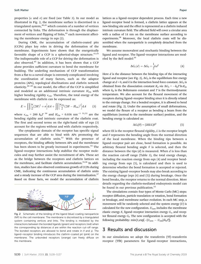
lattices as a ligand–receptor dependent process. Each time a newligand–receptor bond is formed, a clathrin lattice appears at thenew binding site and the effect is represented as a clathrin inducedintrinsic curvature field. The affected field will cover a circular areawith a radius of 14 nm on the membrane surface according toexperiments.58 Moreover, the local clathrin coats will be dis-sembled when the nanoparticle is completely detached from themembrane.
We assume monovalent and stochastic binding between theligand and receptor. The ligand–receptor interactions are mod-eled by the Bell model:59
DGr dð Þ ¼ DG0 þ1
2kd2 (4)
Here d is the distance between the binding tips of the interactingligand and receptor (see Fig. 2), DG0 is the equilibrium free energychange at d = 0, and k is the interaction bond force constant. DG0 isobtained from the dissociation constant Kd via DG0 = �kBT ln Kd,where kB is the Boltzmann constant and T is the thermodynamictemperature. We also account for the flexural movement of thereceptors during ligand–receptor binding since it is directly relatedto the entropy change. For a bonded receptor, it is allowed to bendand rotate (Fig. 2). Under the assumption of small deformations,we model the flexure of a receptor as bending a beam from theequilibrium (normal to the membrane surface) position, and thebending energy is calculated as:
DGf(y) = (2EI/L)y2 (5)
where EI is the receptor flexural rigidity, L is the receptor lengthand y represents the bending angle from the normal directionof the local membrane. When the binding tips between aligand–receptor pair are close, bond formation is possible. Anarbitrary flexural bending angle y is selected, and then thedistance between the tips (d) is measured. When d is less thanthe reaction cut-off range (Table 3), the total energy change,including the reaction energy from eqn (4) and receptor bend-ing energy from eqn (5), is calculated and then is used todetermine whether the bond formation is accepted or rejected.The existing ligand–receptor bonds may also break according tothe energy change (eqn (4) and (5)) during breakage. Once thebond breaks, the receptor returns to the normal direction. Moredetails regarding the clathrin-mediated endocytosis model canbe found in our previous publication.32
The simulations contain four types of Monte Carlo (MC) steps:receptor diffusion, particle translation or rotation, bond formationor breakage, and membrane surface evolution. In each MC step, amovement will be randomly selected and the system energy (U) iscalculated for the new configuration. Unew includes the membraneelastic energy E, ligand–receptor interaction energy Gr, and recep-tor flexural energy Gf. The new configuration is accepted with thefollowing probability: min{1,exp[�(Unew � Uold)/kBT]}.
3 Results and discussion
In our simulations we adopt the transferrin (Tf)–transferrinreceptor (TfR) parameters for ligand–receptor interactions,
Fig. 2 Schematic of the binding of the ligand (blue) coating nanoparticle(NP) to the cell membrane. The membrane is discretised by a triangulatedsystem containing vertices and links. The binding is mediated by theinteractions between the bonded ligands (green) and receptors (red) whenthe corresponding tip distances d are within the reaction cut-off range.The bonded receptors are allowed to bend and rotate in y and f. Theligand–receptor binding introduces the clathrin-coated pit (pink) on themembrane. The unbonded receptors (orange) can freely diffuse onthe membrane.
Soft Matter Paper

This journal is©The Royal Society of Chemistry 2019 Soft Matter, 2019, 15, 5128--5137 | 5131
since they are well known for triggering CME and have beenextensively studied for drug delivery across the blood–brainbarrier (BBB).60–64 We focus on ellipsoidal nanoparticlesbecause they have been used for drug delivery purposes.4
However, this model can be easily extended to study othernonspherical nanoparticles. The ellipsoidal nanoparticles inour study have the same volume as 80 nm-diameter sphericalparticles. Based on our previous studies,32 80 nm-diameterligand-coated spherical particles are highly likely to be inter-nalized during CME. The ligand density for all various shapeparticles is set to be 5300 mm�2.65 The initial attack angles areset at az = 0, which means the vector n is parallel to the z-axis atthe beginning of the simulations (Fig. 1). For each case, we runat least 5 independent simulations for statistical consistency.Some of the other parameters used in the simulation are shownin Appendix A.
3.1 Endocytosis of oblate-shaped ellipsoids
We first consider the CME of oblate-shaped nanoparticles with1 o AR o 3. Table 1 shows the aspect ratio and size parametersused for the simulations. The number of ligands coated on theparticle surface is calculated based on the surface area of theparticle and the ligand density. The a and b are the length ofthe major and minor axes in eqn (1).
The CME of oblate ellipsoids shows two entry modes: atip-first mode with minimal rotation (o15 degrees) and a tiltedentry mode with moderate rotation (15–45 degrees) as shown inFig. 3a. But no laying-down (445 degrees) pattern has beenobserved in this study. Both the sphere and high AR oblateellipsoids (AR = 2.08 and AR = 2.62) rotate less than 10 degreesthroughout the simulations. The sphere has less rotation due tothe homogeneously distributed curvatures. For high AR oblateellipsoids, the flat tip with low curvature helps stabilize thenanoparticle on the membrane with less rotation. Small to inter-mediate AR oblate ellipsoids (AR = 1.17 and AR = 1.47) are capableof taking advantage of both tip-first and tilted entry modes.
As shown in Fig. 3b, the number of ligand–receptor bondsfor spherical nanoparticles continuously increases without sig-nificant interruptions. The nanoparticle is fully endocytosedafter 1.2 � 109 MC steps. For the tip-first and tilted entries ofparticles with AR = 1.17 and AR = 1.47, the number of bondsshows a two-step growth pattern with two major bond formingperiods separated by a plateau in between. The oblate ellipsoidwith AR = 1.17 has an initial rapid bond forming periodbecause of the large contact area of the flat tip surface. Butthe number of bonds reaches a plateau when half of the coatedligands are bonded. This period ends with further growth of theCCP and membrane deformation. The nanoparticle is quickly
wrapped with the number of bonds jumping from 40 to 80. Theellipsoid with AR = 1.47 follows a similar two-step bondformation pattern. The main difference is that the first plateaulasts for many more MC steps due to sharper edges. For the tip-first entry of high AR ellipsoids (AR = 2.08 and AR = 2.62), thenumber of bonds is saturated at around 40 after initial bondformation on the flat tip surface near the membrane.
The normalized CCP ratio, RCCP, reflects a similar trend tothe number of bonds formed (Fig. 3c). RCCP is defined as thearea of the CCP over the area of the minimal ellipsoidthat encapsulates the particle. For AR = 1.17 and AR = 1.47oblate particles, the CCP growth rate significantly drops afterRCCP B 0.5. The flat tip of oblate ellipsoids makes it easy towrap half of the particle. The growth rate is reduced because ofthe highly curved edges. After overcoming the curved edge, theCCP growth is accelerated until full wrapping with RCCP B 1.0.For high aspect ratio ellipsoids, the CCP area also grows fast atthe beginning until RCCP B 0.5. It slowly reaches the maximumvalue of RCCP B 0.8 during the rest of the simulation. As shownin Fig. 3d, the decrease of the total energy is mainly driven bythe increase of existing bonds in the simulation. Therefore, theenergy change follows a subsequent two-step decrease patternfor low to intermediate AR particles. In contrast, the systemenergy of high AR particles reaches equilibrium rapidly after aninitial decrease.
Fig. 3e, g and h show the equilibrium profiles of particleswith a tip-first entry mode. The AR = 1.17 particle shows a fullywrapped profile (Fig. 3e) with a symmetric vesicle at equili-brium. For high aspect ratio ellipsoids with AR = 2.08 andAR = 2.62, we observe firm attachment of the particle to themembrane but no internalization (Fig. 3g and h). Themembrane partially wraps the oblate with RCCP o 1.0 and
Table 1 Shape and size parameters for oblate ellipsoids
Aspect ratio (AR) a (nm) b (nm) Number of ligands
1 40 40 1061.17 42 36 1061.47 45.5 31 1102.08 51 24.5 1182.62 55 21 126
Fig. 3 Endocytosis of sphere and oblate ellipsoids with various aspectratios. (a) The rotation of the nanoparticle (az); (b) the number of ligand–receptor bonds (the equilibrium profile of a spherical nanoparticle is alsoshown); (c) the normalized CCP area ratio RCCP; and (d) the total energy ofthe system U as a function of MC steps during CME. The equilibriumparticle–membrane profiles for ellipsoids with (e) AR = 1.17, (f) AR = 1.47,(g) AR = 2.08 and (h) AR = 2.62. The CCP (pink region), bonded TfR (reddots), free TfR (black dots), bonded Tf (green dots) and unbonded Tf (bluedots) are all shown in the profiles.
Paper Soft Matter
5132 | Soft Matter, 2019, 15, 5128--5137 This journal is©The Royal Society of Chemistry 2019
further bending is suppressed by the highly curved edges. Theellipsoid shows minimal rotation (az o 10 degrees) and sym-metric wrapping. This is due to an initial large contact areawith respect to the membrane surface, which leads to higheradhesion stability and less rotation. The results are consistentwith the theoretical work from Bahrami,66 in which higheradhesion strength is required for high aspect ratio ellipsoids tobe internalized. Another theoretical study also proposed asimilar prediction that an increased aspect ratio makes it easierfor attachment but more difficult to achieve a completelywrapped state for the ellipsoidal nanoparticle.30 The vesicleformed by the oblate ellipsoid with AR = 1.47 through a tiltedentry mode is shown in Fig. 3f. In this situation, the nanopar-ticle adjusts itself to a tilted position around 20–25 degrees tohelp bond formation on one side of the edge. Similar rotationof oblate-shaped ellipsoids during RME has also been observedin ref. 25 and 29. The tilting of the ellipsoid leads to anasymmetric wrapping of the membrane.
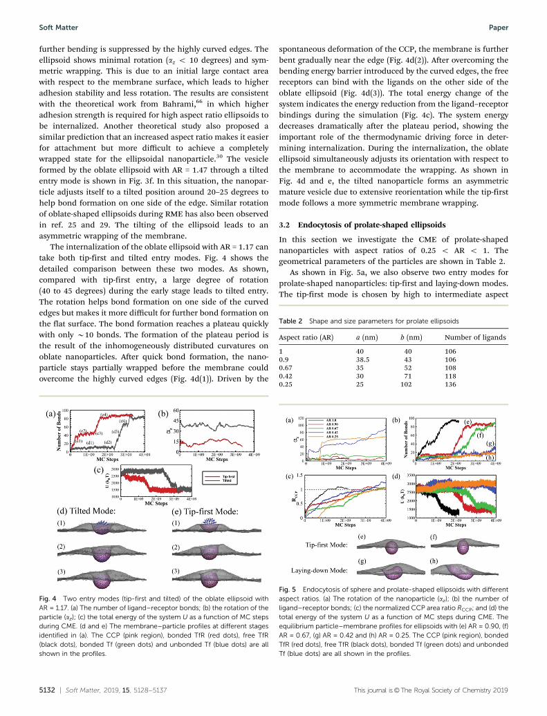
The internalization of the oblate ellipsoid with AR = 1.17 cantake both tip-first and tilted entry modes. Fig. 4 shows thedetailed comparison between these two modes. As shown,compared with tip-first entry, a large degree of rotation(40 to 45 degrees) during the early stage leads to tilted entry.The rotation helps bond formation on one side of the curvededges but makes it more difficult for further bond formation onthe flat surface. The bond formation reaches a plateau quicklywith only B10 bonds. The formation of the plateau period isthe result of the inhomogeneously distributed curvatures onoblate nanoparticles. After quick bond formation, the nano-particle stays partially wrapped before the membrane couldovercome the highly curved edges (Fig. 4d(1)). Driven by the
spontaneous deformation of the CCP, the membrane is furtherbent gradually near the edge (Fig. 4d(2)). After overcoming thebending energy barrier introduced by the curved edges, the freereceptors can bind with the ligands on the other side of theoblate ellipsoid (Fig. 4d(3)). The total energy change of thesystem indicates the energy reduction from the ligand–receptorbindings during the simulation (Fig. 4c). The system energydecreases dramatically after the plateau period, showing theimportant role of the thermodynamic driving force in deter-mining internalization. During the internalization, the oblateellipsoid simultaneously adjusts its orientation with respect tothe membrane to accommodate the wrapping. As shown inFig. 4d and e, the tilted nanoparticle forms an asymmetricmature vesicle due to extensive reorientation while the tip-firstmode follows a more symmetric membrane wrapping.
3.2 Endocytosis of prolate-shaped ellipsoids
In this section we investigate the CME of prolate-shapednanoparticles with aspect ratios of 0.25 o AR o 1. Thegeometrical parameters of the particles are shown in Table 2.
As shown in Fig. 5a, we also observe two entry modes forprolate-shaped nanoparticles: tip-first and laying-down modes.The tip-first mode is chosen by high to intermediate aspect
Fig. 4 Two entry modes (tip-first and tilted) of the oblate ellipsoid withAR = 1.17. (a) The number of ligand–receptor bonds; (b) the rotation of theparticle (az); (c) the total energy of the system U as a function of MC stepsduring CME. (d and e) The membrane–particle profiles at different stagesidentified in (a). The CCP (pink region), bonded TfR (red dots), free TfR(black dots), bonded Tf (green dots) and unbonded Tf (blue dots) are allshown in the profiles.
Table 2 Shape and size parameters for prolate ellipsoids
Aspect ratio (AR) a (nm) b (nm) Number of ligands
1 40 40 1060.9 38.5 43 1060.67 35 52 1080.42 30 71 1180.25 25 102 136
Fig. 5 Endocytosis of sphere and prolate-shaped ellipsoids with differentaspect ratios. (a) The rotation of the nanoparticle (az); (b) the number ofligand–receptor bonds; (c) the normalized CCP area ratio RCCP; and (d) thetotal energy of the system U as a function of MC steps during CME. Theequilibrium particle–membrane profiles for ellipsoids with (e) AR = 0.90, (f)AR = 0.67, (g) AR = 0.42 and (h) AR = 0.25. The CCP (pink region), bondedTfR (red dots), free TfR (black dots), bonded Tf (green dots) and unbondedTf (blue dots) are all shown in the profiles.
Soft Matter Paper

This journal is©The Royal Society of Chemistry 2019 Soft Matter, 2019, 15, 5128--5137 | 5133
ratio particles with AR = 0.9 and AR = 0.67, characterized by atwo-step growth pattern of bonds (Fig. 5b). Since the tip of theprolate particle is highly curved, the bond formation reaches aplateau quickly at B2 � 108 MC steps with less than 20 ligand–receptor bonds. The periods of the plateau are similar for bothAR = 0.9 and AR = 0.67 ellipsoids. This is quite different fromthe oblate-shaped cases where a higher AR particle has asignificantly longer plateau period than a smaller AR one(see Fig. 3b). After 2.0 � 109 MC steps, the number of bondsquickly increases to more than 80 for both cases due to theeasiness of bond formation on the flat edges of the prolateellipsoid. The RCCP and system energy U also follow a similarstep-wise change in the simulations (Fig. 5c and d). During thewrapping, the ellipsoid remains perpendicular to the membranesurface with little rotation. The vesicle formed by the nanoparticlewith AR = 0.9 is mostly spherically shaped, while it becomes anellipsoidal shape for the ellipsoid with AR = 0.67 due to the loweraspect ratio (Fig. 5e and f).
On the other hand, the low aspect ratio nanoparticles withAR = 0.42 and AR = 0.25 use the laying-down entry mode duringwhich the particles rotate to a nearly horizontal position (Fig. 5a).The number of bonds during the uptake is much lower than thetip-first mode (Fig. 5b). As a result, the total energies of the systemsU do not decrease (Fig. 5d). However, RCCP still keeps growingduring the simulation (Fig. 5c). The AR = 0.42 ellipsoid is fullywrapped while the AR = 0.25 ellipsoid does not completely inter-nalize (Fig. 5g and h). Though the RCCP of the AR = 0.25 particlealso reaches 1.0, the CCP cannot bend itself to fully wrap theparticle because of the extremely low aspect ratio.
Our results indicate that prolate ellipsoids with low ARprefer to take the laying-down mode to enter cells. Experimentshave observed both tip-first and laying-down entry modes forviruses with low AR.67,68 Coarse-grained molecular dynamics(CGMD) studies also showed a similar perpendicular to parallelreorientation of low AR spherocylinders in RME.69 A morerecent theoretical study also showed that the parallel orienta-tion of ellipsoids is more energetically favorable than the tip-first orientation because of a higher bending energy cost perarea at the tip.24 However, the theoretical work from Yi et al.70
and CGMD simulations from Shen et al.25 showed that thelow AR ellipsoids preferred the tip-first entry mode at lowmembrane tension. In ref. 70, the ligand–receptor interactionshave been modeled as direct adhesion within a certain area andthe membrane wrapping was driven by the energy reductioncaused by the adhesion. In ref. 25, discrete ligand–receptorinteractions have been considered with a relatively high bind-ing strength (50 kBT) and very long interaction cutoff (37.5 nm).In our model, the ligand–receptor interaction cutoff is muchshorter, therefore it is much harder to continuously form bondsfrom a tip-oriented position with high curvatures. Laying-downhelps bond formation on the flat side and further reduces theenergy of the system.
Another interesting observation with AR = 0.42 and AR = 0.25particles is that RCCP continuously increases while the numberof ligand–receptor bonds remains nearly constant at a relativelylow value (less than 30) (Fig. 5b and c). In our model, the
recruitment and accumulation of clathrin are stimulated by theformation of new ligand–receptor bonds (see the methods section).Laying-down of the particle allows continuous new ligand–receptorbond formation at new binding sites. However, the existing bondsbreak more frequently at the same time. This is due to the fact thatthe radius of curvature on the membrane caused by the clathrin(Hcla) is B30 nm; this is significantly smaller than the b values ofthe ellipsoids (71 nm and 102 nm for AR = 0.42 and AR = 0.25particles). Therefore, the bending of the membrane caused byclathrin tends to break the existing ligand–receptor bonds. As aresult, the prolate ellipsoid with AR = 0.42 is fully wrapped withonly B30 bonds, less than half of the spherical counterpart atequilibrium. As shown in Fig. 5d, we do not observe a significanttotal energy decrease, which is similar to the high AR oblateellipsoid case in Fig. 3d. The ability to be internalized with fewerligand–receptor bonds is important to transcellular drug delivery.For transcellular drug delivery, the nanoparticles need to first enterthe cell at one side and then release from the cell at the other side.Fewer ligand–receptor bonds during internalization facilitates theparticle release during expulsion. Indeed, experiments havedemonstrated that spherical nanoparticles with lower avidity havebetter efficiency for transcytosis across blood–brain barriers.65,71 Inaddition, it has also been observed in in vivo experiments that it iseasier for Tf-coated nanorods to release from the cell thanspherical ones.7
Moreover, for the ellipsoid with intermediate aspect ratioAR = 0.67, we find that the particle can be internalized by allthree modes. Fig. 6 presents the entry of tip-first and laying-down modes. Compared with the two-step growth of bonds inthe tip-first mode, the laying-down mode features a continuousincrease of bonds until it is fully endocytosed. The internaliza-tion of the laying-down ellipsoid takes less MC steps than thetip-first mode. This is because the rotation of the particle allowsthe ligands on the flat side to interact with the receptors on themembrane. The equilibrium energy of the tip-first mode islower than the laying-down mode, meaning that the tip-firstmode is a more energetically favorable status for this ellipsoidalnanoparticle.
3.3 Overall endocytosis comparison
Fig. 7 summarizes our findings of endocytosis for ellipsoidparticles. As shown in Fig. 7a, there are three major patterns forthe entry of ellipsoids with different aspect ratios. Character-ized by the degrees of rotation during the entry, we observe atip-first mode (o15 degrees), tilted entry mode (15–45 degrees)and laying-down mode (445 degrees). The oblate ellipsoid withAR 4 1.47 only takes the tip-first entry mode. On the otherhand, the low aspect ratio prolate nanoparticles with AR = 0.25and AR = 0.42 only use the laying-down entry mode. Intermedi-ate aspect ratio ellipsoids with 0.67 r AR r 1.47 may chooseeither the tip-first mode or the tilted entry mode. Interestinglythe prolate ellipsoid with AR = 0.67 shows flexibility and maytake any of the three entry modes for endocytosis. The cap-ability of taking any entry mode is helpful for drug deliveryapplications. In realistic biological scenarios, the initial contactdirection between the particle and cell membrane is random.
Paper Soft Matter

5134 | Soft Matter, 2019, 15, 5128--5137 This journal is©The Royal Society of Chemistry 2019
Therefore particles that can adopt all entry modes may havebetter opportunities to enter the cells. Overall the prolatenanoparticles rotate more to enter the cell compared with thesphere and oblate nanoparticles during endocytosis, because ofthe highly curved tip of prolate ellipsoids.
Fig. 7b summarizes the internalization stages of all theparticles with different shapes. We have observed completewrapping (CW) for nanoparticles with aspect ratio 0.42 r AR r1.47. The sphere nanoparticle can be internalized with the leastMC steps indicating the highest efficiency. Although it is moredifficult for ellipsoidal particles to enter the cell, in general theyrequire fewer ligand–receptor bonds compared with sphericalparticles. Especially, the prolate ellipsoid with AR = 0.42 canhave CW with a significantly lower number of bonds (B30).This is especially important for transcellular drug delivery sincefewer bonds during endocytosis may lead to a higher exocytosisefficiency. Overall, our results are consistent with the experi-mental observations;6,7 low AR prolate and high AR oblatenanoparticles, with AR o 0.42 or AR 4 1.47 in our study, onlyshow partial wrapping (PW), indicating a low internalizationefficiency.
The rotation of the particles, different entry modes and wrappingstates during endocytosis on fluid membranes have been studiedthrough various analytical analysis and simulations.24,25,29,30,66,69,72
The distinctive feature of our model is that the deformation of themembrane and wrapping of the particles are driven by the assemblyof CCPs. In our model, the accumulation of CCPs is triggered by theligand–receptor interactions, while the ligand–receptor binding is
modeled as a stochastic process and the modelling parameters aredirectly obtained from experiments. Therefore, the entire particleinternalization process is stochastic and dictated by many inter-correlated events, such as ligand–receptor bond formation/break-age, particle translation/rotation, clathrin assembly/dissembly,membrane deformation, etc. As a result, our simulations pro-vide additional information. For example, our results indicatethat the internalization of particles may not always correlatewith continuous increasing of bonds in CME (see Fig. 3). Therotation (or entry mode) of the same nanoparticle may bedifferent due to localized CCP recruitment. Indeed, our resultsindicate that multiple entry modes may be taken by specificnanoparticles. Due to the stochastic nature, the wrapping stateof certain particles may also become scattered.
4 Conclusions
CME is the fundamental biological mechanism for the cellmetabolism and intracellular transport of nanoparticles. Theadvancement of nanotechnology makes the manufacturing ofnanoparticles with different shapes possible for biomedical
Fig. 6 Two entry modes of the prolate ellipsoid with AR = 0.67. (a) Thenumber of ligand–receptor bonds; (b) the rotation of the particle (az); and(c) the total energy of the system U as a function of MC steps during CME.(d and e) The membrane–particle profiles at different stages identified in(a). The CCP (pink region), bonded TfR (red dots), free TfR (black dots),bonded Tf (green dots) and unbonded Tf (blue dots) are all shown in theprofiles.
Fig. 7 Equilibrium rotation angles and clathrin coated area for sphericaland ellipsoidal nanoparticles. (a) The nanoparticles show three entrypatterns: tip-first mode (o15 degrees), tilted entry mode (15–45 degrees)and laying-down mode (445 degrees) depending on the degree ofrotation. The error bars represent the minimal and maximal degrees ofrotation observed in 5 independent simulations. (b) The clathrin area plotshows the influence of the shape of nanoparticle on the internalizationcapability. The error bars are based on the standard deviation from 5independent simulations. The nanoparticle is able to be completelywrapped (CW) within 0.42 r AR r 1.47 while only partially wrapped(PW) for AR o 0.42 or AR 4 1.47. The equilibrium membrane profiles fornanoparticles with AR = 0.25 and 2.62 are shown respectively.
Soft Matter Paper

This journal is©The Royal Society of Chemistry 2019 Soft Matter, 2019, 15, 5128--5137 | 5135
application. But how the different shape nanoparticles interactwith the cell membrane is complicated and still under debate.In this work, we systematically investigated the CME oftransferrin-coated spherical and ellipsoidal nanoparticles throughmodeling and simulations. Our stochastic model takes intoaccount the membrane deformation, clathrin lattice accumulationand transferrin–transferrin receptor interactions based on MonteCarlo simulations. In our model, the membrane deformationand particle internalization are primarily driven by the clathrinpolymerization, which is stimulated from the ligand–receptorinteractions.
Through our simulations, we found three entry modes forthe CME of spheres and ellipsoids including tip-first, tiltedentry and laying-down modes. Each mode is characterized bydistinctive degrees of rotation during the wrapping of themembrane. High aspect ratio oblate ellipsoids with AR 4 2use only the tip-first entry mode. Small to intermediateaspect ratio ellipsoids with 0.67 r AR r 1.47 are able to takethe tip-first and tilted entry modes. Low aspect ratio prolatenanoparticles with AR o 0.5 only internalize through thelaying-down mode. Moreover, we have observed that certainmoderate aspect ratio prolate ellipsoids, such as AR = 0.67 inour study, are able to take advantage of all three modes forinternalization.
The bond formation and CCP growth of both tip-first andtilted modes show a two-step wrapping pattern with a plateauin between. The plateau period depends on the aspect ratio androtation of the nanoparticle. The laying-down mode has acontinuous CCP wrapping pattern, but the equilibrium numberof bonds is highly dependent on the aspect ratio of thenanoparticle. The prolate ellipsoid with AR = 0.42 is interna-lized with more MC steps but much fewer ligand–receptorbonds than other shapes. Internalization of nanoparticles withfewer ligand–receptor bonds may significantly facilitate releaseof the particle during transcellular drug delivery. In addition,we have observed complete wrapping for particles with anintermediate AR range of 0.42 r AR r 1.47. In general, theinternalization of spherical nanoparticles is easier than that ofellipsoidal particles. In summary, our simulation results areconsistent with a variety of experimental measurements andprovide deeper understanding of the fundamental mechan-isms involved in CME of nanoparticles with different shapes.Our model represents a powerful and viable platform forfacilitating the rational design of nanoparticles for targeteddrug delivery.
Conflicts of interest
There are no conflicts to declare.
Appendix A: Simulation parameters
The table below lists some of the simulation parameters usedand the corresponding references:
Acknowledgements
The research reported in this publication was supported by theNational Science Foundation under CBET-1604211 and theNational Institute of General Medical Sciences of the NationalInstitutes of Health under Award Number R01GM122081.Computational resources were provided in part by the ExtremeScience and Engineering Discovery Environment (XSEDE)under grant No. MCB170012.
References
1 B. D. Harrison, T. M. A. Wilson and A. Klug, Philos. Trans.R. Soc. London, Ser. B, 1999, 354, 531–535.
2 G. Wanger, T. C. Onstott and G. Southam, Geobiology, 2008,6, 325–330.
3 N. Doshi and S. Mitragotri, PLoS One, 2010, 5, e10051.4 Y. Liu, J. Tan, A. Thomas, D. Ou-Yang and V. R. Muzykantov,
Ther. Delivery, 2012, 3, 181–194.5 L. Xu, H. Kuang, L. Wang and C. Xu, J. Mater. Chem., 2011,
21, 16759–16782.6 B. D. Chithrani, A. A. Ghazani and W. C. W. Chan, Nano
Lett., 2006, 6, 662–668.7 B. D. Chithrani and W. C. W. Chan, Nano Lett., 2007, 7,
1542–1550.8 L. Florez, C. Herrmann, J. M. Cramer, C. P. Hauser,
K. Koynov, K. Landfester, D. Crespy and V. Mailander,Small, 2012, 8, 2222–2230.
9 Y. Zhang, S. Tekobo, Y. Tu, Q. Zhou, X. Jin, S. A. Dergunov,E. Pinkhassik and B. Yan, ACS Appl. Mater. Interfaces, 2012,4, 4099–4105.
10 S. Barua, J.-W. Yoo, P. Kolhar, A. Wakankar, Y. R. Gokarnand S. Mitragotri, Proc. Natl. Acad. Sci. U. S. A., 2013, 110,3270–3275.
11 X. Huang, X. Teng, D. Chen, F. Tang and J. He, Biomaterials,2010, 31, 438–448.
12 H. Meng, S. Yang, Z. Li, T. Xia, J. Chen, Z. Ji, H. Zhang,X. Wang, S. Lin, C. Huang, Z. H. Zhou, J. I. Zink andA. E. Nel, ACS Nano, 2011, 5, 4434–4447.
Table 3 Summary of some parameters used in the simulation
Parameters Value Ref.
Size of membrane surface 910 nm � 910 nmMembrane bending rigidity k 20 kBT 73Membrane characteristic tension s 0.001 pN nm�1 74Clathrin bending rigidity kcla 200 kBT 75Clathrin intrinsic curvature Hcla 0.036 nm�1 51Transferrin receptor length 9.3 nm 76Transferrin receptor radius 5 nm 76Antibody length 9 nm 77Antibody radius 2.5 nm 77Number of transferrin receptors onthe luminal side
300 78
Equilibrium free energy change DG0 �8.64 � 10�20 J 79Reactive compliance (reaction cut-offdistance)
0.9 nm 79
Receptor flexural rigidity EI 7000 pN nm2 80System temperature 298 K
Paper Soft Matter
5136 | Soft Matter, 2019, 15, 5128--5137 This journal is©The Royal Society of Chemistry 2019
13 S. E. A. Gratton, P. A. Ropp, P. D. Pohlhaus, J. C. Luft,V. J. Madden, M. E. Napier and J. M. DeSimone, Proc. Natl.Acad. Sci. U. S. A., 2008, 105, 11613–11618.
14 R. Agarwal, V. Singh, P. Jurney, L. Shi, S. V. Sreenivasan andK. Roy, Proc. Natl. Acad. Sci. U. S. A., 2013, 110, 17247–17252.
15 R. Agarwal, P. Jurney, M. Raythatha, V. Singh, S. V.Sreenivasan, L. Shi and K. Roy, Adv. Healthcare Mater.,2015, 4, 2269–2280.
16 S. Dasgupta, T. Auth and G. Gompper, J. Phys.: Condens.Matter, 2017, 29, 373003.
17 S. Zhang, H. Gao and G. Bao, ACS Nano, 2015, 9, 8655–8671.18 C. Kinnear, T. L. Moore, L. Rodriguez-Lorenzo, B. Rothen-
Rutishauser and A. Petri-Fink, Chem. Rev., 2017, 117,11476–11521.
19 J. Zhao and M. H. Stenzel, Polym. Chem., 2018, 9, 259–272.20 R. Qiao, A. P. Roberts, A. S. Mount, S. J. Klaine and P. C. Ke,
Nano Lett., 2007, 7, 614–619.21 K. Yang and Y.-Q. Ma, Nat. Nanotechnol., 2010, 5, 579.22 S. Nangia and R. Sureshkumar, Langmuir, 2012, 28,
17666–17671.23 R. Vacha, F. J. Martinez-Veracoechea and D. Frenkel, Nano
Lett., 2011, 11, 5391–5395.24 S. Dasgupta, T. Auth and G. Gompper, Nano Lett., 2014, 14,
687–693.25 Z. Shen, H. Ye, X. Yi and Y. Li, ACS Nano, 2019, 13, 215–228.26 Y. Li, T. Yue, K. Yang and X. Zhang, Biomaterials, 2012, 33,
4965–4973.27 C. Huang, Y. Zhang, H. Yuan, H. Gao and S. Zhang, Nano
Lett., 2013, 13, 4546–4550.28 L. Zhang, Y. Zhao and X. Wang, ACS Appl. Mater. Interfaces,
2017, 9, 26665–26673.29 H. Tang, H. Zhang, H. Ye and Y. Zheng, J. Phys. Chem. B,
2018, 122, 171–180.30 S. Dasgupta, T. Auth and G. Gompper, Soft Matter, 2013, 9,
5473–5482.31 L. Chen, S. Xiao, H. Zhu, L. Wang and H. Liang, Soft Matter,
2016, 12, 2632–2641.32 H. Deng, P. Dutta and J. Liu, Biochim. Biophys. Acta, Gen.
Subj., 2018, 1862, 2104–2111.33 W. Helfrich, Z. Naturforsch., C: Biochem., Biophys., Biol.,
Virol., 1973, 28, 693–703.34 M. Deserno, Phys. Rev. E: Stat., Nonlinear, Soft Matter Phys.,
2004, 69, 031903.35 J. Agudo-Canalejo and R. Lipowsky, ACS Nano, 2015, 9,
3704–3720.36 Q. Yu, S. Othman, S. Dasgupta, T. Auth and G. Gompper,
Nanoscale, 2018, 10, 6445–6458.37 J. Liu, R. Tourdot, V. Ramanan, N. J. Agrawal and
R. Radhakrishanan, Mol. Phys., 2012, 110, 1127–1137.38 O. L. Mooren, B. J. Galletta and J. A. Cooper, Annu. Rev.
Biochem., 2012, 81, 661–686.39 B. Qualmann, M. M. Kessels and R. B. Kelly, J. Cell Biol.,
2000, 150, F111–F116.40 G. Gompper and D. M. Kroll, J. Phys.: Condens. Matter, 1997,
9, 8795–8834.41 D. M. Kroll and G. Gompper, Science, 1992, 255, 968.
42 N. Ramakrishnan, P. B. S. Kumar and J. H. Ipsen, Phys. Rev.E: Stat., Nonlinear, Soft Matter Phys., 2010, 81, 041922.
43 J. H. Hurley, E. Boura, L. A. Carlson and B. Rozycki, Cell,2010, 143, 875–887.
44 A. Fotin, Y. F. Cheng, P. Sliz, N. Grigorieff, S. C. Harrison,T. Kirchhausen and T. Walz, Nature, 2004, 432, 573–579.
45 L. Hinrichsen, A. Meyerhoiz, S. Groos and E. J. Ungewickell,Proc. Natl. Acad. Sci. U. S. A., 2006, 103, 8715–8720.
46 R. J. Mashl and R. F. Bruinsma, Biophys. J., 1998, 74,2862–2875.
47 T. Kohyama, D. M. Kroll and G. Gompper, Phys. Rev. E: Stat.,Nonlinear, Soft Matter Phys., 2003, 68, 061905.
48 M. F. Hagan and D. Chandler, Biophys. J., 2006, 91, 42–54.49 M. Giani, W. K. den Otter and W. J. Briels, Biophys. J., 2016,
111, 222–235.50 A. Banerjee, A. Berezhkovskii and R. Nossal, Biophys. J.,
2012, 102, 2725–2730.51 S. Zaremba and J. H. Keen, J. Cell Biol., 1983, 97, 1339–1347.52 M. G. J. Ford, I. G. Mills, B. J. Peter, Y. Vallis,
G. J. K. Praefcke, P. R. Evans and H. T. McMahon, Nature,2002, 419, 361–366.
53 J. S. Bonifacino and L. M. Traub, Annu. Rev. Biochem., 2003,72, 395–447.
54 D. Ricotta, S. D. Conner, S. L. Schmid, K. von Figura andS. Honing, J. Cell Biol., 2002, 156, 791–795.
55 J. Schlessinger, Cell, 2000, 103, 211–225.56 J. Chen, J. Wang, K. R. Meyers and C. A. Enns, Traffic, 2009,
10, 1488–1501.57 M. Ehrlich, W. Boll, A. van Oijen, R. Hariharan,
K. Chandran, M. L. Nibert and T. Kirchhausen, Cell, 2004,118, 591–605.
58 J. Heuser and T. Kirchhausen, J. Ultrastruct. Res., 1985, 92,1–27.
59 G. I. Bell, M. Dembo and P. Bongrand, Biophys. J., 1984, 45,1051–1064.
60 A. Dautry-Varsat, A. Ciechanover and H. F. Lodish, Proc.Natl. Acad. Sci. U. S. A., 1983, 80, 2258–2262.
61 Z. M. Qian, H. Y. Li, H. Z. Sun and K. Ho, Pharmacol. Rev.,2002, 54, 561–587.
62 H. Y. Li, H. Z. Sun and Z. M. Qian, Trends Pharmacol. Sci.,2002, 23, 206–209.
63 D. Peer, J. M. Karp, S. Hong, O. C. FaroKhzad, R. Margalitand R. Langer, Nat. Nanotechnol., 2007, 2, 751–760.
64 A. I. Khan, J. Liu and P. Dutta, Biochim. Biophys. Acta, Gen.Subj., 2018, 1862, 1168–1179.
65 D. T. Wiley, P. Webster, A. Gale and M. E. Davis, Proc. Natl.Acad. Sci. U. S. A., 2013, 110, 8662–8667.
66 A. H. Bahrami, Soft Matter, 2013, 9, 8642–8646.67 T. Noda, H. Ebihara, Y. Muramoto, K. Fujii, A. Takada,
H. Sagara, J. H. Kim, H. Kida, H. Feldmann and Y. Kawaoka,PLoS Pathog., 2006, 2, e99.
68 S. Welsch, L. Kolesnikova, V. Krahling, J. D. Riches, S. Beckerand J. A. G. Briggs, PLoS Pathog., 2010, 6, e1000875.
69 R. Vacha, F. J. Martinez-Veracoechea and D. Frenkel, NanoLett., 2011, 11, 5391–5395.
70 X. Yi, X. Shi and H. Gao, Nano Lett., 2014, 14, 1049–1055.
Soft Matter Paper

This journal is©The Royal Society of Chemistry 2019 Soft Matter, 2019, 15, 5128--5137 | 5137
71 Y. J. Yu, Y. Zhang, M. Kenrick, K. Hoyte, W. Luk, Y. M. Lu,J. Atwal, J. M. Elliott, S. Prabhu, R. J. Watts andM. S. Dennis, Sci. Transl. Med., 2011, 3, 84ra44.
72 D. M. Richards and R. G. Endres, Proc. Natl. Acad. Sci. U. S. A.,2016, 113, 6113–6118.
73 E. Evans and W. Rawicz, Phys. Rev. Lett., 1990, 64, 2094–2097.74 J. E. Hassinger, G. Oster, D. G. Drubin and P. Rangamani,
Proc. Natl. Acad. Sci. U. S. A., 2017, 114, E1118–E1127.75 A. J. Jin, K. Prasad, P. D. Smith, E. M. Lafer and R. Nossal,
Biophys. J., 2006, 90, 3333–3344.
76 H. Fuchs, U. Lucken, R. Tauber, A. Engel and R. Geßner,Structure, 1998, 6, 1235–1243.
77 H. M. Berman, J. Westbrook, Z. Feng, G. Gilliland,T. N. Bhat, H. Weissig, I. N. Shindyalov and P. E. Bourne,Nucleic Acids Res., 2000, 28, 235–242.
78 J. D. Bleil and M. S. Bretscher, EMBO J., 1982, 1, 351–355.79 A. Yersin, T. Osada and A. Ikai, Biophys. J., 2008, 94,
230–240.80 S. Weinbaum, X. B. Zhang, Y. F. Han, H. Vink and S. C.
Cowin, Proc. Natl. Acad. Sci. U. S. A., 2003, 100, 7988–7995.
Paper Soft Matter