Embed Size (px)
Citation preview
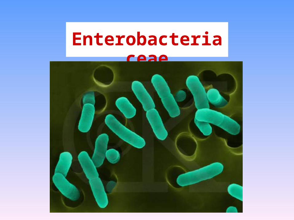
Enterobacteriaceae
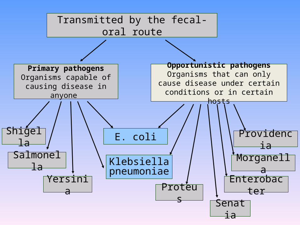
Transmitted by the fecal-oral route
Primary pathogensOrganisms capable of causing
disease in anyone
Opportunistic pathogensOrganisms that can only cause disease
under certain conditions or in certain hosts
Shigella
Salmonella
Yersinia
Klebsiellapneumoniae
E. coli
Senatia
Morganella
Providencia
EnterobacterProteus
Characteristics shared by all Characteristics shared by all
members of family members of family
EnterobacteriaceaeEnterobacteriaceae
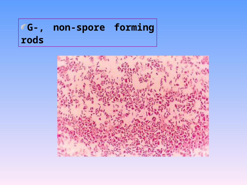
G-, non-spore forming rods
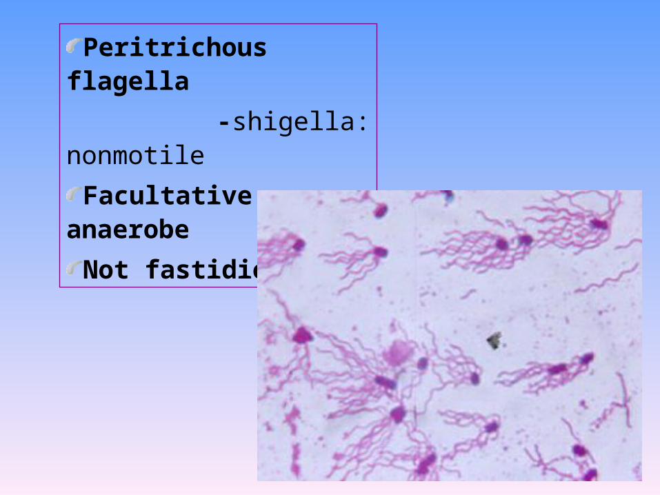
Peritrichous flagella
-shigella: nonmotile
Facultative anaerobe
Not fastidious
Active metabolism
- All ferment glucose;
- All reduce nitrates to nitrites;
- All oxidase negative
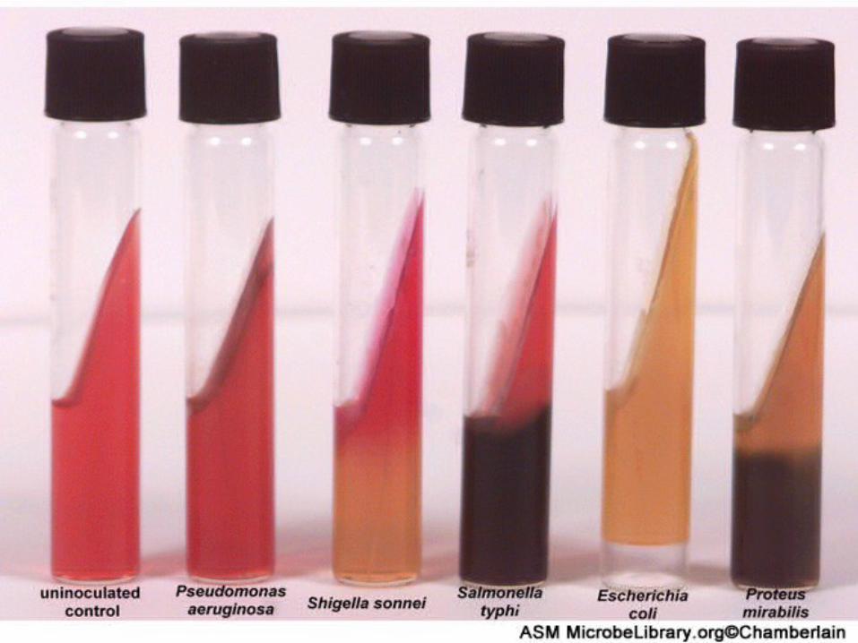
- Lactose fermentation: the key measure to isolate
and identify the Enterobacteriaceae
+: nonpathogenic
-: pathogenic
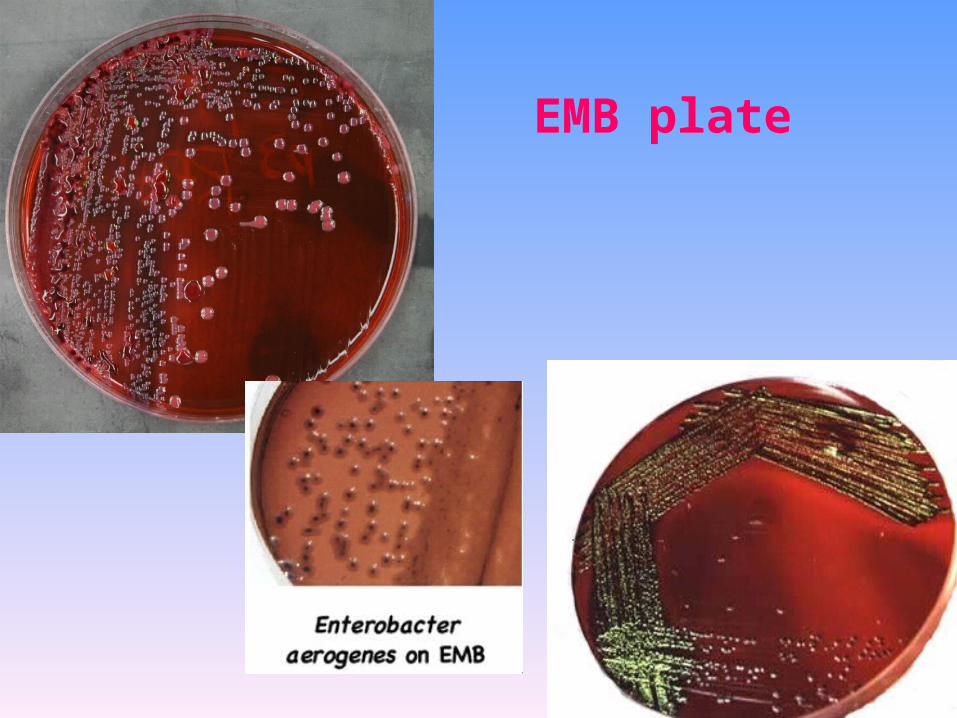
EMB plate
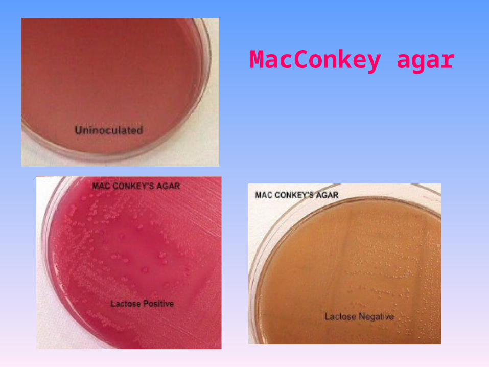
MacConkey agar

Triple sugar iron agar (TSI)
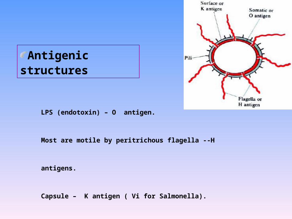
Antigenic structures
LPS (endotoxin) – O antigen.
Most are motile by peritrichous flagella --H antigens.
Capsule – K antigen ( Vi for Salmonella).
Cell envelope (wall)
various outer membrane proteins.
Pili - various antigen types, some encoded by plasmids
Enterobacteriaceae: gastrointestinal diseases
– Escherichia coli – Salmonella– Shigella – Yersinia entercolitica
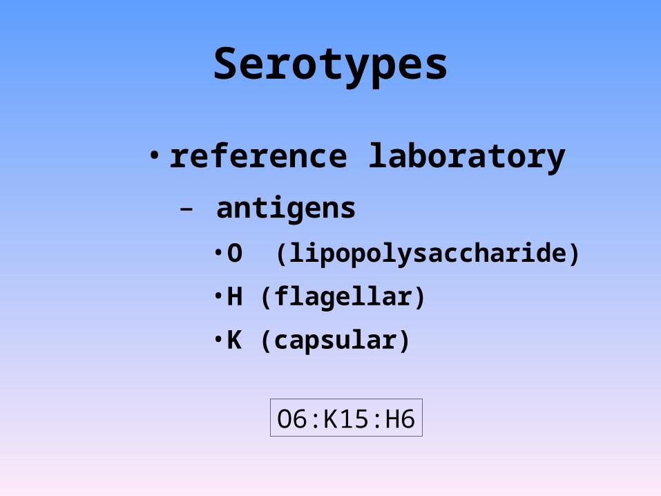
Serotypes
• reference laboratory
– antigens
• O (lipopolysaccharide)
• H (flagellar)
• K (capsular)
O6:K15:H6

(I) Escherichia coli

Medical significance
Normal flora
E. coli is the majority of GI normal flora
Opportunistic pathogen
Extraintestinal infection
Urinary tract infectionSepticemia
Neonatal meningitis
pathogen
Intestinal tract infection
ETECEIECEPECEHEC
EAggEC
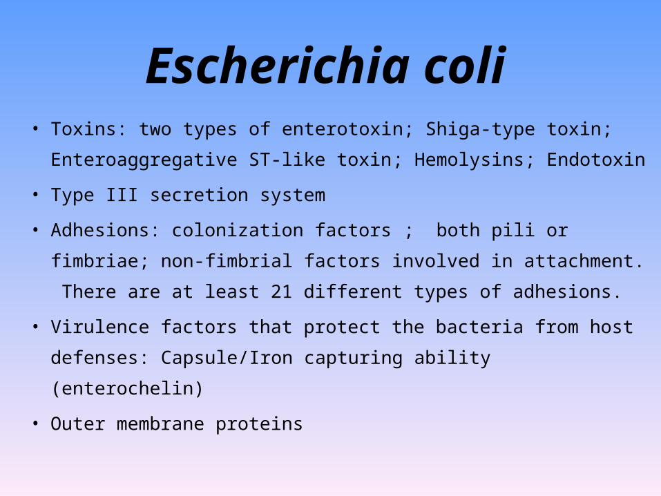
Escherichia coli• Toxins: two types of enterotoxin; Shiga-type toxin;
Enteroaggregative ST-like toxin; Hemolysins; Endotoxin
• Type III secretion system
• Adhesions: colonization factors ; both pili or fimbriae; non-
fimbrial factors involved in attachment. There are at least 21
different types of adhesions.
• Virulence factors that protect the bacteria from host defenses:
Capsule/Iron capturing ability (enterochelin)
• Outer membrane proteins
What is the pathogenesis of these five
groups of pathogenic E. coli?
Gastroenteritis caused by E. coli

Enterotoxigenic E. coli (ETEC)
• A watery diarrhea, nausea, abdominal cramps and low-grade fever for 1-5 days.
• Travellers diarrhea and diarrhea in children in developing countries
• Transmission is via contaminated food or water.• diarrhea like cholera • milder • nursery travellers diarrhea • caused by LT, ST, or LT/ST.
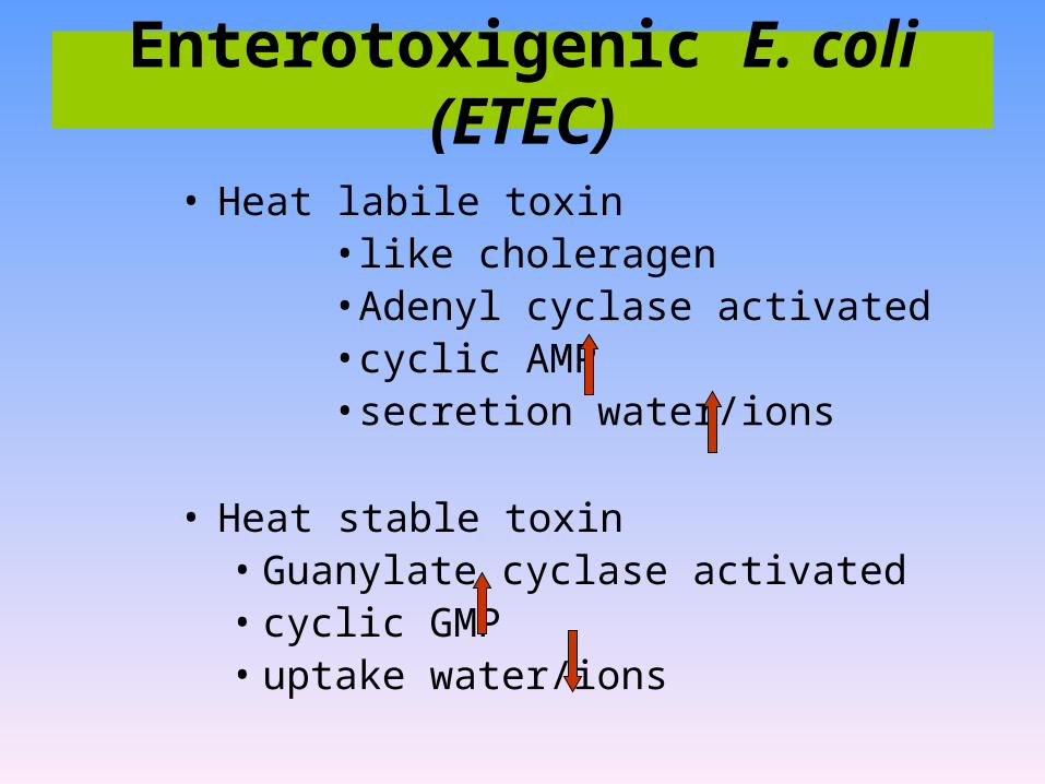
• Heat labile toxin• like choleragen• Adenyl cyclase activated • cyclic AMP • secretion water/ions
• Heat stable toxin• Guanylate cyclase activated • cyclic GMP• uptake water/ions
Enterotoxigenic E. coli (ETEC)
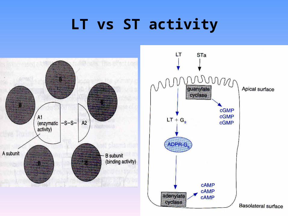
LT vs ST activity

• The organism attaches to the intestinal mucosa via pili
• Outer membrane proteins are involved in direct penetration,
invasion of the intestinal cells, and destruction of the
intestinal mucosa.
• There is lateral movement of the organism from one cell to
adjacent cells.
• Symptoms include fever, severe abdominal cramps, malaise,
and watery diarrhea followed by scanty stools containing
blood, mucous, and pus.
• resembles shigellosis
Enteroinvasive E. coli (EIEC)
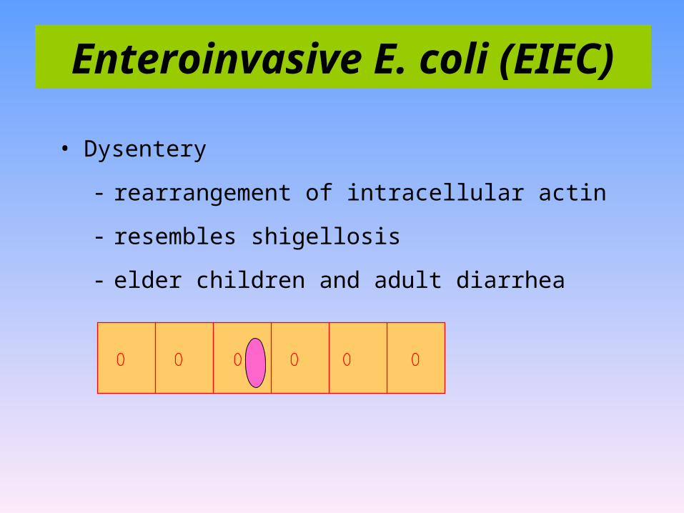
• Dysentery
- rearrangement of intracellular actin
- resembles shigellosis
- elder children and adult diarrhea
Enteroinvasive E. coli (EIEC)
• Malaise and low grade fever diarrhea, vomiting, nausea, non-
bloody stools
• Bundle forming pili are involved in attachment to the
intestinal mucosa.
• This leads to changes in signal transduction in the cells,
effacement of the microvilli, and to intimate attachment via a
non-fimbrial adhesion called intimin.
• This is a problem mainly in hospitalized infants and in day
care centers.
Enteropathogenic E. coli (EPEC)
• fever• infant diarrhea
• vomiting
• nausea • non-bloody stools
• Destruction of surface microvilli
- loose attachment mediated by bundle forming pili (Bfp);
- Stimulation of intracellular calcium level;
- Rearrangement of intracellular actin
Enteropathogenic E. coli (EPEC)
Enterohemorrhagic E. coli (EHEC) • Hemorrhagic – bloody, copious diarrhea
– few leukocytes
– afebrile
• Hemolytic-uremic syndrome – hemolytic anemia
– thrombocytopenia (low platelets)
– kidney failure

• Vero toxin – “shiga-like”
• Hemolysins
• younger than 5 years old,causing
hemorrhagic colitis
Enterohemorrhagic E. coli (EHEC)
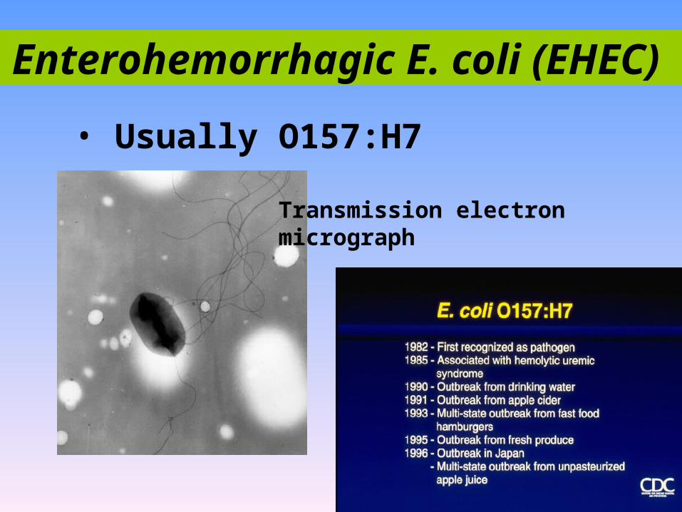
• Usually O157:H7
Transmission electron micrograph
Enterohemorrhagic E. coli (EHEC)

• a cause of persistent, watery diarrhea with vomiting and
dehydration in infants.
• That is autoagglutination in a ‘stacked brick’ arrangement.
• the bacteria adheres to the intestinal mucosa and elaborates
enterotoxins (enteroaggregative heat-stable toxin, EAST).
• The result is mucosal damage, secretion of large amounts of
mucus, and a secretory diarrhea.
Enteroaggregative E. coli (EaggEC)
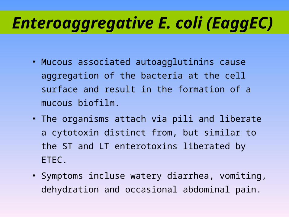
• Mucous associated autoagglutinins cause aggregation of
the bacteria at the cell surface and result in the formation
of a mucous biofilm.
• The organisms attach via pili and liberate a cytotoxin
distinct from, but similar to the ST and LT enterotoxins
liberated by ETEC.
• Symptoms incluse watery diarrhea, vomiting, dehydration
and occasional abdominal pain.
Enteroaggregative E. coli (EaggEC)

Summary of E. coli strains
that cause gastroenteritis.
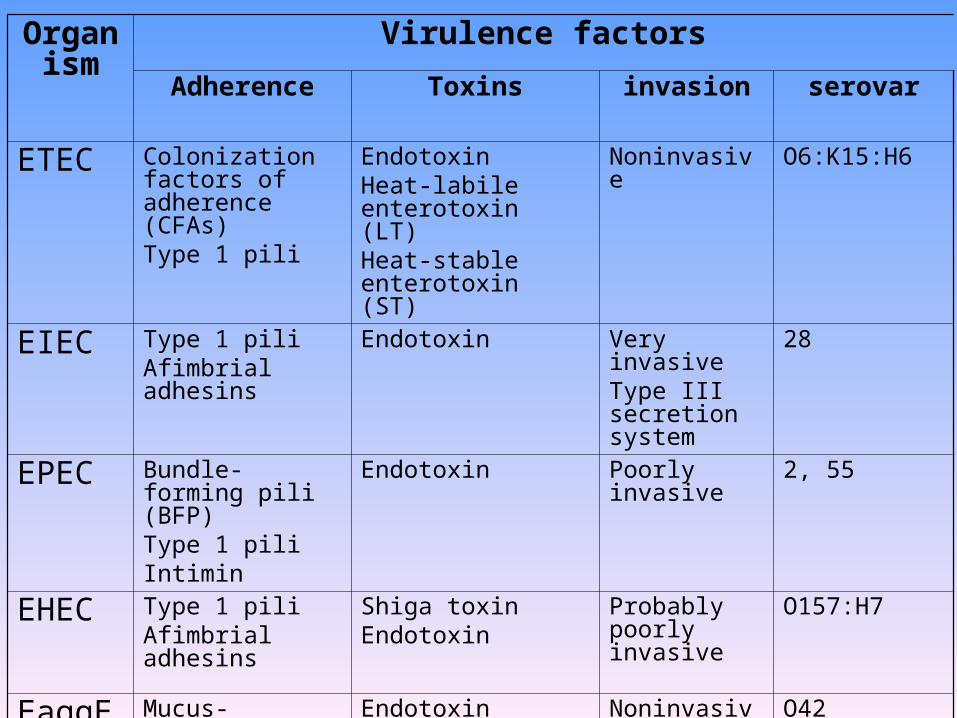
Organism
Virulence factorsAdherence Toxins invasion serovar
ETEC Colonization factors of adherence (CFAs)Type 1 pili
Endotoxin Heat-labile enterotoxin (LT)Heat-stable enterotoxin (ST)
Noninvasive O6:K15:H6
EIEC Type 1 pili Afimbrial adhesins
Endotoxin Very invasiveType III secretion system
28
EPEC Bundle-forming pili (BFP)Type 1 piliIntimin
Endotoxin Poorly invasive 2, 55
EHEC Type 1 pili Afimbrial adhesins
Shiga toxinEndotoxin
Probably poorly invasive
O157:H7
EaggEC Mucus-associated autoagglutinationType 1 pili
EndotoxinCytotoxin (enteroaggregative ST-like toxin)
Noninvasive O42

organism Site Disease pathogenesis
Enterotoxigenic E. coli (ETEC)
Small intestine
Traveler’s diarrhea, infant diarrhea in under developed countries, watery diarrhea, cramps, nausea, low-grade fever
Heat-stable and/or heat-labile enterotoxins, stimulate guanylate or adenylate cyclase activity with fluid and electrolyte loss
Enteroinvasive E. coli (EIEC)
Large intestine
Fever, cramping, watery diarrhea followed by development of dysentery with scant, bloody stools
Plasmid-mediated invasion and destruction of epithelial cells lining colon
Enteropathogenic E. coli (EPEC)
Small intestine
Infant diarrhea with fever, nausea, vomiting, nonbloody stools
plasmid-mediated adherence and destruction of epithelial cells
Enterohemorrhagic E. coli (EHEC)
Large intestine
Hemorrhagic colitis with severe abdominal cramps, watery diarrhea initially, followed by grossly bloody diarrhea, little or no fever, hemolytic uremic syndrome (HUS)
Mediated by cytotoxic “vero-toxin”
Enteroaggregative E. coli (EAggEC)
Small intestine
Persistent infant diarrhea, sometimes with gross blood, low-grade fever
Aggregative adherence mediated by plasmid
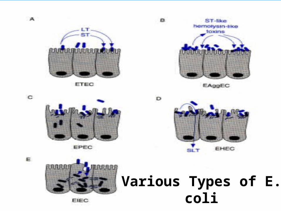
Various Types of E. coli
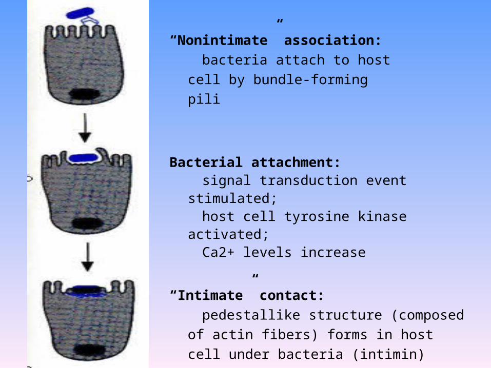
“Nonintimate” association:
bacteria attach to host cell by bundle-
forming pili
Bacterial attachment: signal transduction event stimulated; host cell tyrosine kinase activated; Ca2+ levels increase
“Intimate” contact:
pedestallike structure (composed of actin fibers)
forms in host cell under bacteria (intimin)
Escherichia coli• In the diagnostic laboratory generally the groups are not
differentiated and treatment would be on symptomatology.
• Generally fluid replacement is the primary treatment.
• Antibiotics are generally not used except in severe disease or disease
that has progressed to a systemic stage (e.g.hemolytic-uremia
syndrome).
• Two major classes of pili are produced by E. coli : mannose sensitive
and mannose resistant pili. The former bind to mannose containing
glyocoproteins and the latter to cerebrosides on the host epithelium
allowing attachment. This aids in colonization by E. coli.
Sanitary significance
• Total bacterial number: number of bacteria
contained per ml or gm of the sample; the standard
of drinking water is less than 100.
• Coliform bacteria index: the number of coliform
bacteria detected out per 1000 ml sample; the
standard of drinking water is less than 3
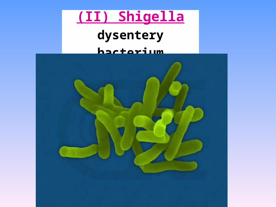
(II) Shigelladysentery bacterium
Genral features
• Pili.
• Most strains can not ferment lactose; S. sonnei
can slowly ferment lactose.
• According to O antigen, 4 groups
• Easily causing drug-resistence.

Classification
Serogroup Species Number of serotypes
A S. dysenteriae 10
B S. flexneri 13
C S. boydii 18
D S. sonnei 1
The classification is based on O antigens
What disease is caused by Shigella species?
Bacillary dysentery
shigellosis
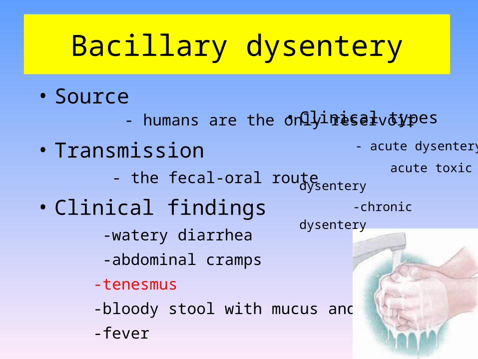
Bacillary dysentery
• Source - humans are the only reservoir
• Transmission - the fecal-oral route
• Clinical findings -watery diarrhea
-abdominal cramps
-tenesmus
-bloody stool with mucus and pus
-fever
• Clinical types
- acute dysentery
acute toxic dysentery
-chronic dysentery
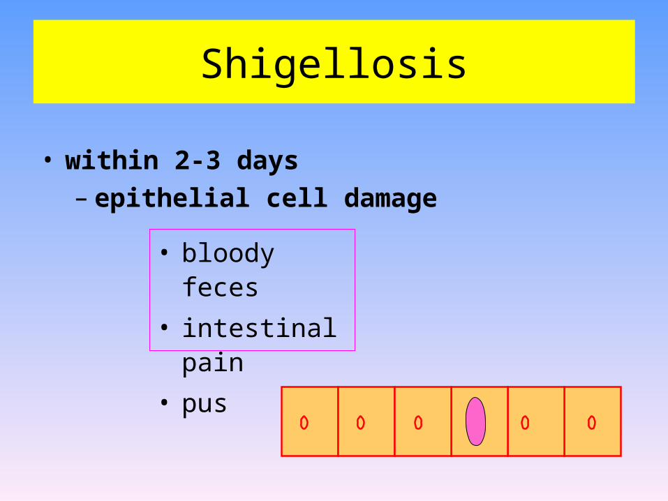
Shigellosis
• within 2-3 days– epithelial cell damage
• bloody feces
• intestinal pain
• pus
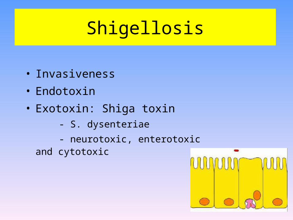
Shigellosis
• Invasiveness
• Endotoxin
• Exotoxin: Shiga toxin - S. dysenteriae
- neurotoxic, enterotoxic and cytotoxic
Shiga toxin
• enterotoxic• cytotoxic• inhibits protein synthesis– lysing 28S rRNA
Clinical significance
• man only "reservoir"
• mostly young children
– fecal to oral contact
– children to adults
• transmitted by adult food handlers
– unwashed hands
Clinical significance• The infective dose required to cause infection is very low (10-
200 organisms).
• There is an incubation of 1-7 days followed by fever,
cramping, abdominal pain, and watery diarrhea (due to the
toxin)for 1-3 days.
• This may be followed by frequent, scant stools with blood,
mucous, and pus (due to invasion of intestinal mucosa).
• Is is rare for the organism to disseminate.
• The severity of the disease depends upon the species one is
infected with. S. dysenteria is the most pathogenic followed
by S. flexneri, S. sonnei and S. boydii.
• Primary immunity defense - SIgA
• Immunity intensity - Limited
- reasons
surface infection
various types
immunity
Diagnosis of Shigella infection
• Specimen: stool.
• Culture and Identification
• Quick immunological methods:
- Immunofluorescent “ball” test;
- Coagglutination.
Prevention & Treatment
• manage dehydration
• patients respond to antibiotics , Problem of drug-
resistance
– disease duration diminished
• streptomycin dependent (SD) dysentery vaccine.
Summary Shigella
• Shigella (4 species; S. flexneri, S. boydii, S. sonnei, S.
dysenteriae) all cause bacillary dysentery or shigellosis, (bloody
feces associated with intestinal pain).
• The organism invades the epithelial lining layer, but does not
penetrate.
• Usually, within 2-3 days, dysentery results from bacteria
damaging the epithelium lining layers of the intestine often with
release of mucus and blood (found in the feces) and attraction of
leukocytes (also found in the feces as "pus").
Summary Shigella
• Shiga toxin (chromosomally encoded) is neurotoxic, enterotoxic
and cytotoxic plays a role. The toxin inhibits protein synthesis
(acting on the 80S ribosome and lysing 28S rRNA).
• This is primarily a disease of young children occurring by fecal-
oral contact. Adults can catch this disease from children.
However it can be transmitted by infected adult food handlers,
contaminating food. The source in each case is unwashed hands.
Man is the only "reservoir".
• Patients with severe dysentery are usually treated with antibiotics
(e.g. ampicillin). In contrast to salmonellosis, patients respond to
antibiotic therapy and disease duration is diminished.
(III) Salmonella(III) Salmonella

•More than 2000 serotypes
•Transmitted by the fecal-oral routed
•Salmonellosis may present as one of several
syndromes including gastroenteritis, enteric
(typhoid) fever or septicemia.
•Zoonosis
Salmonella
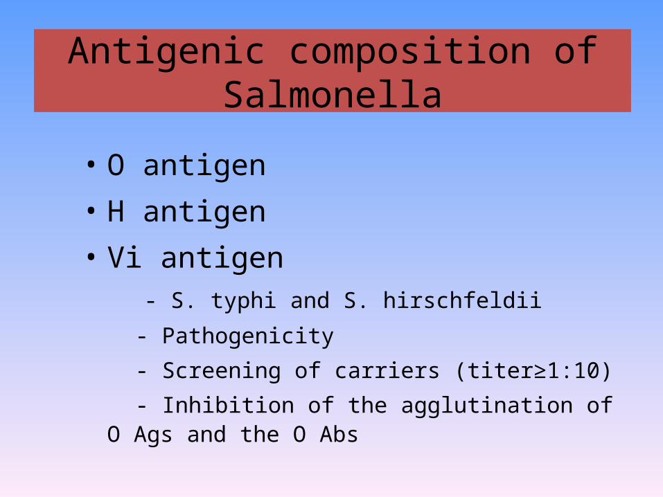
• O antigen• H antigen• Vi antigen - S. typhi and S. hirschfeldii
- Pathogenicity
- Screening of carriers (titer≥1:10)
- Inhibition of the agglutination of O Ags and the O Abs
Antigenic composition of Salmonella

The antigenic structures of salmonellae used in serologic typing
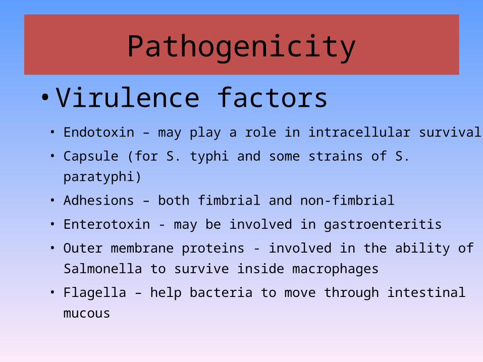
• Virulence factors
Pathogenicity
• Endotoxin – may play a role in intracellular survival
• Capsule (for S. typhi and some strains of S. paratyphi)
• Adhesions – both fimbrial and non-fimbrial
• Enterotoxin - may be involved in gastroenteritis
• Outer membrane proteins - involved in the ability of Salmonella
to survive inside macrophages
• Flagella – help bacteria to move through intestinal mucous

• Virulence factors
Pathogenicity
• Type III secretion systems and effector molecules –
2 different systems may be found:
– One type is involved in promoting entry into intestinal
epithelial cells
– The other type is involved in the ability of Salmonella to
survive inside macrophages

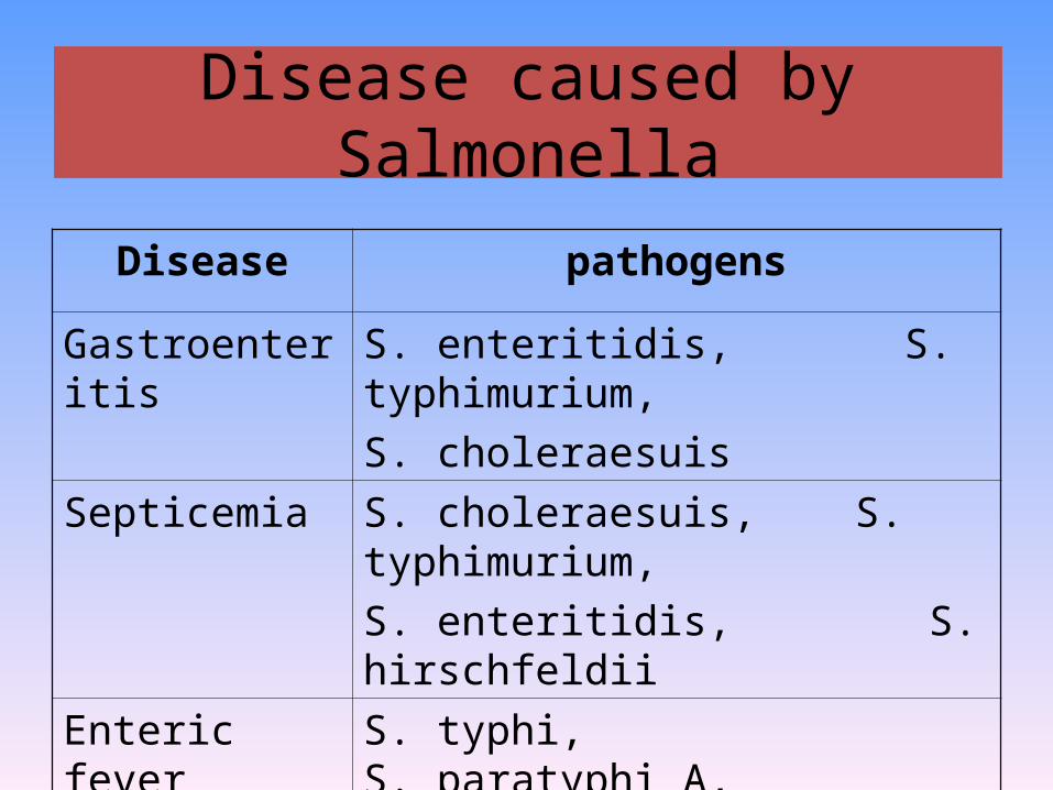
Disease caused by Salmonella
Disease pathogens
Gastroenteritis S. enteritidis, S. typhimurium,
S. choleraesuis
Septicemia S. choleraesuis, S. typhimurium,
S. enteritidis, S. hirschfeldii
Enteric fever S. typhi, S. paratyphi A,
S. schottmuelleri, S. hirschfeldii
Gastroenteritis
• The most common form of salmonellosis and generally requires an 8-48 hour incubation period and may last from 2-5 days.
• Symptoms include nausea, vomiting and diarrhea (non-bloody stool). Salmonella enteritidis is the most common isolate.
• poultry, eggs. no human reservoir
• self-limiting (2 - 5 days)
Gastroenteritis
• The most common form of salmonellosis (70%)
• Self-limited (2-5 d)
• Not enter into blood
• Culture - blood (-)
- stool (+)
Septicemia
• Salmonella septicemia (bacteremia) may be
caused by any species but S. choleraesuis is
common.
• This disease resembles other G- septicemias and
is characterized by a high, remittent fever with
little gastrointestinal involvement.
Septicemia
• 5-10% of salmonellosis
• Intestinal manifestations: often absent
• Culture - blood (+)
- stool (-)
The pathogenesis of typhoid
Enteric fever
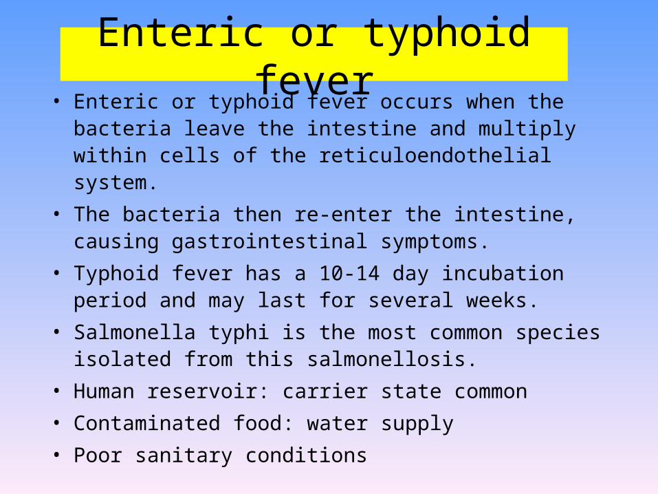
Enteric or typhoid fever• Enteric or typhoid fever occurs when the bacteria leave the
intestine and multiply within cells of the reticuloendothelial system.
• The bacteria then re-enter the intestine, causing gastrointestinal symptoms.
• Typhoid fever has a 10-14 day incubation period and may last for several weeks.
• Salmonella typhi is the most common species isolated from this salmonellosis.
• Human reservoir: carrier state common
• Contaminated food: water supply
• Poor sanitary conditions

Typhoid -Therapy
• Antibiotics– essential
• Vaccines Vi (capsular) antigen :protective

What does the pathogenesis imply in terms of collection of clinical samples?
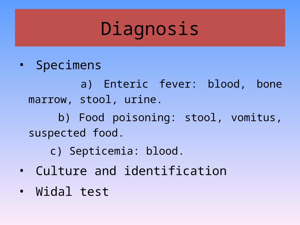
Diagnosis
• Specimens
a) Enteric fever: blood, bone marrow, stool, urine.
b) Food poisoning: stool, vomitus, suspected food.
c) Septicemia: blood.
• Culture and identification
• Widal test
A quantitative agglutination test for typhoid and
paratyphoid, in which detects a patient’s
antibodies to the specific O antigen of S. typhi
and H antigens of S. typhi, S. paratyphi A, S.
schottmuelleri and S. hirschfeldii
Widal test

How to interpret the results of Widal test?
• Consider the manifestaton, course, history,
and local epidemiological conditions

How to interpret the results of Widal test?
TO<1:80,TH<1:160, PH<1:80 Normal value
TO≥1:80 & TH≥1:160 or
TO≥1:80 & PH≥1:80
Typhoid fever
Paratyphoid fever
TO≥1:80 & TH <1:160 or
TO≥1:80 & PH <1:80
Early stage of infection or cross-
reaction of O antigen with other
salmonellae
TO < 1:80 & TH ≥1:160 or
TO < 1:80 & PH ≥ 1:80
Vaccination or nonspecific memory
reaction
How to interpret the results of Widal test?
• Dynamic observation
-antibody titer give a rise gradually
-titer of convalescence serum≥4 times than that of
early specimen
• False negative
-pre-antobiotic
-immunosuppressed
Primary immunity defense
- CMI
Immunity intensity
- strong and permanent
immunity

Salmonella• Using appropriate antibodies more than 2000 antigenic “types”
have been recognized. There are, however, only a few types that are commonly associated with characteristic human diseases (most simply referred to as S. enteritidis, S. cholerae-suis and S. typhi).
• Salmonellosis, the common salmonella infection, is caused by a variety of serotypes (S. enteritidis) and is transmitted from contaminated food (such as poultry and eggs). It does not have a human reservoir and usually presents as gastroenteritis (nausea, vomiting and non-bloody stools). The disease is usually self-limiting (2-5 days). Like Shigella they invade the epithelium and do not produce systemic infection. In uncomplicated cases of salmonellosis, which are the vast majority, antibiotic therapy is not useful. S. choleraesuis (seen much less commonly) causes septicemia after invasion. In this case, antibiotic therapy is required. .
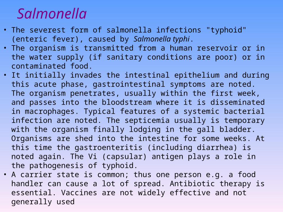
Salmonella• The severest form of salmonella infections "typhoid" (enteric
fever), caused by Salmonella typhi. • The organism is transmitted from a human reservoir or in the water
supply (if sanitary conditions are poor) or in contaminated food. • It initially invades the intestinal epithelium and during this acute
phase, gastrointestinal symptoms are noted. The organism penetrates, usually within the first week, and passes into the bloodstream where it is disseminated in macrophages. Typical features of a systemic bacterial infection are noted. The septicemia usually is temporary with the organism finally lodging in the gall bladder. Organisms are shed into the intestine for some weeks. At this time the gastroenteritis (including diarrhea) is noted again. The Vi (capsular) antigen plays a role in the pathogenesis of typhoid.
• A carrier state is common; thus one person e.g. a food handler can cause a lot of spread. Antibiotic therapy is essential. Vaccines are not widely effective and not generally used
Summary
• Common members of Enterobacteriaceae to
cause human diseases
• Common properties of Enterobaceriaceae
• The medical significance of E. coli
• The opposite traits of Shigella and Salmonella
in terms of pathogenesis and immunity
• Widal test: definition, result determination