Embed Size (px)
Citation preview
Neonatal enteral feeding tube as loci for
Enterobacteriaceae colonisation and risk
to neonatal health
by
HALEMA ABUDALLA
A thesis submitted in partial fulfilment of the requirements
of Nottingham Trent University for the degree
of Doctor of Philosophy
MAY 2014
School of Science and Technology
Nottingham Trent University
Clifton Lane
Nottingham
NG11 8NS
United Kingdom

ii
STATEMENT
I hereby certify that all the research work presented here in this thesis is the result of my
own work except where the references have been made to the published literature or
previous research work.
HALEMA ABUDALLA
iii
Abstract
The incidence of neonatal infections caused by Enterobacteriaceae has been increasing
in recent years, and they are now recognised as the predominant causative agents in
neonatal intensive care unit (NICU) outbreaks. Klebsiella spp. and Serratia spp. are the
most common causative pathogens, and E. coli is one of the leading causes of neonatal
meningitis and sepsis. The infant intestinal flora is influenced by the feeding regime.
This study focuses on assessing the risk to neonates from the ingestion of
the Enterobacteriaceae such as; Enterobacter hormaechei, Enterobacter ludwigii,
Enterobacter aerogenes, Enterobacter cloacae and Klebsiella oxytoca. The strains under
study were isolated from two sources; human mastic breast milk (MBM) and neonatal
nasogastric enteral feeding tubes (EFT).
The overall aim was to evaluate the risk to neonates posed by the ingestion of these
organisms either from contaminated breast milk or from infant formula.
Due to the lack of adequate source information, it was necessary to first confirm the
identity of the strains under investigation. This was achieved using standard biochemical
profiles (phenotyping) and where necessary 16S rDNA sequence analysis. Secondly, it
was necessary to determine whether all strains were unique or if any were multiple
isolations of the same strain. This was achieved using Pulsed-Field Gel Electrophoresis
(PFGE). To determine the potential exposure of neonates to these organisms, a range of
physiological and virulence related assays were undertaken; heat tolerance to 55°C,
biofilm formation, capsule formation and acidic pH survival (pH 3.5).
The potential virulence of the strains was assessed using attachment-invasion assays of
human Caco-2 intestinal cells, human brain microvascular endothelial cells (HBMEC)
and rat brain capillary endothelial cell line (rBCEC4); and also persistence of bacteria in
macrophages by using U937 cells. Patterns of adherence of Enterobacteriaceae to
Caco-2 cells was investigated. The presence of the virulence factors of strains was
determined by identifying haemolytic activity, serum resistance, siderophore production
and antimicrobial susceptibility. The iron uptake genes were also investigated.
iv
The results by PFGE showed that neonatal enteral feeding tubes and mastic
human breast milk were contaminated by twenty-one and three pulsotypes of
Enterobacteriaceae, respectively. Furthermore, the same pulsotypes were spread among
enteral feeding tubes of infants in the same NICUs; indicating the same origins, such as:
environment, milk or carer. Similarly, the MBM strains were isolated from the same
mother. The identification of strains by using 16S rDNA sequence analysis (genotyping)
was more accurate than phenotyping (API technique) and the clustering of strains by
PFGE is a suitable technique for strains relatedness.
The physiological features of the strains in the current study were investigated. The
ability of strains to survive at 55ºC was studied and most of the strains were able to
survive at 55ºC for >30 minutes. Biofilm formation was investigated as this may be a
factor of organism persistence in the neonatal intensive care unit (via milk, environment
or workers) and attachment to enteral feeding tubes. All strains formed biofilms and this
was, in general, enhanced at 37°C compared with room temperature (20ºC) in all types
of formula. The highest levels of biofilm were in casein-based infant formula. Most
strains produced capsular material at 37ºC on all types of formula. However, capsular
material was produced by all strains in soya infant formula. All strains were able to
survive at pH 3.5 for up to 2 hours.
All strains were able to attach to Caco-2, HBMEC and rBCEC4 cells lines, while there
was variation between strains ability to invade mammalian cells. In particular, most of
Ent. hormaechei strains were able to invade the three types of cells lines and one
Ent. ludwigii strain 1439 was only able to invade the rBCEC4 cell line. Ent. ludwigii
strain 1439 was isolated from a case of neonatal meningitis. Three out of eight strains of
Ent. hormaechei and two strains of Ent. cloacae strains survived within macrophages.
Haemolysin production, serum resistance and siderophore production were also studied
and all strains were positive.
Genes encoding for iron uptake irp1, irp2 and fyuA were detected whereas irp1, irp2
genes were absent in all strains while fyuA was present in 4/6 of Ent. ludwigii strains, 1/8
of Ent. hormaechei, 2 of Ent. cloacae, 1 of Ent. aerogenes and 3/6 of K. oxytoca. Three

v
out of eight strains of Ent. hormaechei showed resistance to even the 3rd
generation
cephalosporins, ceftazidime and cefotaxime and were ESBL-positive.

vi
ACKNOWLEDGEMENTS
First of all I would like to thank Allah for helping me all the way during my study, and
giving a great opportunity of coming to the highly prestigious Institute for my doctoral
degree.
I would like to express my deepest gratitude to my supervisors Prof. Steve Forsythe, Mr.
Philip Cheetham and Dr. Michael Loughlin for their consistent help and valuable
guidance during my study.
I am very grateful to Prof Nadia Chuzhanova and Dr. Chrisopher Terrell-Nield for their
help in the statistical analysis. The help of the all my laboratory colleagues in
microresearch especially, Summya, Hana, Naqash, Alya, Abiyad and Fahad provided me
with regular support in the lab work.
The technical support from Michael Brice and Pamela Horne have made my work
possible.
My life away from my family was tough but their support and love gave me strength. I
wanted to express my thanks and love which I have for them. My special thanks to my
lovely mum and my sisters.
Since my time was very difficult, Allah sent angels in my life in the shape of my brother
Sajid and his wife Riffat and their children Usman, Haider and Umar.
The presence of my friends Inas and Binafsha was a source of oxygen in my life. So my
special thanks to them. I pay thanks to my little sister Aisha Alhudiri for her company
and sometimes providing me with distractions in my monotonous life.
I shared my office with great friends and colleques Wesam Alwarfaly, Marion Limo and
Estefania Boix Lopez. We shared the best and the difficult moments. My very special
thanks to them.
During my stay in United Kingdom I met many unforgettable people who in some way
or other gave me a lesson or support and I am thankful to all of them.
Last but not the least I was to thank the Libyan Ministry of Higher Education for
sponsoring my study, and the Libyan embassy in London who looked after me during all
this period.

vii
DEDICATION
This thesis is lovingly dedicated to the memory of my father who will always be missed.
Special feeling of gratitude to my mother Sharefa, who taught me that even the most
difficult of tasks can be accomplished with determination. It is also dedicated to my
lovely sisters without whose encouragement and support, I would not have completed
this work.
viii
TABLE OF CONTENTS
STATEMENT .................................................................................................................................................... ii
Abstract ........................................................................................................................................................ iii
ACKNOWLEDGEMENTS ............................................................................................................................... vi
DEDICATION ................................................................................................................................................. vii
TABLE OF CONTENTS ................................................................................................................................ viii
LIST OF TABLES ............................................................................................................................................ xv
LIST OF FIGURES ........................................................................................................................................ xvii
APPENDICES ................................................................................................................................................. xix
Chapter 1: INTRODUCTION ................................................................................................................. 1
1.1 INTRODUCTION ................................................................................................................................ 2
1.2 Impact of Enterobacteriaceae on neonates ........................................................................................... 3
1.3 Enterobacter spp. as a cause of neonatal infection ............................................................................... 3
1.3.1 Ent. cloacae ...................................................................................................................................... 4
1.3.2 Ent. hormaechei ............................................................................................................................... 5
1.3.3 Ent. aerogenes .................................................................................................................................. 7
1.3.4 Ent. ludwigii ..................................................................................................................................... 8
1.4 Klebsiella spp. as a cause of neonatal infection .................................................................................... 9
1.5 Influence of the existing gut microbiome on neonates health ............................................................... 9
1.6 Potential sources of infection to neonate in NICU .............................................................................. 10
1.6.1 Bacterial exposure of neonates human breast milk ........................................................................ 11
1.6.2 Bacterial exposure of neonates through powdered infant formula ................................................. 12
1.6.3 Bacterial exposure through neonatal feeding tube ......................................................................... 13
ix
1.6.4 Bacterial exposure of neonates health-care workers or immediate environment ........................... 15
1.7 Commonly used infant formula .......................................................................................................... 16
1.8 Conditions and concerns in the NICU ................................................................................................ 20
1.9 Bacterial mechanisms of pathogenicity .............................................................................................. 21
1.9.1 The interaction of bacteria with mammalian cells ......................................................................... 21
Adhesion to mammalian cells ........................................................................................ 23 1.9.1.1
Invading mammalian cells ............................................................................................. 24 1.9.1.2
Intracellular lifestyles .................................................................................................... 24 1.9.1.3
1.10 Biofilm formation ............................................................................................................................... 25
1.10.1 Specialized attachment structures/surface properties of the cell .................................................... 26
1.10.2 Extracellular polymeric substances (EPS) ..................................................................................... 28
1.10.3 Cell–cell communication ............................................................................................................... 30
1.11 Siderophores ....................................................................................................................................... 30
1.12 Diversity of antibiotics resistance in Enterobacteriaceae ................................................................... 32
1.13 Objectives ........................................................................................................................................... 36
1.14 Project Aims ....................................................................................................................................... 38
Chapter 2: MATERIALS AND METHODS ........................................................................................ 39
2.1 GENERAL MATERIALS .................................................................................................................. 40
2.1.1 Bacterial strains .............................................................................................................................. 40
2.1.2 Bacterial cultivation ....................................................................................................................... 50
Tryptone soya agar (TSA) ............................................................................................. 50 2.1.2.1
Tryptone soya broth (TSB) ............................................................................................ 50 2.1.2.2
Milk agar ....................................................................................................................... 50 2.1.2.3
Saline distilled water ..................................................................................................... 50 2.1.2.4
Luria-Bertani broth (LB) ............................................................................................... 50 2.1.2.5
Iso-Sensitest agar (ISO) ................................................................................................. 51 2.1.2.6

x
Long term storage of strains .......................................................................................... 51 2.1.2.7
2.1.3 Mammalian cell lines ..................................................................................................................... 51
Human colonic carcinoma epithelial (Caco-2) cell line ................................................. 51 2.1.3.1
Human brain microvascular endothelial cells (HBMEC) .............................................. 51 2.1.3.2
Rat brain capillary endothelial cell line (rBCEC4) ........................................................ 52 2.1.3.3
Macrophage cell line (U937) ......................................................................................... 52 2.1.3.4
2.1.4 Medium of tissue culture experiments ........................................................................................... 52
Growth medium for human colonic carcinoma epithelial (Caco-2) cell line ................. 52 2.1.4.1
Infection medium for Caco-2 cell line ........................................................................... 52 2.1.4.2
Growth medium for human brain microvascularendothelial cells (HBMEC) and rat 2.1.4.3
brain capillary endothelial cell line (rBCEC4) ............................................................................................. 52
Infection medium for HBMEC and rBCEC4................................................................. 53 2.1.4.4
Growth medium for macrophage cell line (U937) ......................................................... 53 2.1.4.5
Infection medium for macrophage cell line (U937) ...................................................... 53 2.1.4.6
2.1.5 Buffers and detergents .................................................................................................................... 53
Dulbecco’s Phosphate Buffered Saline (PBS) ............................................................... 53 2.1.5.1
Triton- X 0.5% ............................................................................................................... 53 2.1.5.2
TE Buffer ....................................................................................................................... 53 2.1.5.3
Tris base- Boric acid – EDTA (TBE) Buffer 10X preparation (1 Litre distilled water) 54 2.1.5.4
TAE 1X ......................................................................................................................... 54 2.1.5.5
Cell Suspension Buffer .................................................................................................. 54 2.1.5.6
Cell lysis buffer ............................................................................................................. 54 2.1.5.7
Iron III solution .............................................................................................................. 55 2.1.5.8
Chrome azurol sulphate (CAS) solution ........................................................................ 55 2.1.5.9
Hexadecyltrimethylammonium bromide (HDTMA) ..................................................... 55 2.1.5.10
Sodium hydroxide solution ............................................................................................ 55 2.1.5.11
2.1.6 Molecular studies ........................................................................................................................... 55
Genomic DNA extraction .............................................................................................. 55 2.1.6.1
xi
PCR product purification ............................................................................................... 55 2.1.6.2
Agarose gel electrophoresis ........................................................................................... 56 2.1.6.3
2.1.7 Safety considerations ..................................................................................................................... 56
2.2 Characterisation experiments .............................................................................................................. 56
2.2.1 Phenotypic characterisation............................................................................................................ 56
2.2.2 Genotypic characterisation ............................................................................................................. 56
2.3 Identification of strains by using 16S rDNA sequence analysis ......................................................... 58
2.3.1 Preparation of FTA® Elute cards ................................................................................................... 58
2.3.2 AccuPRO-ID Bacterial Identification ............................................................................................ 58
2.3.3 PCR 16S rDNA sequence analysis ................................................................................................. 58
2.4 Physiological experiments .................................................................................................................. 59
2.4.1 Heat tolerance ................................................................................................................................ 59
2.4.2 Biofilm formation .......................................................................................................................... 59
2.4.3 Capsule formation .......................................................................................................................... 60
2.4.4 Acid tolerance of organisms to pH 3.5 (HCl acidified formula) .................................................... 60
2.5 Virulence factors ................................................................................................................................. 61
2.5.1 Bacterial attachment and invasion of host cells ............................................................................. 61
Preparation of bacterial inoculum .................................................................................. 61 2.5.1.1
Mammalian cell culture ................................................................................................. 61 2.5.1.2
Attachment assay ........................................................................................................... 62 2.5.1.3
Gentamicin protection invasion assay ........................................................................... 62 2.5.1.4
Gentamicin protection assay for U937 macrophage uptake and persistence ................. 63 2.5.1.5
The adhesion patterns of bacteria with Caco-2 human epithelial cells .......................... 64 2.5.1.6
2.5.2 Haemolysin production .................................................................................................................. 65
2.5.3 Serum sensitivity determination ..................................................................................................... 65
2.5.4 Siderophore production .................................................................................................................. 65
2.5.5 Determination of high pathogenicity island ................................................................................... 66
xii
2.5.6 Antimicrobial susceptibility testing and ESBLs detection ............................................................. 67
2.5.7 Data analysis .................................................................................................................................. 68
Chapter 3: CHARACTERISATION AND IDENTIFICATION OF BACTERIAL ISOLATES .... 69
3.1 INTRODUCTION .............................................................................................................................. 70
3.2 Materials and Methods ........................................................................................................................ 72
3.3 Results................................................................................................................................................. 72
3.3.1 Characterisation of Enterobacteriaceae strains using the PFGE technique ................................... 72
Characterisation of enteral feeding tubes strains ........................................................... 73 3.3.1.1
Characterisation of Enterobacteriaceae isolated from mastic breast milk .................... 85 3.3.1.2
Characterisation of K. pneumoniae strains isolated from EFT in Jordan....................... 87 3.3.1.3
3.3.2 Comparison of phenotypic and genotypic techniques for identification of Enterobacteriaceae ... 90
Identification of Enterobacteriaceae strains isolated from EFT .................................... 90 3.3.2.1
Identification of Enterobacteriaceae isolated from mastic human breast milk ............ 95 3.3.2.2
3.4 DISCUSSION ..................................................................................................................................... 97
Chapter 4: PHYSIOLOGICAL TRAITS OF BACTERIAL ISOLATES ....................................... 104
4.1 Introduction ....................................................................................................................................... 105
4.2 Materials and methods ...................................................................................................................... 107
4.3 Results............................................................................................................................................... 108
4.3.1 Heat tolerance .............................................................................................................................. 108
4.3.2 Biofilm formation by Enterobacteriaceae on three types of formula milk at different
temperatures ...... …………………………………………………………………………………………..119
Whey-based formula .................................................................................................... 119 4.3.2.1
Casein-based formula .................................................................................................. 122 4.3.2.2
Soya-Powder Infant formula........................................................................................ 122 4.3.2.3
4.3.3 Acid tolerance of organisms to pH 3.5 (HCl acidified formula) .................................................. 133
xiii
4.4 DISCUSSION ................................................................................................................................... 138
Chapter 5: VIRULENCE FACTORS ................................................................................................. 142
5.1 INTRODUCTION ............................................................................................................................ 143
5.2 Materials and methods ...................................................................................................................... 148
5.3 Results............................................................................................................................................... 149
Bacterial attachment and invasion of host cells ......................................................................................... 149
5.3.1 Bacterial attachment to Caco-2 human epithelial cells ................................................................ 149
5.3.2 Bacterial invasion Caco-2 human epithelial cells ........................................................................ 153
5.3.3 Bacterial attachment HBMEC ...................................................................................................... 156
5.3.4 Bacterial invasion of HBMEC ..................................................................................................... 156
5.3.5 Bacterial attachment rBCEC4 ...................................................................................................... 159
5.3.6 Bacterial invasion of rBCEC4 ...................................................................................................... 159
5.3.7 U937 macrophage uptake and persistence studies ....................................................................... 162
5.3.8 Summary of potential risk for Enterobacteriaceae ...................................................................... 164
5.3.9 Patterns of adherence of Enterobacteriaceae to Caco-2 cells ...................................................... 166
5.3.10 Haemolysin production ................................................................................................................ 169
5.3.11 Serum sensitivity .......................................................................................................................... 169
5.3.12 Siderophore production ................................................................................................................ 169
5.3.13 Determination of high pathogenicity island ................................................................................. 169
5.3.14 Determination of antibiotic susceptibility testing by the disc diffusion method .......................... 172
5.4 DISCUSSION ................................................................................................................................... 175
Chapter 6: GENERAL DISCUSSION ................................................................................................ 181
Chapter 7: CONCLUSION AND FUTURE WORK ......................................................................... 197
7.1 CONCLUSION ................................................................................................................................. 198
7.2 Suggested preventive measures for the transmission of bacteria among neonates in NICU ............. 200

xiv
7.3 Limitations and recommendations for future research ...................................................................... 201
REFERENCES ........................................................................................................................................... 203
APPENDICES ........................................................................................................................................... 240
xv
LIST OF TABLES
Table 1.1: Most common species Enterobaceriaceae causing clinical infectious disease. ........................... 3
Table 1.2: The compositions of based infant formula ................................................................................. 19
Table 1.3: The important siderophores produced by bacteria ..................................................................... 31
Table 1.4: Diversity of β-lactam resistance in Enterobacteriaceae. ............................................................ 34
Table 2.1: Description of Ent. hormaechei and Ent. aerogenes strains isolated from enteral feeding tubes
at neonatal intensive care units in Nottingham. ............................................................................................ 41
Table 2.2: Description of Ent. cancerogenus strains isolated from enteral feeding tubes at neonatal
intensive care units in Nottingham ............................................................................................................... 42
Table 2.3: Description of Klebsiella spp. strains isolated from enteral feeding tubes at neonatal intensive
care units in Nottingham .............................................................................................................................. 43
Table 2.4: Description of strains isolated from Spain from MBM.. ............................................................ 44
Table 2.5: Description of K. pneumoniae strains isolated from neonatal EFT used in Jordan, part 1 ......... 45
Table 3.1: Summary of PFGE analysis of Ent. hormaechei strains. ........................................................... 75
Table 3.2: Summary of PFGE analysis of Ent. cancerogenus strains: ........................................................ 78
Table 3.3: Summary of PFGE analysis of Klebsiella spp. strains. .............................................................. 80
Table 3.4: Summary of PFGE analysis of Ent. cloacae and Ent. aerogenes. .............................................. 83
Table 3.5: Summary of PFGE analysis of Enterobacteriaceae isolated from mastic human breast milk
(MBM). ........................................................................................................................................................ 86
Table 3.6: Summary of PFGE analysis of K. pneumoniae strains isolated from Jordan hospital. ............... 88
Table 3.7: Identification Enterobacteriaceae isolated from enteral feeding tubes by using phenotyping and
genotyping techniques; part 1 ...................................................................................................................... 92
Table 4.1: D-value for Ent. ludwigii and Ent. cancerogenus strains in two different types of formula milk.
.................................................................................................................................................................... 116
Table 4.2: D-value for Ent. hormaechei and K. oxytoca in two different types of formula milk. ............. 118
Table 4.3: Biofilm formation level on based-infant formula (whey, casein and soya) by EFT strains and
MBM strains .............................................................................................................................................. 127

xvi
Table 4.4: Capsule production on based infant formula (whey, casein and soya) by EFT strains and MBM
strains. ........................................................................................................................................................ 132
Table 5.1: Summary of virulence factors for Ent. hormaechei, Ent. ludwigii, Ent. cloacae, Ent. aerogenes
and K. oxytoca. ........................................................................................................................................... 165
Table 5.2: Description patterns of adherence of Enterobacteriaceae to Caco-2 cells. .............................. 167
Table 5.3: Description of haemolysin production (horse/sheep blood), serum sensitivity determination and
siderophore production. .............................................................................................................................. 171
Table 5.4: Description of antibiotic resistance profiles of Enterobacteriaceae isolates ............................ 174
xvii
LIST OF FIGURES
Figure 1.1: Sources of transmission of bacteria within a NICU setting. ..................................................... 11
Figure 1.2: The processes governing biofilm formation. ............................................................................ 26
Figure 1.3: Genetic and structure organization of the high pathogenicity island of Y. enterocolitica......... 32
Figure 3.1: PFGE cluster analysis of Ent. hormaechei strains (as performed by Alkeskas). ...................... 74
Figure 3.2: PFGE cluster analysis of Ent. cancerogenus strains (as performed by Alkeskas). ................... 77
Figure 3.3: PFGE cluster analysis of Klebsiella spp. (as performed by Alkeskas). .................................... 80
Figure 3.4: PFGE cluster analysis of Ent. cloacae and Ent.aerogenes (as performed by Alkeskas). ......... 83
Figure 3.5: PFGE cluster analysis of MBM strains. .................................................................................... 85
Figure 3.6: PFGE cluster analysis of K. pneumoniae strains isolated from EFT in Jordan......................... 87
Figure 4.1: Survival of Ent. ludwigii in (A) whey-based infant formul and (B) in casein-based infant
formula at 55°C. ......................................................................................................................................... 110
Figure 4.2: Survival of Ent. cancerogenus in (A) whey-based infant formul and (B) in casein based-infant
formula at 55°C. ......................................................................................................................................... 112
Figure 4.3: Survival of Ent. hormaechei, Ent. cloacae strains (779 and 789) and Ent. aerogenes strain
1056 in (A) whey-based infant formul and (B) in casein-based infant formula at 55°C. .......................... 114
Figure 4.4: Survival of K. oxytoca in (A) whey-based infant formul and (B) in casein-based infant formula
at 55°C. ...................................................................................................................................................... 116
Figure 4.5: : Biofilm formation in whey-based infant formula at 20°C and 37°C. ................................... 121
Figure 4.6: Biofilm formation in casein-based infant formula at 20°C and 37°C. .................................... 124
Figure 4.7: Biofilm formation in soya powder infant formula at 20°C and 37°C. .................................... 125
Figure 4.8: Capsule formation on milk agar by Enterobacteriaceae strains. ............................................ 131
Figure 4.9: Survival of Ent. ludwigii in whey-based infant formula at pH 3.5 for 2h. .............................. 134
Figure 4.10: Survival Ent. ludwigii in casein-based infant formula at pH 3.5 for 2h. ............................... 134
Figure 4.11: Survival of Ent. hormaechei, Ent. cloacae and Ent.aerogenes strains in whey-based infant
formula at pH 3.5 for 2h. ............................................................................................................................ 135
Figure 4.12: Survival of Ent. hormaechei, Ent. cloacae and Ent. aerogenes strains in casein-based infant
formula at pH 3.5 for 2h. ............................................................................................................................ 135
xviii
Figure 4.13: Survival of Ent. cancerogenus in whey-based infant formula at pH 3.5 for 2h. ................... 136
Figure 4.14: Survival of Ent. cancerogenus in casein-based infant formula at pH 3.5 for 2h. ................. 136
Figure 4.15: Survival of K. oxytoca in whey-based infant formula at pH 3.5 for 2h. ............................... 137
Figure 4.16: Survival of K. oxytoca in casein-based infant formula at pH 3.5 for 2h. .............................. 137
Figure 5.1: Attachment of Ent. ludwigii, Ent. hormaechei, Ent. aerogenes, Ent. cloacae and K. oxytoca to
the mammalian Caco-2 cell line. .............................................................................................................. 151
Figure 5.2: Attachment of Ent. cancerogenus to the mammalian Caco-2 cell line. .................................. 152
Figure 5.3: Invasion of Ent. ludwigii, Ent. hormaechei, Ent. aerogenes, Ent. cloacae and K. oxytoca to the
mammalian Caco-2 cell line. ...................................................................................................................... 154
Figure 5.4: Invasion of Ent. cancerogenus the mammalian Caco-2 cell line….………………………... 155
Figure 5.5: Attachment of Ent. ludwigii and Ent. hormaechei to the mammalian HBMEC cell line. ...... 157
Figure 5.6: Invasion of Ent. ludwigii and Ent. hormaechei to the mammalian HBMEC cell line. ........... 158
Figure 5.7: Attachment of Ent. ludwigii and Ent. hormaechei to the mammalian rBCEC4 cell line. ....... 160
Figure 5.8: Invasion of Ent. ludwigii and Ent. hormaechei to the mammalian rBCEC4 cell line. ............ 161
Figure 5.9: Ent. ludwigii, Ent. hormaechei, Ent. aerogenes, Ent. cloacae and K. oxytoca persistence in
U937 macrophage cells .............................................................................................................................. 163
Figure 5.10: Light microscopy of Giemsa stained Caco-2 human epithelial cells, showing the three types
of bacteria adherence patterns, after 3 hours incubation of 108 bacteria. ................................................... 168
Figure 5.11: lane 1: ladder 100bp; lanes 2 negative control; lane 3: PCR product of fyuA gene size 547bp
using Ent. hormaechei, Ent. cloacae, Ent. aerogenes and Ent. ludwigii DNA. ......................................... 170
Figure 5.12: lane 1: ladder 100bp; lanes 2 negative control; lane 3: PCR product of fyuA gene size 547bp
using K. oxytoca DNA. .............................................................................................................................. 170
xix
APPENDICES
Appendix 1: Salmonella serotype Braenderup reference standard (H9812), approximate band sizes in
kilobases ..................................................................................................................................................... 240
Appendix 2: 100 bp PCR sizer ladder. ..................................................................................................... 240
Appendix 3: PFGE cluster analysis of Ent. hormaechei isolated from EFT (UK). ................................... 241
Appendix 4: PFGE cluster analysis of Ent. cancerogenus isolated from EFT(UK). ................................ 242
Appendix 5: PFGE cluster analysis of Klebsiella spp. isolated from EFT (UK). ..................................... 243
Appendix 6: PFGE cluster analysis of Ent. cloacae and Ent. aerogenes isolated from EFT (UK)........... 244
Appendix 7: PFGE cluster analysis of Klebsiella spp. isolated from EFT (Jordan). ................................ 245

xx
ABBREVIATIONS LIST
A. Acinetobacter
Ae. Aerobacter
Aer. Aeromonas
BP Base pairs
Caco-2 Human colonic carcinoma epithelial cells
CDC Centers for Disease Control and Prevention
CFU Colony forming unit
Cit. Citrobacter
Cl. Clostridium
COSHH Control Of Substances Hazardous to Health
CPD Combination of cefpodoxime
DNA Deoxyribonucleic acid
DNase Deoxyribonuclease
dNTP 2’-deoxynucleoside 5’-triphosphate
dNTPs Deoxyribonucleotide Triphosphate
E. Escherichia
EDTA Ethylenediamine tetra-acetic acid
EFT Enteral feeding tubes
EHOS Ent. hormaechei outbreaks
Ent. Enterobacter
ESBLs Extended spectrum β-lactamases
EPS Extracellular polymeric substances
FAO Food and Agriculture Organization
HBMEC Human brain microvascular endothelial cells
HCL Hydrochloric acid
HPA Health Protection Agency
ICUs intensive care units
ISO Iso-Sensitest agar
K. Klebsiella
KAH King Abdulla Hospital

xxi
LBA Luria-Bertani agar
LBA Luria-Bertani Agar
LPS Lipopolysaccharide
MBM Mastic breast milk
Mo. Morganella
MOI Multiplicity of infection
MRSA Methicillin-Resistant Staphylococcus aureus
My. Mycobacterium
NCH Nottingham City Hospital
NICU Neonatal intensive care unit
NNIS National nosocomial infections surveillance system
NTU Nottingham Trent University
OD Optical Density
P. Pseudomonas
PBS Dulbecco’s phosphate buffered saline
PCR Polymerase chain reaction
PFGE Pulsed Field Gel Electrophoresis
PIF Powdered infant formula
Pl. Plesiomonas
Pr. Providencia
PRH Princesses Rahma Hospital
QMC The Nottingham Queen's Medical Centre
R Resistance
rBCEC4 Rat brain capillary endothelial cell line
rpm Revolutions per minute
S Sensitive
S. Streptococcus
Sal. Salmonella
SDS Sodium dodecyl sulphate
Se. Serratia
Sh. Shigella
xxii
Spp. Sepsis
St. Staphylococcus
TAE Tris-acetate-EDTA
TBE Tris base- Boric acid – EDTA
TEB Tris EDTA buffer
TIFF Tagged Image File Format
TSA Tryptone Soya Agar
TSB Tryptone Soya Broth
U Unique
U937 Macrophage Cell Line
UN United Nations
UPGMA Unweight pair group method with arithmetic mean
US United States
USA The United States of America
UV Ultraviolet
V. Vibrio
v/v Volume per volume
w/v Weight per volume
WHO World Health Organization
Y. Yersinia
Α Alpha
Β Beta
γ Gamma

1
Chapter 1: INTRODUCTION
Chapter 1
2
1.1 INTRODUCTION
The Enterobacteriaceae is a family of Gram-negative bacilli, some of which are motile,
and have capsules. Most species are facultative anaerobic, oxidase-negative and
catalase-positive rods. The Enterobacteriaceae which most commonly produce
extended-spectrum β-lactamases (ESBLs) are Klebsiella pneumoniae and Escherichia
coli (Romero et al., 2007). Some members are found in soil, water, plants and animals.
The Enterobacteriaceae group is comprised of more than 100 species of bacteria that
normally inhabit the intestines of humans and animals. Many Enterobacteriaceae species
are lactose fermenting and are commonly referred to as “coliforms”. Exotoxins are
produced by some pathogenic strains, whereas others produce enterotoxins which affect
the intestinal tract, causing diarrhoea and body fluid loss. Some Enterobacteriaceae (i.e.
K. pneumoniae) are able to cause pneumonia and urinary tract infections. Wound
infections and other nosocomial (hospital acquired) infections are caused by
Enterobacteriaceae. They are also recognized as a major cause of bacteraemia and
meningitis.
In recent years, the incidence of neonatal infections due to Enterobacteriaceae has been
increasing and they are now recognised as the predominant causative agents of infection
in neonatal intensive care unit (NICU) outbreaks (Gastmeier et al., 2007; McGuire et al.,
2004; Kaufman et al., 2004). The most common neonatal Enterobacteriaceae pathogens
reported are Klebsiella spp. and Serratia spp.; 23.4% and 13.8% cases respectively
(Gastmeier et al., 2007). There are also other pathogenic species which, although they
occur less frequently, have a high severity following infection. For example, pathogenic
strains of E. coli are a leading cause of neonatal meningitis and sepsis (Stoll et al., 2005),
and Cronobacter sakazakii can cause necrotizing enterocolitis and meningitis. Neonates
may be particularly prone to Gram negative infection (i.e. Enterobacteriaceae) as their
innate immune cells have lower responses to lipopolysaccharide (LPS), the outer portion
of the bacterial cell wall (Townsend et al., 2007). However, Nanthakumar et al. (2000)
reported that the human colonic carcinoma epithelial (Caco-2) cells secrete less IL8
(LPS, 8-fold; IL-1b, 20-fold) than fetal cells after inflammatory stimulation.

Chapter 1
3
1.2 Impact of Enterobacteriaceae on neonates
Enterobacteriaceae are the most common opportunistic pathogens particularly E. coli,
Enterobacter spp., Klebsiella spp., Serratia spp., and Salmonella. They are significantly
implicated with morbidity and mortality (Friedland et al., 2003; Adamson et al., 2012).
Many different types of infections can be caused by these organisms, such as sepsis,
brain meningitis, pneumonia, and urinary tract infections particularly in intensive care
units (ICUs) (Kollef et al., 1999; Ibrahim et al., 2000). Enterobacteriaceae such as E.
coli, Klebsiella, Proteus, Serratia and Citrobacter spp. are able to cause nosocomial
infections. Most of these strains are members of normal flora such as E. coli and Ent.
cloacae, and probably cause infections through selection following an empiric
antimicrobial regime (Iversen et al., 2004a; Stoll et al., 2005). The most common human
diseases which are caused by a family of Enterobacteriaceae are presented in Table 1.1
(Liu et al., 2012; Podschun and Ullmann, 1998; Pazhani et al., 2005; Fraser and Arnett,
2006; Chaudhry et al., 2007; Hammerum and Heuer, 2009; Bisi-Johnson et al., 2011).
Table 1.1: Most common species Enterobaceriaceae causing clinical infectious disease.
1.3 Enterobacter spp. as a cause of neonatal infection
The Enterobacter genus belongs to the family of Enterobacteriaceae and are known as
facultative anaerobic Gram-negative strains. This genus are saprophytic bacteria which
Clinical species Clinical Infection Presentation
Cit. freundiiPneumonia, meningitis, septicaemia, wound and
urinary tract infections
Ent. aerogenes, Ent. cloacaePneumonia, septicaemia, wound and urinary
tract infections
E. coliDiarrhoea, meningitis, septicaemia and urinary
tract infections
K.oxytoca, K. pneumoniaePneumonia, septicaemia and urinary tract
infections
Mo. morganii Septicaemia and urinary tract infections
Pl. shigelloides Diarrhoea and septicaemia
Pr. rettgeri, Pr. stuartii Urinary tract infections
Sal. enteriticaDiarrhoea, typhoid fever, septicaemia,
osteomyelitis and urinary tract infections
Se. marcescens, Se. liquefaciensPneumonia, septicaemia, wound and urinary
tract infections
Sh. sonnei, Sh. flexneri Diarrhoea
Y. pestis, Y. enterocolitica Diarrhoea, septicaemia plague and enteritis
Chapter 1
4
are widely encountered in nature, such as in soil and sewage and are commensal
in the enteric flora in the human gastrointestinal tract (Mazari-Hiriart et al., 2008;
Quintanilha et al., 2007). There are 14 known species of Enterobacter, some of these
have been implicated as causes of diseases in humans and the most commonly
encountered species are Ent. aerogenes, Ent. cloacae, Ent. agglomerans, and
C. sakazakii (Andresen et al., 1994; Burchard et al., 1986; Chow et al., 1991; Gallagher
1990; Gaston 1988; Haddy et al., 1991; Hawkins et al., 1991; Stenhouse et al., 1992).
Ent. aerogenes and Ent. cloacae are considered the most frequently encountered human
pathogens within the genus Enterobacter (Andresen, et al., 1994; Burchard et al., 1986;
Haddy et al., 1991; Burchard et al., 1994; Karnad et al., 1987; Weischer and Kolmos,
1992). The major nosocomial pathogens often found in intensive care settings are
Enterobacter species. From 1976 to 1989 in the United States of America (USA), the
National Nosocomial Infections Surveillance System (NNIS) reported data on
nosocomial bacteremia. In 2008 the National Healthcare Safety Network reported that
Enterobacter spp. accounted for approximately 5% of nosocomial bacteremia cases,
between 1995 and 2002, a study by Wisplinghoff et al. (2004) from different states in
USA demonstrated the most commonly isolated nosocomial pathogens with a higher rate
in intensive care unit wards was Enterobacter species (Wisplinghoff et al., 2004).
Previous studies by v Dijk et al. (2002) and van den Berg et al. (2000) reported that
neonatal intensive care units (NICUs) have outbreaks of Enterobacter spp.
1.3.1 Ent. cloacae
Ent. cloacae are members of the genus Enterobacter. As other Enterobacteriacae, they
are saprophytic microorganisms of the normal digestive flora in humans and are the most
commonly isolated clinical species (Thomas et al., 1993; Wang et al., 1991). Recently,
six species have been classified in the Ent. cloacae complex, including Ent. cloacae,
Ent. asburiae, Ent. hormaechei, Ent. kobei, Ent. ludwigii and Ent. nimipressuralis. The
biochemical and molecular studies on E. cloacae have shown genomic heterogeneity,
comprising six species. Species of the Ent. cloacae complex are widely encountered in
nature, but they can act as pathogens (Mezzatesta et al., 2012).
Chapter 1
5
Ent. cloacae has emerged as an alarming pathogen for healthcare institutions globally. It
accounts for up to 5% of hospital-acquired sepsis, 5% of nosocomial pneumonias,
4% of nosocomial urinary tract infections and 10% of postsurgical peritonitis cases
(Paauw et al., 2008a and Hoffmann and Roggenkamp, 2003). In different clinical
settings, including neonatal NICUs, outbreaks due to exogenous Ent. cloacae infection
have been reported. In 1998, van Nierop et al. reported an outbreak of Ent. cloacae in a
NICUs that caused nine deaths. Also, Kuboyama et al. (2003) reported an overall 34%
mortality rate associated with Ent. cloacae in unrecognized outbreaks in NICU. This
bacterium was possibly transmitted to neonates by contaminated intravenous fluids,
whole parenteral nutrition solutions and medical equipment.
The healthcare workers may play a role in cross-transmission of many single-clone
outbreaks and inpatients may also act as a reservoir (Dalben et al., 2008). In hospitalized
neonates, Ent. cloacae can cause a range of infections such as urinary tract infection,
bloodstream infection, pneumonia and bronchopulmonary dysplasia (Grattard et al.,
1994; Modi et al., 1987). Enterobacteriaceae has been isolated from powder infant
formula (PIF), such as Ent. cloacae, K. pneumoniae, K. oxytoca, Ent. hormaechei, Cit.
freundii, and E. coli (Muytjens et al., 1988; Townsend et al., 2008a).
Only a few of the studies have reported enteral feeding tubes (EFT) as a site of the
bacterial colonisations. Bacterial contamination of the EFT with >103cfu/ml in 71/125
EFTs was reported by Mehall et al. (2002a) from infants over four months of age.
Staphylococcus epidermidis, S. aureus, Enterococcus faecalis, Ent. cloacae and K.
pneumoniae were detected in their study. They also showed that seven infants older than
four months who were fed with tubes containing >105cfu/ml developed necrotizing
enterocolitis. Their study also identified methicillin-resistant Staphylococcus aureus
(MRSA) contamination in the EFT (Mehall et al., 2002b).
1.3.2 Ent. hormaechei
Ent. hormaechei is a Gram-negative rod in the Ent. cloacae complex, and is most
frequently isolated from clinical sources (Townsend et al., 2008a). The species have
been defined within the Ent. cloacae complex via deoxyribonucleic acid (DNA) cross-
Chapter 1
6
hybridization, naming three new subspecies of Ent. hormaechei: Ent. hormaechei subsp.
steigerwalti, Ent. hormaechei subsp. hormaechei and Ent. hormaechei subsp. oharae
(Hoffmann et al., 2005a).
Using biochemical identification, Ent. hormaechei strains are catalase positive, oxidase
and deoxyribonuclease (DNase) negative and fermentative. Ent. hormaechei form non-
pigmented colonies after 18 to 24 hours at 15 to 42°C, with an optimum for growth at
36°C, on all non-selective media, such as Colombia agar with 5% sheep blood, chocolate
agar, tryptic soy agar (TSA), Luria-Bertani agar (LBA), and brain heart infusion agar, as
well as on semi-selective media such as MacConkey and ENDO agar (Hoffmann and
Roggenkamp, 2003).
Ent. hormaechei is the most commonly isolated nosocomial pathogen (Paauw et al.,
2008a). It has been a cause of septicaemia in an outbreak occurring in six NICUs in five
hospitals in Brazil, and due to contamination of parental nutrition solutions which all
originated from the same manufacturer (Campos et al., 2007). Ciprofloxacin resistance
was closely associated with the prior use of fluoroquinolones and broad-spectrum
cephalosporin in Enterobacter species isolates causing bacteremia (Kang et al., 2005).
A previous study conducted by the Post-doctoral researcher Dr Stacy Townsend at
Nottingham Trent University (NTU) identified Ent. hormaechei from isolates received
from a United States (US) hospital that had been misidentified as ‘Cronobacter’ strains.
Further analysis, with Pulsed Field Gel Electrophoresis (PFGE) and 16S rDNA
sequencing revealed that the strains were clonal and were from an NICU outbreak which
the hospital had not detected (Townsend et al., 2008a). Hence, the hospital had not only
failed recognising that they had an outbreak, but had also misidentified the organisms.
Muytjens et al. (1988) identified Cronobacter strains from PIF using biochemical
profiling. However, by using 16S rDNA sequence analysis some of the strains were
reidentified as Ent. hormaechei. This organism has been reported to be resistant to
quinolone highlighting the difficulty of treatment (Townsend et al., 2008a). The study of
this organism is significant in the clinical setting as it is difficult to treat and identify.
Enterobacteriaceae isolates identified from EFT from the Nottingham Queen's Medical
Chapter 1
7
Centre (QMC) and Nottingham City Hospital (NCH) included Ent. hormaechei (Hurrell
et al., 2009b). Many of these strains were resistant to the 3rd
generation cephalosporins,
ceftazidime and cefotaxime, and ESBLs were detected. This demonstrates that neonates
are directly exposed to antibiotic resistant strains in NICUs during feeding.
Comparative bacterial thermal death rates of Enterobacteriaceae revealed that an
Ent. hormaechei strain isolated from a neonatal enteral feeding tube was more heat
resistant that Sal. seftenberg. This latter organism is recognised as the most heat resistant
Enterobacteriaceae and its detection is used by industry to ensure their heat treatment
processes are efficient (Forsythe, 2009). This shows that Ent. hormaechei has a greater
chance of surviving during the reconstitution of infant formula than other organisms, and
that preparation may need to be modified to take this into account.
1.3.3 Ent. aerogenes
Ent. aerogenes is known as part of the normal flora of the human gastrointestinal tract
that is in most cases endogenously acquired (Flynn et al., 1987). In addition,
Ent. aerogenes is an opportunistic colonizing pathogen which can cause infection in
hospitalized patients (Sanders and Sanders, 1997). This organism is one of the more
commonly described Gram-negative bacteria causes of nosocomial respiratory tract
infections (Arpin et al., 1996; Davin-Regli et al., 1996a). Recently, Ent. aerogenes
has emerged as a significant hospital pathogen (de Champs et al., 1991; Flynn et al.,
1987; Mellencamp et al., 1990; Meyers et al., 1988). Because of the widespread
prescription of broad-spectrum antibiotics, especially extended-spectrum cephalosporins,
Ent. aerogenes has emerged as a pathogen (Shlaes, 1993).
The presence of medical devices such as endotracheal tubes or central venous catheters
are associated with multiresistant Ent. aerogenes, which facilitate colonization (Chow et
al., 1991; Weinstein, 1986).
Ent. aerogenes has been associated with nosocomial infections during the last 5 years.
Several outbreaks linked with multiresistant strains of Ent. aerogenes have been reported
in ICUs in France (Arpin et al., 1996; Davin-Regli et al., 1996 a and b; Grattard et al.,
Chapter 1
8
1995; Neuwirth et al., 1996), Belgium (De Gheldre et al., 1997; Jalaluddin et al., 1998),
Austria (Allerberger et al., 1996) and the United States (Georghiou et al., 1995).
Ent. aerogenes species are not commonly isolated in NICUs however, a previous study
by Gallagher (1990) reported that in a pediatric population during a 5 year period, 22
episodes of nosocomially acquired Enterobacter bacteria were identified: 17 were
Ent. cloacae, three were Ent. aerogenes and two were Ent. agglomerans. Another study
also reported that Ent. aerogenes was isolated in a NICU from the blood culture of a 5
day old neonate; and 12 more episodes were found 10 weeks later which caused
nosocomial outbreaks of septicemia in the NICU (Loiwal et al., 1999).
1.3.4 Ent. ludwigii
Ent. ludwigii is a Gram-negative rod, belonging to the Enterobacteriaceae family, and is
motile, catalase positive, oxidase and DNase negative, fermentative, and non-pigmented.
Ent. ludwigii has the ability to ferment 3-0-methyl-D-gluco-pyranose and myo-inositol.
Ent. ludwigii has been reported to produce a Bush class 1 beta-lactamase rendering
resistance to ampicillin, amoxicillin plus clavulanic acid and cefoxitin in the disk
diffusion tests. In addition, Ent. ludwigii is susceptible to cefepime, trimethoprim plus
sulphamethoxazole, gentamicin, and resistant to piperacillin, piperacillin plus
tazobactam, cefoxitin, cefotaxime, ceftazidime (Hoffmann et al., 2005b).
There have been few studies of this organism, possibly due to the lack of a reliable
identification scheme. However, during the second year of this project, we received an
additional Ent. ludwigii strain (NTU1439) from Nottingham QMC hospital which had
been isolated from a case of neonatal meningitis. This strain was included in the project
as it reinforced the importance of evaluating the risk of neonatal infection by Ent.
ludwigii.
In previous studies, Ent. ludwigii has been isolated from various sources such as clinical
specimens (Hoffmann et al., 2005b), and from plants (Shoebitz et al.,2009). Delgado
et al. (2008) isolated Ent. ludwigii from human milk. However, as will be reported here,
it was originally identified as K. oxytoca using classical tests. By using 16S rDNA
sequence analysis, the Delgado K. oxytoca isolates were reidentified as Ent. ludwigii at
Chapter 1
9
NTU. By coincidence, NTU already had an Ent. ludwigii strain in its collection which
was initially identified as Cronobacter; this strain isolated from a case of neonatal
meningitis in Nottingham QMC hospital.
1.4 Klebsiella spp. as a cause of neonatal infection
The most commonly isolated members of the genus Klebsiella (family
Enterobacteriaceae) are K. pneumoniae and K. oxytoca which consist of Gram-negative,
non-motile, encapsulated rods (Green et al., 2009), and the colonies of Klebsiella have a
characteristic mucoid appearance because they produce polysaccharide capsules
(Podschun and Ullmann, 1998). Klebsiella spp. are opportunistic pathogens that can
cause a variety of illnesses including pneumonia, urinary tract infections, septicaemia,
soft tissue, intravenous, meningitis, liver abscess, and gastrointestinal disease common in
immunocompromised or those with underlying conditions (Green et al., 2009).
To cause infection, microorganisms must adhere to the mucosal surfaces in order to
reach host cells and can use surface appendages to achieve this (Podschun and Ullmann,
1998). Klebsiella is abundant in the environment (e.g., soil and water) but can also be
isolated from skin, mucous membranes, and the intestines of humans and animals
(Podschun and Ullmann, 1998). In a study of bacterial colonisation in a NICU, there was
a higher risk of the acquisition of Klebsiella spp., and Enterobacter spp., in infants
receiving antibiotics for more than 3 days and associated with an increased duration of
NICU stay. Klebsiella spp. is recognised to survive on the skin and is more resistant to
desiccation than other Enterobacteriaceae (Gastmeier et al., 2007).
1.5 Influence of the existing gut microbiome on neonates health
Colonization of the human gut with microbes begins during birth by microbes from the
environment. In the first few hours of life, usually the most important source of
inoculum is the mother’s vaginal and fecal microbiomes (Gueimonde et al., 2006 and
Vaishampayan et al., 2010). However, some data reported that infants delivered by C-
section-delivered have lower gut microbial richness and variety at 4 months of age,
Chapter 1
10
compared to vaginally delivered infants (Azad et al., 2013). The great functional and
phylogenetic difference observed between infant gut microbiomes possibly because of
random colonization happenings, variances in immune responses to the colonizing
microbes, alterations in host behaviour, or other features of host lifestyle (Palmer et al.,
2007; Dethlefsen et al., 2006).
The previous studies report that the microbiome is implicated in human health
(Turnbaugh et al., 2007). Gut microbiotas can contribute to excess host adiposity
(Turnbaugh and Gordon 2009; Turnbaugh et al., 2006), protect against the improvement
of type 1 diabetes (Wen et al., 2008), and induce colitis (Garrett et al., 2007) and
metabolic syndrome (Stockman, 2012). Hence, the microbiota has been proposed as a
target for therapeutic intervention for numerous chronic diseases (Turnbaugh et al., 2007;
Zaneveld et al., 2008).
The microbiotas of adult are assumed to be relatively stable over time which imparts
resilience to disturbance, confirming continued gut function (Turnbaugh and Gordon
2009; Ley et al., 2006; Dethlefsen et al., 2008). In contrast, in the infant (full-term 2-5
years old) the chaotic shifts in the microbiome are affected with life events, such as
drastic diet changes or antibiotic treatments, which result in large changes in the relative
large quantity of taxonomic groups. Moreover, they found that the 2.5 years old human
gut microbiome has several of the functional features of the adult microbiome (Koenig et
al., 2010).
1.6 Potential sources of infection to neonate in NICU
Neonates characterise a unique and highly exposed patient population. Improvements in
medical technology that have occurred over the last few decades have developed the
survival and quality of life for neonates, particularly those infants born with extreme
prematurity or with congenital defects. However, infants hospitalized in the NICU can
be exposed and acquire health care-associated infections from both human and inanimate
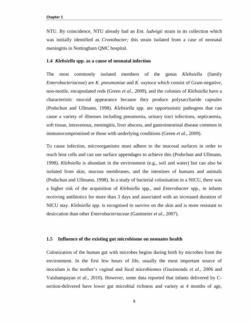
sources (Brady, 2005). Evidence supporting each of these ways is given below (Figure
1.1):
Chapter 1
11
Figure 1.1: Sources of transmission of bacteria within a NICU setting.
1.6.1 Bacterial exposure of neonates human breast milk
Nutrition is necessary to the health and growth of infants and children. Breastfeeding is
the best nutritional choice for infants, superior to infant formula feeding, as breast milk
possesses immune factors that are effective for long term protection against infections,
diseases and influences metabolism later in life (Oddy, 2001). In the NICU, most
mothers are providing their breast milk to feed their premature infants (Morales and
Schanler, 2007). However, breast milk has been recognized as the source of numerous
bacterial infections in neonates (Qutaishat et al., 2003; Revathi et al., 1995), including
Salmonella (Anonymous, 1978; Ryder et al., 1977). Several clinicians request bacterial
cultures of milk from the mother for screening before allowing the milk to be fed to
neonates (Ng et al., 2004). A previous in-vitro study by Lenati et al. (2008) found that
Cronobacter spp. was resistant to the antimicrobial properties of breast milk, for more
details see Figure 1. 11.
In the last few years, there have been many studies which have demonstrated that breast
milk is a prevalent source of bacteria to the infant gut and plays a role in the initiation
Chapter 1
12
and growth of gut microbiota. Staphylococci, streptococci, lactococci, lactobacilli and
enterococci were isolated more frequently from this biological fluid in healthy women
(Heikkila et al., 2003; Martín et al., 2003).
Sometimes transmission of serious bacterial diseases to the neonate can occur through
breast milk, for example, Salmonella species (Qutaishat et al., 2003), group B
streptococcus (Kotiw et al., 2003), Listeria species (Svabic-Vlahovic et al., 1988),
methicillin-resistant St. aureus (Gastelum et al., 2005), and Mycobacterium tuberculosis
(Pronczuk et al., 2002), which is the most common species documented in cases of
infection by this route, Figure 1.1
The report by Widger et al. (2010) states that three cases of late onset neonatal
septicaemia were investigated in their unit, including one that resulted in death, which
were more likely to be caused by contaminated expressed breast milk. Another report
demonstrated that the contamination of breast milk has been causing fatality in neonates,
but these have been related to concurrent mastitis (Gastelum et al., 2005; Kotiw et al.,
2003). Recent study by Urbaniak et al. (2014) informed that Enterobacteriaceae were
isolated from breast tissue of women with and without cancer in Canada and Ireland. The
women were not had any symptoms of infection, however the viable of bacteria was
confirmed in some samples by culture.
1.6.2 Bacterial exposure of neonates through powdered infant formula
Recently, the Food and Agriculture Organization/World Health Organization (FAO/
WHO, 2004 and 2006) have focused attention on the microbiological safety of PIF. The
considerable concern for PIF was due to neonatal infections through Cronobacter and
Salmonella that were associated with contaminated PIF (Caubilla-Barron et al., 2007;
Iversen et al., 2008). PIF are not sterile however, they comply with the international
microbiological standards (Codex Alimentarius Commission 2008). Furthermore, the
report by US Centers for Disease Control and Prevention (CDC) report that in a NICU,
an outbreak of C. sakazakii infection was associated with the use of powdered infant
formula (CDC, 2002). The report also emphasised that infection through this organism
often leads to a fatal disease, highlighting the significance of careful preparation,
handling and use of infant formula products in the health care setting, Figure 1.1
Chapter 1
13
Many types of bacteria have been isolated from PIF and the most common organisms
isolated from PIF were Clostridium perfringens, St. aureus, Bacillus cereus and
Enterobacteriaceae (Forsythe, 2005). Enterobacteriaceae including Ent. cloacae,
K. pneumoniae, K. oxytoca, Ent. hormaechei, Cit. freundii, and E. coli have been
isolated from PIF (Muytjens et al., 1988; Townsend et al., 2008a). These organisms have
been categorised by the FAO/WHO (2004 and 2006) as category B which is 'causality
plausible, but not yet demonstrated' with consideration of their potential
to cause neonatal infections via the ingestion of reconstituted PIF. Though
these organisms are opportunistic pathogens, there was no evidence to confirm any
outbreaks in NICUs associated with their presence in contaminated PIF. The most
common organisms isolated from PIF were Cl. perfringens, St. aureus, Bacillus cereus
and Enterobacteriaceae (Forsythe, 2005).
There are approximately 2,000 serovars of the Salmonella bacterium that can cause
disease in humans, and symptoms include diarrhoea, fever and vomiting which can cause
serious infection in infants (Crawley and Westland, 2012). A previous study by
Rodríguez-Urrego et al. (2010) in 2008 in Spain reported that 31 cases of Salmonella
infection in infants occurred because of infant formula contamination, and 10 of these
infants needed hospitalisation.
Powdered infant formula is made from pasteurized liquid and then spray dried. However,
it is more likely that, the contamination of formula occurs from the preparer or
preparation environment than from the manufacturing procedure (FAO/WHO 2004). The
previous study by Weir (2002) recommended that in NICUs, the preparation of PIF
should be carried out following the manufacturer’s instructions by trained people under
clean and aseptic conditions in a designated area. The FAO/WHO (2004) recommended
that reconstitution of powdered infant formula with water >70°C, decreasing the time
between reconstitution and feeding (< 2 hours), and not storing reconstituted feed at
ambient temperature could reduce the risk of bacterial infection.
1.6.3 Bacterial exposure through neonatal feeding tube
In NICUs, enteral feeding tubes are commonly used to feed infants who are unable to
swallow milk or formula. As per other medical inserting devices, EFT have to be clean
Chapter 1
14
and sterile. Recent studies at NTU in collaboration with local NICUs revealed that
organisms are present in EFT from neonates on non-formula feeding regimes (Hurrell et
al., 2009b). This indicates that neonate exposure to Cronobacter may not be exclusively
through reconstituted formula, and that increased exposure to bacterial pathogens may be
linked to more general feeding practices, Figure 1.1.
Hurrell et al. (2009a and b) studied 129 neonatal enteral feeding tubes from two local
NICUs. They found that irrespective of feeding regime, neonatal enteral feeding tubes
can act as loci for the bacterial attachment and multiplication of a wide range of
Enterobacteriaceae and, hence, revealed a potentially greater risk to neonatal health
than Cronobacter alone. The organisms frequently isolated included E. coli,
Ent. cancerogenus, Ent. hormaechei, K. pneumoniae, K. oxytoca, Raoutella spp.,
Se. liquefaciens and S. marcescens. Additionally, Cronobacter spp. and Yersinia
enterocolitica were recovered. All S. marcescens strains were resistant to amoxillin and
co-amoxiclav.
Of additional importance was that a quarter of Ent. hormaechei isolates were resistant to
the 3rd
generation cephalosporins ceftazidime and cefotaxime. During the period of the
study, K. pneumoniae and S. marcescens caused infections in the two NICUs and ESBLs
were detected in two of these strains (Hurrell et al., 2009b).
Hurrell et al. (2009b) isolated bacteria from EFT used by infants on two NICUs. The
infants had received different feeding regimes: reconstituted PIF, breast milk, fortified
breast milk, ready to feed formula and a mixed feeding regime. A nil by mouth group
was selected as a control cohort. Enterobacteriaceae was isolated from 76% of the
enteral feeding tubes. The highest Enterobacteriaceae biofilm was isolated from infants
fed reconstituted PIF; an average 4.2 log10 cfu/tube. In fact, the PIF was reconstituted at
room temperature and not with water at >70°C, as recommended by FAO/WHO. These
bacteria could multiply while feeding on the formula during the time the tube was in
place (48 hours) and because, unlike breast milk, PIF contains no antibacterial agents.
The study found that the lowest levels of bacteria were obtained from the EFT of infants
fed breast milk 52%, whereas the others ranged from 78 to 88% for mixed feeding
regime and reconstituted PIF. Bacteria were obtained from the EFT of infants fed breast
Chapter 1
15
milk (maximum of 5.3 log10cfu/tube) and infants that were nil by mouth had a maximum
value of 2.7 log10 cfu/tube. The maximum value of breast milk was higher than the other
neonates in the group and may be due to the high near neutral gastric pH (6.0).
Enterobacteriaceae biofilms were recovered from 81% of neonates fed with sterile
ready-to-feed formula. It is thought that the bacteria may have originated from the throat
due to gastroesophageal reflux.
In parallel studies, Hurrell et al. (2009a) showed in situ that bacteria can grow in the
nasogastric tubes to cell densities of 107 colony forming units/tube within 8 h, and
109cfu/tube within 24 hours. It is plausible that in vivo the biofilm will both inoculate
subsequent routine feeds (every 2-3 hours) and as the biofilm ages, clumps of cells will
detach and survive passage through the neonate’s stomach. Subsequently, these
organisms will enter the stomach as a bolus with each feed.
A major conclusion of this study was that the microbiological safety of neonatal feeds
should not exclusively focus on reconstituted infant formula, and Cronobacter spp., but
also on the general preparation and practices of enteric feeding to reduce the risk of
exposure to other Enterobacteriaceae some of which may carry antibiotic resistance
factors. Therefore, the practice of prolonged placement of EFT in neonates needs to be
considered with respect to the increased risk of exposure to bacterial pathogens. Hence,
biofilm formation on EFT is an important risk factor to consider with respect to neonatal
infections (Hurrell et al., 2009a and b). The sources of these potentially pathogenic
Enterobacteriaceae need to be evaluated.
1.6.4 Bacterial exposure of neonates health-care workers or immediate
environment
As Shown in Figure 1.1, healthcare workers and patients are the most possible sources of
infectious agents and are also the most common susceptible hosts. Residents in NICU
can also become infected with health care-associated infections by horizontal
transmission of microorganisms spread through aerosol or contact (direct or indirect)
transmission. Despite at the present time, the enhanced safety of blood products
administered in hospitals, the frequent use of blood products in the stabilization of
critically ill newborns allows for the potential transmission of bloodborne pathogens,
Chapter 1
16
currently identified and those yet to be identified (Yeager, 1974 and Saulsbury et al.,
1987). Likewise, Transmission of infections on contaminated medical devices is also
potential and outbreaks of hospital-acquired infections have been associated to devices
such as electronic thermometers, blood pressure cuffs, stethoscopes, latex gloves, masks,
neckties, pens, badges and lanyards, and white coats (WHO, 2009; Uneke et al., 2008;
Treakle et al., 2009).
Some studies have found that pathogens bacteria can be transmitted from out-of-hospital
sources to patients through health-care workers’ hands. For instance, an outbreak of
postoperative Se. marcescens wound infections was refer to a contaminated jar of
exfoliant cream in a nurse’s home. An investigation suggested that the bacterial was
transmitted to patients by the hands of the nurse who wore artificial fingernails (Passaro
et al., 1997). Se. marcescens was transferred from contaminated soap to patients through
health-care wokers’hands (Sartor et al., 2000).
Multidrug-resistant bacteria cause a important ratio of hospital-associated infections
(Hidron et al., 2008; Morgan et al., 2010). These organisms are generally transmitted
from patient-to-patient in the healthcare system via transiently contaminated healthcare
workers, equipment, and the environment (Siegel et al., 2007). An outbreak of
multidrug-resistant Acinetobacter baumannii, strains from patients, healthcare workers’
hands, and the environment were identical (El Shafie et al., 2004).
Health-care waste is considered as a source of pathogenic microorganisms, that can
cause contamination and give increase to infection. These microorganisms can be
transmitted by direct contact, in the air, or through a diversity of vectors (Pr ss, ).
1.7 Commonly used infant formula
The World Health Organization (WHO, 2003) and health departments across the
developed and developing world recommend breastfeeding for feeding infants in the first
six months of life as the best feeding method (Crawley and Westland, 2012). Infant
formulas as breast milk substitutes are available for mothers who cannot or choose not to
breastfeed. However, infant formulas are an artificial substitute for breast milk having
inherent differences (Renfrew et al., 2012) such as the exact chemical makeup of breast
Chapter 1
17
milk is still unknown and cannot be manufactured. Breast milk contains mother’s
antibodies and numerous other defensive factors. These mature factors from the mother’s
immune system help the baby’s immature immune system to fight against infection. In
addition, infant formula requires industrial storage and delivery systems with important
quality control problems. Furthermore, infant formula has no positive influence on
maternal health (Crawley and Westland, 2012) whereas breast feeding leaves a life-long
impact on the mother’s health.
The essential components of any infant milk, irrespective of the format (powder or
ready-to feed), are proteins, fats, carbohydrates, vitamins and minerals. The major infant
formula manufacturers improve their own brands with a combination of each of these
components. The essential nutritional properties of the majority of infant formula are
very similar. Most infant formula are based on modified cows’ milk (skimmed or full-fat,
liquid or powder, or using whey protein concentrates) by adding lactose or other
carbohydrates, vegetable and other oils, vitamins and minerals (Crawley and Westland,
2012).
Infant formula manufacturers have modified cow’s milk protein (whey based) or first
milks by adjusting the casein to whey ratio which is similar to breast milk (40:60). Infant
formula are suitable from birth which can be either 'whey-based' or 'casein-based'. The
whey-based infant formula may also have a relatively poor mineral content, specifically
sodium and potassium which are required for new-born babies who have immature
kidneys.
In addition, whey-based infant formula are usually the first choice if a mother does not
breast-feed. While in casein-based infant formula the ratio of casein to whey is the same
as cows' milk (80:20), casein-based infant formula are normally used if the baby seems
not to be satisfied on a whey based product. Also, it can be helpful to delay the
introduction of solids before the recommended age up to 4 to 6 months (Forsythe, 2009).
Other infant formula are based on soy protein from soya beans which is nutritionally
similar to cows' milk formula. Soya infant formula is formulated for infants who cannot
tolerate cows' milk because they have an allergy to the proteins in cow’s milk and/or are

Chapter 1
18
unable to digest the lactose in cows' milk. Usually, glucose syrups are used in soya infant
formulas as a source of carbohydrates. This type of formula is used by parents who wish
to feed their baby with a vegetarian product (Forsythe, 2009). The compositions of based
infant formula are given in Table (1.2).

Chapter 1
19
Table 1.2: The compositions of based infant formula.
Nutrition Whey Casein Soya
Lactose Lactose Glucose syrup
Vegtable oils (contains soya lecituin) Vegtable oils Vegetable oils
skimmed milk skimmed milk _
Demineralised water water _
_ Emulsifier (monoglycerides of fatty
acids and soya lecithin)Emulsifler (soya lecithin)
Whey protin concentrate _ Soya protein
Galacto-oligosaccharids (GOS) _ _
Fructo-olgosaccharids (FOS) _ _
Fish oil _ _
Gluconate _ _
Thiamin (B1) Thiamin (B1) Thiamin (B1)
L-carnitine L-cysteine L- carnitine
Niacin (B3) Niacin (B3) Niacin (B3)
Taurine Taurine Taurin
Biotin Biotin Biotin
Folic acid Folic acid Folic acid
Riboflavin (B2) Riboflavin (B2) Riboflavin (B2)
Vitamin A Vitamin A Vitamin A
Vitamin B6 Vitamin B6 Vitamin B6
Vitamin B12 Vitamin B12 Vitamin B12
Vitamin C Vitamin C Vitamin C
Vitamin D3 Vitamin D Vitamin D3
Vitamin E Vitamin E Vitamin E
Vitamin K1 Vitamin K Vitamin K1
Pantothenic acid Pantothenic acid Pantothenic acid
Potassium Chloride Potassium chloride Potassium chloride
Copper Copper sulphate Copper sulphate
Magnesium Carbonate Magnesium Carbonate Magnesium hydrogen phosphate
Zinc sulphate Zinc sulphate Zinc sulphate
Maganese sulphate Manganese sulphate Manganese sulphate
Choline chloride Choline Chloride Choline Chloride
Potassium iodide Potassium iodide Potassium iodide
Sodium selenite Sodium selenite Sodium selenite
Inositol Inositol Inositol
Cytidine -5`-monophosphate Cytidine- 5`-monophosphate _
Guanosine -5`-monophosphate
disodium salt
Guanosine-5`-monophosphate
disodium
_
Uridine -5`-monophosphate disodium
salt Uridine -5`-monophosphate disodium
_
Adenosine -5`-monophosphate
disodium salt Adenosine -5`-monophosphate
_
Inosine -5`-monophosphate disodium
salt Inosine -5`-monophosphate disodium
_
Sodium Chloride Sodium bicarbonate _
Potassium hydroxide Potassium hydroxide _
Calcium Citrate Sodium citrate Sodium Citrate
_ Ferrous sulphate _
Calcium hydroxide _ Calcium Carbonate
Iron lactate _ Iron Sulphate
Calcium Phosphate _ Calcium phosphate
Potassium Citrate _ Potassium citrate
Acid regulator (Citric acid) _ _
Minerals
Ingredients of based infant formula
Major nutrients
Vitamins
Chapter 1
20
1.8 Conditions and concerns in the NICU
Premature babies are those born before 37 weeks of gestation (or before the 37th week of
pregnancy). They are at risk for a variety of problems such as breathing, feeding,
difficulties or both, and infections. Thus, they need special medical care that only exists
within NICUs or Special Care Nurseries (SCNs) (American Academy of Pediatrics,
2014). Furthermore, for premature infants, feeding by mouth is not safe and sometimes
not possible because of neurological immaturity or respiratory problems. Milk in such
infants is usually given through a nasogastric feeding tube. Enteral feeding is generally
preferred over parenteral nutrition in the first few days of life and it helps promote
endocrine adaptation and the maturation of bowel motility (McGuire et al., 2004).
The WHO guidelines for hygienic preparation of PIF plan to decrease the number of
bacteria in the reconstituted product by using hot water for preparation and a short time
storage for any surviving organisms to grow (Codex Alimentarius Commission, 2008).
However, a broader perspective is that neonates are frequently fed by using enteral
feeding tubes. These tubes are commonly placed for long periods of time (more than
several days) to decrease distress to the neonate by gag reflexes (Holý and Forsythe,
2014).
However, Enterobacteriaceae can adhere to and colonize tubes at 37°C receiving fresh
feeds at regular intervals (Hurrell et al. 2009a). This is related to all neonates with
inserted nasogastric tubes, and not only for those who are receiving reconstituted PIF. In
fact, Enterobacteriaceae have been isolated from nasogastric tubes from neonates
receiving breast milk and other feeding regimes at a rate ≥ 107 cfu/tube (Hurrell et al.
2009a). Thus, regardless of the type of feed the hygienic practices and avoidance of
temperature abuse are important and vital (Holý and Forsythe, 2014).
The recommendation for reconstitution of PIF with hot water (>70°C) by the FAO/WHO
is not followed in all countries (e.g. USA). To avoid the contamination problems when
dipping a thermometer into the reconstituted formula, the advice in the UK is to use the
water that has been boiled in a kettle and left to cool for 30 minutes. With consideration
of the difference in cooling curves depending on the volume of water and kind of kettle,
Chapter 1
21
this is impractical for premature babies who only need small volumes of formula, and are
fed at 2-hourly intervals.
The PIF includes breast milk fortifiers which are added to complement the nutritional
value of breast milk. These products are not reconstituted with water and cannot be
exposed to heat treatment to kill intrinsic bacteria (Holý and Forsythe, 2014).
Recently, specific UK national guidelines (under consultation) on the prevention and
management of Gram-negative sepsis in neonates were developed because of a NICU
outbreak in the UK (Wise, 2012).
1.9 Bacterial mechanisms of pathogenicity
Pathogenic bacteria are able to cause disease in human hosts using a number of
mechanisms. Bacterial pathogens have numerous molecules that facilitate attachment to
host cell targets, causing a diversity of different host responses. The molecular
mechanisms through which bacteria can interact with the host can be unique to specific
pathogens or conserved across several different species. The identification and
characterisation of all these different strategies is the key to fighting bacterial diseases.
This study focuses on mechanisms used by pathogenic bacteria to cause infectious
disease.
1.9.1 The interaction of bacteria with mammalian cells
The colonisation of intestinal bacteria on the surface of intestinal walls is one of the
basic ways bacteria are able to interact with mammalian cells. The intestinal walls of
newborn babies are sterile, but are quickly colonised by bacteria entering the body with
food and from the environment. E. coli is one of the first microorganisms which
colonises the surface of intestinal walls of newborn babies (Grajek and Olejnik, 2004).
Nevertheless, the microorganisms on the host epithelium can be affected by diverse
factors, for instance, nutrition, antibiotic treatment, environmental exposure to
microorganisms, and microbial colonization in the neonatal period (Lindhorst and
Oscarson, 2009; Abraham and Medzhitov, 2011).
Chapter 1
22
The gastrointestinal tract also has innate defence mechanisms that act against microbial
survival and dissemination inside the host by washing microbes from surfaces,
consisting of epithelial integrity, the rapid regeneration of old epithelial cells, fast
propulsion of infected cells, autophagy, and innate immune responses (Kim et al., 2010).
However, bacteria can invade epithelial cells and cause infections (Townsend et al.,
2008b; Kim et al., 2010). Besides that, the organisms are able to form biofilms which
possibly protect cells from environmental stresses and antimicrobial agents (Kim et al.,
2008).
The preterm babies have a stratum corneum which is immature and thin as compared to
that found in full term babies, children or adults. The mature stratum corneum consists of
between 10 and 20 layers of cells, whereas the stratum corneum of preterm babies less
than 30 weeks may have only 2 to 3 layers, which could increase the chance of invasion
by harmful microbes (Evans and Rutter, 1986 and Hoeger, 2006). However, many
diverse bacterial pathogens have the ability to adhere, invade, and cause damage to host
cells and tissues, in addition to surviving the host defences and causing an infection
(Wilson et al., 2002). Furthermore, several bacterial pathogens are able to secrete
various components and toxins to avoid the defences of immune systems (Kim et al.,
2010). To avoid these factors, pathogenic bacteria are also capable of creating an
antiphagocytic surface layer which consists of polysaccharides or by secreting their
adhesins on polymeric structures which extend out from the cell surface (Kline et al.,
2009).
The infection of the human intestine by enteropathogenic bacteria such as E. coli,
Sal. enterica, Shigella flexneri, and Y. enterocolitica is based on the capability of
bacteria to adhere, invade and colonize the intestinal epithelium, survive intracellularly
and be transmitted from cell to cell (Reis and Horn, 2010).Two different strategies are
used by these bacteria to cause an infection in the host; firstly these bacteria are able to
resist the host attack through macrophages by releasing the T3SS system proteins.
Secondly they are able to survive within macrophages because of their ability to induce
changes within the intracellular environment of the cell (Reis and Horn, 2010;
Sansonetti, 2002).
Chapter 1
23
Bacteria can interact with the host cells in two ways: attachment to the host cell without
any modification to the membrane or by using the surface appendages such as microvilli
(Greiffenberg et al., 2000). In Gram-negative bacteria, the outer membrane is an
important factor which helps in the attachment of the bacteria with the external
environment (Mogensen and Otzen, 2005).
Adhesion to mammalian cells 1.9.1.1
The expression of adhesive molecules on the surfaces of bacteria is an essential virulence
factor. Most bacterial pathogens use adhesion to facilitate their interaction with host
tissue cells. These surfaces include skin, mucous membranes, such as oral cavity,
nasopharynx, urogenital tract, and deeper tissues; lymphoid tissue, gastric and intestinal
epithelia, alveolar lining, endothelial tissue. The host acts to wash microbes from these
surfaces by producing various mechanical forces, such as saliva secretion, coughing,
sneezing, mucous flow, peristalsis, and blood flow (Wilson et al., 2002).
Microbial adherence factors can be produced from polypeptides (proteins) or
polysaccharides (carbohydrates or sugars) which form the capsule. Protein adhesins are
divided into two categories: fimbrial and afimbrial. Fimbriae (pili) are appendages that
protrude from the bacterial surface and consist of proteins that are tightly packed into an
array as a helical cylinder. In particular, Gram-negative bacterial pathogens depend
on fimbriae for adherence such as E. coli (for both urinary tract infections and
gastroenteritis), Vibrio cholerae, Pseudomonas aeruginosa, and Neisseria species
(Donnenberg, 2000; Hahn, 1997).
In afimbrial adhesins made of proteins that are known as adherence factors, the contact
of afimbrial adhesins with the host cell is more intimate and occur over a shorter domain
than with fimbriae. Gram-negative (Y. pseudotuberculosis, enteropathogenic E coli,
Neisseria spp.), Gram-positive (Staphylococcus spp. and Streptococcus spp.) and
mycobacterial pathogens express afimbrial adhesins (Donnenberg, 2000; Merz and So,
2000; Joh et al., 1999; Bermudez and Sangari, 2000). The interactions between bacterial
and the mucosa host such as adherence and invasion are commonly the cause of bacterial
disease in the gut (Hu and Kopecko, 2008).
Chapter 1
24
Invading mammalian cells 1.9.1.2
Adhesion of bacteria to host surfaces is the first step in the pathogenesis of most
infections in man and animals. Some pathogens have deeper access into the host to
continue the infection cycle. This pathogenic strategy can be divided into two categories:
extracellular and intracellular (Wilson et al., 2002).
Extracellular invasion happens when a microbe breaks down the barriers of a tissue to
distribute enzymes inside the host and the microbe remains outside of host cells. This is
a strategy used by several pathogenic bacteria such as group A β-haemolytic
Streptococcus and St. aureus which produce toxins such as haemolysin (Walker, 1998).
Haemolysin are divided into three different types, α-hemolysin, β-hemolysin and γ-
hemolysin. Clinical isolates, such as E. coli and Serratia spp. are able to secrete α-
hemolysin (König et al., 1987; Welch, 1987; Schmidt et al., 1995). The haemolysins
expressed by these species have the capability to destruct cells however, other cell types
may also contribute to their spread in host tissues (Wilson et al., 2002).
The ability of microbes to penetrate the cells of a host tissue and survive within this
situation is known as intracellular invasion. Numerous Gram-negative, Gram-positive,
and mycobacterial pathogens have the capability to invade host cells (Finlay and Falkow,
1997; Cleary and Cue, 2000; Bermudez and Sangari, 2000; Dehio et al., 2000).
Cronobacter strains have the ability to attach and invade Caco-2 human epithelial cells,
and invade rat brain capillary endothelial cells (Townsend et al., 2008b).
Intracellular lifestyles 1.9.1.3
After invasion, many bacterial pathogens have the ability to survive and replicate within
host cells which survive invasion such as epithelial, endothelial and macrophages. The
host cells have mechanisms to damage ingested bacteria which include the creation of
reactive oxidative intermediates, the decrease of pH vacuoles that contain bacteria, and
the activation of destruction proteases. However, bacteria have the ability to avoid these
killing mechanisms, such as survival in low acidity (Wilson et al., 2002). For example,
Adherent-invasive E. coli (AIEC) isolated from Crohn's disease patients was able to
replicate within macrophages in large vacuoles (Bringer et al., 2006). Bacteria that
Chapter 1
25
survive intracellularly are possibly able to replicate and transmit between cells in the
same area of infection or transfer to other areas of the body (Wilson et al., 2002).
1.10 Biofilm formation
The biofilm was defined as "an assemblage of microbial cells that adhere to abiotic or
biotic surfaces. Also, biofilm known as irreversibly associated (not removed by gentle
rinsing) with a surface and enclosed in a matrix of primarily polysaccharide material"
(Donlan, 2002; Davey and O’Toole 2000). In Gram-negative bacteria, several of the
polysaccharides are neutral or polyanionic. The existence of ketal-linked pyruvates or
uronic acids enhances their anionic properties, thus it allows association of divalent
cations such as calcium and magnesium to increase the binding force in a developed
biofilm (Donlan, 2002; Sutherland, 2001a).
Biofilm protect microorganisms from shear forces and low pH environments such as in
the stomach. Inside the host, biofilm gives microorganisms a competitive advantage due
to its protects biofilm bacteria from exposure to innate immune defences (such as
opsonization and phagocytosis) and antibiotic treatments antibiotics (Scher et al., 2005).
The process of biofilm formation is characterized by several stages that have been
identified include in Figure 1.2.
The adhesion surface is pre-conditioned by the macromolecules which are present in the
bulk liquid, 2. The planktonic cells in the bulk liquid are transferred to the surface, 3.
Cells are adsorbed on the surface, 4. Those cells which are adsorbed reversibly are
described, 5. Some bacterial cells adsorb irreversibly on the surface, 6. Cell-cell
signalling molecules are produced, 7. Transfer of substrate to and within the biofilm, 8.
Cells bound to the biofilm metabolise the substrate and transfer the products out of the
biofilm, 9. All of these processes occur in parallel with EPS production, replication and
cell growth (Simo˜es et al., 2010).
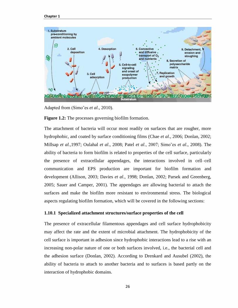
Chapter 1
26
Adapted from (Simo˜es et al., 2010).
Figure 1.2: The processes governing biofilm formation.
The attachment of bacteria will occur most readily on surfaces that are rougher, more
hydrophobic, and coated by surface conditioning films (Chae et al., 2006; Donlan, 2002;
Millsap et al.,1997; Oulahal et al., 2008; Patel et al., 2007; Simo˜es et al., 2008). The
ability of bacteria to form biofilm is related to properties of the cell surface, particularly
the presence of extracellular appendages, the interactions involved in cell–cell
communication and EPS production are important for biofilm formation and
development (Allison, 2003; Davies et al., 1998; Donlan, 2002; Parsek and Greenberg,
2005; Sauer and Camper, 2001). The appendages are allowing bacterial to attach the
surfaces and make the biofilm more resistant to environmental stress. The biological
aspects regulating biofilm formation, which will be covered in the following sections:
1.10.1 Specialized attachment structures/surface properties of the cell
The presence of extracellular filamentous appendages and cell surface hydrophobicity
may affect the rate and the extent of microbial attachment. The hydrophobicity of the
cell surface is important in adhesion since hydrophobic interactions lead to a rise with an
increasing non-polar nature of one or both surfaces involved, i.e., the bacterial cell and
the adhesion surface (Donlan, 2002). According to Drenkard and Ausubel (2002), the
ability of bacteria to attach to another bacteria and to surfaces is based partly on the
interaction of hydrophobic domains.
Chapter 1
27
Numerous cells are able to produce extracellular filamentous appendages, which may
play a role in the attachment process. In fact, many of such structures are known to
present – flagella, pili or fimbriae, prothecae, stalks and hold-fast (Harbron and Kent,
1988).
Flagella are responsible for the motility of bacteria. Numerous types of bacteria have
flagella. It is probable that the flagellum itself may form an adhesive bond with the
adhesion surface (Harbron and Kent, 1988).
The primary function of flagella in biofilm formation is supposed to be in transport and
in initial cell–surface interactions (Sauer and Camper, 2001). Flagella-mediated motility
is assumed to overcome repulsive forces at the surface of the substratum and, as a result,
a monolayer of cells forms on the adhesion surface (Daniels et al., 2004).
Pili or fimbriae are found on several Gram-negative bacteria including Pseudomonas
species. They are fine, filamentous appendages ranging in width from 4–35 nm and in
length up to several micrometers (Harbron and Kent, 1988). These two structures are
usually straight, and are not involved in motility. Their function is only known to make
cells more adhesive, because bacteria with pili can adhere strongly to other bacterial
cells and inorganic particles (Harbron and Kent, 1988). According to Sauer and Camper
(2001), pili and pilus-associated structures have been appeared to be significant for the
adhesion to and colonization of surfaces, possibly by overcoming the initial electrostatic
repulsion barrier that are present between the cell and the substratum.
The expression of fimbria is variable between pathogenic and environmental isolates.
(Gerstel and Römling 2001) establish that phosphate and nitrogen depletion are signals
which able to switch on the agfD promoters in the stationary phase in Salmonella
typhimurium. Fimbria expression has been correlated with strain pathogenicity. It was
discovered in 100% of E. coli responsible for sudden infant death syndrome, and also 55%
of bovine mastitis isolates, and 38% of E. coli non O157 STEC (Olsén et al., 1989;
Goldwater and Bettelheim, 2002; Cookson et al., 2002). Zogaj and researchers detected
the production and genes responsible for cellulose and fimbria using Citrobacter spp.
Enterobacter species and Klebsiella species. Their results also display that Citrobacter
Chapter 1
28
species are capable to produce curli fibers but Enterobacter and Klebsiella species are
unable to do. On the other hand, they discovered that genes responsible for curli fibers
production were existent in Citrobacter species and Enterobacter species but not in
Klebsiella species (Zogaj et al., 2003). Ten fimbrial related genes were present in all C.
sakazakii and 38% of C. malonaticus, but were absent in other Cronobacter species
(Healy et al., 2009).
A number of genes have been detected as requirements for the formation of curli fibers.
The previous study by Zogaj et al (2003) has isolated csg genes from some species
belong to Enterobacteriaceae such as Citrobacter, Cronobacter and Klebsiella spp.
Furthermore, Sakellaris et al. (2000) isolated them from Shigella spp. The expression of
csg genes is not only associated with curli expression but they are connected also with
bacterial resistance to environmental factors such as desiccation. The csg genes were
detected in strains which were unable to express curli fimbria; also not all Salmonella
strains and E. coli are capable to express curli fimbria even under suitable environmental
conditions (Zogaj et al., 2003).
The essential regulator of csgBAC is possibly the transcriptional regulator csgD. The
increase of biofilm formation, invasion and bacterial virulence factors have been
associated with the rise of csgD gene activity, being strongly controlled with genetic and
environmental factors (Uhlich et al., 2002). It has been determined that csgD operon is
being influenced by regulators that are grouped as csgDEFG and csgBA as divergent
operons (Hammar et al., 1995). Gualdi and researchers (2008) found that overexpression
of csgD can overcome temperature dependent control of curli encoding csgBA operon
1.10.2 Extracellular polymeric substances (EPS)
EPS are responsible to bind cells and other particulate materials together (cohesion) and
to the surface (adhesion) (Allison, 2003; Sutherland, 2001b). The EPS comprises of
polysaccharides, proteins, nucleic acids, lipids, phospholipids and humic substances
(Jahn and Nielsen, 1998; Sutherland, 2001b; Wingender et al., 1999). Proteins and
polysaccharides amount for 75–89% of the biofilm EPS composition, indicating that
they are the predominant components (Tsuneda et al., 2003).
Chapter 1
29
Biofilms form a gel phase around microorganisms which live inside it (Sutherland,
2001a; Wingender et al., 1999). The EPS matrix functions as a barrier in which diffusive
transport prevails over convective transport (Sutherland, 2001b). EPS has general
protective effect on biofilm forming microorganisms against adverse conditions, for
example, it has frequently been shown that biofilm cells can tolerate high concentrations
of biocides (Mah and O’Toole, 200 ; Simo˜es and Vieira, 200 ; Simo˜es et al., 2005).
The EPS matrix acts to delay or prevent antimicrobials from reaching target bacteria
within the biofilm by diffusion limitation and/or chemical interaction with the
extracellular proteins and polysaccharides (Heinzel, 8; Mah and O’Toole, 200 ).
Furthermore, inside the EPS matrix the molecules necessary for cell–cell communication
and community behaviour may accumulate at concentrations high enough to be effective
(Pearson et al., 1999; Sutherland, 2001b).
Lipids and nucleic acids might have important effect on the rheological properties and
thus the stability of biofilms (Neu, 1996). The extracellular DNA is required for the
initial formation of biofilms by Pseudomonas aeruginosa and probably for biofilms
formed by other bacteria that specifically release DNA (Whitchurch et al., 2002).
Colonic acid and cellulose are essential EPS components to form biofilm and have a
similar influence on bacterial attachment to mammalian cells. Matthysse et al. (2008)
reported that the ability of E. coli O157:H7 to attach to plastic surfaces as well as alfalfa
sprouts and Caco-2 cell lines based on EPS. They used E. coli O157:H7 with deletions of
pgaC, yhjN and wcaD the genes encoding for poly-ß-1,6-N-acetylglucosamine, cellulose
and colonic acid respectively. Their result reported that the attachment of E. coli
O157:H7 to alfalfa sprouts was blocked in the pgac mutant; the attachment was
decreased 100 and 10 fold compared to the wild type strain for yhjN mutants and was
reduced 1,000 and 100 fold for wcaD mutants. The attachment to Caco-2 cell line was
significantly reduced for pgaC and yhjN mutants but not for wcaD mutant. The E. coli
O157:H7 strains were not capable to form significant biofilm on PVC in LB agar.
Mutation in the polysaccharides genes pgaC, wacaD and yhjO were capable to form
third of the amount of biofilm compared to the wild type (Matthysse et al., 2008).
Chapter 1
30
1.10.3 Cell–cell communication
Cell–cell signalling plays an important role in cell attachment and detachment from
biofilms (Daniels et al., 2004; Donlan, 2002). Bacteria are considered to be colonial by
nature and exploit elaborate systems of intercellular interactions and communications to
enable their adaptation to altering environments (Davies et al., 1998; Fuqua and
Greenberg, 2002; Sauer and Camper, 2001). Accordingly to Daniels et al. 2004 the
successful adaptation of bacteria to changing natural conditions is based on their
capability to sense and respond to the external environment and modify gene expression.
Quorum sensing is a process of cell–cell communication that based on the process of
autoinduction (Eberhard et al., 1981). The process of quorum sensing acts to provide a
mechanism for self-organization and regulation of microbial cells (Parsek and Greenberg,
2005). It includes an environmental sensing system that allows bacteria to monitor and
respond to their behaviour according to population density.
1.11 Siderophores
Bacterial pathogens require iron for survival and a diverse range of metabolic processes
including amino acid synthesis, oxygen transport, respiration, nitrogen fixation,
methanogenesis, the citric acid cycle and DNA biosynthesis (Sandy and Butler, 2009).
The iron in human cells is very low and is bound by lactoferrin, transferrin, and ferritin
(Brooks et al., 2004). Pathogenic bacteria synthesize and secrete small organic
molecules called siderophores, which are low-molecular-weight chelators that solubilize
and acquire iron from eukaryotic iron-binding proteins such as lactoferrin and transferrin
(Ratledge and Dover, 2001). Thus, siderophores are considered as major virulence
factors in the pathogenicity of organisms (Eijkelkamp et al., 2011).
The majority of aerobic and facultative anaerobic microorganisms produce at least one
siderophore (Neil, 1995). So far nearly 500 siderophores have been found from selected
microorganisms and several important siderophores produced by bacteria are presented
in Table 1.3 (Balagurunathan and Radhakrishnan, 2007).
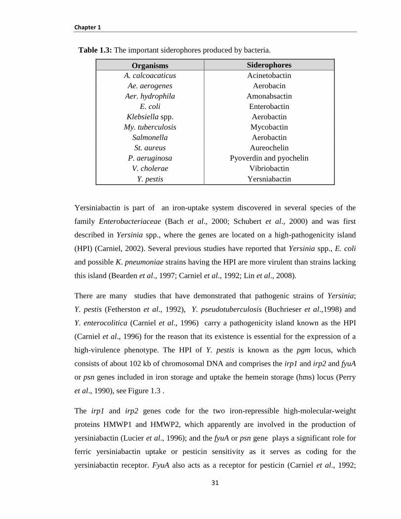
Chapter 1
31
Table 1.3: The important siderophores produced by bacteria.
Organisms Siderophores
A. calcoacaticus Acinetobactin
Ae. aerogenes Aerobacin
Aer. hydrophila Amonabsactin
E. coli Enterobactin
Klebsiella spp. Aerobactin
My. tuberculosis Mycobactin
Salmonella Aerobactin
St. aureus Aureochelin
P. aeruginosa Pyoverdin and pyochelin
V. cholerae Vibriobactin
Y. pestis Yersniabactin
Yersiniabactin is part of an iron-uptake system discovered in several species of the
family Enterobacteriaceae (Bach et al., 2000; Schubert et al., 2000) and was first
described in Yersinia spp., where the genes are located on a high-pathogenicity island
(HPI) (Carniel, 2002). Several previous studies have reported that Yersinia spp., E. coli
and possible K. pneumoniae strains having the HPI are more virulent than strains lacking
this island (Bearden et al., 1997; Carniel et al., 1992; Lin et al., 2008).
There are many studies that have demonstrated that pathogenic strains of Yersinia;
Y. pestis (Fetherston et al., 1992), Y. pseudotuberculosis (Buchrieser et al.,1998) and
Y. enterocolitica (Carniel et al., 1996) carry a pathogenicity island known as the HPI
(Carniel et al., 1996) for the reason that its existence is essential for the expression of a
high-virulence phenotype. The HPI of Y. pestis is known as the pgm locus, which
consists of about 102 kb of chromosomal DNA and comprises the irp1 and irp2 and fyuA
or psn genes included in iron storage and uptake the hemein storage (hms) locus (Perry
et al., 1990), see Figure 1.3 .
The irp1 and irp2 genes code for the two iron-repressible high-molecular-weight
proteins HMWP1 and HMWP2, which apparently are involved in the production of
yersiniabactin (Lucier et al., 1996); and the fyuA or psn gene plays a significant role for
ferric yersiniabactin uptake or pesticin sensitivity as it serves as coding for the
yersiniabactin receptor. FyuA also acts as a receptor for pesticin (Carniel et al., 1992;
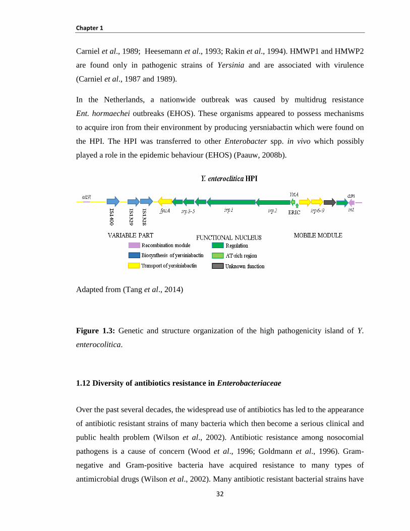
Chapter 1
32
Carniel et al., 1989; Heesemann et al., 1993; Rakin et al., 1994). HMWP1 and HMWP2
are found only in pathogenic strains of Yersinia and are associated with virulence
(Carniel et al., 1987 and 1989).
In the Netherlands, a nationwide outbreak was caused by multidrug resistance
Ent. hormaechei outbreaks (EHOS). These organisms appeared to possess mechanisms
to acquire iron from their environment by producing yersniabactin which were found on
the HPI. The HPI was transferred to other Enterobacter spp. in vivo which possibly
played a role in the epidemic behaviour (EHOS) (Paauw, 2008b).
Adapted from (Tang et al., 2014)
Figure 1.3: Genetic and structure organization of the high pathogenicity island of Y.
enterocolitica.
1.12 Diversity of antibiotics resistance in Enterobacteriaceae
Over the past several decades, the widespread use of antibiotics has led to the appearance
of antibiotic resistant strains of many bacteria which then become a serious clinical and
public health problem (Wilson et al., 2002). Antibiotic resistance among nosocomial
pathogens is a cause of concern (Wood et al., 1996; Goldmann et al., 1996). Gram-
negative and Gram-positive bacteria have acquired resistance to many types of
antimicrobial drugs (Wilson et al., 2002). Many antibiotic resistant bacterial strains have
Chapter 1
33
acquired multidrug resistance, which represents a cause for significant concern,
including diarrhoeal pathogens, for example, Shigella, Salmonella, E. coli, and
Enterococcus faecium; respiratory pathogens such as K. pneumoniae and P. aeruginosa;
urinary tract pathogens like E. coli, and M. tuberculosis, which remains the leading cause
of death from a single infectious disease worldwide (Mazel and Davies, 1999; Wood et
al., 1996).
Enterobacteriaceae with E. coli are by far the most important pathogens for humans and
have been known as the most common source of community and hospital-acquired
infections. They have the facility to spread easily among humans by hand carriage and
contaminated food and water and have the ability to acquire genetic material by
horizontal gene transfer, mediated mostly by plasmids and transposons (Partridge, 2011;
Stokes and Gillings, 2011; Toleman and Walsh, 2011). For these reasons, the emerging
multidrug resistance in Enterobacteriaceae is of most importance for clinical therapy.
The β-lactams are different antibiotic molecules that are composed of four groups:
penicillins, cephalosporins, monobactam, and carbapenems, according to their chemical
structures. The latest developed molecules that have the broadest spectrum of activity are
carbapenems (imipenem, ertapenem, meropenem, and doripenem) (Nordmann et al.,
2012). Multidrug-resistant Enterobacteriaceae produce ESBLs which are the most
important contributing factor to β-lactam resistance (Livermore, 2003). β-lactamases are
enzymes produced by bacteria that act to inactivate β-lactam antibiotics by hydrolysis.
These enzymes are divided into four groups based on their activity profile as shown in
Table 1.4 (Nordmann et al., 2012).
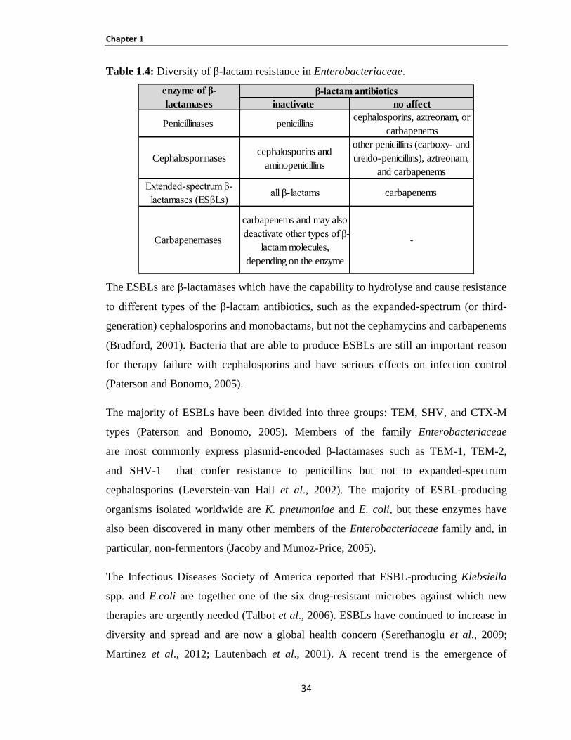
Chapter 1
34
Table 1.4: Diversity of β-lactam resistance in Enterobacteriaceae.
The ESBLs are β-lactamases which have the capability to hydrolyse and cause resistance
to different types of the β-lactam antibiotics, such as the expanded-spectrum (or third-
generation) cephalosporins and monobactams, but not the cephamycins and carbapenems
(Bradford, 2001). Bacteria that are able to produce ESBLs are still an important reason
for therapy failure with cephalosporins and have serious effects on infection control
(Paterson and Bonomo, 2005).
The majority of ESBLs have been divided into three groups: TEM, SHV, and CTX-M
types (Paterson and Bonomo, 2005). Members of the family Enterobacteriaceae
are most commonly express plasmid-encoded β-lactamases such as TEM-1, TEM-2,
and SHV-1 that confer resistance to penicillins but not to expanded-spectrum
cephalosporins (Leverstein-van Hall et al., 2002). The majority of ESBL-producing
organisms isolated worldwide are K. pneumoniae and E. coli, but these enzymes have
also been discovered in many other members of the Enterobacteriaceae family and, in
particular, non-fermentors (Jacoby and Munoz-Price, 2005).
The Infectious Diseases Society of America reported that ESBL-producing Klebsiella
spp. and E.coli are together one of the six drug-resistant microbes against which new
therapies are urgently needed (Talbot et al., 2006). ESBLs have continued to increase in
diversity and spread and are now a global health concern (Serefhanoglu et al., 2009;
Martinez et al., 2012; Lautenbach et al., 2001). A recent trend is the emergence of
inactivate no affect
Penicillinases penicillinscephalosporins, aztreonam, or
carbapenems
Cephalosporinases cephalosporins and
aminopenicillins
other penicillins (carboxy- and
ureido-penicillins), aztreonam,
and carbapenems
Extended-spectrum β-
lactamases (ESβLs)all β-lactams carbapenems
Carbapenemases
carbapenems and may also
deactivate other types of β-
lactam molecules,
depending on the enzyme
enzymeofβ-
lactamases
β-lactamantibiotics
Chapter 1
35
community-onset bloodstream infections caused by ESBL-producing bacteria,
particularly CTX-M producing E. coli. These infections are now rare, but it is possible
that, in the near future possible the clinicians will be regularly confronted with hospital
varieties of bacteria causing infections in patients from the community (Thenmozhi et al.,
2014).
Mostly phenotypic methods are used in clinical diagnostic laboratories because these
tests are easy to do, are cost effective, and have been included in most automated
sensitivity systems, making them widely accessible (Wiegand et al., 2007). However,
phenotypic techniques are not able to differentiate between the specific enzymes
responsible for ESBL production types (SHV, TEM, and CTX-M). Numerous research
or reference laboratories are using genotypic methods to identify the specific gene
responsible for the production of the ESBL (Woodford and Sundsfjord, 2005).
Since 2000, many surveys from different European countries, including Spain
(Rodríguez-Baño et al., 2004), Italy (Mugnaioli et al., 2006), Greece (Pournaras et al.,
2004), the UK (Woodford et al., 2004) and Canada (Pitout et al., 2007) have reported an
alarming increase in resistance of bacteria to other classes of antimicrobial agents
between ESBL-producing bacteria isolated from community sites. These surveys
exhibited co-resistance to co-trimoxazole, tetracycline, gentamicin, and ciprofloxacin.
For example, in Canada more than 66% of isolates were resistant to ciprofloxacin (Pitout
et al., 2007). These studies also presented that strains producing CTX-M enzymes were
significantly more resistant to ciprofloxacin than strains missing polymerase chain
reaction (PCR) evidence for blaCTX-M genes (Pitout et al., 2007; Pitout et al., 2004).
Hurrell et al. (2009b) determined the antibiograms for the Enterobacteriaceae strains
isolated from EFT in NICU and they found that all the S. marcescens strains were
resistant to amoxillin and co-amoxiclav. Also, a quarter of Ent. hormaechei isolates were
resistant to the 3rd
generation cephalosporins, ceftazidime and cefotaxime. During the
study period K. pneumoniae and S. marcescens caused infections in the two NICUs. In
addition, ESBLs were discovered in three of these strains (Hurrell et al., 2009b).
Chapter 1
36
The infant intestinal flora is influenced by the feeding regime. Enterobacteriaceae are
present in neonatal nasogastric enteral feeding tubes in NICU, but there is no study
determined the risk of these strains. This study focuses on assessing the risk to neonates
from opportunistic bacterial pathogens (Enterobacteriaceae) which have been isolated
from two sources; human mastic breast milk and also neonatal nasogastric enteral
feeding tubes. The overall purpose of the current study was to evaluate the risk to
neonates posed by ingestion of specified Enterobacteriaceae either from contaminated
milk or from infant formula.
1.13 Objectives
The initial studies used PFGE to genetically fingerprint the strains isolated from the
NICUs by other researchers. This was determined by the prevalence and persistence of
particular Enterobacteriaceae species, for example, whether certain strains persist in the
NICUs, and verified whether they colonise the tubes of unrelated neonates over
prolonged periods of time. The second part of the project was to focus on the virulence
potential of K. oxytoca, Ent. hormaechei and Ent. ludwigii.
To extend the earlier studies by Hurrell et al. (2009a and b) of Enterobacteriaceae from
EFT, additional strains from mastic breast milk (MBM) were included. These were
obtained from a study by Delgado et al. (2008). Klebsiella strains have been selected as
they are both a common cause of neonatal infections and were frequently isolated from
the EFT. An additional reason is that when Delgado et al. (2008) examined the bacterial
diversity of MBM, they isolated a number of Klebsiella strains, and these have been
made available to us for comparative studies. Despite common expectation, breast milk
is not sterile and can contain a range of bacteria.
There was additional merit for a study to include Klebsiella as it has been isolated from
similar sources from two separate countries (UK and Spain) and this has considerable
comparative value. The second organism of interest, Ent. hormaechei, was isolated from
both the previous Nottingham NICU studies of Hurrell et al. (2009b). This organism had
come to our attention through a series of semi-related studies, as follows:
Chapter 1
37
1. Dr Stacy Townsend (former NTU post-doctoral researcher) acquired ‘Cronobacter’
strains from a hospital for further analysis. Further studies using PFGE revealed that
they were clonal, and were from a NICU outbreak which the hospital was not aware of.
Subsequent 16S rDNA sequencing reidentified the strains as Ent. hormaechei
(Townsend et al., 2008a). Hence, the hospital had not only not recognised an outbreak
but had also misidentified the organisms.
2. NTU acquired a quinolone resistant ‘Cronobacter’ from Poirel et al. (2007) as it is an
unusual trait for the organism. However, 16S rDNA sequencing showed the strain was
Ent. hormaechei and this indicates the difficulty in treatment of infections due to this
organism as well as its identification in the clinical setting.
3. Muytjens et al. (1988) highlighted the incidence of Cronobacter spp. in powdered
infant formula. This paper has been highly cited to date as a key primary study on the
topic. However, the strains were identified in 1988 using biochemical profiling, whereas
NTU used 16S rDNA sequence analysis to reidentify the strains, some of which were
Ent. hormaechei (Townsend et al., 2008a). Hence, this indicates powdered formula as a
source of neonate exposure.
4. Enterobacteriaceae isolates from EFT from QMC and NCH hospitals included
Ent. hormaechei (Hurrell et al., 2009b). Many of these strains were resistant to the 3rd
generation cephalosporins, ceftazidime and cefotaxime, and ESBLs were detected. This
demonstrates that neonates are directly exposed to antibiotic resistant strains in NICUs
during feeding.
5. Comparative bacterial death rates of Enterobacteriaceae revealed that the
Ent. hormaechei strain isolated from neonatal enteral feeding tube (4 above) was more
heat resistant than Sal. Seftenberg. This later organism is normally recognised as the
most heat resistant Enterobacteriaceae and heat treatment processes in industry use this
later organism to ensure their processes are efficient (Forsythe; FSA report 2009). This
shows that the organism has a greater chance of surviving during the reconstitution of
infant formula than other organisms, and that procedures may need to be modified to
take this into account.
Chapter 1
38
In addition to Ent. ludwigii obtained from a study by Delgado et al. (2008), we also
received Ent. ludwigii strain (NTU1439) from Nottingham QMC hospital that was
isolated from a case of neonatal meningitis. This organism was included in the project as
it reinforced the importance of evaluating the risk of neonatal infection by Ent. ludwigii.
Ent. aerogenes and Ent. cloacae were not included in this study because, at the time this
study was initiated, these strains were considered as K. oxytoca. However, later we
identified these strains as Ent. aerogenes and Ent. cloacae using 16S rDNA sequence
analysis.
This study compares the physiological and virulence traits between bacterial species
which neonates are exposed to from MBM and EFT. Only a limited comparison was
made according to the source of strains. However, this was not possible for Ent. ludwigii
and Ent. hormaechei as these were not isolated from MBM and EFT respectively.
Finally, K. pneumoniae strains obtained from Jordan which have been isolated from EFT
were also analysed, but only for PFGE profile due to time constraints.
1.14 Project Aims
In particular, the specific aims are:
1. Characterise Ent. ludwigii, Ent. hormaechei and Klebsiella spp. strains according to
standard phenotyping methods and compare with genotyping.
2. Determine physiological attributes of the strains with respect to heat tolerance, biofilm
formation, cell capsule production and acidic pH survival (pH 3.5).
3. Determine the virulence potential of strains isolated from MBM and EFT using tissue
culture technique and by identifying the presence of virulence factors.
Chapter 2
39
Chapter 2: MATERIALS AND METHODS
Chapter 2
40
2.1 GENERAL MATERIALS
2.1.1 Bacterial strains
A total of 228 strains of Enterobacteriaceae from different sources were used in this
study. One hundred thirty-three strains had been isolated from neonatal EFT from two
local Nottingham hospitals (QMC and NCH), (Table 2.1- 2.3) and reported in a previous
Nottingham NICU study by Hurrell et al. (2009b). Twenty strains had been isolated from
MBM by the lactation consultants at different primary health-care centres in Spain
(Delgado et al., 2008) (Table 2.4). Seventy five EFT and flushed milk isolates of K.
pneumoniae from Jordan were also analysed, but only for PFGE profile due to time
constraints (Table 2.5 part 1 and 2).
The following studies were carried out on the three sets of strains:
Initial experiments used strains from two different sources, EFT and MBM, to
determine their relatedness by using PFGE technique (Tables 2.1-2.4). PFGE
subtyping of EFT isolates from QMC and NCH was by Alkeskas in our research
group as part of his PhD project (Table 2.1- 2.3).
Further experiments used strains from MBM (Table 2.4) and EFT isolates from
QMC and NCH (Table 2.6). These strains were used to determine their
physiological traits, such as heat tolerance, biofilm formation, capsule formation
and acid tolerance at pH 3.5. In addition, these organisms were used to
investigate their ability to attach and invade Caco-2 cell line.
Twenty three strains were chosen for virulence factors determination, as shown
in (Table 2.7). This part of the project focused on the virulence potential of
Ent. ludwigii, Ent. hormaechei, Ent. cloacae, Ent. aerogenes and K. oxytoca
isolated from two different sources.
Ent. hormaechei, and Ent. ludwigii strains from the MBM and EFT isolates
(QMC and NCH) collection were used for determining attachment and invasion
HBMEC and rBCEC4 cell lines (Table 2.8).
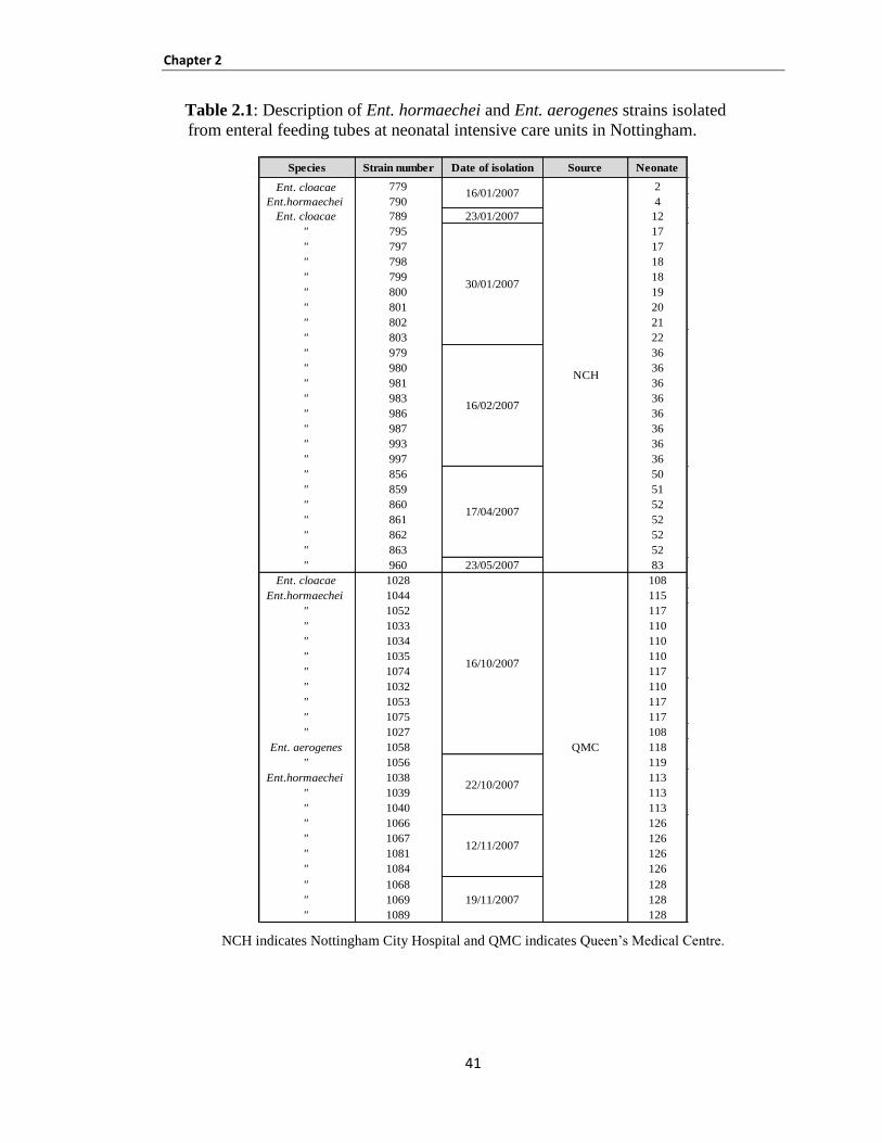
Chapter 2
41
Table 2.1: Description of Ent. hormaechei and Ent. aerogenes strains isolated
from enteral feeding tubes at neonatal intensive care units in Nottingham.
NCH indicates Nottingham City Hospital and QMC indicates Queen’s Medical Centre.
Species Strain number Date of isolation Source Neonate
Ent. cloacae 779 2
Ent.hormaechei 790 4
Ent. cloacae 789 23/01/2007 12
" 795 17
" 797 17
" 798 18
" 799 18
" 800 19
" 801 20
" 802 21
" 803 22
" 979 36
" 980 36
" 981 36
" 983 36
" 986 36
" 987 36
" 993 36
" 997 36
" 856 50
" 859 51
" 860 52
" 861 52
" 862 52
" 863 52
" 960 23/05/2007 83
Ent. cloacae 1028 108
Ent.hormaechei 1044 115
" 1052 117
" 1033 110
" 1034 110
" 1035 110
" 1074 117
" 1032 110
" 1053 117
" 1075 117
" 1027 108
Ent. aerogenes 1058 118
" 1056 119
Ent.hormaechei 1038 113
" 1039 113
" 1040 113
" 1066 126
" 1067 126
" 1081 126
" 1084 126
" 1068 128
" 1069 128
" 1089 128
16/01/2007
NCH
17/04/2007
16/02/2007
30/01/2007
16/10/2007
22/10/2007
12/11/2007
QMC
19/11/2007
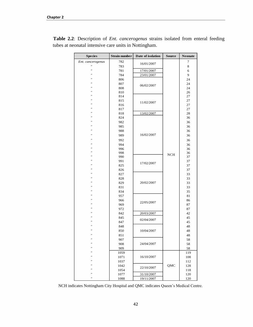
Chapter 2
42
Table 2.2: Description of Ent. cancerogenus strains isolated from enteral feeding
tubes at neonatal intensive care units in Nottingham.
NCH indicates Nottingham City Hospital and QMC indicates Queen’s Medical Centre.
Species Strain number Date of isolation Source Neonate
Ent. cancerogenus 782 7
" 783 8
" 781 17/01/2007 6
" 784 23/01/2007 9
" 806 24
" 807 24
" 808 24
" 810 26
" 814 27
" 815 27
" 816 27
" 817 27
" 818 13/02/2007 28
" 824 36
" 982 36
" 985 36
" 988 36
" 989 36
" 992 36
" 994 36
" 996 36" 998 36
" 990 37
" 991 37
" 825 37
" 826 37
" 827 33
" 828 33
" 829 33
" 831 33
" 834 35
" 957 81
" 966 86
" 969 87
" 972 87
" 842 20/03/2007 42
" 845 45
" 847 45
" 848 48
" 850 48
" 851 48
" 907 58
" 908 58
" 909 58
" 1059 119
" 1071 108
" 1037 112
" 1042 128
" 1054 118
" 1077 31/10/2007 120
" 1088 19/11/2007 120
NCH
20/02/2007
16/10/2007
QMC22/10/2007
06/02/2007
16/02/2007
11/02/2007
17/02/2007
16/01/2007
24/04/2007
10/04/2007
02/04/2007
22/05/2007
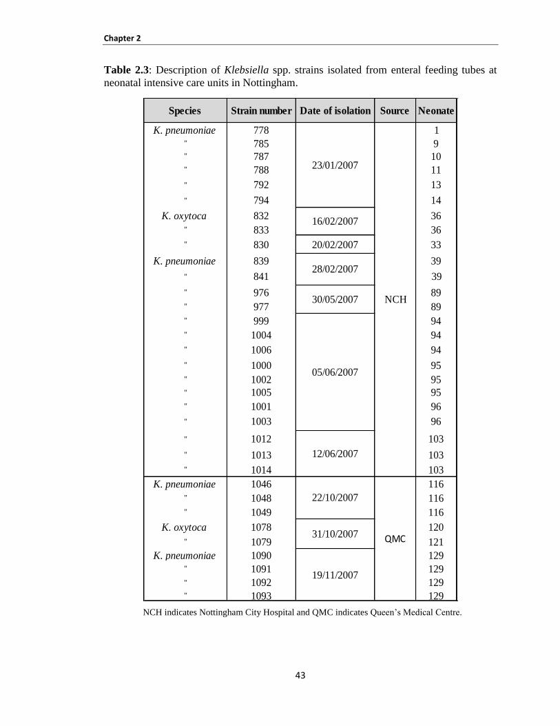
Chapter 2
43
Table 2.3: Description of Klebsiella spp. strains isolated from enteral feeding tubes at
neonatal intensive care units in Nottingham.
NCH indicates Nottingham City Hospital and QMC indicates Queen’s Medical Centre.
Species Strain number Date of isolation Source Neonate
K. pneumoniae 778 1" 785 9" 787 10" 788 11
" 792 13
" 794 14
K. oxytoca 832 36
" 833 36
" 830 20/02/2007 33
K. pneumoniae 839 39
" 841 39
" 976 89
" 977 89
" 999 94
" 1004 94
" 1006 94
" 1000 95
" 1002 95" 1005 95
" 1001 96
" 1003 96
" 1012 103
" 1013 103
" 1014 103
K. pneumoniae 1046 116
" 1048 116
" 1049 116
K. oxytoca 1078 120
" 1079 121
K. pneumoniae 1090 129" 1091 129
" 1092 129" 1093 129
16/02/2007
05/06/2007
30/05/2007
28/02/2007
19/11/2007
22/10/2007
31/10/2007
12/06/2007
QMC
23/01/2007
NCH
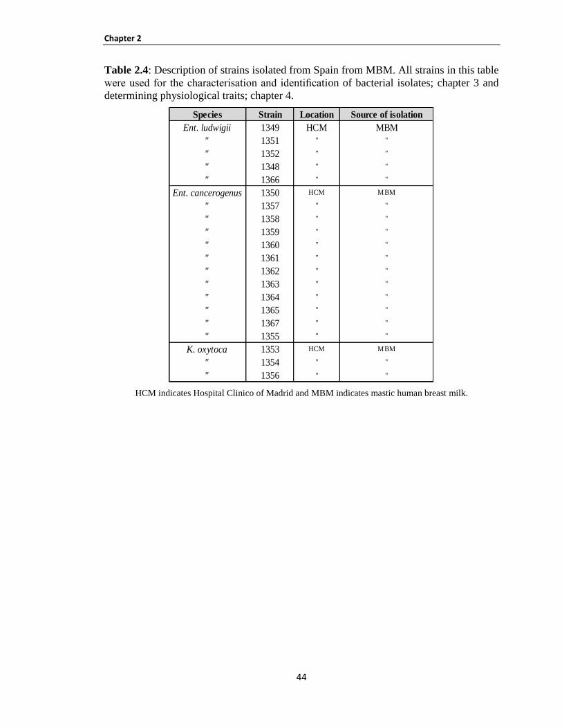
Chapter 2
44
Table 2.4: Description of strains isolated from Spain from MBM. All strains in this table
were used for the characterisation and identification of bacterial isolates; chapter 3 and
determining physiological traits; chapter 4.
HCM indicates Hospital Clinico of Madrid and MBM indicates mastic human breast milk.
Species Strain Location Source of isolation
Ent. ludwigii 1349 HCM MBM
" 1351 " "
" 1352 " "
" 1348 " "
" 1366 " "
Ent. cancerogenus 1350 HCM MBM
" 1357 " "
" 1358 " "
" 1359 " "
" 1360 " "
" 1361 " "
" 1362 " "
" 1363 " "
" 1364 " "
" 1365 " "
" 1367 " "
" 1355 " "
K. oxytoca 1353 HCM MBM
" 1354 " "
" 1356 " "
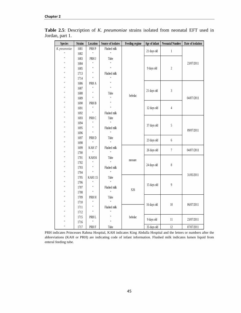
Chapter 2
45
Table 2.5: Description of K. pneumoniae strains isolated from neonatal EFT used in
Jordan, part 1.
PRH indicates Princesses Rahma Hospital, KAH indicates King Abdulla Hospital and the letters or numbers after the
abbreviations (KAH or PRH) are indicating code of infant information. Flushed milk indicates lumen liquid from
enteral feeding tube.
Species Strains Location Source of isolates Feeding regime Age of infant Neonatal Number Date of isolation
K. pneumoniae 1681 PRH P Flushed milk
" 1682 " "
" 1683 PRH J Tube
" 1684 " "
" 1685 " "
" 1713 " Flushed milk
" 1714 " "
" 1686 PRH A "
" 1687 " "
" 1688 " Tube
" 1689 " "
" 1690 PRH B "
" 1691 " "
" 1692 " Flushed milk
" 1693 PRH C Tube
" 1694 " "
" 1695 " Flushed milk
" 1696 " "
" 1697 PRH D Tube
" 1698 " "
" 1699 KAH 17 Flushed milk
" 1700 " "
" 1701 KAH16 Tube
" 1702 " "
" 1703 " Flushed milk
" 1704 " "
" 1705 KAH1 15 Tube
" 1706 " "
" 1707 " Flushed milk
" 1708 " "
" 1709 PRH H Tube
" 1710 " "
" 1711 " Flushed milk
" 1712 " "
" 1715 PRH L "
" 1716 " "
" 1717 PRH F Tube 35 days old 12 07/07/2011
06/07/2011
bebelac9 days old 11 23/07/2011
S26
15 days old 9
16 days old 10
31/05/2011
23/07/2011
24 days old 8
04/07//2011
09/07/2011
04/07//2011
neosure
26 days old 7
bebelac
21 days old 1
9 days old 2
21 days old 3
12 days old 4
37 days old 5
23 days old 6
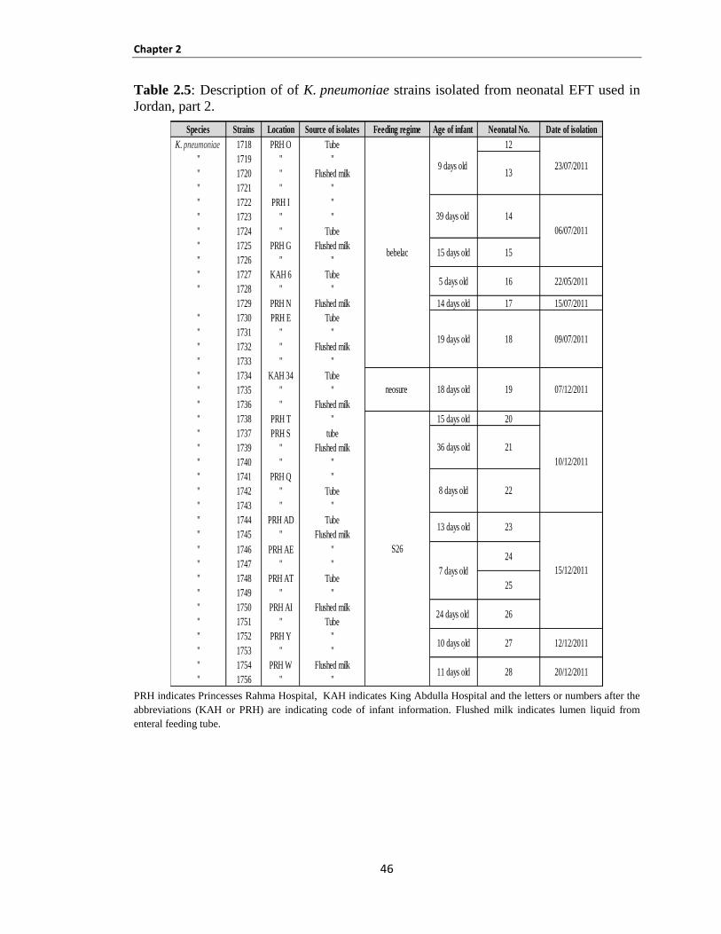
Chapter 2
46
Table 2.5: Description of of K. pneumoniae strains isolated from neonatal EFT used in
Jordan, part 2.
PRH indicates Princesses Rahma Hospital, KAH indicates King Abdulla Hospital and the letters or numbers after the
abbreviations (KAH or PRH) are indicating code of infant information. Flushed milk indicates lumen liquid from
enteral feeding tube.
Species Strains Location Source of isolates Feeding regime Age of infant Neonatal No. Date of isolation
K. pneumoniae 1718 PRH O Tube 12
" 1719 " "
" 1720 " Flushed milk
" 1721 " "
" 1722 PRH I "
" 1723 " "
" 1724 " Tube
" 1725 PRH G Flushed milk
" 1726 " "
" 1727 KAH 6 Tube
" 1728 " "
1729 PRH N Flushed milk 14 days old 17 15/07/2011
" 1730 PRH E Tube
" 1731 " "
" 1732 " Flushed milk
" 1733 " "
" 1734 KAH 34 Tube
" 1735 " "
" 1736 " Flushed milk
" 1738 PRH T " 15 days old 20
" 1737 PRH S tube
" 1739 " Flushed milk
" 1740 " "
" 1741 PRH Q "
" 1742 " Tube
" 1743 " "
" 1744 PRH AD Tube
" 1745 " Flushed milk
" 1746 PRH AE "
" 1747 " "
" 1748 PRH AT Tube
" 1749 " "
" 1750 PRH AI Flushed milk
" 1751 " Tube
" 1752 PRH Y "
" 1753 " "
" 1754 PRH W Flushed milk
" 1756 " "
12/12/2011
09/07/2011
25
24 days old 26
7 days old
24S26
10/12/2011
36 days old 21
8 days old 22
13 days old 23
11 days old 28 20/12/2011
15/12/2011
10 days old 27
neosure 18 days old 19 07/12/2011
bebelac
9 days old 23/07/2011
39 days old 14
06/07/2011
15 days old 15
5 days old 16 22/05/2011
19 days old 18
13
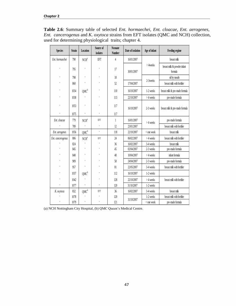
Chapter 2
47
Table 2.6: Summary table of selected Ent. hormaechei, Ent. cloacae, Ent. aerogenes,
Ent. cancerogenus and K. oxytoca strains from EFT isolates (QMC and NCH) collection,
used for determining physiological traits; chapter 4.
(a) NCH Nottingham City Hospital, (b) QMC Queen’s Medical Centre.
Species Strain LocationSource of
isolates
Neonate
NumberDate of isolation Age of infant Feeding regime
Ent. hormaechei 790 NCHa EFT 4 16/01/2007 breast milk
" 795 " " 17breast milk & powder infant
formula
" 798 " " 18 nil by mouth
" 860 " " 52 17/04/2007 breast milk with fortifier
" 1034 QMCb " 110 16/10/2007 1-2 weeks breast milk & pre-made formula
" 1038 " " 113 22/10/2007 > 4 weeks pre-made formula
" 1053 " " 117
" 1075 " " 117
Ent. cloacae 779 NCHa EFT 1 16/01/2007 pre-made formula
" 789 " " 12 23/01/2007 breast milk with fortifier
Ent. aerogenes 1056 QMCb " 118 22/10/2007 < one week breast milk
Ent. cancerogenus 806 NCHa EFT 24 06/02/2007 > 4 weeks breast milk with fortifier
" 824 " " 36 16/02/2007 3-4 weeks breast milk
" 845 " " 45 02/04/2007 2-3 weeks pre-made formula
" 848 " " 48 10/04/2007 > 4 weeks infant formula
" 909 " " 58 24/04/2007 2-3 weeks pre-made formula
" 957 " " 81 22/05/2007 3-4 weeks breast milk with fortifier
" 1037 QMCb " 112 16/10/2007 1-2 weeks
" 1042 " " 128 22/10/2007 > 4 weeks breast milk with fortifier
" 1077 " " 120 31/10/2007 1-2 weeks
K. oxytoca 832 QMCb EFT 36 16/02/2007 3-4 weeks breast milk
" 1078 " " 120 1-2 weeks breast milk with fortifier
" 1079 " " 121 < one week pre-made formula
breast milk & pre-made formula
> 4weeks
2-3weeks
16/10/2007 2-3 weeks
30/01/2007
31/10/2007
> 4 weeks
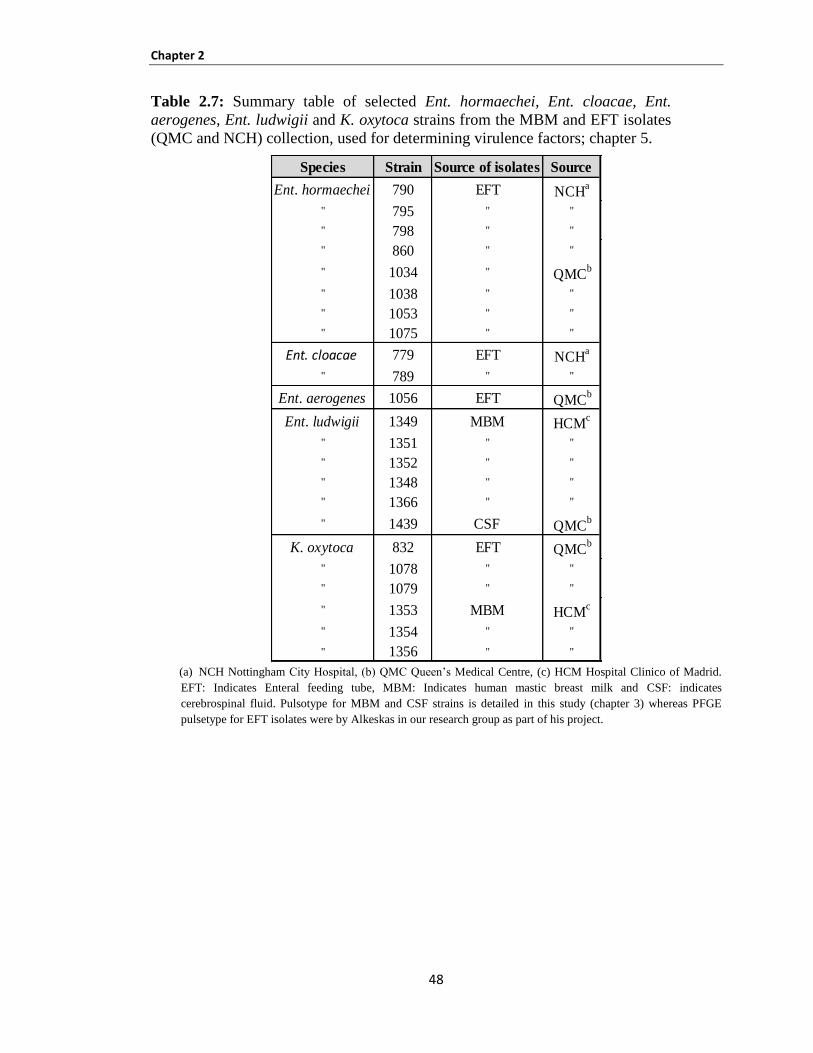
Chapter 2
48
Table 2.7: Summary table of selected Ent. hormaechei, Ent. cloacae, Ent.
aerogenes, Ent. ludwigii and K. oxytoca strains from the MBM and EFT isolates
(QMC and NCH) collection, used for determining virulence factors; chapter 5.
(a) NCH Nottingham City Hospital, (b) QMC Queen’s Medical Centre, (c) HCM Hospital Clinico of Madrid.
EFT: Indicates Enteral feeding tube, MBM: Indicates human mastic breast milk and CSF: indicates
cerebrospinal fluid. Pulsotype for MBM and CSF strains is detailed in this study (chapter 3) whereas PFGE
pulsetype for EFT isolates were by Alkeskas in our research group as part of his project.
Species Strain Source of isolates Source
Ent. hormaechei 790 EFT NCHa
" 795 " "
" 798 " "
" 860 " "
" 1034 " QMCb
" 1038 " "
" 1053 " "
" 1075 " "
Ent. cloacae 779 EFT NCHa
" 789 " "
Ent. aerogenes 1056 EFT QMCb
Ent. ludwigii 1349 MBM HCMc
" 1351 " "
" 1352 " "
" 1348 " "
" 1366 " "
" 1439 CSF QMCb
K. oxytoca 832 EFT QMCb
" 1078 " "
" 1079 " "
" 1353 MBM HCMc
" 1354 " "
" 1356 " "
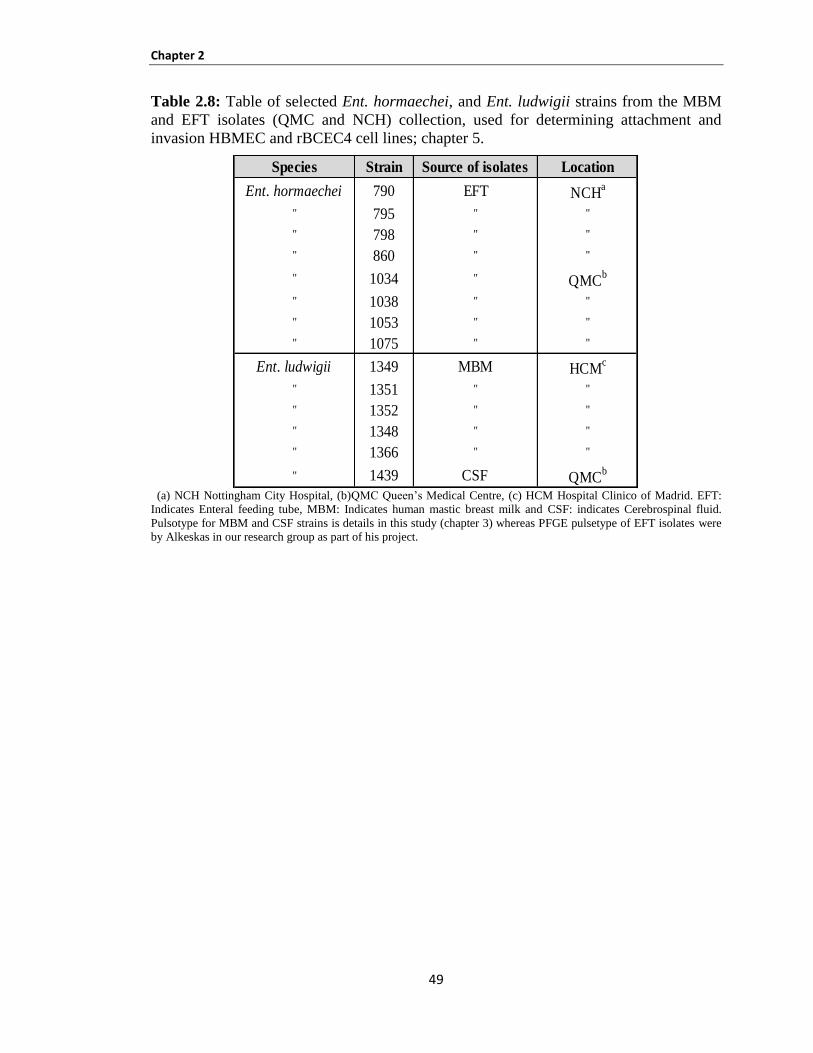
Chapter 2
49
Table 2.8: Table of selected Ent. hormaechei, and Ent. ludwigii strains from the MBM
and EFT isolates (QMC and NCH) collection, used for determining attachment and
invasion HBMEC and rBCEC4 cell lines; chapter 5.
(a) NCH Nottingham City Hospital, (b)QMC Queen’s Medical Centre, (c) HCM Hospital Clinico of Madrid. EFT:
Indicates Enteral feeding tube, MBM: Indicates human mastic breast milk and CSF: indicates Cerebrospinal fluid.
Pulsotype for MBM and CSF strains is details in this study (chapter 3) whereas PFGE pulsetype of EFT isolates were
by Alkeskas in our research group as part of his project.
Species Strain Source of isolates Location
Ent. hormaechei 790 EFT NCHa
" 795 " "
" 798 " "
" 860 " "
" 1034 " QMCb
" 1038 " "
" 1053 " "
" 1075 " "
Ent. ludwigii 1349 MBM HCMc
" 1351 " "
" 1352 " "
" 1348 " "
" 1366 " "
" 1439 CSF QMCb
Chapter 2
50
2.1.2 Bacterial cultivation
Tryptone soya agar (TSA) 2.1.2.1
Tryptone soya agar (CM131B, Oxoid Thermo Fisher Scientific) was prepared according
to the manufacturer’s instructions. Briefly, 40g of Tryptone soya agar was suspended in
1 litre of distilled water, and sterilised at 121°C for 15 minutes. After cooling to 45-50°C
it was mixed gently and dispensed into sterile Petri dishes. These were stored in the
fridge at 4ºC until required.
Tryptone soya broth (TSB) 2.1.2.2
Tryptone soya broth (CM0989B, Oxoid Thermo Fisher Scientific) was prepared by
dissolving 30g of TSB in 1 litre of distilled water, and autoclaving at 121°C for 15
minutes. The media was stored in a cool and dark place until required.
Milk agar 2.1.2.3
To prepare milk agar media, 3g of agar (LP001, Oxoid Thermo Fisher Scientific) and
0.4g of ammonium sulphate were dissolved in 40ml of distilled water, and autoclaved at
121ºC for 15 minutes. 200ml of milk based ready to use formula whey (Cow and Gate
First Infant Milk), casein (SMA Extra Hungry Ready to use) and soy-based (Cow and
Gate infasoy) infant formula was warmed at 55ºC and mixed with the autoclaved agar.
The medium was dispensed into Petri dishes, and stored in a cooled place until required.
Saline distilled water 2.1.2.4
One saline tablet (BR0053G, Oxoid Thermo Fisher Scientific) was dissolved in 500ml
distilled water to prepare 0.85% saline that was autoclaved at 121°C for 15 minutes.
Luria-Bertani broth (LB) 2.1.2.5
Luria-Bertani Broth (L3022, Sigma Aldrich; USA) was prepared by dissolving 25g of
Luria-Bertani in 1 litre distilled water. It was dispensed in the required volumes and
autoclaved at 121ºC for 15 minutes. The media was stored in a cool and dark place until
required.
Chapter 2
51
Iso-Sensitest agar (ISO) 2.1.2.6
Distilled water (1 litre) was mixed with 31.4g ISO agar (CM0471, Oxoid Thermo Fisher
Scientific), brought to boil to dissolve the medium completely and then sterilised by
autoclaving at 121°C for 15 minutes. The plates were stored in a cool place until
required.
Long term storage of strains 2.1.2.7
In a 2ml Eppendorf tube 800µl of TSB broth was mixed with one colony from the TSA
plate and 200µl of 80 % glycerol was added and stored in a -80°C freezer.
2.1.3 Mammalian cell lines
In this study, four mammalian cell lines (Caco-2, HMBEC, rBCEC4 and U937) were
used to determine Enterobacteriaceae virulence towards host cells. The cell lines were
stored in liquid nitrogen, with the date, cell line type and passage numbers clearly
labelled.
Human colonic carcinoma epithelial (Caco-2) cell line 2.1.3.1
Human colonic carcinoma epithelial cell line (Caco-2) was received from the European
Collection of Cell Cultures (ECACC# 86010202). The cell line was originally from a
human colon adenocarcinoma. The cell lines are able to form a well-differentiated cell
monolayer and are similar to small intestinal enterocytes. Therefore, it is widely utilized
in studies of pathogen-host cell interaction (Szymanski et al., 1995). Cells were used
between passages seven and twelve.
Human brain microvascular endothelial cells (HBMEC) 2.1.3.2
The human brain microvascular endothelial HBMEC cell line was obtained from Dr M.
Rittig (University of Nottingham). Cells were used between passages sixty six and
seventy.
Chapter 2
52
Rat brain capillary endothelial cell line (rBCEC4) 2.1.3.3
The rat brain capillary endothelial cell line rBCEC4 was obtained from I. E. Blasig
(Forschungsinstitut für Molekulare Pharmakologie, Berlin; Germany). Cell line rBCEC4
was used between passages twenty and twenty five.
Macrophage cell line (U937) 2.1.3.4
Macrophages cell line (U937) was received from the American Type Culture Collection
(ATCC) (CRL-1593.2). Cells were used between passages seven and eleven.
2.1.4 Medium of tissue culture experiments
Growth medium for human colonic carcinoma epithelial (Caco-2) cell line 2.1.4.1
The growth medium was prepared by using 500ml minimum essential medium (M4655,
Sigma) mixed with 10% fetal calf serum (F7524, Sigma), 1% non-essential amino acid
solution (100x, M7145, Sigma) and 1% penicillin-streptomycin mixture (10.000 units
penicillin and 10mg streptomycin/ml) (P4333, Sigma).
Infection medium for Caco-2 cell line 2.1.4.2
To prepare the Infection media, 500ml minimum essential medium (M4655, Sigma), was
mixed with 10% fetal calf serum and 1% non-essential amino acid solution (100x,
M7145, Sigma).
Growth medium for human brain microvascularendothelial cells (HBMEC) 2.1.4.3
and rat brain capillary endothelial cell line (rBCEC4)
Dulbecco’s modified Eagle’s medium-high glucose DMEM (D6429, Sigma) 500ml was
prepared and supplied with 10% fetal calf serum (F7524, Sigma), 1% non-essential
amino acid solution (100x, M7145, Sigma) and 1% penicillin-streptomycin mixture
(10.000 units penicillin and 10mg streptomycin/ml) (P4333, Sigma).
Chapter 2
53
Infection medium for HBMEC and rBCEC4 2.1.4.4
500ml Dulbecco’s modified Eagle’s medium-high glucose DMEM (D6429, Sigma) was
mixed with 10% fetal calf serum and 1% non-essential amino acid solution (100x,
M7145, Sigma).
Growth medium for macrophage cell line (U937) 2.1.4.5
The growth medium was prepared by using 500ml RPMI-1640 medium (R8758, Sigma)
supplied with 10% fetal calf serum (F7524, Sigma), 1% non-essential amino acid
solution (100x, M7145, Sigma) and 1% penicillin-streptomycin mixture (10.000 units
penicillin and 10mg streptomycin/ml) (P4333, Sigma).
Infection medium for macrophage cell line (U937) 2.1.4.6
500ml RPMI-1640 Medium (R8758, Sigma) was mixed with 10% fetal calf serum and
1% non-essential amino acid solution (100x, M7145, Sigma).
2.1.5 Buffers and detergents
Dulbecco’sPhosphateBufferedSaline (PBS) 2.1.5.1
Phosphate buffered saline containing sodium and magnesium chloride solution (D8662,
Sigma) was used to wash cells during preparation. The phosphate buffered saline was
used for all cell lines used in this study.
Triton- X 0.5% 2.1.5.2
The solution of 0.5 % Triton-X was prepared by adding 500µl of Triton-X (Fisher
BP151-100 Canada) to 99.5ml of sterile distilled water.
TE Buffer 2.1.5.3
The TE Buffer solution was prepared by using 5ml of 1M Tris base (BP152-500,
Thermo Fisher Scientific), with 1ml of 0.5M EDTA , and 494ml distilled water. This
buffer was used to prepare the plug agarose and to wash PFGE plugs.
Chapter 2
54
Tris base- Boric acid – EDTA (TBE) Buffer 10X preparation (1 Litre 2.1.5.4
distilled water)
The mixture was prepared using 108g Tris base (BP152-500, Thermo Fisher Scientific),
55g boric acid (B/3750/53), 40ml 0.5M EDTA pH 8.0 and autoclaved for 20 minutes.
TAE 1X 2.1.5.5
To make up TAE 1X 20ml of TAE 50X (2.0M Tris Acetate + 100 mM Na2EDTA) were
mixed with 980ml of distilled water.
Cell Suspension Buffer 2.1.5.6
To prepare a cell suspension buffer total volume = 3ml* n, Buffer needed as follows:
Tris pH 8.0 = 0.3ml* n
EDTA pH 8.0 = 0.6ml* n
VdH2O = 3ml* n – (V add Tris +V add EDTA)
n indicates the number of samples and V indicates Volume.
Cell lysis buffer 2.1.5.7
To prepare the cell lysis buffer (50 mM Tris:50 mM EDTA, pH 8.0 + 1% Sarcosyl);
1% Sarkosyl NL (N-Dodecanoyl-N-methylglycine sodium salt) Sigma Aldrich, UK
(L-5125), proteinase K 20mg/ml Sigma Aldrich, UK (P2308) and Calculate the
total volume of cell lysis buffer/Proteinase K Buffer needed as follows:
total volume = 5ml *n
Tris, pH 8.0 = 0.25ml * n
EDTA, pH 8.0 = 0.5ml * n
Sarkosyl NL = 0.05g *n
proteinase k = 25µl * n and
VdH2O = 5ml * n – (V Tris + V EDTA + V proteinase k)
(n indicates the number of samples and V indicates Volume).

Chapter 2
55
Iron III solution 2.1.5.8
To prepare iron III solution, 9ml of HCl (100ml of distilled water added to 830µl HCl)
was mixed with 1ml of FeCl3 6H2O solution (1L distilled water mixed with 2.73g of
FeCl3 6H2O).
Chrome azurol sulphate (CAS) solution 2.1.5.9
ChromeazurolS (199532, Sigma) was prepared by mixing 100ml of sterile distilled water
with 0.0121g of CAS.
Hexadecyltrimethylammonium bromide (HDTMA) 2.1.5.10
Distilled water (80ml) was mixed with 0.1458g of HDTMA (H6268, Sigma).
Sodium hydroxide solution 2.1.5.11
Ten grams of 50% w/v NaOH was added to distilled water (20ml).
2.1.6 Molecular studies
Genomic DNA extraction 2.1.6.1
Each bacterial strain was grown aerobically in TSB broth with shaking, at 37°C
incubator. The DNA was carried out with the GenElute™ Bacterial Genomic DNA Kit
(Sigma) for each strain. Nanodrop 2000 (Thermo Scientific, UK) was used to check the
concentration and purity of the eluted DNA.
PCR product purification 2.1.6.2
MinElute PCR Purification Kits (Qiagen, UK) were used to purify the amplified
products by following the manufacturer's protocol. Nanodrop 2000 (Thermo Scientific,
UK) was used to check the concentration and purity of the samples. The final elution
step was conceded out in 20µl of molecular biology grade water (Fisher Scientific, UK).
The sequencing machines were by Accugenix, Delaware (USA), Eurofins MWG Operon
(London, UK) and Source Bioscience (Nottingham, UK). The data was analysed by
Ribosomal Database Project (http://rdp.cme.msu.edu/seqmatch/seqmatch_intro.jsp).
Chapter 2
56
Agarose gel electrophoresis 2.1.6.3
The amplified PCR products were checked by visualisation on a 1% agarose gel (Life
Science ® Company, UK, BIO-41025). The gel, 1% (w/v) agarose was mixed in 1X
Tris-acetate-EDTA (TAE) buffer (Geneflow, UK) and microwaved until melted. From
SYBR® Safe DNA gel stain (Life Technologies–Invitrogen, UK) 0.1µ/ml (v/v) was
added to the molten gel and then poured in a tray and allowed to set. The gel was loaded
with 5µl of sample and 100 bp DNA ladder (Promega, Southampton, UK) was used as a
marker. The gel was run for 45 minutes at 90V in 1X TAE buffer. Under ultraviolet
(UV) light, the gel was viewed to observe the DNA bands, using the InGenius® gel
documentation system (Syngene; UK).
2.1.7 Safety considerations
Health and safety codes of practice were followed for all experiments. All materials used
in the protocols were risk assessed using the suitable control of substances hazardous to
health (COSHH) forms. Good laboratory practices were carried out for microbes, media
and chemicals, as well as for operating laboratory devices. The Hepatitis B vaccination
was received before commencement of tissue culture experiments.
2.2 Characterisation experiments
2.2.1 Phenotypic characterisation
The identification of the strains was initially determined using the ID32E (bioMérieux)
phenotyping kit, according to manufacturers' recommendations, and the apiweb database
(www.apiweb.biomerieux.com).
2.2.2 Genotypic characterisation
Pulsed-field gel electrophoresis (PFGE) was used as a technique for differentiating
strains at the DNA level. The isolates were streaked on TSA and incubated at 37ºC
for 14-18 hours. Each strain was suspended in 3ml cell suspension buffer (0.3ml Tris,
0.6ml EDTA and 2.1ml H2O); section 2.1.5.6. The cell concentrations were adjusted to
an optical density (OD) of 1.35 ± 0.05 at 610nm. The TE buffer was prepared and added
Chapter 2
57
to 1% agarose and 1% SDS and kept at 55°C until required. Bacterial cell suspension
(300µl) was mixed with 300µl of TEB agarose and 15µl of Proteinase K (20mg/ml,
Sigma, P2308) and dispensed in 100µl volumes into each plug.
The plugs were placed in 50ml polypropylene tubes containing 5ml of cell lysis buffer/
Proteinase K (0.25 Tris, 0.50µl EDTA, 1% Sarcosyl and 25µl of Proteinase K); section
2.1.5.7 and left in a water bath shaker at 54ºC for 1.5-2 hours with vigorous shaking at
150-175 rpm. The lysis buffer was removed and 15ml of sterile ultra-pure water was
added to wash the agarose plugs, and incubated in the water bath with shaking at 50°C
for 15 minutes. This was repeated three times. The TE buffer (5ml) was added to the
plugs and incubated in the shaking water bath at 50°C for 15 minutes. The TE buffer was
poured off and the previous step repeated three times. The plugs were stored at 4°C in
5-10ml sterile TE buffer until required.
For restriction digestion of the DNA, 2mm of the plug was cut off and placed it in an
Eppendorff tube containing 135µl of sterile water and 15µl of buffer for 30 minutes at
room temperature. The mixture was removed and 150µl of restriction enzyme mixture
(131.25µl sterilised water, 15µl TB buffer and 3.75µl enzyme Xba1) was added and
incubated at 37°C for 1.5-2 hours. Each plug was put in order in the comb and 100ml of
0.5 TBE buffer (95ml H2O, 5ml TBE and 1g agarose) were poured into the gel support
tray.
Two litres prepared from the TBE buffer 0.5x (1900ml H2O, 100ml TBE) was added to
the running tank. The gel was run at 14°C with pulse times of 2.2 to 63.8 second for 20
hours at 6 V. After the electrophoresis run was over, the gel was stained by 20μl of
ethidium bromide (Sigma, E1510) stock solution (10mg/ml) with 400ml of distilled
water for 20-30 minutes in a covered container. The InGenius® gel documentation
system (Syngene; UK) was used to photograph the gels. The electronic images of the
PFGE patterns were saved in TIFF file format and analysed with Bionumerics software
version 3.5, dice coefficient and unweight pair group method with arithmetic mean
(UPGMA) for cluster analysis. The Salmonella serotype Braenderup reference standard
(H9812) strain 732 was used as the marker.
Chapter 2
58
The tolerance value is expressed as a percentage of the total normalized gel length. Two
bands on different PFGE patterns are considered to be the same if the difference in the
position of the bands is less than the tolerance percentage. If the difference in the
positions of the bands is greater than the tolerance percentage, then the bands are
considered to be two different bands. A low tolerance (0.5%) allows for a minimal
amount of shifting of the bands when comparing bands of different PFGE patterns. In
this study different tolerance values were used when analyzing the PFGE patterns
however the optimum band similarities were obtained at the tolerance values of 1.50%,
1.75% and 1.60% for EFT, MBM and Jordan strains respectively.
2.3 Identification of strains by using 16S rDNA sequence analysis
2.3.1 Preparation of FTA® Elute cards
The FTA Elute card was labelled with the sample code, and 5ml of sterile water was
dispensed into a labelled tube. The suspension of bacteria was equal to 0.5 McFarland
standard and 40µl of the suspension distributed in concentric circular motion within each
circle. The dry cards were placed in an envelope and shipped to Accugenix,
Inc.Delaware, USA.
2.3.2 AccuPRO-ID Bacterial Identification
Cultures were 24-48 hours old on TSA after incubation at 37°C as according to the
recommended methodology from AccuPRO-ID bacterial proteotypic identification
service. Each sterile Eppendorf tube (1.5ml) was labelled for each sample and 300µl of
sterile, nuclease free water was placed in it. Each Eppendorf tube was inoculated with
1 µl loop of bacteria from culture (24-48 hours old). Pure ethanol (900µl) was added to
each suspension and the cap secured in preparation for shipment.
2.3.3 PCR 16S rDNA sequence analysis
Genomic DNA extraction was prepared as described above in section 2.1.6.1. The partial
sequencing of the 16S rDNA loci (528 bp) was performed using primers described
by Iversen et al. (2006). The PCR reaction mixture contained 5µl buffer, 2µl MgCl2, 1µl
2’-deoxynucleoside 5’-triphosphate (dNTP), forward (TGGAGAGTTTGATCCTGGC-
Chapter 2
59
TCAG) primer and reverse (TACCGCGGCTGCTGGCAC) primer (2.5µl each), 0.25µl
TAQ polymerase, 1µl DNA and 10.75µl distilled water. PCR conditions were as follows:
initial denaturation at 95°C for 10 minutes; 30 cycles of denaturation at 95°C for 30
second, primer annealing at 62.6°C for 30 second, extension at 72°C for 45 second;
followed by a final extension step of 72°C for 10 minutes.
2.4 Physiological experiments
2.4.1 Heat tolerance
Overnight cultures were inoculated into whey-based ready to use formula (Cow and Gate
First Infant Milk) and casein (SMA Extra Hungry Ready to use) and incubated at 37°C
overnight. Infant milk was warmed to 55°C for 30 minutes in a water bath. Bacterial
suspensions (1ml) were inoculated into 9ml warmed milk. At zero time 100µl of
suspension were transferred to one well of the 96 well plates. This was repeated every
5 minutes for 30 minutes. Serial dilutions of the bacterial suspension (15µl) were diluted in
saline to 10-7
. The Miles and Misra enumeration technique on TSA plates was used to
determine the number of cells surviving exposure to 55ºC. The plates were incubated at
37ºC overnight. The viable count was plotted against time, and D-values calculated on the
time required for a 1 log10 reduction in the viable count.
2.4.2 Biofilm formation
Bacteria production was determined according to the method described by Stepanović et
al. (2004), with a slight modification. In brief, bacteria were grown at 37°C overnight
without shaking for 24 hours in TSB and then diluted to 104 cfu/ml in sterile saline and
used to inoculate reconstituted formula (casein, whey and soya). Serial dilution of each
type of inoculated formula milk was carried out into saline in the wells of a 96-well flat
bottomed polystyrene microplate. The microplates were incubated aerobically for 24
hours and 48 hours at 20ºC and for 24 hours at 37ºC. The contents of each well were
removed using a vacuum and the remaining biofilm was washed two times with 300µl of
sterile saline and shaken for 15 minutes at 200 rpm. Crystal violet 0.01% (250µl) was
added to the wells for 5 minutes, and then the microplates were washed two times with
250µl sterile distilled water per well. The residual attached bacteria were extracted with
Chapter 2
60
200µl of ethanol (70% v/v) per well, and after 15 minutes 150µl was transferred to new
plates.
The absorbance of each well was measured at 540nm using a GEN5 program, the
triplicate results were averaged and divided by the negative control. The relative
increase in biofilm formation level for each strain was obtained from dividing the OD of
the strain retained by the isolates. The OD of tested strains was determined according to
the method described by Stepanovic et al. (2004) where the threshold for positive
bacterial biofilm OD was defined as three standard fold increase above the mean OD. of
the negative control. Strains were categorised as follows: No biofilm formation: OD
strain = OD control, low biofilm formation: OD strain ≤ 2x OD control, moderate
biofilm formation: OD strain ≤ 4 x OD control and high biofilm formation: OD strain >4
x OD control.
2.4.3 Capsule formation
The preparation of milk agar media was described in section 2.1.2.3. Capsule production
on milk agar by tested strains was determined by colony appearance on milk agar at
20ºC (after 24 hours and 48 hours) and 37ºC (after 24 hours).
2.4.4 Acid tolerance of organisms to pH 3.5 (HCl acidified formula)
Bacteria resistance to pH 3.5 was studied according to the method as already used by
Edelson-Mammel et al. (2006), with some modification. A single colony of the test
organism was inoculated into 5ml of TSB and incubated for 18 hours at 37°C. Two types
of formula were used in this study (whey and casein-based formulas). The milk was
adjusted to pH 3.5 with 1mM of sterile HCl (sterilised by filtration using 0.2µm filters).
Two sterile tubes containing 9ml of pH adjusted formula were placed in a water bath at
37°C to avoid the heat shocking of the inoculated cells.
The pH adjusted infant formula was inoculated with 1ml of the bacterial cell suspension
into the water bath to maintain the temperature. From each tube 100µl of adjusted infant
formula was taken at each time point (0, 5, 10, 15, 20, 30, 45, 60, 90 and 120 minutes)
and then were serially diluted in normal saline and plated on TSA agar plates by using
Chapter 2
61
the Miles and Misra technique to determine the number of viable cells. The plates were
incubated for 18 hours at 37°C and the number of colonies was enumerated as colony
forming units (cfu). The number of colonies was transformed to log (cfu/ml) values for
presentation on the log curve graph.
2.5 Virulence factors
2.5.1 Bacterial attachment and invasion of host cells
Preparation of bacterial inoculum 2.5.1.1
The strains were grown overnight on TSB at 37°C, The overnight culture were then
pelleted by centrifugation for 3 minutes at 1200 rpm. The supernatant was then discarded
and the pellet was re-suspended in 1ml of infection medium. Each strain was added at
~ 106
cfu per well to obtain the multiplicity of infection of 1:100 and incubated with 5%
CO2 at 37°C for 3 hours.
Mammalian cell culture 2.5.1.2
All cell lines were taken from the liquid nitrogen that was used for each of the tissue
culture studies. The frozen ampoule was melted and quickly seeded into 10ml of the
suitable growth medium, and 100µg/ml gentamicin was added to the initial passage.
Bacterial attachment and invasion of host cells were determined by the method of
Townsend et al. (2008b). The seeded cells were incubated at 37°C with 5% CO2. The
cells were centrifuged for 5 minutes at 1500 rpm by using CENTAUR2 centrifuge (MES,
UK). The pellet was re-suspended in 25ml of growth medium and seeded into 75ml
tissue culture flasks. The seeded cells were incubated at 37°C in 5% (v/v) CO2, for 24
hours for rBCEC4 and HBMEC for 48 hours for Caco-2 and U937 to obtain the
confluent layer (70-80%). Mammalian cells were then seeded in 24-well microplate for
48 hours and the concentration in each well was 2 x 10⁴ cells/ml. Five millilitres of
trypsin-EDTA solution was used to detach the confluent monolayer.
Chapter 2
62
Attachment assay 2.5.1.3
The attachment and invasion assay was used to determine the ability of the bacterial
strains to adhere Caco-2, rBCEC4, and HBMEC cell lines. To obtain less variation
between the two assays, the determination of attachment was carried out at the same time
using the same cell line passage of mammalian cell line and same inoculum of bacterial
suspension. Cultivation of the Caco-2, rBCEC4, and HBMEC cell lines was described
in section 2.1.3. The mammalian cells were washed three times using Dulbecco’s
phosphate buffered saline (PBS) (D8662, Sigma) and were infected with each isolate at
~ 106
cfu per well (MOI of 1:100) and incubated with 5% CO2 at 37°C for 3 hours. After
incubation, the cells were washed three times with PBS, and then 100µl 0.5% Triton-
X100 was added to the wells and diluted with PBS to determine the viable count using
the Miles and Misra method.
This assay enumerated the total number of bacteria associated with the mammalian cells
after a 3 hours incubation period. These cells may be either attached to the surface or
located intracellularly. Therefore, the number of attached bacteria was determined by
subtracting the number of intracellular bacteria following invasion using the gentamicin
protection assay. The average number of bacterial cfu/well was used to present data,
given that each well contained 2 × 10⁴ mammalian cells. The attachment efficiency of
the tested isolates was determined by the number of bacteria attached per well/inoculum
size ×100. The experiment was repeated twice in triplicate. Error bars in Figures indicate
the standard deviation value.
The attachment and invasion assays were performed with the breast milk and nasogastric
EFT isolates as listed in Table 2.5. E. coli 1230 and Sal. Enteritidis strain 358 were used
as negative control and positive control respectively for Caco-2 cell line, whereas
Cit. koseri strain 48 was used as positive control for rBCEC4 and HMBEC cell lines.
Gentamicin protection invasion assay 2.5.1.4
For the invasion assay, the mammalian cell lines Caco-2, rBCEC4, and HBMEC were
used as described in section 2.1.4. These cells were washed three times with PBS and
were infected with the organism at 2 x 10⁶
cfu per well (MOI of 1:100) and incubated
Chapter 2
63
with 5% CO2 at 37°C for 3 hours. After incubation, the cells were washed three times
with PBS and then 500µl of warm tissue culture media with 125µg/ml gentamicin
(G1397, Sigma) were added to each well and incubated for 1 hour. Triton-X100 solution
(100µl) was added to each well after removing the liquid and washing three times with
PBS. Serial dilutions were carried out of each sample for enumeration following the
Miles and Misra method on TSA plates. The invasion efficiency of tested strains was
calculated by dividing the number of the internalised bacteria per well (cfu) by the initial
bacterial cell number (cfu) and multiplying by 100. The experiment was repeated twice
in triplicate. Error bars in figures indicate the standard deviation.
Gentamicin protection assay for U937 macrophage uptake and persistence 2.5.1.5
The ability of bacterial strains to internalise and persist in human macrophages was
studied. The macrophage cells were cultivated in RPMI 1640 medium with 2mM L-
glutamine, and modified to contain 10mM HEPES, 1mM sodium pyruvate, 4.5g/L
glucose, 1.5g/L sodium bicarbonate, and supplemented with 10% foetal bovine serum.
At 37°C and under 5% CO2, the cells were treated with 0.1μg/ml of phorbol 2-myristate
13-acetate (PMA; Sigma) and placed in tissue culture plates to adhere and become
activated. The period before infection was at least 24 hours.
RPMI medium was added prior to infection for washing the cells to remove residual
PMA. The strains were grown overnight in TSB, and then 120µl was transferred to 5ml
of fresh RPMI 1640 medium. The suspension was incubated for 2 hours at 37ºC in a
shaking incubator at 160 rpm. The OD was measured by using a spectrophotometer
(JENWAH, UK) and adjusted at 600nm to between 0.3-0.5nm. Bijoux bottles containing
3ml of tissue culture media were inoculated with bacteria with a final OD of 0.005nm.
The bacteria were inoculated at ~ 105 cfu per well and U937 human macrophages were
seeded at
2 x 10⁴ cells per well to obtain the multiplicity of infection of 1:10 for 45 minutes at
37°C in 5% CO2 (v/v) incubator. The cells were then washed three times with PBS.
The persistence of bacterial cells in macrophages up to 48 hours was investigated by the
maintenance of two sets that were infected for 45 minutes. After the incubation period,
Chapter 2
64
the macrophages were resuspended in U937 medium supplemented with 00μg/ml and
10μg/ml of gentamicin for 24 hours and 48 hours respectively and incubated at 37°C in 5%
CO2. Macrophages were washed three times with PBS and then lysed with 0.5% Triton-
X100, pipetted to homogenise, decimally-diluted and then plated on TSA to determine
the number of intracellular bacteria by using the Miles and Misra technique. The uptake
and persistence efficiency of bacterial strains was calculated by using the number of the
internalised bacteria per well (cfu) divided by the total of bacterial cells (cfu) in the
inoculum and multiplying by 100.
The U937 macrophage uptake and persistence assays were performed with the MBM
and nasogastric EFT isolates as listed in Table 2.5. E. coli strain 1230 and Cit. koseri
strain 48 were used as negative control and positive control respectively. The standard
deviation is indicated by the mean of uptake and persistence efficiency of each isolate
for two independent experiments in triplicates.
The adhesion patterns of bacteria with Caco-2 human epithelial cells 2.5.1.6
Six well microplates were seeded with 2 x 10⁴
Caco-2 cells and then incubated at 37°C
with 5% CO2 for 48 hours and checked for adherence and confluent growth. Overnight
bacterial cultures were centrifuged and resuspended in 8ml of appropriate complete
medium DMEM. The tissue culture cells were infected with ~ 108 bacteria per well (4ml)
giving a MOI 1:10000. The infected Caco-2 cells were incubated at 37°C with 5% CO2
for 3 hours. The medium was aspirated after the incubation period and the cells washed
three times with sterile PBS. The coverslip for control slide was prepared by removing
the medium immediately from the chamber slide, then the coverslip was removed and
the cells washed three times with 5ml sterile PBS.
Absolute methanol was used for 5 minutes to fix the slides, and these were air-dried.
Giemsa staining was performed following a modified method described by Mange et al.
(2006). The Giemsa stain was diluted 1: 20 (5%) with distilled water, then the cells were
stained for 15 minutes, then washed with distilled water and allowed to air dry. The
slides were viewed using light microscopy under oil immersion.
Chapter 2
65
2.5.2 Haemolysin production
Tryptic soy agar (500ml) was prepared according to the manufacturer’s instructions and
sterilised at 121°C for 15 minutes. After cooling to 45-50°C, 25ml of sterile sheep blood
defibrinated (Oxoid, SR0051) or horse blood (Oxoid, SR0050) were added to create
a 5% suspension of sheep or horse blood and mixed gently and dispensed into sterile
Petri dishes. A single colony was harvested from a TSA plate and streaked onto blood
agar plate, and incubated for 18 hours at 37°C. Following the haemolytic reaction on a
blood agar plate, strains were categorised as follows: α-partial haemolysis as greenish
colour around the colonies, ß-indicated by clearing around colonies on blood agar and
γ - indicated by a lack of clearing or another change around colonies on blood agar.
S. pyogenes NCTC 9994 was used as positive control.
2.5.3 Serum sensitivity determination
Serum resistance was determined according to the method described by Hughes et al.
(1982), with a slight modification. Serum was obtained from human male AB plasma
(Sigma, H4522). The strains were grown overnight in LB Broth and were centrifuged
(1500g for 5 minutes), the pellet was diluted to l06 cfu/ml in 5ml of phosphate buffered
saline. An inoculum (0.5ml) of suspension cells was added to 1.5ml of undiluted serum,
equal volumes (0.2ml) of this suspended and human serum were mixed before
incubation at 37°C.
Viable counts were obtained at the beginning and after 1, 2, 3 and 4 hours of incubation
at 37°C. Serially diluted cells were plated on TSA agar plates using the Miles and Misra
technique and incubated aerobically at 37°C for 18 hours before the number of colonies
was counted. The experiment was in duplicate. E. coli strain 1230 and Salm. Enteritidis
strains 358 were used as negative control and positive control respectively.
2.5.4 Siderophore production
For siderophore production the method of Gundogan et al. (2011) was used with slight
modification. Chrome azurolsulphate (CAS) agar was prepared by using two solutions.
The first solution (dark blue liquid) was prepared by using 50ml of CAS solution (see
Chapter 2
66
section 2.1.5.9), 10ml of iron III solution (section 2.1.5.8) and 40ml of HDTMA before
autoclaving at 121°C for 15 minutes (100ml in total of dark solution). The second
solution was prepared by mixing 900ml of distilled water, 15g agar, 30.24g pipes and
12g NaOH and then autoclaving at 121°C for 15 minutes. After autoclaving, the first
solution was mixed with the second solution and then the media was poured into the
plates.
Immediately before use, 5mm diameter holes were punched into the agar. To prepare the
bacterial suspension, five colonies were taken from TSA and inoculated into LB broth
containing 200µM of 2,2´-dipyridyl (31.236mg in 1L of LB broth) and incubated with
shaking (170 rpm) at 37°C overnight. After incubation, the sample was centrifuged
at 5000 rpm for 10 minutes and 70µl of the supernatant was added into the holes. The
agar was incubated at 37ºC for 4-8 hours. The observance of an orange zone
around the hole indicated that the strain was positive for siderophore production.
Y. enterocolitica strain 8081 was used as a positive control.
2.5.5 Determination of high pathogenicity island
Each bacterial strain was grown aerobically in TSB broth with shaking, at 37°C
incubator. The DNA was carried out with the GenElute™ Bacterial Genomic DNA Kit
(Sigma) for each strain. Amplifications were done in a 25µl reaction 2 mM MgCl2, 1µl
Deoxyribonucleotide Triphosphate (dNTPs) mix, 1µl of each primer, and 0.25 U of Taq
Flexi DNA polymerase (Promega, UK). The method was as previously described
by Schubert et al. (2000). The initial denaturation step (94°C, 5 minutes) was followed
by 30 cycles of denaturation (94ºC, 1 minute), annealing (annealing temperature,
1 minute), and extension (72ºC, 1 minute) with 1 final extension step (72ºC, 8 minutes).
The sequences of the forward primers and reverse primers, the annealing temperature,
and the size of the amplified fragment are presented as follows:
irp1 primer forward TGAATCGCGGGTGTCTTATGC and reverse
TCCCTCAATAAAGCCCGCT (product size 238 bp; temperature, 57ºC).
irp2 primer forward AAGGATTCGCTGTTACCGGAC and reverse
TCGTCGGGCAGCGTTTCTTCT, (product size 287 bp; temperature, 57ºC) and;

Chapter 2
67
fyuA primer forward GCGACGGGAAGCGATGATTTA and reverse
TAAATGCCAGGTCAGGTCACT (product size 547 bp; temperature, 56ºC).
Samples were electrophoresed in 1.5% agarose gels which were stained with SYBR®
Safe DNA Gel Stain, and a 100 bp DNA ladder (Promega, UK) to determine product size
[Appendix 2]. The running tank was filled with 1X TAE buffers and gels were run for
45 minutes at 90 volts. Under the UV light, using the InGenius® gel documentation
system (Syngene; UK), DNA samples were visualized. Y. enterocolitica strain 8081 was
used as positive control.
2.5.6 Antimicrobial susceptibility testing and ESBLs detection
The disk diffusion method was used to determine bacterial susceptibility in accordance
with standardised HPA (2006) and BSAC methods (2008) for the following antibiotics:
amikacin (30µg), ampicillin (10µg), cefotaxime (30µg), ceftazidime (30µg), cephalothin
(30µg), ciprofloxacin (1µg), gentamicin (10µg), imipenem (10µg), cefuroxime (30µg),
chloramphenicol (30µg), streptomycin (10µg), ceftriaxone (30µg), cefoperazone (30µg),
doxycycline (30µg), cefotetan (30µg), cefpodoxime (10µg), co-amoxiclav (20+10µg;
zone diameter based on a 2:1 ratio of amoxicillin:clavulanate) and trimethoprim (2.5µg).
ESBLs production was determined by the combination of cefpodoxime (CPD) kit (30-μg)
(Oxoid, DD0029) and cefpodoxime-plus-clavulanate (CD CV) (30- plus 10-μg) disks,
ceftazidime (CAZ) (30-μg) and ceftazidime-plus-clavulanate (CAZ CV) (30- plus 10-μg)
disks and cefotaxime (CTX) (30-μg) and cefotaxime-plus-clavulanate (CTX CV) (30-
plus 10-μg) disks. From overnight cultures, 4-5 colonies were transferred into a tube
containing 5ml of a sterile saline and mixed thoroughly via vortex to give just visible
turbidity. The suspension was adjusted to the density of the 0.5 McFarland standards.
ISO-sensitest agar (Oxoid, CM0471) was streaked by adjusted suspension of organisms.
The disk tests were performed with confluent growth on ISO-sensitest agar (Oxoid,
CM0471). After incubation for 18 hours at 37°C, a zone of inhibition can be observed. E.
coli NCTC 10418 was used as a control organism as it is sensitive to all antimicrobial
drugs. An ESBL was detected by measuring a difference of ≥ 5mm between the zones of
combination disc compared to that of the individual antibiotic cefpodoxime/ceftazidime
and cefotaxime.
Chapter 2
68
2.5.7 Data analysis
In this study, two comparisons were analysed using ANOVA one way (Tukey method)
analysis test and a P value of less than 0.05 (p < 0.05) was considered to indicate
statistical significance. The first comparison was between biofilm levels in each type of
infant formula (whey, casein and soya) at two temperatures 20°C and 37°C by each
strain. Additionally, ANOVA one way (Tukey methods) analysis test was used in order
to compare between levels of biofilm for the species in different types of formula and
different temperatures. Furthermore, one way ANOVA also was used to find the
consistence of the independent experiments for the strains to adhere Caco-2, rBCEC4,
and HBMEC cell lines, the significance was set at p < 0.05. All experiments were
formed in triplicate except for experiments of acid tolerance of organisms at pH 3.5
which were performed in duplicate. Data are presented as the average of two
independent experiments. Error bars in figures represent the standard deviation.

Chapter 3
69
Chapter 3: CHARACTERISATION AND
IDENTIFICATION OF BACTERIAL
ISOLATES
Chapter 3
70
3.1 INTRODUCTION
There are a variety of phenotypic and genotypic methods to determine if isolates are
indistinguishable or closely related. These tests include PFGE and multilocus enzyme
electrophoresis, as described by Tenover et al. (1995). The most common technique,
which is still considered the gold standard, for bacterial strain characterisation is PFGE.
The PFGE technique is used by many laboratories to determine strain relatedness to
identify the source of a strain or outbreak, and to confirm an outbreak of a bacterial
disease (Agasan et al., 2002; Bender et al., 2001). This technique relies on its ability to
separate and visualise DNA molecules based on their size. In addition, PFGE has shown
an excellent ability to separate DNA molecules ranging in size from a few kilobases (kb)
to greater than 10 megabase pairs (Mb) (Steward et al., 1988; Burmeister et al., 1992).
PFGE technique is able to separate large of DNA molecules by the use of an alternating
electrical field, such that greater size resolution can be obtained when compared to
normal agarose gel electrophoresis. PFGE is often employed to track pathogens, such as
Salmonella, Shigella, E. coli (including O157) ( Peters, 2009). However, current
technology has been certain that whole-genome sequencing to be a highly discriminating
and efficient method for strain typing of outbreak samples. it has been recently applied to
subtyping analysis, and it bridges the gap between PFGE and whole-genome sequencing.
Whole-genome sequencing uses restriction site analysis arranges 200 to 500 bands in the
order they appear on the chromosome while PFGE utilizes ∼20 bands sorted by size.
The most important limitation of whole-genome sequencing at the present time is the
cost time in spent preparing longer than the time required for PFGE analysis, sequencing,
and analysing samples (Miller, 2013; Pendleton et al., 2013).
The Centres for Disease Control and Prevention (CDC) and PulseNet possess
standardized protocols to compare PFGE patterns for most foodborne pathogens, such as
Salmonella, E. coli O157:H7, Shigella, Campylobacter jejuni, Listeria monocytogenes
and Yersinia pestis (CDC, 2004; Cooper et al., 2006).
There are several commercially available restriction enzymes, such as Sma I, Spe I, Xba I,
Nhe I, Apa I and Avr II, with the ability to cleave DNA at specific sites on the genome
Chapter 3
71
(Basim and Basim, 2001). The enzymes are regularly utilized to improve
characterisation and discrimination between bacteria (Tenover et al., 1995). The
restriction enzymes act to cut the genomic DNA into small fragments (8 – 25) which are
separated by using agarose gel electrophoresis. Then the electronic images of PFGE (‘tiff’
format) can be analysed using commercially available software such as BioNumerics
(CDC, 2004).
To interpret the PFGE patterns in epidemiological investigations for the analysis of
restriction patterns, the following criterion suggested, by Tenover et al. (1995), is often
used:
Indistinguishable: strains are considered indistinguishable if their restriction
patterns have the same band numbers and those bands are identical in size.
Closely related: strains have between one to three bands different from each
other.
Possibly related: isolates are considered possibly related if they are differ by four
to six band differences.
Unrelated strains: The isolates are considered unrelated if the strains differ by 7
or more bands.
Although phenotyping is a quick method for isolate identification, it is prone to the
subjective reading of results, and dependence upon an external database. 16S rDNA
sequence analysis is more reliable, although it requires a longer period of time before the
results are obtained. In addition, because of its universal distribution among bacteria
16S rDNA sequences is a useful alternative when phenotypic characterisation methods
fail. 16S rRNA gene sequences are used to study bacterial phylogeny and taxonomy for
many reasons, specifically that all bacteria have a 16S rRNA gene, often existing as a
multigene family or operons. Moreover, there is no change in the function of 16S rRNA
as well as the large size of 16S rRNA gene (1500bp) which is really helpful in the
bioinformatics analysis (Patel et al., 2001).
This study used twenty bacterial strains isolated from mastic human breast milk by
lactation consultants at different primary health-care centres in Spain (Delgado et al.,
2008). Initial studies were, therefore, necessary to confirm the identity of these strains
Chapter 3
72
and to determine if any were clones, i.e., that same strain may have been isolated more
than once. Hence, the strains were analysed using ID32E (phenotyping), pulsed-field gel
electrophoresis and 16S rDNA sequence analysis. In addition, seventy five EFT isolates
of K. pneumoniae (from Jordan) were also analysed, but only for PFGE profile due to
time constraints.
Initial PFGE subtyping for one hundred and thirty-three strains isolated from neonatal
EFT from two local Nottingham hospitals (QMC and NCH) was undertaken by Alkeskas
(former NTU PhD student) as part of his research (see Figure 3.1-4). His study was taken
further by myself and the PFGE fingerprinting was re-analysed and the strains were re-
identified using 16S rRNA. While MBM strains and Jordan strains were analysed using
PFGE technique by myself. DNA fingerprinting via PFGE for these strains was used to
determine their genotypic similarities and the variation between isolates. Furthermore,
this study investigated if the same strains were isolated from the same baby on more than
one occasion, or from different neonates, and to identified if the same strains
were isolated at different times from the NICUs. Previous research at NTU has also
identified these strains by using ID20E, ID32E and 16S rDNA sequence analysis. The
MBM strains were analysed by using ID32E, and 16S rDNA sequence analysis.
3.2 Materials and Methods
The preparation of culture media and conditions for this chapter were described
previously in section 2.2 of chapter 2 ’Materials and Methods’. Briefly, the dendogram
was created by BioNumerics software, version 3.5, dice coefficient and unweight pair
group method with arithmetic mean (UPGMA) for cluster analysis. The analysis of the
PFGE data was interpreted using criteria proposed by Tenover et al. (1995).
3.3 Results
3.3.1 Characterisation of Enterobacteriaceae strains using the PFGE technique
The tolerance and optimisation for the bands were 1.50%, 1.75% and 1.60% for EFT
strains, MBM strains and Jordan strains respectively. As according to a previous study
by Hunter et al. (2005) Salmonella serotype Braenderup strain H9812 (NTU collection
Salmonella strain 732) was chosen as the universal size standard; see [Appendix 1]. This
Chapter 3
73
strain had coverage of a large range of DNA fragment sizes, even distribution of bands,
and optimises stability of the PFGE pattern.
The universal size standard was used to establish reference positions (visible restriction
fragments in the pattern), which are used to normalise the electronic images (‘tiff’
format). The protocol that was used in this experiment has been standardised by the
CDC and PulseNet (2004).
Characterisation of enteral feeding tubes strains 3.3.1.1
One hundred thirty-three Enterobacteriaceae strains isolated from neonatal enteral
feeding tubes from two local hospitals in Nottingham (NCH and QMC) were analysed.
These were Ent. hormaechei, Ent. cancerogenus, Klebsiella spp., Ent. cloacae and
Ent. aerogenes. As shown in Figure 3.1, forty four Ent. hormaechei strains formed
seven distinguishable pulsotypes and two unique strains. Fifty one strains of Ent.
cancerogenus formed 6 distinguishable pulsotypes and there were two unique strains,
see figure 3.2. In Figure 3.3, thirty three Klebsiella spp. strains formed 6 distinguishable
pulsotypes and one unique strain. Finally, the remaining species are presented in one
Figure (Figure 3.4) which were three strains of Ent. cloacae and two strains of Ent.
aerogenes. They were clustered into two distinguishable pulsotypes, and there is one
which was unique.
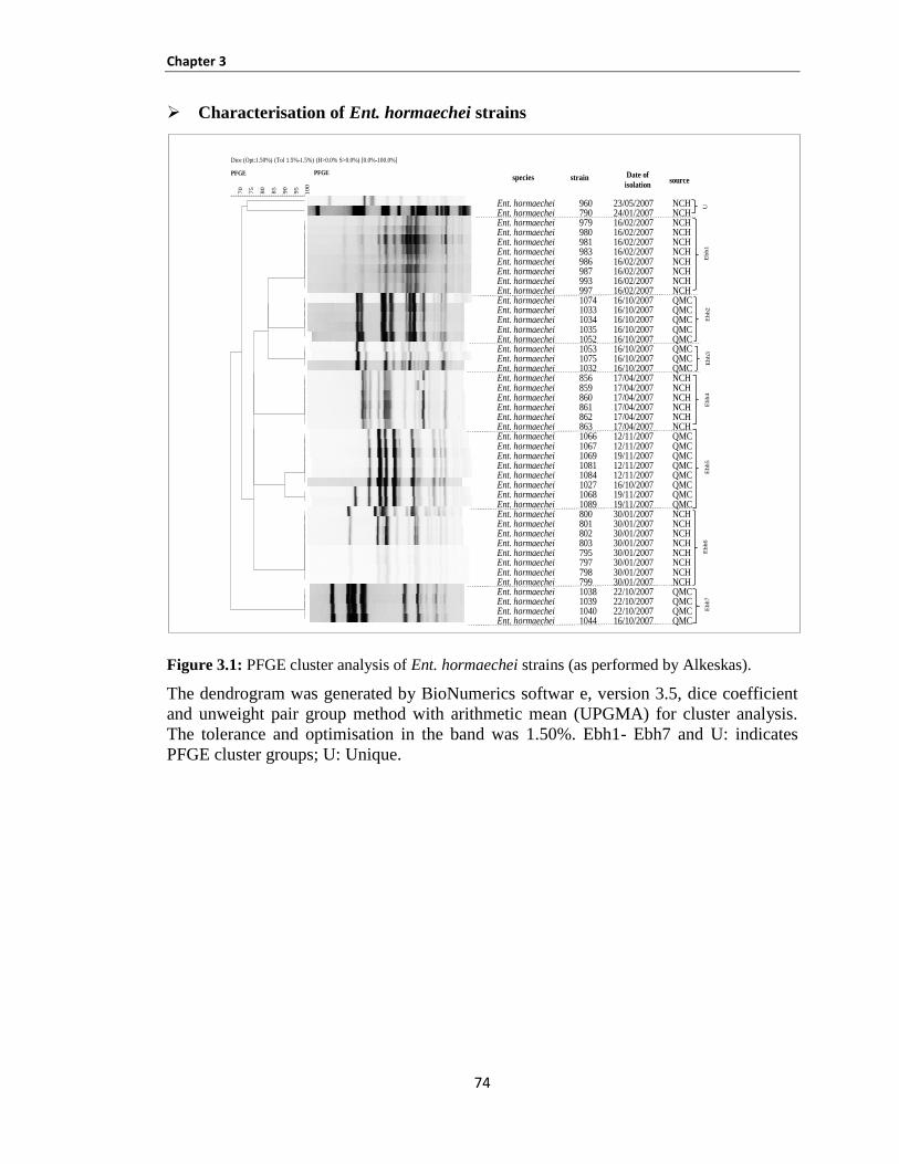
Chapter 3
74
Characterisation of Ent. hormaechei strains
Figure 3.1: PFGE cluster analysis of Ent. hormaechei strains (as performed by Alkeskas).
The dendrogram was generated by BioNumerics softwar e, version 3.5, dice coefficient
and unweight pair group method with arithmetic mean (UPGMA) for cluster analysis.
The tolerance and optimisation in the band was 1.50%. Ebh1- Ebh7 and U: indicates
PFGE cluster groups; U: Unique.
Dice (Opt:1.50%) (Tol 1.5%-1.5%) (H>0.0% S>0.0%) [0.0%-100.0%]
PFGE
10
0
95
90
85
80
75
70
PFGE
.
.
.
.
.
.
.
.
.
.
.
.
.
.
.
.
.
.
.
.
.
.
.
.
.
.
.
.
.
.
.
.
.
.
.
.
.
.
.
.
.
.
.
.
Ent. hormaecheiEnt. hormaecheiEnt. hormaecheiEnt. hormaecheiEnt. hormaecheiEnt. hormaecheiEnt. hormaecheiEnt. hormaecheiEnt. hormaecheiEnt. hormaecheiEnt. hormaecheiEnt. hormaecheiEnt. hormaecheiEnt. hormaecheiEnt. hormaecheiEnt. hormaecheiEnt. hormaecheiEnt. hormaecheiEnt. hormaecheiEnt. hormaecheiEnt. hormaecheiEnt. hormaecheiEnt. hormaecheiEnt. hormaecheiEnt. hormaecheiEnt. hormaecheiEnt. hormaecheiEnt. hormaecheiEnt. hormaecheiEnt. hormaecheiEnt. hormaecheiEnt. hormaecheiEnt. hormaecheiEnt. hormaecheiEnt. hormaecheiEnt. hormaecheiEnt. hormaecheiEnt. hormaecheiEnt. hormaecheiEnt. hormaecheiEnt. hormaecheiEnt. hormaecheiEnt. hormaecheiEnt. hormaechei
96079097998098198398698799399710741033103410351052105310751032856859860861862863106610671069108110841027106810898008018028037957977987991038103910401044
23/05/200724/01/200716/02/200716/02/200716/02/200716/02/200716/02/200716/02/200716/02/200716/02/200716/10/200716/10/200716/10/200716/10/200716/10/200716/10/200716/10/200716/10/200717/04/200717/04/200717/04/200717/04/200717/04/200717/04/200712/11/200712/11/200719/11/200712/11/200712/11/200716/10/200719/11/200719/11/200730/01/200730/01/200730/01/200730/01/200730/01/200730/01/200730/01/200730/01/200722/10/200722/10/200722/10/200716/10/2007
NCHNCHNCHNCHNCHNCHNCHNCHNCHNCHQMCQMCQMCQMCQMCQMCQMCQMCNCHNCHNCHNCHNCHNCHQMCQMCQMCQMCQMCQMCQMCQMCNCHNCHNCHNCHNCHNCHNCHNCHQMCQMCQMCQMC
UE
bh1
Ebh2
Ebh3
Ebh4
Ebh5
Ebh6
Ebh7
Date of
isolationstrainspecies source
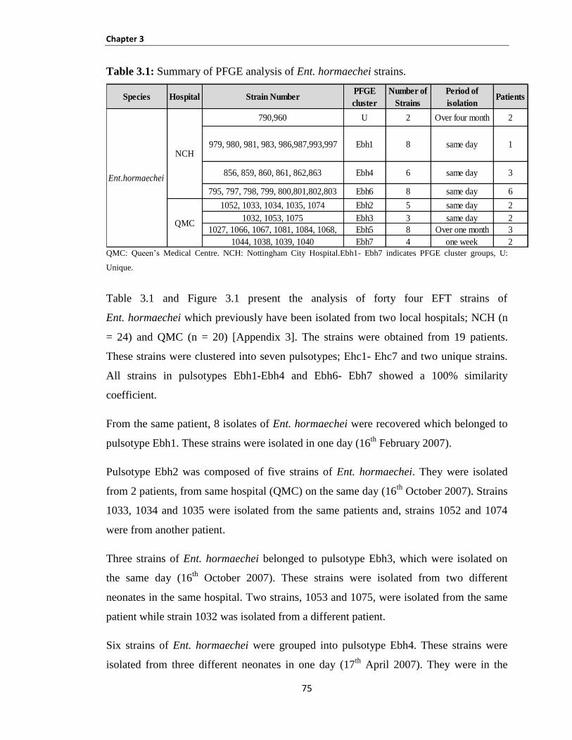
Chapter 3
75
Table 3.1: Summary of PFGE analysis of Ent. hormaechei strains.
QMC: Queen’s Medical Centre. NCH: Nottingham City Hospital.Ebh1- Ebh7 indicates PFGE cluster groups, U:
Unique.
Table 3.1 and Figure 3.1 present the analysis of forty four EFT strains of
Ent. hormaechei which previously have been isolated from two local hospitals; NCH (n
= 24) and QMC (n = 20) [Appendix 3]. The strains were obtained from 19 patients.
These strains were clustered into seven pulsotypes; Ehc1- Ehc7 and two unique strains.
All strains in pulsotypes Ebh1-Ebh4 and Ebh6- Ebh7 showed a 100% similarity
coefficient.
From the same patient, 8 isolates of Ent. hormaechei were recovered which belonged to
pulsotype Ebh1. These strains were isolated in one day (16th
February 2007).
Pulsotype Ebh2 was composed of five strains of Ent. hormaechei. They were isolated
from 2 patients, from same hospital (QMC) on the same day (16th
October 2007). Strains
1033, 1034 and 1035 were isolated from the same patients and, strains 1052 and 1074
were from another patient.
Three strains of Ent. hormaechei belonged to pulsotype Ebh3, which were isolated on
the same day (16th
October 2007). These strains were isolated from two different
neonates in the same hospital. Two strains, 1053 and 1075, were isolated from the same
patient while strain 1032 was isolated from a different patient.
Six strains of Ent. hormaechei were grouped into pulsotype Ebh4. These strains were
isolated from three different neonates in one day (17th
April 2007). They were in the
Species Hospital Strain NumberPFGE
cluster
Number of
Strains
Period of
isolationPatients
790,960 U 2 Over four month 2
979, 980, 981, 983, 986,987,993,997 Ebh1 8 same day 1
856, 859, 860, 861, 862,863 Ebh4 6 same day 3
795, 797, 798, 799, 800,801,802,803 Ebh6 8 same day 6
1052, 1033, 1034, 1035, 1074 Ebh2 5 same day 2
1032, 1053, 1075 Ebh3 3 same day 2
1027, 1066, 1067, 1081, 1084, 1068, Ebh5 8 Over one month 3
1044, 1038, 1039, 1040 Ebh7 4 one week 2
NCH
QMC
Ent.hormaechei
Chapter 3
76
same hospital. Four strains, 860-863, were isolated from the same patient whereas strains
856 and 859 were isolated from two different patients.
A total of eight Ent. hormaechei strains belonging to pulsotype Ebh5 were isolated
during a period of one month (16th
October and 12th
and 19th
November 2007) from 3
different patients in the same hospital (QMC). Pulsotype Ebh5 was composed of two
sub-clusters. The similarity coefficient of each sub-cluster was 100% validating the
clonality of the isolates in each sub-cluster. While the similarity coefficient of strains
1068 and 1089 with the other 6 strains (1066, 1067, 1069, 1081, 1084 and 1027) was 91%
which again validates that these isolates are higly identical. The difference between these
strains was only one band.
Eight Ent. hormaechei strains belonged to pulsotype Ebh6 which were isolated on the
same day (30th
January 2007) from six different patients in the same hospital (NCH).
Four strains out of eight strains were isolated from two different patients. Strains 795 and
797 were isolated from the same patient while strains 798 and 799 were isolated from
another patient.
Four Ent. hormaechei strains were isolated during a one week period (16th
and 22nd
October 2007) and belonged to pulsotype Ebh7. These were isolated from 2 different
patients in the same hospital (QMC). Three strains of pulsotype Ebh7 were isolated from
the same patient on the same day (22nd
October 2007) while one strain was isolated from
another patient on different day (16th
October 2007). The similarity coefficient of the
cluster was 100%.
The remaining two Ent. hormaechei strains (790 and 960) were unique and were isolated
from two different patients from the same hospital.
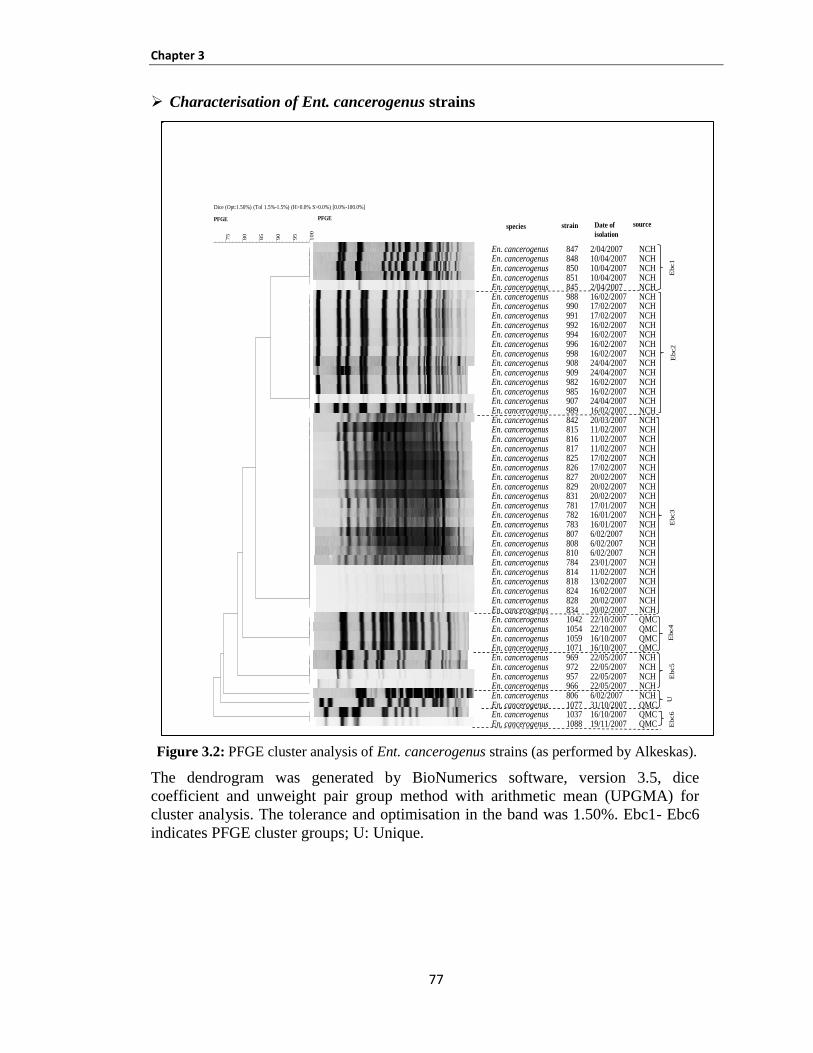
Chapter 3
77
Characterisation of Ent. cancerogenus strains
Figure 3.2: PFGE cluster analysis of Ent. cancerogenus strains (as performed by Alkeskas).
The dendrogram was generated by BioNumerics software, version 3.5, dice
coefficient and unweight pair group method with arithmetic mean (UPGMA) for
cluster analysis. The tolerance and optimisation in the band was 1.50%. Ebc1- Ebc6
indicates PFGE cluster groups; U: Unique.
Dice (Opt:1.50%) (Tol 1.5%-1.5%) (H>0.0% S>0.0%) [0.0%-100.0%]
PFGE
10
0
95
90
85
80
75
PFGE
.
.
.
.
.
.
.
.
.
.
.
.
.
.
.
.
.
.
.
.
.
.
.
.
.
.
.
.
.
.
.
.
.
.
.
.
.
.
.
.
.
.
.
.
.
.
.
.
.
.
.
En. cancerogenusEn. cancerogenusEn. cancerogenusEn. cancerogenusEn. cancerogenusEn. cancerogenusEn. cancerogenusEn. cancerogenusEn. cancerogenusEn. cancerogenusEn. cancerogenusEn. cancerogenusEn. cancerogenusEn. cancerogenusEn. cancerogenusEn. cancerogenusEn. cancerogenusEn. cancerogenusEn. cancerogenusEn. cancerogenusEn. cancerogenusEn. cancerogenusEn. cancerogenusEn. cancerogenusEn. cancerogenusEn. cancerogenusEn. cancerogenusEn. cancerogenusEn. cancerogenusEn. cancerogenusEn. cancerogenusEn. cancerogenusEn. cancerogenusEn. cancerogenusEn. cancerogenusEn. cancerogenusEn. cancerogenusEn. cancerogenusEn. cancerogenusEn. cancerogenusEn. cancerogenusEn. cancerogenusEn. cancerogenusEn. cancerogenusEn. cancerogenusEn. cancerogenusEn. cancerogenusEn. cancerogenusEn. cancerogenusEn. cancerogenusEn. cancerogenus
8478488508518459889909919929949969989089099829859079898428158168178258268278298317817827838078088107848148188248288341042105410591071969972957966806107710371088
2/04/200710/04/200710/04/200710/04/20072/04/200716/02/200717/02/200717/02/200716/02/200716/02/200716/02/200716/02/200724/04/200724/04/200716/02/200716/02/200724/04/200716/02/200720/03/200711/02/200711/02/200711/02/200717/02/200717/02/200720/02/200720/02/200720/02/200717/01/200716/01/200716/01/20076/02/20076/02/20076/02/200723/01/200711/02/200713/02/200716/02/200720/02/200720/02/200722/10/200722/10/200716/10/200716/10/200722/05/200722/05/200722/05/200722/05/20076/02/200731/10/200716/10/200719/11/2007
NCHNCHNCHNCHNCHNCHNCHNCHNCHNCHNCHNCHNCHNCHNCHNCHNCHNCHNCHNCHNCHNCHNCHNCHNCHNCHNCHNCHNCHNCHNCHNCHNCHNCHNCHNCHNCHNCHNCHQMCQMCQMCQMCNCHNCHNCHNCHNCHQMCQMCQMC
Ebc1
Ebc3
Ebc4
Ebc5
UE
bc6
Date of
isolation
strainspecies source
Ebc2
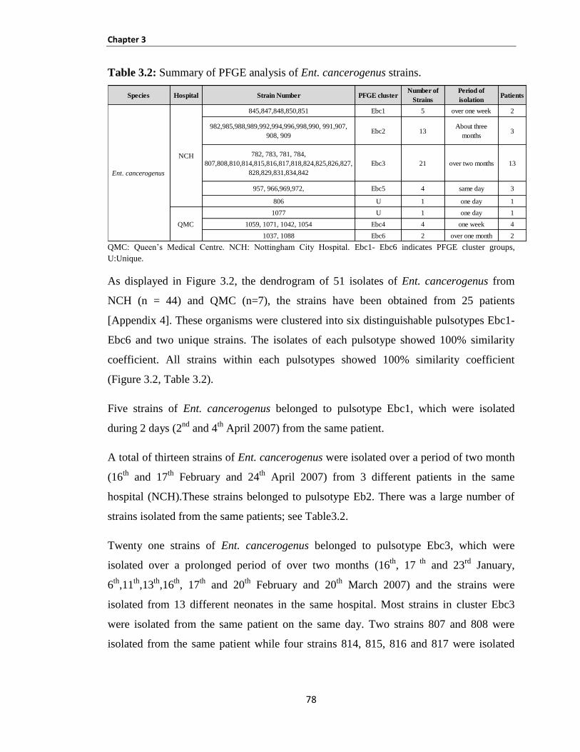
Chapter 3
78
Table 3.2: Summary of PFGE analysis of Ent. cancerogenus strains.
QMC: Queen’s Medical Centre. NCH: Nottingham City Hospital. Ebc1- Ebc6 indicates PFGE cluster groups,
U:Unique.
As displayed in Figure 3.2, the dendrogram of 51 isolates of Ent. cancerogenus from
NCH (n = 44) and QMC (n=7), the strains have been obtained from 25 patients
[Appendix 4]. These organisms were clustered into six distinguishable pulsotypes Ebc1-
Ebc6 and two unique strains. The isolates of each pulsotype showed 100% similarity
coefficient. All strains within each pulsotypes showed 100% similarity coefficient
(Figure 3.2, Table 3.2).
Five strains of Ent. cancerogenus belonged to pulsotype Ebc1, which were isolated
during 2 days (2nd
and 4th
April 2007) from the same patient.
A total of thirteen strains of Ent. cancerogenus were isolated over a period of two month
(16th
and 17th
February and 24th
April 2007) from 3 different patients in the same
hospital (NCH).These strains belonged to pulsotype Eb2. There was a large number of
strains isolated from the same patients; see Table3.2.
Twenty one strains of Ent. cancerogenus belonged to pulsotype Ebc3, which were
isolated over a prolonged period of over two months (16th
, 17 th
and 23rd
January,
6th
,11th
,13th
,16th
, 17th
and 20th
February and 20th
March 2007) and the strains were
isolated from 13 different neonates in the same hospital. Most strains in cluster Ebc3
were isolated from the same patient on the same day. Two strains 807 and 808 were
isolated from the same patient while four strains 814, 815, 816 and 817 were isolated
Species Hospital Strain Number PFGE clusterNumber of
Strains
Period of
isolationPatients
845,847,848,850,851 Ebc1 5 over one week 2
982,985,988,989,992,994,996,998,990, 991,907,
908, 909Ebc2 13
About three
months3
782, 783, 781, 784,
807,808,810,814,815,816,817,818,824,825,826,827,
828,829,831,834,842
Ebc3 21 over two months 13
957, 966,969,972, Ebc5 4 same day 3
806 U 1 one day 1
1077 U 1 one day 1
1059, 1071, 1042, 1054 Ebc4 4 one week 4
1037, 1088 Ebc6 2 over one month 2
Ent. cancerogenus
NCH
QMC
Chapter 3
79
from one patient. In addition, two strains 825 and 826 were isolated from the same
patient. Also strains 827, 828, 829 and 831 were isolated from the same patients.
There were four strains of Ent. cancerogenus in pulsotype Ebc4, which were isolated in
one week period (16th
and 22nd
October 2007) from 4 different patients in the same
hospital (QMC).
Four strains of Ent. cancerogenus belonged to pulsotype Ebc5, which were isolated on
the same day (22nd
May 2007) and the strains were isolated from three different neonates
in the same hospital. Two strains 969 and 972 were isolated from the same patient while
strain 957 and 966 were isolated from different patients.
Pulsotypes Ebc6 was composed of two strains of Ent. cancerogenus isolated over a
prolonged period of one month (16th
October 2007 and 19th
November 2007) isolated
from 2 different patients in the same hospital (QMC).
Two strains (806 and 1077) were unique and were isolated from two different patients
from different hospitals.
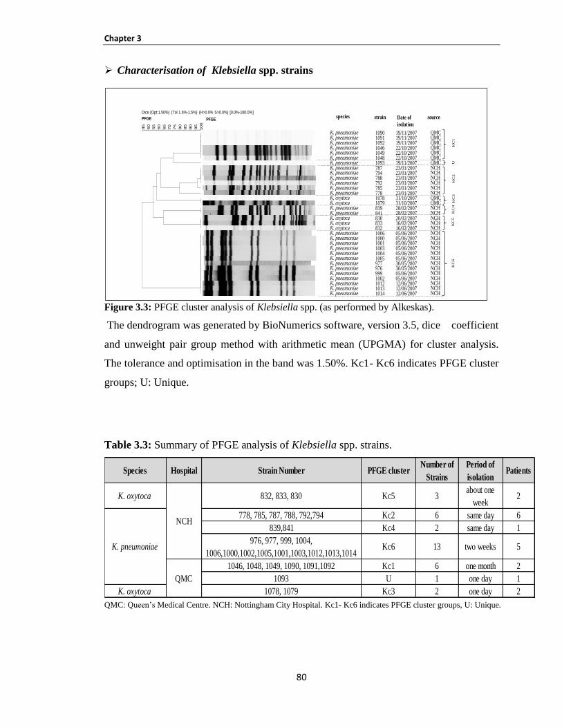
Chapter 3
80
Characterisation of Klebsiella spp. strains
Figure 3.3: PFGE cluster analysis of Klebsiella spp. (as performed by Alkeskas).
The dendrogram was generated by BioNumerics software, version 3.5, dice coefficient
and unweight pair group method with arithmetic mean (UPGMA) for cluster analysis.
The tolerance and optimisation in the band was 1.50%. Kc1- Kc6 indicates PFGE cluster
groups; U: Unique.
Table 3.3: Summary of PFGE analysis of Klebsiella spp. strains.
QMC: Queen’s Medical Centre. NCH: Nottingham City Hospital. Kc1- Kc6 indicates PFGE cluster groups, U: Unique.
Dice (Opt:1.50%) (Tol 1.5%-1.5%) (H>0.0% S>0.0%) [0.0%-100.0%]
PFGE
100
95
90
85
80
75
70
65
60
55
50
45
PFGE
.
.
.
.
.
.
.
.
.
.
.
.
.
.
.
.
.
.
.
.
.
.
.
.
.
.
.
.
.
.
.
.
.
K. pneumoniaeK. pneumoniaeK. pneumoniaeK. pneumoniaeK. pneumoniaeK. pneumoniaeK. pneumoniaeK. pneumoniaeK. pneumoniaeK. pneumoniaeK. pneumoniaeK. pneumoniaeK. pneumoniaeK. oxytocaK. oxytocaK. pneumoniaeK. pneumoniaeK. oxytocaK. oxytocaK. oxytocaK. pneumoniaeK. pneumoniaeK. pneumoniaeK. pneumoniaeK. pneumoniaeK. pneumoniaeK. pneumoniaeK. pneumoniaeK. pneumoniaeK. pneumoniaeK. pneumoniaeK. pneumoniaeK. pneumoniae
1090109110921046104910481093787794788792785778107810798398418308338321006100010011003100410059779769991002101210131014
19/11/200719/11/200719/11/200722/10/200722/10/200722/10/200719/11/200723/01/200723/01/200723/01/200723/01/200723/01/200723/01/200731/10/200731/10/200728/02/200728/02/200720/02/200716/02/200716/02/200705/06/200705/06/200705/06/200705/06/200705/06/200705/06/200730/05/200730/05/200705/06/200705/06/200712/06/200712/06/200712/06/2007
QMCQMCQMCQMCQMCQMCQMCNCHNCHNCHNCHNCHNCHQMCQMCNCHNCHNCHNCHNCHNCHNCHNCHNCHNCHNCHNCHNCHNCHNCHNCHNCHNCH
KC
1U
KC
2K
C3
KC
4K
C5
KC
6
Date of
isolation
strainspecies source
Species Hospital Strain Number PFGE clusterNumber of
Strains
Period of
isolationPatients
K. oxytoca 832, 833, 830 Kc5 3about one
week2
778, 785, 787, 788, 792,794 Kc2 6 same day 6
839,841 Kc4 2 same day 1
976, 977, 999, 1004,
1006,1000,1002,1005,1001,1003,1012,1013,1014Kc6 13 two weeks 5
1046, 1048, 1049, 1090, 1091,1092 Kc1 6 one month 2
1093 U 1 one day 1
K. oxytoca 1078, 1079 Kc3 2 one day 2
NCH
QMC
K. pneumoniae
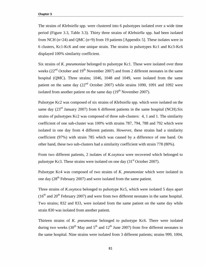
Chapter 3
81
The strains of Klebsiella spp. were clustered into 6 pulsotypes isolated over a wide time
period (Figure 3.3, Table 3.3). Thirty three strains of Klebsiella spp. had been isolated
from NCH (n=24) and QMC (n=9) from 19 patients [Appendix 5]. These isolates were in
6 clusters, Kc1-Kc6 and one unique strain. The strains in pulsotypes Kc1 and Kc3-Kc6
displayed 100% similarity coefficient.
Six strains of K. pneumoniae belonged to pulsotype Kc1. These were isolated over three
weeks (22nd
October and 19th
November 2007) and from 2 different neonates in the same
hospital (QMC). Three strains; 1046, 1048 and 1049, were isolated from the same
patient on the same day (22nd
October 2007) while strains 1090, 1091 and 1092 were
isolated from another patient on the same day (19th
November 2007).
Pulsotype Kc2 was composed of six strains of Klebsiella spp. which were isolated on the
same day (23rd
January 2007) from 6 different patients in the same hospital (NCH).Six
strains of pulsotypes Kc2 was composed of three sub-clusters: 4, 1 and 1. The similarity
coefficient of one sub-cluster was 100% with strains 787, 794, 788 and 792 which were
isolated in one day from 4 different patients. However, these strains had a similarity
coefficient (97%) with strain 785 which was caused by a difference of one band. On
other hand, these two sub-clusters had a similarity coefficient with strain 778 (80%).
From two different patients, 2 isolates of K.oxytoca were recovered which belonged to
pulsotype Kc3. These strains were isolated on one day (31st
October 2007).
Pulsotype Kc4 was composed of two strains of K. pneumoniae which were isolated in
one day (28th
February 2007) and were isolated from the same patient.
Three strains of K.oxytoca belonged to pulsotype Kc5, which were isolated 5 days apart
(16th
and 20th
February 2007) and were from two different neonates in the same hospital.
Two strains; 832 and 833, were isolated from the same patient on the same day while
strain 830 was isolated from another patient.
Thirteen strains of K. pneumoniae belonged to pulsotype Kc6. There were isolated
during two weeks (30th
May and 5th
and 12th
June 2007) from five different neonates in
the same hospital. Nine strains were isolated from 3 different patients; strains 999, 1004,

Chapter 3
82
1006 from one patient, another three strains 1000, 1002 and 1005 were from the second
and strains 1012, 1013 and 1014 from third patient. In addition, four strains were isolated
from two different patients; 976, 977, 1001 and 1003.
One strain, 1093, was unique and was isolated from one patient.
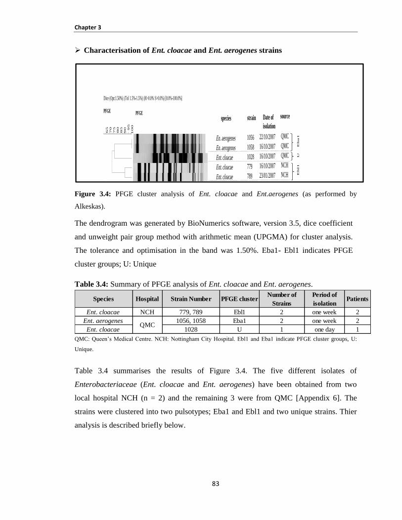
Chapter 3
83
Characterisation of Ent. cloacae and Ent. aerogenes strains
Figure 3.4: PFGE cluster analysis of Ent. cloacae and Ent.aerogenes (as performed by
Alkeskas).
The dendrogram was generated by BioNumerics software, version 3.5, dice coefficient
and unweight pair group method with arithmetic mean (UPGMA) for cluster analysis.
The tolerance and optimisation in the band was 1.50%. Eba1- Ebl1 indicates PFGE
cluster groups; U: Unique
Table 3.4: Summary of PFGE analysis of Ent. cloacae and Ent. aerogenes.
QMC: Queen’s Medical Centre. NCH: Nottingham City Hospital. Ebl and Eba indicate PFGE cluster groups, U:
Unique.
Table 3.4 summarises the results of Figure 3.4. The five different isolates of
Enterobacteriaceae (Ent. cloacae and Ent. aerogenes) have been obtained from two
local hospital NCH (n = 2) and the remaining 3 were from QMC [Appendix 6]. The
strains were clustered into two pulsotypes; Eba1 and Ebl1 and two unique strains. Thier
analysis is described briefly below.
Species Hospital Strain Number PFGE clusterNumber of
Strains
Period of
isolationPatients
Ent. cloacae NCH 779, 789 Ebl1 2 one week 2
Ent. aerogenes 1056, 1058 Eba1 2 one week 2
Ent. cloacae 1028 U 1 one day 1QMC
Dice (Opt:1.50%) (Tol 1.5%-1.5%) (H>0.0% S>0.0%) [0.0%-100.0%]
PFGE
10
0
95
90
85
80
75
70
65
PFGE
.
.
.
.
.
.
En. aerogenes
En. aerogenes
Ent. cloacae
Ent. cloacae
Ent. cloacae
Shig. flexneri
1056
1058
1028
779
789
1094
22/10/2007
16/10/2007
16/10/2007
16/10/2007
23/01/2007
16/10/2007
QMC
QMC
QMC
NCH
NCH
QMC
Eba1
UE
bl1
U
Date of
isolation
strainspecies sourceD
ice
(Opt
:1.5
0%)
(Tol
1.5%
-1.5
%)
(H>
0.0%
S>
0.0%
) [0
.0%
-100
.0%
]
PF
GE
100
95
90
85
80
75
70
65
PF
GE
. . . . . .
En.
aer
ogen
es
En.
aer
ogen
es
Ent
. clo
acae
Ent
. clo
acae
Ent
. clo
acae
Shig
. fle
xner
i
1056
1058
1028
779
789
1094
22/1
0/20
07
16/1
0/20
07
16/1
0/20
07
16/1
0/20
07
23/0
1/20
07
16/1
0/20
07
QM
C
QM
C
QM
C
NC
H
NC
H
QM
C
Eba1 U Ebl1 U
Dat
e of
isol
atio
n
stra
insp
ecie
sso
urce
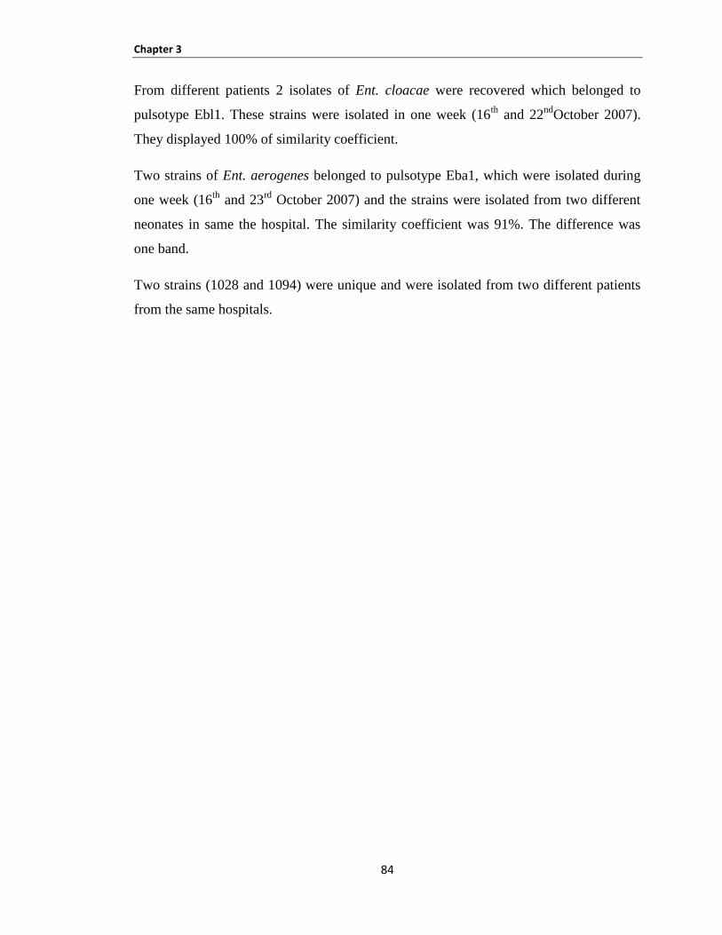
Chapter 3
84
From different patients 2 isolates of Ent. cloacae were recovered which belonged to
pulsotype Ebl1. These strains were isolated in one week (16th
and 22nd
October 2007).
They displayed 100% of similarity coefficient.
Two strains of Ent. aerogenes belonged to pulsotype Eba1, which were isolated during
one week (16th
and 23rd
October 2007) and the strains were isolated from two different
neonates in same the hospital. The similarity coefficient was 91%. The difference was
one band.
Two strains (1028 and 1094) were unique and were isolated from two different patients
from the same hospitals.
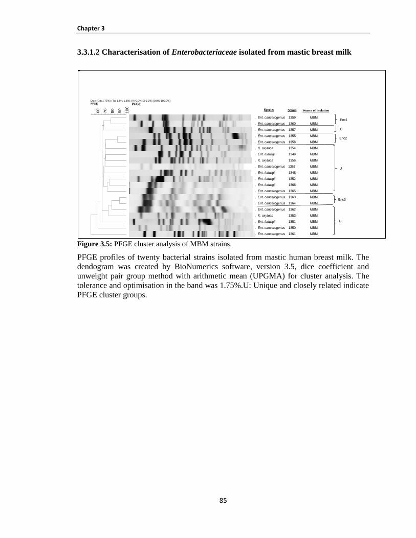
Chapter 3
85
Characterisation of Enterobacteriaceae isolated from mastic breast milk 3.3.1.2
Figure 3.5: PFGE cluster analysis of MBM strains.
PFGE profiles of twenty bacterial strains isolated from mastic human breast milk. The
dendogram was created by BioNumerics software, version 3.5, dice coefficient and
unweight pair group method with arithmetic mean (UPGMA) for cluster analysis. The
tolerance and optimisation in the band was 1.75%.U: Unique and closely related indicate
PFGE cluster groups.
Dice (Opt:1.75%) (Tol 1.8%-1.8%) (H>0.0% S>0.0%) [0.0%-100.0%]PFGE
10
0
90
80
70
60
PFGE
.
.
.
.
.
.
.
.
.
.
.
.
.
.
.
.
.
.
.
.
Ent. cancerogenus
Ent. cancerogenus
Ent. cancerogenus
Ent. cancerogenus
Ent. cancerogenus
K. oxytoca
Ent. ludwigii
K. oxytoca
Ent. cancerogenus
Ent. ludwigii
Ent. ludwigii
Ent. ludwigii
Ent. cancerogenus
Ent. cancerogenus
Ent. cancerogenus
Ent. cancerogenus
K. oxytoca
Ent. ludwigii
Ent. cancerogenus
Ent. cancerogenus
1359
1360
1357
1355
1358
1354
1349
1356
1367
1348
1352
1366
1365
1363
1364
1362
1353
1351
1350
1361
MBM
MBM
MBM
MBM
MBM
MBM
MBM
MBM
MBM
MBM
MBM
MBM
MBM
MBM
MBM
MBM
MBM
MBM
MBM
MBM
Species Strain Source of isolation
Enc1
U
Enc2
U
Enc3
U

Chapter 3
86
Table 3.5: Summary of PFGE analysis of Enterobacteriaceae isolated from mastic
human breast milk (MBM).
Enc1-Enc3 indicate PFGE cluster groups, U: Unique.
Table 3.5 is a summary of Figure 3.5 which presents the analysis of PFGE of twenty
strains of Ent. ludwigii, Ent. cancerogenus and K. oxytoca from MBM in Spain. The
MBM isolates were clustered into 3 pulsotypes Enc1, Enc2 and Enc3 and 14 unique
strains (Figure 3.5, Table 3.5). The PFGE of the MBM isolates revealed that two strains;
1359 and 1360 (Enc1), showed 78% of similarity coefficient while two strains; 1355 and
1358 (Enc2), were 89% similar according to Dice analysis and differed by only 3 bands
(Figure 3.5). The similarity coefficient of two strains (1363 and 1364) with the others
showed a similarity coefficient of 92%, which were clustered as Enc3. Therefore, they
are probably six isolates of the same clone (Tenover et al., 1995). Since the original
sampling by Delgado was confidential and little information exists with respect to the
clustering and typing of strains we do not know if each of the two strains were from the
same mother, but this was deemed highly likely. All other strains were < 80% similar
and were therefore regarded as unique isolates, i.e. not clonal.
Species Strain Number PFGE clusterGenetic
relatedness
1359, 1360 Enc1
1355, 1358 Enc2
1363, 1364 Enc3
1350,1357, 1361, 1362, 1365
& 1367
Ent. ludwigii1348,1349,1351, 1352 &
1366
K.oxytoca 1353, 1354 & 1356
closely related
Unrelated
Ent. cancerogenus
U
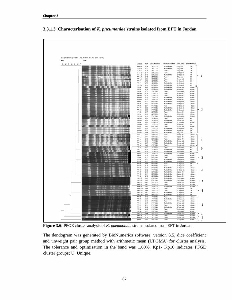
Chapter 3
87
Characterisation of K. pneumoniae strains isolated from EFT in Jordan 3.3.1.3
Figure 3.6: PFGE cluster analysis of K. pneumoniae strains isolated from EFT in Jordan.
The dendogram was generated by BioNumerics software, version 3.5, dice coefficient
and unweight pair group method with arithmetic mean (UPGMA) for cluster analysis.
The tolerance and optimisation in the band was 1.60%. Kp1- Kp10 indicates PFGE
cluster groups; U: Unique.
Kp
1K
p2
Kp
3K
p5
Kp
9K
p8
Kp
7K
p6
Kp
4
strain
Dice (Opt:1.60%) (Tol 1.6%-1.6%) (H>0.0% S>0.0%) [0.0%-100.0%]
pfge
100
95
90
85
80
75
70
pfge
.
.
.
.
.
.
.
.
.
.
.
.
.
.
.
.
.
.
.
.
.
.
.
.
.
.
.
.
.
.
.
.
.
.
.
.
.
.
.
.
.
.
.
.
.
.
.
.
.
.
.
.
.
.
.
.
.
.
.
.
.
.
.
.
.
.
.
.
.
.
.
.
.
.
.
PRH AE
PRH AE
PRH AT
PRH AD
PRH AD
PRH AT
PRH AI
PRH Y
PRH Y
PRH W
PRH P
PRH L
PRH F
PRH O
PRH O
PRH O
PRH O
KAH I
KAH I
KAH I
PRH G
PRH G
KAH 6
KAH 6
KAH 34
PRH Q
PRH AI
PRH P
PRH Q
PRH A
PRH S
PRH Q
PRH W
PRH J
PRH J
PRH J
PRH A
PRH A
PRH A
PRH S
PRH T
PRH S
PRH N
PRH E
PRH E
PRH E
PRH E
PRH H
PRH H
PRH H
PRH H
PRHJ
PRHJ
PRH L
KAH 17
KAH 17
KAH 16
KAH 16
KAH 16
KAH1 15
KAH1 15
KAH1 15
KAH1 15
PRH B
PRH B
PRH C
PRH C
PRH C
PRH C
PRH D
PRH D
KAH 16
KAH 34
KAH 34
PRHB
.
.
.
.
.
.
.
.
.
.
.
.
.
.
.
.
.
.
.
.
.
.
.
.
.
.
.
.
.
.
.
.
.
.
.
.
.
.
.
.
.
.
.
.
.
.
.
.
.
.
.
.
.
.
.
.
.
.
.
.
.
.
.
.
.
.
.
.
.
.
.
.
.
.
.
1746
1747
1748
1744
1745
1749
1751
1752
1753
1754
1681
1716
1717
1718
1719
1720
1721
1722
1723
1724
1725
1726
1727
1728
1736
1743
1750
1682
1742
1689
1740
1741
1756
1683
1684
1685
1686
1687
1688
1737
1738
1739
1729
1730
1731
1732
1733
1709
1710
1711
1712
1713
1714
1715
1699
1700
1701
1702
1703
1705
1706
1707
1708
1691
1692
1693
1695
1694
1696
1698
1697
1704
1735
1734
1690
.
.
.
.
.
.
.
.
.
.
.
.
.
.
.
.
.
.
.
.
.
.
.
.
.
.
.
.
.
.
.
.
.
.
.
.
.
.
.
.
.
.
.
.
.
.
.
.
.
.
.
.
.
.
.
.
.
.
.
.
.
.
.
.
.
.
.
.
.
.
.
.
.
.
.
15/12/2011
15/12/2011
15/12/2011
15/12/2011
15/12/2011
15/12/2011
15/12/2011
12/12/2011
12/12/2011
20/12/2011
23/07/2011
23/07/2011
7/07/2011
23/07/2011
23/07/2011
23/07/2011
23/07/2011
6/07/2011
6/07/2011
6/07/2011
6/07/2011
6/07/2011
22/05/2011
22/05/2011
7/12/2011
10/12/2011
15/12/2011
23/07/2011
10/12/2011
4/07/2011
10/12/2011
10/12/2011
20/12/2011
23/07/2011
23/07/2011
23/07/2011
4/07/2011
4/07/2011
4/07/2011
10/12/2011
10/12/2011
10/12/2011
15/07/2011
9/07/2011
9/07/2011
9/07/2011
9/07/2011
6/07/2011
6/07/2011
6/07/2011
6/07/2011
23/07/2011
23/07/2011
23/07/2011
4/07/2011
4/07/2011
31/05/2011
31/05/2011
31/05/2011
31/05/2011
31/05/2011
31/05/2011
31/05/2011
4/07/2011
4/07/2011
9/07/2011
9/07/2011
9/07/2011
9/07/2011
9/07/2011
9/07/2011
31/05/2011
7/12/2011
7/12/2011
4/07/2011
flushed tube
flushed tube
Tube
Tube
flushed tube
Tube
Tube
Tube
flushed tube
flushed tube
flushed tube
flushed tube
Tube
Tube
Tube
flushed tube
flushed tube
flushed tube
flushed tube
Tube
flushed tube
flushed tube
Tube
Tube
flushed tube
Tube
flushed tube
flushed tube
Tube
Tube
flushed tube
flushed tube
flushed tube
Tube
Tube
Tube
flushed tube
flushed tube
Tube
Tube
flushed tube
flushed tube
flushed tube
Tube
Tube
flushed tube
flushed tube
Tube
Tube
flushed tube
flushed tube
flushed tube
flushed tube
flushed tube
flushed tube
flushed tube
Tube
Tube
flushed tube
Tube
Tube
flushed tube
flushed tube
Tube
flushed tube
Tube
flushed tube
Tube
flushed tube
Tube
Tube
flushed tube
Tube
Tube
Tube
7days old
7days old
7days old
13 days old
13 days old
7days old
24days old
10 days old
10 days old
11 days old
21days old
9 days old
35 days old
9 days old
9 days old
9 days old
9 days old
39 days old
39 days old
39 days old
15 days old
15 days old
5 days old
5 days old
18 days old
8 days old
24days old
21 days old
8 days old
21days old
36 days old
8 days old
7days old
9 days old
9 days old
9 days old
21days old
21days old
21days old
36 days old
15 days old
36 days old
14 days old
19 days old
19 days old
19 days old
19 days old
16 days old
16 days old
16 days old
16 days old
9 days old
9 days old
9 days old
26 days old
26 days old
24days old
24days old
24days old
15 days old
15 days old
15 days old
15 days old
12 days old
12 days old
37days old
37days old
37days old
37days old
23 days old
23 days old
24days old
18 days old
18days old
12 days old
S26
S26
S26
S26
S26
S26
S26
S26
S26
S26
bebelac
bebelac
bebelac
bebelac
bebelac
bebelac
bebelac
bebelac
bebelac
bebelac
bebelac
bebelac
bebelac
bebelac
neosure
S26
S26
bebelac
S26
bebelac
S26
S26
S26
bebelac
bebelac
bebelac
bebelac
bebelac
bebelac
S26
S26
S26
bebelac
bebelac
bebelac
bebelac
bebelac
S26
S26
bebelac
bebelac
bebelac
bebelac
bebelac
neosure
neosure
neosure
neosure
neosure
S26
S26
S26
S26
bebelac
bebelac
bebelac
bebelac
bebelac
bebelac
bebelac
bebelac
neosure
neosure
neosure
bebelac
Location Date of isolation Source of isolation Age of infant Milk information
Kp
10
UU
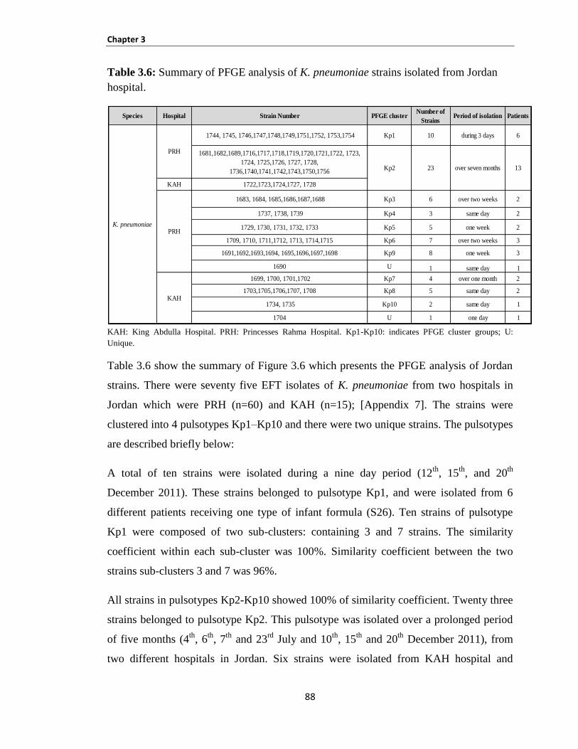
Chapter 3
88
Table 3.6: Summary of PFGE analysis of K. pneumoniae strains isolated from Jordan
hospital.
KAH: King Abdulla Hospital. PRH: Princesses Rahma Hospital. Kp1-Kp10: indicates PFGE cluster groups; U:
Unique.
Table 3.6 show the summary of Figure 3.6 which presents the PFGE analysis of Jordan
strains. There were seventy five EFT isolates of K. pneumoniae from two hospitals in
Jordan which were PRH (n=60) and KAH (n=15); [Appendix 7]. The strains were
clustered into 4 pulsotypes Kp1–Kp10 and there were two unique strains. The pulsotypes
are described briefly below:
A total of ten strains were isolated during a nine day period (12th
, 15th
, and 20th
December 2011). These strains belonged to pulsotype Kp1, and were isolated from 6
different patients receiving one type of infant formula (S26). Ten strains of pulsotype
Kp1 were composed of two sub-clusters: containing 3 and 7 strains. The similarity
coefficient within each sub-cluster was 100%. Similarity coefficient between the two
strains sub-clusters 3 and 7 was 96%.
All strains in pulsotypes Kp2-Kp10 showed 100% of similarity coefficient. Twenty three
strains belonged to pulsotype Kp2. This pulsotype was isolated over a prolonged period
of five months (4th
, 6th
, 7th
and 23rd
July and 10th
, 15th
and 20th
December 2011), from
two different hospitals in Jordan. Six strains were isolated from KAH hospital and
Species Hospital Strain Number PFGE clusterNumber of
StrainsPeriod of isolation Patients
1744, 1745, 1746,1747,1748,1749,1751,1752, 1753,1754 Kp1 10 during 3 days 6
1681,1682,1689,1716,1717,1718,1719,1720,1721,1722, 1723,
1724, 1725,1726, 1727, 1728,
1736,1740,1741,1742,1743,1750,1756
KAH 1722,1723,1724,1727, 1728
1683, 1684, 1685,1686,1687,1688 Kp3 6 over two weeks 2
1737, 1738, 1739 Kp4 3 same day 2
1729, 1730, 1731, 1732, 1733 Kp5 5 one week 2
1709, 1710, 1711,1712, 1713, 1714,1715 Kp6 7 over two weeks 3
1691,1692,1693,1694, 1695,1696,1697,1698 Kp9 8 one week 3
1690 U 1 same day 1
1699, 1700, 1701,1702 Kp7 4 over one month 2
1703,1705,1706,1707, 1708 Kp8 5 same day 2
1734, 1735 Kp10 2 same day 1
1704 U 1 one day 1
KAH
K. pneumoniae
13
PRH
Kp2
PRH
23 over seven months
Chapter 3
89
seventeen strains were isolated from PRH hospital. These strains were isolated from 13
different neonates where eight patients received bebelac, one received neosure and four
received S26 infant formula. The Kp2 pulsotypes strains were isolated from a large
number of neonates over a wide time period from two different hospitals. The result
appears unlikely and needs further confirmation by using another restriction enzyme.
Six strains of K. pneumoniae were grouped into pulsotype Kp3. These strains were
isolated from two different neonates over two weeks (4th
and 23rd
July 2011). The
hospitals used bebelac for infant feed.
Three strains of K. pneumoniae belonged to pulsotype Kp4, which were isolated on the
same day (10th
December 2011) and the strains were isolated from two different neonates,
receiving S26 infant formula.
Pulsotype Kp5 was composed of five strains of K. pneumoniae. There were isolated on
one week (9th
and 15th
July 2011) from 2 patients, receiving bebelac infant formula.
Seven strains of K. pneumoniae formed pulsotype Kp6, which were isolated over a two
week period (6th
and 23rd
July 2011) from 3 different patients receiving S26 and bebelac
infant formula.
From 2 different patients, 4 isolates of K. pneumoniae were recovered which belonged to
pulsotype Kp7. These strains were isolated over one months (31st May and 4
th July
2011).The babies were received neosure infant formula.
Five strains of K. pneumoniae belonged to pulsotype Kp8, which were isolated on the
same day (31st May 2011) from two different patients, receiving different types of infant
formula (S26 and bebelac).
At one week period (4th
and 9th
July 2011) eight strains of K. pneumoniae were isolated
which belonged to pulsotype Kp9. These were isolated from 3 different patients,
receiving bebelac infant formula.
Two strains of K. pneumoniae belonged to pulsotype Kp10, which were isolated on the
same day (7th
December 2011) and from the same patient receiving neosure infant
formula.
Chapter 3
90
Two strains (1704 and 1690) were unique and were isolated from two different patients
receiving neosure and bebelac infant formula respectively.
3.3.2 Comparison of phenotypic and genotypic techniques for identification of
Enterobacteriaceae
All strains were identified using biochemical profiles based on API 20E and ID32E
methods. Genotyping identification was also used to identify the isolates by using 16S
rDNA sequence analysis. In addition, the phenotypic and genotypic results were
compared with PFGE results.
IdentificationofEnterobacteriaceae strains isolated from EFT 3.3.2.1
Table 3.7 part1 shows the comparison between phenotyping and genotyping
identification technique. Most Ent. cloacae strains were re-identified by 16S rDNA
sequence analysis and were subsequently re-assigned as Ent. hormaechei. There were
clustered into 7 clusters Ebh1-Ebh7 and four unique strains by using PFGE technique.
An interesting result was that two strains which were initially identified as K. oxytoca by
ID32E, but were re-identified by 16S rDNA sequence analysis as Ent. aerogenes. There
were clustered in one cluster Eba1. Another two strains were identified by ID32E tests as
Ent. cloacae and Ent. amnigenus however, were re-identified by 16S rDNA sequence
analysis as Ent. hormaechei. They were clustered in one cluster Ebh7.
In Table 3.7 part 2 shows that fifty one strains were previously identified as Klebsiella
spp. and E. coli; six strains were identified as K. pneumoniae and 44 strains were
identified as K. oxytoca, whereas one strain was E. coli by using API20E/ID32E tests.
While in this study these strains were re-identified by 16S rDNA sequence analysis as
Ent. cancerogenus, but their pulsotypes clustered into 6 clusters Ebc1-Ebc6 and two
unique strains.
All K. pneumoniae and 15 of K. oxytoca strains belonged to one cluster; pulsotype Ebc2.
On the other hand, strains 1037 and 1088 were identified as K. oxytoca and E. coli
respectively by ID32E, but they were identified as Ent. cancerogenus by 16S rDNA gene
sequence analysis and both belonged to cluster Ebc6.

Chapter 3
91
All strains in table 3.8 were identified as Klebsiella spp. by both API20E/ID32E tests
and were in 6 pulsotypes Kc1-Kc6 and one unique strain. The exception was in clusters
Kc1 and Kc6 which consisted of only one E. coli and 3 Ent. aerogenes strains
respectively, as identified by ID32E test. However when re-identified by 16S rDNA, the
strains in both Kc1 and Kc6 clusters were found to be K. pneumoniae.
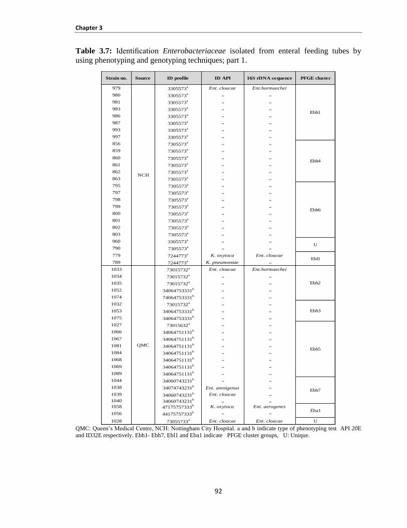
Chapter 3
92
Table 3.7: Identification Enterobacteriaceae isolated from enteral feeding tubes by
using phenotyping and genotyping techniques; part 1.
QMC: Queen’s Medical Centre, NCH: Nottingham City Hospital. a and b indicate type of phenotyping test API 20E
and ID32E respectively. Ebh1- Ebh7, Ebl1 and Eba1 indicate PFGE cluster groups, U: Unique.
Strain no. Source ID profile ID API 16S rDNA sequence PFGE cluster
979 3305573a Ent. cloacae Ent.hormaechei
980 3305573a „ „
981 3305573a „ „
983 3305573a „ „
986 3305573a „ „
987 3305573a „ „
993 3305573a „ „
997 3305573a „ „
856 7305573a „ „
859 7305573a „ „
860 7305573a „ „
861 7305573a „ „
862 7305573a „ „
863 7305573a „ „
795 7305573a „ „
797 7305573a „ „
798 7305573a „ „
799 7305573a „ „
800 7305573a „ „
801 7305573a „ „
802 7305573a „ „
803 7305573a „ „
960 3305573a „ „
790 7305573a „ „
779 7244773a K. oxytoca Ent. cloacae
789 7244773a K. pneumoniae „
1033 73015732a Ent. cloacae Ent.hormaechei
1034 73015732a „ „
1035 73015732a „ „
1052 34064753331b „ „
1074 74064753331b „ „
1032 73015732a „ „
1053 34064753331b „ „
1075 34064753331b „ „
1027 73015632a „ „
1066 34064751131b „ „
1067 34064751131b „ „
1081 34064751131b „ „
1084 34064751131b „ „
1068 34064751131b „ „
1069 34064751131b „ „
1089 34064751131b „ „
1044 34060743231b „ „
1038 34074743231b
Ent. amnigenus „
1039 34060743231b Ent. cloacae „
1040 34060743231b „ „
1058 47175757333b K. oxytoca Ent. aerogenes
1056 44175757333b „ „
1028 73055733a Ent. cloacae Ent. cloacae U
QMC
Eba1
Ebh6
Ebh5
U
Ebh2
Ebh7
Ebl1
NCH
Ebh1
Ebh3
Ebh4
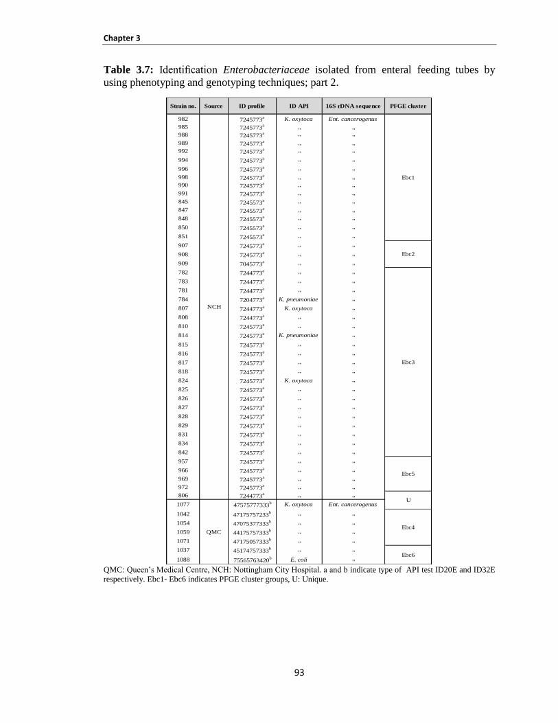
Chapter 3
93
Table 3.7: Identification Enterobacteriaceae isolated from enteral feeding tubes by
using phenotyping and genotyping techniques; part 2.
QMC: Queen’s Medical Centre, NCH: Nottingham City Hospital. a and b indicate type of API test ID20E and ID32E
respectively. Ebc1- Ebc6 indicates PFGE cluster groups, U: Unique.
Strain no. Source ID profile ID API 16S rDNA sequence PFGE cluster
982 7245773a K. oxytoca Ent. cancerogenus
985 7245773a „ „
988 7245773a „ „
989 7245773a „ „
992 7245773a „ „
994 7245773a „ „
996 7245773a „ „
998 7245773a „ „
990 7245773a „ „
991 7245773a „ „
845 7245573a „ „
847 7245573a „ „
848 7245573a „ „
850 7245573a „ „
851 7245573a „ „
907 7245773a „ „
908 7245773a „ „
909 7045773a „ „
782 7244773a „ „
783 7244773a „ „
781 7244773a „ „
784 7204773a K. pneumoniae „
807 7244773a K. oxytoca „
808 7244773a „ „
810 7245773a „ „
814 7245773a K. pneumoniae „
815 7245773a „ „
816 7245773a „ „
817 7245773a „ „
818 7245773a „ „
824 7245773a K. oxytoca „
825 7245773a „ „
826 7245773a „ „
827 7245773a „ „
828 7245773a „ „
829 7245773a „ „
831 7245773a „ „
834 7245773a „ „
842 7245773a „ „
957 7245773a „ „
966 7245773a „ „
969 7245773a „ „
972 7245773a „ „
806 7244773a „ „
1077 47575777333b K. oxytoca Ent. cancerogenus
1042 47175757233b „ „
1054 47075377333b „ „
1059 44175757333b „ „
1071 47175057333b „ „
1037 45174757333b „ „
1088 75565763420b E. coli „
Ebc1
Ebc2
QMC
U
Ebc3
Ebc6
Ebc4
Ebc5
NCH
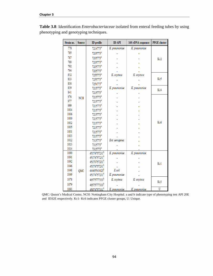
Chapter 3
94
Table 3.8: Identification Enterobacteriaceae isolated from enteral feeding tubes by using
phenotyping and genotyping techniques.
QMC: Queen’s Medical Centre, NCH: Nottingham City Hospital. a and b indicate type of phenotyping test API 20E
and ID32E respectively. Kc1- Kc6 indicates PFGE cluster groups, U: Unique.
Chapter 3
95
IdentificationofEnterobacteriaceae isolated from mastic human breast 3.3.2.2
milk
The strains previously isolated from MBM were further analysed by phenotyping using
ID32E following the procedure in section 2.2.1. The results are summarised in Table 3.9.
As shown, all strains were identified as K. oxytoca. 16S rDNA gene sequence analysis
was used to identify the recovered bacterial strains. As shown in Table 3.9, there was
variation between ID32E results and 16S identification. The results obtained for the
MBM isolates showed that out of the twenty strains identified as K. oxytoca by ID32E,
twelve were Ent. cancerogenus and five were Ent. ludwigii with the more reliable 16S
rDNA sequence analysis; see Table 3.9.
The interesting finding was that six strains which were Ent. cancerogenus (1359, 1360,
1355, 1358, 1363 and 1364) were identified as K. oxytoca by ID32E test and their ID
profile was different; Table 3.9. However, these strains were identified by 16S rDNA
sequence analysis as Ent. cancerogenus and their pulsotypes were Enc1, Enc2 and Enc3.
Each of the two strains belonged to one cluster respectively; see Table 3.9.
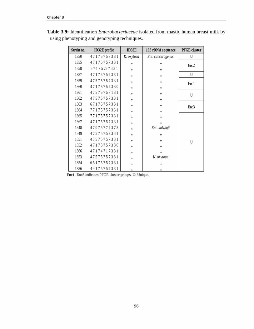
Chapter 3
96
Table 3.9: Identification Enterobacteriaceae isolated from mastic human breast milk by
using phenotyping and genotyping techniques.
Enc1- Enc3 indicates PFGE cluster groups, U: Unique.
Strain no. ID32E profile ID32E 16S rDNA sequence PFGE cluster
1350 4 7 1 7 5 7 5 7 3 3 1 K. oxytoca Ent. cancerogenus U
1355 4 7 1 7 5 7 5 7 3 3 1 „ „
1358 5 7 1 7 5 75 7 3 3 1 „ „
1357 4 7 1 7 5 7 5 7 3 3 1 „ „ U
1359 4 7 5 7 5 7 5 7 3 3 1 „ „
1360 4 7 1 7 5 7 5 7 3 3 0 „ „
1361 4 7 5 7 5 7 5 7 1 3 1 „ „
1362 4 7 5 7 5 7 5 7 3 3 1 „ „
1363 6 7 1 7 5 7 5 7 3 3 1 „ „
1364 7 7 1 7 5 7 5 7 3 3 1 „ „
1365 7 7 1 7 5 7 5 7 3 3 1 „ „
1367 4 7 1 7 5 7 5 7 3 3 1 „ „
1348 4 7 0 7 5 7 7 7 3 7 3 „ Ent. ludwigii
1349 4 7 5 7 5 7 5 7 3 3 1 „ „
1351 4 7 5 7 5 7 5 7 3 3 1 „ „
1352 4 7 1 7 5 7 5 7 3 3 0 „ „
1366 4 7 1 7 4 7 1 7 3 3 1 „ „
1353 4 7 5 7 5 7 5 7 3 3 1 „ K. oxytoca
1354 6 5 1 7 5 7 5 7 3 3 1 „ „
1356 4 4 1 7 5 7 5 7 3 3 1 „ „
Enc1
Enc2
Enc3
U
U
Chapter 3
97
3.4 DISCUSSION
This project investigated the neonatal of infection with Enterobacteriaceae from MBM
from Spain and EFT using strains from QMC and NCH (UK) and Jordan. The aim of
this investigation was to assess neonatal exposure to bacteria. These studied strains do
not have any associated clinical details; nevertheless, the aim of assessing and
characterising strains from these three sources is important. EFT isolates of K.
pneumoniae (from Jordan) were also analysed but only for PFGE profiling due to time
constraints. Strains isolated from MBM and EFT included bacteria which are potentially
pathogenic to neonates. The present study is a continuation the studies of Hurrell et al.
(2009b) and Delgado et al. (2008) and has further investigated and characterised their
strains. PFGE was applied to the EFT from QMC and NCH (UK), EFT isolates of K.
pneumoniae (from Jordan) and MBM (Spain).
In this study, one hundred and thirty-three Enterobacteriaceae strains have been
obtained from two local hospitals in Nottingham, NCH and QMC. These strains were
isolated from neonatal enteral feeding tubes; forty four of Ent. hormaechei, fifty one of
Ent. cancerogenus, thirty three of Klebsiella spp., three strains of Ent. cloacae and two
strains of Ent. aerogenes. These strains were isolated from 60 patients during 10 months
and the period was from 17th
January to 19th
November 2007. This study applied PFGE
as a DNA fingerprinting technique to identify the source of isolation, determine if the
same strains were isolated at different times from the NICU and to confirm whether
these strains caused the contamination of EFT which in turn increased the risk of
exposure to neonates.
The results of this investigation showed that the Ent. hormaechei strains were clustered
into 7 pulsotypes (Ehc1-Ehc7) in addition to the two unique strains identified (Figure 3.1,
Table 3.1). Ent. cancerogenus strains were clustered together into 6 pulsotypes (Ebc1–
Ebc6) and two unique strains; (Figure 3.2, Table 3.2). Klebsiella spp. were grouped into
6 pulsotypes and one unique strain (Figure 3.3, Table 3.3). The last group consisted of
multiple isolates, Ent. cloacae and Ent. aerogenes which were clustered into two
pulsotypes (Eba1 and Ebl1) and one unique strain. Each Eba1 and Ebl1 comprised of 2
isolates; (Figure 3.4, Table 3.4).
Chapter 3
98
The majority of the pulsotypes were isolated from the same patient in one day. For
example, eight strains of Ent. hormaechei and six strains of K. pneumoniae (Figure 3.1,
Table 3.1) were isolated from the same patient on 16th
February 2007 and 30th
January
2007 respectively. These strains were clustered in Ebh1 and Kc2 (Figures 3.1 and 3.3). It
is possible that the strain multiplied and contaminated the feeding tubes in a short time.
The most explicable finding in this study is the identification of clusters Ebh6 and Kc2.
The cluster Ebh6 comprised of eight strains which were isolated from six patients on the
same day, while cluster Kc2 consisted of six strains isolated on the same day from six
different patients. The results of this study indicate that one clone of each cluster
potentially cross contaminated the neonatal enteral feeding tubes with diverse pulsotypes
of Ent. hormaechei and K. pneumoniae during one week (23rd
and 30th
January 2007).
One of the significant findings in this study is that three different clusters were isolated
over a wide time period. There were eight strains of Ent. hormaechei in pulsotype Ebh5
which were isolated from 3 patients in one month (16th
October and 12th
and 19th
November 2007), twenty one strains of Ent. cancerogenus pulsotypes Ebc3 were isolated
from 13 patients over two months (16th
, 17 th
and 23rd
January, 6th
,11th
,13th
,16th
, 17th
and
20th
February and 20th
March 2007) and 6 strains of K. pneumoniae pulsotype Kc1 were
isolated from two patients in one month (22nd
October and 19th
November 2007). Taken
together, these results suggest the cross-transmission of indistinguishable clones of each
pulsotype between different patients over a long period of time in the same NICUs. This
finding may indicate the same origins, such as: environment, infant formula or carers.
Furthermore, the collection of seventy five K. pneumoniae strains isolated from the EFT
of neonates in NICUs from two hospitals in Jordan were analysed by using PFGE. The
objective was to detect strains that were isolated at different times from the NICUs and
to determine whether the strains have colonised the neonatal EFT which might have
caused the colonisation in the NICUs leading to potentially increased exposure and risk
to the neonates.
The PFGE result of Jordan cultures indicated genetically indistinguishable
K. pneumoniae strains, forming two small pulsotype clusters, Kp1 and Kp4, containing
Chapter 3
99
ten and two strains respectively. It also showed that large groups Kp2 and Kp3 were
comprised of forty and twenty three strains respectively.
The Kp2 pulsotype was isolated over a prolonged period of seven months (22nd
May, 4th
,
7th
, 23rd
July, 7th
, 10th
, 15th
and 20th
December 2011) from two different hospitals; KAH
and RAH. A total of 23 strains belonged to Kp2 cluster isolated from 13 different
patients; six of the strains were isolated from KAH hospital, while seventeen strains
were from RAH hospital.
Similarly, Kp3 pulsotype had formed one large pulsotype group which included 6 strains.
This cluster was isolated over two weeks period (4th
and 23rd
July 2011) from 6 patients
and the babies were fed the same type of infant formula, bebelac.
In addition, Kp2 and Kp3 were isolated from thirteen and two patients respectively and
bebelac infant formula was used to feed the babies. This finding indicates the existence
of a clone for each group which showed 100% of similarity coefficient. Furthermore, the
cross-transmission of an indistinguishable clone was determined from different neonates
over a long period.
Of particular interest was the Kp2 pulsotype; the result is quite unlikely since the strains
were isolated from different of two hospitals. However, the most likely explanation
seems to be the likelihood that both of these hospitals in Jordan used the same brand of
infant formula “bebelac” which could have been contaminated with bacterial strains in
Kp2 cluster. The KAH hospital fed the babies with bebelac in May and the PRH hospital
started using this brand in July. So this clone possibly cross contaminated the neonatal
enteral feeding tubes over a wide time period (from May to December) and infected a
large number of neonates. However, this assumption needs verification of these results
using a different restriction enzyme in the PFGE.
In this study, an interesting finding is the indistinguishable clone of K. pneumoniae Kp2
and Kp3 clusters contaminating the feeding tubes in two different hospitals which could
have originated from the same source. The possible sources could be the powdered
infant formula, preparation of PIF for infants in NICU or the hands of care workers. This
Chapter 3
100
can result in the contamination of fresh feed in the tube lumen leading to further bacterial
multiplication.
In this study, the findings of results for two sources of EFT from QMC and NCH (UK)
and EFT from Jordan were consistent with FAO/WHO (2004, 2006) which found the
PIF is not a sterile product and maybe an important source of bacterial pathogens that
can cause serious illness in infants (i.e., children aged <1 year). In addition, previous
studies by Muytjens et al. (1988) and Townsend et al. (2008a) reported that
Enterobacteriaceae isolated from PIF include Ent. cloacae, K. pneumoniae, K. oxytoca,
Ent. hormaechei, Cit. freundii, and E. coli. Furthermore, studies by Caubilla-Barron et
al. (2007) and Iversen et al. (2008) found that C. sakazakii and Salmonella were
associated with contaminated PIF which caused neonatal infections.
The reconstitution temperature of PIF for infants in NICUs and the storage period could
increase the growth of any bacteria and cross contamination in NICUs is then very likely.
Forsythe (2009) reported that in a neonatal care unit, PIF (fortified breast milk and
reconstituted powdered infant formula) was reconstituted at room temperature when
required, not at higher temperatures. This procedure in NICUs across the UK may differ.
A further research study in French hospitals by Rosset et al. (2007) reported that similar
practices were used. They stated that samples were prepared with cold water (room
temperature), kept at room temperature for <15 minutes and then stored in a cold cabinet
at 4°C or below. In a water bath, multiple bottle heating was used to reheat samples for
30 minutes. At the end of reheating, the temperatures were less than 47°C for 55% of the
samples. The recommendation by FAO/WHO in 2006 is to limit the time from
preparation to consumption and also prepared feeds should be stored at temperatures not
more than 5°C.
Another potential source of bacteria could be through a contaminated healthcare worker,
which could lead to bacterial transmission between the patients. This may be consistent
with previous studies that report the source of a nosocomial outbreak of colonisation and
infection with a strain of Se. marcescens was from health care workers. This occurred in

Chapter 3
101
an ICU in a Dutch University Medical Center from May 2002 through March 2003 (de
Vries et al., 2006).
The healthcare workers suffering from chronic otitis external have been linked
epidemiologically with the outbreak of Methicillin-Resistant Staphylococcus aureus
colonisation and infection in a NICU (Bertin et al., 2006). A study by Waters et al.
(2004) detected Gram-negative bacilli previously associated with infections in neonates
on the nurses’ hands, even after the hand hygiene was performed.
Out of the twenty strains isolated from MBM, six strains of Ent. cancerogenus were
divided into three clusters; Enc1- Enc3. The interesting finding is strains 1355 and 1358
(Enc2), were found to have 89% similarity patterns (Figure 3.5), and two strains 1359
and 1360 (Enc1) showed 78% of similarity coefficient, while cluster Enc3 (1363 and
1364) showed a similarity coefficient of 92%. These six strains were also found to be
nearly identical by using 16S rDNA sequence analysis; additionally, the strains differ by
only 3 bands by PFGE (Tenover et al., 1995). All other strains showed pattern < 80%
similar patterns and, therefore, were regarded as unique isolates, i.e. not clonal. The
results for the MBM isolates showed six strains were of particular interest which
belonged to three pulsotypes. They were probably the same clone and most likely
obtained from the same mother.
Two strains, 779 and 789, were isolated from EFT (QMC and NCH) which were then
identified using phenotypic and genotypic techniques. The phenotypic methods,
including API20E and ID32E, identified the strains as Ent. cloacae and K. pneumoniae,
while the genotypic method 16S identified both strains as Ent. cloacae. PFGE clustered
both of these strains in Ebl1. This confirms that the 16S identification is more reliable
than the phenotypic methods.
Another important finding is that (Table 3.7 parts 1 and 2) there were two clusters, Kc1
and Kc6, which included one E. coli and three strains of Ent. aerogenes respectively.
These strains were initially identified by the ID32E test, but by 16S rDNA sequence
analysis, they were re-assigned as K. pneumoniae in both clusters.
Chapter 3
102
Also strains from MBM were characterised using standard phenotyping methods and
compared with genotyping. These included Ent. cancerogenus, which previously had
been misidentified as K. oxytoca by using the more common biochemical identification
kits, compared with 16S sequence analysis (Table 3.9).
It is of particular interest that six strains of Ent. cancerogenus were clustered into three
pulsotypes ( Ecn1- Ecn3) and were misidentified as K. oxytoca by ID32E. This finding
indicted that the phenotypic method incorrectly identified most of the strains compared
with the genotypic technique. This finding was confirmed with PFGE results which
showed similarity classification. The identities of these strains are presented in Table 3.7
parts 1, 2 and 3.8 and Table 3.9.
The most probable explanation is that genotypic identification is more accurate than
phenotyping and characterisation of bacteria by PFGE is a suitable technique for strain
relatedness.
The findings of the current study were consistent with Iversen et al. (2004b and 2004c),
who reported that the API 20E and ID32E tests are imperfect techniques to utilise for
identification of bacteria because they could give false-negative and false-positive results.
Moreover, 16S rDNA sequence analysis results in the current study have agreed with the
findings of Iversen et al. (2004b and 2006b), who demonstrated that some strains
identified as C. sakazakii by a biochemical test belonged to diverse species, including
two species Ent. amnigenus and Ent. cloacae.
Furthermore, Townsend et al. (2008a) acquired ‘Cronobacter’ (C. sakazakii) strains
from a hospital for further analysis which were identified by using the phenotypic
technique. Townsend et al. (2008a), using PFGE, revealed that they were clonal and
were isolated from a NICU outbreak which the hospital was not aware of. Subsequent
16S rDNA sequencing re-identified the strains as Ent. hormaechei. Hence, the hospital
had not only not recognised they had an outbreak but had also misidentified the
organisms by using incorrect techniques.
Additionally, Muytjens et al. (1988) highlighted the incidence of Cronobacter spp. (C.
sakazakii) in powdered infant formula. Initially these strains were identified in 1988
Chapter 3
103
using biochemical profiling, whereas Townsend et al. (2008a) have used 16S rDNA
sequence analysis to re-identify the strains, some of which were Ent. hormaechei. This
indicated that Muytjens et al. (1988) had misidentified the organisms which were present
in powdered formula that could be a source of exposure to neonates because of using
unsuitable identification methods.
In summary, neonatal enteral feeding tubes and mastic human breast milk were
contaminated by a total of twenty-four pulsotypes of Enterobacteriaceae; 21 EFT and 3
MBM. The same pulsotypes spread among the EFT of infants in the same NICUs,
indicating the same origins, such as: environment, infant formula or carers. Genotypic
identification is a more reliable technique to identify strains than phenotyping and
characterisation of bacteria by PFGE is a suitable technique for strains relatedness.
104
Chapter 4: PHYSIOLOGICAL TRAITS
OF BACTERIAL ISOLATES
Chapter 4
105
4.1 Introduction
Enteral feeding tubes are widely used to feed low birth weight infants in NICUs and
may be in situ for several days. A previous study demonstrated that the enteral feeding
tube may be a very important source of bacteria entering neonates, and could also be
a place for opportunistic intestinal pathogens to multiply (Hurrell et al., 2009b).
Enterobacteriaceae are able to form biofilms on enteral feeding tubes as they can attach
to inert surfaces and grow (Murga et al., 2001; Thompson et al., 2006; Zogaj et al.,
2003).
Lately, considerable attention has been focused on the microbiological safety of PIF.
The FAO/WHO (2004) carried out a risk assessment of the organisms associated with
neonatal infections and PIF. They categorised the organisms into groups: Category 1 as
‘clear evidence of causality’, which were Cronobacter spp. (formely Enterobacter
sakazakii) and Salmonella serovars, while other Enterobacteriaceae were considered
Category 2 ‘causality plausible’.
In 2006 FAO/WHO added Acinetobacter spp. to the latter group. These organisms are
opportunistic pathogens, although at present there is no evidence of any outbreaks in
NICUs of these organisms. NTU, in collaboration with local NICUs, discovered that the
organism was present in EFT from neonates on non-formula feeding regimes (Hurrell et
al., 2009b). This indicates that neonate exposure to Cronobacter may not exclusively be
due to reconstituted formula, and the increased exposure to bacterial pathogens may be
linked to more general feeding practices.
From two local NICUs in Nottingham, the bacteria from 129 neonatal enteral feeding
tubes were studied by Hurrell et al. (2009a and b). According to their study,
Enterobacteriaceae due to their ability to adhere and multiply on feeding tubes, posed a
far greater risk to neonatal health than Cronobacter alone. This was evident from the
isolation of a wide range of Enterobacteriaceae from 76% of the feeding tubes which
included E. coli, Ent. cancerogenus, Ent. hormaechei, K. pneumoniae, K. oxytoca,
Raoutella spp., Se. liquefaciens and Se. marcescens, in addition to Cronobacter and
Yersinia spp.
Chapter 4
106
The infants fed reconstituted PIF showed the highest amount of Enterobacteriaceae
biofilm formation averaging 4.2 log10 cfu/tube. Longer duration (48 hours) of milk in the
feeding tube and the absence of antibacterial agents, unlike breast milk, may allow these
bacteria to multiply and grow. Compared to the tubes of infants fed fortified breast milk
(3.6 log10 cfu/tube), the lowest number of bacteria was obtained from the EFT of infants
fed unfortified breast milk (1.4 log10 cfu/tube).
Biofilms of Enterobacteraceae were recovered from 81% of neonates fed sterile ready to
feed formula. It was proposed that the gastroesophageal reflux from the throat could be
the source of bacteria. The study by Hurrell et al. (2009a) also indicated the ability of
bacteria to grow in the nasogastric tubes of 107cfu/tube within 8 hours which increased
to 109
cfu/tube within 24 hours, in situ. It is plausible that the bacterial biofilm in vivo
may contaminate the routine feeds and on ageing may detach and survive the passage
through the neonate’s stomach ultimately reaching the intestial as a bolus with the feed.
Therefore, the biofilm formation on the EFT pose a great risk to the neonate’s health and
should be considered as an important risk factor.
It was concluded from this study that it was important to focus on the microbiological
safety of neonatal feed, not only with respect to Cronobacter spp. and infant formula but
that attention should also be given to the general practices of the feed preparation (Holý
and Forsythe, 2014). Subsequently, this may reduce the neonate’s exposure to a wide
variety of Enterobacteraceae, some of which could be resistant to antibiotics. Similarly,
the placement of feeding tube in neonates for prolonged durations should also be
considered to reduce the exposure to bacterial pathogens. Thus, the biofilm formation on
the EFT represents a significant risk factor for neonatal infections.
Acidity of stomach acts as an important line of defence in protecting against infections
through ingestion (Martinsen et al., 2005; Smith, 2003). According to Hurrell et al.
(2009b) reported that the gastric stomach's pH value of neonates fed breast milk, ready-
to-feed and powdered infant formulas ranges from 2.5, 3.5 and 4.3, respectively. The
several of bacteria are killed at a pH less than 2.0 (Zhu et al., 2006). The bacterial killing
in the stomach is also simplified by the proteolytic activity of the gastric enzymes as they

Chapter 4
107
cause the release of peptides from, for instance, lactoferrin play important role as
antimicrobials (Nibbering et al., 2001; Ryley, 2001).
The aims of this chapter was to determine the pertinent physiological traits of Ent.
ludwigii, Ent. hormaechei, Ent. cancerogenus, Ent. cloacae Ent. aerogenes and K.
oxytoca. Biofilm formation (Stepanovic et al., 2004) on plastic surfaces was determined
using casein, whey and soy- based infant formulas at 20°C and 37°C. Previous studies
on Cronobacter (NTU, unpublished) have shown that biofilm formation is affected by
formula type and temperature, such that more attachment occurs at body temperature
and, therefore, when the organism comes in contact with the enteral feeding tube in situ.
Biofilm formation analysed using the cell wall structures could be adherence to surfaces;
such as capsule. Heat tolerance to 55°C was used as a representative temperature used
for the reconstitution of powdered infant formulas. The survival curve of strains at pH
3.5 (HCl acidified formula) was determined as this is a representative of the neonatal
stomach conditions on ingestion (Hurrell et al., 2009b). The collation of this information
is useful in identifying the potential exposure of neonates to strains due to heat tolerance,
biofilm formation and acidic pH survival as potential risks to neonates in NICUs.
4.2 Materials and methods
The detailed preparation of culture media and experimental conditions have been
described previously in section 2.4.
Selected strains of Ent. ludwigii, Ent. hormaechei, Ent. cancerogenus, Ent. cloacae,
Ent. aerogenes and K. oxytoca were used in this chapter. Their sources and designated
culture collection numbers are preceded in Tables 2.7 (chapter 2). Three types of formula,
whey (Cow and Gate), casein (SMA1), and soy-based (Cow and Gate infasoy) infant
formula were used to grow bacteria cultures for 18-20 hours (stationary phase) at
different temperatures depending on the experimental protocol. The procedure for this
chapter has been described earlier:
- Heat tolerance in section 2.4.1
- Biofilm formation in section 2.4.2
- Capsule formation in section 2.4.3
Chapter 4
108
- Acid tolerance in section 2.4.4
Pathogenic bacteria in PIF should not be considered safe at any level because illnesses
resulting from the management of contaminated PIF do not need a high number of
pathogens, as even small numbers can colonize the neonate and cause disease. However, in
this study, the heat tolerance of the isolate was carried out by D-value determination which
was calculated as the time required for a 1 log reduction in the viable count at 55ºC. The
results were then classified as following:
1- The D- value of less than 4 minutes indicates that the strains are heat sensitive.
2- The D-value between 4 minutes to 15 minutes indicates that the strains are moderatley
tolerant to heat.
3- The D- value of more than 15 minutes indicate that the strains are heat resistant.
In the UK, to prepare PIF it sould use water that has been boiled in a kettle and left to cool
for 30 minutes, These products are not reconstituted with water and cannot be exposed to
heat treatment to kill intrinsic bacteria. Thus, the time required to kill the bacteria less than
15 minutes is still milk hot to fed neonates.
4.3 Results
4.3.1 Heat tolerance
Ent. ludwigii
As shown in Table 4.1, Ent.ludwigii strain 1352 was more heat resistant than the
remaining strains. It had a D-value of > 50 minutes at 55°C in whey and casein-based
infant formula as presented in Figures 4.1 A and B. Whereas, Ent.ludwigii strain 1348
showed a very different death rate D55 =11.5 minutes in casein-based infant formula
compared with D55 =5 minutes in whey-based infant formula. Ent. ludwigii strains 1349
and 1351 showed moderate heat resistance and the viability decline was linear in casein-
based infant formula (D values were D55 = 6 and 9.5 minutes respectively). Strain 1349
showed clear difference in survival in whey-based infant formula with D55 ≤ 4 minutes,
and was deemed sensitive to heat. Furthermore, Ent. ludwigii strains 1366 and 1439
showed sensitivity to heat and subsequently had lower D-values in whey and casein-

Chapter 4
109
based infant formula (Table 4.1, Figures 4.1 B and B). In general, Ent. ludwigii heat
treated in whey-based infant formula product had similar D- values to those determined
in the casein-based infant formula product. However, in casein-based infant formulas the
Ent.ludwigii strains were more heat tolerant and had shorter log-periods.
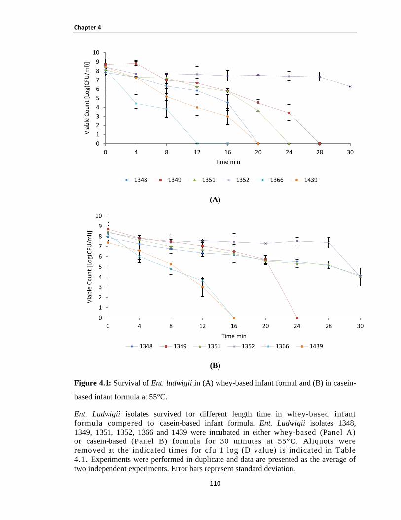
Chapter 4
110
(A)
(B)
Figure 4.1: Survival of Ent. ludwigii in (A) whey-based infant formul and (B) in casein-
based infant formula at 55°C.
Ent. Ludwigii isolates survived for different length time in whey-based infant
formula compered to casein-based infant formula. Ent. Ludwigii isolates 1348,
1349, 1351, 1352, 1366 and 1439 were incubated in either whey-based (Panel A)
or casein-based (Panel B) formula for 30 minutes at 55°C. Aliquots were
removed at the indicated times for cfu 1 log (D value) is indicated in Table
4.1. Experiments were performed in duplicate and data are presented as the average of
two independent experiments. Error bars represent standard deviation.
0
1
2
3
4
5
6
7
8
9
10
0 4 8 12 16 20 24 28 30
Via
ble
Co
un
t [L
og(
CFU
/ml)
]
Time min
1348 1349 1351 1352 1366 1439
0
1
2
3
4
5
6
7
8
9
10
0 4 8 12 16 20 24 28 30
Via
ble
Co
un
t [L
og(
CFU
/ml)
]
Time min
1348 1349 1351 1352 1366 1439
Chapter 4
111
Ent. cancerogenus
There was variation in thermotolerance between Ent. cancerogenus strains.
Ent. cancerogenus strains were differentiated into three groups based on their capability
of heat resistance.
Heat resistant: Ent. cancerogenus strains 909 and 1350 showed high thermoresistance in
whey-based infant formula and moderate heat resistance in casein-based infant formula
with D55 >50 minutes, (Figures 4.2 A and B). In contrast, strain 1367 showed heat
resistance at 55ºC in casein and had moderate heat resistance in whey.
Heat moderately tolerant: The Ent. cancerogenus strains 1037, 1355, 1358, 1360-1362
and 1365 were moderately resistant to heat at 55°C in whey and casein-based infant
formula, as seen in Table 4.1.
Heat sensitive: Ent. cancerogenus strains 806, 824, 845, 848, 957, 1042, 1077, 1357,
1359, 1363 and 1364 were more sensitive to heat in both whey and casein-based infant
formula except strain 1042 and 1357 which were moderate to heat in whey-based infant
formula with D55= 6.5 and 6 minutes, respectively. (See Figure 4.2 A and B, Table 4.1).
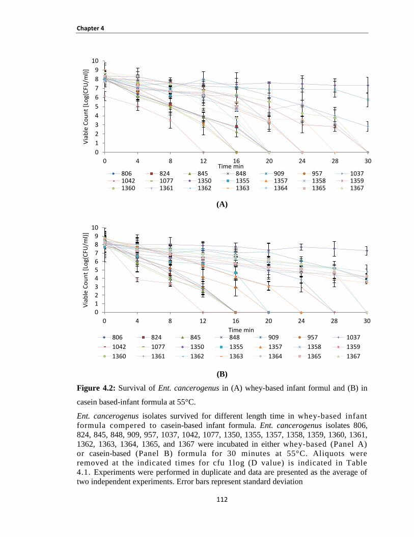
Chapter 4
112
(A)
(B)
Figure 4.2: Survival of Ent. cancerogenus in (A) whey-based infant formul and (B) in
casein based-infant formula at 55°C.
Ent. cancerogenus isolates survived for different length time in whey-based infant
formula compered to casein-based infant formula. Ent. cancerogenus isolates 806,
824, 845, 848, 909, 957, 1037, 1042, 1077, 1350, 1355, 1357, 1358, 1359, 1360, 1361,
1362, 1363, 1364, 1365, and 1367 were incubated in either whey-based (Panel A)
or casein-based (Panel B) formula for 30 minutes at 55°C. Aliquots were
removed at the indicated times for cfu 1log (D value) is indicated in Table
4.1. Experiments were performed in duplicate and data are presented as the average of
two independent experiments. Error bars represent standard deviation
0
1
2
3
4
5
6
7
8
9
10
0 4 8 12 16 20 24 28 30
Via
ble
Co
un
t [L
og(
CFU
/ml)
]
Time min 806 824 845 848 909 957 10371042 1077 1350 1355 1357 1358 13591360 1361 1362 1363 1364 1365 1367
0
1
2
3
4
56
78
9
10
0 4 8 12 16 20 24 28 30
Via
ble
Co
un
t [L
og(
CFU
/ml)
]
Time min 806 824 845 848 909 957 1037
1042 1077 1350 1355 1357 1358 1359
1360 1361 1362 1363 1364 1365 1367
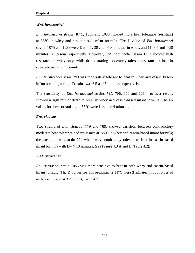
Chapter 4
113
Ent. hormaechei
Ent. hormaechei strains 1075, 1053 and 1038 showed more heat tolerance (resistant)
at 55°C in whey and casein-based infant formula. The D-value of Ent. hormaechei
strains 1075 and 1038 were D55= 11, 20 and >50 minutes in whey, and 11, 8.5 and >50
minutes in casein respectively. However, Ent. hormaechei strain 1053 showed high
resistance in whey only, while demonstrating moderately tolerant resistance to heat in
casein-based infant formula.
Ent. hormaechei strain 790 was moderately tolerant to heat in whey and casein based-
infant formula, and the D-value was 6.5 and 5 minutes respectively.
The sensitivity of Ent. hormaechei strains 795, 798, 860 and 1034 to heat results
showed a high rate of death to 55°C in whey and casein-based infant formula. The D-
values for these organisms at 55°C were less then 4 minutes.
Ent. cloacae
Two strains of Ent. cloacae, 779 and 789, showed variation between contradictory
moderate heat tolerance and resistance at 55ºC in whey and casein-based infant formula;
the exception was strain 779 which was moderately tolerant to heat in casein-based
infant formula with D55 > 10 minutes; (see Figure 4.3 A and B, Table 4.2).
Ent. aerogenes
Ent. aerogenes strain 1056 was more sensitive to heat in both whey and casein-based
infant formula. The D-values for this organism at 55°C were 2 minutes in both types of
milk; (see Figure 4.3 A and B, Table 4.2).

Chapter 4
114
(A)
(B)
Figure 4.3: Survival of Ent. hormaechei, Ent. cloacae strains (779 and 789) and Ent.
aerogenes strain 1056 in (A) whey-based infant formul and (B) in casein-based infant
formula at 55°C.
Ent. hormaechei, Ent. cloacae and Ent. aerogenes isolates survived for different length
time in whey-based infant formula compered to casein-based infant formula.
Ent. hormaechei isolates 790, 795, 798, 860, 1034, 1038, 1053 and 1075, Ent. cloacae
isolates 779 and 789 and Ent. aerogenes isolate 1056 were incubated in either whey-
based (Panel A) or casein-based (Panel B) formula for 30 minutes at 55°C.
Aliquots t were removed at the indicated times for cfu 1 log (D value) is
indicated in Table 4.2. Experiments were performed in duplicate and data are
presented as the average of two independent experiments. Error bars represent standard
deviation.
0
1
2
3
4
5
6
7
8
9
10
0 4 8 12 16 20 24 28 30
Via
ble
Co
un
t [L
og(
CFU
/ml)
]
Time min
790 795 798 860 1034 1038
1053 1075 779 789 1056
0123456789
10
0 4 8 12 16 20 24 28 30
Via
ble
Co
un
t [L
og(
CFU
/ml)
]
Time min
790 795 798 860 1034 1038
Chapter 4
115
K. oxytoca
Differences in the survival of K. oxytoca at 55ºC were noted in the two types of formula
(whey and casein-based infant formula). The K. oxytoca strain 1356 was thermoresistant
at 55ºC in whey and casein-based infant formula with a D-value more than 50 minutes
and was nonlinear. Whereas moderate resistance to heat in whey based infant formula
was demonstrated by K. oxytoca strain 1353 with D55 =5.5, and sensitive to heat in
casein-based infant formula with a D-value undetected D55=3 minutes; (presented in
Figure 4.4 A and B, Table 4.2).
K. oxytoca strain 1354 showed moderate resistance to heat in whey and casein-based
infant formula.
In Table 4.2, Figures 4.4 A and 4.4 B the K. oxytoca strains 832, 1078 and 1079 showed
sensitive behavior to heat at 55ºC in whey and casein-based infant formula and the D-
values were not detected before 4 minutes.
Summary
In this study across all the bacterial species,1/6 strain of Ent. ludwigii, 2/21 strains of
Ent. cancerogenus and 1/8 strains of Ent. hormaechei and 1/6 strains of K. oxytoca
showed the highest thermal resistance on both based infant formula at 55°`C. These
strains were not affected by both formula types at 55°`C.
Clonally related strains (same pulsotype) showed the same death rate at 55ºC. For
instance, Ent. cancerogenus strains 845, 848, 1363, 1364, 795 and 798 and K. oxytoca
strains 1078 and 1079 were sensitive to heat tolerance at 55ºC on whey and casein-based
infant formula. These belong to four clusters; Enc1, Enc3, Ebh6 and Kc3 with each
pulsotype comprising two strains respectively (Tables 4.1 and 4.2).
Also, two strains (1355, 1358) of Ent. cancerogenus showed the same response to heat at
55ºC in whey and casein-based infant formula, which was moderately resistant, and they
belonged to the same pulsotype (Enc2); see Table 4.1.
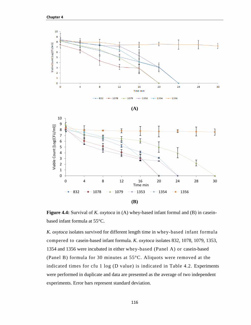
Chapter 4
116
(A)
(B)
Figure 4.4: Survival of K. oxytoca in (A) whey-based infant formul and (B) in casein-
based infant formula at 55°C.
K. oxytoca isolates survived for different length time in whey-based infant formula
compered to casein-based infant formula. K. oxytoca isolates 832, 1078, 1079, 1353,
1354 and 1356 were incubated in either whey-based (Panel A) or casein-based
(Panel B) formula for 30 minutes at 55°C. Aliquots were removed at the
indicated times for cfu 1 log (D value) is indicated in Table 4.2. Experiments
were performed in duplicate and data are presented as the average of two independent
experiments. Error bars represent standard deviation.
0
1
2
3
4
5
6
7
8
9
10
0 4 8 12 16 20 24 28 30
Via
ble
Co
un
t [L
og(
CFU
/ml)
]
Time min
832 1078 1079 1353 1354 1356
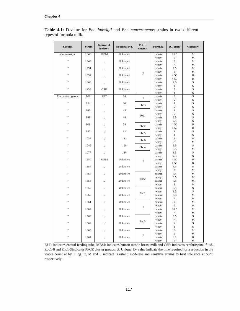
Chapter 4
117
Table 4.1: D-value for Ent. ludwigii and Ent. cancerogenus strains in two different
types of formula milk.
EFT: Indicates enteral feeding tube, MBM: Indicates human mastic breast milk and CSF: indicates cerebrospinal fluid.
Ebc1-6 and Enc1-3indicates PFGE cluster groups, U: Unique. D- value indicate the time required for a reduction in the
viable count at by 1 log. R, M and S indicate resistant, moderate and sensitive strains to heat tolerance at 55ºC
respectively.
Ent.ludwigii 1348 MBM Unknown casein 11.5 M
whey 5 M
" 1349 „ Unknown casein 6 M
whey 4 M
" 1351 „ Unknown casein 9.5 M
whey 5 M
" 1352 „ Unknown casein > 50 R
whey > 50 R
" 1366 „ Unknown casein 2.5 S
whey 1 S
" 1439 CSF Unknown casein 2 S
whey 1 S
Ent.cancerogenus 806 EFT 24 casein 2 S
whey 2 S
" 824 „ 36 casein 1 S
whey 2 S
" 845 „ 45 casein 1 S
whey 2 S
" 848 „ 48 casein 2.5 S
whey 2.5 S
" 909 „ 58 casein > 50 R
whey > 50 R
" 957 „ 81 casein 1 S
whey 1 S
" 1037 „ 112 casein 8 M
whey 9 M
" 1042 „ 128 casein 3.5 S
whey 6.5 M
" 1077 „ 118 casein 1.5 S
whey 2.5 S
" 1350 MBM Unknown casein > 50 R
whey > 50 R
" 1357 „ Unknown casein 3.5 S
whey 6 M
" 1358 „ Unknown casein 7.5 M
whey 6.5 M
" 1355 „ Unknown casein 7.5 M
whey 8 M
" 1359 „ Unknown casein 0.5 S
whey 3.5 S
" 1360 „ Unknown casein 8.5 M
whey 6 M
" 1361 „ Unknown casein 7 M
whey 9 M
" 1362 „ Unknown casein 10.5 M
whey 4 M
" 1363 „ Unknown casein 3.5 S
whey 4 M
" 1364 „ Unknown casein 2 S
whey 1 S
" 1365 „ Unknown casein 9 M
whey 6 M
" 1367 „ Unknown casein 19 R
whey 5 M
D55 (min) Category
Ebc1
Ebc3
Source of
isolatesNeonatal No.
PFGE
cluster Formula
U
U
Enc2
Enc1
Enc3
Ebc2
Ebc5
Ebc6
Ebc4
StrainSpecies
U
U
U

Chapter 4
118
Table 4.2: D-value for Ent. hormaechei and K. oxytoca in two different types of formula
milk.
EFT: Indicates enteral feeding tube, MBM: Indicates human mastic breast milk and CSF: indicates cerebrospinal fluid.
Ebh2-4, 6 and 7; Ebl1; Eba1 and Kc3 and 5 indicates PFGE cluster groups, U: Unique. D- value indicate the time
required for a reduction in the viable count at by 1 log. R, M and S indicate resistant, moderate and sensitive strains to
heat tolerance at 55ºC respectively.
Ent. hormaechei 790 EFT 4 casein 5 M
whey 6.5 M
" 795 „ 17 casein 2 S
whey 2 S
" 798 „ 18 casein 2 S
whey 1 S
" 860 „ 52 casein 1 S
whey 1.5 S
" 1034 „ 110 casein 2.5 S
whey 1.5 S
" 1038 „ 113 casein > 50 R
whey > 50 R
" 1053 „ 117 casein 8.5 M
whey 20 R
" 1075 „ 117 casein 11 M
whey 11 M
Ent. cloacae 779 EFT 2 casein 4 M
whey > 50 R
" 789 „ 12 casein 13 M
whey 10.5 M
Ent. aerogenes 1056 EFT 118 casein 2 S
whey 2 S
K.oxytoca 832 EFT 36 casein 3 S
whey 2 S
" 1078 „ 120 casein 2.5 S
whey 2.5 S
" 1079 „ 121 casein 2 S
whey 1 S
" 1353 MBM Unknown casein 3 S
whey 5.5 M
" 1354 „ Unknown casein 6.5 M
whey 4.5 M
" 1356 „ Unknown casein > 50 R
whey > 50 R
Formula D55 (min) Category StrainSpecies Source of isolates Neonatal No.PFGE
cluster
U
Ebh7
Ebh3
Ebl1
U
Ebh6
Ebh4
Ebh2
Eba1
Kc5
Kc3
Chapter 4
119
4.3.2 Biofilm formation by Enterobacteriaceae on three types of formula milk at
different temperatures
The biofilm formation values had to be corrected for the non-specific binding of crystal
violet to the microtitre tray and un-inoculated formula. The relative biofilm formation
value was determined by dividing the strain value by the corresponding control value. The
results were then categorised as:
1- No biofilm formation: OD strain = OD control
2- Low biofilm formation: OD strain ≤ 2x OD control
3- Moderate biofilm formation: OD strain ≤ 4x OD control
4- High biofilm formation: OD strain > 4x OD control.
Twenty strains isolated from MBM and twenty three strains isolated from EFT (Tables 2.2
and 2.4 respectively) in section 2.1.1 were tested for biofilm formation in three types of
formula milk (whey, casein and soya-based infant formula) at 20°C and 37°C.
The results are summarised below and summarised in Table 4.3.
Whey-based formula 4.3.2.1
At 20°C:
Twenty four out of fourty four strains were able to form biofilm at 20°C including
4/6 strains of Ent. ludwigii, 4/8 strains of Ent. hormaechei, 2 strains of Ent. cloacae, 17/21
strains of Ent. cancerogenus and 3/6 strains K. oxytoca formed relatively low levels of
biofilm. The rest of strains were not able to form biofilm at 20°C; (Figure 4.9).
At 37°C:
All strains formed biofilms on whey-based infant formula at 37°C. The high biofilm level
was produced by Ent. ludwigii strain 1439 and K. oxytoca strain 1353. Moderate levels of
biofilm were produced by 3/6 strains of Ent. ludwigii, 6/8 strains of Ent. hormaechei, 2
strains of Ent. cloacae, 1 strain of Ent. aerogenes, 17/21 strains of Ent. cancerogenus and
4/6 strains of K. oxytoca. The remaining strains only formed low levels of biofilm; (see
Figure 4.9).

Chapter 4
120
Interestingly, the same level of biofilm was observed by Ent. hormaechei strains 1053
and 1075 in whey at 37ºC which were isolated from the same patient and belonged to
pulsotype Ebh3; (Table 4.3). In addition, Ent. cancerogenus strains (1355 and 1358)
and Ent. cancerogenus strains (1359 and 1360) showed moderate biofilm level in
whey-based infant formula at 37°C and belonged to pulsotypes Enc2 and Enc1
respectively. Ent. cancerogenus 1363 and 1364 also showed moderate biofilm
formation and belonged to pulsotype Enc3 (Table 4.3).
The biofilm formation at 37°C was significantly higher than the formation at 20°C
(p<0.05).
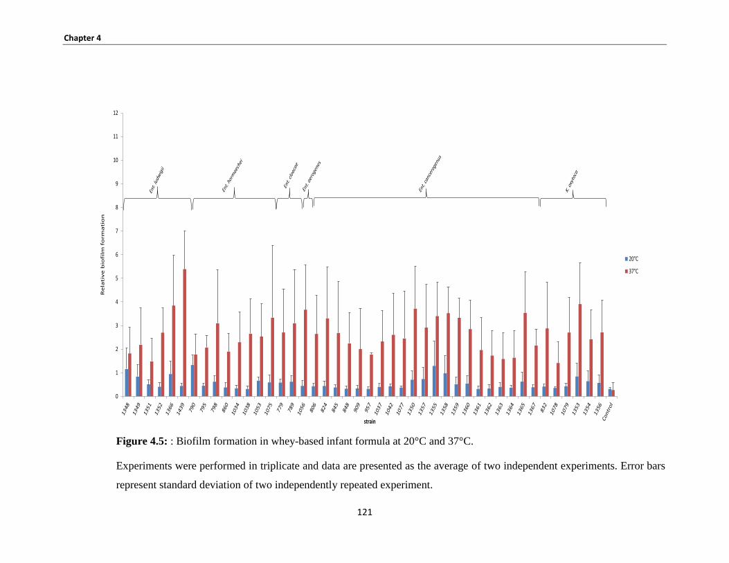
Chapter 4
121
Figure 4.5: : Biofilm formation in whey-based infant formula at 20°C and 37°C.
Experiments were performed in triplicate and data are presented as the average of two independent experiments. Error bars
represent standard deviation of two independently repeated experiment.
0
1
2
3
4
5
6
7
8
9
10
11
12
Re
lati
ve
bio
film
fo
rma
tio
n
strain
20°C
37°C
Chapter 4
122
Casein-based formula 4.3.2.2
At 20°C:
There was no high biofilm on casein-based infant formula at 20°C. Moderate biofilms
were produced by 1/6 strains of Ent. ludwigii, 2/8 strains of Ent. hormaechei and 3/21
strains of Ent. cancerogenus. Low level of biofilm was formed by 5/6 strains of
Ent. ludwigii, 1/8 strains of Ent. hormaechei, two strains of Ent. cloacae, 3/21 strains of
Ent. cancerogenus and 1/6 strains of K. oxytoca. The rest of strains showed low biofilm
at this temperature; see Figure 4.10.
At 37°C:
High biofilm formation was seen by 2/6 strains of Ent. ludwigii, 1/8 strains of
Ent. hormaechei, 8/21 strains of Ent. cancerogenus and 1/6 strains of K. oxytoca.
Moderate biofilm levels were formed by 1/6 strains of Ent. ludwigii,, 3/8 strains of
Ent. hormaechei, 2 strains of Ent. cloacae, 1 strain of Ent. aerogenes, 8/21 strains of
Ent. cancerogenus and 3/6 strains of K. oxytoca (Figure 4.10, Table 4.3).
Low biofilm levels were produced by 3/6 strains of Ent. ludwigii, 4/8 strains of
Ent. hormaechei, 5/21 strains of Ent. cancerogenus and 2 /6 strains of K. oxytoca; (Figure
4.10). The difference in the quantity of biofilm formation between the two temperatures
(20°C and 37°C) was statistically significant in most strains. Results are summarised in
Figure 4.10 and Table 4.3.
It was also noted that most strains which belonged to the same pulsotype showed the same
biofilm result; for example, Ent. cancerogenus strains 1359 and 1360 belonged to cluster
Enc1 and formed high biofilm on infant formula at 37°C and produced low biofilm at
20°C. Also, Ent. cancerogenus strains 1363 and 1364 belonged to group Enc3 which
formed low biofilm at 20°C and 37°C (Table 4.3).
Soya-Powder Infant formula 4.3.2.3
At 20°C:
None of the tested strains produced high levels of biofilm formation in soya milk at both
temperatures 20°C and 37°C. There was low biofilm formation by all Ent. ludwigii,

Chapter 4
123
Ent. hormaechei, Ent. cloacae, Ent. aerogenes, Ent. cancerogenus and K. oxytoca strains
at 20°C (Figure 4.11, Table 4.3).
At 37°C:
Three Ent. cancerogenus strains (1355, 1357 and 1358) and 1 K. oxytoca strain (1356)
formed moderate biofilm levels at 37°C, and low biofilm was formed by all strains of
Ent. ludwigii, Ent. hormaechei, Ent. cloacae, Ent. aerogenes, Ent. cancerogenus and
K. oxytoca strains at 37°C (see Figure 4.11, Table 4.3).
Biofilm formation in soya milk was statistically significantly different at 20°C and 37°C
(p<0.05).
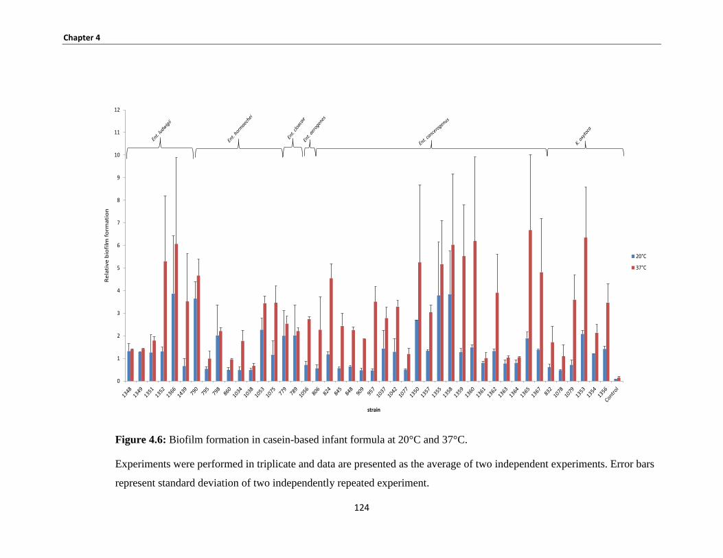
Chapter 4
124
Figure 4.6: Biofilm formation in casein-based infant formula at 20°C and 37°C.
Experiments were performed in triplicate and data are presented as the average of two independent experiments. Error bars
represent standard deviation of two independently repeated experiment.
0
1
2
3
4
5
6
7
8
9
10
11
12R
ela
tiv
e b
iofi
lm f
orm
ati
on
strain
20°C
37°C
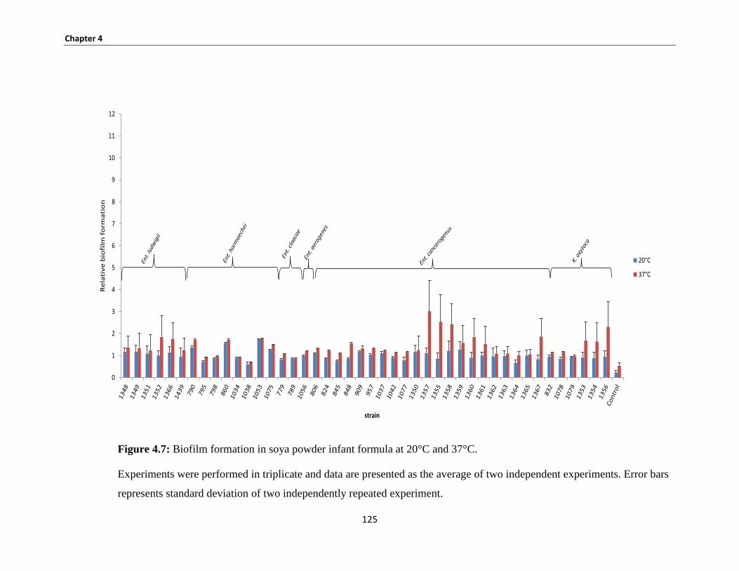
Chapter 4
125
Figure 4.7: Biofilm formation in soya powder infant formula at 20°C and 37°C.
Experiments were performed in triplicate and data are presented as the average of two independent experiments. Error bars
represents standard deviation of two independently repeated experiment.
0
1
2
3
4
5
6
7
8
9
10
11
12
Re
lati
ve
bio
film
fo
rma
tio
n
strain
20°C
37°C
Chapter 4
126
4.3.2.4 Comparison of different levels of biofilm formation at 37°C
In Ent. ludwgii, biofilm formation was significantly higher in casein and whey-
based infant formula than soya-based infant formula (p < 0.001), while there was
no significant difference between casein and soya-based formula.
In Ent. cancergenus, biofilm formation was significantly higher in casein based-
infant formula than both whey and soya-based infant formula (p < 0.001);
however, the biofilm level was significantly higher in whey compared with soya-
based formula.
In Ent. hormaechei and K. oxytoca, biofilm formation was significantly higher in
casein and whey-based infant formula than soya-based infant formula (p<0.001).
In Ent. cloacae and Ent. aerogenes, biofilm formation was not significantly
different between the three types of formula (whey, casein and soya) .
In summary, it was found that the relative biofilm formation was higher at 37ºC
compared with 20ºC by most tested strains in all types of formula (Table 4.3).
The quantity of biofilm was detected on three types of formula which was higher
on casein-based infant formula compared with whey and soya-based infant
formula in all strains.
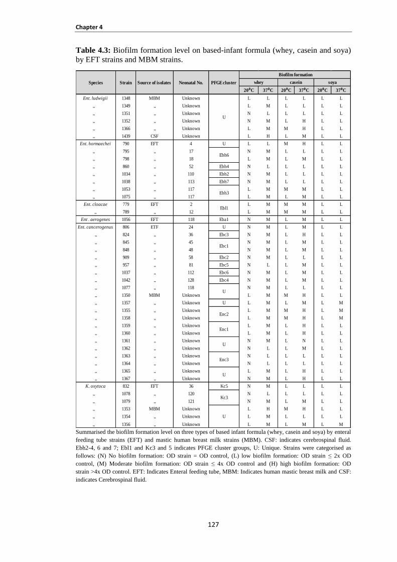
Chapter 4
127
Table 4.3: Biofilm formation level on based-infant formula (whey, casein and soya)
by EFT strains and MBM strains.
Summarised the biofilm formation level on three types of based infant formula (whey, casein and soya) by enteral
feeding tube strains (EFT) and mastic human breast milk strains (MBM). CSF: indicates cerebrospinal fluid.
Ebh2-4, 6 and 7; Ebl1 and Kc3 and 5 indicates PFGE cluster groups, U: Unique. Strains were categorised as
follows: (N) No biofilm formation: OD strain = OD control, (L) low biofilm formation: OD strain ≤ 2x OD
control, (M) Moderate biofilm formation: OD strain ≤ 4x OD control and (H) high biofilm formation: OD
strain >4x OD control. EFT: Indicates Enteral feeding tube, MBM: Indicates human mastic breast milk and CSF:
indicates Cerebrospinal fluid.
20⁰C 37⁰C 20⁰C 37⁰C 20⁰C 37⁰C
Ent. ludwigii 1348 MBM Unknown L L L L L L
„ 1349 „ Unknown L M L L L L
„ 1351 „ Unknown N L L L L L
„ 1352 „ Unknown N M L H L L
„ 1366 „ Unknown L M M H L L
„ 1439 CSF Unknown L H L M L L
Ent. hormaechei 790 EFT 4 U L L M H L L
„ 795 „ 17 N M L L L L
„ 798 „ 18 L M L M L L
„ 860 „ 52 Ebh4 N L L L L L
„ 1034 „ 110 Ebh2 N M L L L L
„ 1038 „ 113 Ebh7 N M L L L L
„ 1053 „ 117 L M M M L L
„ 1075 „ 117 L M L M L L
Ent. cloacae 779 EFT 2 L M M M L L
„ 789 „ 12 L M M M L L
Ent . aerogenes 1056 EFT 118 Eba1 N M L M L L
Ent. cancerogenus 806 ETF 24 U N M L M L L
„ 824 „ 36 Ebc3 N M L H L L
„ 845 „ 45 N M L M L L
„ 848 „ 48 N M L M L L
„ 909 „ 58 Ebc2 N M L L L L
„ 957 „ 81 Ebc5 N L L M L L
„ 1037 „ 112 Ebc6 N M L M L L
„ 1042 „ 128 Ebc4 N M L M L L
„ 1077 „ 118 N M L L L L
„ 1350 MBM Unknown L M M H L L
„ 1357 „ Unknown U L M L M L M
„ 1355 „ Unknown L M M H L M
„ 1358 „ Unknown L M M H L M
„ 1359 „ Unknown L M L H L L
„ 1360 „ Unknown L M L H L L
„ 1361 „ Unknown N M L N L L
„ 1362 „ Unknown N L L M L L
„ 1363 „ Unknown N L L L L L
„ 1364 „ Unknown N L L L L L
„ 1365 „ Unknown L M L H L L
„ 1367 „ Unknown N M L H L L
K. oxytoca 832 EFT 36 Kc5 N M L L L L
„ 1078 „ 120 N L L L L L
„ 1079 „ 121 N M L M L L
„ 1353 MBM Unknown L H M H L L
„ 1354 „ Unknown L M L L L L
„ 1356 „ Unknown L M L M L M
Biofilm formation
whey casein soya
U
Species Strain Source of isolates Neonatal No. PFGE cluster
Ebh6
Ebh3
Ebl1
Ebc1
Enc3
Kc3
U
U
U
Enc2
Enc1
U
Chapter 4
128
4.3.3 Capsule formation on milk agar
Bacterial capsule production was determined by colony appearance on milk agar
(whey, casein and soya-based) at 20°C (after 24 hours and 48 hours) and 37°C (after
24 hours). The results were classified into three categories according to the relative
scale of high, medium and low capsule production which are represented by ≥ +++,
++ and + respectively, Figure 4.8.
The amount of capsular material varied according to the incubation temperature,
time, and milk agar composition; (Table 4.4). No highly mucoid colonies were
apparent after 24 hours incubation at 20°C, but these did appear after 48 hours
incubation. Capsular strains were found in Ent. ludwigii, Ent. hormaechei, Ent.
cancerogenus, and K. oxytoca. However, the number of capsular strains varied
between species.
On whey milk agar the capsule production was recorded for all Ent. ludwigii
at 20ºC and 37ºC, except strain 1366 which produced medium capsular
material at 37ºC only. The high level of capsular material was by
5/6 Ent. ludwigii at 37ºC and by 1 strain at 20ºC, whereas 4/6 Ent. ludwigii
showed medium level of capsular material at 20ºC; see Table 4.4.
With respect to Ent. hormaechei, 4/8 strains produced high capsular material
on whey at 20ºC and 1 strain of Ent. hormaechei strain (1075) produced high
capsule production at 37ºC. Three strains of Ent. hormaechei produced
medium capsular material at 20ºC and 1 strain produced low capsular
material at 37ºC (Table 4.4, Figure 4.8).
Two strains of Ent. cloacae (779 and 789) formed low capsular material
at 20ºC, while at 37ºC they produced medium/high capsular material
respectively.
Ent. aerogenes strain 1056 produced high capsular material at 20°C, but it
was not able to form capsular material at 37°C; Table 4.4, Figure 4.8.
Ent. cancerogenus strains 1364, 806, 957 and 1361 produced high capsular
material at 20ºC, while at 37ºC the high capsular material was by strains
1364, 1361, 1357, 1363 and 1037. The majority of Ent. cancerogenus, 5/21
and 9/21 strains, produced medium capsular material at 20°C and 37°C
respectively (Table 4.4, Figure 4.8). Of particular note was that most Ent.
Chapter 4
129
cancerogenus produced capsular material at 37ºC.
Some K. oxytoca (2/6) strains produced high capsular material in whey-
based infant formula at 37ºC and another 2/6 strains produced medium
capsular material at 37ºC , and low capsular material was by K. oxytoca 3/6
and 1/6 strains at 20°C and 37°C respectively (Table 4.4, Figure 4.8). No
capsule production was formed by 1/6 Ent. ludwigii, 11/21 Ent.
cancerogenus and 1/6 K. oxytoca in whey-based infant formula at 20°C,
while 6/8 Ent. hormaechei, 3/21 strains of Ent. cancerogenus, 1/6 K. oxytoca
and 1 strain of Ent. aerogenes were not able to produce capsules in whey
milk agar at 37°C. In contrast, Ent. hormaechei strain 798, Ent.
cancerogenus strains 1358 and 909 and K. oxytoca strain 832 were not able
to produce capsules in whey milk agar at both temperatures.
On casein milk agar most species were able to produce capsular material at
20ºC and 37ºC. 3/6 and 5/6 Ent. ludwigii strains produced high capsular
material at 20ºC and 37ºC respectively, while 3/6 and 1/6 Ent. ludwigii
produced medium capsular material at 20ºC and 37ºC respectively.
Ent. hormaechei strains 1034, 790, 860,1053 and 798 produced high
capsular material at 20ºC, while strain 1075 formed high capsular material at
37ºC. Medium capsular material was formed by Ent. hormaechei strains
1075 and 860 at 20ºC and 37ºC respectively. Ent. hormaechei strain 1038
produced low capsular material at 37ºC. Of particular note was strain 1034
which produced considerable capsular material at 20ºC after 48 hours (Table
4.4, Figure 4.8).
Ent. cloacae strains 779 and 789 formed low capsular material at 20ºC,
whereas at 37ºC medium/high capsular material was produced respectively.
The high capsular material was produced by Ent. aerogenes strain 1056 at
20°C; however, it was not able to produce capsular material at 37°C; (Table
4.4, Figure 4.8).
The high capsular material was formed by 5/21 and 10/21 strains
of Ent. cancerogenus at 20°C and 37ºC respectively. While the medium
capsular material was produced by 8/21 and 10/21 strains of
Ent. cancerogenus at 20°C and 37ºC respectively (Table 4.4, Figure 4.8).
Chapter 4
130
K. oxytoca strains 1078, 1354 and 1356 produced high capsular material at
20°C and 37ºC. The exception was strain 1356 which formed low capsule at
20°C. The rest of strains produced medium capsular material at 37°C (Table
4.4, Figure 4.8).
On soya powder infant formula agar the capsule production was produced by
most species, the exception was by Ent. cancerogenus strain 909 which was
not able to produce capsular material at both temperatures. 3/6 and 5/6 of
Ent. ludwigii produced high capsular material at 20ºC and 37ºC respectively.
All Ent. hormaechei produced high capsular material at 20ºC, while 5/8 of
Ent. hormaechei formed high capsular material at 37ºC (Table 4.4, Figure
4.8).
Ent. cloacae strains 779 and 789 produced medium capsular material
at 20ºC, whereas at 37ºC these strains formed high/medium capsular
material respectively. The high/medium capsular material was produced by
Ent. aerogenes strain 1056 at 20°C and 37°C respectively (Table 4.4, Figure
4.8).
The high capsule production was by 4/21 and 6/21 of Ent. cancerogenus at
20ºC and 37ºC respectively. Furthermore, 2/6 and 2/6 of K. oxytoca formed
high capsular material at 20ºC and 37ºC respectively (Table 4.4, Figure 4.8).
The ability of strains to produce capsular material at 20ºC and 37ºC was
observed in casein-based infant formula and soya powder more than in whey-
based infant formula and the capsular material production appeared on milk
agar (whey, casein and soya based) at 20°C after 48 hours by most strains.
Ent. ludwigii, Ent. cancerogenus Ent. cloacae, Ent. aerogenes, and
K. oxytoca strains showed the ability to produce more capsular material on
milk agar (whey, casein and soya based) at 20ºC and 37ºC, compared with
Ent. hormaechei strain (see Table 4.4, Figure 4.8).
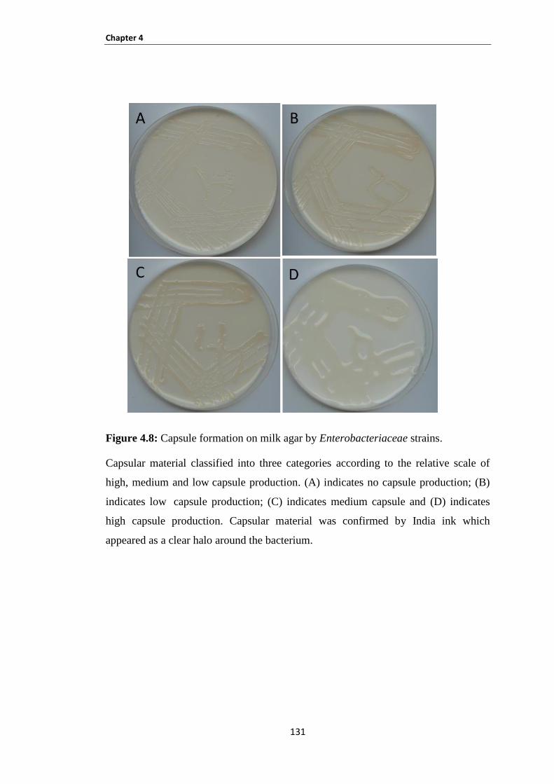
Chapter 4
131
Figure 4.8: Capsule formation on milk agar by Enterobacteriaceae strains.
Capsular material classified into three categories according to the relative scale of
high, medium and low capsule production. (A) indicates no capsule production; (B)
indicates low capsule production; (C) indicates medium capsule and (D) indicates
high capsule production. Capsular material was confirmed by India ink which
appeared as a clear halo around the bacterium.
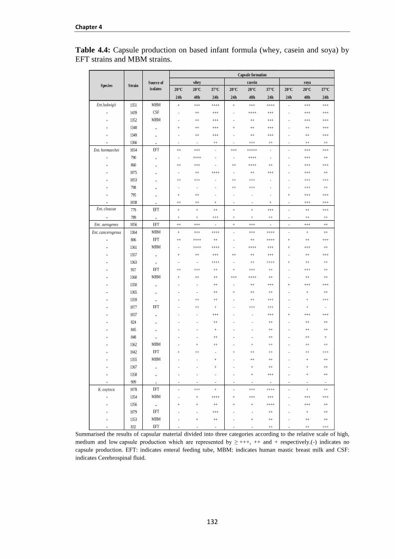
Chapter 4
132
Table 4.4: Capsule production on based infant formula (whey, casein and soya) by
EFT strains and MBM strains.
Summarised the results of capsular material divided into three categories according to the relative scale of high,
medium and low capsule production which are represented by ≥ +++, ++ and + respectively.(-) indicates no
capsule production. EFT: indicates enteral feeding tube, MBM: indicates human mastic breast milk and CSF:
indicates Cerebrospinal fluid.
20°C 20°C 37°C 20°C 20°C 37°C 20°C 20°C 37°C
24h 48h 24h 24h 48h 24h 24h 48h 24h
Ent.ludwigii 1351 MBM + +++ ++++ + +++ ++++ - +++ +++
„ 1439 CSF - ++ +++ - ++++ +++ - +++ +++
„ 1352 MBM - ++ +++ - ++ +++ - +++ +++
„ 1348 „ + ++ +++ + ++ +++ - ++ +++
„ 1349 „ - ++ +++ - ++ +++ - ++ +++
„ 1366 „ - - ++ - +++ ++ - ++ ++
Ent. hormaechei 1034 EFT ++ +++ - +++ +++++ - - +++ +++
„ 790 „ - ++++ - - ++++ - - +++ ++
„ 860 „ ++ +++ - ++ ++++ ++ - +++ +++
„ 1075 „ - ++ ++++ - ++ +++ - +++ ++
„ 1053 „ ++ +++ - ++ +++ - - +++ +++
„ 798 „ - - - ++ +++ - - +++ ++
„ 795 „ + ++ - - - - + +++ +++
„ 1038 „ ++ ++ + - - + - +++ +++
Ent. cloacae 779 EFT + + ++ + + +++ - ++ +++
„ 789 „ + + +++ + + ++ - ++ ++
Ent . aerogenes 1056 EFT ++ +++ - + +++ - - +++ ++
Ent. cancerogenus 1364 MBM + +++ ++++ - +++ ++++ - + ++
„ 806 EFT ++ ++++ ++ - ++ ++++ + ++ +++
„ 1361 MBM - ++++ ++++ - ++++ +++ + +++ ++
„ 1357 „ + ++ +++ ++ ++ +++ - ++ +++
„ 1363 „ - - ++++ - ++ ++++ + ++ ++
„ 957 EFT ++ +++ ++ + +++ ++ - +++ ++
„ 1360 MBM + ++ ++ +++ ++++ ++ - ++ ++
„ 1350 „ - - ++ - ++ +++ + +++ +++
„ 1365 „ - - ++ + ++ ++ - + ++
„ 1359 „ - ++ ++ - ++ +++ - + +++
„ 1077 EFT - ++ + - +++ +++ - + -
„ 1037 „ - - +++ - - +++ + +++ +++
„ 824 „ - - ++ - - ++ - ++ ++
„ 845 „ - - + - - ++ - ++ ++
„ 848 „ - - ++ - - ++ - ++ +
„ 1362 MBM - + ++ - + ++ - ++ ++
„ 1042 EFT + ++ - + ++ ++ - ++ +++
„ 1355 MBM - - + - ++ ++ - + ++
„ 1367 „ - - + - + ++ - + ++
„ 1358 „ - - - - + +++ - + ++
„ 909 „ - - - - - - - - -
K. oxytoca 1078 EFT - +++ + - +++ ++++ - + ++
„ 1354 MBM - + ++++ + +++ +++ - +++ +++
„ 1356 „ + + ++ + + ++++ - +++ ++
„ 1079 EFT - - +++ - - ++ - + ++
„ 1353 MBM - + ++ - + ++ - ++ ++
„ 832 EFT - - - - - ++ - ++ +++
Species StrainSource of
isolates
Capsule formation
whey casein soya
Chapter 4
133
4.3.3 Acid tolerance of organisms to pH 3.5 (HCl acidified formula)
The ability of Ent. ludwigii, Ent. hormaechei, cloacae, Ent. aerogenes,
Ent. cancerogenus and K. oxytoca to tolerate acidic conditions at pH 3.5 was
determined. These strains were exposed to two types of formula (whey and casein)
which were adjusted to pH 3.5 for two hours at body temperature.
All strains were clearly resistant to two hours exposure to pH 3.5 in whey and casein
infant formula: the viable counts were observed over the course of the 2 hours
exposure to pH 3.5 for all strains. The variation across the strains in each type of
formula was very broad. There was no correlation between the source of the strains
(that is, enteral feeding tube isolated compared with mastic human breast milk
strains) and their acid resistance. The results are presented in Figures 4.12-4.19.
These results will serve as a base for bacterial attachment and invasion of host cells
studies in chapter 5. The ability of organisms to survive at pH 3.5 (similar to the
acidic environment of the stomach) means that these organisms are most likely to be
pathogenic and able to invade the intestine, thus causing human infections.
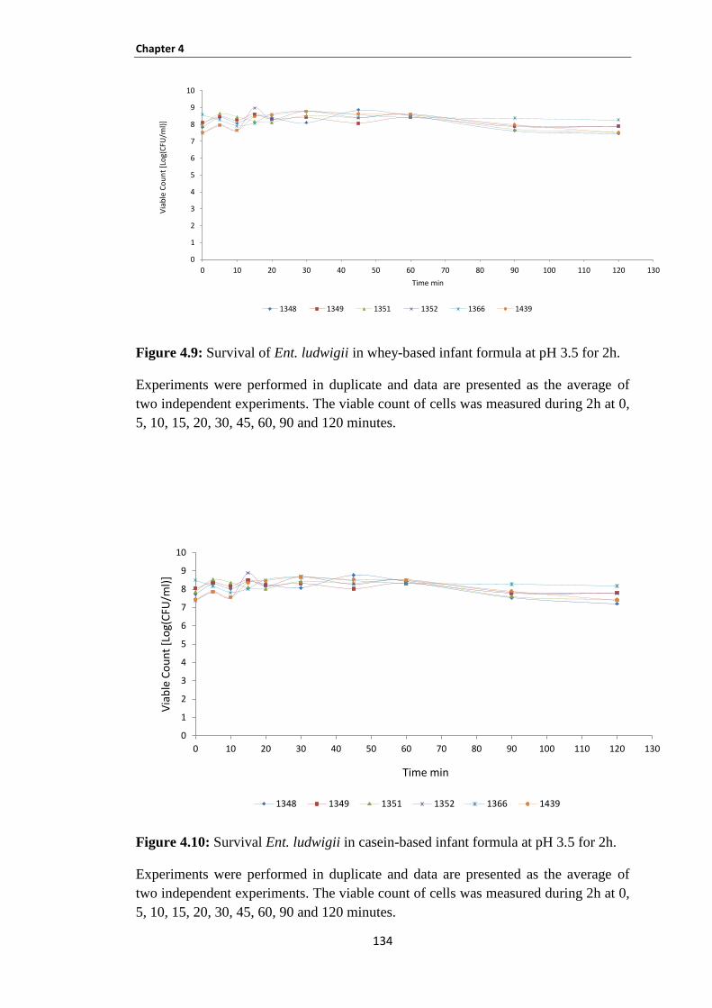
Chapter 4
134
Figure 4.9: Survival of Ent. ludwigii in whey-based infant formula at pH 3.5 for 2h.
Experiments were performed in duplicate and data are presented as the average of
two independent experiments. The viable count of cells was measured during 2h at 0,
5, 10, 15, 20, 30, 45, 60, 90 and 120 minutes.
Figure 4.10: Survival Ent. ludwigii in casein-based infant formula at pH 3.5 for 2h.
Experiments were performed in duplicate and data are presented as the average of
two independent experiments. The viable count of cells was measured during 2h at 0,
5, 10, 15, 20, 30, 45, 60, 90 and 120 minutes.
0
1
2
3
4
5
6
7
8
9
10
0 10 20 30 40 50 60 70 80 90 100 110 120 130
Via
ble
Co
un
t [L
og(
CFU
/ml)
]
Time min
1348 1349 1351 1352 1366 1439
0
1
2
3
4
5
6
7
8
9
10
0 10 20 30 40 50 60 70 80 90 100 110 120 130
Via
ble
Co
un
t [L
og(
CFU
/ml)
]
Time min
1348 1349 1351 1352 1366 1439
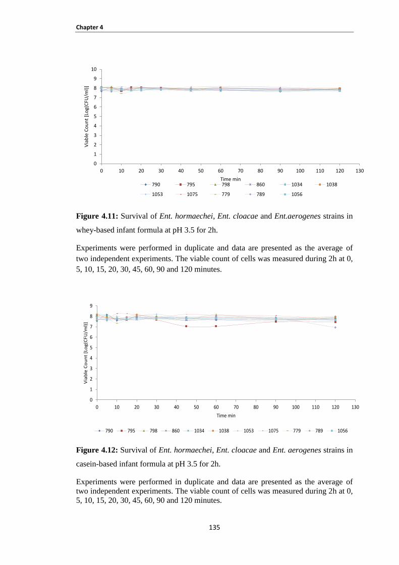
Chapter 4
135
Figure 4.11: Survival of Ent. hormaechei, Ent. cloacae and Ent.aerogenes strains in
whey-based infant formula at pH 3.5 for 2h.
Experiments were performed in duplicate and data are presented as the average of
two independent experiments. The viable count of cells was measured during 2h at 0,
5, 10, 15, 20, 30, 45, 60, 90 and 120 minutes.
Figure 4.12: Survival of Ent. hormaechei, Ent. cloacae and Ent. aerogenes strains in
casein-based infant formula at pH 3.5 for 2h.
Experiments were performed in duplicate and data are presented as the average of
two independent experiments. The viable count of cells was measured during 2h at 0,
5, 10, 15, 20, 30, 45, 60, 90 and 120 minutes.
0
1
2
3
4
5
6
7
8
9
10
0 10 20 30 40 50 60 70 80 90 100 110 120 130
Via
ble
Co
un
t [L
og(
CFU
/ml)
]
Time min790 795 798 860 1034 1038
1053 1075 779 789 1056
0
1
2
3
4
5
6
7
8
9
0 10 20 30 40 50 60 70 80 90 100 110 120 130
Via
ble
Co
un
t [L
og
(CF
U/m
l)]
Time min
790 795 798 860 1034 1038 1053 1075 779 789 1056

Chapter 4
136
Figure 4.13: Survival of Ent. cancerogenus in whey-based infant formula at pH 3.5
for 2h.
Experiments were performed in duplicate and data are presented as the average of
two independent experiments. The viable count of cells was measured during 2h at 0,
5, 10, 15, 20, 30, 45, 60, 90 and 120 minutes.
Figure 4.14: Survival of Ent. cancerogenus in casein-based infant formula at pH 3.5
for 2h.
Experiments were performed in duplicate and data are presented as the average of
two independent experiments. The viable count of cells was measured during 2h at 0,
5, 10, 15, 20, 30, 45, 60, 90 and 120 minutes.
0
1
2
3
4
5
6
7
8
9
10
0 10 20 30 40 50 60 70 80 90 100 110 120 130
Via
ble
Co
un
t [L
og
(CF
U/m
l)]
Time min
806 824 845 848 909 957 1037 1042 1077 1350 1355
1357 1358 1359 1360 1361 1362 1363 1364 1365 1367
0
1
2
3
4
5
6
7
8
9
10
0 10 20 30 40 50 60 70 80 90 100 110 120 130
Via
ble
Co
un
t [L
og(
CFU
/ml)
]
Time min
806 824 845 848 909 957 1037 1042 1077 1350 1355
1357 1358 1359 1360 1361 1362 1363 1364 1365 1367
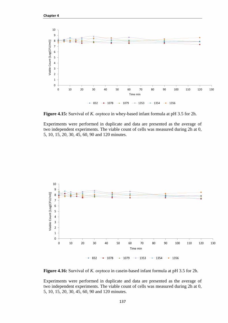
Chapter 4
137
Figure 4.15: Survival of K. oxytoca in whey-based infant formula at pH 3.5 for 2h.
Experiments were performed in duplicate and data are presented as the average of
two independent experiments. The viable count of cells was measured during 2h at 0,
5, 10, 15, 20, 30, 45, 60, 90 and 120 minutes.
Figure 4.16: Survival of K. oxytoca in casein-based infant formula at pH 3.5 for 2h.
Experiments were performed in duplicate and data are presented as the average of
two independent experiments. The viable count of cells was measured during 2h at 0,
5, 10, 15, 20, 30, 45, 60, 90 and 120 minutes.
0
1
2
3
4
5
6
7
8
9
10
0 10 20 30 40 50 60 70 80 90 100 110 120 130
Via
ble
Co
un
t [L
og
(CFU
/ml)
]
Time min
832 1078 1079 1353 1354 1356
0
1
2
3
4
5
6
7
8
9
10
0 10 20 30 40 50 60 70 80 90 100 110 120 130
Via
ble
Co
un
t [L
og
(CF
U/m
l)]
Time min
832 1078 1079 1353 1354 1356

Chapter 4
138
4.4 DISCUSSION
The present study was designed to determine the physiological attributes for
representative strains defined by using PFGE, with respect to stress survival which was
affected by formula type and temperature at 55°C, biofilm formation, capsule
production and acid tolerance of organisms to pH 3.5 (HCl acidified formula).
Most strains survived at 55°C (Tables 4.1 and 4.2). In this study, the considerable
finding was that same death rate at 55ºC was demonstrated by strains which belonged
to the same cluster by PFGE analysis. For example, the pulsotypes of Enc1, Enc2,
Enc3, Ebh6 and Kc3 Ent. cancerogenus strains 845, 848, 1355, 1358, 1363 and 1364,
Ent. hormaechei strains 795 and 798, and K. oxytoca strains 1078 and 1079,
respectively were sensitive to heat tolerance at 55ºC on whey and casein-based infant
formula.
This finding may suggest that these strains was possibly originated from the same
source and the same clone contaminated different feeding tubes. For the MBM strains
Ent. cancerogenus strains (1355, 1358, 1363 and 1364), it was probably that these
organisms were isolated from the same mother. Therefore, the neonates could be
exposed to the ingestion of these organisms either from contaminated milk or from
infant formula. The findings of this study were consistent with Forsythe (2009), who
found that in a neonatal care unit, fortified breast milk and reconstituted powdered
infant formula were reconstituted at room temperature when required, and not at
higher temperatures. Similar procedures were reported by Rosset et al. (2007) in a
study of French hospitals in that samples were reconstituted with room temperature
water.
On the other hand, the ability of tested strains to survive at 55ºC may become a
sourse of infection of neonates in NICUs which was previously described by Iversen
et al. (2004a). They found that C. sakazakii is capable of growing in PIF during
storage at refrigeration temperatures and adhere to infant-feeding equipment, which
probably becomes a sourse of infection. In addition, C. sakazakii strains were able to
grow in PIF between 6° and 45°C, with the optimum being 37° to 43°C (Iversen et
al., 2004a). The meeting by FAO and WHO in 2006 acknowledged that the
reconstitution of PIF with water between 40° and 50°C (unless used immediately),

Chapter 4
139
holding bottles at room temperature and extended feeding periods were linked with a
rise in relative risk.
Biofilm formation was investigated as this may be a means of organism persistence
in the NICUs and attachment to enteral feeding tubes. This study found that the
relative biofilm formation was higher at 37ºC compared with 20ºC by most tested
strains in all types of formula (Figures 4.9, 4.10 and 4.11). The notable result was
that the Ent. hormaechei strains 1053 and 1075 and Ent. cancerogenus strains 1355,
1358, 1359, 1360, 1363 and 1364 were clustered into 4 pulsotypes, Ebh3, Enc2, Enc1
and Enc3 (each pulsotype consists of two strains) respectively, and formed the same
quantity of biofilm in whey and casein-based infant formula at 37ºC and 20ºC.
However, Ent. hormaechei strains 1053 and 1075 only exhibited the same level of
biofilm in whey-based infant formula at 37ºC (Table 4.3). This finding probably
indicates that the same clone was cross contaminated between different feeding tubes
and the prevalence of same clone was recovered from the same mother (MBM
strains).
The quantity of biofilm that was detected on the three types of formula was higher in
casein-based infant formula compared with whey and soya-based infant formula in
all strains. These observations indicate the importance of the nutrient type present in
milk in biofilm formation. Furthermore, these findings are in agreement with other
studies (Kim et al., 2006; Meadows, 1971). Research has shown that nutrients and
other media components influence the attachment of bacteria to surfaces of various
materials (i.e biofilm formation) (Hood and Zottola, 1997). Meadows (1971) found
that casein enhanced the attachment of E. coli, P. fluorescens and Aeromonas
liquefaciens to glass surfaces. Whey protein has shown the same effect in stainless
steel surfaces (Speers et al., 1985).
In general, across all the bacterial species that were tested, this study confirmed the
previous research (Hurrell et al., 2009) which demonstrated that the organisms may
originate from the attached bacterial biofilm, and break off in clumps. The bacterial
multiplication occurs at room temperature during the feeding period compared with
the tube at 37°C (which can be in place for >48 hours). This can result in the
contamination of fresh feed in the tube lumen leading to further bacterial
multiplication. Bacteria enter the neonate’s stomach as clumps of cells which may
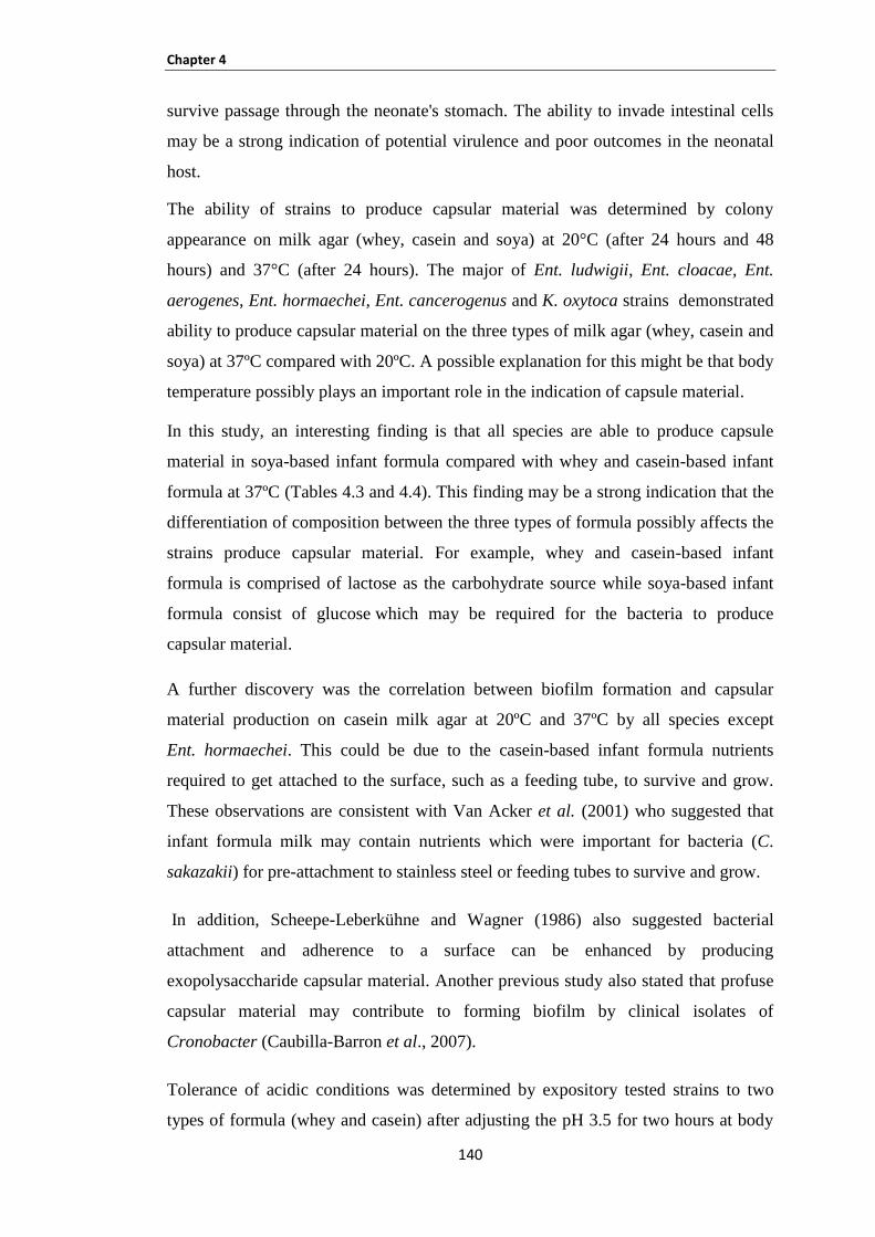
Chapter 4
140
survive passage through the neonate's stomach. The ability to invade intestinal cells
may be a strong indication of potential virulence and poor outcomes in the neonatal
host.
The ability of strains to produce capsular material was determined by colony
appearance on milk agar (whey, casein and soya) at 20°C (after 24 hours and 48
hours) and 37°C (after 24 hours). The major of Ent. ludwigii, Ent. cloacae, Ent.
aerogenes, Ent. hormaechei, Ent. cancerogenus and K. oxytoca strains demonstrated
ability to produce capsular material on the three types of milk agar (whey, casein and
soya) at 37ºC compared with 20ºC. A possible explanation for this might be that body
temperature possibly plays an important role in the indication of capsule material.
In this study, an interesting finding is that all species are able to produce capsule
material in soya-based infant formula compared with whey and casein-based infant
formula at 37ºC (Tables 4.3 and 4.4). This finding may be a strong indication that the
differentiation of composition between the three types of formula possibly affects the
strains produce capsular material. For example, whey and casein-based infant
formula is comprised of lactose as the carbohydrate source while soya-based infant
formula consist of glucose which may be required for the bacteria to produce
capsular material.
A further discovery was the correlation between biofilm formation and capsular
material production on casein milk agar at 20ºC and 37ºC by all species except
Ent. hormaechei. This could be due to the casein-based infant formula nutrients
required to get attached to the surface, such as a feeding tube, to survive and grow.
These observations are consistent with Van Acker et al. (2001) who suggested that
infant formula milk may contain nutrients which were important for bacteria (C.
sakazakii) for pre-attachment to stainless steel or feeding tubes to survive and grow.
In addition, Scheepe-Leberkühne and Wagner (1986) also suggested bacterial
attachment and adherence to a surface can be enhanced by producing
exopolysaccharide capsular material. Another previous study also stated that profuse
capsular material may contribute to forming biofilm by clinical isolates of
Cronobacter (Caubilla-Barron et al., 2007).
Tolerance of acidic conditions was determined by expository tested strains to two
types of formula (whey and casein) after adjusting the pH 3.5 for two hours at body
Chapter 4
141
temperature (Figures 4.12-4.19). All of the tested strains were able to tolerate the 2
hours exposure time at pH 3.5. The viable counts were no difference between the two
types of infant formula.
The results of this research support the idea that gastric juice is playing an important
role in controlling the outcome of food-borne infections. The capability of organisms
to survive in the acidic environment of the stomach is an indictor that these
organisms may be pathogenic bacteria which could be able to invade the intestine,
thus causing human illness. Previous studies found that the reduction in gastric
acidity promotes survival rates of some common food-borne pathogens (Peterson,
1989) and with a reduction of the infective dose (Cash et al., 1974; Schlech et al.,
1993).
142
Chapter 5: VIRULENCE FACTORS
Chapter 5
143
5.1 INTRODUCTION
The ability of bacterial strains to cause infection depends upon a number of factors
including (but not limited to) adhesion, colonisation, activation of disease process,
serum resistance and the production of toxins. However, the essential factor is the
capability of the micro-organism to attach to the host surfaces, such as mucous
membranes, gastric and intestinal epithelial or endothelial tissue (Boyle et al., 2003;
Finlay and Falkow, 1997; Klemm and Schembri, 2000). There is significant
evidence which indicates that markedly diverse microbial pathogens utilize common
strategies to cause infection and disease. For example, numerous different bacterial
pathogens share common mechanisms in expression of their abilities to attach, invade
and cause damage to host cells and tissues, in addition to surviving host defences,
thereby causing infection (Wilson et al., 2002 ).
In order to study the host, pathogens and intestinal micro-flora interactions tumour
derived models such as Caco-2, T84 and HT-29 are widely used (Cencič and
Langerholc, 2010). Caco-2 cell lines are used as the model cell lines for the small
intestine while HT-29 for the large intestine in vitro to determine the ability of
bacterial adhesion and invasion to human cells and tissues (Grajek and Olejnik,
2004). Enterotoxigenic E. coli is one example causing diarrhoea by secretion of
enterotoxin after attaching to the intestinal mucosa (Gaastra, 1996).
The host immune response utilizes phagocytic cells as a critical line of defence
against infection. The capability of a pathogen to persist and replicate within
macrophages is a potent way of evading the defence mechanisms of the host. Sal.
Typhimurium is one of the examples which enters and replicates within the
macrophages and causes damage to them (Lindgren et al., 1996). C. koseri causing
neonatal infection is another bacterium that displays the ability to survive and
replicate within macrophages which in turn may contribute to the establishment of
chronic central nervous system infections including brain abscesses (Townsend et al.,
2003). A further study by Townsend et al. (2008a) found that Cronobacter spp.
strains showed the ability to persist and even replicate within human U937
macrophages.
Chapter 5
144
Despite the availability of effective antimicrobial therapy, neonatal bacterial
meningitis remains a disease with unacceptable rates of morbidity and mortality.
Community-acquired bacterial meningitis in Europe and the United States has a
strong association with Se. pneumoniae and Neisseria meningitidis, both of which are
considered as the most common etiologic agents; Se. pneumoniae alone is associated
with two-thirds of cases (Arda et al., 2008; Brouwer et al., 2010; van de Beek et al.,
2004). Additionally, fatal infections of neonates, such as necrotizing enterocolitis,
septicaemia and meningitis, have also been associated with Cronobacter (Bowen and
Braden, 2006 and 2008).
The blood-brain barrier model of HBMEC is used to study the interaction between
pathogenic bacterial and HBMEC in vitro. The blood-brain barrier is responsible for
preserving biochemical homeostasis within the central nervous system (CNS). It
occurs as a single layer of specialised brain microvascular endothelial cells (BMEC)
which display continuous tight junctions and a conspicuous absence of pinocytosis
(Betz and Goldstein, 1986; Patrick et al., 1992).
A study by Badger et al. (1999) demonstrated that Citrobacter has shown the ability
to invade and replicate within HBMEC. Histological studies of infant rat brains
infected with C. koseri had shown that this model closely mimics the course of
infection observed in humans (Kline et al., 1988). Previous studies have shown that
E. coli K1 and group B streptococci (GBS) invaded brain microvascular endothelial
cells (BMEC) in vitro and were able to penetrate the blood brain barrier in the
experimental newborn rat model of haematogenous meningitis (Badger et al., 1998;
Kim et al., 1992; Nizet et al., 1997).
Similarly, C. sakazakii strains isolated from a NICU outbreak were able to invade
rBCEC4 (Townsend et al., 2008a).Virulence traits were investigated also for Ent.
hormaechei strains by Townsend et al. (2008b), who demonstrated that Ent.
hormaechei strains were shown to invade both gut epithelial (Caco-2) and blood–
brain barrier endothelial cells (rBCEC4), and to persist in macrophages (U937).
The most common feature of the invasive bacterium is its ability to avoid the
bactericidal effects of host serum (Hoe et al., 1999; Hol et al., 1995; Porto et al.,
1989). Briefly, a bacterium that has the ability to survive on human blood possesses
the potential to spread to different organs, avoiding the killing mechanism of
Chapter 5
145
complement and antibody-mediated opsonization. In addition, serum resistance in
species comprising of pathogens and commensals is often attributed to the pathogens
as an acquired trait that allow them to cause disease in their host (Williams et al.,
2001).
Specifically, serum resistance is a common trait of meningococci isolated from blood
or cerebrospinal fluid (Figueroa and Densen, 1991). Clinical isolates of V. vulnificus
were able to survive in human serum (Bogard and Oliver, 2007). Furthermore, a
previous study by Townsend et al. (2008b) reported that Ent. hormaechei strains
isolated from different neonates during a nosocomial outbreak in a California hospital
were serum-resistant.
The production of haemolysis is also associated with virulence in different bacterial
species. Haemolysins produce a group of lytic toxins which then play an essential
role in the disease production. For example, E. coli produces α- or β haemolysins.
These two types of haemolysin are required for the exponential growth, but the
calcium ions are required for lytic activity by α- haemolysins (Chart et al., 8). β
haemolytic group A streptococi and S. aureus break down the barriers of a tissue to
spread in the host while the microbes remain outside the host cells: this strategy is
called extracellular invasion. These species are able to secrete some enzymes that
degrade host cell molecules such as hyaluronidase (cleaves proteoglycans in
connective tissue), streptokinase and staphylokinase (breaks down fibrin clots), lipase
(degrades accumulated host oils), and nuclease (digests released RNA and DNA)
(Wilson et al., 2002).
Iron is an essential element of bacterial pathogenesis for growth, replication and
progression of infection (Braun and Winkelman, 1987). The ability of pathogenic
bacteria to obtain iron from the host is one of the determinants for bacterial virulence.
Bacteria require iron for a variety of metabolic processes, such as amino acid
synthesis, oxygen transport, respiration, nitrogen fixation, methanogenesis, the citric
acid cycle, photosynthesis and DNA biosynthesis, and the amount of free iron
available in the human host is not enough for bacterial growth because of the
presence of iron-binding glycoproteins such as transferrin and lactoferrin (Sandy and
Butler, 2009).

Chapter 5
146
The microorganisms act to synthesize and secrete siderophores,
low-molecular-weight, affinities for ferric iron, which act to sequester and solubilise
the iron from host sources and facilitate its uptake by the bacterium (Pal and Karuna,
2010; Schwyn and Neilands, 1987; Crosa, 1987). Yersiniabactin is a part of an iron-
uptake system which has been first described in Yersinia spp., and the genes are
located on a High-Pathogenicity Island (HPI) (Carniel, 2002). Lately, the existence of
yersiniabactin-mediated iron uptake system has been discovered in several species of
the family Enterobacteriaceae (Bach et al., 2000; Schubert et al., 2000). Previously
the studies have suggested that Yersinia spp., E. coli and probably K. Pneumonia
isolated including the HPI are more virulent than strains lacking this island (Bearden
et al., 1997; Carniel et al., 1992; Lin et al., 2008).
The HPI of Y. pestis is known as the pgm locus, which contains about 102 kb
of chromosomal DNA and comprises of the irp1 and irp2 and fyuA or psn genes
involved in iron storage and uptake the hemein storage (hms) locus
(Perry et al., 1990). The irp1 and irp2 genes coding for the two iron-repressible
high-molecular-weight proteins HMWP1 and HMWP2, which seemingly are
involved in the production of yersiniabactin (Lucier et al., 1996); and the fyuA or psn
gene plays an important role for ferric yersiniabactin uptake or pesticin sensitivity
where it serves as coding for the yersiniabactin receptor. FyuA also acts as a receptor
for pesticin (Carniel et al., 1992; Carniel et al., 1989; Heesemann et al.,1993; Rakin
et al.,1994).
In Gram-negative pathogens, the most important contributing factor to β-lactam
resistance is β-lactamase production (Livermore, 2003). β-lactams are various
antibiotic molecules that are categorised based on their chemical structures (Pitout
and Laupland, 2008). The β-Lactam family of molecules is constituted of four
groups: penicillins, cephalosporins, monobactam, and carbapenems. Carbapenems
(imipenem, ertapenem, meropenem, and doripenem) are the most recently developed
molecules having a broad spectrum of antibacterial activity (Nordmann et al., 2012),
While β lactamases are enzymes produced by bacteria that inactivate β-lactam
antibiotics by hydrolysis.
These enzymes are divided into four groups depending on their activity profile.
Description of these enzymes is given below:
Chapter 5
147
(i) Penicillinases inactivate penicillins, but do not affect cephalosporins, aztreonam,
or carbapenems;
(ii) Cephalosporinases preferentially inactivate cephalosporins and aminopenicillins,
but do not degrade other penicillins (carboxy- and ureido-penicillins), aztreonam, and
carbapenems;
(iii) Extended-spectrum β-lactamases (ESBLs) act to inactivate all β-lactams except
carbapenems and are inhibited by clavulanic acid; and
(iiii) Carbapenemases deactivate carbapenems and may also deactivate other types of
β-lactam molecules, depending on the enzyme (Nordmann et al., 2012).
The ESBLs are one group of β lactamases which have the capability to hydrolyse and
cause resistance to different types of the newer β-lactam antibiotics, comprising the
expanded-spectrum (or third-generation) cephalosporins (eg, cefotaxime, ceftriaxone,
ceftazidime) and monobactams (eg, aztreonam), but not the cephamycins (eg,
cefoxitin and cefotetan) and carbapenems (eg, imipenem, meropenem, and
ertapenem) (Bradford, 2001).
The ability of organisms to produce ESBLs remains an important reason for therapy
failure with cephalosporins and possess major consequences for infection control
(Paterson and Bonomo, 2005). Furthermore, most of ESBLs can be divided into three
groups: TEM, SHV, and CTX-M types (Paterson and Bonomo, 2005). The major
ESBLs-producing organisms isolated worldwide are K. pneumoniae and E. coli, but
these enzymes have also been identified in some other members of
Enterobacteriaceae and in particular non-fermenters (Jacoby and Munoz-Price,
2005). In a Further discovery by Hurrell et al. (2009b), the antibiograms for the
Enterobacteriaceae were isolated where all the Se. marcescens strains were resistant
to amoxillin and co-amoxiclav. In addition, a quarter of Ent. hormaechei isolates
were resistant to the 3rd
generation cephalosporins, ceftazidime and cefotaxime.
During the period of study, K. pneumoniae and Se. marcescens caused infections in
the two NICUs.
The ESBLs were detected in three of these strains (Hurrell et al., 2009b). The
antibiotic resistance patterns of the remaining strains could be due to repressed
chromosomal AMPC β-lactamase production (Bonnet, 2004). It was proposed that
Chapter 5
148
the empirical use of antibiotics may be selecting for ESBL organisms such as Se.
marcescens and K. pneumoniae. Although no link was established (due to
restrictions in sampling) with feeding tube isolates, it was notable that these two
species were also responsible for neonatal infections in both NICUs during the study
period.
Bacteria are known to attach and grow in feeding tubes, but further details regarding
their potential virulence is still awaited. Hence, this part of the project was
focused on the virulence potential of Ent. ludwigii, Ent. hormaechei, Ent. cloacae,
Ent. aerogenes and K .oxytoca.
5.2 Materials and methods
Thirty four Enterobacteriaceae composed of Ent. ludwigii, Ent. hormaechei,
Ent. aerogenes, Ent. cloacae, Ent. cancerogenus and K. oxytoca were isolated from
both the previous Nottingham NICU study (Hurrell et el., 2009b) and from Delgado
et al. (2008). They were used to determine the ability of the strains to attach to the
human epithelial cells, as given in Tables 2.4 and 2.6. The attachment and invasion of
Caco-2 human epithelial cells was determined after a 3 hours exposure period. E. coli
1230 and Salm. Enteritidis strain 358 were used as negative and positive controls
respectively. Salm. Enteritidis is a documented enteric pathogen capable of attaching
and invading Caco-2 cells, the procedure described in section 2.5.1.3 and 2.5.1.4.
Selected strains listed in Table 2.7 section 2.1.1 in chapter 2, were used in this study.
As Ent. hormaechei is associated with cases of bacterial meningitis and also we have
Ent. ludwigii strain (NTU1439) from Nottingham QMC hospital that was isolated
from a case of neonatal meningitis during the period of study, thus we carried out an
in vitro study to investigate the ability of these strains to invade capillary endothelial
cells which comprise the HBMEC the procedure described in section 2.5.1.3 and
2.5.1.4. Ent. aerogenes and Ent. cloacae were not included in this study because, at
the time this study was initiated, these strains were considered as K. oxytoca.
However, later we identified these strains as Ent. aerogenes and Ent. cloacae using
16S rDNA sequence analysis. The attachment and invasion of HBMEC was
determined after a 3 hours exposure period the procedure described in section 2.5.1.3
and 2.5.1.4. E. coli 1230 and C. koseri strain 48 were used as negative and positive

Chapter 5
149
controls respectively. The in vitro study we carried out to determine the ability of
Ent. ludwigii, and Ent. hormaechei to invade rBCEC4 was compared with the
HBMEC cell line. The positive and negative controls were C. koseri 48 and E. coli
1230 respectively.
The U937 macrophage cell lines were used to investigate the ability of tested strains
to persist in macrophages for up to 48 hours. Ent. ludwigii, Ent. hormaechei,
Ent. cloacae, Ent. aerogenes and K. oxytoca strains were used to infect the U937
macrophage cell line. These strains were used with previous cell lines. Strains of
C. koseri 48 and E. coli 1230 were used as positive and negative controls respectively;
section 2.5.1.5. Giemsa staining was used to stain Caco-2 human epithelial cells
after 3 hours incubation of ~108 bacteria. As part of a study, the virulence factor,
haemolysin production, was examined and the method was described in section 2.5.2.
The ability of bacteria to resist serum was detected because serum resistance is
attributed to disease in their host. The method is described in section 2.5.3.
Siderophore production was carried out to determine the ability of strains to obtain
iron from the host. The method is described in section 2.5.4. The antimicrobial
susceptibility of each strain to several classes of antibiotics was tested by using the
disc diffusion method. As recommended by Health Protection Agency (2006), E. coli
NCTC 10418 was used as the control strain. A PCR detection analysis was used to
determine the high pathogenicity island genes. The preparations of culture media and
conditions for this chapter were described previously in section 2.5 in chapter 2 of
materials and methods.
5.3 Results
Bacterial attachment and invasion of host cells
The tested bacteria strains showed interacted of bacteria with different mammalian
cells and the results are given in the following section:
5.3.1 Bacterial attachment to Caco-2 human epithelial cells
The Caco-2 cell line was used to determine the ability of the strains to attach to the
human epithelial cells. To facilitate the classification of strains, attachment levels
were divided into three groups (high, medium and low) the cut of point. The cut off
points were experimental decision because of the variation between results of strains.
Chapter 5
150
A cut off value of more than 10% of recovered cfu/ml was considered as the high
attachment of strains to Caco-2 human epithelial cells, medium attachment refers to
the attachment of strains between 5% and 10% cfu/ml, while less than 5% cfu/ml was
considered as low attachment.
As seen in Figure 5.1, all strains showed the ability to attach to the Caco-2 human
epithelial cells. Two of the K. oxytoca strains, 1356 and 1079, showed a high level of
attachment. A medium level of attachment was shown by 4/6 strains of Ent. ludwigii,
3/8 strains of Ent. hormaechei and 1/6 strains of K. oxytoca, whereas low attachment
was reported by 2/10 strains of Ent. ludwigii, 5/8 strains of Ent. hormaechei, 3/6
strains of K. oxytoca and all Ent. aerogenes and Ent. cloacae strains,
As shown in Figure 5.2, all Ent. cancerogenus strains attached to Caco-2 human
epithelial cells and the high level of attachment was shown by strains 1350, 1077,
1037, 806 and 1360, whereas the medium level of attachment was observed by
strains 845, 957, 1359, 1355 and 1364. The rest of Ent. cancerogenus strains showed
low attachment to Caco-2 human epithelial cells. Using one-way ANOVA, Ent.
Ludwigii; strains 1348, 1349, 1351 and 1366, Ent. hormaechei strains; 798, 860 and
1034,
Ent. cancerogenus strains; 806, 845, 957, 1037, 1077, 1350, 1355, 1359, 1360 and
1364 and k. oxytoca strains; 1353, 1356 and 1079 showed significantly more
attachment than E. coli 1230 (p < 0.05).
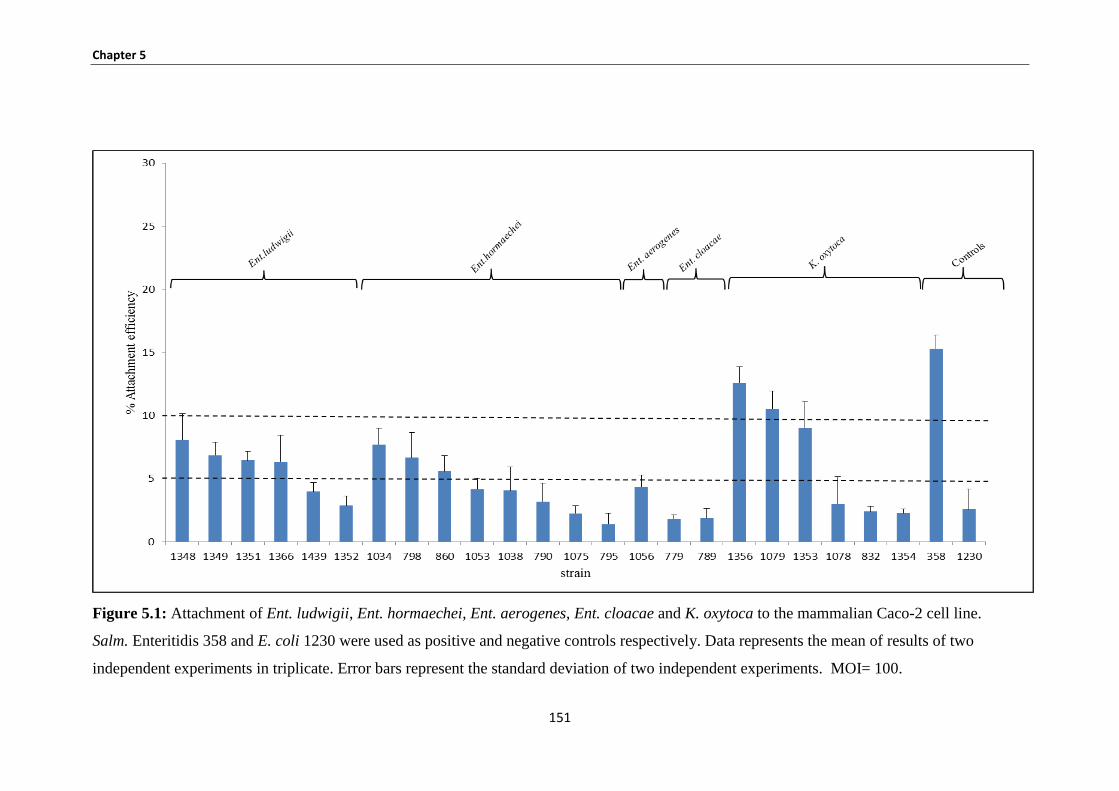
Chapter 5
151
Figure 5.1: Attachment of Ent. ludwigii, Ent. hormaechei, Ent. aerogenes, Ent. cloacae and K. oxytoca to the mammalian Caco-2 cell line.
Salm. Enteritidis 358 and E. coli 1230 were used as positive and negative controls respectively. Data represents the mean of results of two
independent experiments in triplicate. Error bars represent the standard deviation of two independent experiments. MOI= 100.

Chapter 5
152
Figure 5.2: Attachment of Ent. cancerogenus to the mammalian Caco-2 cell line.
Salm. Enteritidis 358 and E. coli 1230 were used as positive and negative controls respectively. Data represents the mean of results of two
independent experiments in triplicate. Error bars represent the standard deviation of two independent experiments. MOI= 100.

Chapter 5
153
5.3.2 Bacterial invasion Caco-2 human epithelial cells
To determine the ability of strains to invade Caco-2 human epithelial cells,
gentamicin protection assay was used. In Figures 5.3 and 5.4 most strains invaded
Caco-2 human epithelial cells at very low levels, however Ent. hormaechei strain
1053 showed four fold less invasion to Caco-2 human epithelial cells than Salm.
Enteritidis.
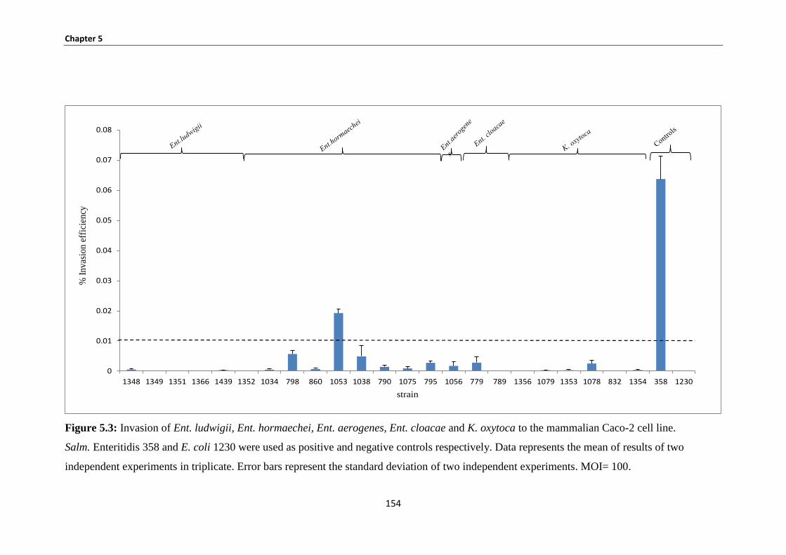
Chapter 5
154
Figure 5.3: Invasion of Ent. ludwigii, Ent. hormaechei, Ent. aerogenes, Ent. cloacae and K. oxytoca to the mammalian Caco-2 cell line.
Salm. Enteritidis 358 and E. coli 1230 were used as positive and negative controls respectively. Data represents the mean of results of two
independent experiments in triplicate. Error bars represent the standard deviation of two independent experiments. MOI= 100.
0
0.01
0.02
0.03
0.04
0.05
0.06
0.07
0.08
1348 1349 1351 1366 1439 1352 1034 798 860 1053 1038 790 1075 795 1056 779 789 1356 1079 1353 1078 832 1354 358 1230
% I
nvas
ion
eff
icie
ncy
strain
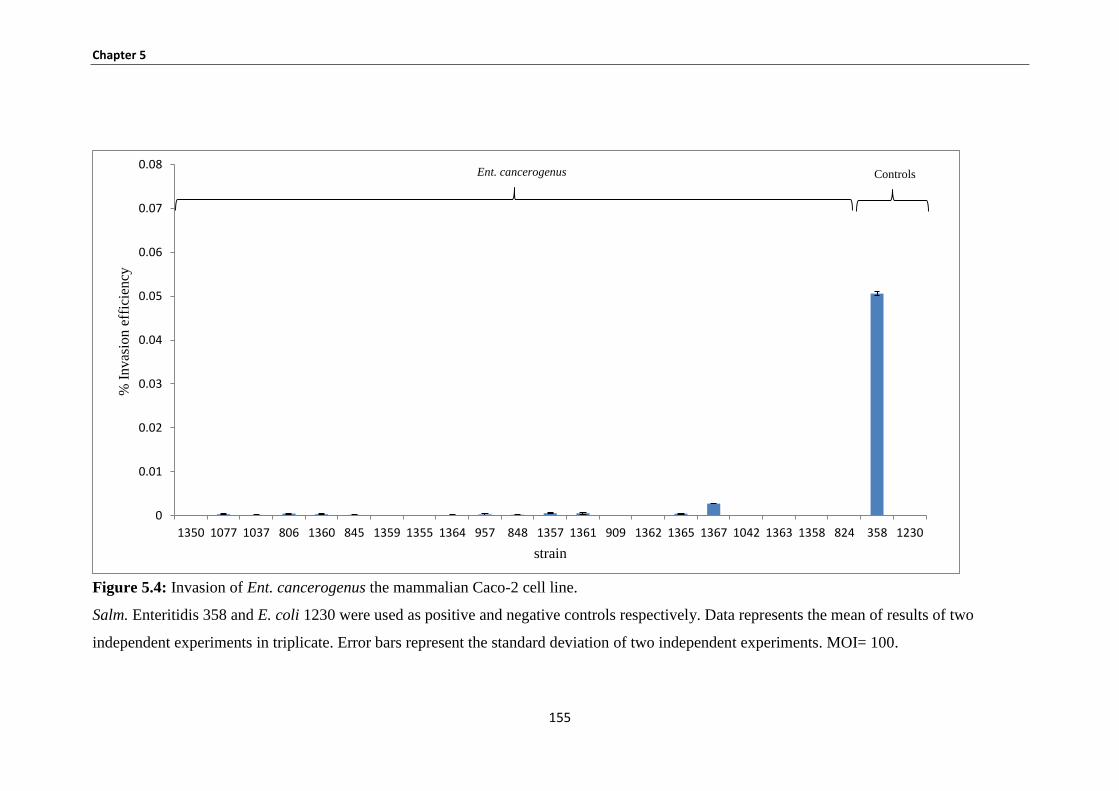
Chapter 5
155
Figure 5.4: Invasion of Ent. cancerogenus the mammalian Caco-2 cell line.
Salm. Enteritidis 358 and E. coli 1230 were used as positive and negative controls respectively. Data represents the mean of results of two
independent experiments in triplicate. Error bars represent the standard deviation of two independent experiments. MOI= 100.
0
0.01
0.02
0.03
0.04
0.05
0.06
0.07
0.08
1350 1077 1037 806 1360 845 1359 1355 1364 957 848 1357 1361 909 1362 1365 1367 1042 1363 1358 824 358 1230
% I
nvas
ion
eff
icie
ncy
strain
Ent. cancerogenus Controls
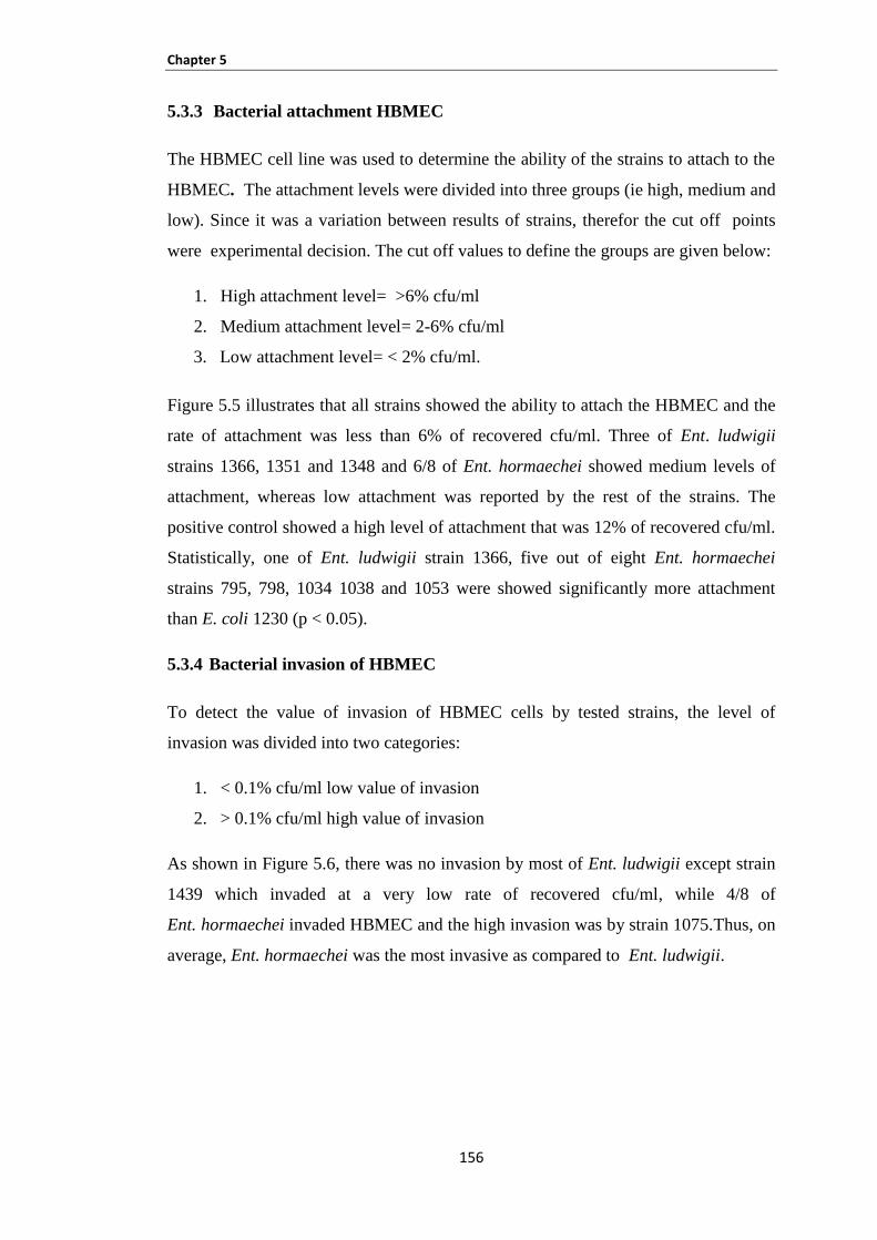
Chapter 5
156
5.3.3 Bacterial attachment HBMEC
The HBMEC cell line was used to determine the ability of the strains to attach to the
HBMEC. The attachment levels were divided into three groups (ie high, medium and
low). Since it was a variation between results of strains, therefor the cut off points
were experimental decision. The cut off values to define the groups are given below:
1. High attachment level= >6% cfu/ml
2. Medium attachment level= 2-6% cfu/ml
3. Low attachment level= < 2% cfu/ml.
Figure 5.5 illustrates that all strains showed the ability to attach the HBMEC and the
rate of attachment was less than 6% of recovered cfu/ml. Three of Ent. ludwigii
strains 1366, 1351 and 1348 and 6/8 of Ent. hormaechei showed medium levels of
attachment, whereas low attachment was reported by the rest of the strains. The
positive control showed a high level of attachment that was 12% of recovered cfu/ml.
Statistically, one of Ent. ludwigii strain 1366, five out of eight Ent. hormaechei
strains 795, 798, 1034 1038 and 1053 were showed significantly more attachment
than E. coli 1230 (p < 0.05).
5.3.4 Bacterial invasion of HBMEC
To detect the value of invasion of HBMEC cells by tested strains, the level of
invasion was divided into two categories:
1. < 0.1% cfu/ml low value of invasion
2. > 0.1% cfu/ml high value of invasion
As shown in Figure 5.6, there was no invasion by most of Ent. ludwigii except strain
1439 which invaded at a very low rate of recovered cfu/ml, while 4/8 of
Ent. hormaechei invaded HBMEC and the high invasion was by strain 1075.Thus, on
average, Ent. hormaechei was the most invasive as compared to Ent. ludwigii.
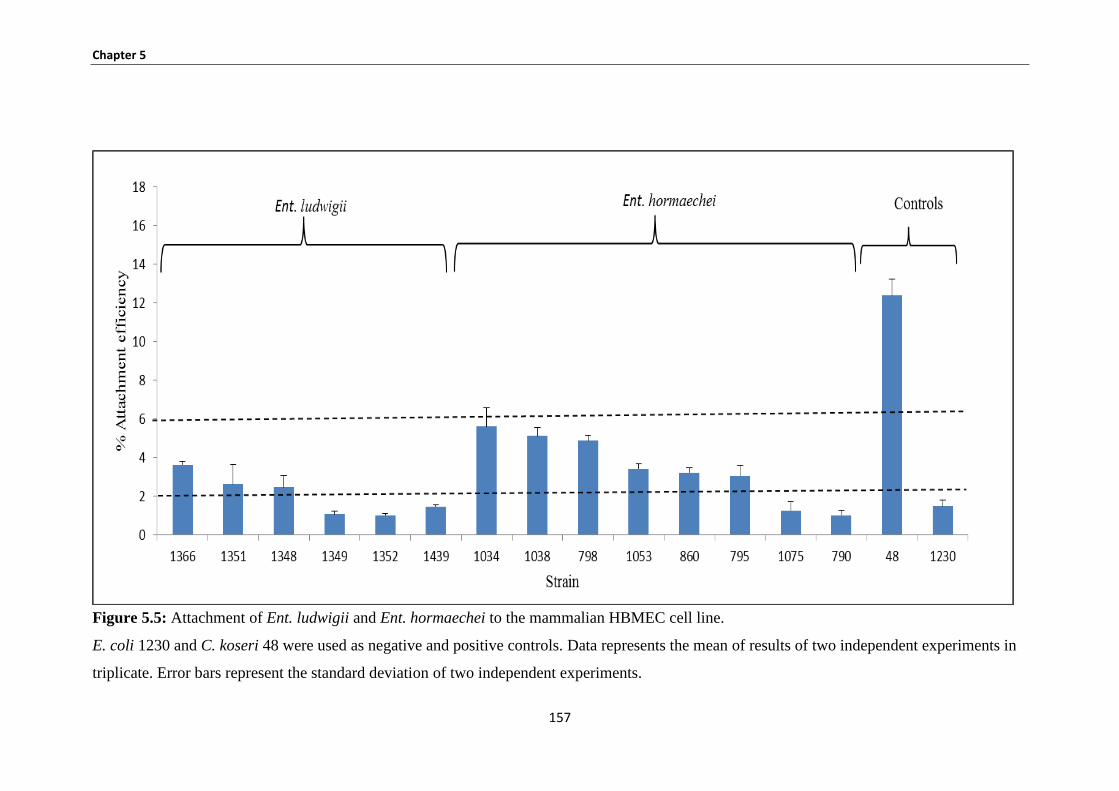
Chapter 5
157
Figure 5.5: Attachment of Ent. ludwigii and Ent. hormaechei to the mammalian HBMEC cell line.
E. coli 1230 and C. koseri 48 were used as negative and positive controls. Data represents the mean of results of two independent experiments in
triplicate. Error bars represent the standard deviation of two independent experiments.
Chapter 5
158
Figure 5.6: Invasion of Ent. ludwigii and Ent. hormaechei to the mammalian HBMEC cell line.
E. coli 1230 and C. koseri 48 were used as negative and positive controls. Data represents the mean of results of two independent experiments in
triplicate. Error bars represent the standard deviation of two independent experiments.
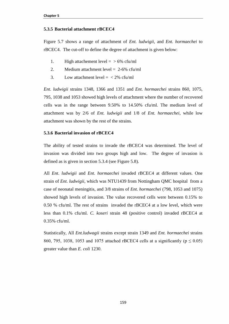
Chapter 5
159
5.3.5 Bacterial attachment rBCEC4
Figure 5.7 shows a range of attachment of Ent. ludwigii, and Ent. hormaechei to
rBCEC4. The cut-off to define the degree of attachment is given below:
1. High attachement level = > 6% cfu/ml
2. Medium attachment level = 2-6% cfu/ml
3. Low attachment level = < 2% cfu/ml
Ent. ludwigii strains 1348, 1366 and 1351 and Ent. hormaechei strains 860, 1075,
795, 1038 and 1053 showed high levels of attachment where the number of recovered
cells was in the range between 9.50% to 14.50% cfu/ml. The medium level of
attachment was by 2/6 of Ent. ludwigii and 1/8 of Ent. hormaechei, while low
attachment was shown by the rest of the strains.
5.3.6 Bacterial invasion of rBCEC4
The ability of tested strains to invade the rBCEC4 was determined. The level of
invasion was divided into two groups high and low. The degree of invasion is
defined as is given in section 5.3.4 (see Figure 5.8).
All Ent. ludwigii and Ent. hormaechei invaded rBCEC4 at different values. One
strain of Ent. ludwigii, which was NTU1439 from Nottingham QMC hospital from a
case of neonatal meningitis, and 3/8 strains of Ent. hormaechei (798, 1053 and 1075)
showed high levels of invasion. The value recovered cells were between 0.15% to
0.50 % cfu/ml. The rest of strains invaded the rBCEC4 at a low level, which were
less than 0.1% cfu/ml. C. koseri strain 48 (positive control) invaded rBCEC4 at
0.35% cfu/ml.
Statistically, All Ent.ludwagii strains except strain 1349 and Ent. hormaechei strains
860, 7 5, 038, 053 and 075 attached rBCEC4 cells at a significantly (p ≤ 0.05)
greater value than E. coli 1230.
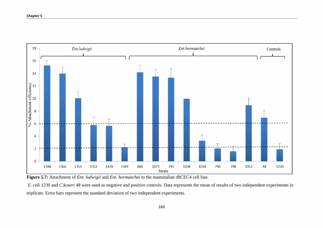
Chapter 5
160
Figure 5.7: Attachment of Ent. ludwigii and Ent. hormaechei to the mammalian rBCEC4 cell line.
E. coli 1230 and C.koseri 48 were used as negative and positive controls. Data represents the mean of results of two independent experiments in
triplicate. Error bars represent the standard deviation of two independent experiments.
Chapter 5
161
Figure 5.8: Invasion of Ent. ludwigii and Ent. hormaechei to the mammalian rBCEC4 cell line.
E. coli 1230 and C. koseri 48 were used as negative and positive controls. Data represents the mean of results of two independent experiments in
triplicate. Error bars represent the standard deviation of two independent experiments.
Chapter 5
162
5.3.7 U937 macrophage uptake and persistence studies
After invasion, a number of bacterial pathogens have the ability to survive and
replicate within host cells. The host immune response utilizes phagocytic cell as a
critical line of defence against infection. The capability of a pathogen to persist and
replicate within macrophages is a potent way of evading the defence mechanisms of
the host.
Three strains of Ent. hormaechei out of eight strains and 1/2 strain of Ent. cloacae
persisted in macrophages for 48 hours. In contrast, Ent. cloacae strain 779 did not
persist after a 24 hours period. Ent. hormaechei 798 strain demonstrated a high level
of persistence at 24 hours, while Ent. cloacae 789 strain showed high replication at
48 hours.
Ent. hormaechei 798 strain displayed a high level of replication. All Ent. ludwigii and
K. oxytoca strains showed no uptake by U937 macrophage cells, or were rapidly
killed following uptake (Figure 5.9).
The only significant replicate was observed at 48 minutes for strain 798 (P < 0.05).
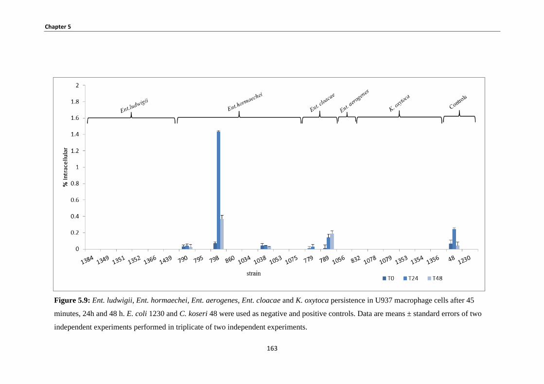
Chapter 5
163
Figure 5.9: Ent. ludwigii, Ent. hormaechei, Ent. aerogenes, Ent. cloacae and K. oxytoca persistence in U937 macrophage cells after 45
minutes, 24h and 48 h. E. coli 1230 and C. koseri 48 were used as negative and positive controls. Data are means ± standard errors of two
independent experiments performed in triplicate of two independent experiments.
Chapter 5
164
5.3.8 Summary of potential risk for Enterobacteriaceae
Evaluation of the ability of opportunistic pathogenic bacteria to attach and invade
Caco-2, HBMEC and rBECE4 cells and survive within macrophages are an
important consideration. The data for these results was summarised in Table 5.1. The
potential risk for 23 strains of Enterobacteriaceae was determined and categorised as
high, moderate and low with regard to their virulence potential. Six strains showed
high potential risk; 3 of the strains persisted within macrophages which included Ent.
hormaechei strains 798, 1038 and 790, while an other 3 strains, Ent. hormaechei
1053 and 1075 and Ent. ludwigii 1439, were able to invade rBMCE4 at high levels.
Ent. hormaechei 798 and 1075 strains also showed high invasion to rBMCE4 and
HBMEC respectively. Two strains of Ent. cloacae (779 and 789) showed moderate
risk. The rest of the strains showed low potential risk.
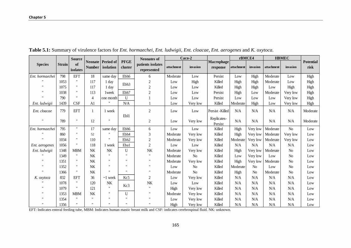
Chapter 5
165
Table 5.1: Summary of virulence factors for Ent. hormaechei, Ent. ludwigii, Ent. cloacae, Ent. aerogenes and K. oxytoca.
EFT: Indicates enteral feeding tube, MBM: Indicates human mastic breast milk and CSF: indicates cerebrospinal fluid. NK: unknown.
attachment invasion attachment invasion attachment invasion
Ent. hormaechei 798 EFT 18 same day Ebh6 6 Moderate Low Persist Low High Moderate Low High
" 1053 " 117 1 day 2 Low High Killed High High Moderate Low High
" 1075 " 117 1 day 2 Low Low Killed High High Low High High
" 1038 " 113 1week Ebh7 2 Low Low Persist High Low Moderate Very low High
" 790 " 4 one month U 1 Low Low Persist Low Low Low Very low High
Ent. ludwigii 1439 CSF A1 " N/A 1 Low Very low Killed Moderate High Low Very low High
Ent. cloacae 779 EFT 1 1 week 2 Low Low Persist -Killed N/A N/A N/A N/A Moderate
" 789 " 12 " 2 Low Very lowReplicates-
PersistN/A N/A N/A N/A Moderate
Ent. hormaechei 795 " 17 same day Ebh6 6 Low Low Killed High Very low Moderate No Low
" 860 " 52 " Ebh4 3 Moderate Very low Killed High Very low Moderate Very low Low
" 1034 " 110 " Ebh2 2 Moderate Very low Killed Moderate Very low Moderate Very low Low
Ent. aerogenes 1056 " 118 1 week Eba1 2 Low Low Killed N/A N/A N/A N/A Low
Ent. ludwigii 1348 MBM NK NK U NK Moderate Very low Killed High Very low Moderate No Low
" 1349 " NK " " " Moderate No Killed Low Very low Low No Low
" 1351 " NK " " " Moderate Very low Killed High Very low Moderate No Low
" 1352 " NK " " " Low No Killed Moderate No Low No Low
" 1366 " NK " " " Moderate No Killed High No Moderate No Low
K. oxytoca 832 EFT 36 ~1 week Kc5 2 Low Very low Killed N/A N/A N/A N/A Low
" 1078 " 120 NK NK Low Low Killed N/A N/A N/A N/A Low
" 1079 " 121 " " High Very low Killed N/A N/A N/A N/A Low
" 1353 MBM NK " U " Moderate Very low Killed N/A N/A N/A N/A Low
" 1354 " " " " " Low Very low Killed N/A N/A N/A N/A Low
" 1356 " " " " " High Very low Killed N/A N/A N/A N/A Low
Neonates of
patients isolates
represented
Species Strain
Source
of
isolates
Neonate
Number
Period of
isolation
Kc3
Caco-2
Ebh3
Ebl1
Potential
risk
rBMCE4 HBMECMacrophage
response
PFGE
cluster
Chapter 5
166
5.3.9 Patterns of adherence of Enterobacteriaceae to Caco-2 cells
Giemsa staining was used to determine the adhesion patterns of Ent. ludwigii,
Ent. hormaechei, Ent. cloacae, Ent. aerogenes and K. oxytoca to Caco-2 human
epithelial cells. Three different adhesion patterns could be distinguished on Caco-2
human epithelial cells:
(i) Aggregative adhesions: Large clusters of adhering bacteria which cover the cell
surface, observed for 6/8 strains of Ent. hormaechei and 1/6 strains of K. oxytoca (see
Table 5.2, Figure 5.10 (A)).
(ii) Diffuse adhesions: Bacteria distributed over the cell surface; observed in only one
strain ie, Ent. aerogenes (see Table 5.2, Figure 5.10 (B)).
(iii) Localized adhesions: Distribution in small groups; observed in all Ent. ludwigii
and Ent. cloacae strains, 5/6 strains of K. oxytoca, 2/8 strains of Ent. hormaechei;
(see Table 5.2, Figure 5.10 (C)).
The strains showed the same adhesion patterns belonging to the same pulsotype (see
Table 5.2). Examples of the different patterns of adhesion as observed on Caco-2
human epithelial cells can be seen in Figure 5.10 A, B and C.
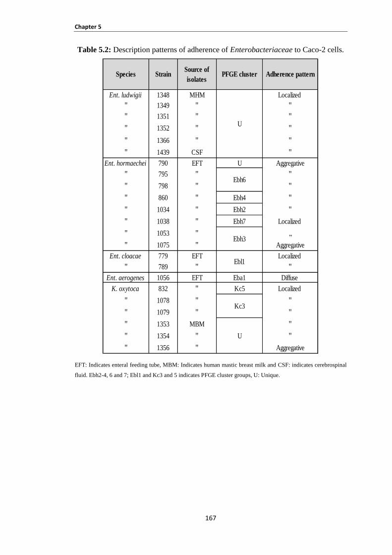
Chapter 5
167
Table 5.2: Description patterns of adherence of Enterobacteriaceae to Caco-2 cells.
EFT: Indicates enteral feeding tube, MBM: Indicates human mastic breast milk and CSF: indicates cerebrospinal
fluid. Ebh2-4, 6 and 7; Ebl1 and Kc3 and 5 indicates PFGE cluster groups, U: Unique.
Ent. ludwigii 1348 MHM Localized
" 1349 " "
" 1351 " "
" 1352 " "
" 1366 " "
" 1439 CSF "
Ent. hormaechei 790 EFT U Aggregative
" 795 " "
" 798 " "
" 860 " Ebh4 "
" 1034 " Ebh2 "
" 1038 " Ebh7 Localized
" 1053 " „
" 1075 " Aggregative
Ent. cloacae 779 EFT Localized
" 789 " "
Ent. aerogenes 1056 EFT Eba1 Diffuse
K. oxytoca 832 " Kc5 Localized
" 1078 " "
" 1079 " "
" 1353 MBM "
" 1354 " "
" 1356 " Aggregative
U
Ebh6
Ebh3
Ebl1
Kc3
U
Species StrainSource of
isolatesAdherence patternPFGE cluster
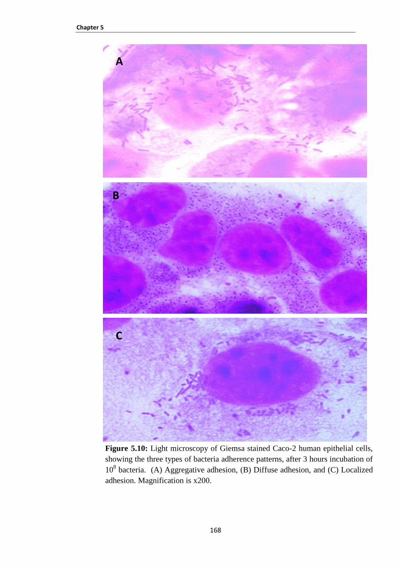
Chapter 5
168
Figure 5.10: Light microscopy of Giemsa stained Caco-2 human epithelial cells,
showing the three types of bacteria adherence patterns, after 3 hours incubation of
108 bacteria. (A) Aggregative adhesion, (B) Diffuse adhesion, and (C) Localized
adhesion. Magnification is x200.
A
B
C
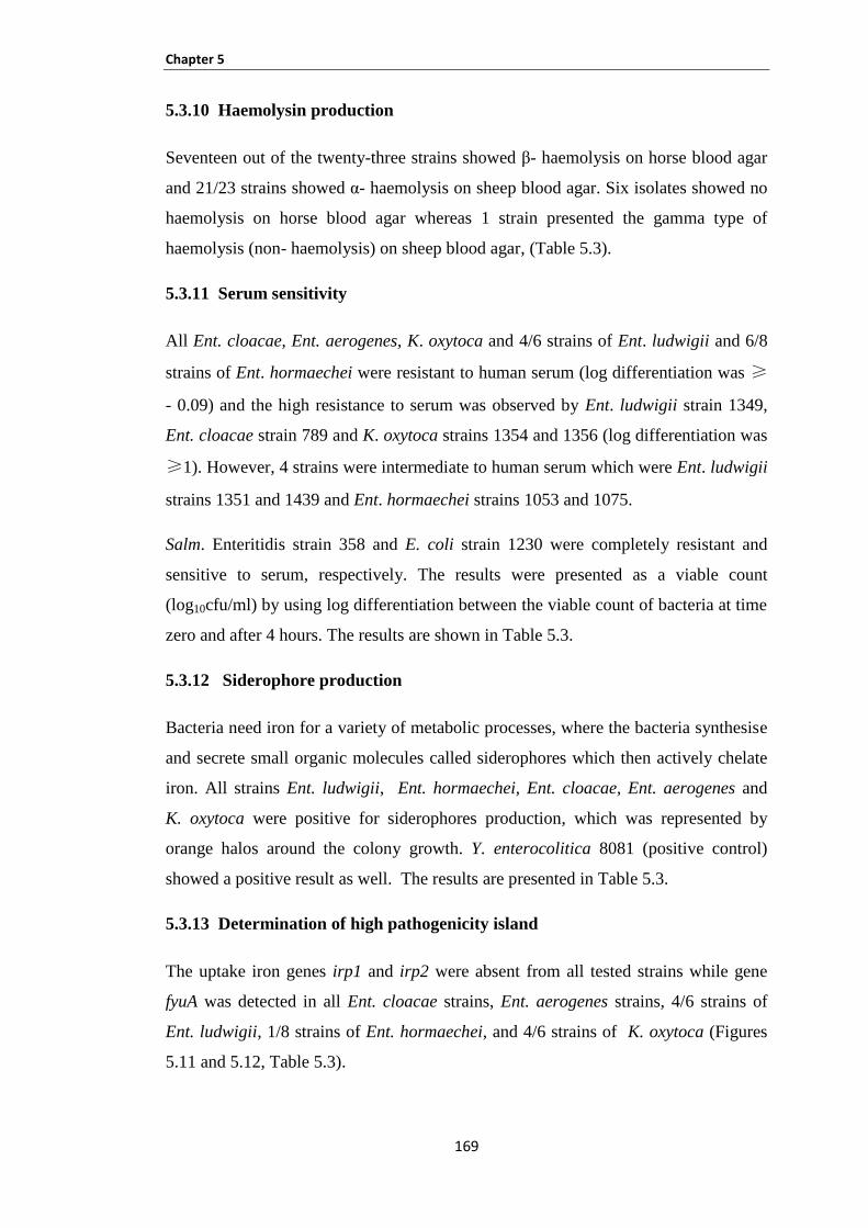
Chapter 5
169
5.3.10 Haemolysin production
Seventeen out of the twenty-three strains showed β- haemolysis on horse blood agar
and 21/23 strains showed α- haemolysis on sheep blood agar. Six isolates showed no
haemolysis on horse blood agar whereas 1 strain presented the gamma type of
haemolysis (non- haemolysis) on sheep blood agar, (Table 5.3).
5.3.11 Serum sensitivity
All Ent. cloacae, Ent. aerogenes, K. oxytoca and 4/6 strains of Ent. ludwigii and 6/8
strains of Ent. hormaechei were resistant to human serum (log differentiation was ≥
- 0.09) and the high resistance to serum was observed by Ent. ludwigii strain 1349,
Ent. cloacae strain 789 and K. oxytoca strains 1354 and 1356 (log differentiation was
≥1). However, 4 strains were intermediate to human serum which were Ent. ludwigii
strains 1351 and 1439 and Ent. hormaechei strains 1053 and 1075.
Salm. Enteritidis strain 358 and E. coli strain 1230 were completely resistant and
sensitive to serum, respectively. The results were presented as a viable count
(log10cfu/ml) by using log differentiation between the viable count of bacteria at time
zero and after 4 hours. The results are shown in Table 5.3.
5.3.12 Siderophore production
Bacteria need iron for a variety of metabolic processes, where the bacteria synthesise
and secrete small organic molecules called siderophores which then actively chelate
iron. All strains Ent. ludwigii, Ent. hormaechei, Ent. cloacae, Ent. aerogenes and
K. oxytoca were positive for siderophores production, which was represented by
orange halos around the colony growth. Y. enterocolitica 8081 (positive control)
showed a positive result as well. The results are presented in Table 5.3.
5.3.13 Determination of high pathogenicity island
The uptake iron genes irp1 and irp2 were absent from all tested strains while gene
fyuA was detected in all Ent. cloacae strains, Ent. aerogenes strains, 4/6 strains of
Ent. ludwigii, 1/8 strains of Ent. hormaechei, and 4/6 strains of K. oxytoca (Figures
5.11 and 5.12, Table 5.3).
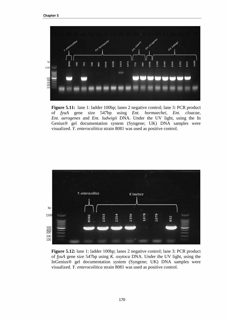
Chapter 5
170
Figure 5.11: lane 1: ladder 100bp; lanes 2 negative control; lane 3: PCR product
of fyuA gene size 547bp using Ent. hormaechei, Ent. cloacae,
Ent. aerogenes and Ent. ludwigii DNA. Under the UV light, using the In
Genius® gel documentation system (Syngene; UK) DNA samples were
visualized. Y. enterocolitica strain 8081 was used as positive control.
Figure 5.12: lane 1: ladder 100bp; lanes 2 negative control; lane 3: PCR product
of fyuA gene size 547bp using K. oxytoca DNA. Under the UV light, using the
InGenius® gel documentation system (Syngene; UK) DNA samples were
visualized. Y. enterocolitica strain 8081 was used as positive control.
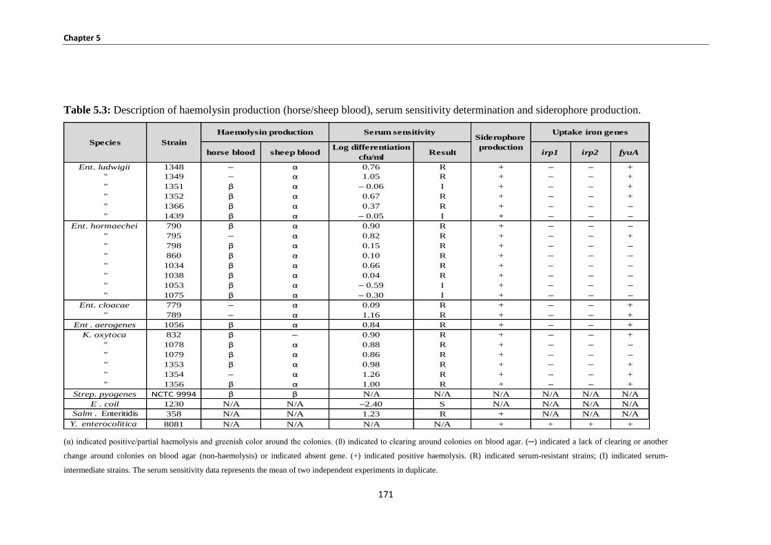
Chapter 5
171
Table 5.3: Description of haemolysin production (horse/sheep blood), serum sensitivity determination and siderophore production.
(α) indicated positive/partial haemolysis and greenish color around the colonies. (ß) indicated to clearing around colonies on blood agar. (─) indicated a lack of clearing or another
change around colonies on blood agar (non-haemolysis) or indicated absent gene. (+) indicated positive haemolysis. (R) indicated serum-resistant strains; (I) indicated serum-
intermediate strains. The serum sensitivity data represents the mean of two independent experiments in duplicate.
horse blood sheep bloodLog differentiation
cfu/mlResult irp1 irp2 fyuA
Ent. ludwigii 1348 ─ α 0.76 R + ─ ─ +
" 1349 ─ α 1.05 R + ─ ─ +
" 1351 β α ─ 0.06 I + ─ ─ +
" 1352 β α 0.67 R + ─ ─ +
" 1366 β α 0.37 R + ─ ─ ─
" 1439 β α ─ 0.05 I + ─ ─ ─
Ent. hormaechei 790 β α 0.90 R + ─ ─ ─
" 795 ─ α 0.82 R + ─ ─ +
" 798 β α 0.15 R + ─ ─ ─
" 860 β α 0.10 R + ─ ─ ─
" 1034 β α 0.66 R + ─ ─ ─
" 1038 β α 0.04 R + ─ ─ ─
" 1053 β α ─ 0.59 I + ─ ─ ─
" 1075 β α ─ 0.30 I + ─ ─ ─
Ent. cloacae 779 ─ α 0.09 R + ─ ─ +
" 789 ─ α 1.16 R + ─ ─ +
Ent . aerogenes 1056 β α 0.84 R + ─ ─ +
K. oxytoca 832 β ─ 0.90 R + ─ ─ +
" 1078 β α 0.88 R + ─ ─ ─
" 1079 β α 0.86 R + ─ ─ ─
" 1353 β α 0.98 R + ─ ─ +
" 1354 ─ α 1.26 R + ─ ─ +
" 1356 β α 1.00 R + ─ ─ +
Strep. pyogenes NCTC 9994 β β N/A N/A N/A N/A N/A N/A
E . coil 1230 N/A N/A ─2.40 S N/A N/A N/A N/A
Salm . Enteritidis 358 N/A N/A 1.23 R + N/A N/A N/A
Y. enterocolitica 8081 N/A N/A N/A N/A + + + +
Species Strain
Uptake iron genesSerum sensitivityHaemolysin productionSiderophore
production
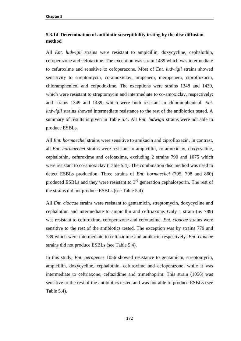
Chapter 5
172
5.3.14 Determination of antibiotic susceptibility testing by the disc diffusion
method
All Ent. ludwigii strains were resistant to ampicillin, doxycycline, cephalothin,
cefoperazone and cefotaxime. The exception was strain 1439 which was intermediate
to cefuroxime and sensitive to cefoperazone. Most of Ent. ludwigii strains showed
sensitivity to streptomycin, co-amoxiclav, imipenem, meropenem, ciprofloxacin,
chloramphenicol and cefpodoxime. The exceptions were strains 1348 and 1439,
which were resistant to streptomycin and intermediate to co-amoxiclav, respectively;
and strains 1349 and 1439, which were both resistant to chloramphenicol. Ent.
ludwigii strains showed intermediate resistance to the rest of the antibiotics tested. A
summary of results is given in Table 5.4. All Ent. ludwigii strains were not able to
produce ESBLs.
All Ent. hormaechei strains were sensitive to amikacin and ciprofloxacin. In contrast,
all Ent. hormaechei strains were resistant to ampicillin, co-amoxiclav, doxycycline,
cephalothin, cefuroxime and cefotaxime, excluding 2 strains 790 and 1075 which
were resistant to co-amoxiclav (Table 5.4). The combination disc method was used to
detect ESBLs production. Three strains of Ent. hormaechei (795, 798 and 860)
produced ESBLs and they were resistant to 3rd
generation cephalosporin. The rest of
the strains did not produce ESBLs (see Table 5.4).
All Ent. cloacae strains were resistant to gentamicin, streptomycin, doxycycline and
cephalothin and intermediate to ampicillin and ceftriaxone. Only 1 strain (ie. 789)
was resistant to cefuroxime, cefoperazone and cefotaxime. Ent. cloacae strains were
sensitive to the rest of the antibiotics tested. The exception was by strains 779 and
789 which were intermediate to ceftazidime and amikacin respectively. Ent. cloacae
strains did not produce ESBLs (see Table 5.4).
In this study, Ent. aerogenes 1056 showed resistance to gentamicin, streptomycin,
ampicillin, doxycycline, cephalothin, cefuroxime and cefoperazone, while it was
intermediate to ceftriaxone, ceftazidime and trimethoprim. This strain (1056) was
sensitive to the rest of the antibiotics tested and was not able to produce ESBLs (see
Table 5.4).
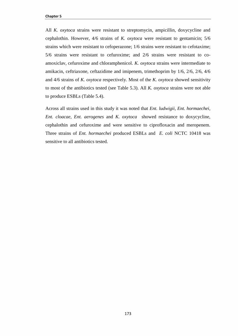
Chapter 5
173
All K. oxytoca strains were resistant to streptomycin, ampicillin, doxycycline and
cephalothin. However, 4/6 strains of K. oxytoca were resistant to gentamicin; 5/6
strains which were resistant to cefoperazone; 1/6 strains were resistant to cefotaxime;
5/6 strains were resistant to cefuroxime; and 2/6 strains were resistant to co-
amoxiclav, cefuroxime and chloramphenicol. K. oxytoca strains were intermediate to
amikacin, ceftriaxone, ceftazidime and imipenem, trimethoprim by 1/6, 2/6, 2/6, 4/6
and 4/6 strains of K. oxytoca respectively. Most of the K. oxytoca showed sensitivity
to most of the antibiotics tested (see Table 5.3). All K. oxytoca strains were not able
to produce ESBLs (Table 5.4).
Across all strains used in this study it was noted that Ent. ludwigii, Ent. hormaechei,
Ent. cloacae, Ent. aerogenes and K. oxytoca showed resistance to doxycycline,
cephalothin and cefuroxime and were sensitive to ciprofloxacin and meropenem.
Three strains of Ent. hormaechei produced ESBLs and E. coli NCTC 10418 was
sensitive to all antibiotics tested.
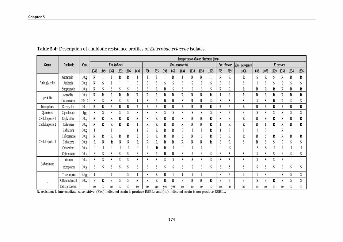
Chapter 5
174
Table 5.4: Description of antibiotic resistance profiles of Enterobacteriaceae isolates.
R, resistant; I, intermediate; s, sensitive. (Yes) indicated strain is produce ESBLs and (no) indicated strain is not produce ESBLs.
Ent . aerogenes
1348 1349 1351 1352 1366 1439 790 795 798 860 1034 1038 1053 1075 779 789 1056 832 1078 1079 1353 1354 1356
Gentamicin 10µg R I I R R I I I I R I R R I R R R S R S R R R
Amikacin 30µg R S I I I S S S S S S S S S S I S I S S S S S
Streptomycin 10µg R S S S S S S R R S S S S S R R R R R R R R R
Ampicillin 10µg R R R R R R R R R R R R R R I I R R R R R R R
Co-amoxiclav 20+10 S S S S S I S R R R S R R S S S S S S R R S S
Tetracyclines Doxycycline 30µg R R R R R R R R R R R R R R R R R R R R R R R
Quinolones Ciprofloxacin 1µg S S S S S S S S S S S S S S S S S S S S S S S
Cephalosporins 1 Cephalothin 30µg R R R R R R R R R R R R R R R R R R R R R R R
Cephalosporins 2 Cefuroxime 30µg R R R R R I R R R R R R R R I R R R I R R R R
Ceftriaxone 30µg I I I I I I S R R R S I I R I I I I S I R I I
Cefoperazone 30µg R R R R R S S R R R S R S R S R R R S R R R R
Cefotaxime 30µg R R R R R R R R R R R R R R S R S R S S S S S
Ceftazidime 30µg I I I I I I I R R I I I I I I S I S S I I I I
Cefpodoxime 10µg S S S S S S S R R R S S S S S S S S S S S S S
Imipenem 10µg S S S S S S S S S S S S S S S S S S S S S I I
meropenem 10µg S S S S S S S S S S S S S S S S S S S S S S S
Trimethoprim 2.5µg I I I I S I S R R I I I I I S S I I S I S S S
Chloramphenicol 30µg S R S S S R R R R R S R R R S S S S S R R S S
ESBL production no no no no no no no yes yes yes no no no no no no no no no no no no no
Antibiotic Con.
Interpretation of zone diameters (mm)
Ent. ludwigii Ent. hormeachei Ent. cloacae K. oxytoca
Aminoglycoside
penicillin
Group
_
Carbapenems
Cephalosporins 3
Chapter 5
175
5.4 DISCUSSION
Pathogenic bacteria use a number of strategies to cause disease in human hosts, some
of which include adhesion, colonisation, activation of disease process, serum
resistance and toxins production. The most important factor is the ability of bacteria
to attach to the host surfaces, such as mucous membranes, gastric and intestinal
epithelial or endothelial tissue (Boyle et al., 2003; Finlay and Falkow, 1997; Klemm
and Schembri, 2000). Enterobacteriaceae are the most common opportunistic
pathogens, particularly E. coli, Enterobacter spp., Klebsiella spp., Serratia spp., and
Salmonella, which are considerably implicated with a wide range of morbidity and
mortality (Friedland et al., 2003; Adamson et al., 2012).
In the present study, an attempt was made to determine the relationship between
haemolysin production and other virulence associated factors in selected strains of
Enterobacteraceae. There was a relationship between haemolysin production and
other factors, such as serum resistance and siderophore production, which may be
associated with neonates’ infection.
This study was focused on investigating the potential virulence of
Enterobacteriaceae from EFT and MBM by determining whether or not they pose
a risk of infection to neonates. Forty three Enterobacteriaceae composed of
Ent. ludwigii, Ent. hormaechei, Ent. aerogenes, Ent. cloacae, Ent. cancerogenus and
K. oxytoca strains were chosen to be part of the study as they were the most
representative of the species isolated. The potential of virulence for the 43 strains to
mammalian cells was determined using attachment and invasion studies of Caco-2
tissue culture cells, while the attachment and invasion rBECE4 and HBMEC tissue
culture cells were only studied for 14 strains (Ent. ludwigii, Ent. hormaechei).
Macrophage survival was studied for 23 strains (Ent. ludwigii, Ent. hormaechei, Ent.
aerogenes, Ent. cloacae and K. oxytoca using the U937 cell line of human monocyte
cells.
This study demonstrated that all strains showed the ability to attach to epithelial and
endothelial cell lines. The viable counts obtained exhibit the ability of strains to
attach to Caco-2, HBMEC and rBECE4 cells. There were differences in the ability of
Ent. ludwigii, Ent. hormaechei, Ent. cancerogenus Ent. aerogenes, Ent. cloacae and

Chapter 5
176
K. oxytoca to attach to intestinal epithelial cells (Caco-2) (see Figures 5.1-5.4). The
viable counts showed that Ent. cancerogenus and K. oxytoca strains were able to
attach to Caco-2 cells at relatively high levels compared with the rest of strains
(Figure 5.2). Whereas Ent. ludwigii and Ent. hormaechei strains attached to rBECE4
cells at higher numbers compared to their ability to attach to HBMEC cells (Figures
5.5, and 5.7). However, all strains were able to attach to the different types of
epithelial and endothelial cell lines.
The ability of bacteria to adhere to host surfaces, such as skin, mucous membranes
(oral cavity, nasopharynx, urogenital tract), and deeper tissues (lymphoid tissue,
gastric and intestinal epithelia, alveolar lining, endothelial tissue), is a key step in the
host-pathogen interaction. Hosts produce numerous mechanical forces, such as saliva
secretion, coughing, sneezing, mucous flow, peristalsis, and blood flow, which act to
wash microbes away from susceptible surfaces. However, microbial pathogens
express factors that bind to molecules on various host tissue cells and render the
microbe resistant to these mechanical washing forces (Podschun and Ullmann, 1998).
As a result, microbial biochemical processes cause diseases, including proliferation,
toxin secretion, host cell invasion, and activation of host cell signalling cascades
(Wilson et al., 2002).
Once adhered to a host surface, some pathogenic bacteria can gain access deep into
the host and continue the infection cycle. In this study, there were variations between
species in their invasion of Caco-2 cells; for example, all Ent. hormaechei were able
to invade Caco-2 cells compared to the remaining species. Additionally, there was no
direct association between attachment and invasion rates of Caco-2 cells; for example,
Ent. cancerogenus strains 1350, 1077, 1037, 806 and 1360 and K. oxytoca strains
1356 and 1079 had the highest attachment rates, yet 1053 had the highest invasion
rates (Figures. 5.1 and 5.3).
Previous studies reported that K. oxytoca had been associated with septicaemia, soft
tissue, intravenous, meningitis, liver abscess, and gastrointestinal disease. This is
commonly observed in immunocompromised hosts or those with underlying
conditions (Green et al., 2009). Townsend et al. (2008a) demonstrated that virulence
traits were associated with Ent. hormaechei, and they are capable of invading deep
tissues (causing systemic infection and the symptoms that it causes to spread
Chapter 5
177
throughout the systems of the body). Furthermore, Ent. hormaechei associated with
sepsis were able to invade gut epithelial cells. This demonstrates the virulence
potential of Ent. hormaechei for this host site and suggests that its ability to invade
intestinal cells may be a strong indication of potential virulence and pathogenicity in
the neonatal host.
According to Table 5.1, six isolates were classed as high risk, indicating they could
increase the exposure of infants to the bacteria from infant formula administered
through EFT. These strains were Ent. hormaechei strains 790, 798, 1038, 1053 and
1075 and Ent. ludwigii strain 1439. In this study, the level of risk was determined
according to the ability of strains to invade mammalian cells (Caco-2, HBMEC and
rBECE4 cells) and to persist in human macrophages. Ent. hormaechei strains 790,
798, 1038, 1053 and 1075 and Ent. ludwigii strain 1439 showed low levels of
invasion to Caco-2 cells, whereas Ent. hormaechei strain 1053 showed a high level of
invasion to Caco-2 cells.
On the other hand, the virulence potential of Ent. hormaechei and Ent. ludwigii
strains to invade brain capillary endothelial cells was determined using HBMEC and
rBECE4 cells. Interestingly, three strains of Ent. hormaechei (789, 1053 and 1075)
showed similar invasion to both cell lines (HBMEC and rBECE4); however, Ent.
ludwigii strain 1439 was able to invade rBECE4 cell line at much higher levels than
HBMEC cell line. Ent. ludwigii strain 1439 was isolated from a case of neonatal
meningitis (CSF) that was obtained from Nottingham QMC hospital. This study is an
in vitro appraisal evaluating the virulence of the isolate in relationship to clinical
symptoms. There is a strong likelihood that the baby could have acquired the strain
from the environment, through the skull, as its skull was opened for the brain surgery.
It was possible to directly associate clinical symptoms and results with in vitro
studies. For Ent. hormaechei strains isolated from EFT, there was no clinical data for
the patients; however, the ability of Ent. hormaechei strains to invade HBMEC and
rBECE4 cell lines may be a strong indication of potential virulence and pathogenicity
in the neonatal host to cause meningitis. Hervas et al. (2001) reported that
Ent. hormaechei may be associated with meningitis because sepsis and meningitis
were associated with Enterobacter spp. In addition, this result is consistent with a
previous study by Townsend et al. (2008b), who studied virulence assays of clinical
Chapter 5
178
isolates with associated patient details and clinical symptoms. They found that C.
sakazakii strains were able to invade HBMEC and rBCEC4 cells.
Also demonstrated was the ability of selected strains of Ent. ludwigii,
Ent. hormaechei, Ent. aerogenes, Ent. cloacae and K. oxytoca to persist in human
macrophages. Three strains of Ent. hormaechei (790, 1038 and 798) and 2 strains of
Ent. cloacae (779 and 789) were able to persist and replicate in macrophages for 48
hours. However, Ent. cloacae strain 779 showed decreased levels of persistence after
24 hours while Ent. hormaechei strain 798 persisted at high levels within the
macrophages up to 48 hours and then was killed (Figure 5.9). Ent. ludwigii,
Ent. aerogenes and K. oxytoca strains were killed by macrophages.
These results compared the ability of selected strains to invade mammalian cells. The
important finding was that three Ent. hormaechei strains (790, 798 and 1038) were
able to invade three types of cell lines (Caco-2, HBMEC and rBECE4 cells) (Table
5.1, Figures. 5.3, 5.6, 5.8 and 5.9). These results indicate there could be a relationship
between the invasion of epithelial and brain endothelial cells, and also persistence
and replication in human macrophage cells. The ability of strains to invade capillary
endothelial cells may give access to the brain by crossing the blood-brain barrier
which may led to infected neonates. Moreover, the persistance and replication of the
strains in human macrophage cells could be important to facilitate host immune
evasion and dissemination.
These results were consistent with the study conducted by Townsend et al. (2008b),
who studied a NICU outbreak of C. sakazakii infections in France. They found that
all C. sakazakii strains were able to attach and invade Caco-2 human epithelial cells,
and invade rat brain capillary endothelial cells, and also most of the strains persisted
in macrophage cells for 48 hours. They suggested that these features act to facilitate
host immune evasion and spreading.
Patterns of adherence of Enterobacteriaceae to Caco-2 cells were studied. Selected
strains of Ent. ludwigii, Ent. hormaechei, Ent. aerogenes, Ent. cloacae and K.
oxytoca did adhere to Caco-2 cells, and there were different patterns of adherence.
Whether the pattern of adherence relates to the pathogenicity of the strains is
unknown. In this study, there was no relationship between types of adherence
Chapter 5
179
patterns and ability to invade the Caco-2 human epithelial cells (as shown in Table
5.2, Figure 5.10 A, B and C).
A number of virulence factors, that including haemolysins production, serum
sensitivity determination, siderophore production and antimicrobial susceptibility,
were investigated.
Haemolysin production is one of the toxins that are secreted by pathogenic bacteria
and may play an essential role in disease. Haemolysins are classified to three
different types of haemolysins: α-haemolysin, β-haemolysin and γ-haemolysin. These
are found in most clinical isolates, such as E. coli and Serratia species (König et al.,
1987; Welch, 1987; Schmidt et al., 1995). The present study shows that all Ent.
ludwigii, Ent. hormaechei, Ent. aerogenes, Ent. cloacae and K. oxytoca strains were
α-haemolytic on sheep blood agar, except K. oxytoca strain 832, while β-haemolysin
was observed on horse blood by 17/23 strains. Previous studies have shown that the
most important virulence factor commonly expressed by extra-intestinal pathogenic
bacteria, such as E. coli, is α-haemolysin (Balsalobre et al., 2006).
Another study by Ring et al. (2002) demonstrated that β-hemolysin plays an
important role for the result of group B streptococcal sepsis by contributing to liver
failure and high mortality. Thus, these results suggest that the strains have the ability
to cause haemolysis and thereby damage the cell membrane of RBCs through
production of β-haemolysis which is a potential risk factor to neonatal health.
The sensitivity to normal human serum of isolated Enterobacteriaceae was
determined. All Ent. ludwigii, Ent. hormaechei, Ent. aerogenes, Ent. cloacae, and K.
oxytoca strains were serum resistant, except four strains, Ent. ludwigii strains 1351
and 1439 and Ent. hormaechei strains 1053 and 1075, which survived in blood serum
at an intermediate rate. A common feature of many different species causing
bacteremia is the capability to avoid the bactericidal effects of human serum (Porto et
al., 1989; Hol et al., 1995; Hoe et al., 1999).
Several previous studies found that a common feature of meningococci isolated from
blood or cerebrospinal fluid is serum resistance (Figueroa and Densen, 1991).
Similarly, the clinical isolated of V. vulnificus exhibited the ability to survive in
human serum (Bogard and Oliver, 2007). Furthermore, Ent. hormaechei nosocomial
Chapter 5
180
outbreak strains isolated from different neonates in a California hospital showed
serum-resistance (Townsend et al., 2008b). The finding in the current study could be
explained by the ability of strains to survive in blood serum which possibly indicates
an increased risk to neonates.
Iron is an important element for pathogenic bacteria to grow, replicate and progress
an infection (Braun and Winkelman, 1987). Bacterial ability to obtain iron from the
host is one of the determinants for the virulence of a pathogen. All
Enterobacteriaceae isolates showed active siderophores production. The
identification and characterization of the molecular virulence factors of genes that
allow pathogens to cause infection in the hospital pathogenic bacteria have been used
(Wilson et al., 2002). Since all of the investigated Enterobacteriaceae produce
siderophores, the irp1, irp2, fyuA genes in HPI were detected as uptake iron genes.
The siderophore yersiniabactin production is connected with virulence in Yersinia
species (Schubert et al., 2000).
The uptake iron gene fyuA was detected in all Ent. cloacae strains, and
Ent. aerogenes strains, 4/6 strains of Ent. ludwigii, 1/8 strains of Ent. hormaechei,
and 4/6 strains of K. oxytoca, while genes irp1 and irp2 were absent from all tested
strains (Figures 5.11 and 5.12, Table 5.2). The interesting finding is that the strains of
Ent. ludwigii, Ent. hormaechei, and K. oxytoca produced siderophores
phenotypically, while genetically lost genes irp1, irp2 and fyuA. This finding may
suggest that these strains possibly use different iron uptake systems.
All Ent. ludwigii, Ent. hormaechei, Ent. aerogenes, Ent. cloacae, and K. oxytoca
strains showed multiple resistances to a broad spectrum of antibiotics. The
interesting finding was three strains of Ent. hormaechei (795, 798 and 860) exhibited
resistant to aminoglycoside, pencillin, tetracycline, chloramphenicol, quinolones and
cephalosporins 1, 2 and 3, but were sensitive to carbapenems. In addition, the high
levels of cephalosporins resistance in Enterobacteriaceae may be because of hyper
production of chromosomal AMPC. This finding is consistent with Hurrell et al.
(2009b) who reported that many of Ent. hormaechei strains isolated from ENT from
QMC and NCH hospitals were resistant to the 3rd
generation cephalosporins,
ceftazidime and cefotaxime, and ESBLs were detected. This demonstrates that
neonates are directly exposed to antibiotic resistant strains in NICUs during feeding.

Chapter 6
181
Chapter 6
182
Recently, the incidence of neonatal infections caused by Enterobacteriaceae has been
increasing and they are now considered as major causative agents in NICU outbreaks
(Gastmeier et al., 2007; McGuire et al., 2004; Kaufman et al., 2004). E. coli,
Enterobacter spp., Klebsiella spp., Serratia spp. and Salmonella are considered as the
most common opportunistic pathogens which are implicated with morbidity and
mortality (Friedland et al., 2003; Adamson et al., 2012). These organisms are able to
cause many different types of infections such as sepsis, brain meningitis, pneumonia
and urinary tract infections, particularly in intensive care units (ICU) (Kollef et al.,
1999; Ibrahim et al., 2000). Enterobacteriaceae such as E. coli, Klebsiella, Proteus,
Serratia and Citrobacter spp. have the ability to cause nosocomial infections. Several
of these species are the members of human normal flora such as E. coli and Ent.
cloacae and possibly can cause infections via selection following an empiric
antimicrobial regime (Iversen and Forsythe, 2004a; Stoll et al., 2005).
This project concerns the risk assessment to neonates from ingested
Enterobacteriaceae from MBM and EFT. This study was set up with the aim of
assessing and characterising strains from these two sources and also, to study
physiological traits associated with exposure and to determine virulence traits. The
strains from the EFT of neonates in intensive care units from the UK have been
published by NTU (Hurrell et al., 2009b), whereas the strains from MBM were from
an external source from Spain (Delgado et al., 2008). Strains isolated from MBM and
EFT included bacteria which are potentially pathogenic to neonates. The present
study is a continuation of these studies and has further investigated and characterised
these strains. Later on, an additional member of Enterobacteriaceae, K. pneumoniae
isolated from EFT (obtained from Jordan) was also included in this study. These
K. pneumoniae strains were analysed, but only for PFGE profiling due to time
constraints. Enterobacteriaceae from EFT and MBM in this study were initially
identified using phenotyping methods and compared with genotyping.
The PFGE technique was used to determine if isolates are indistinguishable or
closely related. This technique is based on multilocus enzyme restriction; as
described by Tenover et al. (1995). There are many laboratories which use PFGE to
determine strain relatedness and to identify the source of a strain or outbreak (Agasan
et al., 2002; Bender et al., 2001).
Chapter 6
183
A total of thirty-four pulsotypes of Enterobacteriaceae from EFT (21 from the UK
and 10 from Jordan) and 3 MBM pulsotypes were identified from neonatal enteral
feeding tubes and mastic human breast milk using PFGE. The same pulsotypes were
disseminated across enteral feeding tubes of different infants in the same NICUs.
Furthermore, the same pulsotypes were spread between enteral feeding tubes of
infants in the same NICUs, indicating the same origins, such as: environment, infant
formula or carers. The exception was one cluster of Jordanian strains which was the
Kp2 pulsotype. This pulsotype showed an unexpected result because the strains were
isolated from two different hospitals (the KAH and PRH Jordan). According in
Figure 1.1 in section 1.6 the explanation for this observation could be that the same
brand of infant formula “bebelac” was used in the two hospitals in Jordan which
could have been contaminated with bacterial strains in the Kp2 cluster. The KAH and
PRH hospitals fed the babies with bebelac in May and July respectively and this
clone may have cross contaminated the neonatal enteral feeding tubes for a long time
period (from May to December) and infected an enormous number of neonates. In
addition, health care worker (e,g. Doctors, Nurses) may be working in tow hospital
and may transmission of strains between two hospital. Another source could be
medical equipment supplier that used medical equipment, healthcare equipment,
hospital equipment, and more. These two hospitals may be provided with equipment
from same company. However, this assumption needs verification by using a
different restriction enzyme in the PFGE.
These findings are consistent with previous studies which stated that PIF is not a
sterile product and can possibly be contaminated with pathogens that can lead to
serious disease in infants who are less than one year old. The potential for
contamination of PIF during production has been studied by FAO/WHO (2004 and
2006) and several surveys of the PIF drew attention towards the pathogens that may
contaminate this infant foodstuff (Iversen and Forsythe, 2004; Muytjens et al., 1988).
In 2004, the report by the Food and Agriculture Organization of the United Nations
and the World Health Organization concluded that Sal. Enterica and C. sakazakii are
the microorganisms of greatest concern in PIF (FAO/WHO, 2004 and 2006).
Another previous study by Morgan et al. (2012) is agreement with this study they
reported that the contamination of healthcare workers' protective clothing during
routine care of patients with multidrug-resistant organisms is most frequent with A.
Chapter 6
184
baumannii. The major determinant of transmission to healthcare workers' gloves or
gowns was environmental contamination.
The EFT strains (UK) were isolated over the period from January to November 2007.
This study demonstrated that twenty-one pulsotypes of Enterobacteriaceae were
isolated from contaminated neonatal enteral feeding tubes for which no clear
sources were known. These pulsotypes included; seven Ent. hormaechei, six
Ent. cancerogenus, six K. oxytoca, one Ent. cloacae and one Ent. aerogenes
pulsotype. Previous studies stated that some of these most commonly faced species;
Ent. aerogenes, Ent. cloacae, Ent. agglomerans and C. sakazakii have been
associated with infections in humans (Andresen et al., 1994; Burchard et al., 1986;
Chow et al., 1991; Gallagher, 1990; Gaston 1988; Haddy et al., 1991; Hawkins et al.,
1991; Stenhouse et al., 1992).
This study has shown that three different clusters of K. oxytoca, Ent. cancerogenus
and Ent. hormaechei strains were isolated over a wide time period and comprised of a
large number of strains. This indicates that a large number of strains were colonising
the enteral feeding tubes; for instance, pulsotypes group Kc1 and Kc6 of K. oxytoca
comprised 6 and 13 strains, isolated over a one month period and a two weeks period
and isolated from two and five patients, respectively [Appendix 5]. Similarly, for two
large pulsotype groups Ebc2 and Ebc3 of Ent. cancerogenus, each group included 13
and 21 strains and was isolated over a prolonged period of three months and two
months, respectively [Appendix 4]. In addition, pulsotype group Ebh5 of Ent.
hormaechei comprised 8 strains, isolated during a one month period from 16 October
to 19 November from three patients.
These findings may indicate the cross-transmission of indistinguishable clones of
each pulsotype among different patients for long periods in the same NICUs.
However, the rest of the pulsotypes for the Ent. hormaechei, Ent. cancerogenus and
Klebsiella spp. showed a large variety of PFGE profiles (Figures 3.1-3 and Figure
3.6). This indicates the enteral feeding tubes were colonised by a broad range of
Enterobacteriaceae. The diversity in PFGE profiles indicates that the EFT could be
contaminated from different sources. The significant explanation for this finding is
that the same NICU was contaminated by the same pulsotypes which spread between
the enteral feeding tubes of infants and colonised different patients. Thus, the
Chapter 6
185
contamination of feeding tubes could be from the same sources, such as:
environment, infant formula or carers, (Figure 1.1 in section 1.6).
These findings are in agreement with Muytjens et al. (1988) who isolated
Enterobacteriaceae from PIF including Ent. hormaechei, Ent. cloacae, E. coli,
K. pneumoniae and K. oxytoca. Likewise, studies by Caubilla-Barron et al. (2007)
and Iversen et al. (2008) recovered C. sakazakii and Salmonella from contaminated
PIF and linked them with neonatal infections.
The increase in bacterial growth and cross contamination in NICUs is likely to be
affected by the reconstitution temperature of PIF for infants. Forsythe, (2009)
reported that in NICUs, PIF (fortified breast milk and reconstituted powdered infant
formula) was prepared at room temperature, not at the higher temperatures required
when the formula was needed. The procedure used within NICUs may be different
across the UK.
Another potential source of bacterial cross transmission between the patients is the
contaminated health care worker. The most common organisms which can be
transmitted from patient to patient on the hands of the health care team are the
Enterobacteriaceae (Brady, 2005). Health care workers were considered as the
source of a nosocomial outbreak of colonisation and infection with a strain of
Se. marcescens (de Vries et al., 2006). Paper can be source for cross-contamination
of bacterial pathogens in medical settings if current recommendations on hand
hygiene aren't meticulously followed (H"ubner et al., 2011).
Outbreaks of MRSA colonisations and infections in NICUs have previously been
epidemiologically linked with health care workers infected with chronic otitis
external (Bertin et al., 2006). Another study indicated that Gram-negative bacilli
which were previously linked with infection in neonates could be observed on the
nurses’s hands even after normal hand hygienic practices were followed (Waters et
al., 2004). Likewise, the immediate environment of the patient, such as patient
gowns, bed linen, bedside furniture, and other objects, become contaminated with
patient flora (Bonten et al., 1996; Samore et al,. 1996).
In this study, a collection of twenty Enterobacteriaceae strains isolated from MBM
were also characterised by the PFGE technique. Six strains of Ent. cancerogenus
Chapter 6
186
were divided into three pulsotypes; Enc1- Enc3, and the rest of the strains were
unique. The interesting finding is each pulsotype (Enc1- Enc3) consisted of two
strains and were found to have 78%, 89% and 92% similarity patterns, respectively
(Figure 3.5). The PFGE result showed the strains differ by only 3 bands and they are
probably two isolates of the same clone for each pulsotype. Additionally, these six
strains were also found to be nearly identical by using 16S rDNA sequence analysis.
The results of the MBM isolates indicated clonality of six strains belonging to three
pulsotypes which are likely to be isolated from the same mother. On the another
hand, the explanation for this observation could be that the same clone may have
cross contaminated human milk from nurse or breast pump, see Figure 1.1 in section
1.6.
Strains used in this study were identified using both phenotypic (API20E and ID32E)
and genotypic (16S rDNA sequence analysis) techniques. Using phenotypic
techniques API 20E and ID32E, the strains isolated from EFT, were clustered as
Ebl1 and Ebh7 respectively (Table 3.7, part 1 and 2). Each cluster had two strains;
Ent. cloacae and K. pneumoniae belonging to pulsotype Ebl1 and Ent. amnigenus
and Ent. cloacae were clustered under Ebh7. Interestingly, using 16S rDNA sequence
analysis, the strains were re-identified as Ent. cloacae and Ent. hormachei,
respectively.
Similarly, four strains misidentified using the phenotypic method ID32E, one of the
strains was identified as E. coli and three other strains as Ent. aerogenes (Table 3.8).
However, using PFGE it was shown that all the four strains belonged to Kc1 and Kc6
pulsotype, respectively. Furthur analysis using 16S rDNA sequence analysis, all of
these four were re-identified as K. pneumoniae.
The MBM strains were also characterised using standard phenotyping methods and
compared with genotyping techniques (Table 3.8). The interesting finding was that
six strains of Ent. cancerogenus were clustered into three pulsotypes (Ecn1- Ecn3)
and had previously been misidentified as K. oxytoca using a more common
biochemical identification kit, ID32E.
Overall, these findings confirm that 16S rDNA sequence analysis is a more reliable
identification method in comparison to phenotypic techniques. This is in agreement
with previous studies which reported that API 20E and ID32E tests are imperfect
Chapter 6
187
techniques for the identification of bacteria because they could give false-negative
and false-positive results (Iversen et al., 2004a and b). As in this study, strain
misidentification was reported by Iversen et al. (2004a and 2004b) who demonstrated
that some strains identified as Ent. sakazakii using biochemical test actually belonged
to diverse species, including two species Ent. amnigenus and Ent. cloacae.
A selection of species from PFGE analysis were chosen to determine the
physiological attributes with respect to stress survival. These species included
Ent. ludwigii, Ent. hormaechei, Ent. cancerogenus, Ent. cloacae, Ent. aerogenes and
K. oxytoca. The effect of formula type and temperature at 55°C on stress survival of
bacteria was determined.
Biofilm formation on plastic surfaces was determined using casein, whey and soy-
based infant formulas at 20°C and 37°C, capsule production at 20°C and 37°C and
acid tolerance of organisms to pH 3.5 (HCl acidified formula).
The ability of strains to survive at 55°C were determined and most strains survived at
55°C (Table 4.1 and 4.2). An interesting finding was that strains showing the same
death rate at 55°C also belonged to the same pulsotype. For example, pulsotypes of
Ebc1, Ebc2, Enc3, Ebh6 and Kc3 for Ent. cancerogenus strains 845, 848, 1355, 1358,
1363 and 1364, Ent. hormaechei strains 795 and 798, and K. oxytoca strains 1078
and 1079, each pulsotype comprising two strains respectively, were sensitive to heat
tolerance at 55ºC on whey and casein-based infant formula.
Based upon this finding, it may be concluded that these strains have the same source
of origin and various feeding tubes were possibly contaminated by the same clone.
As the MBM Ent. cancerogenus strains (1355, 1358, 1363 and 1364) were isolated
from the same mother too, or breast pump, there is also a possibility of ingestion of
these strains by neonates where they could have been exposed to the contaminated
milk or the infant formula.
This finding is consistent with previous studies that have demonstrated fortified
breast milk and reconstituted PIF were prepared in a neonatal care unit at room
temperature when required, and not at higher temperatures (Forsythe, 2009). Another
study by Rosset et al. (2007) reported that, in a study of French hospitals, similar
procedures were used by reconstituting the samples with room temperature water.
Chapter 6
188
Besides, the survival of strains at 55ºC possibly is one of the significant traits
resulting in infections of neonates in NICUs which was previously defined by Iversen
and Forsythe, (2004a) who demonstrated that C. sakazakii strains can grow in PIF
between 6° and 45°C and the optimisation temperature of growth being 37°C to
43°C. According to a FAO and WHO meeting (2006), when lukewarm water is used
for the reconstitution of PIF (if not used immediately) or when the bottles were stored
at room temperature and prolonged feeding periods, the relative risk was found to be
enhanced.
The most important goals of this study were to determine the ability of bacteria to
form a biofilm on the surface of feeding tubes at different temperatures (20°C and
37°C), as well as to investigate whether the biofilm formation is affected by different
types of formula, more details in section 1.10, Figure 1.2. The temperature and type
of formula may be a mean for the persistent survival of the organism in the neonatal
intensive care unit and the factor for possible attachment of the organism to the
surface of the enteral feeding tubes. In the current study, most strains in all types of
formula formed a high level of biofilm at 37ºC compared with 20ºC, (Figures 4.9,
4.10 and 4.11).
All the three kinds of formula were subjected to biofilm quantification, where all the
strains resulted in high biofilm formation in casein-based infant formula when
compared with whey or soya-based infant formula. This shows the biofilm formation
was affected by the type of nutrition present in the milk and these findings
corroborate the previous studies (Kim et al., 2006; Meadows, 1971)
A previous study has reported that the nutrient sources and other media components
affect the attachment of the biofilm forming bacteria on the surface of various
materials (Hood and Zottola, 1997). Meadows (1971) has shown that casein
enhanced the attachment of various organisms (E. coli, Pseudomonas fluorescens and
Aeromonas liquefaciens) to glass surfaces and the same result was also observed for
stainless steel surfaces when whey was present (Speers et al., 1985).
On the other hand, the strains in the same pulsotype formed the same quantity of
biofilm in whey and casein-based infant formula at 37ºC and 20ºC. For example,
Ent. hormaechei strains 1053 and 1075 and Ent. cancerogenus strains 1355, 1358,
1359, 1360, 1363 and 1364 belonged to corresponding to pulsotypes respectively viz.
Chapter 6
189
Ebh3, Enc2, Enc1 and Enc3 (a pulsotype has two strains). The strains formed
equivalent biofilm formation at 37ºC and 20ºC in both the whey and casein-based
infant formula. Ent. hormaechei strains 1053 and 1075 only exhibited the same level
of biofilm in whey-based infant formula at 37ºC; Table 4.3. Based upon these
findings, it can be concluded that the same strain contaminated different feeding
tubes and the same clone has been recovered from the same mother (MBM strains).
The findings of this investigation are in agreement with the study previously
conducted by Hurrell et al. (2009b) where bacteria were shown to have the potential
to break away from the biofilm in clumps and plausibly be a souce of contamination.
Therefore when fresh feed is added into the tube lumen, bacterial multiplication and
contamination could further be enhanced. When injested by the neonate, the bacteria
may be able to survive in the stomach and endure passage to the intestines. The
ability of the bacteria to pass through the intestinal cells may be an implication of
either the bacteria’s virulence or poor immunity in the neonatal host.
Several characteristics of bacteria may add to their capacity to form biofilms and
make them resistant to environmental pressures. the ability of bacterial to form
biofilm and attachment to mammalian cells based on EPS which is component of
colonic acid and cellulose. Similarity, the genes pgaC, yhjN and wcaD have
associated with the ability of E. coli O157:H7 to form biofilms on plastic surfaces as
well as alfalfa sprouts and Caco-2 cell lines, as their deletion resulted in significant
reduction in biofilm formation (Matthysse et al. 2008). Moreover, EPS expression in
the wells (neonates EFT) could be linked with the capability of bacteria to form
biofilm.
Production of capsular material was determined for Ent. ludwigii, Ent. cloacae,
Ent. aerogenes, Ent. hormaechei, Ent. cancerogenus and K. oxytoca strains by colony
appearance on milk agar (whey, casein and soya) at 20°C (after 24 hours and 48
hours) and 37°C (after 24 hours). Most strains produce capsular material on the three
types of milk agar (whey, casein and soya) at 37ºC compared with 20ºC. These
results indicate that production of capsules around the bacteria depends on the body
temperature.
Interestingly, all the species under investigation were able to synthesize capsular
material in soya-based infant formula at 37ºC (Table 4.3 and 4.4, Figuer 4.8) rather
Chapter 6
190
than in whey-based or casein-based infant formula. This finding also demonstrates
that the composition of the infant formula also influences the synthesis
of the capsular material by the bacteria under investigation. For instance, whey-
based or casein-based formula were comprised of lactose and soya-based formula
contained glucose (section 1.7, Table 1.2). Lactose is a disaccharide derived from the
condensation of galactose and glucose, the latter might be needed for the synthesis of
the capsular material. Glucose could only be obtained from whey-based or casein-
based formula upon breaking down of the oxygen bond, a process which may have
slowed down capsular formation in comparison to soya based formula.
This study also revealed the correlation between the formation of the biofilm and
the synthesis of the capsular material by all the species of bacteria except
Ent. hormaechei on casein milk agar at both the temperatures (20ºC and 37ºC).
This may be due to the nutrient required for the synthesis of the material which
results in the attachment survival and growth of the bacteria on the surface of the
feeding tubes. This is in agreement with the previous studies by Van Acker et al.
(2001) where they reported that infant formula milk may contain a factor which aids
the bacteria in attaching to the surface of the stainless steel or feeding tubes for their
growth and survival.
Moreover, it is also in agreement with the report by Scheepe-Leberkühne and
Wagner (1986), who suggested that the attachment and adherence by bacteria may be
due to the synthesis of exopolysaccharides. One other earlier study has also suggested
that the copious synthesis of the capsular material contributes to the formation of
biofilm in the clinical isolates of Cronobacter (Caubilla-Barron et al., 2007). Hence
the biofilm produced by the bacteria may protect it from desiccation and also aid in
masking the bacteria from the host’s immune response, resulting in the development
of the disease.
There was a correlation between capsular material production by strains on milk agar
and uptake and persistence of strains within macrophages. For example, Ent. cloacae
779 and 789 produced capsules on three types of infant formula and those strains
persisted in microphages. Previous study by Guerry and Szymanski, (2008)
demonstrated that capsular material productions may play a role in resistance to
Chapter 6
191
desiccation, serum activity and contribute to the survival of pathogenic bacteria after
ingestion by phagocyte cells.
As previously mentioned, the bifilm formation on EFT may ultimately lead to
colonisation of neonate’s stomach as they the bacteria may get detached as cluster
thereby contaminating the feed.
As previously discussed, the organisms originating from the bacterial biofilm on EFT
surface detach and break off in clumps. These biofilms and clumps will contaminate
any fresh feed in the tube lumen leading to further bacterial multiplication. Bacteria
enter the neonate stomach, and clumps of cells may survive passage through the
neonate's stomach. Although the acidity in the stomach of an adult is normally high,
and kills the majority of ingested bacteria, for the neonates this is not true. The
gastric pH of neonate was 3.5 to 4.3 for the residual feeding regimes (Hurrell et al.,
2009b).
In this study, all strains were able to survive on two types of infant formula (whey
and casein) after adjusting to pH 3.5 for two hours at 37ºC, (Figures 4.12-4.19).
Gastric juice is reported to play a pivotal role in controlling the food-borne infections
and this research also backs up this point. The competence of bacteria in enduring the
acidic conditions of the stomach implies that the bacteria might be pathogenic, which
may result in disease. Earlier studies have shown that the survival rates of a few
common food-borne infections were increased when the gastric acidity fell (Peterson,
1989) accompanied by the decrease in the infective dose (Cash et al., 1974; Schlech
et al., 1993).
The next part of study determined the virulence potential of strains isolated from
MBM and EFT using tissue culture techniques and identified the presence of
virulence factors. Mammalian cell lines Caco-2 (colon) are used as representatives of
the human intestinal tract whereas HBMEC and rBCEC4 are used as representatives
of the human brain. Forty three Enterobacteriaceae including Ent. ludwigii, Ent.
hormaechei, Ent. aerogenes, Ent. cloacae, Ent. cancerogenus and K. oxytoca strains
were chosen to determine their attachment and invasion of Caco-2 tissue culture
cells. Fourteen organisms were checked for the ability to attach and invade through
the surface of rBECE4 and HBMEC cells. Moreover, the ability of 23 strains to

Chapter 6
192
survive inside the macrophage was investigated by using the U937 human monocyte
cell line.
All species exhibited the ability to attach to epithelial and endothelial cell lines
(Caco-2, HBMEC and rBECE4 cells). However, there were differences between
species in their attachment to mammalian cell lines; (see Figures 5.1-5.4). For
example, Ent. cancerogenus and K. oxytoca strains showed relatively high levels of
attachment to Caco-2 cells compared with Ent. ludwigii, Ent. hormaechei, Ent.
aerogenes and Ent. cloacae strains, (Figure 5.2). While, Ent. ludwigii and Ent.
hormaechei strains showed higher levels of attachment to rBECE4 cells compared to
their ability to attach to HBMEC cells (Figures 5.5 and 5.7). This finding is a clear
indication of the potential of these bacteria to cause disease.
When the pathogen attaches firmly with the host surface, it then starts to penetrate
deep into the host. As it infects the host, the cycle goes on. Although a pathogen
might possess an enhanced attachment rate, this may not be the case for invasion.
Similar results were obtained in this study. Initially, all Ent. hormaechei were found
to invade Caco-2 cells. Whereas Ent. cancerogenus strains 1350, 1077, 1037, 806
and 1360 and K. oxytoca strains 1356 and 1079 which were observed to have highest
attachment rates, did not imply the same in invasion rate. The invasion rate was
found to be maximum for strain 1053 (Figures 5.1 and 5.3).
In earlier study, the organism K. oxytoca has been linked with various conditions
such as septicaemia, meningitis, gastrointestinal disease, soft tissue and even in the
intravenous region where the host was either immuno-compromised or with
conditions that favour pathogen infection conditions (Green et al., 2009). Virulent
Ent. hormaechei, which has the ability to invade into innermost tissues, resulting in
systemic infection has been reported by Townsend et al. (2008a). This invasive
process by Ent. hormaechei also includes gut epithelial cells. Therefore, the
pathogenicity and virulence of strains can be determined by the ability of the strain to
invade through the gut barrier.
Moreover, in addition to determine the potential pathogenicity of strains, the ability
of certain strains to survive inside human macrophages was measured and this
included Ent. ludwigii, Ent. hormaechei, Ent. aerogenes, Ent. cloacae and K.
oxytoca. Five strains, including three of Ent. hormaechei (790, 1038 and 798) and
Chapter 6
193
two of Ent. cloacae (779 and 789), were able to survive inside macrophages up to 48
hours. Among the investigated strains, Ent. cloacae 779 strain showed signs of a
decrease in persistence after 24 hours, whereas Ent. hormaechei 798 strain showed
the maximum persistence of 48 hours in macrophages after which later on they were
killed (Figure 5.9). The remaining three organisms, namely Ent. ludwigii, Ent.
aerogenes and K. oxytoca, were destroyed by macrophages.
In this study, there was a wide variation between strains in intra-macrophage survival
data, which is plausibly reflective of the levels of pathogenicity in neonates.
According to a previous study, the Sal. Enteritidis and Sal. Typhimurium, commonly
causing localized infections in humans, such as mild gastroenteritis, were not able to
survive well in human macrophages when tested in vitro (Schwan et al., 2000).
To determine the pathogenicity, the invasive capability through epithelial or brain
endothelia and persistence of the bacteria inside macrophages were compared for
Ent. hormaechei strains 790, 798 and 1038. The mentioned strains could survive in
macrophages for extended periods and also had the capability to invade the
monolayer of three cell lines (Caco-2, HBMEC and rBECE4 cells) (Table 5.1,
Figures. 5.3, 5.6, 5.8 and 5.9). These results substantiate the initial claim. This could
also be the factor that aids the bacteria to penetrate the nearly unbreakable barrier,
that is, the blood-brain barrier and escape from the host’s immune response, and to
disseminate.
The aforementioned results corroborate the previous study of Townsend et al.
(2008a), which investigated the Cronobacter spp. outbreak in NICUs in France. That
study highlighted the ability of the Cronobacter spp., to attach and invade the Caco-2
human epithelial cell barrier. They were found to cross the rat brain capillary
endothelial cells and persist inside macrophages for 48 hours. Half of the neonates
exhibited clinical symptoms. This places more emphasis on the features which are
used to evade the host’s immune response and are able to spread inside hosts.
Enterobacteriaceae were also examined to establish any pattern in adherece
to the surface of Caco-2 cells. The strains Ent. ludwigii, Ent. hormaechei,
Ent. aerogenes, Ent. cloacae and K. oxytoca were found to adhere to the Caco-2
cells, whereas no definite pattern could be observed. Therefore, it can be concluded
Chapter 6
194
that the bacteria did not adhere with a definite pattern with the Caco-2 human
epithelial cells and do nevertheless invade (Table 5.2, Figure 5.10 A, B and C).
The other virulence factors which were studied were the ability to produce
haemolysins and siderophores, serum sensitivity and antimicrobial susceptibility.
Haemolysin is a toxin produced by the bacteria which also determines its
pathogenicity. According to the previous study, α-haemolysin is more virulent among
the three and it is expressed by various extra-intestinal pathogenic bacteria including
E. coli (Balsalobre et al., 2006). Among the bacteria studied, except K. oxytoca strain
832, all the other strains were found to be α-haemolytic on sheep blood and 17 strains
were β-haemolytic on horse blood.
Similarly, Ring et al. (2002) have shown that β-hemolysin is an important factor for
the sepsis induced by group B Streptococcus spp., resulting in liver failure and death
in serious cases. Therefore, these results imply that the strains have ability to produce
both α-haemolysin and β-hemolysin, which may lysis the RBC cell membranes and
cause disease in the neonates.
Another virulence trait of bacterial sensitivity to normal human serum was
investigated. Serum resistance was observed in all the strains of Ent. ludwigii, Ent.
hormaechei, Ent. aerogenes, Ent. cloacae, and K. oxytoca, whereas Ent. ludwigii
strains 1351 and 1439 and Ent. hormaechei strains 1053 and 1075 showed
intermediate resistance to human serum. This is in agreement with the previous
reports which claim that the most common trait of meningococci isolated from blood
or cerebrospinal fluid is serum resistance (Figueroa and Densen, 1991). Clinical
isolates of V. vulnificus showed serum resistance (Bogard and Oliver, 2007). Ent.
hormaechei strains isolated from different neonates during a nosocomial outbreak in
a California hospital were able to survive in human serum (Townsend et al., 2008b).
Next on the list of virulence factors is the requirement for the element iron, which is
important for the growth and development of the bacteria that cause the infection
(Braun and Winkelman, 1987). This can be identified based on the ability of the
bacteria to produce siderophores, a protein which helps in the uptake of iron into the
bacteria. In this study, all the bacteria were observed to produce active siderophores.
Apart from siderophore production, in the present scenario, molecular
characterization was also performed to confirm the pathogenicity of the infecting

Chapter 6
195
strains (Wilson et al., 2002). Genes that are responsible for the uptake of iron, viz. the
irp1, irp2, fyuA were detected in HPI, the genetic and structure organiation of HPI are
presented in section 1.11 Figure 1.3. Yersiniabactin, a siderophore produced by
Yersinia spp., is also a factor which confers virulence to it (Schubert et al., 2000).
Johnson and Stell (2000) have shown that the genes for urosepsis, fyuA, is highly
prevalent in virulent E. coli. Moreover, Yersinia HPI can be used as a marker for the
detection of E. coli and other Enterobacteriaceae which cause extra-intestinal
infection (Schubert et al., 2002). In this study, the fyuA gene was identified in
all Ent. aerogenes strains, Ent. cloacae strains, 4/6 Ent. ludwigii strains, 1/8
Ent. hormaechei strains, 4/6 K. oxytoca strains, whereas the other two genes under
investigation were not detected in any of the strains in the study. The fascinating
aspect of this study is that some strains of Ent. ludwigii, Ent. hormaechei, and K.
oxytoca were able to synthesize siderophores, where no genes previously reported for
siderophore production (irp1, irp2 and fyuA) could be detected genetically. This
shows that the organism has gained another siderophore systems such as pyochelin or
anguibactin
lack the hms locus and therefore are pigmentation negative but do carry a 45-kb
stretch of chromosomal DNA comprising the irp1-irp2 and fyuA genes.
In addition, all strains of Ent. ludwigii, Ent. hormaechei, Ent. aerogenes, Ent.
cloacae, and K. oxytoca have acquired resistance to a broad spectrum of antibiotics.
One of the noteworthy results found was that three strains of Ent. hormaechei (795,
798 and 860) which exhibited resistance to aminoglycoside, penicillin, tetracycline,
chloramphenicol, quinolones and cephalosporins 1, 2 and 3 and were found to be
sensitive for carbapenems. Furthermore, most strains showed high levels of
cephalosporins resistance in the absence of ESBLs producing genes, which may be
because of the hyper production of chromosomal AMPC. AMPC β-lactamases are
bacterial enzymes which are clinically important that hydrolyse 3rd
generation
extended spectrum cephalosporins and cephamycins causing resistance to these types
of antibiotic (Jacoby, 2009).
These findings were in agreement with the previous study of Hurrell et al. (2009b),
who reported the isolation of Ent. hormaechei from the surface of enteral feeding
tubes at QMC and NCH. Most of the isolates were resistant to even the 3rd
generation
Chapter 6
196
cephalosporins, ceftazidime and cefotaxime and were also found to be ESBL-
positive. This shows that the neonates were exposed to the strains which were highly
resistant to antibiotics.
Hemolysin production may also be associated with the other factors of virulence such
as serum resistance and siderophore secretions which may result in neonatal
infection.
Finally, in accordance with table 5.1, six isolates can be categorized as high risk as
they enhance the chance of infecting the neonates through the contaminated EFT
administered infant formula. Those are Ent. hormaechei strains 790, 798, 1038, 1053
and 1075 and Ent. ludwigii strain 1439.
The risk factor in this analysis was ascertained based on the capacity of the strains
under investigation to invade through various mammalian cell lines (Caco-2,
HBMEC and rBECE4 cells) and their persistence in the human macrophages. The
Ent. hormaechei strain 1053 exhibited a relatively high invasive capability in
comparison with the other strains, Ent. hormaechei strains 790, 798, 1038 and 1075
and Ent. ludwigii strain 1439, which exhibited less invasive capability in Caco-2
cells.
HBMEC and rBECE4 cells were used as model systems to investigate the potential
of the strains to cross the brain capillary endothelial cell barrier. For three strains of
Ent. hormaechei, (789, 1053 and 1075), a similar invasive rate was observed in both
the cell lines, yet this was not the case of Ent. ludwigii strain 1439. The Ent. ludwigii
strain 1439 exhibited higher levels of invasion in rBECE4 cells rather than HBMEC
cell line. The strain was isolated from a neonatal meningitis case at QMC,
Nottingham.
There is also a chance that the strain could have infected the neonate as the skull was
opened for a brain surgery. However, since this study aimed at evaluating the
virulence of the bacteria that cause the disease with the clinical symptoms in vitro, it
may be a better indication that the bacteria is potentially virulent and can potentially
cause meningitis, because of the ability of the strains Ent. hormaechei to invade
through the HBMEC and rBECE4 cell line barrier.

Chapter7
197
Chapter 7: CONCLUSION AND
FUTURE WORK
Chapter7
198
7.1 CONCLUSION
The present study indicates the potential exposure of neonates to particular genotypes
of strains due to heat tolerance, biofilm formation and acidic pH survival. The study
also attempted to describe whether those genotypes have a greater propensity to
invade neonatal intestines and invade the brain blood barrier and to evade the
immature immune system. The key findings of this project are listed below.
• Twenty-one EFT and 3 MBM pulsotypes of Enterobacteriaceae were
associated with contaminated neonatal enteral feeding tubes and mastic
human breast milk, respectively. This indicates that infants are susceptible to
colonisation by Enterobacteriaceae.
• The same pulsotypes were spread among the enteral feeding tubes of infants
in the same NICUs, indicating the same origins, such as: environment, milk,
carers or healthcare equipment.
• Genotypic identification of bacterial strains is more accurate than the
phenotypic technique and the profiling of bacteria by PFGE used in this study
is a suitable technique for studying strain relatedness.
• Most strains were able to survive at 55ºC for >30 minutes and this indicated
that neonates might have been exposed to these organisms either from breast
milk or from reconstituted infant formula.
• Strains belonging to the same pulsotypes (Enc1, Enc2, Enc3, Ebh6 and Kc3
pulsotypes) by PFGE analysis showed the same death rate at 55ºC. These
clonal strains may have the same source of origin which possibly
contaminated different feeding tubes. Similarly, the MBM strains probably
were isolated from the same mother or breast pomp. There is also a possibility
of ingestion of these strains by neonates where they could have been exposed
to the contaminated milk or the infant formula.
• Biofilm formation on plastic materials was influenced by incubation
temperature and type of formula in all tested strains.
• The quantity of biofilm was high in casein-based infant formula compared
with whey- and soya-based infant formula in all strains. These observations
Chapter7
199
may indicate the importance of the nutrient type present in PIF for biofilm
formation.
• Most strains produced capsule material at 37ºC. This result is strong evidence
that body temperature may play an important role in producing capsule
material.
• The ability of organisms to survive at pH 3.5 is an indicator that these
organisms may pass through the stomach and colonise the intestine which
may cause human illness.
• All strains showed an ability to attach Caco-2, HBMEC and rBCEC4 cell
lines.
• Many strains invaded Caco-2, HBMEC and rBCEC4 cell lines and persisted
and replicated within macrophages which may play a significant role towards
morbidity and mortality in NICUs.
• Most Ent. hormaechei strains were able to invade HBMEC cell lines at high
rates compared to Ent. ludwigii strains.
• Many of the Ent. hormaechei strains were able to survive within macrophages
compared to the rest of the strains.
• The adhesion of bacteria is significant for its ability to cause infection in
neonates. All strains did adhere to Caco-2 cells and they showed three
different adhesion patterns; aggregative, diffused and localized adhesions.
Some strains belonging to the same pulsotype showed the same adhesion
patterns. This is an indicator that the neonate was exposed by the same clone
which could be from the same source.
• The detection of haemolytic activity, serum resistance and siderophore
production in all strains in this study indicate an increased risk to neonates.
• Three of Ent. hormaechei strains showed resistance to even the 3rd
generation
cephalosporins, ceftazidime and cefotaxime and were ESBL-positive. This
illustrates the fact that the neonates were exposed to strains which were highly
resistant to antibiotics.
Chapter7
200
7.2 Suggested preventive measures for the transmission of bacteria among
neonates in NICU
Hand hygiene is considered the most important procedures in the prevention
of cross contamination and cross infection by bacteria. Social hand washing
should be before starting work in NICU, also after changing the gloves.
Change gloves during neonate care if moving from patient to patient. If the
gloves contaminated body site to either another body site (including nonintact
skin, mucous membrane, or medical device) within the same patient or the
environment you should remove it.
The health care should use sterile disposal coat in NICU.
For the waste, do not touch bin lid with hands and dispose of the waste
without de-contaminating hands.
The EFT should be placed in the neonates less than 24 hours to avoid bacteria
colonisation.
Sterile medical equipment (thermometer or stethoscope) should be use in
NICU between potions.
Sterile breast pump should be used in NICU, nipple and mammary areola
should be cleaned with soap and sterile water and then express the milk using
sterile-gloves.
Holding different neonates with same coat and gloves should be avoided.
Chapter7
201
7.3 Limitations and recommendations for future research
It is important to note the methodological limitations of this study. The limitations
include poor information about source of MBM strains because we did not know
which was obtained from which mother.
In this study all the isolates from EFT were analysed. However, for comparative
isolates should have been obtained from diverse sources in NICU such as nurse,
breast pump, powder infant formula, water, surface of preparation area, and medical
equipment to determine source of contamination in NICU, as described in Figure 1.1.
Also, there was no clinical infection reported for the infants and also it was a long
time (from 2007) between collection of strains and this study.
The potentially of risk to neonatal health from Enterobacteriaceae strains for EFT
strains and MBM strains should be studied in UK and Spain separately, as
comparative study in each country.
Another important limitation of this study is Ent. aerogenes and Ent. cloacae were
not to test their ability to attach and invest HBMEC and rBCEC4 cell lines because,
at the time this study was initiated, these strains were considered as K. oxytoca.
On the another hand, there was one cluster, Kp2 pulsotype (Figure 3.6, section
3.3.1.3 was determined in two different hospitals (the KAH and PRH Jordan), this is
an unexpected result. Thus this hypothesis needs verification by using a different
restriction enzyme in the PFGE. Furthermore, more isolates should have been
collected from different source as described in Figure 1.1.
The current study did not examine responsible genes for biofilm formation, It is
important to determine the expression of several biofilm genes affected by
temperature on biofilm formation on plastic material. Such as, csgABCDEFG operon
and mlrA. Moreover, responsible genes to form biofilms on plastic surfaces as well as
alfalfa sprouts and Caco-2 cell lines, such as genes pgaC, yhjN and wcaD should be
determined.
Accordind to current study results for the synthesis of the capsular material should
determine that if the glucose more importand for producing capsular material than
galactose or are they equal sorces to support bacterial capsular formation?
Chapter7
202
Determination of survival curve of strains a range of at pH 2.5, 3.5 and 4.3 (HCl
acidified formula) as this is representative of the neonatal stomach conditions on
ingestion (Hurrell et al., 2009b). Besides, evaluated intragastric pH changes and acid
reflux in response to feeding in infants with normal gastric emptying (Omari, 2003).
Test the ability of organisms to metabolise sialic acid after passing through the
blood–brain barrier, as this could be an important virulence factor.
Study an apoptosis due to Ent. hormaechei, as this is an important mechanism
contributing to the development of diseases.
Finally, molecular methods for detecting of carbapenem resistance genes such as
blaKPC gene. Carbapenem resistance among Enterobacteriaceae, in particular
among Klebsiella pneumoniae and Escherichia coli, is an emerging problem
worldwide.
REFERENCES
203
REFERENCES
Abraham, C., and Medzhitov, R. (2011). Interactions between the host innate
immune system and microbes in inflammatory bowel disease. Gastroenterology, 140
(6), 1729-1737.
Adamson, V., Mitt, P., Pisarev, H., Metsvaht, T., Telling, K., Naaber, P., and
Maimets, M. (2012). Prolonged outbreak of Serratia marcescens in Tartu University
Hospital: a case–control study. BioMed Central Infectious Diseases, 12 (1), 281-286.
Agasan, A., Kornblum, J., Williams, G., Pratt, C.C., Fleckenstein, P., Wong, M., and
Ramon, A. (2002). Profile of Salmonella enterica subsp. enterica (subspecies I)
serotype 4,512:i:- strains causing food-borne infections in New York City. Journal of
Clinical of Microbiology, 40 (6), 1924-192.
Allerberger, F., Koeuth, T., Lass-Flörl, C., Dierich, P., Putensen, C., Scmutzhard, E.,
Mohsenipour, I., Grundmann, H., Hartung, D., Bauernfeind, A., Eberlein, E., and
Lupski, R. (1996). Epidemiology of infection due to multiresistant Enterobacter
aerogenes in a university hospital. European Journal of Clinical Microbiology and
Infectious Diseases, 15 (6), 517-520.
Allison, G. (2003). The biofilm matrix. Biofouling, 19 (2), 139–150.
Andresen, J., Asmar, I., and Dajani, S. (1994). Increasing Enterobacter bacteremia in
pediatric patients. Pediatr. Infectious Disease Journal, 13 (9), 787-792.
Anonymous. (1978). Nosocomial salmonellosis in infants associated with
consumption of contaminated human milk: summary of two outbreaks. National
Nosocomial Infections Study report, 14-15.
American Academy of Pediatrics (2014). Common conditions concerns and
equipment in the NICU. (http://www2.aap.org/sections/perinatal/pdf/NICUjournal
Tab6.pdf, last accessed 5 February 2014).
REFERENCES
204
Arda, B., Sipahi, R., Atalay, S., and Ulusoy, S. (2008). Pooled analysis of 2,408 cases
of acute adult purulent meningitis from Turkey. Medical Principles and Practice, 17
(1), 76-79.
Arpin, C., Coze, C., Rogues, A., Gachie, J., Bebear, C., and Quentin, C. (1996).
Epidemiological study of an outbreak due to multidrug-resistant Enterobacter
aerogenes in a medical intensive care unit. Journal of Clinical Microbiology, 34 (9),
2163-2169.
Azad, B., Konya, T., Maughan, H., Guttman, S., Field J., Chari, S., Sears, R., Becker,
B., Scott, A., Kozyrskyj, L. (2013). Gut microbiota of healthy Canadian infants:
profiles by mode of delivery and infant diet at 4 months. Canadian Medical
Association Journal, 185(5), 385–394
Bach, S., de Almeida, A., and Carniel, E. (2000). The Yersinia high-pathogenicity
island is present in different members of the family Enterobacteriaceae. FEMS
Microbiology Letters, 183 (2), 289-294.
Badger, J.L., and Kim, K.S. (1998). Environmental growth conditions influence the
ability of Escherichia coli K1 to invade brain microvascular endothelial cells and
confer serum resistance. Journal of Infection and Immunity, 66 (12), 5692-5697.
Badger, J.L., Stins, M.F., and Kim, K.S. (1999). Citrobacter freundii invades and
replicates in human brain microvascular endothelial cells. Journal of Infection and
Immunity, 67 (8), 4208 -4215.
Balsalobre, C., Silván, J.M., Berglund, S., Mizunoe, Y., Uhlin, B.E. and Wai, S.N.
(2006). Release of the type I secreted α-haemolysin via outer membrane vesicles
from Escherichia coli. Molecular Microbiology, 59 (1), 99-112.
Basim, E., and Basim, H. (2001). Pulsed-field gel electrophoresis (PFGE) technique
and its use in molecular biology. Turkish Journal of Biology, 25 405-418.
Bearden, S.W., Fetherston, J.D., and Perry, R.D. (1997). Genetic organization of the
yersiniabactin biosynthetic region and construction of avirulent mutants in Yersinia
pestis. Journal of Infection and Immunity, 65 (5), 1659-1668.
Bender, J.B., Hedberg, C.W., Boxrud, D.J., Besser, J.M., Wicklund, J.H., Smith,
K.E., and Osterholm, M.T. (2001). Use of molecular subtyping in surveillance for
REFERENCES
205
Salmonella enterica serotype typhimurium. The New England Journal of Medicine,
344 (3), 189-195.
Bermudez, L., and Sangari, F. (2000). Mycobacterial invasion of epithelial cells.
Springer, (33), 231-249.
Bertin, M.L., Vinski, J., Schmitt, S., Sabella, C., Danziger-Isakov, L., McHugh, M.,
Procop, G.W., Hall, G., Gordon, S.M., and Goldfarb, J. (2006). Outbreak of
methicillin-resistant Staphylococcus aureus colonization and infection in a neonatal
intensive care unit epidemiologically inked to a healthcare worker with chronic otitis.
Infection Control and Hospital Epidemiology, 27(6), 581-585.
Betz, A.L., and Goldstein, G.W. (1986). Specialized properties and solute transport in
brain capillaries. Annual Review of Physiology, 48, 241-250.
Bisi-Johnson, M.A., Obi, C.L., Vasaikar, S.D., Baba, K.A., and Hattori, T. (2011).
Molecular basis of virulence in clinical isolates of Escherichia coli and Salmonella
species from a tertiary hospital in the Eastern Cape, South Africa. Gut Pathogens, 3
(1), 9-16.
Bogard, R.W., and Oliver, J.D. (2007). Role of iron in human serum resistance of the
clinical and environmental Vibrio vulnificus genotypes. Journal of Applied and
Environmental Microbiology, 73 (23), 7501-7505.
Bonnet, R. (2004). Growing group of extended-spectrum β-lactamases: The CTX-M
enzymes. Antimicrobial Agents and Chemotherapy, 48 (1), 1-14.
Bonten, J., Hayden, K., Nathan, C., Van Voorhis, J., Matushek, M., Slaughter, S.,
Rice, T., and Weinstein, A. (1996). Epidemiology of colonisation of patients and
environment with vancomycin-resistant enterococci. The Lancet, 348 (9042), 1615-
1619.
Bowen, A.B., and Braden C.R. (2008). Enterobacter sakazakii disease and
epidemiology. In: Farber, J.M., Forsythe, S.J. editors. Emerging issues in food safety.
Enterobacter sakazakii. Washington: ASM Press, Chapter 4, pp 101-125
Bowen, A. B., and Braden, C. R. (2006). Invasive Enterobacter sakazakii disease in
infants. Emerging Infectious Diseases, 12 (8), 1185e1189.
REFERENCES
206
Boyle, C., and Finlay, B. (2003). Bacterial pathogenesis: exploiting cellular
adherence. Current Opinion in Cell Biology, 15 (5), 633-639.
Bradford, A. (2001). Extended-spectrum β-lactamases in the 21st century:
characterization, epidemiology, and detection of this important resistance threat.
Clinical Microbiology Reviews, 14 (4), 933-951.
Brady, M.T. (2005). Health care-associated infections in the neonatal intensive care
unit. American Journal of Infection Control, 33 (5), 268-275.
Braun, V., and Winkelman, G. (1987). Microbial iron transport: structure and
function of siderophores. Clinical Biochemistry, 5, 67-99.
Bringer, M., Glasser, A., Tung, C., Méresse, S., and Darfeuille-Michaud, A. (2006).
The Crohn's disease-associated adherent-invasive Escherichia coli strain lf82
replicates in mature phagolysosomes within J774 macrophages. Cellular
Microbiology, 8 (3), 471-484.
British Society for Antimicrobial Chemotherapy (2008). BSAC disc diffusion
method for antimicrobial susceptibility testing. (http://bsac.org.uk/wp-
content/uploads/2012/02/version-7-1-february-2008.pdf, last accessed 14 April
2012).
Brooks, G.F.; Butel, JS. And Morse, S.A. (2004). Jawetz, Melnick, and Adelberg's
Medical Microbiology, 23rd
edn, Lange Medical Books/McGraw-Hill Medical
Publishing Division, New York.
Balagurunathan, R., and Radhakrishnan, M. (2007). Microbial siderophores- gateway
for iron removal. Envis Centre Newsletter, 5.
Brouwer, C., Mcintyre, P., De Gans, J., Prasad, K., and Van De Beek, D. (2010).
Corticosteroids for acute bacterial meningitis. The Cochrane Database of Systematic
Reviews, 9.
Buchrieser, C., Brosch, R., Bach, S., Guiyoule, A., and Carniel, E. (1998). The high-
pathogenicity island of Yersinia pseudo tuberculosis can be inserted into any of the
three chromosomal asnt RNA genes. Molecular Microbiology, 30 (5), 965-978.
REFERENCES
207
Burchard, W., Barrall, T., Reed, M., and Slotman, J. (1986). Enterobacter bacteremia
in surgical patients. Surgery, 100 (5), 857-861.
Burchard, W., Yu, L., and Shlaes, M. (1994). Epidemiologic perspectives on
Enterobacter for the infection control professional. American Journal of Infection
Control. 22 (4), 195-201.
Campos, C., Lobianco, F., Seki, M., Santos, R., and Asensi, D. (2007).
Outbreak of Enterobacter hormaechei septicaemia in newborns caused by
contaminated parenteral nutrition in Brazil. Journal of Hospital Infection, 66 (1), 95-
97.
Carniel, E. (2002). Plasmids and pathogenicity islands of Yersinia. Current Topics in
Microbiology and Immunology, 264 (1), 89-108.
Carniel, E., Guilvout, I., and Prentice, M. (1996). Characterization of a large
chromosomal" high-pathogenicity island" in biotype 1B Yersinia enterocolitica.
Journal of Bacteriology, 178 (23), 6743-6751.
Carniel, E., Guiyoule, A., Guilvout, I., and Mercereau-Puijalon, O. (1992). Molecular
cloning, iron-regulation and mutagenesis of the irp2 gene encoding HMWP2, a
protein specific for the highly pathogenic Yersinia. Molecular Microbiology, 6 (3),
379-388.
Carniel, E., Mazigh, D., and Mollaret, H. (1987). Expression of iron-regulated
proteins in Yersinia species and their relation to virulence. Journal of Infection and
Immunity, 55 (1), 277-280.
Carniel, E., Mercereau-Puijalon, O., and Bonnefoy, S. (1989). The gene coding for
the 190,000-dalton iron-regulated protein of Yersinia species is present only in the
highly pathogenic strains. Journal of Infection and Immunity, 57 (4), 1211-1217.
Cash, A., Music, I., Libonati, P., Snyder, J., Wenzel, P., and Hornick, B. (1974).
Response of man to infection with Vibrio cholera. I. Clinical, serologic and
bacteriologic responses to a known inoculum. Journal of Infectious Diseases, 129
(1), 45-52.

REFERENCES
208
Caubilla-Barron, J., Hurrell, E., Townsend, S., Cheetham, P., Loc-Carrillo, C., Fayet,
O., Prere, M-F., Forsythe, S. (2007). Genotypic and phenotypic analysis of
Enterobacter sakazakii strains from an outbreak resulting in fatalities in a neonatal
intensive care unit in France. Journal of Clinical Microbiology, 45 (12), 3979-3985.
Cencič, A., and Langerholc, T. (2010). Functional cell models of the gut and their
applications in food microbiology-A review. International Journal of Food
Microbiology, 141 (Supplement1), S4-S14.
Centers for Disease Control and Prevention (2002). Enterobacter sakazakii infections
associated with the use of powdered infant formula -Tennessee, 2001. Morbidity and
Mortality Weekly Report 2002; 51(14), 298-300.
Centers for Disease Control and Prevention (2004). One-day (24-28 h) standardized
laboratory protocol for molecular subtyping of Escherichia coli O157:H7, non-
typhoidal Salmonella serotypes, and Shigella sonnei by pulsed field gel
electrophoresis. [Online.]http://www.cdc.gov/pulsenet/protocols.htm.
Chae, S., Schraft, H., Truelstrup, L., and Mackereth, R. (2006). Effects of
physicochemical surface characteristics of Listeria monocytogenes strains on
attachment to glass. Food Microbiology, 23 (3), 250–259.
Chart, H., Jenkins, C., Smith, HR., Hedges, D., and Rowe, B. (1998). Haemolysin
production by strains of Verocytotoxin-producing Escherichia coli. Microbiology,
144 (1), 103-107.
Chaudhry, A., Saeed, S., and Tariq, P. (2007). Antibacterial effects of oregano
(Origanum vulgare) against Gram negative bacilli. Pakistan Journal of Botany, 39
(2), 609-613.
Chow, J., Fine, M., Shlaes, D., Quinn, J., Hooper, D., Johnson, M., Ramphal, R.,
Wagener, M., Miyashiro, D., and Victor, L. (1991). Enterobacter bacteremia: clinical
features and emergence of antibiotic resistance during therapy. Annals of Internal
Medicine, 115 (8), 585-590.
Cleary, P., and Cue, D. (2000). High frequency invasion of mammalian cells by
β-hemolytic streptococci. Subcellular Biochemistry, (33), 137-66.
REFERENCES
209
Codex Alimentarius Commission (2008). Code of hygienic practice for powdered
formulae for infants and young children. CAC/RCP 66. Italy: Joint FAO/WHO Food
Standards Programme. (http://www.codexalimentarius.org/standards/list-of-
standards/en/?provide=standards&orderField=fullReference&sort=asc&num1=CAC/
RCP, last accessed 13 April 2014).
Cookson, L., Cooley, A., and Woodward, J. (2002). The role of type 1 and curli
fimbriae of Shiga toxin-producing Escherichia coli in adherence to abiotic surfaces.
International Journal of Medical Microbiology, 292 (3-4), 195-205.
Crawley, H., and Westland, S. (2012). Infant Milks in the UK: A Practical Guide for
Health Professionals. (http://www.firststepsnutrition.org/pdfs/Infant_milks.pdf, last
accessed 10 November 2013).
Crosa, J.H. (1987). Flanking and internal regions of chromosomal genes mediating
aerobactin uptake systems in enteroinvasive Escherichia coli and Shigella exneri.
Journal of General Microbiology, 133 (8), 2269-2278.
Dalben, M., Varkulja, G., Basso, M., Krebs, L., Gibelli, A., van der Heijden,
I., Rossi, F., Duboc, G., Levin, S., and Costa, F. (2008). Investigation of an outbreak
of Enterobacter cloacae in a neonatal unit and review of the literature. Journal of
Hospital Infection, 70 (1), 7-14.
Daniels, R., Vanderleyden, J., and Michiels, J. (2004). Quorum sensing and
swarming migration in bacteria. FEMS Microbiology Reviews, 28 (3), 261–289.
Davey, E. and O'toole, A. (2000). Microbial biofilms: from ecology to molecular
genetics. Microbiology and molecular biology reviews, 64 (4), 847-867.
Davies, G., Parsek, R., Pearson, P., Iglewski, H., Costerton, W., and Greenberg, P.
(1998). The involvement of cell-to-cell signals in the development of a bacterial
biofilm. Science, 280, 295–298.
Davin-Regli, A., Monnet, D., Saux, P., Bosi, C., Charrel, R., Barthelemy, A., and
Bollet, C. (1996a). Molecular epidemiology of Enterobacter aerogenes acquisition:
1-year prospective study in two intensive care units. Journal of Clinical
Microbiology, 34 (6), 1474-1480.
REFERENCES
210
Davin-Regli, A., Saux, P., Bollet, C., Gouin, F., and De Micco, P. (1996b).
Investigation of outbreaks of Enterobacter aerogenes colonisation and infection in
intensive care units by random amplification of polymorphic DNA. Journal of
Medical Microbiology, 44 (2), 89-98.
de Champs, C., Guelon, D., Joyon, D., Sirot, D., Chanal, M., and Sirot, J. (1991).
Treatment of a meningitis due to Enterobacter aerogenes producing a derepressed
cephalosporinase and a Klebsiella pneumoniae producing an extended-spectrum β-
lactamase. Infection, 19 (3), 181-183.
De Gheldre, Y., Maes, N., Rost, F., De Ryck, R., Clevenbergh, P., Vincent, J., and
Struelens, M. (1997). Molecular epidemiology of an outbreak of multidrug-resistant
Enterobacter aerogenes infections and in vivo emergence of imipenem resistance.
Journal of Clinical Microbiology, 35 (1), 152-160.
de Vries, J.J., Baas, W.H., van der Ploeg, K., Heesink, A., Degener, J.E., and Arends,
J.P. (2006). Outbreak of Serratia marcescens colonization and infection traced to a
healthcare worker with long-term carriage on the hands. Infection Control and
Hospital Epidemiology, 27 (11), 1153-1158.
Dehio, C., Gray-Owen, S., Meyer, T. (2000). Host cell invasion by pathogenic
Neisseriae. Subcellular Biochemistry, (33), 61-96.
Delgado, S., Arroyo, R., Martin, R., and Rodriguez, M. (2008). PCR-DGGE
assessment of the bacterial diversity of breast milk in women with lactational
infectious mastitis. BMC Infectious Diseases, 8 (1), 51.
Dethlefsen, L., Eckburg, P., Bik, E., and Relman, D. (2006). Assembly of the human
intestinal microbiota. Trends in Ecology and Evolution, 21 (9), 517-523.
Dethlefsen, L., Huse, S., Sogin, M., and Relman, D. (2008). The Pervasive Effects of
an Antibiotic on the Human Gut Microbiota, as Revealed by Deep 16S rRNA
Sequencing. PLoS Biology, 6 (11), e280.
Donlan, M. (2002). Biofilms: Microbial life on surfaces. Emerging Infectious
Diseases, 8 (9), 881-890.
Donnenberg, M. (2000). Pathogenic strategies of enteric bacteria. Nature, 406 (6797),
REFERENCES
211
768 -774.
Drenkard, E., and Ausubel, M. (2002). Pseudomonas biofilm formation and antibiotic
resistance are linked to phenotypic variation. Nature, 416 (18), 740–743.
Eberhard, A., Burlingame, L., Eberhard, C., Kenyon, L., Nealson, H., and
Oppenheimer, J. (1981). Structural identification of autoinducer of Photobacterium
fischeri luciferase. Biochemistry, 20 (9), 2444–2449.
Edelson-Mammel, S., Porteous, K., and Buchana, R. (2006). Acid resistance of
twelve strains of Enterobacter sakazakii, and the impact of habituating the cells to an
acidic environment. Journal of Food Science, 71 (6), 201-207
Eijkelkamp, A., Hassan, A., Paulsen, T., and Brown, H. (2011). Investigation of the
human pathogen Acinetobacter baumannii under iron limiting conditions. BMC
Genomics.12.
El Shafie, S., Alishaq, M., Leni Garcia, M. (2004). Investigation of an outbreak of
multidrug-resistant Acinetobacter baumannii in trauma intensive care unit. Journal of
hospital infection, 56 (2), 101–05.
Evans, N., and Rutter, N. (1986). Development of the epidermis in the newborn.
Neonatology, 49 (2), 74-80.
FAO/WHO (Food and Agriculture Organization/World Health Organization),
(2006). Enterobacter sakazakii and Salmonella in powdered infant formula. World
Health Organization: Geneva, Switzerland.
FAO/WHO (Food and Agriculture Organization/World Health Organization),
(2004). Enterobacter sakazakii and other microorganisms in powdered infant
formula. World Health Organization: Geneva, Switizerland.
Fetherston, J.D., Schuetze, P., and Perry, R.D. (1992). Loss of the pigmentation
phenotype in Yersinia pestis is due to the spontaneous deletion of 102 kb of
chromosomal DNA which is flanked by a repetitive element. Molecular
Microbiology, 6 (18), 2693-2704.
Figueroa, J., and Densen, P. )1991(. Infectious diseases associated with complement
deficiencies. Clinical Microbiology Reviews, 4 (3), 359-395.
REFERENCES
212
Finlay, B.B., and Falkow, S. (1997). Common themes in microbial pathogenicity
revisited. Microbiology and Molecular Biology Reviews, 61 (2), 136-169.
Flynn, D., Weinstein, R., Nathan, C., Gaston, M., and Kabins, S. (1987). Patients'
endogenous flora as the source of “nosocomial” Enterobacter in cardiac surgery.
Journal of Infectious Diseases, 156 (2), 363-368.
Forsythe, S. (2005). Enterobacter sakazakii and other bacteria in powdered infant
milk formula. Maternal and Child Nutrition, 1 (1), 44-50.
Forsythe, S. (2009). Bacteriocidal preparation of powdered infant milk formulae.
(http://www.foodbase.org.uk/results.php?f_report_id=395, last accessed 13 April
2011).,
Fraser, S.L., and Arnett, M. (2006). Enterobacter infections. EMedicine, August, 6.
Friedland, I., Stinson, L., Ikaiddi, M., Harm, S., and Woods, L. (2003). Resistance in
Enterobacteriaceae: results of a multicenter surveillance study, 1995-2000. Infection
Control and Hospital Epidemiology, 24 (8), 607-612.
Fuqua, C., and Greenberg, P. (2002). Listening in on bacteria acyl-homoserine
lactone signalling. Nature Reviews in Molecular Cell Biology, 3 (9), 685–695.
Gaastra, W., and Svennerholm, A. (1996). Colonization factors of human
enterotoxigenic Escherichia coli (ETEC). Trends in Microbiology, 4 (11), 444-452.
Gallagher, P. (1990). Enterobacter bacteremia in pediatric patients. Review of
Infectious Diseases, 12 (5), 808-812.
Garrett, W., Lord, G., Punit, S., Lugo-Villarino, G., Mazmanian, S., Ito, S., Glickman,
J., and Glimcher, L. (2007). Communicable Ulcerative Colitis Induced by T-bet
Deficiency in the Innate Immune System. Cell, 131 (1), 33-45.
Gastelum, T., Dassey, D., Mascola, L., and Yasuda, M. (2005). Transmission of
community associated methicillin resistant Staphylococcus aureus from breast milk
in the neonatal intensive care unit. The Pediatric Infectious Disease Journal, 24 (12),
1122-1124.
REFERENCES
213
Gastmeier, P., Loui, A., Stamm-Balderjahn, S., Hansen, S., Zuschneid, I., Sohr, D.,
Behnke, M., Obladen, M., Vonberg, P., and Rüden, H. (2007). Outbreaks in neonatal
intensive care units-They are not like others. American Journal of Infection Control,
35 (3), 172-176.
Gaston, A. (1988). Enterobacter: an emerging nosocomial pathogen. Journal of
Hospital Infection. 11 (3), 197-208.
Georghiou, P., Hamill, R., Wright, C., Versalovic, J., Koeuth, T., Watson, D., and
Lupski, J. (1995). Molecular epidemiology of infections due to Enterobacter
aerogenes: identification of hospital outbreak-associated strains by molecular
techniques. Clinical Infectious Diseases, 20 (1), 84-94.
Gerstel, U. and Romling, U. (2001). Oxygen tension and nutrient starvation are major
signals that regulate agfD promoter activity and expression of the multicellular
morphotype in Salmonella typhimurium. Environmental microbiology, 3 (10), 638-
648.
Goldmann, A., Weinstein, A., Wenzel, P., Tablan, C., Duma, J., Gaynes, P.,
Schlosser, J., and Martone, J. (1996). Strategies to prevent and control the emergence
and spread of antimicrobial-resistant microorganisms in hospitals: a challenge to
hospital leadership. Journal of the American Medical Association, 275 (3), 234-240.
Goldwater, N. and Bettelheim, A., (2002). Correspondence: Curliated Escherichia
coli, soluble curlin and the sudden infant death syndrome (SIDS). Journal of medical
microbiology, 51(11), 1009 -1012.
Grajek, W., and Olejnik, A. (2004). Epithelial cell cultures in vitro as a model to
study functional properties of food. Polish Journal of Food and Nutrition Sciences,
13 (54), 5-24.
Grattard, F., Pozzetto, B., Berthelot, P., Rayet, I., Ros, A., Lauras, B., and Gaudin, G.
(1994). Arbitrarily primed PCR, ribotyping, and plasmid pattern analysis applied to
investigation of a nosocomial outbreak due to Enterobacter cloacae in a neonatal
intensive care unit. Journal of Clinical Microbiology, 32 (3), 596-602.
REFERENCES
214
Grattard, F., Pozzetto, B., Tabard, L., Petit, M., Ros, A., and Gaudin, O. (1995).
Characterization of nosocomial strains of Enterobacter aerogenes by arbitrarily
primed-PCR analysis and ribotyping. Infection Control and Hospital Epidemiology,
224-230.
Green, M., Tran, R., and Janda, M. (2009). Klebsiella and gastrointestinal syndromes
with particular emphasis on Klebsiella oxytoca antibiotic-associated colitis. Clinical
Microbiology Newsletter, 31(15), 111-116.
Greiffenberg, L., Goebel, W., Kim, S., Daniels, J., and Kuhn, M. (2000). Interaction
of Listeria monocytogenes with human brain microvascular endothelial cells: an
electron microscopic study. Journal of Infection and Immunity, 68 (6), 3275-3279.
Gualdi, L., Tagliabue, L., Bertagnoli, S., Ierano, T., De Castro, C. and Landini, P.
(2008). Cellulose modulates biofilm formation by counteracting curli-mediated
colonization of solid surfaces in Escherichia coli. Microbiology, 154 (7), 2017-2024.
Gueimonde, M., Sakata, S., Kalliom??ki, M., Isolauri, E., Benno, Y., and Salminen,
S. (2006). Effect of Maternal Consumption of Lactobacillus GG on Transfer and
Establishment of Fecal Bifidobacterial Microbiota in Neonates. Journal of Pediatric
Gastroenterology and Nutrition, 42 (2), 166-170.
Guerry, P., and Szymanski, M. (2008). Campylobacter sugars sticking out. Trends in
Microbiology, 16 (9), 428-435.
Haddy, I., Cecil, L., Norris, L., and Markert, J. (1991). Enterobacter bacteremia in
the community hospital. Journal of Family Practice. 32 (6), 601-606.
Hahn, H. (1997). The type-4 pilus is the major virulence-associated adhesin of
Pseudomonas aeruginosa—a review. Gene, 192 (1), 99-108.
Hammar, M., Arnqvist, A., Bian, Z., Olsen, A. and Normark, S. (1995). Expression
of two csg operons is required for production of fibronectin and Congo red-binding
curli polymers in Escherichia coli K-12. Molecular microbiology, 18 (4), 661-670.

REFERENCES
215
Hammerum, A.M., and Heuer, O.E. (2009). Human health hazards from
antimicrobial-resistant Escherichia coli of animal origin. Clinical Infectious Diseases,
48 (7), 916-921.
Harbron, S., and Kent, A. (1988). Aspects of cell adhesion. In L. F. Melo, T. R. Bott,
and C. A. Bernardo (Eds.), NATO ASI series, Vol. 145 ( 125–140).
Hawkins, E., Lissner, R., and Sanford, P. (1991). Enterobacter sakazakii bacteremia
in an adult. Southern Medical Journal, 84 (6), 793–795.
Health Protection Agency, (2006). Laboratory detection and reporting of bacteria
with extended spectrum beta-lactamases. National Standard Method QSOP 51, issue
2. http://www.hpa-standardmethods.org.uk/pdf_sops.asp.
Healy, B., Huynh, S., Mullane, N., O'Bbrien, S., Iversen, C., Lehner, A., Stephan, R.,
Parker, C. and Fanning, S. (2009). Microarray-based comparative genomic indexing
of the Cronobacter genus (Enterobacter sakazakii). International journal of food
microbiology, 136 (2), 159-164.
Heesemann, J., Hantke, K., Vocke, T., Saken, E., Rakin, A., Stojiljkovic, I., and
Berner, R. (1993).Virulence of Yersinia enterocolitica is closely associated with
siderophore production, expression of an iron-repressible outer membrane protein of
65000 Da and pesticin sensitivity. Molecular Microbiology, 8 (2), 397- 408.
Heikkila, P., and Saris, E. (2003). Inhibition of Staphylococcus aureus by the
commensal bacteria of human milk. Journal of Applied Microbiology, 95 (3), 471-
478.
Heinzel, M. (1998). Phenomena of biocide resistance in microorganisms.
International Biodeterioration and Biodegradation, 41 (3-4), 225–234.
Hervas, A., Ballesteros, F., Alomar, A., Gil, J., Benedi, J., and Alberti, S. (2001).
Increase of Enterobacter in neonatal sepsis: a twenty-two-year study. The Pediatric
Infectious Disease Journal, 20 (2), 134-140.
Hidron, I., Edwards, R., Patel, J., Horan, C., Sievert, M., Pollock, A., Fridkin, K.;
National healthcare safety network team; participating national healthcare safety
network facilities. (2008). NHSN annual update: Antimicrobial-resistant pathogens
REFERENCES
216
associated with healthcare-associated infections: Annual summary of data reported to
the National Healthcare Safety Network at the Centers for Disease Control and
Prevention, 2006–2007 Infection Control and Hospital Epidemiology, 29 (11), 996–
1011.
Hoe, N.P., Nakashima, K., Lukomski, S., Grigsby, D., Liu, M., Kordari, P., Dou, S.J.,
Pan, X., Vuopio-Varkila, J., Salmelinna, S., McGeer, A., Low, D.E., Schwartz, B.,
Schuchat, A., Naidich, S., De Lorenzo, D., Fu, Y.X, and Musser, J.M. (1999(. Rapid
selection of complement-inhibiting protein variants in group A Streptococcus
epidemic waves. Nature Medicine, 5 (8), 924-929.
Hoeger, P. (2006). Physiology of neonatal skin. In: Harper, J., Oranje, A., Prose, N.,
eds. Textbook of pediatric dermatology, vol. 2, Blackwell Publishing. 42
Hoffmann, H., and Roggenkamp, A. (2003). Population genetics of the nomenspecies
Enterobacter cloacae. Applied and Environmental Microbiology, 69 (9), 5306-5318.
Hoffmann, H., Stindl, S., Ludwig, W., Stumpf, A., Mehlen, A., Monget, D., Pierard,
D., Ziesing, S., Heesemann, J., Roggenkamp, A., and Schleifer, H. (2005a).
Enterobacter hormaechei subsp. oharae subsp. nov., E. hormaechei subsp.
hormaechei comb. nov., and E. hormaechei subsp. Steigerwaltii subsp. nov., three
new subspecies of clinical importance. Journal of Clinical Microbiology, 43 (7),
3297-3303.
Hoffmann, H., Stindl, S., Stumpf, A., Mehlen, A., Monget, D., Heesemann, J.,
Schleifen, K., and Roggenkamp, A. (2005b). Description of Enterobacter ludwigii sp.
nov., a novel Enterobacter species of clinical relevance. Systematic and Applied
Microbiology, 28 (3), 206-212.
Hol, C., Verduin, C.M., Van Dijke, E.E., Verhoef, J., Fleer, A., and van Dijk, H.
)1995(. Complement resistance is a virulence factor of Branhamella (Morexella)
catarrhalis. FEMS Immunology and Medical Microbiology. 11 (3), 207-212.
Holý, O., and Forsythe, S. (2014). Cronobacter spp. as emerging causes of
healthcare-associated infection. Journal of Hospital Infection. 86 (3), 169-177.
REFERENCES
217
Hood, S. K., and Zottola, E. A. (1997). Growth media and surface conditioning
influence the adherence of Pseudomonas fragi, Salmonella typhimurium, and Listeria
monocytogenes cells to stainless steel. Journal of Food Protection, 60 (9), 1034-
1037.
Hu, L., and Kopecko, J. (2008). Cell biology of human host cell entry by
Campylobacter jejuni. In: Nachamkin, I., Szymanski, C.M., and Blaser M.J. editors.
Campylobacter 3rd
ed. Washington, DC: ASM Press. 297–314.
H"ubner, N., H"ubner, C., Kramer, A., and Assadian, O. (2011). Original Research:
Survival of Bacterial Pathogens on Paper and Bacterial Retrieval from Paper to
Hands: Preliminary Results. AJN The American Journal of Nursing, 111(12), 30-34.
Hughes, C., Phillips, R., and Roberts, A. (1982). Serum resistance among
Escherichia coli strains causing urinary tract infection in relation to 0 type and the
carriage of hemolysin, colicin, and antibiotic resistance determinants. Journal of
Infection and Immunity. 35 (1), 270-275.
Hurrell, E., Kucerova, E., Loughlin, M., Caubilla-Barron, J., and Forsythe, S.
(2009a). Biofilm formation on enteral feeding tubes by Cronobacter sakazakii,
Salmonella serovars and other Enterobacteriaceae. International Journal of Food
Microbiology, 136 (2), 227-231.
Hurrell, E., Kucerova, E., Loughlin, M., Caubilla-Barron, J., Hilton, A., Armstrong,
R., Smith, C., Grant, J., and Forsythe, S. (2009b). Neonatal enteral feeding tubes as
loci for colonisation by members of the Enterobacteriaceae. BMC Infectious
Diseases, (9), 146.
Ibrahim, E.H., Sherman, G., Ward, S., Fraser, V.J., and Kollef, M.H. (2000). The
Influence of Inadequate Antimicrobial Treatment of Bloodstream Infections on
Patient Outcomes in the ICU Setting. Chest Journal, 118 (1), 146-155.
Iversen C, Lane, M., and Forsythe, S. (2004a). The growth profile, thermotolerance
and biofilm formation of Enterobacter sakazakii grown in infant formula milk.
Letters in Applied Microbiology, 38 (5), 378-382.
REFERENCES
218
Iversen, C., and Forsythe, S. (2004). Isolation of Enterobacter sakazakii and other
Enterobacteriaceae from powdered infant formula milk and related products. Food
Microbiology, 21 (6), 771-777.
Iversen, C., Dale, H., Druggan, P., Waddington, M., Crawford, J., and Forsythe, S.
(2004b). The identification of Enterobacter sakazakii using partial 16S sequencing
and biochemical techniques, abstract presented at the 104th
General Meeting of the
American Society for Microbiology. New Orleans, Louisiana.
Iversen, C., Druggan, P., and Forsythe, S. (2004c). A selective differential medium
for Enterobacter sakazakii a preliminary study. International Journal of Food
Microbiology, 96 (2), 133-139.
Iversen, C., Mullane, M., McCardell, B., Tall, D., Lehner, A., Fanning, S., Stephan,
R., and Joosten, H. (2008). Cronobacter gen. nov., a new genus to accommodate the
biogroups of Enterobacter sakazakii, and proposal of Cronobacter sakazakii gen.
nov., comb. nov., C. malonaticus sp. nov.C. turicensis, sp. nov.,C. muytjensii sp. nov.,
C. dublinensis sp. nov.,Cronobacter enomospecies 1, and of three subspecies.C.
dublinensis sp. nov. subsp. Dublinensis subsp.nov. C. dublinensis sp. nov. subsp.
Lausannensis subsp. nov., and C. dublinensis sp. nov. subsp. lactaridi subsp. Nov.
International Journal System Evolutionar Microbiology, 58 (6), 1442-1447.
Jacoby, G., (2009). AmpC β-Lactamases. Clinical Microbiology Reviews, 22 (1),
161-182.
Jacoby, A., and Munoz-Price, S. (2005). The new β-lactamases. The New England
Journal of Medicine, 352 (4), 380-391.
Jalaluddin, S., Devaster, M., Scheen, R., Gerard, M., and Butzler, P. (1998).
Molecular epidemiological study of nosocomial Enterobacter aerogenes isolates in a
Belgian hospital. Journal of Clinical Microbiology, 36 (7), 1846-1852.
Joh, D., Wann, E., Kreikemeyer, B., Speziale, P., and Hook, M. (1999). Role of
fibronectin-binding MSCRAMMs in bacterial adherence and entry into mammalian
cells. Matrix Biology, 18 (3), 211-23.
REFERENCES
219
Johnson, J.R., and Stell, A.L. (2000). Extended virulence genotypes of Escherichia
coli strains from patients with urosepsis in relation to phylogeny and host
compromise. Journal of Infectious Diseases, 181 (1), 261-272.
Kang, C., Kim, S., Park, B., Lee, K., Kim, H., Kim, E., Oh, M., and Choe, K. (2005).
Clinical epidemiology of ciprofloxacin resistance and its relationship to Broad-
spectrum cephalosporin resistance in bloodstream infections caused by Enterobacter
species. Infection Control and Hospital Epidemiology, 26 (1), 88-92.
Karnad, A., Alvarez, S., and Berk, L. (1987). Enterobacter pneumonia. Southern
Medical Journal. 80 (5), 601-604.
Kaufman, D., and Fairchild, D. (2004). Clinical microbiology of bacterial and fungal
sepsis in very-low-birth-weight infants. Clinical Microbiology Reviews, 17 (3), 638-
680.
Kim, H., Bang, J., Beuchat, R., and Ryu, H. (2008). Fate of Enterobacter sakazakii
attached to or in biofilms on stainless steel upon exposure to various temperatures or
relative humidities. Journal of Food Protection, 71 (5), 940-945.
Kim, H., Ryu, H., and Beuchat, R. (2006). Attachment of and biofilm formation by
Enterobacter sakazakii on stainless steel and enteral feeding tubes. Applied and
Environmental Microbiology, 72 (9), 5846-5856.
Kim, K., Kim, P., Choi, J., Lim, A., Lee, J., Hwang, S., and Ryu, S. (2010). Outer
membrane proteins A (OmpA) and X (OmpX) are essential for basolateral invasion
of Cronobacter sakazakii. Applied and Environmental Microbiology, 76 (15), 5188-
5198.
Kim, S., Itabashi, H., Gemski, P., Sadoff, J., Warren, L., and Cross, S. (1992). The
K1 capsule is the critical determinant in the development of Escherichia coli
meningitis in the rat. Journal of Clinical Investigation. 90 (3), 897-905.
Klemm, P., and Schembri, M.A. (2000). Bacterial adhesins: function and structure.
International Journal of Medical Microbiology, 290 (1), 27-35.
REFERENCES
220
Kline, A., Fälker, S., Dahlberg, S., Normark, S., and Henriques-Normark, B. (2009).
Bacterial adhesins in host-microbe interactions. Cell Host and Microbe, 5 (6), 580-
592.
Kline, M. W., Kaplan, S.L., Hawkins, E.P., and Mason, E.O. (1988). Pathogenesis of
brain abscess formation in an infant rat model of Citrobacter diversus bacteremia and
meningitis. Journal of Infectious Diseases, 157 (1), 106-112.
Koenig, J., Spor, A., Scalfone, N., Fricker, A., Stombaugh, J., Knight, R., Angenent,
L., and Ley, R. (2010). Succession of microbial consortia in the developing infant gut
microbiome. Proceedings of the National Academy of Sciences, 108 (Supplement_1),
4578-4585.
Kollef, M.H., Sherman, G., Ward, S., and Fraser, V.J. (1999). Inadequate
Antimicrobial treatment of infections: a risk factor for hospital mortality among
critically Ill patients. Chest Journal, 115 (2), 462-474.
König, W., Faltin, Y., Scheffer, J., Schöffler, H., and Braun, V. (1987). Role of cell-
bound hemolysin as a pathogenicity factor for Serratia infections. Journal of
Infection and Immunity, 55 (11), 2554-2561.
Kotiw, M., Zhang, W., Daggard, G., Reiss-Levy, E., Tapsall, W., and Numa, A.
(2003). Late-onset and recurrent neonatal Group B streptococcal disease associated
with breast milk transmission. Pediatric and Developmental Pathology, 6 (25), 251-
256.
Kuboyama, H., de Oliveira, B., and Moretti-Branchini, L. (2003). Molecular
epidemiology of systemic infection caused by Enterobacter cloacae in a high-risk
neonatal intensive care unit. Infection Control and Hospital Epidemiology, 24 (7),
490-494.
Lautenbach, E., Strom, L., Bilker, B., Patel, B., Paul, H., Edelstein, H., Fishman, O.
(2001). Epidemiological Investigation of Fluoroquinolone Resistance in Infections
Due to Extended-Spectrum β –Lactamase – Producing Escherichia coli and
Klebsiella pneumonia . Clinical Infectious Diseases, 33 (8),1288 – 1294.
Lenati, F., O'Connor, L., Hébert, C., Farber, M., and Pagotto, J. (2008). Growth and
survival of Enterobacter sakazakii in human breast milk with and without fortifiers
REFERENCES
221
as compared to powdered infant formula. International Journal of Food
Microbiology, 122 (1-2), 171-179.
Leverstein-van Hall, M., Fluit, A., Paauw, A., Box, A., Brisse, S., and Verhoef, J.
(2002). Evaluation of the Etest ESBL and the BD Phoenix, VITEK 1, and VITEK 2
automated instruments for detection of extended-spectrum beta-lactamases in
multiresistant Escherichia coli and Klebsiella spp. Journal of clinical microbiology,
40(10), 3703-3711.
Ley, E., Turnbaugh, J., Klein, S., and Gordon, I. (2006). Microbial ecology: human
gut microbes associated with obesity. Nature, 21 (7122), 1022-1023.
Lin, T.L., Lee, C.Z., Hsieh, P.F., Tsai, S.F., and Wang, J.T. (2008). Characterization
of integrative and conjugative element ICEKp1-associated genomic heterogeneity in
a Klebsiella pneumoniae strain isolated from a primary liver abscess. Journal of
Bacteriology, 190 (2), 515.
Lindgren, S.W., Stojiljkovic, I., and Heffron, F. (1996). Macrophage killing is an
essential virulence mechanism of Salmonella typhimurium. Proceedings of The
National Academy of Sciences, 93 (9), 4197- 4201.
Lindhorst, T.K., and Oscarson, S. (2009). Glycoscience and microbial adhesion.
Springer-Verlag Berlin Heidelberg.
Liu, L., Johnson, H.L., Cousens, S., Perin, J., Scott, S., Lawn, J.E., Rudan, I.,
Campbell, H., Cibulskis, R., and Li, M. (2012). Global, regional, and national causes
of child mortality: an updated systematic analysis for 2010 with time trends since
2000. The Lancet, 379 (9832), 2151-2161.
Livermore, D.M. (2003). Bacterial resistance: origins, epidemiology, and impact.
Clinical Infectious Diseases, 36 (1), S11-S23.
Loiwal, V., Kumar, A., Gupta, P., Gomber, S., and Ramachandran, G. (1999).
Enterobacter aerogenes outbreak in a neonatal intensive care unit. Pediatrics
International, 41 (2), 157-61.
REFERENCES
222
Lucier, T.S., Fetherston, J.D., Brubaker, R.R., and Perry, R.D. (1996). Iron uptake
and iron-repressible polypeptides in Yersinia pestis. Journal of Infection and
Immunity, 64 (8), 3023-3031.
Mah, F., and O’Toole, A. (200 ). Mechanisms of biofilm resistance to antimicrobial
agents. Trends in Microbiology, 9 (1), 34–39.
Martín, R., Langa, S., Reviriego, C., Jiménez, E., Marín, L., Xaus, J., Fernández, L.,
and Rodríguez, M. (2003). Human milk is a source of lactic acid bacteria for the
infant gut. Journal of Pediatrics, 143 (6), 754 -758.
Martinsen, C., Bergh, K., and Waldum, L. (2005). Gastric juice: a barrier against
infectious diseases. Basic and Clinical Pharmacology and Toxicology, 96, 94-102.
Martinez, P., Garzón, D., Mattar, S., (2012). CTX-M-producing Escherichia coli and
Klebsiella pneumonia isolated from community-acquired urinary tract infections in
Valledupar, Colombia. Brazilian Society of Infectious Diseases, 16 (5), 420–425.
Matthysse, G., Deora, R., Mishra, M., and Torres, G., 2008. Polysaccharides
cellulose, poly-β-1, 6-N-Acetyl-D-glucosamine, and colanic acid are required for
optimal binding of Escherichia coli O157: H7 strains to alfalfa sprouts and K-12
strains to plastic but not for binding to epithelial cells. Applied and Environmental
Microbiology, 74(8), 2384-2390.
Mazari-Hiriart, M., Ponce-de-Leon, S., Lopez-Vidal, Y., Islas-Macias, P., Amieva-
Fernandez, R.I., and Quinones-Falconi, F. (2008). Microbiological implications of
periurban agriculture and water reuse in Mexico City. PLoS ONE, 3 (5), e2305
Mazel, D., and Davies, J. (1999). Antibiotic resistance in microbes. Cellular and
Molecular Life Sciences CMLS, 56 (9-10), 742-754.
McGuire, W., Clerihew, L., and Fowlie, P. W. (2004). Infection in the preterm infant.
British Medical Journal, 329, 1277-1280.
Meadows, P. S. (1971). The attachment of bacteria to solid surfaces. Archives of
Microbiology, 75 (4), 374-381.
REFERENCES
223
Mehall, R., Kite, A., Saltzman, A., Wallett, T., Jackson, J., and Smith, D. (2002a).
Prospective study of the incidence and complications of bacterial contamination of
enteral feeding in neonates. Journal of Pediatric Surgery, 37 (8), 1177-1182.
Mehall, R., Kite, A., Gilliam, H., Jackson, J., and Smith, D. (2002b). Enteral feeding
tubes are a reservoir for nosocomial antibiotic-resistant organisms. Journal of
Pediatric Surgery, 37 (7), 1011-1012.
Mellencamp, A., Roccaforte, S., Preheim, C., Sanders, C., Anene, A., and Bittner, J.
(1990). Isolation of Enterobacter aerogenes susceptible to beta-lactam antibiotics
despite high level beta-lactamase production. European Journal of Clinical
Microbiology and Infectious Diseases, 9 (11), 827-830.
Merz, A., and So, M. (2000). Interactions of pathogenic neisseriae with epithelial cell
membranes. Annual Review of Cell and Developmental Biology, 16 (1), 423-457.
Meyers, B., Fontanilla, E., and Mascola, L. (1988). Risk factors for development of
sepsis in a hospital outbreak of Enterobacter aerogenes. American Journal of
Infection Control, 16 (3), 118-122.
Mezzatesta, M., Gona, F., and Stefani, S. (2012). Enterobacter cloacae complex:
clinical impact and emerging antibiotic resistance. Future Microbiology, 7 (7), 887-
902.
Miller, J. (2013). Whole-genome mapping: a new paradigm in strain-typing
technology. Journal of Clinical Microbiology, 51 (4), 1066-1070.
Millsap, W., Reid, G., van der Mei, C., and Busscher, J. (1997). Adhesion of
Lactobacillus species in urine and phosphate buffer to silicone rubber and glass under
flow. Biomaterials, 18 (1), 87–91.
Modi, N., Damjanovic, V., and Cooke, R. (1987). Outbreak of cephalosporin
resistant Enterobacter cloacae infection in a neonatal intensive care unit. Archives of
Disease in Childhood, 62 (2), 148-151.
Mogensen, E., and Otzen, E. (2005). Interactions between folding factors and
bacterial outer membrane proteins. Molecular Microbiology, 57 (2), 326-346.
REFERENCES
224
Morales, Y., and Schanler, J. (2007). Human milk and clinical outcomes in VLBW
infants: how compelling is the evidence of benefit? Seminars in perinatology, 31 (2),
83-88.
Morgan, D., Liang, S., Smith, C., Johnson, J., Harris, A., Furuno, J., Thom, K.,
Snyder, G., Day, H., and Perencevich, E. (2010). Frequent multidrug-resistant
Acinetobacter baumannii contamination of gloves, gowns, and hands of healthcare
workers. Infection control and hospital epidemiology: the official journal of the
Society of Hospital Epidemiologists of America, 31(7), 716.
Morgan, J., Rogawski, E., Thom, A., Johnson, K., Perencevich, N., Shardell, M.,
Leekha, S., Harris, D. (2012). Transfer of multidrug-resistant bacteria to healthcare
workers' gloves and gowns after patient contact increases with environmental
contamination. Critical Care Medicine, 40 (4),1045-1051
Mugnaioli, C., Luzzaro, F., De Luca, F., Brigante, G., Perilli, M., Amicosante, G.,
Stefani, S., Toniolo, A., and Rossolini, M. (2006). CTX-M-type extended-spectrum
β-actamases in Italy: molecular epidemiology of an emerging countrywide problem.
Antimicrobial Agents and Chemotherapy, 50 (8), 2700-2706.
Murga, R., Miller, M., and Donlan, M. (2001). Biofilm formation by Gram-negative
bacteria on central venous catheter connectors: effect of conditioning films in a
aboratory model. Journal of Clinical Microbiology, 39 (6), 2294-2297.
Muytjens, L., Roelofs-Willemse, R., and Jaspar, H. (1988). Quality of powdered
substitutes for breast milk with regard to members of the family Enterobacteriaceae.
Journal of Clinical Microbiology, 26 (4), 743-746.
Nanthakumar, N., Fusunyan, D., Sanderson, I., and Walker, A. (2000). Inflammation
in the developing human intestine: A possible pathophysiologic contribution to
necrotizing enterocolitis. Proceedings of the National Academy of Sciences of the
United States of America, 97 (11), 6043-6048.
Neil, J. (1995). Siderophores: structure and function of microbial iron transport
compounds. Journal of Biological Chemistry, 270 (45), 26723-26726.
Neu, R. (1996). Significance of bacterial surface-active compounds in interaction of
bacteria with interfaces. Microbiological Reviews, 60 (1), 151–166.
REFERENCES
225
Neuwirth, C., Siebor, E., Lopez, J., Pechinot, A., and Kazmierzak, A. (1996).
Outbreak of TEM-24-producing Enterobacter aerogenes in an intensive care unit and
dissemination of the extended-spectrum b-lactamase to other members of the family
Enterobacteriaceae. Journal of Clinical Microbiology, 34 (1), 76-79.
Ng, K., Lee, Y., Leung, C., Wong, F., and Ho, C. (2004). Bacteriological screening
of expressed breast milk revealed a high rate of bacterial contamination in Chinese
women. Journal of Hospital Infection, 58 (2), 146-150.
Nibbering, H., Ravensbergen, E., Welling, M., van Berkel, A., van Berkel, H.,
Pauwels, K., and Nuijens, H. (2001). Human lactoferrin and peptides derived from its
N terminus are highly effective against infections with antibiotic-resistant bacteria.
Infection and Immunity, 69, 1469-1476.
Nizet, V., Kim, K.S., Stins, M., Jonas, M., Chi, E.Y., Nguyen, D., and Rubens, C.E.
(1997). Invasion of brain microvascular endothelial cells by group B streptococci.
Journal of Infection Immunity, 65 (12), 5074-5081.
Nordmann, P., Dortet, L., and Poirel, L. (2012). Carbapenem resistance in
Enterobacteriaceae: here is the storm. Trends in Molecular Medicine Review, 18 (5),
263-272.
Oddy, W. (2001). Breastfeeding protects against illness and infection in infants and
children: review of the evidence. Breastfeeding review: professional publication of
the Nursing Mothers' Association of Australia, 9 (2), 11-18.
Olsén, A., Jonsson, A. and Normark, S., (1989). Fibronectin binding mediated by a
novel class of surface organelles on Escherichia coll. Nature, 338 (20), 652-655.
Omari, T. (2003). Multipoint measurement of intragastric pH in healthy preterm
infants. Archives of Disease in Childhood - Fetal and Neonatal Edition, 88 (6), 517F-
520.
Oulahal, N., Brice, W., Martial, A., and Degraeve, P. (2008). Quantitative analysis of
survival Staphylococcus aureus or Listeria innocua on two types of surfaces:
propylene and stainless steel in contact with three different dairy products. Food
Control, 19 (2), 178–185.
REFERENCES
226
Passaro, D., Waring, L., Armstrong, R., Bolding, F., Bouvier, B., Rosenberg, J.,
Reingold, A., McQuitty, M., Philpott, S., Jarvis, W. Werner, B., Tompkins, S., and
Vugia, J. (1997). Postoperative Serratia marcescens wound infections traced to an
out-of-hospital source. Journal of Infectious Diseases, 175 (4), 992-995.
Paauw, A. (2008b). Enterobacter hormaechei from isolate to epidemic. Ph.D. Thesis,
University of Utrecht, Netherlands. [Online]. Available at:
(http://dspace.library.uu.nl/handle/1874/31342, last accessed 14 March 2014).
Paauw, A., Caspers, M., Schuren, J., Leverstein-van Hall, A., Delétoile, A., Montijn,
C., Verhoef, J., and Fluit, C. (2008a). Genomic diversity within the Enterobacter
cloacae complex. PLoS ONE, 3 (8), e3018.
Pal, R., and Gokarn, K. (2010). Siderophores and pathogenecity of microorganisms.
Journal of Bioscience and Technology, 1 (3), 127-134.
Palmer, C., Bik, E., DiGiulio, D., Relman, D., and Brown, P. (2007). Development of
the Human Infant Intestinal Microbiota. PLoS Biology, 5 (7), e177.
Parsek, R., and Greenberg, P. (2005). Sociomicrobiology: the connections between
quorum sensing and biofilms. Trends in Microbiology, 13 (1), 27–33.
Partridge, R. (2011). Analysis of antibiotic resistance regions in Gram-negative
bacteria. FEMS Microbiology Reviews, 35, 820-855.
Pr ss, A., Giroult, E., and ushbrook, P. ( ). Hospital hygiene and infection
control. Safe management of wastes from health-care.
http://www.who.int/water_sanitation_health/medicalwaste/148to158.pdf, last
accessed 26 October 2014).
Patel, D., Ebert, M., Ward, R., and Anderson, M. (2007). S. epidermidis biofilm
formation: effects of biomaterial surface chemistry and serum proteins. Journal of
Biomedical Materials Research A, 80 (1), 742–751.
Paterson, D., and Bonomo, A. (2005). Extended-spectrum β-lactamases: a clinical
update. Clinical Microbiology Reviews, 18 (4), 657-686.
Patrick, D., Betts, J., Frey, E.A., Prameya, R., Dorovini-Zis, K., and Finlay, B.B.
(1992). Haemophilus influenzae lipopolysaccharide disrupts confluent monolayers of
REFERENCES
227
bovine brain endothelial cells via a serum-dependent cytotoxic pathway. The Journal
of Infectious Diseases, 165 (5), 865-872.
Pazhani, G.P., Ramamurthy, T., Mitra, U., Bhattacharya, S.K., and Niyogi, S.K.
(2005). Species diversity and antimicrobial resistance of Shigella spp. isolated
between 2001 and 2004 from hospitalized children with diarrhoea in Kolkata
(Calcutta), India. Epidemiology and Infection, 133 (6), 1089-1095.
Pearson, P., Delden, V., and Iglewski, H. (1999). Active efflux and diffusion are
involved in transport of Pseudomonas aeruginosa cell-to-cell signals. Journal of
Bacteriology, 181 (4), 1203–1210.
Pendleton, S., Hanning, I., Biswas, D., and Ricke, S. (2013). Evaluation of whole-
genome sequencing as a genotyping tool for Campylobacter jejuni in comparison
with pulsed-field gel electrophoresis and flaA typing. Poultry Science, 92 (2), 573-
580.
Perry, R.D., Pendrak, M.L., and Schuetze, P. (1990). Identification and cloning of a
hemin storage locus involved in the pigmentation phenotype of Yersinia pestis.
Journal of Bacteriology, 172 (10), 5929-5937.
Peterson, W., Mackowlak, P., Barnett, C., Marling-Cason, M., and Haley, C. (1989).
The human gastric bactericidal barrier: mechanisms of action, relative antibacterial
activity, and dietary influences. Journal of Infectious Diseases, 159 (5), 979-983.
Peters, T. (2009). Pulsed-field gel electrophoresis for molecular epidemiology of
food pathogens. Springer, 59-70.
Pitout, D., Church, L., Gregson, B., Chow, L., McCracken, M., Mulvey, R., and
Laupland, B. (2007). Molecular epidemiology of CTX-M-producing Escherichia coli
in the Calgary Health Region: emergence of CTX-M-15-producing isolates.
Antimicrobial Agents and Chemotherapy, 51 (4), 1281-1286.
Pitout, D., Hanson, D., Church, L., and Laupland, B. (2004). Population-
basedlaboratory surveillance for Escherichia coli-producingextended-spectrum β-
lactamases: importance of community isolates with blaCTX-M genes. Clinical
Infectious Diseases, 38 (12), 1736-1741.
REFERENCES
228
Pitout, J.D. and Laupland, K.B. (2008). Extended-spectrum β-lactamase- producing
Enterobacteriaceae: an emerging public-health concern. The Lancet Infectious
Diseases, 8 (3), 159-166.
Podschun, R., and Ullmann, U. (1998). Klebsiella spp. as nosocomial pathogens:
Epidemiology, taxonomy, typing methods, and pathogenicity factors. Clinical
Microbiology Reviews, 11 (4), 589-603.
Poirel, L., Nordmann, P., De Champs, C., and Eloy, C. (2007). Nosocomial spread of
QnrA-mediated quinolone resistance in Enterobacter sakazakii. International
Journal of Antimicrobial Agents, 29 (2), 223-224.
Porto, M.H., Noel, G.J., Edelson, P.J. and the Brazilian purpuric fever study groupt
(1989). Resistance to serum bactericidal activity distinguishes Brazilian Purpuric
Fever (BPF) case strains of haemophilus influenza biogroup aegyptius (H. aegyptius)
from Non-BPF strains. Journal of Clinical Microbiology, 27 (4), 792-794.
Pournaras, S., Ikonomidis, A., Sofianou, D., Tsakris, A., and Maniatis, A. (2004).
CTX-M-type β-lactamases affect community Escherichia coli treatment, Greece.
Emerging Infectious Diseases, 10 (6), 1163-1164.
Pronczuk, J., Akre, J., Moy, G., and Vallenas, C. (2002). Global perspectives in
breast milk contamination: infectious and toxic hazards. Environmental health
perspectives, 110 (6), A349-A351.
Quintanilha, G., Zilberstein, B., Santos, A., Pajecki, D., Moura, G., Alves, R., Maluf-
Filho, F., and Cecconello, I. (2007). A novel sampling method for the investigation of
gut mirobiota. World Journal of Gastroenterology, 13 (29), 3990-3995.
Qutaishat, S., Stemper, E., Spencer, K., Borchardt, A., Opitz, C., Monson,
A., Anderson, L., and Ellingson, L. (2003). Transmission of Salmonella enterica
serotype Typhimurium DT 04 to infants through mother’s breast milk. Pediatrics,
111 (6 Pt1), 1442-1446.
Rakin, A., Saken, E., Harmsen, D., and Heesemann, J. (1994). The pesticin receptor
of Yersinia enterocolitica: a novel virulence factor with dual function. Molecular
Microbiology, 13 (2), 253-263.
REFERENCES
229
Ratledge, C., and Dover, G. (2001). Iron metabolism in pathogenic bacteria. Annual
Review of Microbiology, 54, 881-94
Reis, R.S., and Horn, F. (2010). Enteropathogenic Escherichia coli, Samonella,
Shigella and Yersinia: cellular aspects of host-bacteria interactions in enteric
diseases. Gut Pathogens, 2 (1), 1-12.
Renfrew, M.J., Pokhrei, S., Quigley, M., McCOrmick, F., Fox-Rushby, J., Dodds, R.,
Duffy, S., Trueman, P., and Williams, A. (2012). Preventing disease and saving
resources: the potential contribution of breastfeeding rates in the UK. London.
UNICEF
Revathi, G., Mahajan, R., Faridi, M., Kumar, A., and Talwar, V. (1995).
Transmission of lethal Salmonella senftenberg from mother’s breast-milk to her
baby. Annals of Tropical Paediatrics, 15(2), 159-161.
Ring, A., Braun, J., Pohl, J., Nizet, V., Stremmel, W., and Shenep1, J. (2002). Group
B Streptococcal β-Hemolysin Induces Mortality and Liver Injury in Experimental
Sepsis. Journal of Infectious Diseases, 185 (12), 1745-1753.
Rodríguez-Baño, J., Navarro, D., Romero, L., Martínez-Martínez, L., Muniain, A.,
Perea, J., Pérez-Cano, R., and Pascual, A. (2004). Epidemiology andclinical features
of infections caused by extended-spectrum β-lactamase-producing Escherichia coli in
non-hospitalized patients. Journal of Clinical Microbiology, 42 (3), 1089-94.
Rodríguez-Urrego, J., Herrera-Le'On, S., Echeita-Sarriondia, A., Soler, P., Simon, F.,
Mateo, S., and Investigation team. (2010). Nationwide outbreak of Salmonella
serotype Kedougou associated with infant formula, Spain, 2008. Eurosurveillance, 15
(22), 19582.
Romero, D., Padilla, P., Hernández, H., Grande, P., Vázquez, F., García, G., García-
Rodríguez, A., and Muñoz Bellido, L. (2007). Prevalence of clinical isolates of
Escherichia coli and Klebsiella spp. producing multiple extended-spectrum β-
lactamases. Diagnostic Microbiology and Infectious Disease, 59 (4), 433-437.
REFERENCES
230
Rosset, P., Noel, V., and Morelli, E. (2007). Time - temperature profiles of infant
milk formula in hospitals and analysis of Enterobacter sakazakii growth. Food
Control, 18 (11), 1412-1418.
Ryder, W., Crosby-Ritchie, A., McDonough, B., and Hall, J III. (1977). Human milk
contaminated with Salmonella kottbus. A cause of nosocomial illness in infants.
Journal of the American Medical Association, 238 (14), 1533-1534.
Ryley, C. (2001). Human antimicrobial peptides. Reviews in Medical Microbiology,
12, 177-186.
Sakellaris, H., Hannink, K., Rajakumar, K., Bulach, D., Hunt, M., Sasakawa, C. and
Adler, B. (2000). Curli loci of Shigella spp. Infection and Immunity, 68 (6), 3780-
3783.
Samore, H., Venkataraman, L., Degirolami, C., Arbeit, D., and Karchmer, W. (1996).
Clinical and molecular epidemiology of sporadic and clustered cases of nosocomial
Clostridium difficile. The American Journal of Medicine, 100 (1), 32-40.
Sanders, E., and Sanders, C. (1997). Enterobacter spp. : pathogens poised to flourish
at the turn of the century. Clinical Microbiology Reviews, 10 (2), 220-241.
Sansonetti, P. (2002). Host-pathogen interactions: the seduction of molecular cross
talk. Journal of Gastroenterology and Hepatology, 50 (supplement 3), iii2-iii8.
Sandy, M., and Butler, A. (2009). Microbial iron acquisition: Marine and terrestrial
siderophores. Chemical Reviews (Washington, DC, United States), 109 (10), 4580-
4595.
Sartor, C., Jacomo, V., Duvivier, C., Tissot-Dupont, H., Sambuc, R., and Drancourt,
M. (2000). Nosocomial Serratia marcescens infections associated with extrinsic
contamination of a liquid nonmedicated soap. Infection Control and Hospital
Epidemiology, 21(3), 196-199.
Sauer, K., and Camper, K. (2001). Characterization of phenotypic changes in
Pseudomonas putida in response to surface-associated growth. Journal of
Bacteriology, 183 (22), 6579–6589.
Saulsbury, T., and Wykoff, F., Boyle, J. (1987). Transfusion-acquired human

REFERENCES
231
immunodeficiency virus in twelve neonates: epidemiologic, clinical and
immunological features. Pediatric Infectious Disease Journal, 6 (6), 544-549.
Scheepe-Leberkühne, M., and Wagner, F. (1986). Optimization and preliminary
characterization of an exopolysaccharide synthezised by Enterobacter sakazakii.
Biotechnology Letters, 8 (10), 695-700.
Scher, K., Romling, U., and Yaron, S. (2005). Effect of heat, acidification, and
chlorination on Salmonella enterica serovar Typhimurium cells in a biofilm formed
at the air-liquid interface. Applied and Environmental Microbiology, 71(3), 1163-
1168.
Schlech, F III., Chase, P., and Badley, A. (1993). A model of food-borne Listeria
monocytogenes infection in the Sprague-Dawley rat using gastric inoculation:
development and effect of gastric acidity on infective dose. International Journal of
Food Microbiology, 18 (1), 15-24.
Schmidt, H., Beutin, L., and Karch, H. (1995). Molecular analysis of the plasmid-
encoded hemolysin of Escherichia coli O157: H7 strain EDL 933. Journal of
Infection and Immunity, 63 (3), 1055-1061.
Schubert, S., Cuenca, S., Fischer, D., and Heesemann, J. (2000). High-pathogenicity
island of Yersinia pestis in Enterobacteriaceae isolated from blood cultures and urine
samples: prevalence and functional expression. Journal of Infectious Diseases, 182
(4), 1268-1271.
Schwan, W.R., Huang, X.Z., Hu, L., and Kopecko, D.J. (2000). Differential bacterial
survival, replication, and apoptosis-inducing ability of Salmonella serovars within
human and murine macrophages. Journal of Infection and Immunity. 68 (3), 1005-
1013.
Serefhanoglu, K., Turan, H., Timurkaynak, E., Arslan, H., (2009 ). Bloodstream
Infections Caused by ESBL-Producing E. coli and K. pneumonia : Risk Factors for
Multidrug- Resistance. The Brazilian Journal of Infectious Diseases, 13 (6), 403-407.
Shlaes, D. (1993). The clinical relevance of Enterobacter infections. Clinical
Therapeutics, 15, 21-28.
REFERENCES
232
Shoebitz, M., Ribaudo, M., Pardo, A., Cantore, L., Ciampi, L., and Curá, A. (2009).
Plant growth promoting properties of a strain of Enterobacter ludwigii isolated from
lolium perenne rhizosphere. Soil Biology and Biochemistry, 41 (9), 1768-1774.
Schwyn, B., and Neilands, J.B. (1987). Universal chemical assay for the detection
and determination of siderophores. Analytical Biochemistry, 160 (1), 47-56.
Siegel, D., Rhinehart, E., Jackson, M., Chiarello, L; Health Care Infection Control
Practices Advisory Committee (2007). 2007 Guideline for isolation precautions:
Preventing transmission of infectious agents in health care settings. American
Journal of Infection Control,. 35 (Suppl 2), S65–S164.
Simo˜es, M., and Vieira, M. J. (200 ). Persister cells in Pseudomonas fluorescens
biofilms treated with a biocide. In Proceedings of the international conference
processes in biofilms: Fundamentals to applications (58–62), Davis, CA, USA.
Simo˜es, M., Pereira, O., and Vieira, J. (2005). Effect of mechanical stress on
biofilms challenged by different chemicals. Water Research, 39 (20), 5142–5152.
Simo˜es, M., Simo˜es, L. C., Cleto, S., Pereira, M. O., and Vieira, J. (2008). The
effects of a biocide and a surfactant on the detachment of Pseudomonas fluorescens
from glass surfaces. International Journal of Food Microbiology, 121 (3), 335–341.
Simo˜es, M., Simo˜es, L., and Vieira, M. (20 0). A review of current and emergent
biofilm control strategies. LWT-Food Science and Technology, 43 (4), 573-583.
Smith, L. (2003). The role of gastric acid in preventing foodborne disease and how
bacteria overcome acid conditions. Journal of Food Protection, 66, 1292-1303.
Speers, S., and Gilmour, A. (1985). The influence of milk and milk components on
the attachment of bacteria to farm dairy equipment surfaces. Journal of Applied
Microbiology, 59 (4), 325-332.
Treakle, M., Thom, A., Furuno, P., Strauss, M., Harris, D., Perencevich, N. (2009).
Bacterial contamination of health care workers' white coats. American Journal of
Infection Control, 37 (2), 101-105
Stenhouse, E. (1992). Enterobacter agglomerans as a contaminant of blood.
Transfusion, 32 (1), 86. (Letter.)
REFERENCES
233
Stepanović, S., Cirković, I., anin, L., and Svabić-Vlahović, M. (2004). Biofilm
formation by Salmonella spp. and Listeria monocytogenes on plastic surface. Letters
in Applied Microbiology, 38 (5), 424-432.
Stockman, J. (2012). Metabolic Syndrome and Altered Gut Microbiota in Mice
Lacking Toll-Like Receptor 5. Yearbook of Pediatrics, 2012, 460-462.
Stokes, W., and Gillings, R. (2011). Gene flow, mobile genetic elements and the
recruitment of antibiotic resistance genes into Gram-negative pathogens. FEMS
Microbiology Reviews, 35, 790-819
Stoll, J., Hansen, I., Higgins, D., Fanaroff, A., Duara, S., Goldberg, R., Laptook, A.,
Walsh, M., Oh, W., Hale, E., and National Institute of Child Health and Human
Development (2005). Very low birth weight preterm infants with early onset neonatal
sepsis: The predominance of gram-negative infections continues in the national
institute of child health and human development neonatal research network (2002-
2003). The Pediatric Infectious Disease Journal, 24 (7), 635-639.
Sutherland, W. (2001a). Biofilm exopolysaccharides: a strong and sticky framework.
Microbiology, 147 (1), 3-9.
Sutherland, W. (2001b). The biofilm matrix – an immobilized but dynamic microbial
environment. Trends in Microbiology, 9 (5), 222–227.
Svabic-Vlahovic, M., Pantic, D., Pavicic, M., and Bryner, H. (1988). Transmission of
Listeria monocytogenes from mother’s milk to her babies and to puppies. Lancet, 19
(2), 1201.
Talbot, H., Bradley, J., Edwards, Jr., Gilbert, D., Scheld, M., and Bartlett, G. (2006).
Bad bugs need drugs: an update on the development pipeline from the Antimicrobial
Availability Task Force of the Infectious Diseases Society of America. Clinical
Infectious Diseases, 42 (5), 657-68.
Tang, Y., Sussman, M., Liu, D., Poxton, I. and Schwartzman, J. (2014). Molecular
Medical Microbiology, Three-Volume Set. Burlington: Elsevier Science.
Tenover, C., Arbeit, D., Goering, V., Mickelsen, A., Murray, E., Persing, H., and
Swaminathan, B. (1995). Interpreting chromosomal DNA restriction patterns
REFERENCES
234
produced by pulsed-field gel electrophoresis: Criteria for bacterial strain typing.
Journal of Clinical Microbiology, 33 (9), 2233-2239.
Thenmozhi, S., Moorthy, K., Sureshkumar, B., Suresh, M. (2014). Antibiotic
Resistance Mechanism of ESBL Producing Enterobacteriaceae in Clinical Field: A
Review. International Journal of Pure and Applied Bioscience, 2 (3), 207-226.
Thomas, A., Lalitha, K., Jesudason, V., and John, S. (1993). Transducer related
Enterobacter cloacae sepsis in postoperative cardiothoracic patients. Journal of
Hospital Infection, 25 (3), 211-214.
Thompson, J., Gray, V., Lindsay, D., and von Holy, A. (2006). Carbon: nitrogen:
phosphorus ratios influence biofilm formation by Enterobacter cloacae and
Citrobacter freundii. Journal of Applied Microbiology, 101(5), 1105-1113.
Toleman, A., and Walsh, R. (2011). Combinatorial events of insertion sequences and
ICE in Gram-negative bacteria. FEMS Microbiology Reviews, 35, 912-935
Townsend, S., Caubilla Barron, J., Loc-Carrillo, C., and Forsythe, S. (2007). The
presence of endotoxin in powdered infant formula milk and the influence of
endotoxin and Enterobacter sakazakii on bacterial translocation in the infant rat.
Food Microbiology, 24 (1), 67-74.
Townsend, M., Hurrell, E., Caubilla-Barron, J., Loc-Carrillo, C., and Forsythe, J.
(2008a). Characterization of an extended-spectrum beta-lactamase Enterobacter
hormaechei nosocomial outbreak, and other Enterobacter hormaechei misidentified
as Cronobacter (Enterobacter) sakazakii. Microbiology, 154 (12), 3659-3667.
Townsend, S., Hurrell, E., and Forsythe, S. (2008b). Virulence studies of
Enterobacter sakazakii isolates associated with a neonatal intensive care unit
outbreak. BMC Microbiology, 8 (1), 64.
Townsend, S.M., Pollack, H.A., Gonzalez-Gomez, I., Shimada, H., and Badger, J.L.
(2003). Citrobacter koseri brain abscess in the neonatal rat: survival and replication
within human and rat macrophages. Infection Immunity, 71 (10), 5871-5880.
Townsend, SM., Hurrell, E., Caubilla-Barron, J., Loc-Carrillo, C., and Forsythe, S.
(2008a). Characterization of an extended-spectrum beta-lactamase Enterobacter
REFERENCES
235
hormaechei nosocomial outbreak, and other Enterobacter hormaechei misidentified
as Cronobacter (Enterobacter) sakazakii. Microbiology, 154 (12), 3659-3667.
Tsuneda, S., Aikawa, H., Hayashi, H., Yuasa, A., and Hirata, A. (2003). Extracellular
polymeric substances responsible for bacterial adhesion onto solid surface. FEMS
Microbiology Letters, 223, (2) 287–292.
Turnbaugh, P. and Gordon, J. (2009). The core gut microbiome, energy balance and
obesity. The Journal of Physiology, 587 (17), 4153-4158.
Turnbaugh, P., Ley, R., Hamady, M., Fraser-Liggett, C., Knight, R., and Gordon, J.
(2007). The Human Microbiome Project. Nature, 449 (7164), 804-810.
Turnbaugh, P., Ley, R., Mahowald, M., Magrini, V., Mardis, E., and Gordon, J.
(2006). An obesity-associated gut microbiome with increased capacity for energy
harvest. Nature, 444 (7122), 1027-131.
Uneke, J, Ogbonna, A., Oyibo, G., Ekuma, U. (2008). Bacteriological assessment of
stethoscopes used by medical students in Nigeria: implications for nosocomial
infection control. World-Health-Population,10 (4), 53-61.
Uhlich, A., Keen, E. and Elder, O., (2002). Variations in the csgD promoter of
Escherichia coli O157: H7 associated with increased virulence in mice and increased
invasion of HEp-2 cells. Infection and immunity, 70 (1), 395-399.
United Kingdom Health Protection Agency (2006). Laboratory detection and
reporting of bacteria with extended spectrum-lactamases. In QSOP 51 Health
Protection Agency, London, United Kingdom.
Urbaniak, C., Cummins, J., Brackstone, M., Macklaim, J., Gloor, G., Baban, C., Scott,
L., O’Hanlon, D., Burton, J., Francis, K., Tangney, M., and eid, G. (20 4).
Bacterial microbiota of human breast tissue. Applied and Environmental
Microbiology, doi:10.1128/AEM. [Published ahead of print].
v Dijk, Y., Bik, M., Hochstenbach-Vernooij, S., v d Vlist, J., Savelkoul, H., Kaan, A.,
and Diepersloot, J. (2002). Management of an outbreak of Enterobacter cloacae in a
neonatal unit using simple preventive measures. Journal of Hospital Infection, 51 (1),
21-26.
REFERENCES
236
Vaishampayan, P., Kuehl, J., Froula, J., Morgan, J., Ochman, H., and Francino, M.
(2010). Comparative Metagenomics and Population Dynamics of the Gut Microbiota
in Mother and Infant. Genome Biology and Evolution, 2 (0), 53-66.
van Acker, J., de Smet, F., Muyldermans, G., Bougatef, A., Naessens, A., and
Lauwers, S. (2001). Outbreak of necrotizing enterocolitis associated with
Enterobacter sakazakii in powdered milk formula. Journal of Clinical Microbiology,
39 (1), 293-297.
Van De Beek, D., De Gans, J., Spanjaard, L., Weisfelt, M., Reitsma, B., and
Vermeulen, M. (2004). Clinical features and prognostic factors in adults with
bacterial meningitis. The New England Journal of Medicine, 351 (18) 1849-1859.
Van den Berg, W., Claahsen, L., Niessen, M., Muytjens, L., Liem, K., and Voss, A.
(2000). Enterobacter cloacae outbreak in the NICU related to disinfected
thermometers. Journal of Hospital Infection, 45 (1), 29-34.
Van Nierop, H., Duse., G., Stewart, G., Bilgeri, R., and Koornhof, J. (1998).
Molecular epidemiology of an outbreak of Enterobacter cloacae in the neonatal
intensive care unit of a provincial hospital in Gauteng, South Africa. Journal of
Clinical Microbiology, 36 (10), 3085-3087.
Walker, T. (1998). Microbiology. Philadelphia: WB Saunders Company
Wang, C., Chu, L., Ho, J., and Hwang, C. (1991). Analysis of plasmid pattern in
paediatric intensive care unit outbreaks of nosocomial infection due to Enterobacter
cloacae. Journal of Hospital Infection, 19 (1), 33-40.
Waters, V., Larson, E., Wu, F., San Gabriel, P., Haas, J., Cimiotti, J., Della-Latta, P.,
and Saiman, L. (2004). Molecular epidemiology of gram-negative bacilli from
infected neonates and health care workers’ hands in neonatal intensive care units.
Clinical Infectious Diseases, 38 (12), 1682-1897
Weinstein, R. (1986). Endemic Emergence of Cephalosporin-Resistant Enterobacter:
Relation to Prior Therapy. Infection Control, 7 (2), 120-123.
Weir, E. (2002). Powdered infant formula and fatal infection with Enterobacter
sakazakii. Canadian Medical Association Journal, 166 (12), 1570-1570.
REFERENCES
237
Weischer, M., and Kolmos, J. (1992). Retrospective 6-year study of Enterobacter
bacteraemia in a Danish university hospital. Journal of Hospital Infection. 20 (1), 15-
24.
Welch, R.A. (1987). Identification of two different hemolysin determinants in
uropathogenic Proteus isolates. Journal of Infection and Immunity, 55 (9), 2183-
2190.
Wen, L., Ley, R., Volchkov, P., Stranges, P., Avanesyan, L., Stonebraker, A., Hu, C.,
Wong, F., Szot, G., Bluestone, J., Gordon, J., and Chervonsky, A. (2008). Innate
immunity and intestinal microbiota in the development of Type 1 diabetes. Nature,
455 (7216), 1109-1113.
Whitchurch, B., Tolker-Nielsen, T., Ragas, C., and Mattick, S. (2002). Extracellular
DNA required for bacterial biofilm formation. Science, 295 (22), 1487.
Widger, J., O’Connell, N., and Stack, T. (2010). Breast milk causing neonatal sepsis
anddeath. Clinical Microbiology and Infection, 16 (12), 1796-1798.
Wiegand, I., Geiss, H., Mack, D., Stürenburg, E., and Seifert, H. (2007). Detection of
extended-spectrum β-actamases among Enterobacteriaceae by use of semiautomated
microbiology systems and manual detection rocedures. Journal Clinical of
Microbiology, 45 (4), 1167-74.
Williams, B.J., Morlin, G., Valentine, N., and Smith, A.I. (2001). Serum resistance in
an invasive, nontypeable haemophilus influenzae strain. Journal of Infection and
Immunity, 2 (69), 695-705.
Wilson, J.W., Schurr, M.J., Leblanc, C.L., Ramamurthy, R., Buchanan, K.L., and
Nickerson, C.A. (2002). Mechanisms of bacterial pathogenicity. Postgraduate
Medical Journal, 78 (918), 216-224.
Wingender, J., Neu, R., and Flemming, C. (1999). What are bacterial extracellular
polymeric substances? In J. Wingender, T. R. Neu, and H.-C. Flemming (Eds.),
Microbial extracellular polymeric substances – Characterization, structure and
function (1–19).
Wise, J. (2012). Three babies die in pseudomonas outbreak at Belfast neonatal unit.
REFERENCES
238
British Medical Journal, 344 (7841), 3.
Wisplinghoff, H., Bischoff, T., Tallent, M., Seifert, H., Wenzel, P., and Edmond, B.
(2004). Nosocomial bloodstream infections in us hospitals: analysis of 24,179 cases
from a prospective nationwide surveillance study. Clinical Infectious Diseases, 39 (3),
309.
Wood, J., Gold, S., and Moellering, C. (1996). Antimicrobial-drug resistance. New
England Journal of Medicine, 335 (19), 1445-1453.
Woodford, N., and Sundsfjord, A. (2005). Molecular detection of antibiotic
resistance: when and where? Journal of Antimicrobial Chemotherapy, 56 (2), 259-
261.
Woodford, N., Ward, M., Kaufmann, M., Turton, J., Fagan, E., James, D., Johnson,
A., Pike, R., Warner, M., Cheasty, T., Pearson, A., Harry, S., Leach, B., Loughrey,
A., Lowes, A., Warren, E., Livermore, M. (2004). Community and hospital spread of
Escherichia coli producing CTX-M extended-spectrum β-lactamases in the UK.
Journal of Antimicrobial Chemotherapy, 54 (4), 735-743.
World Health Organization (2009) WHO Guidelines for Hand Hygiene in Health
Care. First Global Patient Safety Challenge Clean Care is Safer Care. Geneva: WHO
270.
Zaneveld, J., Turnbaugh, P., Lozupone, C., Ley, R., Hamady, M., Gordon, J., and
Knight, R. (2008). Host-bacterial coevolution and the search for new drug targets.
Current Opinion in Chemical Biology, 12 (1), 109-114.
Yeager, S. (1974). Transfusion-acquired cytomegalovirus infection in newborn
infants. American Journal of Diseases of Children, 128 (4), 478-83.
Zhu, H., Hart, A., Sales, D., and Roberts, B. (2006). Bacterial killing in gastric juice
effect of pH and pepsin on Escherichia coli and Helicobacter pylori. Journal of
Medical Microbiology, 55, 1265-1270.
Zogaj, X., Bokranz, W., Nimtz, M., and Romling, U. (2003). Production of cellulose
and curli fimbriae by members of the family Enterobacteriaceae isolated from the
human gastrointestinal tract. Journal of Infection and Immunity, 71 (7), 4151-4158.

REFERENCES
239
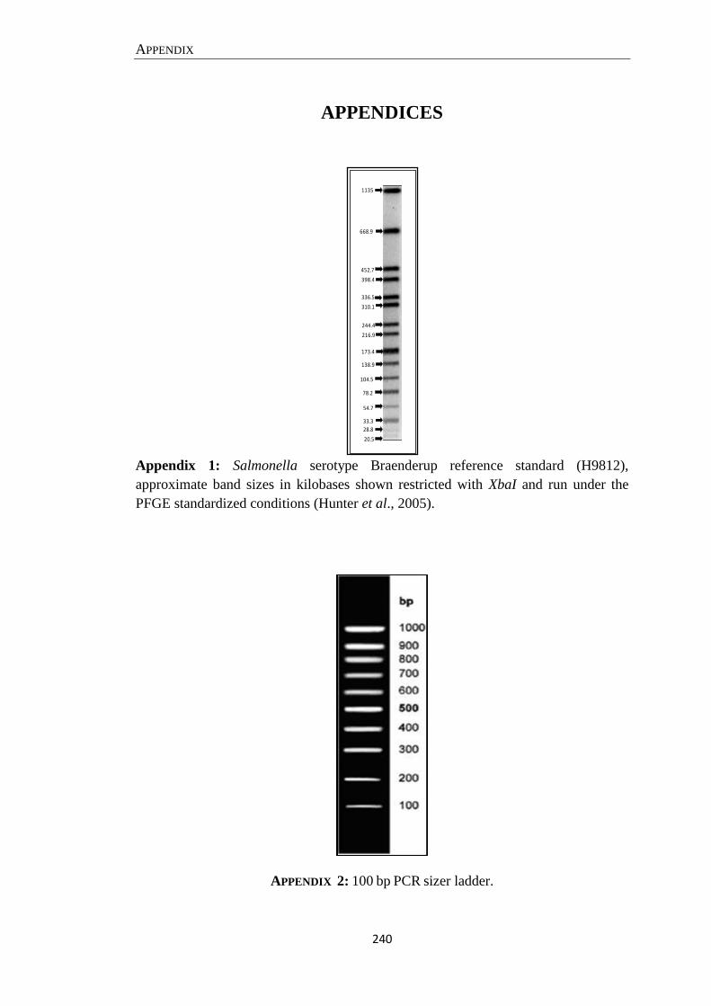
APPENDIX
240
APPENDICES
Appendix 1: Salmonella serotype Braenderup reference standard (H9812),
approximate band sizes in kilobases shown restricted with XbaI and run under the
PFGE standardized conditions (Hunter et al., 2005).
APPENDIX 2: 100 bp PCR sizer ladder.
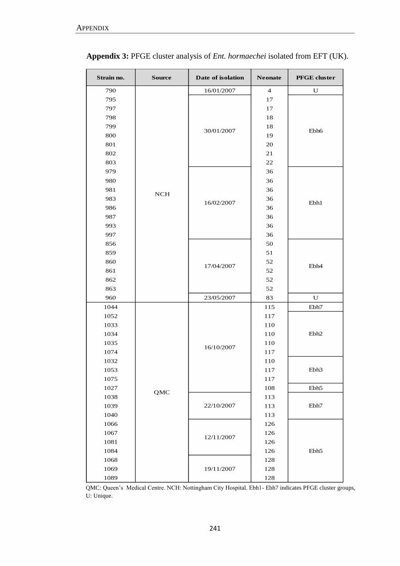
APPENDIX
241
Appendix 3: PFGE cluster analysis of Ent. hormaechei isolated from EFT (UK).
QMC: Queen’s Medical Centre. NCH: Nottingham City Hospital. Ebh - Ebh7 indicates PFGE cluster groups,
U: Unique.
Strain no. Source Date of isolation Neonate PFGE cluster
790 16/01/2007 4 U
795 17
797 17
798 18
799 18
800 19
801 20
802 21
803 22
979 36
980 36
981 36
983 36
986 36
987 36
993 36
997 36
856 50
859 51
860 52
861 52
862 52
863 52
960 23/05/2007 83 U
1044 115 Ebh7
1052 117
1033 110
1034 110
1035 110
1074 117
1032 110
1053 117
1075 117
1027 108 Ebh5
1038 113
1039 113
1040 113
1066 126
1067 126
1081 126
1084 126
1068 128
1069 128
1089 128
Ebh1
12/11/2007
19/11/2007
Ebh4
22/10/2007
NCH
QMC
Ebh2
Ebh3
Ebh7
Ebh5
16/10/2007
30/01/2007
16/02/2007
17/04/2007
Ebh6
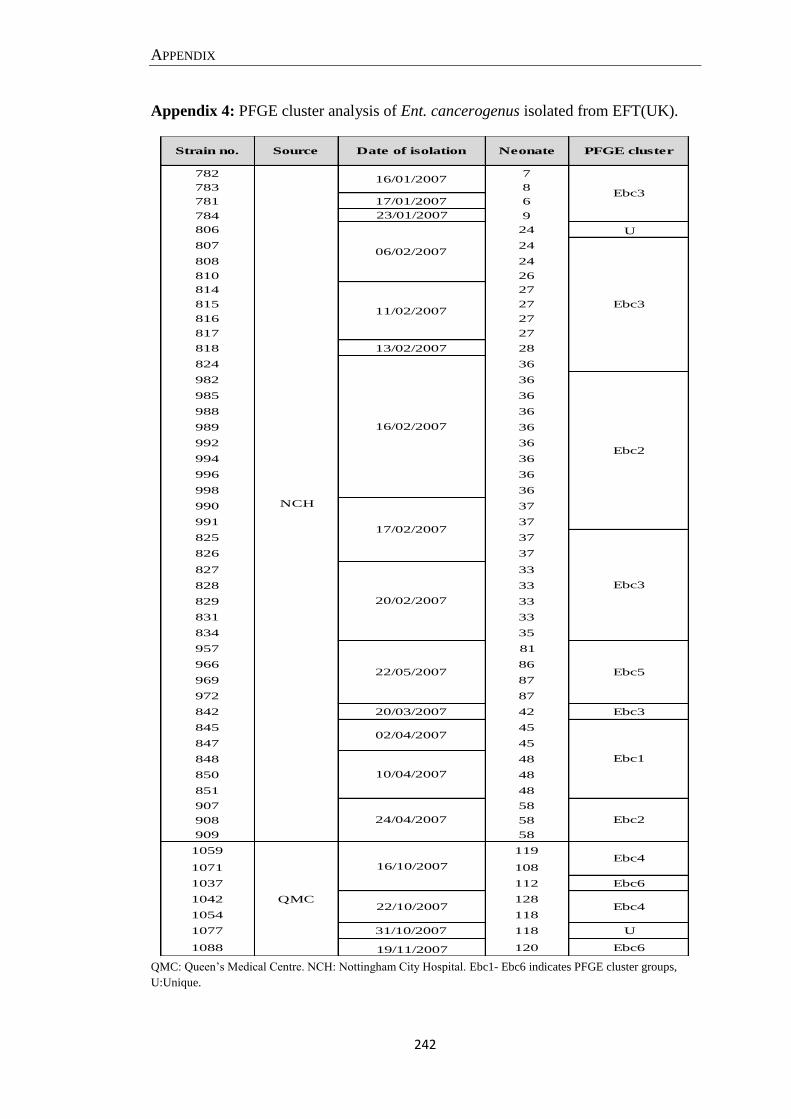
APPENDIX
242
Appendix 4: PFGE cluster analysis of Ent. cancerogenus isolated from EFT(UK).
QMC: Queen’s Medical Centre. NCH: Nottingham City Hospital. Ebc1- Ebc6 indicates PFGE cluster groups,
U:Unique.
Strain no. Source Date of isolation Neonate PFGE cluster
782 7
783 8
781 17/01/2007 6
784 23/01/2007 9
806 24 U
807 24
808 24
810 26
814 27
815 27
816 27
817 27
818 13/02/2007 28
824 36
982 36
985 36
988 36
989 36
992 36
994 36
996 36
998 36
990 37
991 37
825 37
826 37
827 33
828 33
829 33
831 33
834 35
957 81
966 86
969 87
972 87
842 20/03/2007 42 Ebc3
845 45
847 45
848 48
850 48
851 48
907 58
908 58
909 58
1059 119
1071 108
1037 112 Ebc6
1042 128
1054 118
1077 31/10/2007 118 U
1088 19/11/2007 120 Ebc6
16/01/2007
NCH
22/05/2007
16/10/2007
QMC
Ebc3
06/02/2007
Ebc311/02/2007
16/02/2007
Ebc2
17/02/2007
Ebc3
20/02/2007
Ebc4
22/10/2007 Ebc4
Ebc5
02/04/2007
Ebc1
10/04/2007
24/04/2007 Ebc2
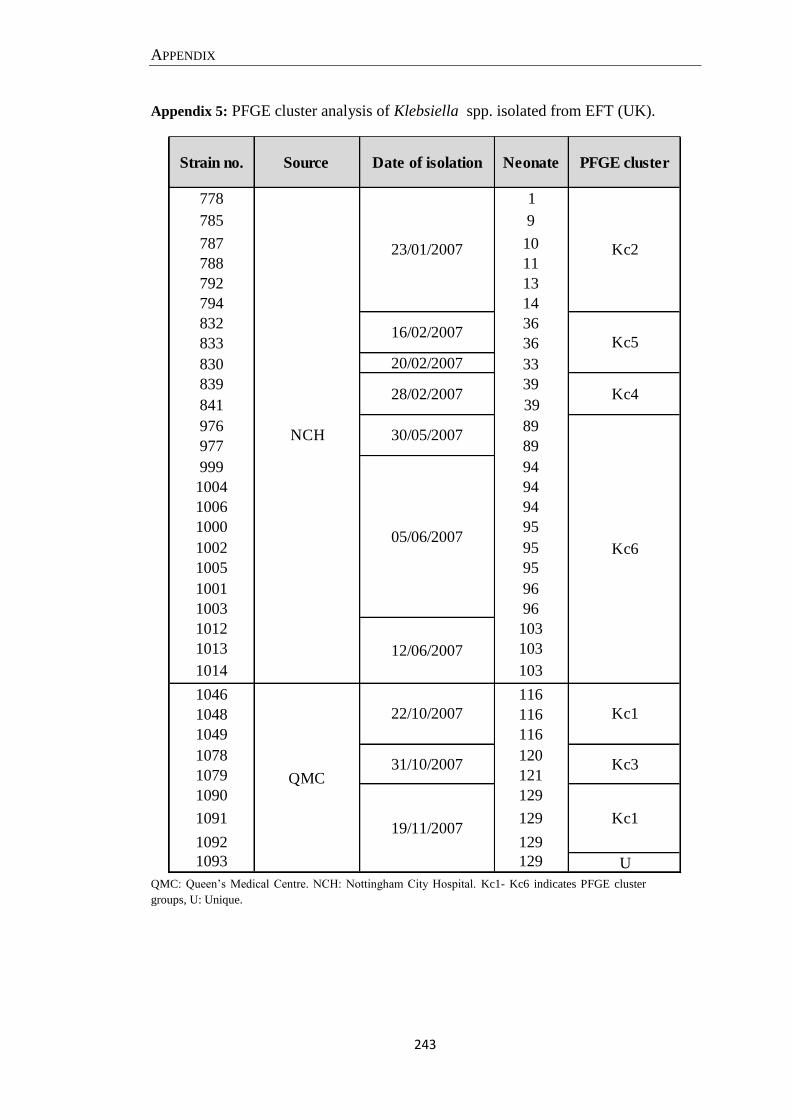
APPENDIX
243
Appendix 5: PFGE cluster analysis of Klebsiella spp. isolated from EFT (UK).
QMC: Queen’s Medical Centre. NCH: Nottingham City Hospital. Kc1- Kc6 indicates PFGE cluster
groups, U: Unique.
Strain no. Source Date of isolation Neonate PFGE cluster
778 1
785 9
787 10
788 11
792 13
794 14
832 36
833 36
830 20/02/2007 33
839 39
841 39
976 89
977 89
999 94
1004 94
1006 94
1000 95
1002 95
1005 95
1001 96
1003 96
1012 103
1013 103
1014 103
1046 116
1048 116
1049 116
1078 120
1079 121
1090 129
1091 129
1092 129
1093 129 U
Kc1
16/02/2007
23/01/2007
Kc5
NCH
QMC
Kc2
Kc4
Kc6
Kc3
Kc1
30/05/2007
05/06/2007
12/06/2007
22/10/2007
31/10/2007
28/02/2007
19/11/2007
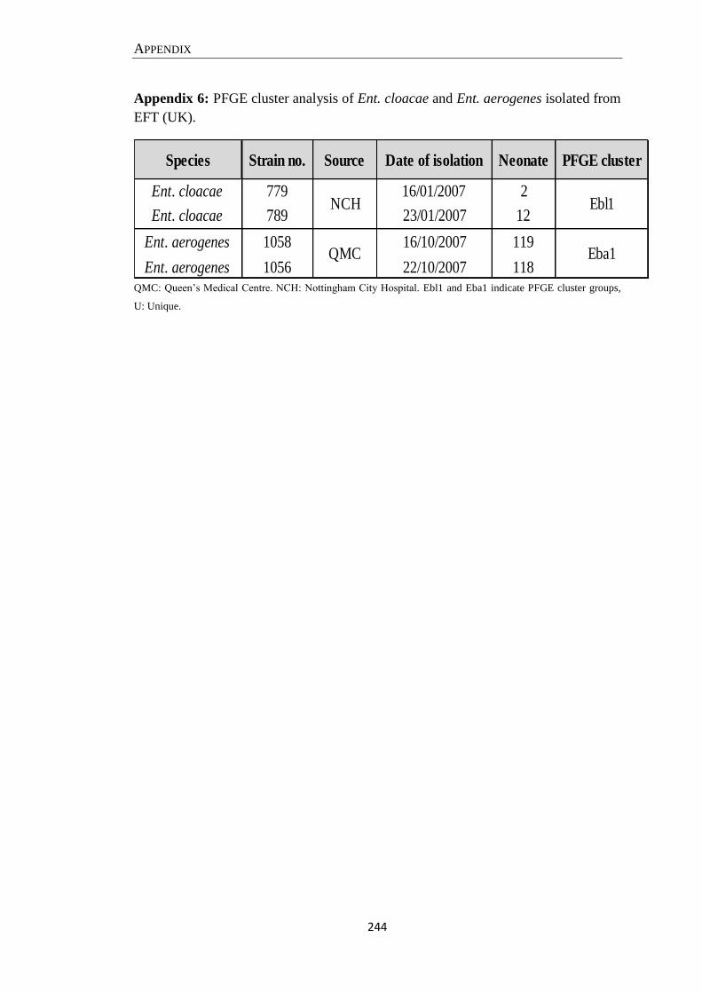
APPENDIX
244
Appendix 6: PFGE cluster analysis of Ent. cloacae and Ent. aerogenes isolated from
EFT (UK).
QMC: Queen’s Medical Centre. NCH: Nottingham City Hospital. Ebl1 and Eba1 indicate PFGE cluster groups,
U: Unique.
Species Strain no. Source Date of isolation Neonate PFGE cluster
Ent. cloacae 779 16/01/2007 2
Ent. cloacae 789 23/01/2007 12
Ent. aerogenes 1058 16/10/2007 119
Ent. aerogenes 1056 22/10/2007 118
NCH Ebl1
Eba1QMC
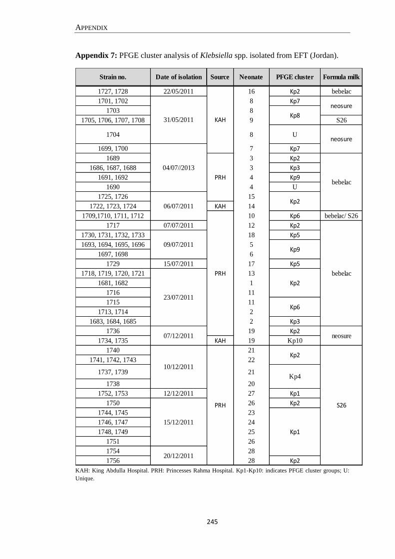
APPENDIX
245
Appendix 7: PFGE cluster analysis of Klebsiella spp. isolated from EFT (Jordan).
KAH: King Abdulla Hospital. PRH: Princesses Rahma Hospital. Kp1-Kp10: indicates PFGE cluster groups; U:
Unique.
1727, 1728 22/05/2011 16 Kp2 bebelac
1701, 1702 8 Kp7
1703 8
1705, 1706, 1707, 1708 9 S26
1704 8 U
1699, 1700 7 Kp7
1689 3 Kp2
1686, 1687, 1688 3 Kp3
1691, 1692 4 Kp9
1690 4 U
1725, 1726 15
1722, 1723, 1724 KAH 14
1709,1710, 1711, 1712 10 Kp6 bebelac/ S26
1717 07/07/2011 12 Kp2
1730, 1731, 1732, 1733 18 Kp5
1693, 1694, 1695, 1696 5
1697, 1698 6
1729 15/07/2011 17 Kp5
1718, 1719, 1720, 1721 13
1681, 1682 1
1716 11
1715 11
1713, 1714 2
1683, 1684, 1685 2 Kp3
1736 19 Kp2
1734, 1735 KAH 19 Kp10
1740 21
1741, 1742, 1743 22
1737, 1739 21
1738 20
1752, 1753 12/12/2011 27 Kp1
1750 26 Kp2
1744, 1745 23
1746, 1747 24
1748, 1749 25
1751 26
1754 28
1756 28 Kp2
Kp831/05/2011
04/07//2013
06/07/2011
PRHbebelac
neosure
KAH
neosure
09/07/2011
PRH
PRH
Kp1
Kp4
Kp2
10/12/2011
15/12/2011
Kp6
Kp2
Kp9
S26
23/07/2011
07/12/2011
20/12/2011
neosure
bebelac
Strain no. Date of isolation Source Neonate PFGE cluster Formula milk
Kp2