Embed Size (px)
Citation preview

1
Chem. Pharm. Bull. 1
Regular Article 2
Enhancing the Solubility and Oral Bioavailability of Poorly Water-Soluble 3
Drugs using Monoolein Cubosomes 4
Md. Ashraf Ali,a,b
Noriko Kataoka,a Abdul-Hackam Ranneh,
a Yasunori Iwao,
a Shuji 5
Noguchi,a Toshihiko Oka,
c and Shigeru Itai*
,a 6
7
aDepartment of Pharmaceutical Engineering & Drug Delivery Science, Graduate School of 8
Integrated Pharmaceutical & Nutritional Sciences, University of Shizuoka; 52–1 Yada, 9
Suruga-ku, Shizuoka 422–8526, Japan. 10
bDepartment of Pharmacy, Faculty of Life Science, Mawlana Bhashani Science and 11
Technology University, Santosh, Tangail-1902, Bangladesh 12
cDepartment of Physics, Faculty of Science and Nanomaterials Research Division, Research 13
Institute of Electronics, Shizuoka University; 836 Oya, Suruga-ku, Shizuoka 422–8529, Japan. 14
15
*Address correspondence to: 16
Shigeru Itai, Ph.D. 17
Professor 18
Department of Pharmaceutical Engineering & Drug Delivery Science 19
Graduate School of Integrated Pharmaceutical & Nutritional Sciences 20
University of Shizuoka 21
52–1 Yada, Suruga-ku, Shizuoka 422–8526, Japan 22
Tel.: +81 54 264 5614, Fax: +81 54 264 5615. 23
E-mail: [email protected] 24

2
25
26
27
28
29
30
31
32
33
34
35
36
37
38
39

3
Summary 40
Monoolein cubosomes containing either spironolactone (SPI) or nifedipine (NI) were 41
prepared using a high-pressure homogenization technique and characterized in terms of their 42
solubility and oral bioavailability. The mean particle size, polydispersity index (PDI), zeta 43
potential, solubility and encapsulation efficiency (EE) values of the SPI- and NI-loaded 44
cubosomes were determined to be 90.4 nm, 0.187, –13.4 mV, 163 µg/mL and 90.2%, and 45
91.3 nm, 0.168, –12.8 mV, 189 µg/mL and 93.0%, respectively, which were almost identical 46
to those of the blank cubosome. Small-angle X-ray scattering analyses confirmed that the 47
SPI-loaded, NI-loaded and blank cubosomes existed in the cubic space group Im3_
m. The 48
lattice parameters of the SPI- and NI-loaded cubosomes were 147.6 and 151.6 Å, respectively, 49
making them almost identical to that of blank cubosome (151.0 Å). The in vitro release 50
profiles of the SPI- and NI-loaded cubosomes showed that they released less than 5% of the 51
drugs into various media over 12–48 h, indicating that most of the drug remained 52
encapsulated within the cubic phase of their lipid bilayer. Furthermore, the in vivo 53
pharmacokinetic results suggested that these cubosomes led to a considerable increase in the 54
systemic oral bioavailability of the drugs compared with pure dispersions of the same 55
materials. Notably, the stability results indicated that the mean particle size and PDI values of 56
these cubosomes were stable for at least 4 weeks. Taken together, these results demonstrate 57
that monoolein cubosomes represent promising drug carriers for enhancing the solubility and 58
oral bioavailability of poorly water-soluble drugs. 59
60
Keywords: cubosome; drug carrier; monoolein; nifedipine; spironolactone; high-pressure 61
homogenization. 62
63
64
4
Introduction 65
The low oral bioavailability of poorly water-soluble drugs remains one of the most 66
challenging aspects of drug development. A wide variety of formulation approaches, 67
including lipid nanoparticles, solid dispersions, complexes with cyclodextrin or chitosan-68
alginates, nanoemulsions and liposomes, have been used to enhance the solubility and 69
bioavailability of several poorly water-soluble drugs.1–5)
Cubosomes have recently attracted 70
considerable interest from formulation scientists in terms of their potential application as drug 71
delivery systems based on their highly ordered, compartmentalized internal structures, high 72
lipid content and large surface area. 73
Cubosomes are inverse bicontinuous cubic lyotropic crystalline nanoparticles that can be 74
loaded with poorly water-soluble drugs in their three-dimensional cubic phases, leading to 75
pronounced increases in the solubility, stability and bioavailability of these drugs.6–9)
76
Cubosomes can be prepared using nontoxic, biocompatible and biodegradable ingredients, 77
and can be readily used to encapsulate lipophilic drugs. Monoolein is a common amphiphilic 78
building block for the preparation of cubosomes,10)
and is relatively cheap compared with 79
other commonly used lipid excipients such as phytantriol. Nonionic triblock copolymer 80
(Pluronic F-127) is an excellent steric stabilizer that can be used to stabilize the structures of 81
cubosomes and prevent particle aggregation for extended periods of time.11)
Monoolein cubic 82
nanoparticles and cubosomes have recently been proposed as potential drug carriers because 83
they can (i) efficiently encapsulate poorly water-soluble drugs and enhance their solubility; 84
(ii) exhibit bio-adhesive properties with sustained release properties; (iii) protect drug 85
molecules and increase their duration of action; and (iv) improve the oral bioavailability of 86
poorly water-soluble drugs.9, 12–14)
The results of several studies have shown that the 87
encapsulation of 20(S)-protopanaxadiol, simvastatin and amphotericin B in monoolein 88
cubosomes and/or cubic nanoparticles led to increases in the oral bioavailabilities of these 89

5
poorly water-soluble drugs.15–17)
However, there have been very few studies in the literature 90
pertaining to the in vivo evaluation of cubosomes. Given that lipid-based formulations have 91
been used in drug delivery to improve the oral absorption of poorly water-soluble drugs for 92
many years,18–20)
we envisaged that cubosomes could also increase their bioavailability. 93
In this study, we used spironolactone (SPI) and nifedipine (NI) as model drugs to prepare 94
SPI- and NI-loaded cubosomes using a high-pressure homogenization technique. The drug 95
loaded cubosomes were subsequently evaluated in terms of their particle size distribution, 96
particle stability, solubility, and encapsulation efficiency characteristics. We also evaluated 97
the in vitro release rate and oral bioavailability properties of the cubosomes for the 98
encapsulated drugs. 99
100
Experimental 101
Materials 102
Monoolein (-OL-100H) was provided as a gift from Riken Vitamin Co., Ltd (Tokyo, 103
Japan). Pluronic F-127 was gifted from BASF (Ludwigshafen, Germany). Spironolactone and 104
nifedipine were purchased from Tokyo Chemical Co., Ltd (Tokyo, Japan) and Sagami Kasei 105
(Tokyo, Japan), respectively. Chloroform, methanol and formic acid were purchased from 106
Wako Chemical Industries, Ltd (Osaka, Japan). All of the other reagents used in this study 107
were purchased as the highest grades available from commercial sources. 108
Preparation of Cubosomes 109
The SPI- and NI-loaded cubosomes were prepared using a high-pressure homogenization 110
technique according to a slightly modified version of a previously published method.21)
111
Briefly, 900 mg of monoolein, 40 mg of SPI/NI and 100 mg of pluronic F-127 were added to 112
a beaker containing 20 mL of chloroform, and the resulting mixture was manually agitated to 113
give a solution. All of the processes to prepare NI-loaded cubosomes were conducted in 114

6
amber glass containers because NI has high light sensitivity. The solvent was removed under 115
reduced pressure on a rotary evaporator at 40–50 °C, and the resulting residue was dried 116
under vacuum for 24 h at 20 °C to allow for complete removal of the organic solvent. The 117
dried sample was dispersed in 200 mL of Milli-Q hot water with 80 °C, and premixed at 6000 118
rpm for 6 min using a Speed Stabilizer (Kinematica® Co., Luzern, Switzerland). The coarse 119
dispersions were also passed through a high-pressure homogenizer (Microfluidizer®
120
Microfluidics Corporation, Newton, MA, USA) at 35 MPa with eight pass cycles. Finally, the 121
cubosome materials were filtered through a 0.45-μm membrane filter unit and stored in the 122
dark at 20 °C prior to use. 123
Particle Size Distribution and Zeta Potential Measurements 124
The mean particle size, PDI and zeta potential values of the different cubosomes were 125
measured by dynamic light scattering (DLS) using a Zetasizer Nano ZS90 (Malvern 126
Instruments, Malvern, UK) at 25 °C. The cubosome samples were diluted with Milli-Q water 127
by 100-fold and measurements were performed at 25 °C. Each experiment was performed in 128
triplicate. 129
Small-angle X-ray Scattering Measurements 130
Small-angle X-ray scattering (SAXS) analyses were conducted to characterize the liquid 131
crystalline phases of the cubosomes using a NANO-Viewer system (Rigaku, Tokyo, Japan) 132
according to a previously reported method.7)
Twenty microliter samples of the cubosomes 133
were placed in a polyimide tube of 1 mm in diameter. The distance between the sample and 134
the detector was set to 510 mm and the system was calibrated using powdered silver behenate. 135
The temperature and exposure time were adjusted to 25 °C and 5 min, respectively. The 136
lattice parameters of the cubic phase were calculated together with their standard error values 137
using the Grafit 5 software (Erithacus®
Software, Horley, Surrey, UK). 138
139

7
Quantitative Evaluation of SPI and NI by HPLC 140
SPI-loaded cubosome samples were diluted as necessary with acetonitrile and thoroughly 141
mixed before being filtered through a 0.45-µm membrane filter unit. The concentration of 142
SPI in each sample was quantified by HPLC analysis using a TSKgel ODS-80Tm® column 143
(4.6 × 150 mm) on a Shimadzu LC-2010C HT® system (Shimadzu Corporation, Kyoto, 144
Japan). The column was eluted with a mobile phase composed of a 7:3 (v/v) mixture of 145
acetonitrile and water at a flow rate of 0.8 mL/min with an injection volume of 40 µL. The 146
column heater and UV-detector were set at 40 °C and 238 nm, respectively. 147
NI-loaded cubosome samples were diluted as necessary with methanol and thoroughly 148
mixed before being filtered through a 0.45-µm membrane filter unit. The concentration of NI 149
in each sample was measured by HPLC analysis using a Cadenza CD-C18 column (3 × 150 150
mm) on a Shimadzu LC-2010C HT® system. The column was eluted with a mobile phase 151
composed of a 3:2 (v/v) mixture of methanol and water containing 0.2% formic acid at a flow 152
rate of 0.4 mL/min with an injection volume of 40 µL. The column heater and UV-detector 153
were set at 40 °C and 236 nm, respectively. All of the samples were quantified in triplicate. 154
Quantitative Evaluation of SPI and NI by LC-MS/MS 155
The SPI concentration was determined by LC-MS/MS using an API-3000 system (Agilent 156
Technologies, Santa Clara, CA, USA) equipped with a Develosil ODS-HG-5 column (4.6 × 157
150 mm). The system was eluted with a mobile phase composed of a 3:2 (v/v) mixture of 158
methanol and water containing 0.1% formic acid at a flow rate of 0.4 mL/min. The column 159
temperature and run time were set to 28 °C and 15 min, respectively. The conditions for the 160
LC-MS/MS were as follows: interface, turbo spray; ionization mode, ESI in the positive ion 161
mode; ion source, nebulizer gas, curtain gas, collision gas; ionspray voltage, 5000 V; 162
temperature, 350 °C; measurement mode, MRM method. The analytes were detected as 163
follows: m/z values of 417.4 and 341.4 for the precursor ion and the product ion of SPI, 164

8
respectively. For in vivo pharmacokinetic study, we measured the concentration of canrenone 165
(CAN), a major metabolite of SPI, using a slightly modified version of this method, where 166
the column temperature, flow rate and sample run time were set to 35 °C, 0.5 mL/min and 25 167
min, respectively. The analytes were detected as follows: m/z values of 341.3 and 107.1 for 168
the precursor ion and the product ion of CAN, respectively. 169
The NI concentration was determined by LC-MS/MS analysis as above using a Cadenza 170
CD-C18 column (3 × 150 mm) column. The system was eluted with a mobile phase 171
composed of a 3:2 (v/v) mixture of methanol and water containing 0.1% formic acid at a flow 172
rate of 0.4 mL/min. The column temperature and sample run time were set to 40 °C and 20 173
min, respectively. The conditions for the LC-MS/MS were as follows: interface, turbo spray; 174
ionization mode, ESI in the positive ion mode; ion source, nebulizer gas, curtain gas, 175
collision gas; ion spray voltage, 5000 V; temperature, 300 °C; measurement mode, MRM 176
method. The analytes were detected as follows: m/z values of 347.1 and 254.2 for the 177
precursor ion and the product ion of NI, respectively. All of the quantification experiments 178
were performed in triplicate. 179
Drug Encapsulation Efficiency 180
The cubosome preparations consisted of a mixture of encapsulated and free drug (un-181
encapsulated) fractions. The drug encapsulation efficiency (EE) of the cubosomes was 182
determined based on the amounts of encapsulated and free drug by the ultra-centrifugation 183
method. Briefly, a 4.0-mL sample of cubosome was placed in an Amicon Ultra-4 Centrifugal 184
Filter-100K device (Merck Millipore Ltd, Carrigtwohill, Ireland) and centrifuged at 1057 ×g 185
for 30 min using an Eppendorf centrifuge (Hamburg, Germany). The concentration of free 186
drug present in the filtrate was determined by LC-MS/MS as described above. The 187
percentage drug encapsulation efficiency of the cubosome was calculated using the following 188
equation: 189

9
EE (%) =𝐷total − 𝐷free
𝐷total× 100
where Dtotal is the total drug concentration of the sample before centrifugation and Dfree is the 190
free drug concentration in the filtrate after centrifugation. These experiments were conducted 191
in triplicate. 192
Stability Studies of the SPI- and NI-loaded Cubosomes 193
The SPI- and NI-loaded cubosomes were stored at 20 °C in the dark prior to use and their 194
mean particle size and PDI values were measured at 0, 1, 2, 3 and 4 weeks using the methods 195
described above. 196
In Vitro Release of SPI and NI from Cubosomes 197
The in vitro release characteristics of the monoolein cubosome samples were examined 198
using a membrane dialysis method with a variety of different release media, including 0.1 M 199
sodium acetate buffer (pH 4.0), fasted state simulated intestinal fluid (FaSSIF) (pH 6.5) and 200
fed state simulated intestinal fluid (FeSSIF) (pH 5.0) at 37 °C. The biorelevent FaSSIF and 201
FeSSIF media were prepared in accordance with a previously reported protocol.22)
Briefly, a 202
1.0-mL sample of cubosome was added into a dialysis membrane bag (MWCO 14 kDa, 203
Viskase Companies Inc., Darien, IL, USA), which was clamped at both ends before being 204
submerged in 200 mL of release medium in a beaker. The beaker was then placed in a shaker 205
bath at 37 °C and shaken horizontally at 100 strokes min-1
. One-milliliter aliquots of release 206
medium were withdrawn for analysis from each beaker at 1, 2, 4, 6, 12, 24, 48, 72 and 96 h, 207
and replaced with the same volume of fresh medium. The collected samples were instantly 208
diluted with acetonitrile for SPI or methanol for NI and filtered through a 0.45-µm membrane 209
filter. Finally, the concentration of drug released into the release medium was determined by 210
HPLC as described above. These experiments were conducted in triplicate. 211
212
213

10
In Vivo Pharmacokinetic Study 214
Male Sprague-Dawley rats (weight: 280–310 g, age: 8–9 weeks; Japan SLC, Shizuoka, 215
Japan) were fasted overnight for 12 h prior to the experiment. All of the procedures used in 216
this study were performed in accordance with the guidelines approved by the Institutional 217
Animal Care and Ethical Committee of the University of Shizuoka, Japan. SPI and NI 218
dispersions were prepared at a concentration of 180 µg/mL by dispersing these materials in 219
Milli-Q water as controls for the SPI- and NI-loaded cubosomes. The SPI- and NI-loaded 220
cubosome, as well as the corresponding controls, were orally administered to the rats at doses 221
corresponding to 1 and 0.5 mg/kg of SPI and NI, respectively. Following the administration 222
of these formulations, blood samples were collected via the tail vein at 1, 5, 10, 30, 60, 90, 223
120, 180 and 240 min in Eppendorf tubes containing the anticoagulant heparin. The blood 224
samples were then centrifuged at 4 °C and 4226 ×g for 10 min to obtain plasma. The plasma 225
samples were immediately treated with methanol and mixed by vortexing for 2 min, before 226
being centrifuged at 4 °C and 4226 ×g for 10 min. The supernatant was collected and filtered 227
through a 0.20-μm membrane filter unit. We determined the concentration of CAN and NI in 228
the filtrate by LC-MS/MS using the method described above. The main pharmacokinetic 229
parameters, including the maximum peak plasma concentration (Cmax), time of peak plasma 230
concentration (Tmax), half-life (t1/2) and the area under the plasma concentration-time curve, 231
were calculated using the linear trapezoidal rule from time zero to infinity (AUC0→∞) and 232
analyzed using the Win Nonlin® Pharmacokinetic program. 233
Statistical Analysis 234
Statistical analyses were performed using the Student’s t-test. A probability value of 235
p<0.05 was considered to indicate statistical significance. 236
237
238

11
Results and Discussion 239
Characterization of SPI- and NI-loaded Cubosomes 240
The results for the characterization of the SPI- and NI-loaded cubosome particles are 241
shown in Table 1. The mean particle size, PDI and zeta potential values of the SPI- and NI-242
loaded cubosomes were almost identical to those of the blank cubosomes. These data show 243
that the encapsulation of drug molecules in the monoolein cubosomes had no impact on their 244
mean particle size, PDI or zeta potential. It is noteworthy that the PDI values of SPI-loaded, 245
NI-loaded and blank cubosomes were less than 0.3, indicating that they were monodispersed. 246
The SAXS patterns and lattice parameters of the SPI-loaded, NI-loaded and blank 247
cubosomes are shown in Fig. 1 and Table 2, respectively. The SAXS patterns of all three 248
materials showed three Bragg peaks with relative positions at spacing ratios of √2:√4:√6. 249
These peaks were indexed according to the Miller indices (hkl) = (110), (200) and (211) 250
reflections, which were indicative of the body-centered cubic phase of the Im3_
m space 251
group.21)
The encapsulation of SPI and NI molecules in the cubosomes had no discernible 252
impact on their cubic space group. The lattice parameters of the cubic phase of the SPI-253
loaded cubosomes were slightly less than those of the blank cubosomes, whereas those of the 254
NI-loaded cubosomes remained largely unchanged. 255
The apparent solubilities of SPI and NI increased when they were encapsulated in the 256
monoolein cubosomes, as shown in Table 1. The encapsulation of SPI in the monoolein 257
cubosomes led to a 6-fold increase in its solubility (28 µg/mL in water at 25 °C).23)
258
Furthermore, the solubility of NI increased around 9-fold (20 µg/mL in water at 25 °C) 259
following its encapsulation in the monoolein cubosomes.24)
The observed increases in the 260
solubilities of these drugs could be attributed to the hydrophobic region of the cubic phase of 261
monoolein. From a structural perspective, monoolein consists of a long hydrophobic aliphatic 262
chain and a hydrophilic glycerol moiety. In the monoolein cubosomes, the hydrophobic 263
12
aliphatic chains would form a lipid bilayer with a cubic phase, with the hydrophilic glycerol 264
moieties forming a water channel. Poorly water-soluble drugs such as SPI and NI would 265
therefore be most likely incorporated into the hydrophobic lipid bilayer of the cubic phase of 266
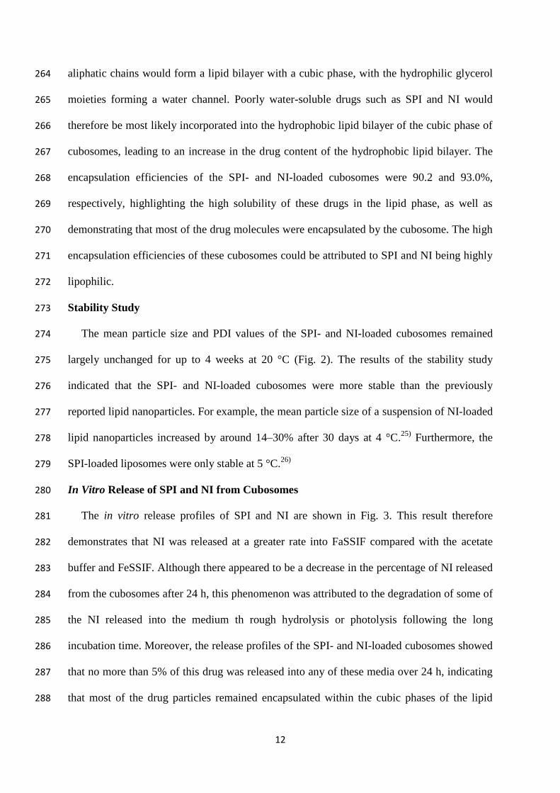
cubosomes, leading to an increase in the drug content of the hydrophobic lipid bilayer. The 267
encapsulation efficiencies of the SPI- and NI-loaded cubosomes were 90.2 and 93.0%, 268
respectively, highlighting the high solubility of these drugs in the lipid phase, as well as 269
demonstrating that most of the drug molecules were encapsulated by the cubosome. The high 270
encapsulation efficiencies of these cubosomes could be attributed to SPI and NI being highly 271
lipophilic. 272
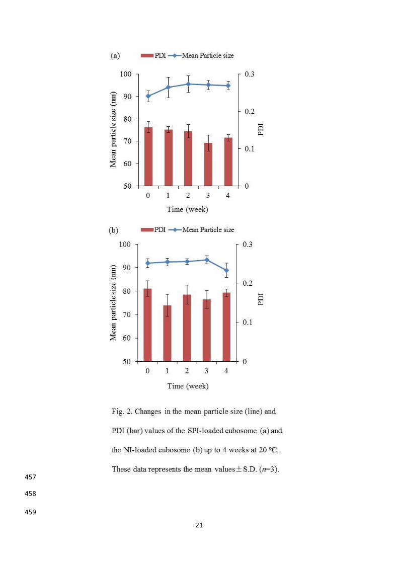
Stability Study 273
The mean particle size and PDI values of the SPI- and NI-loaded cubosomes remained 274
largely unchanged for up to 4 weeks at 20 °C (Fig. 2). The results of the stability study 275
indicated that the SPI- and NI-loaded cubosomes were more stable than the previously 276
reported lipid nanoparticles. For example, the mean particle size of a suspension of NI-loaded 277
lipid nanoparticles increased by around 14–30% after 30 days at 4 °C.25)
Furthermore, the 278
SPI-loaded liposomes were only stable at 5 °C.26)
279
In Vitro Release of SPI and NI from Cubosomes 280
The in vitro release profiles of SPI and NI are shown in Fig. 3. This result therefore 281
demonstrates that NI was released at a greater rate into FaSSIF compared with the acetate 282
buffer and FeSSIF. Although there appeared to be a decrease in the percentage of NI released 283
from the cubosomes after 24 h, this phenomenon was attributed to the degradation of some of 284
the NI released into the medium th rough hydrolysis or photolysis following the long 285
incubation time. Moreover, the release profiles of the SPI- and NI-loaded cubosomes showed 286
that no more than 5% of this drug was released into any of these media over 24 h, indicating 287
that most of the drug particles remained encapsulated within the cubic phases of the lipid 288

13
bilayers of the monoolein cubosomes. This result also suggested that the cubosomes 289
enhanced the solubility of SPI and NI, as stated earlier. The results obtained using the dialysis 290
method for the in vitro release of these drugs from the cubosomes confirmed the drug 291
encapsulation efficiency results determined by the ultra-centrifugation method (>90%). It has 292
been reported that only 1.3% of the SN-38 molecules encapsulated in liposomes were 293
released into the PBS buffer over 30 h, with the value reaching only 1.9% after 120 h.27)
In 294
contrast, the results of a later study showed that around 7% of the SN-38 molecules 295
encapsulated in lipid nanocapsules were released into the PBS buffer over 24 h.28)
Baskaran 296
et al. (2014) reported that only 2.04% of the curcumin molecules encapsulated in monoolein 297
cubic liquid crystalline nanoparticles were released into phosphate buffer saline at pH 7.4, 298
with the rest of the drug molecules remaining unchanged within the nanoparticles.13)
299
In Vivo Pharmacokinetic Study 300
The oral bioavailabilities of the SPI- and NI-loaded cubosomes were investigated in rats 301
and the results were compared with those of the corresponding pure drug dispersions. The 302
pharmacokinetic parameters of these materials are shown in Table 3. The concentration of 303
CAN, a major metabolite of SPI and NI, was determined at different time intervals in all four 304
of these systems, and the results were plotted against time (Fig. 4). These results therefore 305
implied that the oral bioavailabilities of SPI and NI increased considerably following the 306
encapsulation of these drugs in the cubosomes. The mean AUC0–240min and AUC0–∞ values of 307
cubosome-encapsulated drugs increased significantly compared with the corresponding 308
cubosome-free dispersions (Table 3). The oral administration of a solid dispersion of NI (1 309
mg/kg equivalent of NI) to rats was previously reported to give an AUC0–∞ value of 31,676 ng 310
min/mL. In this study, the oral administration of the NI-loaded cubosomes (0.5 mg/kg 311
equivalent of NI) gave an AUC0–∞ value of 58,540 ng min/mL, indicating that the formulation 312
of NI with monoolein cubosomes led to a 4-fold increase in the AUC0–∞ compared with a 313

14
solid dispersion of NI.29)
Furthermore, the half-lives of the SPI- and NI-loaded cubosomes 314
increased around 1.5-fold compared with the corresponding cubosome-free dispersions. 315
Based on these results, we speculated that several factors could be responsible for the 316
observed increases in the absorption characteristics of these two drugs. 317
Several mechanisms, acting in isolation or in combination, can lead to an increase in the 318
oral bioavailability of a drug molecule. In terms of the SPI- and NI-loaded cubosomes 319
prepared in the current study, the observed increases in the oral bioavailability could be 320
attributed to the small size of the cubosome nanoparticles, which would allow them to enter 321
into the intravascular spaces and strongly adhere to the gastrointestinal membrane, leading to 322
an increase in the absorption of the drug molecules. The high affinity of the lipid-like 323
gastrointestinal membrane for these hydrophobic drug molecules could also explain the 324
observed increase in the bioavailability of these compounds. In particular, lipid-like 325
nanoparticles such as the monoolein cubosomes used in this study have mucoadhesive 326
properties that would lead to an increase in their contact time with the gastrointestinal 327
membrane. These properties would therefore lead to an increase in the gastrointestinal 328
residence time of the monoolein cubosomes, resulting in an increase in their oral 329
bioavailability.9, 12)
The plasma drug profiles of SPI and NI indicated that they were 330
immediately absorbed, which could be attributed to the adsorption of the monoolein 331
cubosomes onto the intestinal membranes. Last, the presence of lipids in the gastrointestinal 332
tract such as the monoolein cubosomes can stimulate the secretion of bile into the small 333
intestine from the gall bladder. The cubosomes could then interact with the bile salts to form 334
mixed micelles, which could be absorbed together with the drugs into systemic circulation.30)
335
Furthermore, the SPI- and NI-loaded cubosomes could favor lymphatic transport from the 336
small intestine in a similar manner to that reported for other lipid-based formulations such as 337
liposomes.18–20)
338

15
Although only small amounts of SPI and NI were released (<5%) from the monoolein 339
cubosomes into the different release medium in vitro, the absorption of these drugs increased 340
considerably compared with pure dispersions of the same materials. This disparity between 341
the in vitro release profiles and the in vivo drug absorption characteristics could be attributed 342
to the differences between the in vitro and in vivo environments, the latter of which is much 343
more complex than the former. Furthermore, it is possible that the cubic phase of the 344
monoolein cubosomes could be converted to a different phase (e.g., hexagonal phase) by one 345
of the many compounds found in the gastrointestinal tract.31)
This would result in the rapid 346
release of SPI and NI, leading to an increase in their bioavailability. Furthermore, the 347
absorption of these materials would be regulated by gastric emptying and intestinal transit 348
time, which could explain the differences observed in the in vitro release profiles and in vivo 349
drug absorption characteristics of these materials.32)
We also observed small deviations in the 350
absorption peaks of SPI and NI at 30 and 180 min, respectively, which could be attributed to 351
the complex nature of the in vivo environment. Although the mechanisms responsible for the 352
observed increases in bioavailability of SPI and NI from the cubosomes remain unclear, it is 353
envisaged, based on the results of this study, that these cubosomes could potentially be used 354
as suitable carriers for improving the oral bioavailabilities of SPI and NI. 355
356
Conclusion 357
The results of this study show that monoolein cubosomes containing SPI and NI in the 358
cubic phase of their lipid bilayer enhanced the solubility and oral bioavailability 359
characteristics of both of these drugs. SAXS analyses confirmed that these cubosomes existed 360
in the cubic Im3_
m space group and that they retained their structure after the addition of the 361
drugs. In terms of their physiochemical properties, the mean particle sizes of these 362
cubosomes were less than 100 nm and their PDI values were less than 0.3, which indicated 363

16
that they were monodispersed. The cubosomes also had zeta potentials in the range of –10 to 364
–16 mV. The in vitro release profiles of the SPI- and NI-loaded cubosomes showed that they 365
lost less than 5% of their encapsulated drugs into a variety of different media over 12–48 h. 366
In vivo pharmacokinetic results also suggested that these systems exhibited sustained plasma 367
drug levels and enhanced oral bioavailability. The results of a stability study suggested that 368
the particle size and PDI values of the SPI- and NI-loaded cubosomes remained stable for at 369
least 4 weeks. The SPI- and NI-loaded cubosomes developed in this study therefore represent 370
promising carrier systems for the efficient delivery of drugs for the treatment of hypertension 371
and related diseases. 372
373
Acknowledgements 374
We would like to thank the Ministry of Education, Culture, Sports, Science and 375
Technology (MEXT) of Japan and the Uehara Memorial Foundation for providing doctoral 376
scholarship and research fellowship to Md. Ashraf Ali, respectively. This research work was 377
partly supported by the Japan Society for the Promotion of Science KAKENHI (Grant Nos. 378
26460224, 26460039 and 26460226). We would like to express our gratitude to Dr. Naoto 379
Oku, Professor and Head, Department of Medical Biochemistry, University of Shizuoka for 380
the support of his laboratory in conducting dynamic light scattering (DLS) analysis. 381
382
Conflict of Interest 383
The authors declare no conflict of interest. 384
385
References 386
1. Mehnert W., Mäder K., Adv. Drug Deliv. Rev., 64, 83–101 (2012). 387
2. Leuner C., Dressman J., Eur. J. Pharm. Biopharm., 50, 47–60 (2000). 388

17
3. Khadka P., Ro J., Kim H., Kim I., Kim J. T., Kim H., Cho J. M., Yun G., Lee J., Asian J. 389
Pharm. Sci., 9, 304–316 (2014). 390
4. Anton N., Benoit J. P., Saulnier P., J. Control Release, 128, 185–199 (2008). 391
5. Melis Ç., Ali D. S., Seyda B., “Application of Nanotechnology in Drug Delivery,” Chap. 392
1, ed. by Ali D. S., InTech, Rijeka, 2014, pp. 1–50. 393
6. Hartnett T. E., Ladewig K., O’Connor A. J., Hartley P. G., Mclean K.M., RSC Advances, 394
5, 26543–26549 (2015). 395
7. Ali M. A., Noguchi S., Iwao Y., Oka T., Itai S., Chem. Pharm. Bull., 64, 577–584 (2016). 396
8. Luo Q., Lin T., Zhang C. Y., Zhu T., Wang L., Ji Z., Jia B., Ge T., Peng D., Chen W., Int. 397
J. Pharm., 493, 30–39 (2015). 398
9. Karami Z., Hamidi M., Drug Discovery Today, 21, 789–801 (2016). 399
10. Koynova R., Caffrey M., Chem. Phys. Lipids, 115, 107−219 (2002). 400
11. Chong J. Y. T., Mulet X., Keddie D. J., Waddington L. J., Mudie S. T., Boyd B. J., 401
Drummond C. J., Langmuir, 31, 2615−2629 (2015). 402
12. Nguyen T., Hanley T., Porter C. J. H., Larson I., Boyd B. J., Journal of Pharmacy and 403
Pharmacology, 62, 856–865 (2010). 404
13. Baskaran R., Madheswaran T., Sundaramoorthy P., Kim H. M., Yoo B. K., Int. J. 405
Nanomedicine, 9, 3119–3130 (2014). 406
14. Lai J., Lu Y., Yin Z., Hu F., Wu W., Int. J. Nanomedicine, 5, 13–23 (2010). 407
15. Jin X., Zhang Z. H., Li S. L., Sun E., Tan X. B., Song J., Jia X. B., Fitoterapia, 84, 64–408
71 (2013). 409
16. Lai J., Chen J., Lu Y., Sun J., Hu F., Yin Z., Wu W., AAPS PharmSciTech, 10, 960–966 410
(2009). 411
17. Yang Z., Chen M., Yang M., Chen J., Fang W., Xu P., Int. J. Nanomedicine, 9, 327–336 412
(2014). 413

18
18. Kim H., Kim Y., Lee J., Asian J. Pharm. Sci., 8, 96–103 (2013). 414
19. Porter C. J. H., Trevaskis N. L., Charman W. N., Nature Rev. Drug Discovery, 6, 231–415
248 (2007). 416
20. Trevaskis N. L., Charman W. N., Porter C. J. H., Adv. Drug Deliv. Rev., 60, 702–716 417
(2008). 418
21. Barauskas J., Johnsson M., Joabsson F., Tiberg F., Langmuir, 21, 2569–2577 (2005). 419
22. Marques M., Dissolution Technol., 11, 16 (2004). 420
23. Limayem Blouza I., Charcosset C., Sfar S., Fessi H., Int. J. Pharm., 325, 124–131 421
(2006). 422
24. Hecq J., Deleers M., Fanara D., Vranckx H., Amighi K., Int. J. Pharm., 299, 167–177 423
(2005). 424
25. Funakoshi Y., Iwao Y., Noguchi S., Itai S., Chem. Pharm. Bull., 63, 731–736 (2015). 425
26. Laouini A., Jaafar-Maalej C., Sfar S., Charcosset C., Fessi H., Int. J. Pharm., 415, 53–61 426
(2011). 427
27. Zhang J. A., Xuan T., Parmar M., Ma L., Ugwu S., Ali S., Ahmad I., Int. J. Pharm., 270, 428
93−107 (2004). 429
28. Roger E., Lagarce F., Benoit J. P., Eur. J. Pharm. Biopharm., 79, 181–188 (2011). 430
29. Law S. L., Lo W. Y., Lin F. M., Chaing C. H., Int. J. Pharm., 84, 161–166 (1992). 431
30. Das S., Chaudhury A., AAPS PharmSciTech, 12, 62–76 (2011). 432
31. Chen Y., Ma P., Gui S., Biomed Res. Int., 2014, article ID 815981, 12 pages, (2014). 433
32. Cao X., Deng W. W., Fu M., Wang L., Tong S. S., Wei Y. W., Xu Y., Su W. Y., Xu X. 434
M., Yu J. N., Int. J. Nanomedicine, 7, 753–762 (2012). 435
436
437
438

19
Figure Legends 439
Fig. 1. Intensity vs. the norm of the scattering vector obtained by SAXS measurements 440
for the SPI-loaded, NI-loaded and blank cubosomes (without model drug). q=2 sinθ/λ, 441
where θ is the Bragg angle and λ is the wavelength of X-ray. 442
443
Fig. 2. Changes in the mean particle size (line) and PDI (bar) values of the SPI-loaded 444
cubosome (a) and the NI-loaded cubosome (b) up to 4 weeks at 20 °C. These data 445
represent the mean values ± S.D. (n=3). 446
447
Fig. 3. In vitro release profiles of SPI (a) and NI (b) from the monoolein cubosomes in 448
acetate buffer, FaSSIF and FeSSIF over 96 h at 37 °C. These data represent the mean 449
values ± S.D. (n=3). 450
451
Fig. 4. In vivo drug release, a) plasma CAN concentration-time plot after an oral dose of 452
1.0 mg/kg of the SPI-loaded cubosome and an SPI dispersion, b) plasma NI 453
concentration-time plot after an oral dose of 0.5 mg/kg of the NI-loaded cubosome and 454
an NI dispersion. These data represent the mean values ± S.D. (n=5). 455

20
456
21
457
458
459

22
460 461
462

23
463