Embed Size (px)
Citation preview
Hindawi Publishing CorporationJournal of NanomaterialsVolume 2011, Article ID 126562, 8 pagesdoi:10.1155/2011/126562
Research Article
Enhancement of Oral Bioavailability of Puerarin byPolybutylcyanoacrylate Nanoparticles
Lixia Zhao,1, 2 Anchang Liu,1, 2 Min Sun,1, 3 Jinsong Gu,4 Haigang Wang,2 Shuang Wang,1
Jing Zhang,1 Chenyu Guo,1 Rui Duan,1 and Guangxi Zhai1
1 School of Pharmaceutical Sciences, Shandong University, Jinan 250012, China2 Department of Pharmacy, Qilu Hospital of Shandong University, Jinan 250012, China3 Deparment of Pharmacy, Central Hospital of Zibo, Zibo 255036, China4 Department of Biotechnology, College of Life Science and Technology, University of Jinan, Jinan 250022, China
Correspondence should be addressed to Guangxi Zhai, [email protected]
Received 14 June 2011; Accepted 6 July 2011
Academic Editor: Daxiang Cui
Copyright © 2011 Lixia Zhao et al. This is an open access article distributed under the Creative Commons Attribution License,which permits unrestricted use, distribution, and reproduction in any medium, provided the original work is properly cited.
The interest using novel drug delivery systems to improve oral bioavailability of drug with poor solubility is increasing.In this study, a new oral delivery system, polybutylcyanoacrylate nanoparticles (PBCNs), was introduced to improve theoral bioavailability of puerarin (PUE). PUE-loaded PBCN was successfully prepared by anionic polymerization method.Characterization of PUE-loaded PBCN was evaluated with morphology, size, zeta potential, and in vitro release study. ThePBCN loading PUE exhibited a spherical shape under transmission electron microscopy with an average size of 159.4 nm, andthe zeta potential was −15.0 mV. The in vitro release of PUE-loaded PBCN showed an initial burst release followed by a sustainedrelease. Physicochemical state of PUE in PBCN was investigated by differential scanning colorimetry, X-ray diffraction, and Fouriertransform infrared spectroscopy. The results indicated that PUE in PBCN was in a noncrystalline state. The oral pharmacokineticstudy in rats showed that the relative bioavailability of PUE-encapsulated PBCN to the crude PUE was more than 550%. It can beconcluded that PBCN as an oral drug carrier can significantly improve the oral bioavailability of PUE.
1. Introduction
The bioavailability is an important parameter showing thedegree and rate of drug molecules entering blood circulation,indicating the effectiveness and safety of an extravascularadministration formulation. It can be influenced by drugformulations, food, and physiological factors. However, upto 50% of orally administered drugs present formulationproblems related to poor solubility (0.011 M) [1]. In recentyears, many specific pharmaceutical approaches such asmicro- and nanotechnology have been developed to improvethe bioavailability [2]. As one promising delivery system withimproved bioavailability, polybutylcyanoacrylate nanoparti-cles (PBCNs) have attracted considerable attention [3]. It is atype of solid colloidal particles ranging in size from 10 nm to1000 nm made of biodegradable polymers [4, 5] which couldsignificantly enhance the oral absorption of some drugs suchas thymopentin [6]. The PBCNs that served as the oral
carriers can prevent the destruction of drugs (e.g., peptidedrugs) by the acid and enzymes in gastrointestinal tract[7, 8], improve the drug absorption through Peyer’s patches(PP), the immunization-related tissue in small intestine ofhuman and animals which accounts for about 25% of theintestinal mucosa, and other intestinal lymphoid tissue intothe blood circulation [9], and prolong the residence time invivo because of the small particles and bioadhesive property[6].
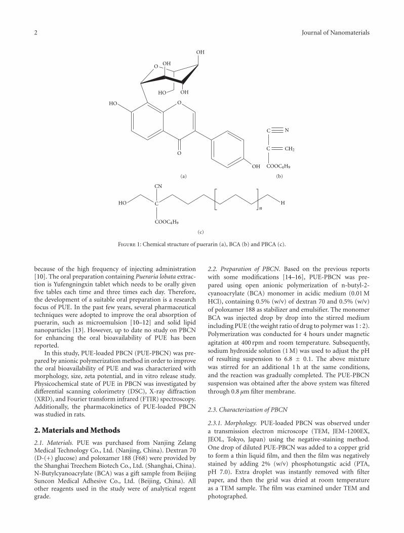
Puerarin (PUE, Figure 1) is the naturally occurringisoflavone C-glycoside extracted from the roots of Puerarialobata (Willd.), Ohwi, and P. thomsonni Benth, which iswidely used in China for the treatment of cerebrovascularand cardiovascular disease. Although PUE has definitetherapeutic effectiveness, its clinical application is limitedby its poor solubility, short elimination half-life, and poororal bioavailability. The commercial available preparationfor PUE is its injection which has poor patient compliance
2 Journal of Nanomaterials
O OH
O
HO
O
OH
HO
OH
OH
(a)
C
C
COOC4H9
N
CH2
(b)
COOC4H9
HO C H
CN
n
(c)
Figure 1: Chemical structure of puerarin (a), BCA (b) and PBCA (c).
because of the high frequency of injecting administration[10]. The oral preparation containing Pueraria lobata extrac-tion is Yufengningxin tablet which needs to be orally givenfive tables each time and three times each day. Therefore,the development of a suitable oral preparation is a researchfocus of PUE. In the past few years, several pharmaceuticaltechniques were adopted to improve the oral absorption ofpuerarin, such as microemulsion [10–12] and solid lipidnanoparticles [13]. However, up to date no study on PBCNfor enhancing the oral bioavailability of PUE has beenreported.
In this study, PUE-loaded PBCN (PUE-PBCN) was pre-pared by anionic polymerization method in order to improvethe oral bioavailability of PUE and was characterized withmorphology, size, zeta potential, and in vitro release study.Physicochemical state of PUE in PBCN was investigated bydifferential scanning colorimetry (DSC), X-ray diffraction(XRD), and Fourier transform infrared (FTIR) spectroscopy.Additionally, the pharmacokinetics of PUE-loaded PBCNwas studied in rats.
2. Materials and Methods
2.1. Materials. PUE was purchased from Nanjing ZelangMedical Technology Co., Ltd. (Nanjing, China). Dextran 70(D-(+) glucose) and poloxamer 188 (F68) were provided bythe Shanghai Treechem Biotech Co., Ltd. (Shanghai, China).N-Butylcyanoacrylate (BCA) was a gift sample from BeijingSuncon Medical Adhesive Co., Ltd. (Beijing, China). Allother reagents used in the study were of analytical regentgrade.
2.2. Preparation of PBCN. Based on the previous reportswith some modifications [14–16], PUE-PBCN was pre-pared using open anionic polymerization of n-butyl-2-cyanoacrylate (BCA) monomer in acidic medium (0.01 MHCl), containing 0.5% (w/v) of dextran 70 and 0.5% (w/v)of poloxamer 188 as stabilizer and emulsifier. The monomerBCA was injected drop by drop into the stirred mediumincluding PUE (the weight ratio of drug to polymer was 1 : 2).Polymerization was conducted for 4 hours under magneticagitation at 400 rpm and room temperature. Subsequently,sodium hydroxide solution (1 M) was used to adjust the pHof resulting suspension to 6.8 ± 0.1. The above mixturewas stirred for an additional 1 h at the same conditions,and the reaction was gradually completed. The PUE-PBCNsuspension was obtained after the above system was filteredthrough 0.8 μm filter membrane.
2.3. Characterization of PBCN
2.3.1. Morphology. PUE-loaded PBCN was observed undera transmission electron microscope (TEM, JEM-1200EX,JEOL, Tokyo, Japan) using the negative-staining method.One drop of diluted PUE-PBCN was added to a copper gridto form a thin liquid film, and then the film was negativelystained by adding 2% (w/v) phosphotungstic acid (PTA,pH 7.0). Extra droplet was instantly removed with filterpaper, and then the grid was dried at room temperatureas a TEM sample. The film was examined under TEM andphotographed.
Journal of Nanomaterials 3
2.3.2. Particle Size and Zeta Potential. The particle size ofPUE-PBCN was analyzed using a particle sizer (Zetasizer3000 HAS, Malvern Instruments Ltd., Malvern, Worcester-shire, UK) with photon correlation spectroscopy (PCS) at afixed angle of 90◦ at a temperature of 25◦C. It was conductedwith He-Ne laser of 3 mW at a wavelength of 633 nm, and theparticle size analysis data were evaluated using the volumedistribution.
Zeta potential of PUE-PBCN was determined using TVmicroscopic electrophoresis system (DXD-II, Optics Co.,Ltd., Jiangsu, China) at room temperature.
2.3.3. Encapsulation Efficiency (EE) and Drug Loading (DL).The measurement for encapsulation efficiency and drugloading of PUE-PBCN was carried out with centrifugationultrafiltration method according to the previous reports[17, 18]. The free PUE was separated from PUE-PBCN bycentrifugal filter tubes (Amicon Ultra-4, Millipore, Ireland)with a molecular cut off of 10 kDa. Briefly, 2 mL of PUE-loaded nanoparticle suspension was added into centrifugalfilter tube and centrifuged at 4000 rpm for 20 min at roomtemperature using a centrifuge (Biofuge primo R. Heraeus,Hanau, Germany). The initial total amount of PUE in thesuspension system and the amount of free drug in the filtratewere measured using UV at the wavelength of 251 nm. Theencapsulation efficiency and drug loading in the PBCN werecalculated as, respectively follows:
EE% = Wtotal drug −Wfree drug
Wtotal drug× 100%, (1)
DL% = Wtotal drug −Wfree drug
Wpolymer× 100%, (2)
where “Wfree drug” is the amount of PUE unloaded in PBCAnanoparticles, “Wtotal drug” is the initial total amount of PUEin the suspension system, and “Wpolymer” is the weight ofbutyl-cyanoacrylate monomer.
2.3.4. DSC and XRD Analysis. The physical state of the drugentrapped in the PBCN was characterized using a differentialscanning calorimeter (CDR-4P, Shanghai Tianping Instru-ment Ltd., Shanghai, China) and an X-ray diffractometer(D/max r-B, Rigaku Co., Tokyo, Japan). Prior to analysis,PUE-PBCN and blank PBCN used for DSC and XRDwere obtained by freeze-drying without any freeze-driedprotectants.
For DSC measurement, about 10 mg of samples (PUE,PUE-PBCN, or blank PBCN) were sealed in the aluminumpan and heated at a scanning rate of 10◦C/min from 30to 400◦C under dry nitrogen atmosphere at a flow rate of0.2 mL/min.
X-ray diffraction patterns were determined for PUE-PBCN, blank PBCN, and cure PUE with a Cu line as thesource of radiation. A radiation at 40 kV voltage and 40 mAcurrent was used, and diffractograms were performed with
a scanning rate of 2◦/min over a 2θ range of from 6◦ to40◦.
2.3.5. Fourier Transform Infrared (FTIR) Spectroscopy. FTIRanalysis was performed to provide further information onthe drug-polymer relationship using an FTIR spectrome-ter (Thermo Electron Scientific Instruments Corp.). FTIRspectra of PUE or BCA monomer or PUE-PBCN (with orwithout drug) were recorded in KBr pellets or KBr cell on anFTIR spectrometer with resolution of 2 cm−1. A total of 64scans were used and data were recorded over the range 4000–400 cm−1.
2.3.6. In Vitro Release Study. The in vitro release of PUEfrom the nanoparticles was studied by dialysis againstphosphate buffer solutions (PBS) with 12–14 kDa molecularcutoff bag pH 7.4 [19]. PUE-PBCN suspension or PUEpropylene glycol solution (containing 3 mg/mL of PUE) wasplaced into dialysis bags, respectively. Then the bags weresuspended in flasks containing 150 mL of PBS as dissolutionmedium at 37◦C in shaking water bath at 100 rpm. 1 mLof dissolution medium was withdrawn at regular timeintervals, and the same volume was added with fresh releasemedium. The concentrations of PUE in dissolution mediumwere measured using UV spectrophotometer at 251 nm. Allexperiments were performed in triplicates.
2.4. Pharmacokinetics Study. The study on the pharmacoki-netics of PUE-PBCN was performed in male Wistar rats(200 ± 20 g) supplied by the Medical Animal Test Center ofShandong University [20]. All animal experiments compliedwith the requirements of the National Act on the use ofexperimental animals (China). The animals were dividedrandomly into 2 groups (n = 5), housed in an environ-mentally controlled breeding room (temperature 25 ± 2 ◦C,humidity 60± 5%, 12 h dark/light cycle) for 7 days and fastedovernight before experiment with free access to water. Group1 was orally administrated PUE suspension (30 mg/kg, PUEdispersed in 0.5% sodium carboxymethylcellulose (CMC-Na) solution), and Group 2 was given PUE-PBCN at thesame dose of 30 mg/kg through the same route. 0.3 mL ofblood was withdrawn from the subclavian vein at 0 min,5 min, 10 min, 20 min, 40 min, 1 h, 2 h, 4 h, 6 h, 8 h, 12 h,and 24 h. The samples were placed into heparinized tubesand isolated immediately by centrifugation at 4000 rpm for10 min. The plasma obtained was stored at −20◦C beforeanalysis.
2.5. Liquid Chromatography Tandem Mass Spectrometry(LC-MS) Analysis. Extraction of PUE from plasma wasconducted as follows: 25 μL of internal standard solution(genistein, 1 μg/mL) and 75 μL of methanol were added to25 μL of plasma and the resulting mixture was vortexed for1 min and then centrifuged at 11,000 rpm for 5 min to obtaina clean supernatant. An aliquot (20 μL) of supernatant wasinjected into the LC-MS for analysis [21].
PUE in plasma was determined by LC-MS analysis usingan Agilent 1200 system equipped with an autosampler,
4 Journal of Nanomaterials
Stir
Surfactant
Monomer
PUE
Micelle
Polymer particle
Hydrophilic layer
Hydrophobic layer
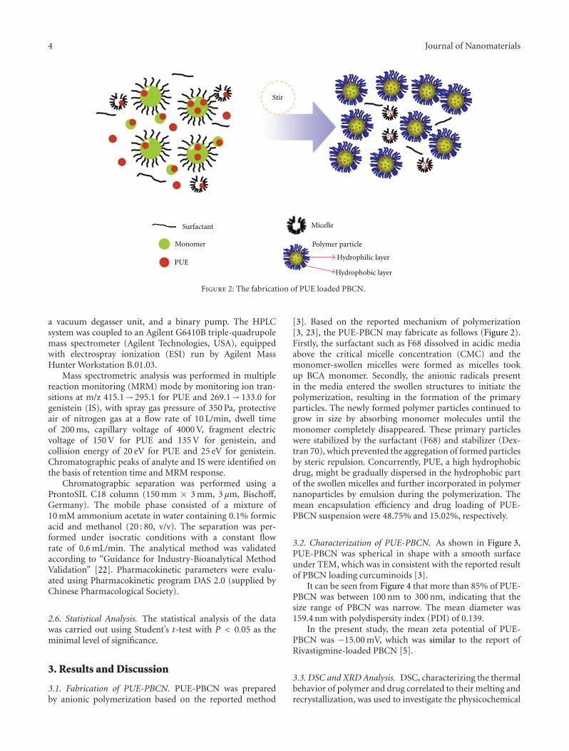
Figure 2: The fabrication of PUE loaded PBCN.
a vacuum degasser unit, and a binary pump. The HPLCsystem was coupled to an Agilent G6410B triple-quadrupolemass spectrometer (Agilent Technologies, USA), equippedwith electrospray ionization (ESI) run by Agilent MassHunter Workstation B.01.03.
Mass spectrometric analysis was performed in multiplereaction monitoring (MRM) mode by monitoring ion tran-sitions at m/z 415.1→ 295.1 for PUE and 269.1→ 133.0 forgenistein (IS), with spray gas pressure of 350 Pa, protectiveair of nitrogen gas at a flow rate of 10 L/min, dwell timeof 200 ms, capillary voltage of 4000 V, fragment electricvoltage of 150 V for PUE and 135 V for genistein, andcollision energy of 20 eV for PUE and 25 eV for genistein.Chromatographic peaks of analyte and IS were identified onthe basis of retention time and MRM response.
Chromatographic separation was performed using aProntoSIL C18 column (150 mm × 3 mm, 3 μm, Bischoff,Germany). The mobile phase consisted of a mixture of10 mM ammonium acetate in water containing 0.1% formicacid and methanol (20 : 80, v/v). The separation was per-formed under isocratic conditions with a constant flowrate of 0.6 mL/min. The analytical method was validatedaccording to “Guidance for Industry-Bioanalytical MethodValidation” [22]. Pharmacokinetic parameters were evalu-ated using Pharmacokinetic program DAS 2.0 (supplied byChinese Pharmacological Society).
2.6. Statistical Analysis. The statistical analysis of the datawas carried out using Student’s t-test with P < 0.05 as theminimal level of significance.
3. Results and Discussion
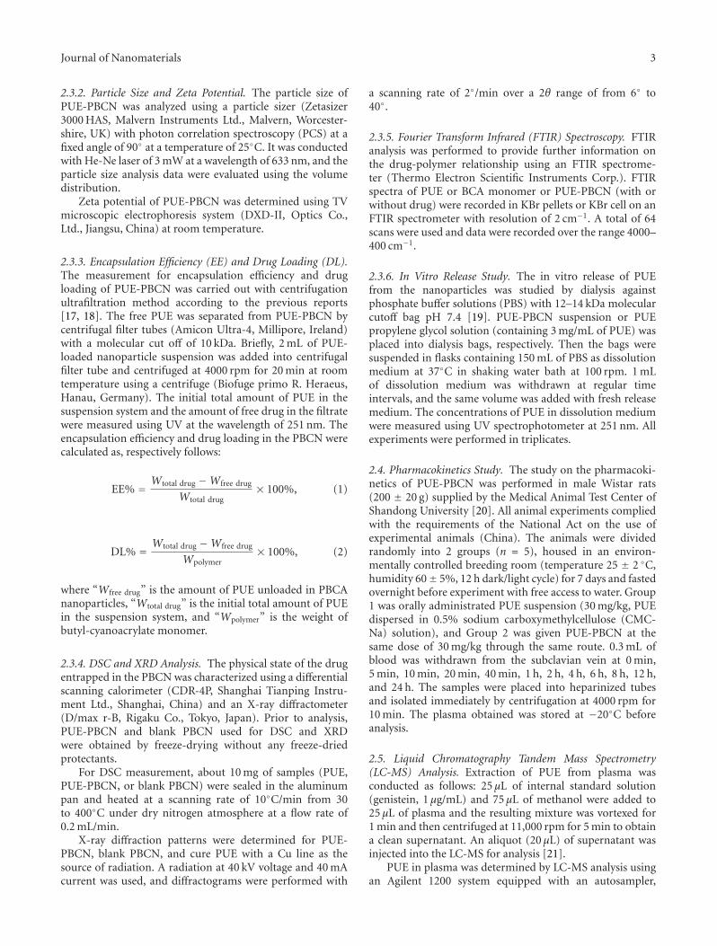
3.1. Fabrication of PUE-PBCN. PUE-PBCN was preparedby anionic polymerization based on the reported method
[3]. Based on the reported mechanism of polymerization[3, 23], the PUE-PBCN may fabricate as follows (Figure 2).Firstly, the surfactant such as F68 dissolved in acidic mediaabove the critical micelle concentration (CMC) and themonomer-swollen micelles were formed as micelles tookup BCA monomer. Secondly, the anionic radicals presentin the media entered the swollen structures to initiate thepolymerization, resulting in the formation of the primaryparticles. The newly formed polymer particles continued togrow in size by absorbing monomer molecules until themonomer completely disappeared. These primary particleswere stabilized by the surfactant (F68) and stabilizer (Dex-tran 70), which prevented the aggregation of formed particlesby steric repulsion. Concurrently, PUE, a high hydrophobicdrug, might be gradually dispersed in the hydrophobic partof the swollen micelles and further incorporated in polymernanoparticles by emulsion during the polymerization. Themean encapsulation efficiency and drug loading of PUE-PBCN suspension were 48.75% and 15.02%, respectively.
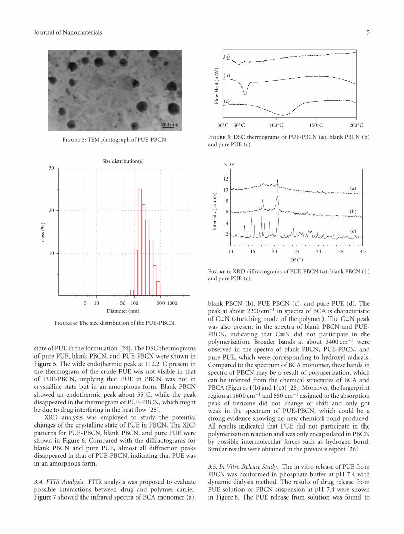
3.2. Characterization of PUE-PBCN. As shown in Figure 3,PUE-PBCN was spherical in shape with a smooth surfaceunder TEM, which was in consistent with the reported resultof PBCN loading curcuminoids [3].
It can be seen from Figure 4 that more than 85% of PUE-PBCN was between 100 nm to 300 nm, indicating that thesize range of PBCN was narrow. The mean diameter was159.4 nm with polydispersity index (PDI) of 0.139.
In the present study, the mean zeta potential of PUE-PBCN was −15.00 mV, which was similar to the report ofRivastigmine-loaded PBCN [5].
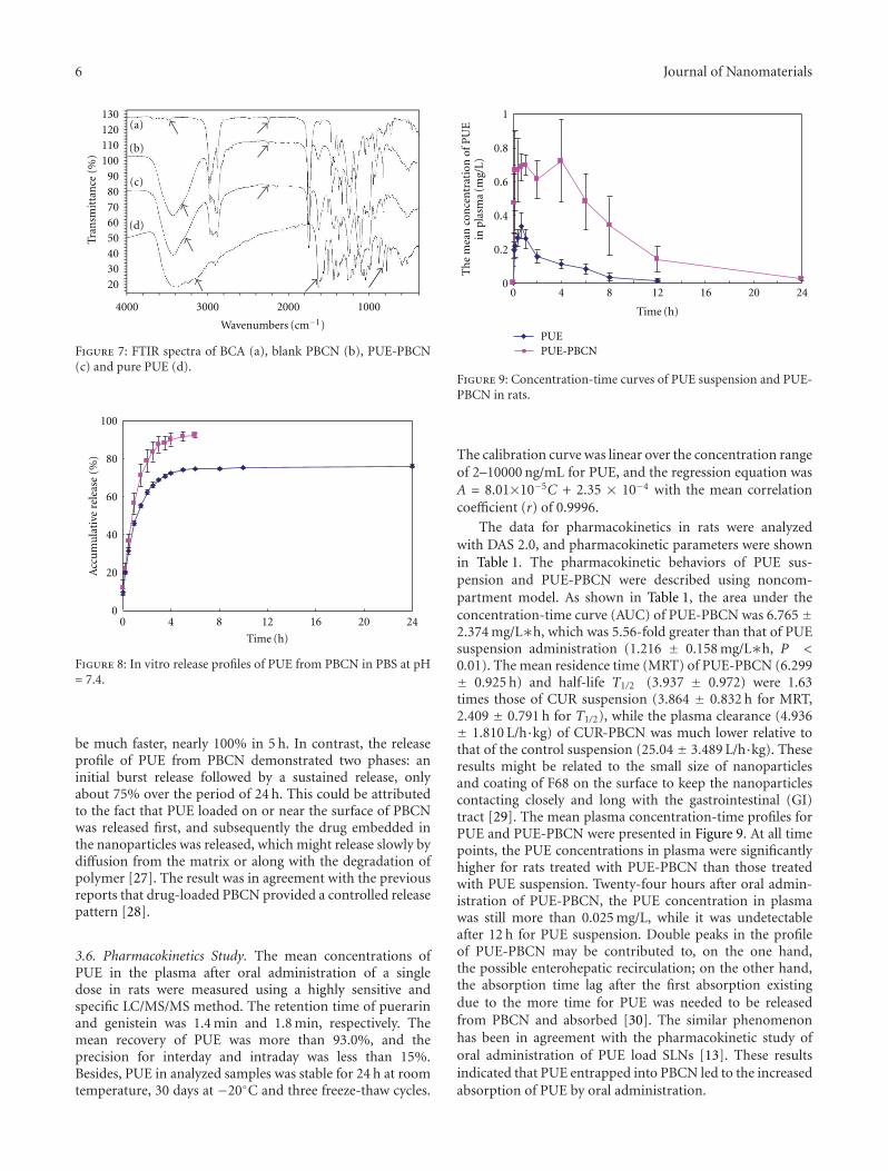
3.3. DSC and XRD Analysis. DSC, characterizing the thermalbehavior of polymer and drug correlated to their melting andrecrystallization, was used to investigate the physicochemical
Journal of Nanomaterials 5
200 nm
Figure 3: TEM photograph of PUE-PBCN.
Size distribution(s)
5 10 50 100 500 1000
Diameter (nm)
10
20
30
clas
s(%
)
Figure 4: The size distribution of the PUE-PBCN.
state of PUE in the formulation [24]. The DSC thermogramsof pure PUE, blank PBCN, and PUE-PBCN were shown inFigure 5. The wide endothermic peak at 112.2◦C present inthe thermogram of the crude PUE was not visible in thatof PUE-PBCN, implying that PUE in PBCN was not incrystalline state but in an amorphous form. Blank PBCNshowed an endothermic peak about 55◦C, while the peakdisappeared in the thermogram of PUE-PBCN, which mightbe due to drug interfering in the heat flow [25].
XRD analysis was employed to study the potentialchanges of the crystalline state of PUE in PBCN. The XRDpatterns for PUE-PBCN, blank PBCN, and pure PUE wereshown in Figure 6. Compared with the diffractograms forblank PBCN and pure PUE, almost all diffraction peaksdisappeared in that of PUE-PBCN, indicating that PUE wasin an amorphous form.
3.4. FTIR Analysis. FTIR analysis was proposed to evaluatepossible interactions between drug and polymer carrier.Figure 7 showed the infrared spectra of BCA monomer (a),
30◦C 50◦C 100◦C 150◦C 200◦C
(a)
(b)
(c)Flow
Hea
t(m
W)
Figure 5: DSC thermograms of PUE-PBCN (a), blank PBCN (b)and pure PUE (c).
(a)
(b)
(c)
12
10
8
6
4
2
10 15 20 25 30 35 40
Inte
nsi
ty(c
oun
ts)
×103
2θ (◦)
Figure 6: XRD diffractograms of PUE-PBCN (a), blank PBCN (b)and pure PUE (c).
blank PBCN (b), PUE-PBCN (c), and pure PUE (d). Thepeak at about 2200 cm−1 in spectra of BCA is characteristicof C≡N (stretching mode of the polymer). The C≡N peakwas also present in the spectra of blank PBCN and PUE-PBCN, indicating that C≡N did not participate in thepolymerization. Broader bands at about 3400 cm−1 wereobserved in the spectra of blank PBCN, PUE-PBCN, andpure PUE, which were corresponding to hydroxyl radicals.Compared to the spectrum of BCA monomer, these bands inspectra of PBCN may be a result of polymerization, whichcan be inferred from the chemical structures of BCA andPBCA (Figures 1(b) and 1(c)) [25]. Moreover, the fingerprintregion at 1600 cm−1 and 650 cm−1 assigned to the absorptionpeak of benzene did not change or shift and only gotweak in the spectrum of PUE-PBCN, which could be astrong evidence showing no new chemical bond produced.All results indicated that PUE did not participate in thepolymerization reaction and was only encapsulated in PBCNby possible intermolecular forces such as hydrogen bond.Similar results were obtained in the previous report [26].
3.5. In Vitro Release Study. The in vitro release of PUE fromPBCN was conformed in phosphate buffer at pH 7.4 withdynamic dialysis method. The results of drug release fromPUE solution or PBCN suspension at pH 7.4 were shownin Figure 8. The PUE release from solution was found to
6 Journal of Nanomaterials
(a)
(b)
(c)
(d)
Tran
smit
tan
ce(%
)
Wavenumbers (cm−1)
2030405060708090
100110120130
4000 3000 2000 1000
Figure 7: FTIR spectra of BCA (a), blank PBCN (b), PUE-PBCN(c) and pure PUE (d).
0
20
40
60
80
100
0 4 8 12 16 20 24
Time (h)
Acc
um
ula
tive
rele
ase
(%)
Figure 8: In vitro release profiles of PUE from PBCN in PBS at pH= 7.4.
be much faster, nearly 100% in 5 h. In contrast, the releaseprofile of PUE from PBCN demonstrated two phases: aninitial burst release followed by a sustained release, onlyabout 75% over the period of 24 h. This could be attributedto the fact that PUE loaded on or near the surface of PBCNwas released first, and subsequently the drug embedded inthe nanoparticles was released, which might release slowly bydiffusion from the matrix or along with the degradation ofpolymer [27]. The result was in agreement with the previousreports that drug-loaded PBCN provided a controlled releasepattern [28].
3.6. Pharmacokinetics Study. The mean concentrations ofPUE in the plasma after oral administration of a singledose in rats were measured using a highly sensitive andspecific LC/MS/MS method. The retention time of puerarinand genistein was 1.4 min and 1.8 min, respectively. Themean recovery of PUE was more than 93.0%, and theprecision for interday and intraday was less than 15%.Besides, PUE in analyzed samples was stable for 24 h at roomtemperature, 30 days at −20◦C and three freeze-thaw cycles.
0 4 8 12 16 20 24
Time (h)
0
0.2
0.4
0.6
0.8
1
PUEPUE-PBCN
Th
em
ean
con
cen
trat
ion
ofP
UE
inpl
asm
a(m
g/L)
Figure 9: Concentration-time curves of PUE suspension and PUE-PBCN in rats.
The calibration curve was linear over the concentration rangeof 2–10000 ng/mL for PUE, and the regression equation wasA = 8.01×10⁻⁵C + 2.35 × 10⁻⁴ with the mean correlationcoefficient (r) of 0.9996.
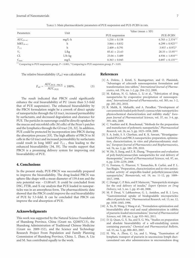
The data for pharmacokinetics in rats were analyzedwith DAS 2.0, and pharmacokinetic parameters were shownin Table 1. The pharmacokinetic behaviors of PUE sus-pension and PUE-PBCN were described using noncom-partment model. As shown in Table 1, the area under theconcentration-time curve (AUC) of PUE-PBCN was 6.765 ±2.374 mg/L∗h, which was 5.56-fold greater than that of PUEsuspension administration (1.216 ± 0.158 mg/L∗h, P <0.01). The mean residence time (MRT) of PUE-PBCN (6.299± 0.925 h) and half-life T1/2 (3.937 ± 0.972) were 1.63times those of CUR suspension (3.864 ± 0.832 h for MRT,2.409 ± 0.791 h for T1/2), while the plasma clearance (4.936± 1.810 L/h·kg) of CUR-PBCN was much lower relative tothat of the control suspension (25.04 ± 3.489 L/h·kg). Theseresults might be related to the small size of nanoparticlesand coating of F68 on the surface to keep the nanoparticlescontacting closely and long with the gastrointestinal (GI)tract [29]. The mean plasma concentration-time profiles forPUE and PUE-PBCN were presented in Figure 9. At all timepoints, the PUE concentrations in plasma were significantlyhigher for rats treated with PUE-PBCN than those treatedwith PUE suspension. Twenty-four hours after oral admin-istration of PUE-PBCN, the PUE concentration in plasmawas still more than 0.025 mg/L, while it was undetectableafter 12 h for PUE suspension. Double peaks in the profileof PUE-PBCN may be contributed to, on the one hand,the possible enterohepatic recirculation; on the other hand,the absorption time lag after the first absorption existingdue to the more time for PUE was needed to be releasedfrom PBCN and absorbed [30]. The similar phenomenonhas been in agreement with the pharmacokinetic study oforal administration of PUE load SLNs [13]. These resultsindicated that PUE entrapped into PBCN led to the increasedabsorption of PUE by oral administration.
Journal of Nanomaterials 7
Table 1: Main pharmacokinetic parameters of PUE suspension and PUE-PCBN in rats.
Parameters UnitValue (mean ± SD)
PUE suspension PUE-PCBN
AUC(0–∞) mg/L·h 1.216 ± 0.158 6.765 ± 2.374∗∗
MRT(0–∞) h 3.864 ± 0.832 6.299 ± 0.925∗∗
T1/2 h 2.409 ± 0.791 3.937 ± 0.972∗
Vz L/kg 85.41 ± 21.63 28.33 ± 13.95∗∗
CL L/h·kg 25.04 ± 3.489 4.936 ± 1.810∗∗
Cmax mg/L 0.363 ± 0.045 0.897 ± 0.135∗∗∗∗
Comparing to PUE suspension group, P < 0.001; ∗Comparing to PUE suspension group, P < 0.05.
The relative bioavailability (Frel) was calculated as
Frel = AUCPUE−PBCN
AUC PUE× 100%. (3)
The result indicated that PBCN could significantlyenhance the oral bioavailability of PU (more than 5.5-foldthat of PUE suspension). The enhanced bioavailability bythe PBCN formulation might be a cowork of direct uptakeof nanoparticles through the GI tract, increased permeabilityby surfactants, and decreased degradation and clearance forPUE. The particles in nanorange could be directly uptaken bythe mucus and microfold cells (M cells) of the Peyer’s patchesand the lymphatics through the GI tract [31, 32]. In addition,PUE could be protected by incorporation into PBCN duringthe absorption process [33]. The high affinity of PBCN to Mcells in the GI tract and increased permeability by surfactantscould result in long MRT and T1/2 , thus leading to theenhanced bioavailability [34, 35]. The results support thatPBCN is a promising delivery system for improving oralbioavailability of PUE.
4. Conclusions
In the present study, PUE-PBCN was successfully preparedto improve the bioavailability. The drug-loaded PBCN wassphere-like shape with a mean diameter of 159.4 nm and thezeta potential was −15.00 mV. It could be concluded fromDSC, FTIR, and X-ray analysis that PUE loaded in nanopar-ticles was in an amorphous form. The pharmacokinetic datashowed that the PBCN could improve the oral bioavailabilityof PUE by 5.5-fold. It can be concluded that PBCN canimprove the oral absorption of PUE.
Acknowledgments
This work was supported by the Natural Science Foundationof Shandong Province, China (Grant no. Q2007C13), theShandong Administration of Traditional Chinese Medicine(Grant no. 2009-152), and the Science and TechnologyResearch Project From Population and Family PlanningCommission of Shandong Province, China. L. Zhao, A. Liuand M. Sun contributed equally to the work.
References
[1] A. Dolenc, J. Kristl, S. Baumgartner, and O. Planinsek,“Advantages of celecoxib nanosuspension formulation andtransformation into tablets,” International Journal of Pharma-ceutics, vol. 376, no. 1-2, pp. 204–212, 2009.
[2] M. Kakran, N. G. Sahoo, L. Li et al., “Fabrication of drugnanoparticles by evaporative precipitation of nanosuspen-sion,” International Journal of Pharmaceutics, vol. 383, no. 1-2,pp. 285–292, 2010.
[3] R. Mulik, K. Mahadik, and A. Paradkar, “Development ofcurcuminoids loaded poly(butyl) cyanoacrylate nanoparticles:physicochemical characterization and stability study,” Euro-pean Journal of Pharmaceutical Sciences, vol. 37, no. 3-4, pp.395–404, 2009.
[4] C. Vauthier and K. Bouchemal, “Methods for the preparationand manufacture of polymeric nanoparticles,” PharmaceuticalResearch, vol. 26, no. 5, pp. 1025–1058, 2009.
[5] S. A. Joshi, S. S. Chavhan, and K. K. Sawant, “Rivastigmine-loaded PLGA and PBCA nanoparticles: preparation, optimiza-tion, characterization, in vitro and pharmacodynamic stud-ies,” European Journal of Pharmaceutics and Biopharmaceutics,vol. 76, no. 2, pp. 189–199, 2010.
[6] W. He, X. Jiang, and Z. R. Zhang, “Preparation and evaluationof poly-butylcyanoacrylate nanoparticles for oral delivery ofthymopentin,” Journal of Pharmaceutical Sciences, vol. 97, no.6, pp. 2250–2259, 2008.
[7] G. Fontana, G. Pitarresi, V. Tomarchio, B. Carlisi, and P. L.San Biagio, “Preparation, characterization and in vitro antimi-crobial activity of ampicillin-loaded polyethylcyanoacrylatenanoparticles,” Biomaterials, vol. 19, no. 11-12, pp. 1009–1017, 1998.
[8] C. Damge, C. P. Reis, and P. Maincent, “Nanoparticle strategiesfor the oral delivery of insulin,” Expert Opinion on DrugDelivery, vol. 5, no. 1, pp. 45–68, 2008.
[9] M. P. Desai, V. Labhasetwar, G. L. Amidon, and R. J. Levy,“Gastrointestinal uptake of biodegradable microparticles:effect of particle size,” Pharmaceutical Research, vol. 13, no. 12,pp. 1838–1845, 1996.
[10] A. Yu, H. Wang, J. Wang et al., “Formulation optimization andbioavailability after oral and nasal administration in rabbitsof puerarin-loaded microemulsion,” Journal of PharmaceuticalSciences, vol. 100, no. 3, pp. 933–941, 2011.
[11] D. Q. Quan, G. X. Xu, and X. G. Wu, “Studies on preparationand absolute bioavailability of a self-emulsifying systemcontaining puerarin,” Chemical and Pharmaceutical Bulletin,vol. 55, no. 5, pp. 800–803, 2007.
[12] H. Wu, A. Zhou, C. Lu, and L. Wang, “Examination oflymphatic transport of puerarin in unconscious lymph duct-cannulated rats after administration in microemulsion drug
8 Journal of Nanomaterials
delivery systems,” European Journal of Pharmaceutical Sciences,vol. 42, no. 4, pp. 348–353, 2011.
[13] C.-F. Luo, M. Yuan, M.-S. Chen et al., “Pharmacokinetics,tissue distribution and relative bioavailability of puerarin solidlipid nanoparticles following oral administration,” Interna-tional Journal of Pharmaceutics, vol. 410, no. 1-2, pp. 138–144,2011.
[14] N. Behan, C. Birkinshaw, and N. Clarke, “Poly n-butylcyanoacrylate nanoparticles: a mechanistic study of polymeri-sation and particle formation,” Biomaterials, vol. 22, no. 11,pp. 1335–1344, 2001.
[15] C. Vauthier, C. Dubernet, E. Fattal, H. Pinto-Alphandary,and P. Couvreur, “Poly(alkylcyanoacrylates) as biodegradablematerials for biomedical applications,” Advanced Drug Deliv-ery Reviews, vol. 55, no. 4, pp. 519–548, 2003.
[16] L. Cai, G. Niu, Z. Hu, W. Jin, J. Wang, and L. Sun,“Polybutylcyanoacrylate magnetic nanoparticles as carriers ofadriamycin,” Journal of Drug Targeting, vol. 17, no. 3, pp. 200–206, 2009.
[17] L. J. Jia, D. R. Zhang, Z. Y. Li et al., “Preparation and character-ization of silybin-loaded nanostructured lipid carriers,” DrugDelivery, vol. 17, no. 1, pp. 11–18, 2009.
[18] Q. Lv, A. Yu, Y. Xi et al., “Development and evaluationof penciclovir-loaded solid lipid nanoparticles for topicaldelivery,” International Journal of Pharmaceutics, vol. 372, no.1-2, pp. 191–198, 2009.
[19] A. Bayat, B. Larijani, S. Ahmadian, H. E. Junginger, and M.Rafiee-Tehrani, “Preparation and characterization of insulinnanoparticles using chitosan and its quaternized derivatives,”Nanomedicine, vol. 4, no. 2, pp. 115–120, 2008.
[20] C. F. Luo, M. Yuan, M. S. Chen, S. M. Liu, and H. Ji,“Metabolites of puerarin identified by liquid chromatographytandem mass spectrometry: similar metabolic profiles in liverand intestine of rats,” Journal of Chromatography B, vol. 878,no. 3-4, pp. 363–370, 2010.
[21] Y. Z. Gu, W. Zhou, and G. X. Zhai, “Preparation of puerarinliposome and its oral absorption in rat,” Journal of ChineseMedicinal Materials, vol. 30, no. 8, pp. 970–973, 2007.
[22] US Food and Drug Administration, “Guidance for Industry:Bioanalytical Method Validation,” 2001, http://www.fda.gov/downloads/Drugs/GuidanceComplianceRegulatoryInforma-tion/Guidances/UCM070107.pdf.
[23] R. S. R. Murthy and L. H. Reddy, “Poly (alkyl cyanocrylate)nanoparticles for delivery of anti-cancer drugs,” in Nanotech-nology, M. M. Amiji, Ed., pp. 251–288, CRC Press, Boca Raton,Fla, USA, 2007.
[24] B. Sarmento, D. Ferreira, F. Veiga, and A. Ribeiro, “Charac-terization of insulin-loaded alginate nanoparticles producedby ionotropic pre-gelation through DSC and FTIR studies,”Carbohydrate Polymers, vol. 66, no. 1, pp. 1–7, 2006.
[25] M. Sun, Y. Gao, C. Guo et al., “Enhancement of transportof curcumin to brain in mice by poly(n-butylcyanoacrylate)nanoparticle,” Journal of Nanoparticle Research, vol. 12, no. 8,pp. 3111–3122, 2010.
[26] M. Simeonova, G. Ivanova, V. Enchev et al., “Physicochemicalcharacterization and in vitro behavior of daunorubicin-loadedpoly(butylcyanoacrylate) nanoparticles,” Acta Biomaterialia,vol. 5, no. 6, pp. 2109–2121, 2009.
[27] F. Ren, R. Chen, Y. Wang, Y. Sun, Y. Jiang, and G. Li,“Paclitaxel-loaded poly(n-butylcyanoacrylate) nanoparticledelivery system to overcome multidrug resistance in ovariancancer,” Pharmaceutical Research, vol. 28, no. 4, pp. 897–906,2011.
[28] L. H. Reddy and R. R. Murthy, “Influence of polymer-ization technique and experimental variables on the par-ticle properties and release kinetics of methotrexate frompoly(butylcyanoacrylate) nanoparticles,” Acta Pharmaceutica,vol. 54, no. 2, pp. 103–118, 2004.
[29] J. Liu, J. Zhu, Z. Du, and B. Qin, “Preparation and pharma-cokinetic evaluation of Tashinone IIA solid lipid nanoparti-cles,” Drug Development and Industrial Pharmacy, vol. 31, no.6, pp. 551–556, 2005.
[30] Y. Li, W. S. Pan, S. L. Chen, H. X. Xu, D. J. Yang, and A. S.C. Chan, “Pharmacokinetic, tissue distribution, and excretionof puerarin and puerarin-phospholipid complex in rats,” DrugDevelopment and Industrial Pharmacy, vol. 32, no. 4, pp. 413–422, 2006.
[31] A. E. Gulyaev, S. E. Gelperina, I. N. Skidan, A. S. Antropov,G. Y. Kivman, and J. Kreuter, “Significant transport ofdoxorubicin into the brain with polysorbate 80- coatednanoparticles,” Pharmaceutical Research, vol. 16, no. 10, pp.1564–1569, 1999.
[32] N. Hussain, V. Jaitley, and A. T. Florence, “Recent advances inthe understanding of uptake of microparticulates across thegastrointestinal lymphatics,” Advanced Drug Delivery Reviews,vol. 50, no. 1-2, pp. 107–142, 2001.
[33] A. Dembri, M. J. Montisci, J. C. Gantier, H. Chacun,and G. Ponchel, “Targeting of 3′-azido 3′-deoxythymidine(AZT)-loaded poly(isohexylcyanoacrylate) nanospheres tothe gastrointestinal mucosa and associated lymphoid tissues,”Pharmaceutical Research, vol. 18, no. 4, pp. 467–473, 2001.
[34] D. Duchene and G. Ponchel, “Bioadhesion of solid oral dosageforms, why and how?” European Journal of Pharmaceutics andBiopharmaceutics, vol. 44, no. 1, pp. 15–23, 1997.
[35] N. Venkatesan, K. Uchino, K. Amagase, Y. Ito, N. Shibata, andK. Takada, “Gastro-intestinal patch system for the delivery oferythropoietin,” Journal of Controlled Release, vol. 111, no. 1-2,pp. 19–26, 2006.

Submit your manuscripts athttp://www.hindawi.com
ScientificaHindawi Publishing Corporationhttp://www.hindawi.com Volume 2014
CorrosionInternational Journal of
Hindawi Publishing Corporationhttp://www.hindawi.com Volume 2014
Polymer ScienceInternational Journal of
Hindawi Publishing Corporationhttp://www.hindawi.com Volume 2014
Hindawi Publishing Corporationhttp://www.hindawi.com Volume 2014
CeramicsJournal of
Hindawi Publishing Corporationhttp://www.hindawi.com Volume 2014
CompositesJournal of
NanoparticlesJournal of
Hindawi Publishing Corporationhttp://www.hindawi.com Volume 2014
Hindawi Publishing Corporationhttp://www.hindawi.com Volume 2014
International Journal of
Biomaterials
Hindawi Publishing Corporationhttp://www.hindawi.com Volume 2014
NanoscienceJournal of
TextilesHindawi Publishing Corporation http://www.hindawi.com Volume 2014
Journal of
NanotechnologyHindawi Publishing Corporationhttp://www.hindawi.com Volume 2014
Journal of
CrystallographyJournal of
Hindawi Publishing Corporationhttp://www.hindawi.com Volume 2014
The Scientific World JournalHindawi Publishing Corporation http://www.hindawi.com Volume 2014
Hindawi Publishing Corporationhttp://www.hindawi.com Volume 2014
CoatingsJournal of
Advances in
Materials Science and EngineeringHindawi Publishing Corporationhttp://www.hindawi.com Volume 2014
Smart Materials Research
Hindawi Publishing Corporationhttp://www.hindawi.com Volume 2014
Hindawi Publishing Corporationhttp://www.hindawi.com Volume 2014
MetallurgyJournal of
Hindawi Publishing Corporationhttp://www.hindawi.com Volume 2014
BioMed Research International
MaterialsJournal of
Hindawi Publishing Corporationhttp://www.hindawi.com Volume 2014
Nano
materials
Hindawi Publishing Corporationhttp://www.hindawi.com Volume 2014
Journal ofNanomaterials