Embed Size (px)
Citation preview
nanomaterials
Article
Enhanced High-Temperature (600 ◦C) NO2 Responseof ZnFe2O4 Nanoparticle-Based Exhaust Gas Sensors
Adeel Afzal 1,* , Adnan Mujahid 2, Naseer Iqbal 1 , Rahat Javaid 3
and Umair Yaqub Qazi 1,4
1 Department of Chemistry, College of Science, University of Hafr Al Batin, P.O. Box 1803,Hafr Al Batin 39524, Saudi Arabia; [email protected] (N.I.); [email protected] (U.Y.Q.)
2 Institute of Chemistry, University of Punjab, Quaid-i-Azam Campus, Lahore 54590, Pakistan;[email protected]
3 Renewable Energy Research Center, Fukushima Renewable Energy Institute, National Institute of AdvancedIndustrial Science and Technology, AIST, 2-2-9 Machiikedai, Koriyama, Fukushima 963-0298, Japan;[email protected]
4 Division of Nanomaterials and Chemistry, Hefei National Laboratory for Physical Sciences at Microscale,University of Science and Technology of China, Hefei 230026, China
* Correspondence: [email protected]; Tel.: +966-13-720-3426 (ext. 1675)
Received: 7 September 2020; Accepted: 21 October 2020; Published: 27 October 2020�����������������
Abstract: Fabrication of gas sensors to monitor toxic exhaust gases at high working temperaturesis a challenging task due to the low sensitivity and narrow long-term stability of the devices underharsh conditions. Herein, the fabrication of a chemiresistor-type gas sensor is reported for thedetection of NO2 gas at 600 ◦C. The sensing element consists of ZnFe2O4 nanoparticles preparedvia a high-energy ball milling and annealed at different temperatures (600–1000 ◦C). The effects ofannealing temperature on the crystal structure, morphology, and gas sensing properties of ZnFe2O4
nanoparticles are studied. A mixed spinel structure of ZnFe2O4 nanoparticles with a lattice parameterof 8.445 Å is revealed by X-ray diffraction analysis. The crystallite size and X-ray density of ZnFe2O4
nanoparticles increase with the annealing temperature, whereas the lattice parameter and volume areconsiderably reduced indicating lattice distortion and defects such as oxygen vacancies. ZnFe2O4
nanoparticles annealed at 1000 ◦C exhibit the highest sensitivity (0.13% ppm–1), sharp response(τres = 195 s), recovery (τrec = 17 s), and linear response to 100–400 ppm NO2 gas. The annealingtemperature and oxygen vacancies play a major role in determining the sensitivity of devices.The plausible sensing mechanism is discussed. ZnFe2O4 nanoparticles show great potential forhigh-temperature exhaust gas sensing applications.
Keywords: annealing temperature; chemiresistors; gas sensors; oxygen vacancies; sensing mechanism;ZnFe2O4 nanoparticles
1. Introduction
Hazardous exhaust gases such as nitrogen dioxide (NO2) and sulfur dioxide (SO2) are the majoratmospheric pollutants [1]. The European Union’s (E.U.) ambient air quality directives have set thehourly NO2 concentration threshold as 200 µg/m3 [2]. According to the European Environment Agency(EEA) report published in 2016, NO2 pollution was responsible for 71,000 premature deaths in theE.U. [3]. Thus, it is important to detect the emission and subsistence of NO2 in indoor and outdoor air.The main source of NO2 pollution is the exhaust emissions as a result of the combustion processesin motor vehicles and manufacturing industries [4]. The direct inspection of the exhaust emissionsrequires devices that can detect NO2 at high temperatures, i.e., usually ≥500 ◦C [5]. In this regard,
Nanomaterials 2020, 10, 2133; doi:10.3390/nano10112133 www.mdpi.com/journal/nanomaterials
Nanomaterials 2020, 10, 2133 2 of 14
metal oxide-based electronic gas sensors are the most sought-after devices for applications in harshenvironments [6–8].
According to a recent review of high-temperature gas sensors, Ghosh et al. [9] noted the majorityof the metal oxide-based gas sensors work at moderately high temperatures only, while the sensitivityof metal oxides is substantially influenced at temperatures above 350 ◦C. Albeit a large number ofmetal oxide-based NO2 gas sensors are reported [10–13], only a few work at high temperatures,i.e., ≥600 ◦C. For instance, Miura et al. [14] reported Yt-stabilized zirconia and spinel ZnFe2O4 sensingelectrodes for the electrochemical detection of NOx at 600–700 ◦C. However, chemiresistive-type NO2
gas sensors for high-temperature applications are rarely reported [13,15,16]. Therefore, the fabricationof high-temperature NO2 gas sensors for harsh environments is highly desired due to their widespreadapplications in all types of combustion systems.
This article reports the first high-temperature thick film chemiresistive gas sensor for NO2 detectionat 600 ◦C. The sensor is based on highly stable spinel zinc ferrite (ZnFe2O4) nanoparticles preparedvia a solid-state, high-energy ball-milling (HEBM) process followed by high-temperature thermalannealing at different temperatures (600, 800, and 1000 ◦C). ZnFe2O4 nanoparticles have been used forthe detection of toxic organic vapors and gases such as acetone at 260 [17] and 275 ◦C [18], ethanol at27 [19] and 220 ◦C [20], toluene at 300 ◦C [21], H2S at 85 ◦C [22] and 135 ◦C [23], and O2 at 180 ◦C [24].Recently, Runa et al. [25] fabricated a chemiresistive NO2 gas sensor using ZnO/ZnFe2O4 compositeswith p-n heterostructure, which revealed excellent selectivity and high gas response toward 0.1–20 ppmNO2 compared to pure ZnO. However, the gas response diminished rapidly at temperatures of≥220 ◦C [25]. In this work, the effects of high-temperature annealing on the crystal structure andNO2 gas sensing properties are studied. The cubic spinel ZnFe2O4 nanoparticles are stable at hightemperatures and demonstrate excellent NO2 sensing capability at 600 ◦C with fast response andrecovery times.
2. Materials and Methods
Iron(III) oxide (Fe2O3 nanopowder) and zinc oxide (ZnO nanopowder) obtained fromMilliporeSigma (Merck KGaA, Darmstadt, Germany) were used to prepare ZnFe2O4 nanoparticles.ZnFe2O4 nanoparticles were synthesized by high-energy ball milling (HEBM) process using a SPEX™8000M Mixer/Mill™ (SPEX® SamplePrep, New Jersey, NJ, USA). The ball mill was equipped with a500-cc stainless steel vessel containing stainless steel balls for mechanical milling of Fe2O3 and ZnO.The mass ratio of steel balls and chemical powders was fixed at 50:1. HEBM was performed underambient conditions for 2 h at 600 rpm. The product was subsequently vacuum annealed at 600, 800,and 1000 ◦C for 2 h, and characterized. Corresponding to the annealing temperature (600–1000 ◦C),the samples were abbreviated as ZnFe2O4-600, ZnFe2O4-800, and ZnFe2O4-1000, respectively.
The crystal structure of the annealed ZnFe2O4 nanoparticles was studied with a STOE STADIP X-ray diffractometer (XRD) (STOE & Cie GmbH, Darmstadt, Germany) using a Cu Kα irradiationsource (λ = 1.5406 Å). The samples were scanned in the 2θ range of 10◦–90◦ with a scan rate of 2◦/min.The crystallite size (D) is determined by the Scherrer’s formula [26] (D = Kλ/Bcosθ), where K is anumerical factor referred to as the crystallite-shape factor with an approximate value of 0.89, λ is thewavelength of the X-rays, B is full-width at half-maximum of the most intense (311) diffraction peak inradians, and θ is the Bragg angle. The experimental lattice parameter (a), X-ray density (ρxrd), and thespecific surface area (SA) are also calculated from the XRD data of annealed ZnFe2O4 nanoparticles,as described elsewhere [27,28].
The microstructure and surface morphology of ZnFe2O4 nanoparticles were studied with a JEOLJSM-6510 scanning electron microscope (SEM) (JEOL Ltd., Tokyo, Japan). The elemental compositionof ZnFe2O4 nanoparticles was determined with the energy-dispersive X-ray spectroscopy (EDS)(JEOL Ltd., Tokyo, Japan).
Thick-film chemiresistor-type gas sensors were fabricated by mixing an appropriate amount ofZnFe2O4-600, ZnFe2O4-800, and ZnFe2O4-1000 nanoparticles with absolute ethanol to make a thick
Nanomaterials 2020, 10, 2133 3 of 14
slurry, which was subsequently drop-coated onto alumina micro-hotplates with vapor-depositedplatinum (Pt) contacts. The devices were placed in a vacuum oven at 80 ◦C for 2 h to dry and stabilizethe sensing element. The chemiresistor-type devices were installed in a gas sensing chamber fittedwith the electrical connections and the gas inlet and outlet. The measurements were performed witha Keithley 6517A electrometer. The sensor responses were measured simultaneously at 600 ◦C for100–400 ppm of NO2 gas. The sensor response (S) is defined as S(%) = (Rg – Ra) × 100/Ra, where Ra andRg are the resistances in air and (100–400 ppm) NO2 gas.
3. Results and Discussion
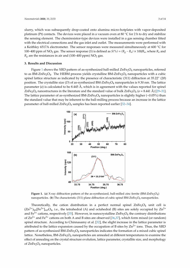
Figure 1 shows the XRD pattern of as-synthesized ball-milled ZnFe2O4 nanoparticles, referredto as BM-ZnFe2O4. The HEBM process yields crystalline BM-ZnFe2O4 nanoparticles with a cubicspinel lattice structure as indicated by the presence of characteristic (311) diffraction at 35.22◦ (2θ)position. The crystallite size (D) of as-synthesized BM-ZnFe2O4 nanoparticles is 9.30 nm. The latticeparameter (a) is calculated to be 8.445 Å, which is in agreement with the values reported for spinelZnFe2O4 nanostructures in the literature and the standard value of bulk ZnFe2O4 (a = 8.441 Å) [29–31].The lattice parameter of as-synthesized BM-ZnFe2O4 nanoparticles is slightly higher (~0.05%) thanthe standard value that may be inherent to the ball-milling process because an increase in the latticeparameter of ball-milled ZnFe2O4 samples has been reported earlier [32–34].
Nanomaterials 2020, 10, x FOR PEER REVIEW 3 of 14
a Keithley 6517A electrometer. The sensor responses were measured simultaneously at 600 °C for 100–400 ppm of NO2 gas. The sensor response (S) is defined as S(%) = (Rg – Ra) × 100/Ra, where Ra and Rg are the resistances in air and (100–400 ppm) NO2 gas.
3. Results and Discussion
Figure 1 shows the XRD pattern of as-synthesized ball-milled ZnFe2O4 nanoparticles, referred to as BM-ZnFe2O4. The HEBM process yields crystalline BM-ZnFe2O4 nanoparticles with a cubic spinel lattice structure as indicated by the presence of characteristic (311) diffraction at 35.22° (2θ) position. The crystallite size (D) of as-synthesized BM-ZnFe2O4 nanoparticles is 9.30 nm. The lattice parameter (a) is calculated to be 8.445 Å, which is in agreement with the values reported for spinel ZnFe2O4 nanostructures in the literature and the standard value of bulk ZnFe2O4 (a = 8.441 Å) [29–31]. The lattice parameter of as-synthesized BM-ZnFe2O4 nanoparticles is slightly higher (~0.05%) than the standard value that may be inherent to the ball-milling process because an increase in the lattice parameter of ball-milled ZnFe2O4 samples has been reported earlier [32–34].
Figure 1. (a) X-ray diffraction pattern of the as-synthesized, ball-milled zinc ferrite (BM-ZnFe2O4) nanoparticles. (b) The characteristic (311) plane diffraction of cubic spinel BM-ZnFe2O4 nanoparticles.
Theoretically, the cation distribution in a perfect normal spinel ZnFe2O4 unit cell is (Zn2+)tet[Fe3+]octO4, i.e., the tetrahedral (A) and octahedral (B) sites are solely occupied by Zn2+ and Fe3+ cations, respectively [35]. However, in nanocrystalline ZnFe2O4 the contrary distributions of Zn2+ and Fe3+ cations on both A and B sites are observed [36,37], which form mixed (or random) spinel structure. According to Chinnasamy et al. [32], the slight increase in the lattice parameter is attributed to the lattice expansion caused by the occupation of B sites by Zn2+ ions. Thus, the XRD pattern of as-synthesized BM-ZnFe2O4 nanoparticles indicates the formation of a mixed cubic spinel lattice. Nonetheless, BM-ZnFe2O4 nanoparticles are annealed at different temperatures to examine the effect of annealing on the crystal structure evolution, lattice parameter, crystallite size, and morphology of ZnFe2O4 nanoparticles.
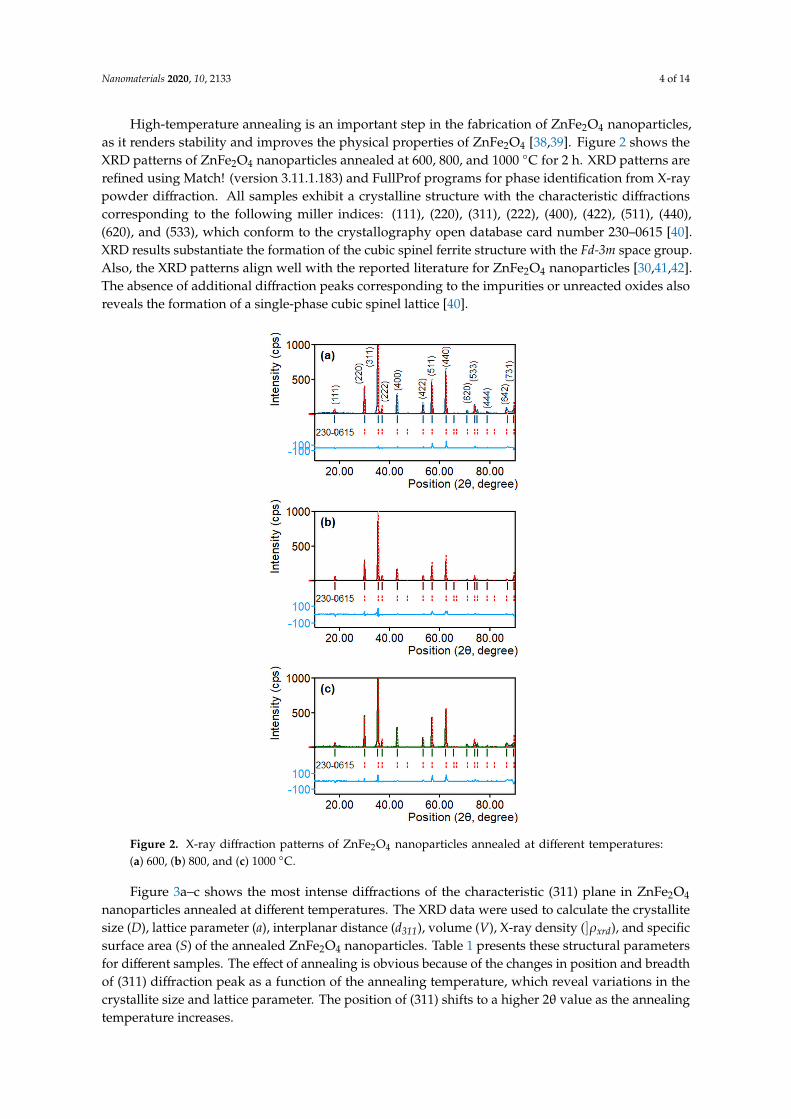
High-temperature annealing is an important step in the fabrication of ZnFe2O4 nanoparticles, as it renders stability and improves the physical properties of ZnFe2O4 [38,39]. Figure 2 shows the XRD patterns of ZnFe2O4 nanoparticles annealed at 600, 800, and 1000 °C for 2 h. XRD patterns are refined
Figure 1. (a) X-ray diffraction pattern of the as-synthesized, ball-milled zinc ferrite (BM-ZnFe2O4)nanoparticles. (b) The characteristic (311) plane diffraction of cubic spinel BM-ZnFe2O4 nanoparticles.
Theoretically, the cation distribution in a perfect normal spinel ZnFe2O4 unit cell is(Zn2+)tet[Fe3+]octO4, i.e., the tetrahedral (A) and octahedral (B) sites are solely occupied by Zn2+
and Fe3+ cations, respectively [35]. However, in nanocrystalline ZnFe2O4 the contrary distributionsof Zn2+ and Fe3+ cations on both A and B sites are observed [36,37], which form mixed (or random)spinel structure. According to Chinnasamy et al. [32], the slight increase in the lattice parameter isattributed to the lattice expansion caused by the occupation of B sites by Zn2+ ions. Thus, the XRDpattern of as-synthesized BM-ZnFe2O4 nanoparticles indicates the formation of a mixed cubic spinellattice. Nonetheless, BM-ZnFe2O4 nanoparticles are annealed at different temperatures to examine theeffect of annealing on the crystal structure evolution, lattice parameter, crystallite size, and morphologyof ZnFe2O4 nanoparticles.
Nanomaterials 2020, 10, 2133 4 of 14
High-temperature annealing is an important step in the fabrication of ZnFe2O4 nanoparticles,as it renders stability and improves the physical properties of ZnFe2O4 [38,39]. Figure 2 shows theXRD patterns of ZnFe2O4 nanoparticles annealed at 600, 800, and 1000 ◦C for 2 h. XRD patterns arerefined using Match! (version 3.11.1.183) and FullProf programs for phase identification from X-raypowder diffraction. All samples exhibit a crystalline structure with the characteristic diffractionscorresponding to the following miller indices: (111), (220), (311), (222), (400), (422), (511), (440),(620), and (533), which conform to the crystallography open database card number 230–0615 [40].XRD results substantiate the formation of the cubic spinel ferrite structure with the Fd-3m space group.Also, the XRD patterns align well with the reported literature for ZnFe2O4 nanoparticles [30,41,42].The absence of additional diffraction peaks corresponding to the impurities or unreacted oxides alsoreveals the formation of a single-phase cubic spinel lattice [40].
Nanomaterials 2020, 10, x FOR PEER REVIEW 4 of 14
using Match! (version 3.11.1.183) and FullProf programs for phase identification from X-ray powder diffraction. All samples exhibit a crystalline structure with the characteristic diffractions corresponding to the following miller indices: (111), (220), (311), (222), (400), (422), (511), (440), (620), and (533), which conform to the crystallography open database card number 230–0615 [40]. XRD results substantiate the formation of the cubic spinel ferrite structure with the Fd-3m space group. Also, the XRD patterns align well with the reported literature for ZnFe2O4 nanoparticles [30,41,42]. The absence of additional diffraction peaks corresponding to the impurities or unreacted oxides also reveals the formation of a single-phase cubic spinel lattice [40].
Figure 2. X-ray diffraction patterns of ZnFe2O4 nanoparticles annealed at different temperatures: (a) 600, (b) 800, and (c) 1000 °C.
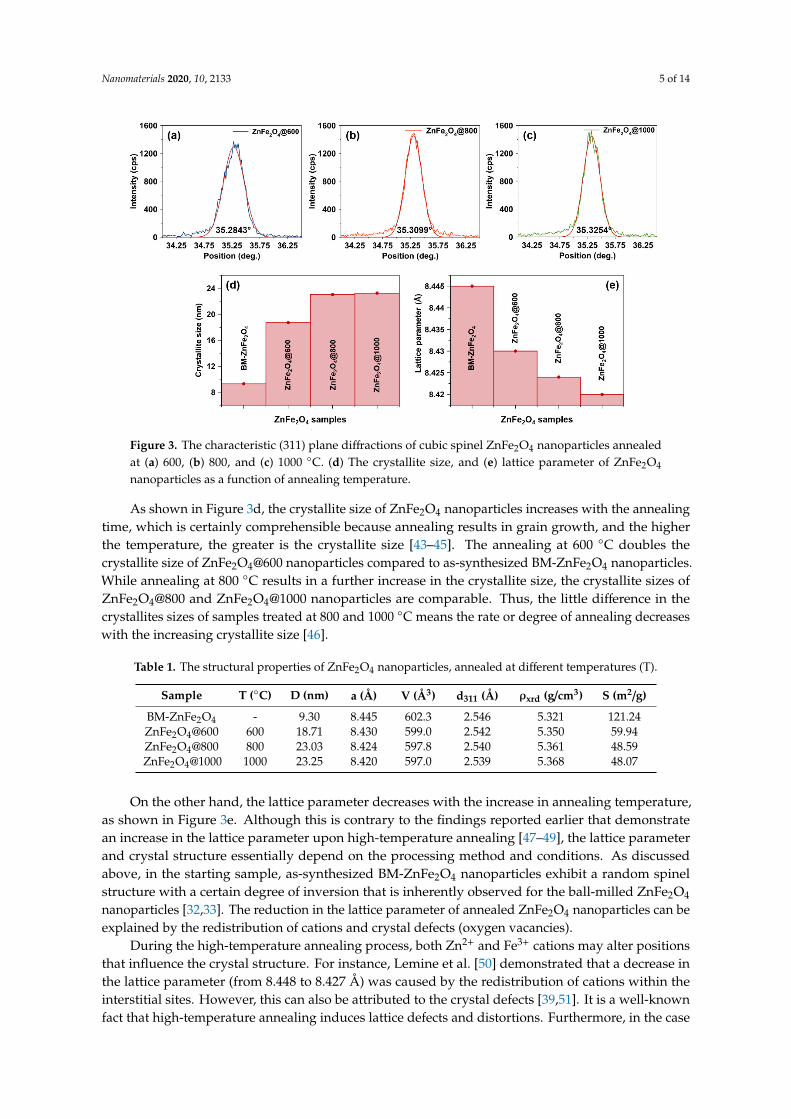
Figure 3a–c shows the most intense diffractions of the characteristic (311) plane in ZnFe2O4 nanoparticles annealed at different temperatures. The XRD data were used to calculate the crystallite size (D), lattice parameter (a), interplanar distance (d311), volume (V), X-ray density (ρxrd), and specific surface area (S) of the annealed ZnFe2O4 nanoparticles. Table 1 presents these structural parameters for different samples. The effect of annealing is obvious because of the changes in position and breadth of (311) diffraction peak as a function of the annealing temperature, which reveal variations in the crystallite size and lattice parameter. The position of (311) shifts to a higher 2θ value as the annealing temperature increases.
Figure 2. X-ray diffraction patterns of ZnFe2O4 nanoparticles annealed at different temperatures:(a) 600, (b) 800, and (c) 1000 ◦C.
Figure 3a–c shows the most intense diffractions of the characteristic (311) plane in ZnFe2O4
nanoparticles annealed at different temperatures. The XRD data were used to calculate the crystallitesize (D), lattice parameter (a), interplanar distance (d311), volume (V), X-ray density (]ρxrd), and specificsurface area (S) of the annealed ZnFe2O4 nanoparticles. Table 1 presents these structural parametersfor different samples. The effect of annealing is obvious because of the changes in position and breadthof (311) diffraction peak as a function of the annealing temperature, which reveal variations in thecrystallite size and lattice parameter. The position of (311) shifts to a higher 2θ value as the annealingtemperature increases.
Nanomaterials 2020, 10, 2133 5 of 14Nanomaterials 2020, 10, x FOR PEER REVIEW 5 of 14
Figure 3. The characteristic (311) plane diffractions of cubic spinel ZnFe2O4 nanoparticles annealed at (a) 600, (b) 800, and (c) 1000 °C. (d) The crystallite size, and (e) lattice parameter of ZnFe2O4 nanoparticles as a function of annealing temperature.
As shown in Figure 3d, the crystallite size of ZnFe2O4 nanoparticles increases with the annealing time, which is certainly comprehensible because annealing results in grain growth, and the higher the temperature, the greater is the crystallite size [43–45]. The annealing at 600 °C doubles the crystallite size of ZnFe2O4@600 nanoparticles compared to as-synthesized BM-ZnFe2O4 nanoparticles. While annealing at 800 °C results in a further increase in the crystallite size, the crystallite sizes of ZnFe2O4@800 and ZnFe2O4@1000 nanoparticles are comparable. Thus, the little difference in the crystallites sizes of samples treated at 800 and 1000 °C means the rate or degree of annealing decreases with the increasing crystallite size [46].
Table 1. The structural properties of ZnFe2O4 nanoparticles, annealed at different temperatures (T).
Sample T (°C) D (nm) a (Å) V (Å3) d311 (Å) ρxrd (g/cm3) S (m2/g) BM-ZnFe2O4 - 9.30 8.445 602.3 2.546 5.321 121.24 ZnFe2O4@600 600 18.71 8.430 599.0 2.542 5.350 59.94 ZnFe2O4@800 800 23.03 8.424 597.8 2.540 5.361 48.59 ZnFe2O4@1000 1000 23.25 8.420 597.0 2.539 5.368 48.07
On the other hand, the lattice parameter decreases with the increase in annealing temperature, as shown in Figure 3e. Although this is contrary to the findings reported earlier that demonstrate an increase in the lattice parameter upon high-temperature annealing [47–49], the lattice parameter and crystal structure essentially depend on the processing method and conditions. As discussed above, in the starting sample, as-synthesized BM-ZnFe2O4 nanoparticles exhibit a random spinel structure with a certain degree of inversion that is inherently observed for the ball-milled ZnFe2O4 nanoparticles [32,33]. The reduction in the lattice parameter of annealed ZnFe2O4 nanoparticles can be explained by the redistribution of cations and crystal defects (oxygen vacancies).
During the high-temperature annealing process, both Zn2+ and Fe3+ cations may alter positions that influence the crystal structure. For instance, Lemine et al. [50] demonstrated that a decrease in the lattice parameter (from 8.448 to 8.427 Å) was caused by the redistribution of cations within the interstitial sites. However, this can also be attributed to the crystal defects [39,51]. It is a well-known fact that high-temperature annealing induces lattice defects and distortions. Furthermore, in the case of nanocrystalline ZnFe2O4, it is believed that Zn2+ ions due to their volatile nature escape from the
Figure 3. The characteristic (311) plane diffractions of cubic spinel ZnFe2O4 nanoparticles annealedat (a) 600, (b) 800, and (c) 1000 ◦C. (d) The crystallite size, and (e) lattice parameter of ZnFe2O4
nanoparticles as a function of annealing temperature.
As shown in Figure 3d, the crystallite size of ZnFe2O4 nanoparticles increases with the annealingtime, which is certainly comprehensible because annealing results in grain growth, and the higherthe temperature, the greater is the crystallite size [43–45]. The annealing at 600 ◦C doubles thecrystallite size of ZnFe2O4@600 nanoparticles compared to as-synthesized BM-ZnFe2O4 nanoparticles.While annealing at 800 ◦C results in a further increase in the crystallite size, the crystallite sizes ofZnFe2O4@800 and ZnFe2O4@1000 nanoparticles are comparable. Thus, the little difference in thecrystallites sizes of samples treated at 800 and 1000 ◦C means the rate or degree of annealing decreaseswith the increasing crystallite size [46].
Table 1. The structural properties of ZnFe2O4 nanoparticles, annealed at different temperatures (T).
Sample T (◦C) D (nm) a (Å) V (Å3) d311 (Å) ρxrd (g/cm3) S (m2/g)
BM-ZnFe2O4 - 9.30 8.445 602.3 2.546 5.321 121.24ZnFe2O4@600 600 18.71 8.430 599.0 2.542 5.350 59.94ZnFe2O4@800 800 23.03 8.424 597.8 2.540 5.361 48.59ZnFe2O4@1000 1000 23.25 8.420 597.0 2.539 5.368 48.07
On the other hand, the lattice parameter decreases with the increase in annealing temperature,as shown in Figure 3e. Although this is contrary to the findings reported earlier that demonstratean increase in the lattice parameter upon high-temperature annealing [47–49], the lattice parameterand crystal structure essentially depend on the processing method and conditions. As discussedabove, in the starting sample, as-synthesized BM-ZnFe2O4 nanoparticles exhibit a random spinelstructure with a certain degree of inversion that is inherently observed for the ball-milled ZnFe2O4
nanoparticles [32,33]. The reduction in the lattice parameter of annealed ZnFe2O4 nanoparticles can beexplained by the redistribution of cations and crystal defects (oxygen vacancies).
During the high-temperature annealing process, both Zn2+ and Fe3+ cations may alter positionsthat influence the crystal structure. For instance, Lemine et al. [50] demonstrated that a decrease inthe lattice parameter (from 8.448 to 8.427 Å) was caused by the redistribution of cations within theinterstitial sites. However, this can also be attributed to the crystal defects [39,51]. It is a well-knownfact that high-temperature annealing induces lattice defects and distortions. Furthermore, in the case
Nanomaterials 2020, 10, 2133 6 of 14
of nanocrystalline ZnFe2O4, it is believed that Zn2+ ions due to their volatile nature escape from thelattice during thermal treatment that successively results in oxygen vacancies [39,52]. Thus, a decreasein the lattice parameter (from 8.445 Å for BM-ZnFe2O4 to 8.420 Å for ZnFe2O4@1000) is attributedto the cationic redistribution (distortion) and lattice compression caused by escaping Zn2+ ions andoxygen vacancies.
Consequently, the interplanar distance and the volume of the annealed ZnFe2O4 nanoparticlesare reduced as a function of the annealing temperature. On the other hand, the X-ray density increases(from 5.321 g cm–3 for BM-ZnFe2O4 to 5.368 g cm–3 for ZnFe2O4@1000) with the increase in annealingtemperature. However, as shown in Table 1, the specific surface area is reduced to 48 m2 g–1 due to anincrease in the crystallite size of the annealed ZnFe2O4 nanoparticles. These results demonstrate thatZnFe2O4@1000 and ZnFe2O4@800 nanoparticles have bigger crystallite size and smaller specific surfacearea, but the greatest number of defect sites (as oxygen vacancies) and a geometrically frustrated [53]or distorted cubic spinel crystal structure compared to as-synthesized BM-ZnFe2O4 nanoparticles.
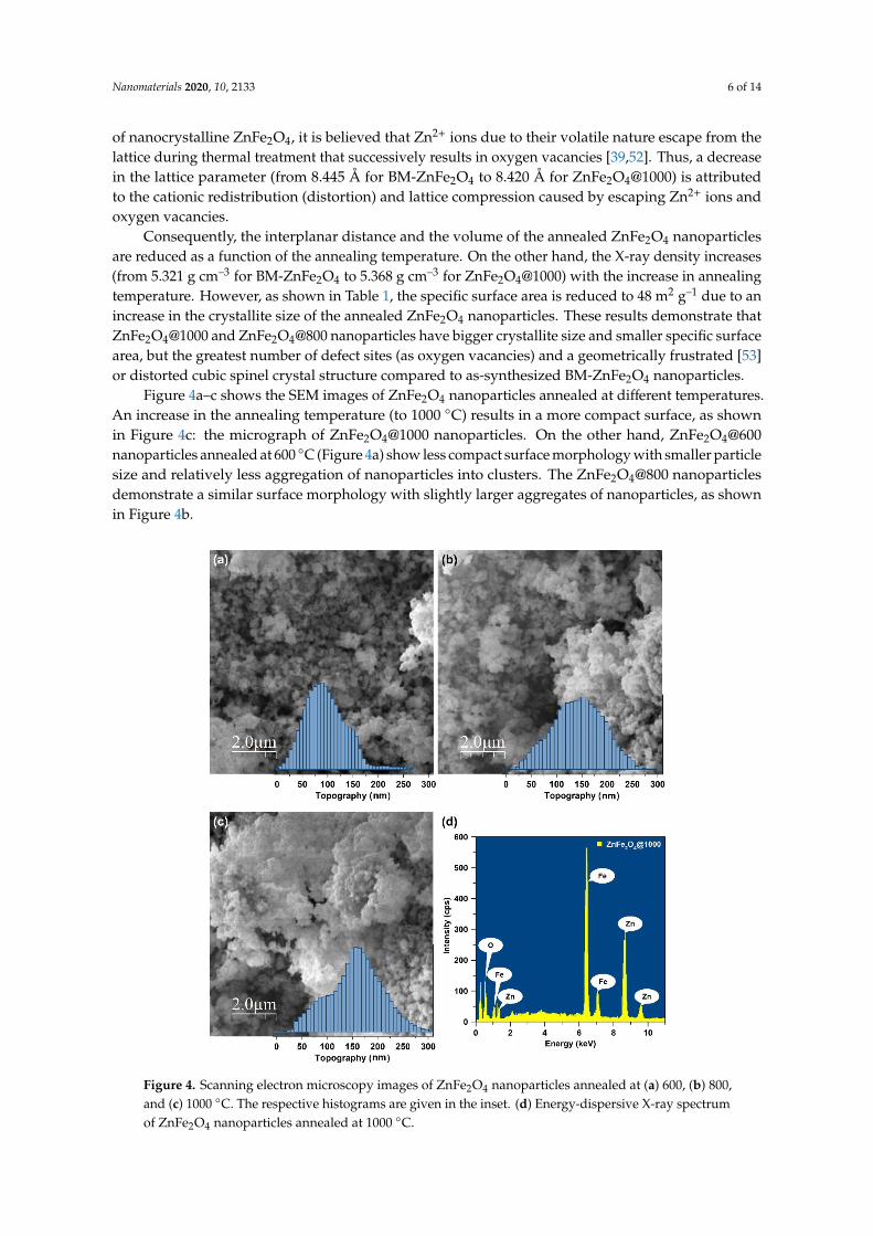
Figure 4a–c shows the SEM images of ZnFe2O4 nanoparticles annealed at different temperatures.An increase in the annealing temperature (to 1000 ◦C) results in a more compact surface, as shownin Figure 4c: the micrograph of ZnFe2O4@1000 nanoparticles. On the other hand, ZnFe2O4@600nanoparticles annealed at 600 ◦C (Figure 4a) show less compact surface morphology with smaller particlesize and relatively less aggregation of nanoparticles into clusters. The ZnFe2O4@800 nanoparticlesdemonstrate a similar surface morphology with slightly larger aggregates of nanoparticles, as shownin Figure 4b.
Nanomaterials 2020, 10, x FOR PEER REVIEW 6 of 14
lattice during thermal treatment that successively results in oxygen vacancies [39,52]. Thus, a decrease in the lattice parameter (from 8.445 Å for BM-ZnFe2O4 to 8.420 Å for ZnFe2O4@1000) is attributed to the cationic redistribution (distortion) and lattice compression caused by escaping Zn2+ ions and oxygen vacancies.
Consequently, the interplanar distance and the volume of the annealed ZnFe2O4 nanoparticles are reduced as a function of the annealing temperature. On the other hand, the X-ray density increases (from 5.321 g cm–3 for BM-ZnFe2O4 to 5.368 g cm–3 for ZnFe2O4@1000) with the increase in annealing temperature. However, as shown in Table 1, the specific surface area is reduced to 48 m2 g–1 due to an increase in the crystallite size of the annealed ZnFe2O4 nanoparticles. These results demonstrate that ZnFe2O4@1000 and ZnFe2O4@800 nanoparticles have bigger crystallite size and smaller specific surface area, but the greatest number of defect sites (as oxygen vacancies) and a geometrically frustrated [53] or distorted cubic spinel crystal structure compared to as-synthesized BM-ZnFe2O4 nanoparticles.
Figure 4a–c shows the SEM images of ZnFe2O4 nanoparticles annealed at different temperatures. An increase in the annealing temperature (to 1000 °C) results in a more compact surface, as shown in Figure 4c: the micrograph of ZnFe2O4@1000 nanoparticles. On the other hand, ZnFe2O4@600 nanoparticles annealed at 600 °C (Figure 4a) show less compact surface morphology with smaller particle size and relatively less aggregation of nanoparticles into clusters. The ZnFe2O4@800 nanoparticles demonstrate a similar surface morphology with slightly larger aggregates of nanoparticles, as shown in Figure 4b.
Figure 4. Scanning electron microscopy images of ZnFe2O4 nanoparticles annealed at (a) 600, (b) 800, and (c) 1000 °C. The respective histograms are given in the inset. (d) Energy-dispersive X-ray spectrum of ZnFe2O4 nanoparticles annealed at 1000 °C.
Figure 4. Scanning electron microscopy images of ZnFe2O4 nanoparticles annealed at (a) 600, (b) 800,and (c) 1000 ◦C. The respective histograms are given in the inset. (d) Energy-dispersive X-ray spectrumof ZnFe2O4 nanoparticles annealed at 1000 ◦C.
Nanomaterials 2020, 10, 2133 7 of 14
The image analysis of the scanning electron micrographs (via WSxM freeware [54]) shows thesize distribution of ZnFe2O4 nanoparticles and the respective histograms are presented as insets inFigure 4a–c. ZnFe2O4@600 nanoparticles exhibit narrow size distribution with an average aggregatesize of 100.2 nm, while ZnFe2O4@800 and ZnFe2O4@1000 nanoparticles reveal a relatively broad sizedistribution and an average aggregate size of 143.8 and 146.5 nm, respectively.
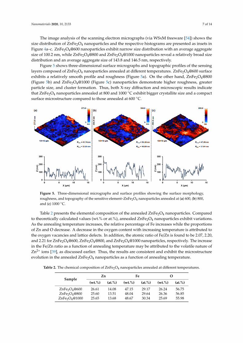
Figure 5 shows three-dimensional surface micrographs and topographic profiles of the sensinglayers composed of ZnFe2O4 nanoparticles annealed at different temperatures. ZnFe2O4@600 surfaceexhibits a relatively smooth profile and roughness (Figure 5a). On the other hand, ZnFe2O4@800(Figure 5b) and ZnFe2O4@1000 (Figure 5c) nanoparticles demonstrate higher roughness, greaterparticle size, and cluster formation. Thus, both X-ray diffraction and microscopic results indicatethat ZnFe2O4 nanoparticles annealed at 800 and 1000 ◦C exhibit bigger crystallite size and a compactsurface microstructure compared to those annealed at 600 ◦C.
Nanomaterials 2020, 10, x FOR PEER REVIEW 7 of 14
The image analysis of the scanning electron micrographs (via WSxM freeware [54]) shows the size distribution of ZnFe2O4 nanoparticles and the respective histograms are presented as insets in Figure 4a–c. ZnFe2O4@600 nanoparticles exhibit narrow size distribution with an average aggregate size of 100.2 nm, while ZnFe2O4@800 and ZnFe2O4@1000 nanoparticles reveal a relatively broad size distribution and an average aggregate size of 143.8 and 146.5 nm, respectively.
Figure 5 shows three-dimensional surface micrographs and topographic profiles of the sensing layers composed of ZnFe2O4 nanoparticles annealed at different temperatures. ZnFe2O4@600 surface exhibits a relatively smooth profile and roughness (Figure 5a). On the other hand, ZnFe2O4@800 (Figure 5b) and ZnFe2O4@1000 (Figure 5c) nanoparticles demonstrate higher roughness, greater particle size, and cluster formation. Thus, both X-ray diffraction and microscopic results indicate that ZnFe2O4 nanoparticles annealed at 800 and 1000 °C exhibit bigger crystallite size and a compact surface microstructure compared to those annealed at 600 °C.
Figure 5. Three-dimensional micrographs and surface profiles showing the surface morphology, roughness, and topography of the sensitive element–ZnFe2O4 nanoparticles annealed at (a) 600, (b) 800, and (c) 1000 °C.
Table 2 presents the elemental composition of the annealed ZnFe2O4 nanoparticles. Compared to theoretically calculated values (wt.% or at.%), annealed ZnFe2O4 nanoparticles exhibit variations. As the annealing temperature increases, the relative percentage of Fe increases while the proportions of Zn and O decrease. A decrease in the oxygen content with increasing temperature is attributed to the oxygen vacancies and lattice defects. In addition, the atomic ratio of Fe/Zn is found to be 2.07, 2.20, and 2.21 for ZnFe2O4@600, ZnFe2O4@800, and ZnFe2O4@1000 nanoparticles, respectively. The increase in the Fe/Zn ratio as a function of annealing temperature may be attributed to the volatile nature of Zn2+ ions [39], as discussed earlier. Thus, the results are consistent and exhibit the microstructure evolution in the annealed ZnFe2O4 nanoparticles as a function of annealing temperature.
Table 2. The chemical composition of ZnFe2O4 nanoparticles annealed at different temperatures.
Sample Zn Fe O
(wt.%) (at.%) (wt.%) (at.%) (wt.%) (at.%) ZnFe2O4@600 26.61 14.08 47.15 29.17 26.24 56.75 ZnFe2O4@800 25.60 13.51 48.04 29.64 26.36 56.85 ZnFe2O4@1000 25.65 13.68 48.67 30.34 25.69 55.98
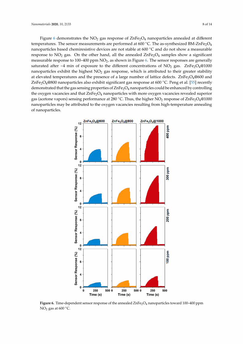
Figure 6 demonstrates the NO2 gas response of ZnFe2O4 nanoparticles annealed at different temperatures. The sensor measurements are performed at 600 °C. The as-synthesized BM-ZnFe2O4
Figure 5. Three-dimensional micrographs and surface profiles showing the surface morphology,roughness, and topography of the sensitive element–ZnFe2O4 nanoparticles annealed at (a) 600, (b) 800,and (c) 1000 ◦C.
Table 2 presents the elemental composition of the annealed ZnFe2O4 nanoparticles. Comparedto theoretically calculated values (wt.% or at.%), annealed ZnFe2O4 nanoparticles exhibit variations.As the annealing temperature increases, the relative percentage of Fe increases while the proportionsof Zn and O decrease. A decrease in the oxygen content with increasing temperature is attributed tothe oxygen vacancies and lattice defects. In addition, the atomic ratio of Fe/Zn is found to be 2.07, 2.20,and 2.21 for ZnFe2O4@600, ZnFe2O4@800, and ZnFe2O4@1000 nanoparticles, respectively. The increasein the Fe/Zn ratio as a function of annealing temperature may be attributed to the volatile nature ofZn2+ ions [39], as discussed earlier. Thus, the results are consistent and exhibit the microstructureevolution in the annealed ZnFe2O4 nanoparticles as a function of annealing temperature.
Table 2. The chemical composition of ZnFe2O4 nanoparticles annealed at different temperatures.
SampleZn Fe O
(wt.%) (at.%) (wt.%) (at.%) (wt.%) (at.%)
ZnFe2O4@600 26.61 14.08 47.15 29.17 26.24 56.75ZnFe2O4@800 25.60 13.51 48.04 29.64 26.36 56.85
ZnFe2O4@1000 25.65 13.68 48.67 30.34 25.69 55.98
Nanomaterials 2020, 10, 2133 8 of 14
Figure 6 demonstrates the NO2 gas response of ZnFe2O4 nanoparticles annealed at differenttemperatures. The sensor measurements are performed at 600 ◦C. The as-synthesized BM-ZnFe2O4
nanoparticles based chemiresistive devices are not stable at 600 ◦C and do not show a measurableresponse to NO2 gas. On the other hand, all the annealed ZnFe2O4 samples show a significantmeasurable response to 100–400 ppm NO2, as shown in Figure 6. The sensor responses are generallysaturated after ~4 min of exposure to the different concentrations of NO2 gas. ZnFe2O4@1000nanoparticles exhibit the highest NO2 gas response, which is attributed to their greater stabilityat elevated temperatures and the presence of a large number of lattice defects. ZnFe2O4@600 andZnFe2O4@800 nanoparticles also exhibit significant gas response at 600 ◦C. Peng et al. [55] recentlydemonstrated that the gas sensing properties of ZnFe2O4 nanoparticles could be enhanced by controllingthe oxygen vacancies and that ZnFe2O4 nanoparticles with more oxygen vacancies revealed superiorgas (acetone vapors) sensing performance at 280 ◦C. Thus, the higher NO2 response of ZnFe2O4@1000nanoparticles may be attributed to the oxygen vacancies resulting from high-temperature annealingof nanoparticles.
Nanomaterials 2020, 10, x FOR PEER REVIEW 8 of 14
nanoparticles based chemiresistive devices are not stable at 600 °C and do not show a measurable response to NO2 gas. On the other hand, all the annealed ZnFe2O4 samples show a significant measurable response to 100–400 ppm NO2, as shown in Figure 6. The sensor responses are generally saturated after ~4 min of exposure to the different concentrations of NO2 gas. ZnFe2O4@1000 nanoparticles exhibit the highest NO2 gas response, which is attributed to their greater stability at elevated temperatures and the presence of a large number of lattice defects. ZnFe2O4@600 and ZnFe2O4@800 nanoparticles also exhibit significant gas response at 600 °C. Peng et al. [55] recently demonstrated that the gas sensing properties of ZnFe2O4 nanoparticles could be enhanced by controlling the oxygen vacancies and that ZnFe2O4 nanoparticles with more oxygen vacancies revealed superior gas (acetone vapors) sensing performance at 280 °C. Thus, the higher NO2 response of ZnFe2O4@1000 nanoparticles may be attributed to the oxygen vacancies resulting from high-temperature annealing of nanoparticles.
Figure 6. Time-dependent sensor response of the annealed ZnFe2O4 nanoparticles toward 100–400 ppm NO2 gas at 600 °C.
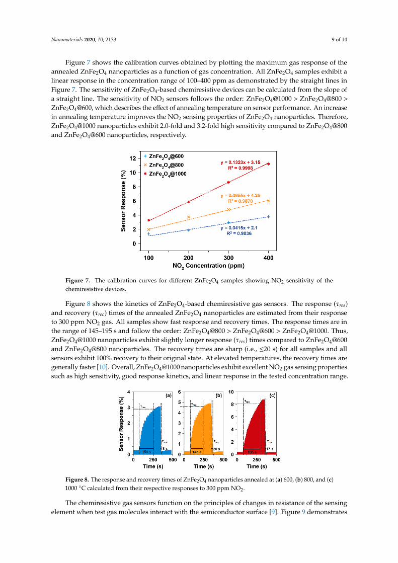
Figure 7 shows the calibration curves obtained by plotting the maximum gas response of the annealed ZnFe2O4 nanoparticles as a function of gas concentration. All ZnFe2O4 samples exhibit a linear response in the concentration range of 100–400 ppm as demonstrated by the straight lines in Figure 7. The sensitivity of ZnFe2O4-based chemiresistive devices can be calculated from the slope of
Figure 6. Time-dependent sensor response of the annealed ZnFe2O4 nanoparticles toward 100–400 ppmNO2 gas at 600 ◦C.
Nanomaterials 2020, 10, 2133 9 of 14
Figure 7 shows the calibration curves obtained by plotting the maximum gas response of theannealed ZnFe2O4 nanoparticles as a function of gas concentration. All ZnFe2O4 samples exhibit alinear response in the concentration range of 100–400 ppm as demonstrated by the straight lines inFigure 7. The sensitivity of ZnFe2O4-based chemiresistive devices can be calculated from the slope ofa straight line. The sensitivity of NO2 sensors follows the order: ZnFe2O4@1000 > ZnFe2O4@800 >
ZnFe2O4@600, which describes the effect of annealing temperature on sensor performance. An increasein annealing temperature improves the NO2 sensing properties of ZnFe2O4 nanoparticles. Therefore,ZnFe2O4@1000 nanoparticles exhibit 2.0-fold and 3.2-fold high sensitivity compared to ZnFe2O4@800and ZnFe2O4@600 nanoparticles, respectively.
Nanomaterials 2020, 10, x FOR PEER REVIEW 9 of 14
a straight line. The sensitivity of NO2 sensors follows the order: ZnFe2O4@1000 > ZnFe2O4@800 > ZnFe2O4@600, which describes the effect of annealing temperature on sensor performance. An increase in annealing temperature improves the NO2 sensing properties of ZnFe2O4 nanoparticles. Therefore, ZnFe2O4@1000 nanoparticles exhibit 2.0-fold and 3.2-fold high sensitivity compared to ZnFe2O4@800 and ZnFe2O4@600 nanoparticles, respectively.
Figure 7. The calibration curves for different ZnFe2O4 samples showing NO2 sensitivity of the chemiresistive devices.
Figure 8 shows the kinetics of ZnFe2O4-based chemiresistive gas sensors. The response (τres) and recovery (τrec) times of the annealed ZnFe2O4 nanoparticles are estimated from their response to 300 ppm NO2 gas. All samples show fast response and recovery times. The response times are in the range of 145–195 s and follow the order: ZnFe2O4@800 > ZnFe2O4@600 > ZnFe2O4@1000. Thus, ZnFe2O4@1000 nanoparticles exhibit slightly longer response (τres) times compared to ZnFe2O4@600 and ZnFe2O4@800 nanoparticles. The recovery times are sharp (i.e., ≤20 s) for all samples and all sensors exhibit 100% recovery to their original state. At elevated temperatures, the recovery times are generally faster [10]. Overall, ZnFe2O4@1000 nanoparticles exhibit excellent NO2 gas sensing properties such as high sensitivity, good response kinetics, and linear response in the tested concentration range.
Figure 8. The response and recovery times of ZnFe2O4 nanoparticles annealed at (a) 600, (b) 800, and (c) 1000 °C calculated from their respective responses to 300 ppm NO2.
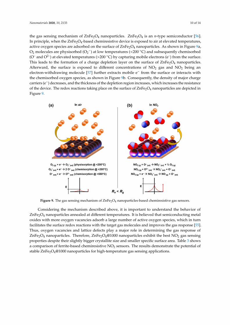
The chemiresistive gas sensors function on the principles of changes in resistance of the sensing element when test gas molecules interact with the semiconductor surface [9]. Figure 9 demonstrates the gas sensing mechanism of ZnFe2O4 nanoparticles. ZnFe2O4 is an n-type semiconductor [56]. In principle, when the ZnFe2O4-based chemiresistive device is exposed to air at elevated temperatures, active oxygen species are adsorbed on the surface of ZnFe2O4 nanoparticles. As shown in Figure 9a, O2 molecules are physisorbed (O2−) at low temperatures (<200 °C) and subsequently chemisorbed (O– and O2–) at elevated temperatures (>200 °C) by capturing mobile electrons (e–) from the surface. This
Figure 7. The calibration curves for different ZnFe2O4 samples showing NO2 sensitivity of thechemiresistive devices.
Figure 8 shows the kinetics of ZnFe2O4-based chemiresistive gas sensors. The response (τres)and recovery (τrec) times of the annealed ZnFe2O4 nanoparticles are estimated from their responseto 300 ppm NO2 gas. All samples show fast response and recovery times. The response times are inthe range of 145–195 s and follow the order: ZnFe2O4@800 > ZnFe2O4@600 > ZnFe2O4@1000. Thus,ZnFe2O4@1000 nanoparticles exhibit slightly longer response (τres) times compared to ZnFe2O4@600and ZnFe2O4@800 nanoparticles. The recovery times are sharp (i.e., ≤20 s) for all samples and allsensors exhibit 100% recovery to their original state. At elevated temperatures, the recovery times aregenerally faster [10]. Overall, ZnFe2O4@1000 nanoparticles exhibit excellent NO2 gas sensing propertiessuch as high sensitivity, good response kinetics, and linear response in the tested concentration range.
Nanomaterials 2020, 10, x FOR PEER REVIEW 9 of 14
a straight line. The sensitivity of NO2 sensors follows the order: ZnFe2O4@1000 > ZnFe2O4@800 > ZnFe2O4@600, which describes the effect of annealing temperature on sensor performance. An increase in annealing temperature improves the NO2 sensing properties of ZnFe2O4 nanoparticles. Therefore, ZnFe2O4@1000 nanoparticles exhibit 2.0-fold and 3.2-fold high sensitivity compared to ZnFe2O4@800 and ZnFe2O4@600 nanoparticles, respectively.
Figure 7. The calibration curves for different ZnFe2O4 samples showing NO2 sensitivity of the chemiresistive devices.
Figure 8 shows the kinetics of ZnFe2O4-based chemiresistive gas sensors. The response (τres) and recovery (τrec) times of the annealed ZnFe2O4 nanoparticles are estimated from their response to 300 ppm NO2 gas. All samples show fast response and recovery times. The response times are in the range of 145–195 s and follow the order: ZnFe2O4@800 > ZnFe2O4@600 > ZnFe2O4@1000. Thus, ZnFe2O4@1000 nanoparticles exhibit slightly longer response (τres) times compared to ZnFe2O4@600 and ZnFe2O4@800 nanoparticles. The recovery times are sharp (i.e., ≤20 s) for all samples and all sensors exhibit 100% recovery to their original state. At elevated temperatures, the recovery times are generally faster [10]. Overall, ZnFe2O4@1000 nanoparticles exhibit excellent NO2 gas sensing properties such as high sensitivity, good response kinetics, and linear response in the tested concentration range.
Figure 8. The response and recovery times of ZnFe2O4 nanoparticles annealed at (a) 600, (b) 800, and (c) 1000 °C calculated from their respective responses to 300 ppm NO2.
The chemiresistive gas sensors function on the principles of changes in resistance of the sensing element when test gas molecules interact with the semiconductor surface [9]. Figure 9 demonstrates the gas sensing mechanism of ZnFe2O4 nanoparticles. ZnFe2O4 is an n-type semiconductor [56]. In principle, when the ZnFe2O4-based chemiresistive device is exposed to air at elevated temperatures, active oxygen species are adsorbed on the surface of ZnFe2O4 nanoparticles. As shown in Figure 9a, O2 molecules are physisorbed (O2−) at low temperatures (<200 °C) and subsequently chemisorbed (O– and O2–) at elevated temperatures (>200 °C) by capturing mobile electrons (e–) from the surface. This
Figure 8. The response and recovery times of ZnFe2O4 nanoparticles annealed at (a) 600, (b) 800, and (c)1000 ◦C calculated from their respective responses to 300 ppm NO2.
The chemiresistive gas sensors function on the principles of changes in resistance of the sensingelement when test gas molecules interact with the semiconductor surface [9]. Figure 9 demonstrates
Nanomaterials 2020, 10, 2133 10 of 14
the gas sensing mechanism of ZnFe2O4 nanoparticles. ZnFe2O4 is an n-type semiconductor [56].In principle, when the ZnFe2O4-based chemiresistive device is exposed to air at elevated temperatures,active oxygen species are adsorbed on the surface of ZnFe2O4 nanoparticles. As shown in Figure 9a,O2 molecules are physisorbed (O2
−) at low temperatures (<200 ◦C) and subsequently chemisorbed(O– and O2–) at elevated temperatures (>200 ◦C) by capturing mobile electrons (e–) from the surface.This leads to the formation of a charge depletion layer on the surface of ZnFe2O4 nanoparticles.Afterward, the surface is exposed to different concentrations of NO2 gas and NO2 being anelectron-withdrawing molecule [57] further extracts mobile e− from the surface or interacts withthe chemisorbed oxygen species, as shown in Figure 9b. Consequently, the density of major chargecarriers (e−) decreases, and the thickness of the depletion region increases, which increases the resistanceof the device. The redox reactions taking place on the surface of ZnFe2O4 nanoparticles are depicted inFigure 9.
Nanomaterials 2020, 10, x FOR PEER REVIEW 10 of 14
leads to the formation of a charge depletion layer on the surface of ZnFe2O4 nanoparticles. Afterward, the surface is exposed to different concentrations of NO2 gas and NO2 being an electron-withdrawing molecule [57] further extracts mobile e− from the surface or interacts with the chemisorbed oxygen species, as shown in Figure 9b. Consequently, the density of major charge carriers (e−) decreases, and the thickness of the depletion region increases, which increases the resistance of the device. The redox reactions taking place on the surface of ZnFe2O4 nanoparticles are depicted in Figure 9.
Figure 9. The gas sensing mechanism of ZnFe2O4 nanoparticles-based chemiresistive gas sensors.
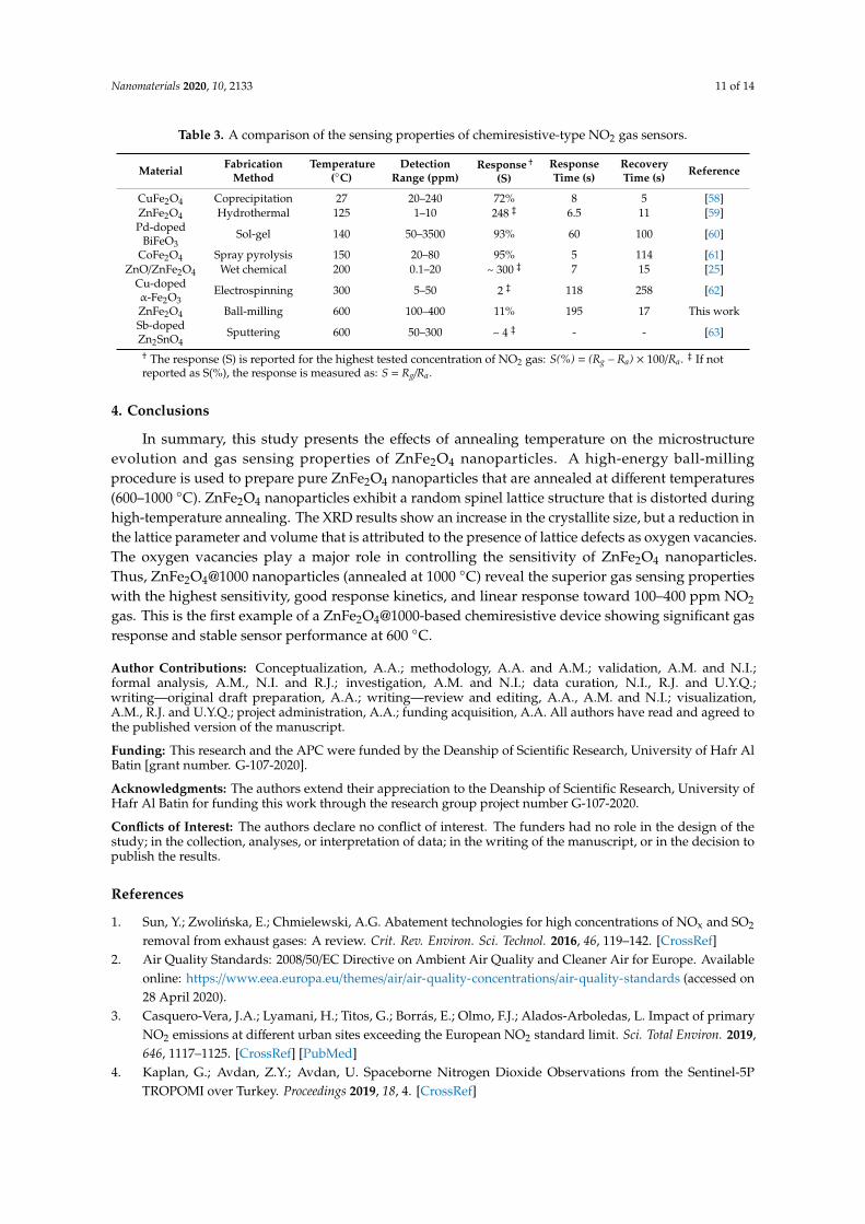
Considering the mechanism described above, it is important to understand the behavior of ZnFe2O4 nanoparticles annealed at different temperatures. It is believed that semiconducting metal oxides with more oxygen vacancies adsorb a large number of active oxygen species, which in turn facilitates the surface redox reactions with the target gas molecules and improves the gas response [55]. Thus, oxygen vacancies and lattice defects play a major role in determining the gas response of ZnFe2O4 nanoparticles. Therefore, ZnFe2O4@1000 nanoparticles exhibit the best NO2 gas sensing properties despite their slightly bigger crystallite size and smaller specific surface area. Table 3 shows a comparison of ferrite-based chemiresistive NO2 sensors. The results demonstrate the potential of stable ZnFe2O4@1000 nanoparticles for high-temperature gas sensing applications.
Table 3. A comparison of the sensing properties of chemiresistive-type NO2 gas sensors.
Material Fabrication
Method Temperature
(°C)
Detection Range (ppm)
Response † (S)
Response Time (s)
Recovery Time (s)
Reference
CuFe2O4 Coprecipitation 27 20–240 72% 8 5 [58] ZnFe2O4 Hydrothermal 125 1–10 248 ‡ 6.5 11 [59]
Pd-doped BiFeO3
Sol-gel 140 50–3500 93% 60 100 [60]
CoFe2O4 Spray pyrolysis 150 20–80 95% 5 114 [61] ZnO/ZnFe2O4 Wet chemical 200 0.1–20 ~ 300 ‡ 7 15 [25] Cu-doped α-
Fe2O3 Electrospinning 300 5–50 2 ‡ 118 258 [62]
ZnFe2O4 Ball-milling 600 100–400 11% 195 17 This work Sb-doped Zn2SnO4
Sputtering 600 50–300 ~ 4 ‡ - - [63]
† The response (S) is reported for the highest tested concentration of NO2 gas: S(%) = (Rg – Ra) × 100/Ra. ‡ If not reported as S(%), the response is measured as: S = Rg/Ra.
Figure 9. The gas sensing mechanism of ZnFe2O4 nanoparticles-based chemiresistive gas sensors.
Considering the mechanism described above, it is important to understand the behavior ofZnFe2O4 nanoparticles annealed at different temperatures. It is believed that semiconducting metaloxides with more oxygen vacancies adsorb a large number of active oxygen species, which in turnfacilitates the surface redox reactions with the target gas molecules and improves the gas response [55].Thus, oxygen vacancies and lattice defects play a major role in determining the gas response ofZnFe2O4 nanoparticles. Therefore, ZnFe2O4@1000 nanoparticles exhibit the best NO2 gas sensingproperties despite their slightly bigger crystallite size and smaller specific surface area. Table 3 showsa comparison of ferrite-based chemiresistive NO2 sensors. The results demonstrate the potential ofstable ZnFe2O4@1000 nanoparticles for high-temperature gas sensing applications.
Nanomaterials 2020, 10, 2133 11 of 14
Table 3. A comparison of the sensing properties of chemiresistive-type NO2 gas sensors.
Material FabricationMethod
Temperature(◦C)
DetectionRange (ppm)
Response †(S)
ResponseTime (s)
RecoveryTime (s) Reference
CuFe2O4 Coprecipitation 27 20–240 72% 8 5 [58]ZnFe2O4 Hydrothermal 125 1–10 248 ‡ 6.5 11 [59]Pd-doped
BiFeO3Sol-gel 140 50–3500 93% 60 100 [60]
CoFe2O4 Spray pyrolysis 150 20–80 95% 5 114 [61]ZnO/ZnFe2O4 Wet chemical 200 0.1–20 ~ 300 ‡ 7 15 [25]
Cu-dopedα-Fe2O3
Electrospinning 300 5–50 2 ‡ 118 258 [62]
ZnFe2O4 Ball-milling 600 100–400 11% 195 17 This workSb-dopedZn2SnO4
Sputtering 600 50–300 ~ 4 ‡ - - [63]
† The response (S) is reported for the highest tested concentration of NO2 gas: S(%) = (Rg – Ra) × 100/Ra. ‡ If notreported as S(%), the response is measured as: S = Rg /Ra.
4. Conclusions
In summary, this study presents the effects of annealing temperature on the microstructureevolution and gas sensing properties of ZnFe2O4 nanoparticles. A high-energy ball-millingprocedure is used to prepare pure ZnFe2O4 nanoparticles that are annealed at different temperatures(600–1000 ◦C). ZnFe2O4 nanoparticles exhibit a random spinel lattice structure that is distorted duringhigh-temperature annealing. The XRD results show an increase in the crystallite size, but a reduction inthe lattice parameter and volume that is attributed to the presence of lattice defects as oxygen vacancies.The oxygen vacancies play a major role in controlling the sensitivity of ZnFe2O4 nanoparticles.Thus, ZnFe2O4@1000 nanoparticles (annealed at 1000 ◦C) reveal the superior gas sensing propertieswith the highest sensitivity, good response kinetics, and linear response toward 100–400 ppm NO2
gas. This is the first example of a ZnFe2O4@1000-based chemiresistive device showing significant gasresponse and stable sensor performance at 600 ◦C.
Author Contributions: Conceptualization, A.A.; methodology, A.A. and A.M.; validation, A.M. and N.I.;formal analysis, A.M., N.I. and R.J.; investigation, A.M. and N.I.; data curation, N.I., R.J. and U.Y.Q.;writing—original draft preparation, A.A.; writing—review and editing, A.A., A.M. and N.I.; visualization,A.M., R.J. and U.Y.Q.; project administration, A.A.; funding acquisition, A.A. All authors have read and agreed tothe published version of the manuscript.
Funding: This research and the APC were funded by the Deanship of Scientific Research, University of Hafr AlBatin [grant number. G-107-2020].
Acknowledgments: The authors extend their appreciation to the Deanship of Scientific Research, University ofHafr Al Batin for funding this work through the research group project number G-107-2020.
Conflicts of Interest: The authors declare no conflict of interest. The funders had no role in the design of thestudy; in the collection, analyses, or interpretation of data; in the writing of the manuscript, or in the decision topublish the results.
References
1. Sun, Y.; Zwolinska, E.; Chmielewski, A.G. Abatement technologies for high concentrations of NOx and SO2
removal from exhaust gases: A review. Crit. Rev. Environ. Sci. Technol. 2016, 46, 119–142. [CrossRef]2. Air Quality Standards: 2008/50/EC Directive on Ambient Air Quality and Cleaner Air for Europe. Available
online: https://www.eea.europa.eu/themes/air/air-quality-concentrations/air-quality-standards (accessed on28 April 2020).
3. Casquero-Vera, J.A.; Lyamani, H.; Titos, G.; Borrás, E.; Olmo, F.J.; Alados-Arboledas, L. Impact of primaryNO2 emissions at different urban sites exceeding the European NO2 standard limit. Sci. Total Environ. 2019,646, 1117–1125. [CrossRef] [PubMed]
4. Kaplan, G.; Avdan, Z.Y.; Avdan, U. Spaceborne Nitrogen Dioxide Observations from the Sentinel-5PTROPOMI over Turkey. Proceedings 2019, 18, 4. [CrossRef]
Nanomaterials 2020, 10, 2133 12 of 14
5. Afzal, A.; Andersson, M.; Di Franco, C.; Ditaranto, N.; Cioffi, N.; Scamarcio, G.; Lloyd Spetz, A.; Torsi, L.Electrochemical deposition of gold on indium zirconate (InZrOx with In/Zr atomic ratio 1.0) for hightemperature automobile exhaust gas sensors. J. Solid State Electrochem. 2015, 19, 2859–2868. [CrossRef]
6. Liu, F.; Wang, B.; Yang, X.; Guan, Y.; Sun, R.; Wang, Q.; Liang, X.; Sun, P.; Lu, G. High-temperature stabilizedzirconia-based sensors utilizing MNb2O6 (M: Co, Ni and Zn) sensing electrodes for detection of NO2.Sens. Actuators B Chem. 2016, 232, 523–530. [CrossRef]
7. Liu, F.; Wang, B.; Yang, X.; Guan, Y.; Wang, Q.; Liang, X.; Sun, P.; Wang, Y.; Lu, G. High-temperature NO2
gas sensor based on stabilized zirconia and CoTa2O6 sensing electrode. Sens. Actuators B Chem. 2017, 240,148–157. [CrossRef]
8. Dai, L.; Shi, M.; Han, W.; Meng, W.; He, Z.; Zhu, L.; Wang, L. High-temperature NO2 sensor based onaluminum/indium co-doped lanthanum silicate oxyapatite electrolyte and cobalt-free perovskite oxidesensing electrode. Sens. Actuators B Chem. 2017, 250, 629–640. [CrossRef]
9. Ghosh, A.; Zhang, C.; Shi, S.Q.; Zhang, H. High-Temperature Gas Sensors for Harsh Environment Applications:A Review. CLEAN Soil Air Water 2019, 47, 1800491. [CrossRef]
10. Afzal, A.; Cioffi, N.; Sabbatini, L.; Torsi, L. NOx sensors based on semiconducting metal oxide nanostructures:Progress and perspectives. Sens. Actuators B Chem. 2012, 171–172, 25–42. [CrossRef]
11. Dey, A. Semiconductor metal oxide gas sensors: A review. Mater. Sci. Eng. B 2018, 229, 206–217. [CrossRef]12. Barsan, N.; Schierbaum, K. Gas Sensors Based on Conducting Metal Oxides: Basic Understanding, Technology and
Applications; Elsevier: Amsterdam, The Netherlands, 2018; ISBN 978-0-12-811225-0.13. Afzal, A. β-Ga2O3 nanowires and thin films for metal oxide semiconductor gas sensors: Sensing mechanisms
and performance enhancement strategies. J. Mater. 2019, 5, 542–557. [CrossRef]14. Miura, N.; Zhuiykov, S.; Ono, T.; Hasei, M.; Yamazoe, N. Mixed potential type sensor using stabilized zirconia
and ZnFe2O4 sensing electrode for NOx detection at high temperature. Sens. Actuators B Chem. 2002, 83,222–229. [CrossRef]
15. Wetchakun, K.; Samerjai, T.; Tamaekong, N.; Liewhiran, C.; Siriwong, C.; Kruefu, V.; Wisitsoraat, A.;Tuantranont, A.; Phanichphant, S. Semiconducting metal oxides as sensors for environmentally hazardousgases. Sens. Actuators B Chem. 2011, 160, 580–591. [CrossRef]
16. Joshi, N.; Hayasaka, T.; Liu, Y.; Liu, H.; Oliveira, O.N.; Lin, L. A review on chemiresistive room temperaturegas sensors based on metal oxide nanostructures, graphene and 2D transition metal dichalcogenides.Microchim. Acta 2018, 185, 213. [CrossRef] [PubMed]
17. Li, L.; Tan, J.; Dun, M.; Huang, X. Porous ZnFe2O4 nanorods with net-worked nanostructure for highlysensor response and fast response acetone gas sensor. Sens. Actuators B Chem. 2017, 248, 85–91. [CrossRef]
18. Lv, L.; Wang, Y.; Cheng, P.; Zhang, B.; Dang, F.; Xu, L. Ultrasonic spray pyrolysis synthesis of three-dimensionalZnFe2O4-based macroporous spheres for excellent sensitive acetone gas sensor. Sens. Actuators B Chem. 2019,297, 126755. [CrossRef]
19. Khurshid, R.; Ali, F.; Afzal, A.; Ali, Z.; Qureshi, M.T. Polyol-mediated coprecipitation and aminosilanegrafting of superparamagnetic, spinel ZnFe2O4 nanoparticles for room-temperature ethanol sensors.J. Electrochem. Soc. 2019, 166, B258–B265. [CrossRef]
20. Nemufulwi, M.I.; Swart, H.C.; Mdlalose, W.B.; Mhlongo, G.H. Size-tunable ferromagnetic ZnFe2O4
nanoparticles and their ethanol detection capabilities. Appl. Surf. Sci. 2020, 508, 144863. [CrossRef]21. Dong, C.; Liu, X.; Xiao, X.; Du, S.; Wang, Y. Monodisperse ZnFe2O4 nanospheres synthesized by a nonaqueous
route for a highly slective low-ppm-level toluene gas sensor. Sens. Actuators B Chem. 2017, 239, 1231–1236.[CrossRef]
22. Gao, X.; Sun, Y.; Zhu, C.; Li, C.; Ouyang, Q.; Chen, Y. Highly sensitive and selective H2S sensor based onporous ZnFe2O4 nanosheets. Sens. Actuators B Chem. 2017, 246, 662–672. [CrossRef]
23. Zhang, H.-J.; Meng, F.-N.; Liu, L.-Z.; Chen, Y.-J.; Wang, P.-J. Highly sensitive H2S sensor based onsolvothermally prepared spinel ZnFe2O4 nanoparticles. J. Alloys Compd. 2018, 764, 147–154. [CrossRef]
24. Fareed, S.; Jamil, A.; Afsar, F.; Sher, F.; Li, C.; Xu, X.; Rafiq, M.A. Selective Oxygen Sensor Prepared UsingNi-doped Zinc Ferrite Nanoparticles. J. Electron. Mater. 2019, 48, 5677–5685. [CrossRef]
25. Runa, A.; Zhang, X.; Wen, G.; Zhang, B.; Fu, W.; Yang, H. Actinomorphic flower-like n-ZnO/p-ZnFe2O4
composite and its improved NO2 gas-sensing property. Mater. Lett. 2018, 225, 73–76. [CrossRef]26. Holzwarth, U.; Gibson, N. The Scherrer equation versus the “Debye-Scherrer equation”. Nat. Nanotechnol.
2011, 6, 534. [CrossRef]
Nanomaterials 2020, 10, 2133 13 of 14
27. Satalkar, M.; Kane, S.N. On the study of Structural properties and Cation distribution of Zn 0.75-x Ni x Mg 0.15
Cu 0.1 Fe 2 O 4 nano ferrite: Effect of Ni addition. J. Phys. Conf. Ser. 2016, 755, 012050. [CrossRef]28. Afzal, A.; Abuilaiwi, F.A.; Javaid, R.; Ali, F.; Habib, A. Solid-state synthesis of heterogeneous
Ni0.5Cu0.5-xZnxFe2O4 spinel oxides with controlled morphology and tunable dielectric properties.J. Mater. Sci. Mater. Electron. 2020, 31, 14261–14270. [CrossRef]
29. Gomes, J.A.; Azevedo, G.M.; Depeyrot, J.; Mestnik-Filho, J.; da Silva, G.J.; Tourinho, F.A.; Perzynski, R.ZnFe2O4 nanoparticles for ferrofluids: A combined XANES and XRD study. J. Magn. Magn. Mater. 2011, 323,1203–1206. [CrossRef]
30. Manikandan, A.; Judith Vijaya, J.; Sundararajan, M.; Meganathan, C.; Kennedy, L.J.; Bououdina, M. Opticaland magnetic properties of Mg-doped ZnFe2O4 nanoparticles prepared by rapid microwave combustionmethod. Superlattices Microstruct. 2013, 64, 118–131. [CrossRef]
31. Ferrari, S.; Kumar, R.S.; Grinblat, F.; Aphesteguy, J.C.; Saccone, F.D.; Errandonea, D. In-situ high-pressurex-ray diffraction study of zinc ferrite nanoparticles. Solid State Sci. 2016, 56, 68–72. [CrossRef]
32. Chinnasamy, C.N.; Narayanasamy, A.; Ponpandian, N.; Chattopadhyay, K. The influence of Fe3+ ions attetrahedral sites on the magnetic properties of nanocrystalline ZnFe2O4. Mater. Sci. Eng. A 2001, 304–306,983–987. [CrossRef]
33. Ehrhardt, H.; Campbell, S.J.; Hofmann, M. Structural evolution of ball-milled ZnFe2O4. J. Alloys Compd.2002, 339, 255–260. [CrossRef]
34. Fella, O.O.; Tamine, M.; Randrianantoandro, N.; Grenèche, J.M. Microstructural Studies of Milled andAnnealed ZnFe2O4 Nanostructures Using X-Ray Diffraction and Mössbauer Spectroscopy. Nanosci. Nanoeng.2013, 1, 1–6.
35. Goldman, A. Crystal Structure of Ferrites. In Handbook of Modern Ferromagnetic Materials; Goldman, A., Ed.;The Springer International Series in Engineering and Computer Science; Springer US: Boston, MA, USA,1999; pp. 207–227. ISBN 978-1-4615-4917-8.
36. Qin, M.; Shuai, Q.; Wu, G.; Zheng, B.; Wang, Z.; Wu, H. Zinc ferrite composite material with controllablemorphology and its applications. Mater. Sci. Eng. B 2017, 224, 125–138. [CrossRef]
37. Fritsch, D. Electronic and optical properties of spinel zinc ferrite: Ab-initio hybrid functional calculations.J. Phys. Condens. Matter 2018, 30, 095502. [CrossRef]
38. Li, G.; Zhu, X.; Song, W.; Yang, Z.; Dai, J.; Sun, Y.; Fu, Y. Annealing Effects on Semitransparent andFerromagnetic ZnFe2O4 Nanostructured Films by Sol–Gel. J. Am. Ceram. Soc. 2011, 94, 2872–2877. [CrossRef]
39. Yadav, R.S.; Kuritka, I.; Vilcakova, J.; Urbánek, P.; Machovsky, M.; Masar, M.; Holek, M. Structural, magnetic,optical, dielectric, electrical and modulus spectroscopic characteristics of ZnFe2O4 spinel ferrite nanoparticlessynthesized via honey-mediated sol-gel combustion method. J. Phys. Chem. Solids 2017, 110, 87–99.[CrossRef]
40. Solano, E.; Frontera, C.; Puig, T.; Obradors, X.; Ricart, S.; Ros, J. Neutron and X-ray diffraction study of ferritenanocrystals obtained by microwave-assisted growth. A structural comparison with the thermal syntheticroute. J. Appl. Crystallogr. 2014, 47, 414–420. [CrossRef]
41. Zhang, J.; Song, J.-M.; Niu, H.-L.; Mao, C.-J.; Zhang, S.-Y.; Shen, Y.-H. ZnFe2O4 nanoparticles: Synthesis,characterization, and enhanced gas sensing property for acetone. Sens. Actuators B Chem. 2015, 221, 55–62.[CrossRef]
42. Xu, X.; Xiao, L.; Haugen, N.O.; Wu, Z.; Jia, Y.; Zhong, W.; Zou, J. High humidity response property of sol–gelsynthesized ZnFe2O4 films. Mater. Lett. 2018, 213, 266–268. [CrossRef]
43. Sintering and Grain Growth. In Ceramic Materials: Science and Engineering; Carter, C.B.; Norton, M.G., Eds.;Springer: New York, NY, USA, 2007; pp. 427–443. ISBN 978-0-387-46271-4.
44. Ranjith Kumar, E.; Arunkumar, T.; Prakash, T. Heat treatment effects on structural and dielectric properties ofMn substituted CuFe2O4 and ZnFe2O4 nanoparticles. Superlattices Microstruct. 2015, 85, 530–535. [CrossRef]
45. Van Hoang, N.; Hung, C.M.; Hoa, N.D.; Van Duy, N.; Van Hieu, N. Facile on-chip electrospinning of ZnFe2O4
nanofiber sensors with excellent sensing performance to H2S down ppb level. J. Hazard. Mater. 2018, 360,6–16. [CrossRef] [PubMed]
46. Wilder, D.R.; Fitzsimmons, E.S. Further Study of Sintering Phenomena. J. Am. Ceram. Soc. 1955, 38, 66–71.[CrossRef]
Nanomaterials 2020, 10, 2133 14 of 14
47. Yadav, R.S.; Havlica, J.; Masilko, J.; Tkacz, J.; Kuritka, I.; Vilcakova, J. Anneal-tuned structural, dielectricand electrical properties of ZnFe2O4 nanoparticles synthesized by starch-assisted sol–gel auto-combustionmethod. J. Mater. Sci. Mater. Electron. 2016, 27, 5992–6002. [CrossRef]
48. Kombaiah, K.; Vijaya, J.J.; Kennedy, L.J.; Bououdina, M. Studies on the microwave assisted and conventionalcombustion synthesis of Hibiscus rosa-sinensis plant extract based ZnFe2O4 nanoparticles and their opticaland magnetic properties. Ceram. Int. 2016, 42, 2741–2749. [CrossRef]
49. Kombaiah, K.; Vijaya, J.J.; Kennedy, L.J.; Bououdina, M. Optical, magnetic and structural properties of ZnFe2O4
nanoparticles synthesized by conventional and microwave assisted combustion method: A comparativeinvestigation. Optik 2017, 129, 57–68. [CrossRef]
50. Lemine, O.M.; Bououdina, M.; Sajieddine, M.; Al-Saie, A.M.; Shafi, M.; Khatab, A.; Al-Hilali, M.; Henini, M.Synthesis, structural, magnetic and optical properties of nanocrystalline ZnFe2O4. Phys. B Condens. Matter2011, 406, 1989–1994. [CrossRef]
51. Nikolic, M.V.; Vasiljevic, Z.Z.; Lukovic, M.D.; Pavlovic, V.P.; Krstic, J.B.; Vujancevic, J.; Tadic, N.; Vlahovic, B.;Pavlovic, V.B. Investigation of ZnFe2O4 spinel ferrite nanocrystalline screen-printed thick films for applicationin humidity sensing. Int. J. Appl. Ceram. Technol. 2019, 16, 981–993. [CrossRef]
52. Sivakumar, N.; Narayanasamy, A.; Ponpandian, N.; Govindaraj, G. Grain size effect on the dielectric behaviorof nanostructured Ni0.5Zn0.5Fe2O4. J. Appl. Phys. 2007, 101, 084116. [CrossRef]
53. Salcedo Rodríguez, K.L.; Stewart, S.J.; Mendoza Zélis, P.M.; Pasquevich, G.A.; Rodríguez Torres, C.E. Role ofdefects on the magnetic behaviour of the geometrically frustrated spinel ZnFe2O4. J. Alloys Compd. 2018, 752,289–295. [CrossRef]
54. Horcas, I.; Fernández, R.; Gómez-Rodríguez, J.M.; Colchero, J.; Gómez-Herrero, J.; Baro, A.M. WSXM:A software for scanning probe microscopy and a tool for nanotechnology. Rev. Sci. Instrum. 2007, 78, 013705.[CrossRef]
55. Peng, S.; Wang, Z.; Liu, R.; Bi, J.; Wu, J. Controlled oxygen vacancies of ZnFe2O4 with superior gas sensingproperties prepared via a facile one-step self-catalyzed treatment. Sens. Actuators B Chem. 2019, 288, 649–655.[CrossRef]
56. Jeseentharani, V.; George, M.; Jeyaraj, B.; Dayalan, A.; Nagaraja, K.S. Synthesis of metal ferrite (MFe2O4,M = Co, Cu, Mg, Ni, Zn) nanoparticles as humidity sensor materials. J. Exp. Nanosci. 2013, 8, 358–370.[CrossRef]
57. Valero, E.L. Advanced Nanomaterials for Inexpensive Gas Microsensors: Synthesis, Integration and Applications;Elsevier: Amsterdam, The Netherlands, 2019; ISBN 978-0-12-814828-0.
58. Rathore, D.; Mitra, S.; Kurchania, R.; Pandey, R.K. Physicochemical properties of CuFe2O4 nanoparticles as agas sensor. J. Mater. Sci. Mater. Electron. 2018, 29, 1925–1932. [CrossRef]
59. Li, K.; Luo, Y.; Liu, B.; Gao, L.; Duan, G. High-performance NO2-gas sensing of ultrasmall ZnFe2O4
nanoparticles based on surface charge transfer. J. Mater. Chem. A 2019, 7, 5539–5551. [CrossRef]60. Waghmare, S.D.; Raut, S.D.; Ghule, B.G.; Jadhav, V.V.; Shaikh, S.F.; Al-Enizi, A.M.; Ubaidullah, M.; Nafady, A.;
Thamer, B.M.; Mane, R.S. Pristine and palladium-doped perovskite bismuth ferrites and their nitrogendioxide gas sensor studies. J. King Saud Univ. Sci. 2020, 32, 3125–3130. [CrossRef]
61. Bagade, A.A.; Rajpure, K.Y. Development of CoFe2O4 thin films for nitrogen dioxide sensing at moderateoperating temperature. J. Alloys Compd. 2016, 657, 414–421. [CrossRef]
62. Wu, R.-A.; Wei Lin, C.; Tseng, W.J. Preparation of electrospun Cu-doped α-Fe2O3 semiconductor nanofibersfor NO2 gas sensor. Ceram. Int. 2017, 43, S535–S540. [CrossRef]
63. Yamada, Y.; Seno, Y.; Masuoka, Y.; Yamashita, K. Nitrogen oxides sensing characteristics of Zn2SnO4 thinfilm. Sens. Actuators B Chem. 1998, 49, 248–252. [CrossRef]
Publisher’s Note: MDPI stays neutral with regard to jurisdictional claims in published maps and institutionalaffiliations.
© 2020 by the authors. Licensee MDPI, Basel, Switzerland. This article is an open accessarticle distributed under the terms and conditions of the Creative Commons Attribution(CC BY) license (http://creativecommons.org/licenses/by/4.0/).