Embed Size (px)
Citation preview
Nagoya J. Med. Sci. 60. 43 - 48, 1997
ENHANCED ACETYLCHOLINESTERASE INCHRONIC SUBDURAL HEMATOMAS
KAZUYA SHIRAISHI
Department of Neurosurgery, Saga Clinic
ABSTRACT
Hematomas and specimens were studied from 13 surgically-operated chronic subdural hematoma cases.Evacuated hematomas contained elevated potassium ions and slightly increased levels of Acetylcholinesterase (AChE), and the dural specimens including hematoma capsules stained positive for AChE using histochemical methods, especially in the inner membrane of the dura where the Masson Trichrome staining revealed damaged and irregular collagen fibers. This article suggests that the initial dural damage caused bytrauma results in minor bleeding, which in turn causes a raised potassium ion concentration in the hemolyticfluid, which may also cause depolarization and elevated AChE in the dura.
Key Words: subdural hematoma, acetylcholinesterase, potassium ion, Masson staining, dural damage
INTRODUCTION
Etiologically, diabetic patients and/or alcoholic people are known to be apt to developchronic subdural hematomas. Most textbooks have mentioned that they seem to have morechances of trauma than ordinary people. The dural reactions have been recognized as a sort ofinflammation. However, it has already been realized among neurosurgeons that many patientshave some minor head traumas, which are not so severe that they cause inflammation in thesubdural space. l ) The cause of the chronic subdural hematoma has been discussed from thestand point of the fibrinolytic system in the capsule. l ,2) Actually, its hematoma contains a highconcentration of fibrin degradation products (FDP). Within the capsule, continuous hemolyticepisodes develop, and the capsule's hematoma expansion often causes brain herniation. Thissituation is why the irrigation operation is generally recommended to reduce the enhanced fibrinolytic cascade in the hematoma capsules. The cause of hematoma expansion should be recognized as a disorder of the fibrinolytic system. But, what is the initial disorder that causes a hematoma capsule? And, then, around the hematoma, what kind of reaction occurs during hematomaexpansion after the initial capsule formation? The author has been interested in the dural reaction in this hematoma and has been investigating the dural specimens and hematoma fluid. Incidentally, the author found enhanced AChE in the hematoma and began to study the relationship between the dural reaction and hematomas with AChE in this clinical practice.
Correspondence: Kazuya Shiraishi, M.D.
Department of Neurosurgery, Soga Clinic
4-14-1 Ohyamacho, Monbetsu City, Hokkaido, 094 Japan
43
44
Kazuya Shiraishi
MATERIALS AND METHODS
Since November 1994, this clinic has treated 24 chronic subdural hematoma cases. Thirteenof these cases were used for this study, each of which had a clear traumatic history and had noassociated metabolic disorders such as diabetes or liver dysfunction. All 13 cases were treated bysingle burr hole irrigation under local anesthesia consisting of 1% lidocaine. In all 13 cases,dural specimens and hematomas were successfully collected. The dura with capsule was excisedwithout coagulation and fixed with 6% paraformaldehyde (pH 7.2). The hematoma was aspirated rapidly to the test tubes. Later, surgical specimens and hematomas were studied with histochemical and biochemical methods. Fixed dura and capsule were stained with H. & E., Masson-Trichrome, and AChE. AChE histochemistry was performed using the Tag03) modificationof the Karnovsky-Roots procedure. 3,4) Briefly, sections were rinsed in O.IM maleate buffer, pH8.0, and then incubated in a solution of 0.053 gm of acetylthiocholine iodide, 0.147 gm of sodium citrate, 0.075 gm of cupric sulfate, and 0.0164 gm of potassium ferricyanide in 1 liter ofthe O.IM maleate buffer. To inhibit nonacetylcholinesterases, 0.0072 gm of ethopropazine wasadded to the incubation solution. After 50-60 min the incubation was stopped with a rinse inO.IM Tris buffer, pH 7.6. The sections were then soaked in a solution of 0.008 gm of cobaltchloride in 100 ml of Tris for 10 min, rinsed again in the Tris buffer, and finally incubated for10 min in a solution of 0.05 gm of diaminobenzidine (DAB) in 100 ml of Tris with 3.3 ml of0.3% H20 2 solution added. The sections were taken through a final rinse, dehydrated andcoverslipped with Permount. Using this method, normal dura did not have AChE staining.
All hematomas were analyzed by the biochemical analyzer (AU 560, Olympus corp.).
Table
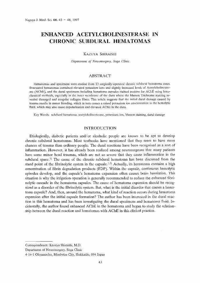
No. Age Sex Days until Hematoma labs resultsoperation K+ mEq AChE lUll T-bil. mg/dl LDH lUll
78 M 40 5.8 152 3.1 450
2 64 M 29 7.1 168 5.1 262
3 72 F 45 4.4 92 4.7 305
4 81 F 48 4.7 90 4.2 250
5 58 M 20 7.5 174 4.7 272
6 77 M 29 7.2 164 3.0 305
7 74 F 34 6.9 165 2.9 208
8 78 M 30 6.5 155 3.8 211
9 68 F 44 5.5 142 5.5 307
10 62 M 20 7.8 162 5.1 406
II 81 M 13 7.9 165 5.0 372
12 74 M 12 7.4 151 3.0 354
13 73 M 42 5.9 102 4.2 330
45
ENHANCED ACHE IN CHRONIC SUBDURAL HEMATOMAS
RESULTS
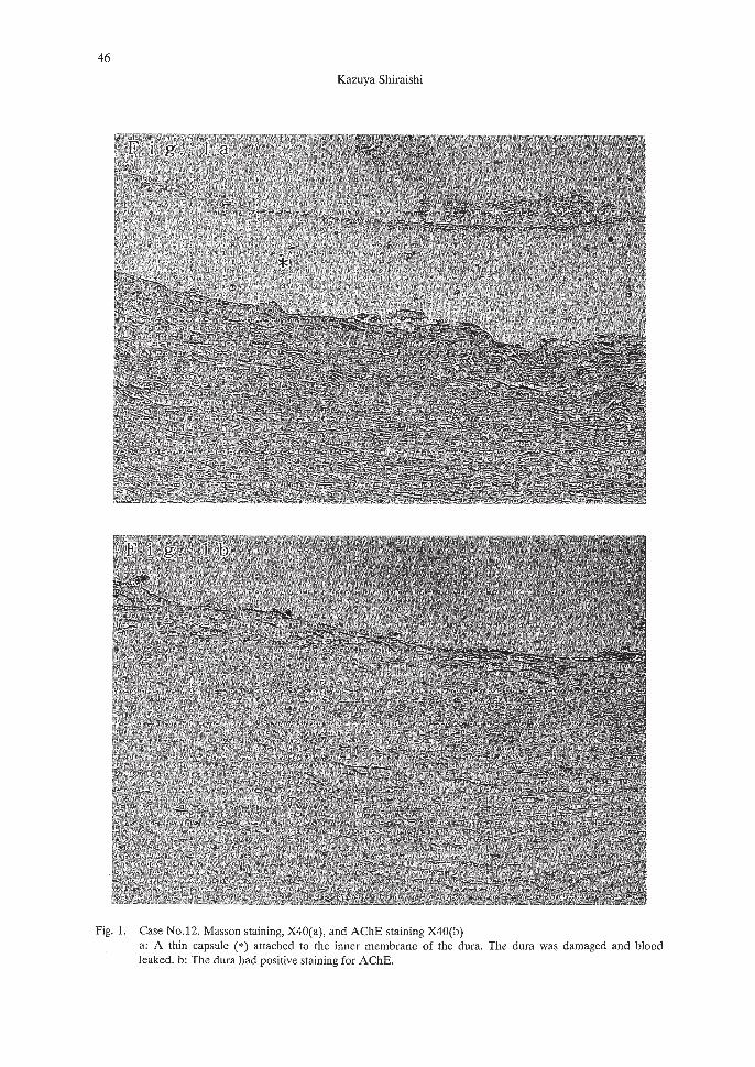
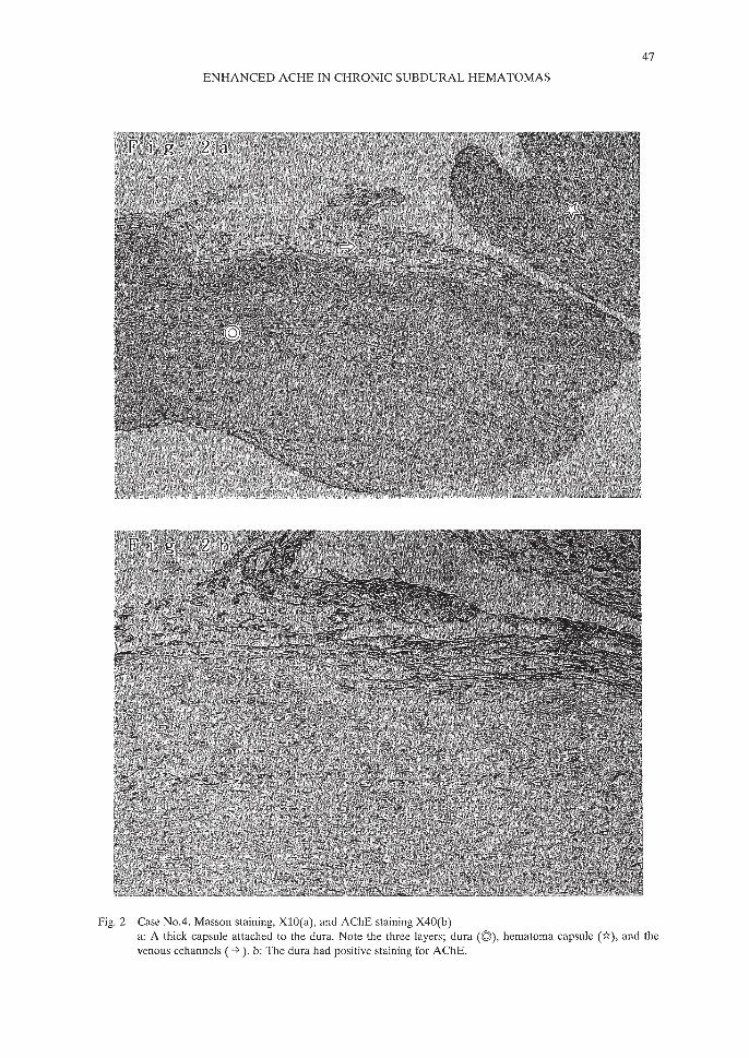
The table shows the data of the selected 13 cases. The characteristics of the hematoma analyses were high potassium (6.51 ± 1.10 mEq/l, 3.3-5.0), T-biI. (4.2 ± 0.9 mg/dl, 0.2-1.3),and a little higher AChE (144.8 ± 28.7 1U1I, 85-180), and normal LDH (310.1 ± 69.4 lUll,320-550) (average & S.D. ,N=13, normal range of this clinic). At the same time other itemswere measured and showed low titers; GOT (7.3 ± 3.1 lUll, 15-40), GPT (8.5 ± 3.0 lUll,10-50), T-ChoI. (20.8 ± 7.2 mg/dl, 130-220), and triglyceride (21.0 ± 5.5 mg/dl, 50-170)(titers, and the normal range of serum). Regarding the pathological study, all cases had AChEactivities in the dura. Figures 1 and 2 show some typical cases. Figure 1 is case No.12, andshows a thin capsule attached to the inner membrane of the dura, which has folded and partiallytorn collagen fibers as demonstrated by Masson staining. Figure 2 is case No.4, and had the longest trauma term among the study group. The case had a thick capsule consisting of venouschannels in the dura. Outside the venous channels, the inner membrane of the dura also showedcorrugated collagen fibers. Both cases demonstrate AChE activity in the dura parenchyma, especially in the inner membrane. AChE activities were positive in the dura of all cases, irrespective of dural thickness or the duration of the hematoma. Control specimens (without maleatebuffer incubation) showed no AChE staining.
DISCUSSION
AChE is known to develop not only in the cholinergic synapses but in the non-cholinergicsystems.5,6) It is one of non-specific enzymes in the central nervous system. In the peripheralnerves, AChE is a marker of axonal transport and with denervation its activities usually decrease.?) The author at first thought that the AChE activities of the hematoma would decreasebecause of dilution by the cerebrospinal fluid and the partial denervation to the dura caused bytrauma, but the data were quite different. Data shows that the fluid has a low content of hemoglobin (1.5 ± 1.1 g/dl), white blood cells (250 ± 120/mm3
), albumin (0.55 ± 0.20 g/dl), andblood sugar (65 ± 34 mg/dl) (N=20). The products related to fibrinolytic cascades, such asFDP, d,d-dimers, and bilirubins, are higher than normal serum. But incidentally, two years ago,the author found that AChE activities in the hematoma are a little higher than normal serum.Other biochemical items, such as GOT, GPT, total cholesterol and triglyceride, decreased in hematomas. In this study, the author has mentioned enhanced AChE in the hematoma and thedura, though the cause of it has not been well known. The secretion of AChE was reported tobe induced by depolarizing agents8) and, in the chronic subdural hematoma, the potassium ion isthe most likely agent because the initial hematoma does develop and the hemolysis cascade isthought to continue during the hematoma expansion. I ,2) Another possibility could be cellulardamage of the brain parenchyma. But the data did not show elevated LDH (one of intracellularproteins), so continuous cellular damage cannot be a plausible reason for enhanced AChE. Actually, in clinical practice, the brain parenchyma of the chronic subdural hematoma is known tohave no damage except compression.9) As Masson stainings showed, dural inner membrane haddamage in collagen fibers and there seemed to be leaking blood in the dural vessels.
Not only at the beginning of the traumatic changes, but in the chronic state, localized vasculardamage of the dura must have existed, after which damaged dura might have induced the production of potassium ions. The author also found cases which showed a granulation formationwithin the dura (data not shown). This fact indicates that the initiation of hematoma capsule canbe in the dura as the result of dural damage, which could induce small granulation in the dura
46
Kazuya Shiraishi
Fig. 1. Case No.12. Masson staining, X40(a), and AChE staining X40(b)a: A thin capsule (*) attached to the inner membrane of the dura. The dura was damaged and bloodleaked. b: The dura had positive staining for AChE.
47
ENHANCED ACHE IN CHRONIC SUBDURAL HEMATOMAS
Fig.2 Case No.4. Masson staining, XlO(a), and AChE staining X40(b)a: A thick capsule attached to the dura. Note the three layers; dura (©), hematoma capsule (*), and thevenous cchannels ( ~). b: The dura had positive staining for AChE.

48
Kazuya Shiraishi
where an elevated potassium ion concentration in turn might induce hemolyitc events in the granulation, accompanying by enhanced AChE in the early stages of the hematoma. Irrespective ofthe size or duration of the hematoma capsules, all cases had positive staining of AChE in thedura, which would support the above-described hypothesis. The hemolytic events and/or continuous activation of potassium ions seemed to be followed by thickened granulation. Repeatedhemolytic events and bleedings have been reported as major causes of hematoma expansion. 1.2)
From the stand point of clinical practice, it is not only necessary to block the hemolytic cascade but also to inhibit potassium ion production, for example, by using the anti-diabetic agent(Sulfonylurea) which might prevent the expansion of the hematoma in order to block the initialgranulation. Actually, diabetic patients suffer from more cases of this hematoma than normalpeople. The treatment of diabetes may be effective not only by decreasing blood sugar but alsoby preventing the initial granulation of chronic subdural hematomas. Controlled diabetic patients are known to have lower mortality than who are not treated.9) A newly developed potassium channel blocker for the dura might be the cure for chronic subdural hematomas.
REFERENCES
1) Ito, H., Komai, T. and Yamamoto, S.: Fibrinolytic enzyme in the lining walls of chronic subdural hematoma.J. Neurosurg., 48, 197-200 (1978).
2) Fujisawa, H., Ito, H., Saito, K., Ikeda, K., Nitta, H. and Yamashita, J.: Immunohistochemical localization ofTissue-type plasminogen activator in the lining wall of chronic subdural hematoma. Surg. Neurol., 35,441-445 (1991).
3) Tago, H., Kimura, H. and Maeda, T.: Visualization of detailed acetylcholinesterase fiber and neuron stainingin rat brain by a sensitive histochemical procedure. 1. Histochm. Cytochem., 34,1431-1438 (1986).
4) Meslam, M.M., Asuncion Moran, M.: Cholinesterases within neurofibrillary tangles related age and Alzheimer's disease. Ann. Neurol., 22, 223-228 (1987).
5) Layer, P.G.: Comparative localization of acetylcholinesterase and psuedocholinesterase during morphogenesis of the chicken brain. Proc. Acad. Natl. Sci. U.S.A., 80, 6413-6417 (1983).
6) Layer, P.G.: Cholinesterase during development of avian nervous system. Cell. Mol. Neurabial., 11, 7-33(1991).
7) Hassig, R., Tavitian, B., Pappalardo, F. and Di Giamberardino, L.: Axonal transport reversal of Acetylcholinesterase molecular forms in transected nerve. J. Neurachem., 57,1913-1920 (1991).
8) Biagioni, S., Bevilacqua, P., Scarsella, G., Vignoli, AL. and Augusti-Tocco, G.: Characterization of Acetylcholinesterase secretion in neuronal cultures and regulation by high K+ and soluble factors from targetcells. J. Neurachem., 64,1528-1535 (1995).
9) Cameron, M.M.: Chronic subdural hematoma: A review of 114 cases. J. Neural. Neurasurg. Psychiatry, 41,834-839 (1978).


![Teske - Anaemia [Compatibiliteitsmodus] · • bleeding (petechiae, ecchymoses, melena, hematuria, hematomas) • fever • splenomegaly Additional Clinical Symptoms: Anaemia Non-Regenerative](https://img.pdfslide.us/doc/110x75/5c8c870a09d3f2804e8c0316/teske-anaemia-compatibiliteitsmodus-bleeding-petechiae-ecchymoses.jpg)