Embed Size (px)
Citation preview
ENGLISH ONLY FINAL
Recommendations for the evaluation of animal cell cultures as substrates for the
manufacture of biological medicinal products and for the characterization of cell
banks
Proposed replacement of TRS 878, Annex 1
© World Health Organization 2010
All rights reserved. Publications of the World Health Organization can be obtained from WHO Press, World Health Organization, 20 Avenue Appia, 1211 Geneva 27, Switzerland (tel.: +41 22 791 3264; fax: +41 22 791 4857; e-mail: [email protected]). Requests for permission to reproduce or translate WHO publications – whether for sale or for noncommercial distribution – should be addressed to WHO Press, at the above address (fax: +41 22 791 4806; e-mail: [email protected]).
The designations employed and the presentation of the material in this publication do not imply the expression of any opinion whatsoever on the part of the World Health Organization concerning the legal status of any country, territory, city or area or of its authorities, or concerning the delimitation of its frontiers or boundaries. Dotted lines on maps represent approximate border lines for which there may not yet be full agreement. The mention of specific companies or of certain manufacturers’ products does not imply that they are endorsed or recommended by the World Health Organization in preference to others of a similar nature that are not mentioned. Errors and omissions excepted, the names of proprietary products are distinguished by initial capital letters.
All reasonable precautions have been taken by the World Health Organization to verify the information contained in this publication. However, the published material is being distributed without warranty of any kind, either expressed or implied. The responsibility for the interpretation and use of the material lies with the reader. In no event shall the World Health Organization be liable for damages arising from its use. The named authors alone are responsible for the views expressed in this publication.
Adopted by the 61st meeting of the WHO Expert Committee on Biological Standardization, 18 to 22 October 2010. A definitive
version of this document, which will differ from this version in editorial but not scientific details, will be published in the WHO
Technical Report Series.
Page 2
Recommendations published by the WHO are intended to be scientific and advisory. Each of the
following sections constitutes guidance for national regulatory authorities (NRAs) and for
manufacturers of biological products. If a NRA so desires, these Recommendations may be adopted
as definitive national requirements, or modifications may be justified and made by the NRA. It is
recommended that modifications to these Recommendations be made only on condition that
modifications ensure that the biological product is at least as safe and efficacious as that prepared in
accordance with the recommendations set out below. The parts of each section printed in small type
are comments for additional guidance intended for manufacturers and NRAs, which may benefit
from those details.
Table of contents
1. Introduction.....................................................................................................................................5
2. Historical overview .........................................................................................................................5
3. Scope.................................................................................................................................................8
4. Definitions........................................................................................................................................9
5. General considerations .................................................................................................................13
• Types of animal cell substrates
Primary cell cultures ......................................................................................................................................................... 13
Diploid cell lines................................................................................................................................................................ 14
Continuous cell lines ........................................................................................................................................................ 15
Stem cell lines .................................................................................................................................................................... 16
Potential risks and risk mitigation associated with biologicals produced In animal cell cultures.................................... 17
Viruses and other transmissible agents ............................................................................................................................. 17
Cellular DNA ................................................................................................................................................................... 19
Cellular RNA ..................................................................................................................................................................... 21
Growth-promoting proteins ............................................................................................................................................. 22
Part A. General recommendations applicable to all types of cell culture production................ 23
A.1 Good manufacturing practices................................................................................................................................... 23
A.2 Principles of good cell culture practices.................................................................................................................... 23
A.2.1 Understanding the cells and the culture system.................................................................................... 23
A.2.2 Manipulation of cell cultures ................................................................................................................ 24
A.2.3 Training and staff.................................................................................................................................. 25
A.2.4 Cell line development and cloning ........................................................................................................ 25
A.2.5 Special considerations for neural cell types.......................................................................................... 27
A.3 Selection of source materials .................................................................................................................................... 27
Page 3
A.3.1 Introduction........................................................................................................................................... 27
A.3.2 Serum and other bovine-derived materials used in cell culture media ................................................ 28
A.3.3 Trypsin and other porcine-derived materials used for preparing cell culture...................................... 30
A.3.4 Medium supplements and general cell culture reagents derived from other sources used for preparing
cell cultures .................................................................................................................................................... 31
A.4 Certifications of cell banks by the manufacturer ....................................................................................................... 32
A.4.1 Cell line data ......................................................................................................................................... 32
A.4.2 Certification of PCCs............................................................................................................................ 32
A.4.3 Certifications of DCLS, CCLs, and SCLs ............................................................................................. 33
A.5 Cryopreservation and cell banking............................................................................................................................ 33
A.5.1 Cryopreservation .................................................................................................................................. 33
A.5.2 Cell banking .......................................................................................................................................... 34
A.5.3 WHO reference cell banks .................................................................................................................... 35
Part B. Recommendations for the characterization of cell banks of animal cell substrates ......37
B.1 General considerations ............................................................................................................................................... 37
B.2 Identity ....................................................................................................................................................................... 39
B.3 Stability ....................................................................................................................................................................... 40
B.4 Sterility ....................................................................................................................................................................... 42
B.5 Viability ...................................................................................................................................................................... 42
B.6 Growth characteristics............................................................................................................................................... 42
B.7 Homogeneity .............................................................................................................................................................. 43
B.8 Tumourigenicity ......................................................................................................................................................... 43
B.9 Oncogenicity .............................................................................................................................................................. 48
B.10 Cytogenetics .............................................................................................................................................................. 50
B.11 Microbial agents ....................................................................................................................................................... 51
B.11.1 General considerations ...................................................................................................................... 51
B.11.2 Viruses................................................................................................................................................ 52
B.11.3 Bacteria, fungi, mollicutes and mycobacteria ................................................................................... 65
B.11.4 Transmissible Spongiform Encephalopathies .................................................................................... 67
B.12 Summary of tests for the evaluation and characterization of animal cell substrates ............................................... 69
B.12.1 Cell seed ............................................................................................................................................. 70
B.12.2 Master Cell seed (MCB) and Working Cell Bank (WCB) .................................................................. 70
Authors............................................................................................................................................... 71
Acknowledgements ........................................................................................................................... 73
References .......................................................................................................................................... 74
Page 4
Appendix 1.............................................................................................................................................
Tests for bovine viruses in serum used to produce cell banks................................ .......................................... 82
Appendix 2.............................................................................................................................................
Tumourigenicity protocol using athymic nude mice to assess mammalian cells.............................................. 86
Appendix 3 ......................................................................................................................................................................
Oncogenicity protocol for the evaluation of DNA and cell lysates.....................................................................90
Abbreviations ................................................................................................................................. .... 93
Page 5
1. Introduction Cell substrates are cells used to manufacture a biological product. It is well established that cell
substrates themselves and events linked to cell growth can affect the characteristics and safety of the
resultant biological products. Therefore, a thorough understanding of the characteristics of the cell
substrate is essential in order to identify points of concern and to develop a quality control system
that addresses those points.
Recent advances in the use and quality control of new animal cell substrates, particularly continuous
cell lines (CCLs) and insect cells, led to the conclusion that an update of the current WHO
Requirements (TRS 878) [ 1] should be prepared. In order to facilitate the resolution of regulatory /
scientific issues related to the use of animal cell cultures, including human, as substrates for the
production of biological products, WHO initiated this revision of its Requirements on cell substrates
by establishing a Study Group (SG). Animal cells refer to cells derived from organisms classified in
the animal kingdom. This document is the result of the SG effort, including wide consultations with
individuals and organizations with expertise in this area. After receiving comments from this
consultative process, as well as from invited reviewers, further revision of the draft
recommendations was undertaken and presented to the ECBS in 2010. During the development of
this document, guidances on this topic issued by other relevant organizations were considered.
Effort was made to be compatible with the existing guidances, whenever possible.
These recommendations provide guidance to National Regulatory Authorities (NRAs), National
Control Laboratories (NCLs) and manufacturers on basic principles and, in some cases, on detailed
procedures that are appropriate to consider in the characterization of animal cells that are proposed
for use in the manufacture of biological products. Although the decision-making authority lies with
the NRA, it is advisable that NCL experts on this topic be consulted.
2. Historical overview Historically, the major concerns regarding the safety of biological medicinal products manufactured
in animal cells have been related to the possible presence of microbial contaminants and, in some
cases, to the properties and components of the cells themselves such as DNA and proteins.
For example, in 1954, an experimental adenovirus vaccine was being developed, and human tumour
cells (HeLa) were rejected as the cell substrate in favour of ”normal” cells [ 2]. At that time,
relatively little was known about the biological mechanism(s) that lead to human cancer, so that the
risks to the recipients of a vaccine based on HeLa cells could not be assessed and quantified
scientifically. Although ”normal” cells were not defined, that decision led to the use of primary cell
cultures (PCCs) from animals such as monkeys, hamsters, and embryonated eggs for vaccine
research and development [ 3].
The first requirements for cell substrates were published by WHO in 1959 for the production of
inactivated poliomyelitis vaccine in PCCs derived from the kidneys of clinically healthy monkeys
[ 4]. Those requirements were revised and published in 1966 [ 5]. Subsequently, other PCCs were
used for the production of other viral vaccines.
In the 1960s, human diploid cells (HDCs) were developed and proposed as an alternative to primary
monkey kidney cell cultures for polio virus vaccine production as well as for other viral vaccines.
Page 6
The rationale for using HDCs was based on the ability to: a) cryogenically preserve the cells at low
population doubling levels (PDLs); b) establish and characterize cryopreserved banks of cells that
later could be expanded to provide a standardized source of cells for many decades; c) extensively
test recovered cells before use in vaccine production; and d) demonstrate that the cells were free
from detectable adventitious agents and that they were unable to form tumors when inoculated into
immunosuppressed animals. Thus, HDCs were normal by all of the then existing criteria. It was
argued that because HDCs were normal and could be standardized, tested, and used for many years,
they were a significant improvement over PCCs.
The pathway to acceptance of HDCs was difficult and lengthy, primarily because some members of
the scientific community believed that HDCs might contain a latent and unknown human oncogenic
agent, and that such a theoretical agent posed a risk to recipients of vaccines produced in HDCs.
Numerous conferences and discussions of new data eventually led to the acceptance of HDCs as a
substrate for viral vaccine production, and they continue to be used by many manufacturers for
various viral vaccines that have a long history of safety and effectiveness. The concept of a master
cell bank (MCB) and working cell bank (WCB) system and characterization of the cell substrate
were introduced during that period [ 6, 7].
Both our understanding of tumor cell biology and the technological tools that were available at that
time were much more limited than they are today. As a result, the proponents of using HDCs for
vaccine production based their argument that the cells were normal, and therefore safe to use, on
four points: a) freedom from detectable adventitious agents; b) the finite life of HDCs; c) the diploid
nature of HDCs; and d) the inability of HDCs to form tumors in various in vivo test systems.
In order to provide a high level of assurance that those four characteristics were stable, the initial lot-
release tests for each batch of a vaccine derived from HDCs included tests of the cell substrate for
adventitious agents, karyology, and tumourigenicity [ 8, 9]. The main question that was being
addressed by the routine use of tumourigenicity tests was whether or not the production cell culture
had undergone a contamination or transformation event such that it contained a mixture of "normal"
and tumourigenic cells. It was eventually agreed that tumourigenicity testing was not sensitive
enough to detect a low level of tumourigenic cells, and that it was wasteful of animals and time in
repeated testing of a cell line that had been well characterized and would be used in the context of a
cell-bank system. Therefore, tumourigenicity tests eventually were required only for the
characterization of a MCB (using cells at the proposed in vitro cell age for production or beyond) for
both HDCs and CCLs [ 10, 11].
In the 1970s, there was a clinical research need for more interferon alpha (IFNα) than could be
produced from primary human lymphocytes. In response to that need, human tumour cells
(Namalwa) grown in vitro were proposed as a cell substrate for the production of IFNα. The primary
concerns about the use of Namalwa cells were that they contained the Epstein-Barr virus (EBV)
genome integrated into the cellular DNA, and that either whole virus or DNA containing viral
elements could be transmitted to the recipients of the IFNα product. Nevertheless, by the end of the
1970s, regulatory agencies had allowed human clinical studies to commence, and the product was
eventually approved in several countries. Among the most important factors that contributed to those
decisions was the fact that IFN, as opposed to live viral vaccines, was not a replicating agent, and
IFNα was being used as a therapeutic rather than a prophylactic product, thus representing different
risk/benefit considerations. In addition, technology had advanced significantly so that IFNα could be
Page 7
highly purified and the purification process could be validated to demonstrate that EBV and cellular
DNA were undetectable in the final product, within the limits of the assays then available, which
permitted risk mitigation.
In the 1980s, advances in science and technology led to the development of recombinant DNA
(rDNA) derived proteins and monoclonal antibodies (MAbs). Animal cells with the capacity to
grow continuously in vitro (CCLs) were the substrates of choice for those products because of the
ease with which they could be transfected and engineered. Also, in contrast to PCCs and HDCs, they
grew rapidly to achieve a high density and expressed a variety of products at high concentrations.
Chinese hamster ovary (CHO) cell lines became widely used for rDNA products, and hybridomas of
various types were required for the production of MAbs. The use of such cells as substrates in the
manufacture of a large array of potentially important biological medicinal products raised safety
concerns once again. A scientific consensus emerged from numerous conferences that there are three
major elements of potential concern related to animal cell substrates: DNA, viruses, and
transforming proteins. In 1986, WHO established a SG to examine cell-substrate issues in greater
depth.
The SG concluded that there is no reason to exclude CCLs from consideration as substrates for the
production of biologicals, and that CCLs are in general acceptable when the manufacturing process
is shown to eliminate potential contaminating viruses pathogenic for humans and to reduce DNA to
acceptable levels and/or to eliminate its biological activity [ 12]. The SG’s emphasis on infectious
agents as the major risk factor was based in large part on actual experience in which virus
transmission and disease had occurred through contaminated biological products (e.g., hepatitis B
virus and human immunodeficiency virus (HIV) in Factor VIII). WHO Requirements for Continuous
Cell Lines used for Biologicals Production were published in 1987 [ 13]. Based on a review of more
recent data, those Requirements were revised in 1998 to raise the acceptable level of rcDNA to 10 ng
per parenteral dose. In addition, it was pointed out that beta-propiolactone, a viral inactivating agent,
may also destroy the biological activity of DNA. Use of this agent therefore provides an additional
level of confidence even when the amount of DNA per dose may be substantial [ 1].
During the 1990s, and on into the 2000s, a variety of CCLs were explored as cell substrates for
biological products in development because, like the cell lines already mentioned, they offered
significant advantages during production (e.g., rapid growth and high expression). These include the
following tumourigenic cell lines: HeLa for adeno-associated virus vectored HIV vaccines; PER.C6
for influenza and HIV vaccines; Madin-Darby Canine Kidney (MDCK) for influenza vaccines; and
293ORF6 for HIV vaccines. More recently insect cell lines and stem cell lines (SCLs) have been
proposed for the manufacture of biological products, and such cells introduce a new set of challenges
for their evaluation and characterisation.
The acceptability of a given cell type (primary, diploid, stem, or continuous) as a substrate for the
production of a specific biological product depends on a variety of factors including an in depth
knowledge of its basic biological characteristics. In that regard, it is important to recognize that the
tumourigenic potential of a CCL is but one of many factors to consider such as the extent to which
the manufacturing process reduces or eliminates cellular factors that may be of concern. An
assessment of the totality of the data available is needed in order to determine whether a product
manufactured in a given cell substrate is potentially approvable.
Page 8
The following recommendations provide guidance to manufacturers and NRAs/NCLs on the
evaluation of animal cell cultures used as substrates for the production of biological medicinal
products, and for the characterization of cell banks.
The main changes from the requirements published in TRS 878, annex 1, include:
1. general manufacturing recommendations applicable to all types of cell culture production
have been updated;
2. some considerations for the evaluation of new cell substrates such as insect cells and stem
cells (SCs) have been added;
3. definitions have been updated and expanded in number and scope, and moved to an earlier
point in the document;
4. the structure of the document has been modified to include more background information,
and the applicability of various sections to different types of cell substrates is highlighted;
5. a new section on risk reduction strategies during the manufacture of biological products has
been added;
6. a section on Good Cell Culture Practice has been added;
7. the section of selection of source materials has been updated, and the detailed methods used
to test for bovine viruses in serum were added in Appendix 1;
8. tumourigenicity testing has been updated, and a model protocol for the nude mouse model
was added in Appendix 2;
9. oncogenicity testing of tumourigenic cell lysates was added, and a model protocol was added
in Appendix 3;
10. recommendations for acceptable levels of residual cellular DNA are product specific and not
specifically addressed; and
11. recommendations for microbial agents testing have been updated.
3. Scope These recommendations supersede previous WHO requirements or recommendations describing
procedures for the use of animal cell substrates for the production of biological medicinal products
[ 1, 13].
Some of these recommendations also may be useful in the quality control of specific biological
products during the manufacturing process, but it is beyond the scope of this document to
recommend quality control release tests. Like-wise, risk-based assessments related to product
approvals are beyond the scope of this document. Requirements or recommendations for individual
products should be consulted in that regard.
Cells modified by recombinant DNA technology have been increasingly used in the manufacture of
novel medicinal products, and specific considerations for those products are addressed elsewhere
[ 1, 10, 14, 15]. Nevertheless, there are a number of generic issues that apply to genetically modified as
well as to other cell substrates.
These recommendations specifically exclude all products manufactured in embryonated eggs,
microbial cells (i.e., bacteria and yeast), and plant cells. Also excluded are whole, viable animal cells
Page 9
such as SCs when they are used directly for therapy by transplantation into patients or when they are
developed into SCLs for the purpose of using them as therapeutic agents by transplantation. In those
cases, characterization tests should be discussed with the NRA/NCL. However, SCLs used for the
production of biological products such as growth factors and vaccines should comply with these
recommendations.
Some of the general recommendations given here (see sections A.1 – A.5) are applicable to all
animal cell substrates. More specific guidance for PCCs can be found in the relevant documents
published by WHO (e.g., production of poliomyelitis vaccine in primary monkey kidney cells) [ 4, 5].
Cell substrates should be developed and used in accordance with applicable requirements of the
NRA/NCL.
In general, it is not a practice consistent with Good Manufacturing Practices to re-test materials that
have already been released for further manufacture, so justification would be necessary before such
re-testing is undertaken. Thus, the scope of this document is intended to cover cell substrates as new
cell banks are established. Specific circumstances under which re-testing of already established and
released cell banks would be appropriate should be discussed with the responsible NRA/NCL.
Recommendations published by WHO are intended to be scientific and advisory in nature. The parts
of each section printed in normal type have been written in the form of recommendations so that,
should a NRA/NCL so desire, they may be adopted as they stand as the basis of national or regional
requirements. The parts of each section printed in small type are comments or additional points that
might be considered in some cases.
4. Definitions The definitions given below apply to the terms used in these recommendations. They may have
different meanings in other contexts.
Adventitious agent: Contaminating microorganisms of the cell culture or source materials including
bacteria, fungi, mycoplasmas/spiroplasmas, mycobacteria, rickettsia, protozoa, parasites,
transmissible spongiform encephalopathies (TSE) agents, and viruses that have been unintentionally
introduced into the manufacturing process of a biological product.
The source of these contaminants may be from the legacy of the cell line,
the raw materials used in the culture medium to propagate the cells (in
banking, in production, or in their legacy), the environment, personnel,
equipment, or elsewhere.
Biological medicinal product: Biological medicinal product is a synonym for biological product or
biological described in WHO Technical Report Series. The definition of a medicinal substance, used
in treatment, prevention or diagnosis, as a "biological" has been variously based on criteria related to
its source, its amenability to characterization by physicochemical means alone, the requirement for
biological assays, or on arbitrary systems of classification applied by regulatory authorities. For the
purposes of WHO, including the present document, the list of substances considered to be
biologicals is derived from their earlier definition as "substances which cannot be fully characterized
by physicochemical means alone, and which therefore require the use of some form of bioassay".
Page 10
However, developments in the utility and applicability of physicochemical analytical methods,
improved control of biological and biotechnology-based production methods, and an increased
applicability of chemical synthesis to larger molecules, have made it effectively impossible to base a
definition of a biological on any single criterion related to methods of analysis, source or method of
production. Nevertheless, many biologicals are produced using in vitro culture systems.
Developers of such medicinal products that do not fit the definition of
biological medicinal product provided in this document should consult the
relevant NRAs for product classification and licensing application pathway.
Biotherapeutic: For the purpose of this document, a biotherapeutic is a biological medicinal product
with the indication of treating human diseases.
Cell bank: A cell bank is a collection of appropriate containers whose contents are of uniform
composition stored under defined conditions. Each container represents an aliquot of a single pool of
cells.
The individual containers (e.g., ampoules, vials) should be representative of
the pool of cells from which they are taken and should be frozen on the
same day by following the same procedure and by using the same
equipment and reagents.
Cell culture: The process by which cells are grown in vitro under defined and controlled conditions
where the cells are no longer organized into tissues.
Cell line: Type of cell population with defined characteristics that originates by serial subculture of a
primary cell population that can be banked.
Cloning and sub-coning steps may be used to generate a cell line. The term
cell line implies that cultures from it consist of lineages of some of the cells
originally present in the primary culture.
Cell seed: A quantity of well-characterized cells stored frozen under defined conditions, such as in
the vapour or liquid phase of liquid nitrogen in aliquots of uniform composition derived from a
single tissue or cell, one or more of which would be used for the production of a master cell bank.
Also referred to as a pre-MCB or seed stock. May be made under Good Manufacturing Practices
(GMP) conditions or under manufacturer’s research and development conditions.
Cell substrate: Cells used to manufacture a biological product.
The cells may be primary or cell lines, and may be grown in monolayer or
suspension culture conditions. Examples of cell substrates include primary
monkey kidney, MRC-5, CHO, and Vero cells.
Cells used to generate essential components of a final product, such as Vero
cells for the generation of “reverse genetics” virus for use in seeding
vaccine production, are considered to be “pre-production” cell substrates.
Whereas cells used to manufacture the bulk product (e.g., packaging cell
lines for gene therapy vectors; Vero cells for vaccine production; CHO cells
for recombinant protein expression) are considered to be “production” cell
substrates.
Continuous cell line (CCL): A cell line having an apparently unlimited capacity for population
doublings. Often referred to as "immortal" and previously referred to as "established".
Page 11
Diploid cell line (DCL): A cell line having a finite in vitro lifespan in which the chromosomes are
paired (euploid) and are structurally identical to those of the species from which they were derived.
While this definition is accurate for standard chromosome preparations, a
given human diploid cell line may contain genetic variations that will be
reflected in a Giemsa-banding pattern that differs from the standard. Gene
expression differences also may be found.
This definition is based on experience and current understanding of the in
vitro behavior of human cells that are not of stem cell origin.
DNA infectivity: The capacity of cellular DNA to generate an infectious virus following introduction
of that DNA into appropriate cells. The viral genome could be integrated or extrachromosomal.
Endogenous virus: A virus whose genome is present in an integrated form in a cell substrate.
Endogenous viruses are present in the genome of the original animal from which the cells were
derived. They may or may not encode an intact or infectious virus.
End of production cells (EOPC): Cells harvested at or beyond the end of a production (EOP) run. In some cases, production cells are expanded under pilot-plant scale or
commercial-scale conditions.
Extended cell bank (ECB): Cells cultured from the MCB or WCB propagated to the proposed in
vitro cell age used for production or beyond.
Functional integrity: The culture sustains the expected performance related to its intended use under
specified conditions (e.g., expression of secreted product at a consistent level; production of
expected yield of virus).
Immortalized: having an apparently unlimited capacity for population doubling.
Indicator cells: Cells of various species used in the in vitro adventitious agent test that are intended
to amplify adventitious viruses to promote their detection. Generally, this would include a human
diploid cell line, such as MRC-5, a monkey kidney cell line, such as Vero cells, and a cell line of the
same species and tissue as the cell bank. The purpose of these cell lines is to indicate a viral
infection of the cell bank either through observation of cytopathic effect during and after an
appropriate observation period or by hemadsorption and/or hemagglutination at the end of the
observation period. Thus, they are referred to as indicator cells. The cell bank may be analyzed on
such indicator cells either by co-cultivation or by passage of cell lysates or spent culture supernatants
from the cell bank onto the indicator cells.
In vitro cell age: Measure of time between thaw of the MCB vial(s) to harvest of the production
vessel measured by elapsed chronological time, by population doubling level of the cells, or by
passage level of the cells when subcultivated by a defined procedure for dilution of the culture.
Latent virus: A virus is considered to be latent when the viral genome is present in the cell without
evidence of active replication, but has the potential to be reactivated.
Master cell bank (MCB): A quantity of well-characterized cells of animal or other origin, derived
from a cell seed at a specific population doubling level (PDL) or passage level, dispensed into
multiple containers, cryopreserved, and stored frozen under defined conditions, such as the vapour or
liquid phase of liquid nitrogen in aliquots of uniform composition. The master cell bank is prepared
from a single homogeneously mixed pool of cells. In some cases, such as genetically engineered
cells, this may be prepared from a selected cell clone established under defined conditions.
Oftentimes, however, the MCB is not clonal. It is considered best practice that the master cell bank
is used to derive working cell banks.
Page 12
Oncogenicity: The capacity of an acellular agent such as a chemical, virus, viral nucleic acid, viral
gene(s), or a subcellular element(s) to cause normal cells of an animal to form tumours.
Oncogenicity is distinct from tumourigenicity (see Tumourigencity). The
tumours that arise in an oncogenicity test are of host origin whereas in a
tumorigenicity test, the tumors are derived from the inoculated cells.
Parental cells: Cells that are manipulated to give rise to a cell substrate. For hybridomas, it is typical
to also describe the parental cells as the cells to be fused.
Manipulation may be as simple as the expansion of a primary cell culture to
provide early passage cells, or a more complex activity such as developing a
hybridoma or transfected clone, and both processes would provide a cell
seed. The parental cells may refer to any stage prior to the preparation of the
cell seed. Examples of a parental cell are: WI-38 and MRC-5 at very early
passage; Vero at passage 121; and CHO before the introduction of a DNA
construct to produce a recombinant cell. In certain situations (e.g., myeloma
cells), there may be a lineage of identified stable parental clones, thus, the
term "parental cell" would normally refer to the cells used immediately
prior to generation of the "cell seed".
Passage: Transfer of cells, with or without dilution, from one culture vessel to another in order to
propagate them, and which is repeated to provide sufficient cells for the production process.
This term is synonymous with “subculture”. Cultures of the same cell line
with the same number of passages in different laboratories are not
necessarily equivalent because of differences in cell culture media, split
ratios, and other variables that may affect the cells. This is a more important
consideration for SCLs and CCLs than for DCLs. Population doubling is the
preferred method of estimating cell line age, and whenever possible, it
should be used instead of “passage”. However, it also may be appropriate to
quantify culture duration of CCLs by the number of subcultivations at a
defined seeding density at each passage or time in days.
Population doubling: A two-fold increase in cell number.
Population doubling level (PDL): The total number of population doublings of a cell line or strain
since its initiation in vitro. A formula to use for the calculation of "population doublings" in a single
passage is: number of population doublings = Log10 (N/No) X 3.33 where: N = number of cells in the
growth vessel at the end of a period of growth. No = number of cells plated in the growth vessel [ 16].
It is best to use the number of viable cells or number of attached cells for this determination.
Primary culture: A culture started from cells, tissues or organs taken directly from one or more
organisms. A primary culture may be regarded as such until it is successfully subcultured for the first
time. It then becomes a "cell line" if it can continue to be subcultured at least several times.
Production cell cultures: A collection of cell cultures used for biological production that have been
prepared together from one or more containers from the working cell bank or, in the case of primary
cell cultures, from the tissues of one or more animals.
Residual cellular DNA (rcDNA): cell substrate DNA present in the final product.
Specific pathogen free (SPF): Animals known to be free of specific pathogenic microorganisms and
reared in an environment that maintains that state. SPF animals usually are raised in biosecure
facilities, and their health status is monitored on an ongoing basis. The SPF status simply provides
Page 13
an assurance that the stock is not infected with the specified pathogens. SPF animals are not disease
free nor are they disease resistant. They may carry pathogens other than those from which they are
specified to be free.
Stem cell line: A continuous cell line generated from stem cells, rather than normal or diseased
differentiated tissue.
Transmissible Spongiform Encephalopathy (TSE): The transmissible spongiform encephalopathies
(TSEs) are a group
of fatal neurodegenerative diseases which include classical and variant
Creutzfeldt–Jakob disease (CJD), Gerstmann-Sträussler-Scheinker syndrome (GSS), fatal familial
insomnia (FFI), and Kuru in humans, bovine spongiform encephalopathy (BSE) in cattle, chronic
wasting disease (CWD) in mule deer and elk, and scrapie in sheep and goats.
Tumourigenicity: The capacity of a cell population inoculated into an animal model to produce a
tumour by proliferation at the site of inoculation and/or at a distant site by metastasis.
Tumourigenicity is distinct from Oncogenicity (see Oncogenicity).
WHO Reference cell bank: A cryopreserved stock of cells prepared from a single, homogeneous
pool of cells prepared under defined conditions and subjected to characterization tests. The purpose
of such a bank is to serve as a well-characterized cell seed for the preparation of master cell banks
that will be extensively characterized by manufacturers, and that has a high probability of meeting
these recommendations.
Working cell bank (WCB): A quantity of well-characterized cells of animal or other origin, derived
from the master cell bank at a specific PDL or passage level, dispensed into multiple containers,
cryopreserved, and stored frozen under defined conditions, such as in the vapour or liquid phase of
liquid nitrogen in aliquots of uniform composition. The working cell bank is prepared from a single
homogeneously mixed pool of cells. One or more of the WCB containers is used for each production
culture.
5. General considerations Types of animal cell substrates
Primary Cell Cultures (PCCs) PCCs have played a prominent role in the development of biology as a science, and of virology in
particular. Cultures of PCCs from different sources have been in worldwide use for the production of
live and inactivated viral vaccines for human use for many decades. For example, PCCs of monkey
kidney cells have been used for the production of inactivated and oral poliomyelitis vaccines since
the 1950s.
Major successes in the control of viral diseases, such as poliomyelitis, measles, mumps and rubella,
were made possible through the wide use of vaccines prepared in PCCs, including those from
chicken embryos and the kidneys of monkeys, dogs, rabbits and hamsters, as well as other tissues.
PCCs are viable cells of disaggregated tissues that are initiated as in vitro cell cultures usually as
adherent cells. Many cell types will be present and a primary culture may be a complex mixture of
cells that can be influenced by the process and conditions under which they were harvested,
disaggregated and introduced to in vitro culture. Not all cells in a primary culture will have the
capacity to replicate. Particular care should be applied to establishing highly reproducible procedures
Page 14
for tissue disaggregation, cell processing and culture initiation and reproducible culture conditions
and nutrition.
PCCs obtained from wild animals usually show a high frequency of viral contamination. For
example, monkey-kidney cell cultures can be contaminated with one or more adventitious agents,
including simian viruses.
If PCCs are necessary for the production of a given biological, then the frequency of contaminated
cell cultures can be significantly reduced by careful screening of the source animals for the absence
of such viruses. Viruses can be detected by molecular tests such as PCR, and by looking for the
presence of circulating antibodies to those viruses in the source animals. The use of animals bred in a
carefully controlled colony, especially those that are specific-pathogen-free, is strongly
recommended. Nevertheless, as suitable alternative cell substrates become available, PCCs are less
likely to be used in the future. WHO has promoted the replacement of animals for experimental
purposes both from an ethical perspective [ 17] and in the interests of progressive improvement in
product safety and quality.
Advantages: (a) they are comparatively easy to prepare using simple media and bovine serum; and
(b) they generally possess a broad sensitivity to various viruses, some of which are cytopathic.
Disadvantages: (a) contamination by infectious agents is a higher risk than with DCLs and CCLs;
(b) the quality and viral sensitivity of cultures obtained from different animals are variable; and (c)
although cell cultures derived from nonhuman primates had been in wide use in the past, it has
become increasingly difficult to obtain and justify the use of such animals for this purpose, (d) they
cannot be tested as extensively as DCLs or CCLs.
Diploid Cell Lines (DCLs) The practicality of using human DCLs for the production of viral vaccines was demonstrated in the
1960s. The experience gained with oral poliomyelitis and other viral vaccines in successfully
immunizing billions of children in many countries has shown clearly that such substrates can be used
in the production of safe and effective vaccines [ 3].
The essential features of DCLs of human (e.g., WI-38, MRC-5) and monkey (i.e., FRhL-2) origin
are: (a) they are cells passaged from primary cultures that have become established as cell lines with
apparently stable characteristics over numerous PDLs; (b) they have a finite capacity for serial
propagation, which ends in senescence, a state in which the culture ceases to replicate, the cells
remain alive and metabolically active, but may show morphological and biochemical changes some
of which begin to appear before replication ceases; (c) they are non-tumourigenic; and (d) they
display diploid cytogenetic characteristics with a low frequency of chromosomal abnormalities of
number and structure until they enter senescence. Substantial experience since the 1960s has been
accumulated on the cytogenetics of WI-38 and MRC-5, and ranges of expected frequencies of
chromosomal abnormalities have been published [ 18, 19]. Similar data are available for FRhL-2 [ 20].
More sophisticated cytogenetic techniques (e.g., high resolution banding, comparative genome
hybridisation) [ 21, 22] have demonstrated the presence of subtle chromosomal abnormalities that
were previously undetectable. Recent studies have shown that subpopulations of human DCLs with
such abnormalities may appear and disappear over time, and that they are non-tumourigenic and
Page 15
undergo senescence in the same manner as the dominant population. Thus, possessing a stable
karyotype might not be such an important characteristic as was previously thought.
Advantages: (a) they can be well characterized and standardized; (b) production can be based on a
cryopreserved cell-bank system that allows for consistency and reproducibility of the reconstituted
cell populations. A cell-bank system usually consists of cell-banks of defined population doubling or
passage levels that generally include a MCB and a working cell-bank (WCB); and (d) unlike the
CCLs and SCLs discussed below, DCLs are not tumourigenic and therefore do not raise the potential
safety issues associated with CCLs and SCLs.
Disadvantages: (a) they are not easy to use in large-scale production, although they have been
cultivated using bioreactor technology employing the microcarrier or multilayer method; (b) in
general, they have more fastidious nutritional requirements than other cell substrates; (c) they may
be difficult to adapt to serum-free growth; and (d) they are more difficult than CCLs to transfect and
engineer, and require immortalization before they can be engineered; (e.g., they are not permissive
for the production of vaccine vectors that require complementation, since they cannot be engineered
readily to express complementing proteins).
Continuous cell lines (CCLs) Some CCLs have been used for the production of safe and effective biotherapeutics and vaccines
since the 1980s.
CCLs have the potential for an apparently indefinite in vitro life span and have been derived by the
following methods: (a) serial subcultivation of a PCC of a human or animal tumour (e.g., HeLa
cells); (b) transformation of a normal cell having a finite life span with an oncogenic virus or viral
sequence (e.g., B lymphocytes transformed by EBV or transfected with viral sequences such as in
PER.C6); (c) serial subcultivation of a primary-cell population derived from normal tissue that
generates a dominant cell population having an apparently indefinite life span, often described as
spontaneous transformation (e.g., Vero, BHK-21, CHO, MDCK, Hi5); (d) fusion between a
myeloma cell and an antibody-producing B lymphocyte to produce a hybridoma cell line; or (e) use
of ectopically expressed genes involved in the cell cycle such as hTERT telomerase gene to enable
indefinite replication of normal human cells.
CCLs may display a consistent modal chromosome number (e.g., MDCK, Vero), and although the
karyotype of individual cells in a culture at any one time-point may vary, the range of chromosome
numbers per cell will usually show characteristic limits. However, other CCLs, such as highly
tumourigenic cells including HeLa may show variation in modal number and a wider drift in the
range of the number of chromosomes per cell.
In the early stage of establishing a cell line, significant diverse karyotypes and changes in karyotype
may be observed, but a characteristic typical chromosome component may emerge with continued
passage presumably as a dominant cell population develops.
Advantages: (a) they can be characterized extensively and their culture conditions standardized; (b)
production can be based on a cell-bank system, which allows consistency and reproducibility of the
reconstituted cell populations for an indefinite period; (c) as a rule, they grow more easily than
Page 16
DCLs using standard media, (d) most can be adapted to grow in serum-free medium; (e) they usually
can be grown on microcarriers for large-scale production in bioreactors; and (f) some can be adapted
to grow in suspension cultures for large-scale production in bioreactors.
Disadvantages: (a) CCLs may express endogenous viruses, and some are tumourigenic in
immunosuppressed animal models; (b) theoretical risks identified by the 1986 Study Group (e.g.,
nucleic acids, transforming proteins, and viruses) need to be taken into account.
Stem Cell Lines (SCLs) SCs differ from other types of cells because they sustain a predominant stem-cell population whilst
simultaneously retaining the capacity to produce cell progenitors of differentiated cell types of
almost all human tissues (i.e., pluripotent). Pluripotent SCLs have an apparent capacity to generate
cell types of all three human germlayers and may be capable of generating in vitro models of any
tissue in the human body. At the time these recommendations are written (2010), two types of
pluripotent SCLs, human embryonic stem cells and induced pluripotency stem cell lines have been
isolated which may have the capabilities to prove useful for manufacturing biologicals. This property
of pluripotency is sustained through numerous cycles of cell division. SCLs may be derived from
early stage embryonic, fetal or adult tissues. Typically, specialized media and environmental
conditions such as the attachment matrix are required for the growth of SCLs in vitro in order for
them to maintain the undifferentiated state. While most stem cell research and development has been
directed towards transplantation of SCs for therapeutic purposes, efforts also have gone into
exploring a variety of SCLs as cell substrates for the production of biologicals.
Key considerations for the culture and control of such cell lines have been developed [ 23]. These
include the fundamental issues common to the maintenance of all cell lines, but also emphasize the
need for appropriate ethical governance regarding donor consent and careful attention to periodic
confirmation of phenotype, absence of non-diploid cells, and sustained pluripotent capacity.
Recently, it has been shown that conditioned medium from SCLs can have regenerative properties,
and such preparations produced from human embryonic SCs have shown regenerative capabilities
including repair of myocardial infarction in animal models [ 24]. This raises the possibility of SCs
being used as a substrate to produce a variety of biologically active molecules. SCLs can, in some
respects, be considered as diploid cells, but they do not appear to have the finite life span
characteristic of human diploid fibroblast cultures. In human embryonic stem-cell cultures, clonal
variants with chromosomal abnormalities are known to arise. Whilst a diploid and non-transformed
nature is to be considered a pre-requisite for cell therapy applications, transformed SCLs might be
considered as a form of CCL for the manufacture of biologicals. Because they do not fall easily
within any one category of substrate already discussed, SCLs are identified separately in this
document.
Advantages
(a) they can be well characterized and their culture conditions standardized; (b) production can be
based on a cell-bank system, which allows consistency and reproducibility of the reconstituted cell
populations for an indefinite period; and (c) some may be adapted to grow in suspension cultures for
large-scale production in bioreactors; (d) they may produce unique proteins of potential importance
Page 17
as biotherapeutics; and (e) they have the potential to generate cells and tissue-like structures that
may permit the expression of agents currently considered "unculturable" in vitro.
Disadvantages
(a) Subculture techniques commonly used for SCLs are laborious; (b) may produce growth proteins
with undefined effects on adult cells/tissues; (c) usually require complex media that may have a TSE
risk; (d) rapid development of differentiated cells also means that they are difficult to control in
vitro; (e) there is little experience with their use as a cell substrate to manufacture biological
products.
Potential risks and risk mitigation associated with biologicals produced in animal cell cultures
The main potential risks associated with the use of biologicals produced in animal cells are directly
related to contaminants from the cells, and they fall into three categories: (a) viruses and other
transmissible agents; (b) cellular nucleic acids (DNA and RNA); and (c) growth-promoting proteins.
In addition, cell-derived inhibiting or toxic substances are theoretically possible. A summary of the
risk assessment for each follows. More comprehensive information has been published elsewhere on
the risks associated with contaminating DNA and growth-promoting proteins
[ 25, 26, 27, 28, 29, 30, 31, 32, 33, 34].
Early in 2010 NRA as well as WHO were made aware of new information regarding the presence of
DNA sequences of porcine circovirus in live attenuated rotavirus vaccines. The detection of these
sequences by the use of advanced analytical methods raised complex questions, e.g., the evaluation
of the potential risk, specific testing of vaccines and the general use of these methods for the
characterization of vaccine cell substrates. The power of the new methodology that was used (i.e.,
massively parallel (deep) sequencing) may uncover the presence of adventitious agents that might
not be detected with current methods. While the implementation for routine use of such methods has
benefits as well as challenges and risks, NRAs need to be prepared for similar situations.
Considerations for making a risk assessment and potentially introducing risk mitigation strategies
may need to be undertaken in such circumstances.
Viruses and other transmissible agents There is a long history of concern regarding the potential transmission of viruses and other infectious
agents that may be present in cell substrates. This area was reviewed most recently by WHO in 1986
by a SG who pointed out that, as described below, cells differ with respect to their potential for
carrying viral agents pathogenic for humans.
Primary monkey kidney cells have been used to produce billions of doses of poliomyelitis vaccines
since they were first developed in the 1950s, and although viruses such as SV40 were discovered in
rhesus monkey kidney cells, control measures were introduced to eliminate or reduce as much as
possible the risk of viral contamination associated with the manufacture of vaccines in cells
containing those viruses. Additional controls may be needed as new viral agents are identified and
technologies to detect them are developed.
Human and nonhuman primate lymphocytes and macrophages may carry latent viruses, such as
herpesviruses and retroviruses. CCLs of non-haematogenous cells from human and nonhuman
Page 18
primates may contain viruses or have viral genes integrated into their DNA. In either case, virus
expression may occur under in vitro culture conditions.
Avian tissues and cells may harbour exogenous and endogenous retroviruses, but there is no
evidence for transmission of disease to humans from products prepared using these substrates. For
example, large quantities of yellow fever vaccines were produced for many years in eggs that
contain avian leukosis viruses, but there is no evidence that these products have transmitted disease
in their long history of use for human immunization. Nevertheless, the potential for transmitting
avian retroviruses should be reduced as much as possible through manufacturing control measures.
Rodents may harbour exogenous and endogenous retroviruses, lymphocytic choriomeningitis virus,
and Hantaviruses, and a range of other potentially zoonotic viruses. While contamination with the
rodent viruses in the cell harvests of biotherapeutic products derived from CHO cell culture has been
reported [ 34, 35, 36], there is no evidence that biological products released for distribution have been
contaminated with rodent viruses, because, if present, they were detected during quality control
testing in compliance with GMPs prior to release. In addition, it is important to note that there have
been no reported cases of transmission of an infectious agent to recipients of recombinant protein
products manufactured in animal cells.
Insect cells recently have been used for vaccine production, and various insect cell lines may be used
for the production of biologicals in the future. Insect viruses tend to be ubiquitous in many insect cell
lines, and are generally unknown and/or uncharacterized. Many insect cell lines have endogenous
transposons and retrovirus-like particles, and some are positive in PERT assays.
HDCs have been used for vaccine production for many years, and although concern was initially
expressed about the possibility of such cells containing a latent pathogenic human virus, no evidence
for such an endogenous agent has been reported, and vaccines produced from this class of cell
substrate have proven to be free from viral contaminants.
In light of the differing potential of the various types of cells mentioned above for transmitting
viruses that are pathogenic in humans, it is essential that the cells being used to produce biological
products be evaluated as thoroughly as possible with respect to infectious agents.
When DCLs, SCLs, or CCLs are used for production, a cell bank system should be used and the cell
banks should be characterized as specified in this document. Efforts to identify viruses by testing for
viral sequences or other viral markers, especially those not detectable by other means, constitute an
important part of the evaluation of cell banks in addition to the standard tests that have been in place
for many years.
When cell lines of rodent or avian origin are examined for the presence of viruses, the major
emphasis in risk assessment should be placed on the results of studies in which transmission to target
cells or animals is attempted. Risk to human recipients should not be assessed solely on
ultrastructural or biochemical/biophysical evidence of the presence of viral or viral-like agents in the
cells.
Page 19
The overall manufacturing process, including the selection and testing of cells and source materials,
any purification procedures used, and tests on intermediate or final products, should ensure the
absence of detectable infectious agents in the final product. When appropriate, validation of
purification procedures should demonstrate adequate reduction of relevant model viruses with a
significant safety factor [ 14]. This is usually required for recombinant protein products.
There may be as yet undiscovered microbial agents for which there is no current evidence or means
of detection. As such agents become identified, it will be important to consider whether to re-
examine cell banks for their presence. In general, it is not a practice consistent with GMPs to re-test
materials that have already been released, so justification would be necessary before such re-testing
is undertaken. Positive findings should be discussed with the NRA/NCL. Whenever new data are
developed with the potential for an impact on the quality, safety or efficacy of a biological product,
it is the responsibility of the manufacturer to provide NRAs all relevant data and information
currently available. This should include confirmation and evaluation of the finding, the
manufacturer’s own risk assessment as well as an investigational and action plan, in order to
facilitate any regulatory action that might be necessary. In addition, new testing methods are likely
to be developed, and as they become available and validated, they should be considered by
manufacturers and NRAs/NCLs for their applicability to the characterization and control of new
animal cell substrates.
Cellular DNA The issue of rcDNA in biological products has been considered by many groups since the 1980s, and
there has been an evolution of consensus on recommendations during that period. The most recent
WHO recommendation (TRS 878) sets the upper limit of rcDNA at 10 ng per parenteral dose. As
stated below, while this value has proved helpful in the past, it does not take into consideration
important factors such as the size of the DNA fragments and any potentially inactivating steps in the
manufacturing process. Therefore, the amount of rcDNA that might be acceptable for a specific
product should take into consideration not only the limit of 10 ng per parenteral dose, but other
factors as well when determining the acceptable level of rcDNA.
PCCs and DCLs have been used successfully for many years for the production of viral vaccines,
and the rcDNA deriving from these cells has not been (and is not) considered to pose any significant
risk. However, with the use of CCLs, which have an apparently indefinite life span, presumably due
to the dysregulation of genes that control growth, and with the ongoing development of products
from cells that are tumourigenic or were derived from tumors, the DNA from such cells has been
considered to have the theoretical potential to confer the capacity for unregulated cell growth, and
perhaps oncogenic activity, upon some cells of a recipient of the biological product. Although the
risk of such DNA has been estimated based on certain assumptions and some experimental data,
assessing the actual risk of such DNA has not been possible until recently when preliminary data
generated from new experimental systems began to quantify those risks [ 37].
The potential risks of DNA arise from both of its biological activities: (a) infectivity, and (b)
oncogenicity. Infectivity could be due to the presence of an infectious viral genome in the cellular
DNA of the cell substrate [ 38, 39, 40]. The viral genome could be that of a DNA virus, whether
integrated or extrachromosomal, or that of a proviral genome of a retrovirus. Both types of viral
DNA have been shown to be infectious in vitro and in several cases, in vivo [ 37, 38, 39]. The
Page 20
oncogenic activity of DNA could arise through its capacity to induce a normal cell to become
transformed and perhaps to become tumourigenic. The major mechanism through which this could
occur would be through the introduction of an active dominant oncogene (e.g., myc, activated ras),
since such dominant oncogenes could directly transform a normal cell. Other mechanisms require
that the rcDNA transform through insertional mutagenesis, and have been considered less likely,
since the frequency of integration of DNA in general is low [ 40]. The frequency of integration at an
appropriate site, such as inactivating a tumor suppressor gene or activating a proto-oncogene, would
be correspondingly lower [ 31].
The 1986 WHO SG addressed the risk posed by the oncogenic activity of rcDNA in biological
products for human use [ 12]. Risk assessment based on a viral oncogene in an animal model
suggested that in vivo exposure to one nanogram (ng) of rcDNA, where 100 copies of an activated
oncogene were present in the genome, would give rise to a transformational event once in 109
recipients [ 26]. On the basis of this and other evidence available at that time, the 1986 SG concluded
that the risk associated with rcDNA in a product is negligible when the amount of such DNA is 100
picograms (pg) or less per parenteral dose. Based on a review of more recent data, those
requirements were revised in 1998 to raise the acceptable level of rcDNA to 10 ng per dose.
Studies in mice using cloned cellular oncogenes also suggest that the risk of neoplastic
transformation by cellular DNA is probably very low [ 33, 42]. However, more recent data have
shown that cloned cellular oncogene DNA can induce tumors in selected strains of mice at levels
below 1 ng. In addition, single oncogenes can also be biologically active [ 42] and initiate the tumour
induction process. Because of these data, and the recent description that genes encoding for certain
micro-RNA species can be oncogenic in vitro [ 44, 45, 46, 47], thus increasing the number of potential
dominant cellular oncogenes, the oncogenic risk of DNA needs to be considered when tumourigenic
cells are considered for use in the production of biologicals. This would be especially important for
live, attenuated viral vaccines where chemical inactivation of the DNA is not possible, and the only
way the biological activity of DNA could be reduced would be by nuclease digestion and the
reduction in the quantity of DNA.
In addition to its oncogenic activity, the infectivity of DNA should be considered. Since a viral
genome once introduced could amplify and produce many infectious particles, the infectivity risk is
likely greater than the oncogenic risk. The polyoma virus genome is infectious in mice at about 50
pg [ 48], and a recent report demonstrated that 1 pg of a proviral copy of a retrovirus is infectious in
vitro [ 49]. Because such low levels of DNA may be biologically active, the amounts of rcDNA
should be factored into safety evaluations when tumourigenic cell substrates are used, especially for
live viral vaccines.
Therefore, considerations that need to be taken into account with respect to rcDNA are: (a) any
reduction in the amount of the contaminating DNA during the manufacturing process; (b) any size
reduction of the contaminating DNA during the manufacturing process; and (c) any chemical
inactivation of the biological activity of contaminating DNA during the manufacturing process. A
product might be considered by a NRA/NCL to have an acceptable level of risk associated with the
DNA of the cell substrate on the basis of (a) and/or (b) and/or (c), when data demonstrate that
appropriate levels have been achieved. For example, data have shown that nuclease digestion of
DNA or chemical inactivation of DNA with beta-propiolactone, a viral inactivating agent, can
Page 21
destroy the biological activity of DNA [ 37, 49, 50]. Therefore, the use of these procedures may
provide an additional level of confidence with respect to DNA risk reduction.
For products such as monoclonal antibodies and subunit vaccines manufactured in tumourigenic cell
substrates, it is necessary to demonstrate the clearance (removal and/or inactivation) of DNA by the
manufacturing process, which may require the validation of the main inactivating or removal steps.
For example, data should be obtained on the effects of DNA-inactivating agents under specific
manufacturing conditions so that firm conclusions on their DNA-inactivating potential for a given
product can be drawn.
There may be instances where CCL DNA is considered to pose a higher level of risk because it
contains specific elements such as infectious retroviral proviral sequences. Under these
circumstances, the steps taken to reduce the risks of rcDNA, such as reducing the size of DNA
fragments, should be set in consultation with the NRA/NCL.
The 1986 WHO SG stated that the risks for rcDNA should be considered negligible for preparations
given orally. This conclusion was based on the finding that polyoma virus DNA was not infectious
when administered orally [ 48]. For such products, the principal requirement is the elimination of
potentially contaminating viruses. Recently, additional data on the uptake of DNA via the oral route
have been published [ 51]. These studies demonstrated that the efficiency of uptake of DNA
introduced orally was significantly lower than that introduced intra-muscularly. Nevertheless, the
specifics of the manufacturing process and the formulation of a given product should be considered
by the NRA/NCL.
With respect to the efficiency of DNA uptake via the nasal route, no data have been published
comparing the nasal route with parenteral routes. However, data presented publically show that
uptake via the intranasal route is less efficient than by the intramuscular route. Limits for a specific
product should be set in consultation with the NRA/NCL.
In general, acceptable limits of rcDNA for specific products should be set in consultation with the
NRA/NCL taking into consideration the characteristics of the cell substrate, the intended use of the
biological product, and most importantly the effect of the manufacturing process on the size,
quantity and biological activity of rcDNA fragments. In general, it has been possible to reduce
rcDNA in biotechnological products to <10 ng per dose, and in many cases <10 pg per dose, because
they can be highly purified. Quantitative PCR for short amplicons has been used to determine total
residual DNA levels as well as residual DNA fragment size distribution. It should be noted that other
methods may give different results for small fragments or for DNA that has been treated with
inactivating agents. Whatever methods are used should be validated. Some products, especially
certain live viral vaccines, are difficult to purify without a significant loss in potency, so that the
amount of rcDNA in those final products may be significantly higher than 10 ng per dose. Such
cases are considered to be exceptional and should be discussed with the NRA/NCL.
Cellular RNA While protein-coding RNA has not been considered to be a risk factor for biological products due to
the unstable nature of RNA and the lack of mechanisms for self-replication, the recent description of
small non-coding RNA molecules – microRNA (miRNA) – that are more stable and have the
Page 22
capacity to modulate gene expression might necessitate a reassessment. Whether these miRNA
molecules can be taken up by cells in vivo is unknown. However, as stated above, because certain
miRNA genes can be oncogenic, DNA containing such sequences may need to be considered along
with oncogenes when assessing the risk of rcDNA (see B.9 Oncogenicity). However, because this is
an evolving area of research, no conclusions can be made regarding the risk of miRNA, and no
recommendations are made to control miRNA at this time.
Growth-promoting proteins Growth factors may be secreted by cells used to produce biologicals, but the risks from these
substances are limited, since their growth-promoting effects are usually transient and reversible, they
do not replicate, and many of them are rapidly inactivated in vivo. In exceptional circumstances,
growth factors can contribute to oncogenesis, but even in these cases, the tumours apparently remain
dependent upon continued administration of the growth factor. Therefore, the presence of known
growth-factor contaminants at ordinary concentrations does not constitute a significant risk in the
preparation of biological products manufactured in animal-cell cultures. However, some SCLs may
secrete higher levels and more potent factors than CCLs. This should be taken into account when
designing characterization studies, and the manufacturing process should be designed to address any
safety issues that are identified.
Page 23
Part A. General recommendations applicable to all types of cell culture
production
A.1 Good manufacturing practices
The general principles of GMP for biologicals should be in place. Requirements or recommendations
have been made by NRAs (e.g., EMA, FDA) and other groups (e.g., ICH). GMP should be applied
from the stage of cell banking.
In the preparation of a cell substrate, it is considered best practice to establish tiered master and
working cell banks (A.5.3) to ensure a reliable and consistent supply of cells that can be fully
characterized and safety tested prior to use for production. By definition, primary-cell cultures
cannot be subjected to such banking regimens. However, some manufacturers have utilized pooled
and cryopreserved primary cultures, which enable completion of lot-release testing as in a tiered
banking system. The strategy for delivery of primary cells or primary cells recovered from
cryopreservation should be based on the quality and safety that can be assured for the final product
according to the overall manufacturing and control processes involved.
A.2 Principles of Good Cell Culture Practices A.2.1 Understanding the cells and the culture system
In all aspects of sourcing, banking and preparing cell cultures, the principles of Good Cell Culture
Practices (GCCPs) should be observed (for example, 52, 53 for SCLs). Fundamental features to be
considered in the development of cell cultures for production or testing are:
1. authenticity, including identity, provenance, and genotypic/phenotypic characteristics
2. absence of contamination with another cell line
3. absence of microbiological contamination, and
4. stability and functional integrity on extended in vitro passage
An important basic principle for all types of cells is that the donor should be free of transmissible
diseases or diseases of uncertain etiology, such as CJD for humans and BSE for cattle. The
NRA/NCL may allow specific exceptions concerning donor health (e.g., myeloma, other tumour
cells).
Cells in culture may change their characteristics in response to changes in culture conditions or on
extended passage under the same culture conditions. The four cell culture types (PCC, DCL, SCL,
CCL) used in manufacture differ in their potential stability, and characterization approaches may
need to be adapted in reflection of these differences.
Cell cultures grow in an in vitro environment that is substantially different from the conditions
experienced by cells in vivo, and it is not unexpected that they may be susceptible to change or
alteration as a result of in vitro culture and processing. It is important to be conscious of the variation
that may arise in the cell-culture environment, as cells may undergo subtle alterations in their cell
biology in response to such changes. It is therefore necessary to try to control key known variables
that could have significant impact on the cell culture. Medium and specific additives (serum, growth
factors, amino acids and other growth promoting compounds) should, where possible, be specified in
Page 24
terms of chemical composition and purity. Where relevant, biological activity of the medium and
additives should be determined before use. New batches of reagents for cell culture should be
supplied with certificates of analysis and origin, which enable their suitability to be evaluated against
the established specification. The use of serum or other poorly defined reagents is not recommended
in the production of new biologicals from cell culture, and wherever possible chemically defined
alternatives should be sought. However, given that our current understanding of cell biology is not
complete, there is a balance to be made between the benefits that defined media bring in the form of
higher reproducibility and reduced risk, and the potential effects of inadequacies of defined-culture
systems that may not meet the full biological needs of cells. Where complex biological reagents,
such as FBS remain necessary, they should be carefully controlled, whenever possible, by pre-use
selection of batches. Such careful selection also should apply to cell-culture surfaces using specified
culture vessels or surface coatings where relevant.
Variation in physical-culture parameters, such as pH, temperature, humidity, and gas composition,
can significantly influence the performance and viability of cells and should be specified with
established tolerances and the relevant equipment calibrated and monitored. In addition, any culture
reagents prepared in the laboratory should be documented, quality controlled and released against an
established specification.
A.2.2 Manipulation of cell cultures
In vitro processing of cells can introduce additional physical and biochemical stresses that could
have an influence on the quality of the final product. Care should be taken to minimize
manipulations, taking into consideration the specifics of the manufacturing process. In all cases, a
consistent process should be demonstrated.
A.2.2.1 Detachment and subculture
Detachment solutions may adversely affect the cells if exposure is not minimized. Cell harvesting
and passaging procedures should be carried out in a reproducible way ensuring consistency in the
confluency of cells when harvested, incubation times, temperature, centrifugation speeds and times,
and post-passage viable cell seeding densities.
A.2.2.2 Cryopreservation (see A.5.1)
A.2.2.3 Introduction of contamination
As already mentioned, the microbiological status of the donor individual, colony, herd, or flock is an
important consideration in the establishment of PCCs. In order to avoid catastrophic failure of the
production process and to avoid infectious hazards for recipients of products, it is important to
minimize the opportunities for contamination of cell cultures. Therefore, cell manipulation and open
processing steps should be minimized, taking into consideration the specifics of the manufacturing
process. It is critical to adopt rigorous aseptic technique and provide appropriate environmental
controls and air quality for cell-culture processing and preparation of growth media. The presence of
any antimicrobial in a biological process or product is discouraged, although a notable exception is
that antibiotic(s) and antifungal(s) may be required for primary-cell cultures. Additionally,
antibiotics may be used in some cell-line selection systems. Where antibiotics have been used,
sterility testing procedures should account for potential inhibitory effects of the antibiotic on
Page 25
contaminating organisms. Penicillin or other beta-lactam antibiotics should not be present in
production-cell cultures.
A.2.3 Training and staff Training in all cell-culture processes is vital to ensure correct procedures are adhered to under
GMPs. Staff should be trained in the underlying principles of cell-culture procedures to give them an
understanding of cell-culture processes that will enable them to identify events and changes that
could impact on the quality of cells [ 52]. Key procedures on which such GCCPs training should
focus include passaging of cells, preparation of sterile media, maintenance and use of biological
safety cabinets, incubators, and cryopreservation.
Cell cultures should be prepared by staff who have not, on the same working day, handled animals
or infectious microorganisms. Furthermore, cell cultures should not be prepared by staff who are
known to be suffering from a transmissible infection. The personnel concerned should undergo a
“return to work” assessment to evaluate any residual risk.
A.2.4 Cell line development and cloning
Wherever a cell culture has passed through a process that may have a significant influence on its
characteristics, such as tumourigenicity, it should be treated as a new (i.e., different) cell line and
should be renamed with a suffix or code to identify this. A MCB should then be prepared from the
post 'treatment' culture. Treatments that may require such rebanking include cell cloning and genetic
manipulation. Any change(s) to the cell-culture process should be demonstrated not to affect product
quality and should be discussed with NRA. In the manufacture of monoclonal antibodies cloning of
hybridoma cultures is particularly important to ensure that a single product is generated since
inclusion of more than one hybridoma cell type could lead to a mixture of different antibody
specificities and classes being present.
The details of cloning and selection may vary, depending on the practices of individual
manufacturers and should be discussed with the NRA/NCL.
For example, in the early stages of cell-line development, a number of different recombinant vector
systems and cell lines may be used. This will essentially be a research activity, but the cell lines and
vectors should originate from well characterized and qualified sources and the cells from an
appropriately qualified seed stock or MCB, which will usually be ‘in-house’ host cells and vectors.
The most promising cell/vector combination will then be used to generate a large number of clones
(100s -1000s) after transfecting the culture with rDNA. Typically, these clones will be screened
based on their productivity, and a number with the highest productivity (10-50) will be taken
forward for further evaluation. Further testing will then be used to select a small number (1-5) for
establishment as small pre-master cell banks, and a final selection will be made, often based on
stability characteristics and amenability to scale-up, before finally generating a MCB and WCB.
Throughout the process, only well characterized and traceable growth media and other critical
reagents will be used (usually the same as for the MCB), and cryopreserved stocks of all working
clones will be made at appropriate stages in the development process (Figure 1).
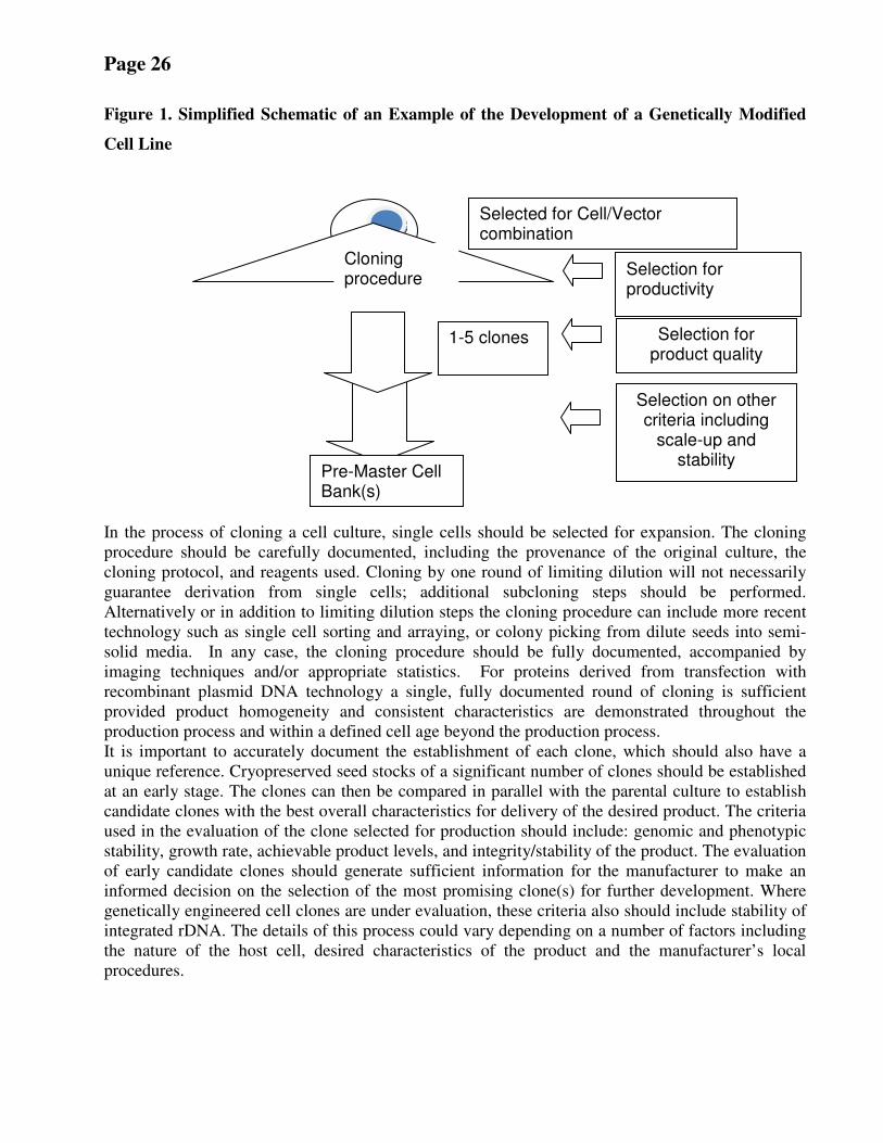
Page 26
Figure 1. Simplified Schematic of an Example of the Development of a Genetically Modified
Cell Line
In the process of cloning a cell culture, single cells should be selected for expansion. The cloning
procedure should be carefully documented, including the provenance of the original culture, the
cloning protocol, and reagents used. Cloning by one round of limiting dilution will not necessarily
guarantee derivation from single cells; additional subcloning steps should be performed.
Alternatively or in addition to limiting dilution steps the cloning procedure can include more recent
technology such as single cell sorting and arraying, or colony picking from dilute seeds into semi-
solid media. In any case, the cloning procedure should be fully documented, accompanied by
imaging techniques and/or appropriate statistics. For proteins derived from transfection with
recombinant plasmid DNA technology a single, fully documented round of cloning is sufficient
provided product homogeneity and consistent characteristics are demonstrated throughout the
production process and within a defined cell age beyond the production process. It is important to accurately document the establishment of each clone, which should also have a
unique reference. Cryopreserved seed stocks of a significant number of clones should be established
at an early stage. The clones can then be compared in parallel with the parental culture to establish
candidate clones with the best overall characteristics for delivery of the desired product. The criteria
used in the evaluation of the clone selected for production should include: genomic and phenotypic
stability, growth rate, achievable product levels, and integrity/stability of the product. The evaluation
of early candidate clones should generate sufficient information for the manufacturer to make an
informed decision on the selection of the most promising clone(s) for further development. Where
genetically engineered cell clones are under evaluation, these criteria also should include stability of
integrated rDNA. The details of this process could vary depending on a number of factors including
the nature of the host cell, desired characteristics of the product and the manufacturer’s local
procedures.
Selected for Cell/Vector combination
Cloning procedure
Selection for productivity
Selection for product quality
1-5 clones
Selection on other criteria including
scale-up and stability
Pre-Master Cell Bank(s)
Page 27
It is important to bear in mind that even following single cell cloning, epigenetic variation can result
in a cloned culture showing evidence of heterogeneity (i.e., more than one clone). This should not
preclude the use of such a culture for production, unless there are indications of instability that could
affect the quality and/or safety of the final product.
A.2.5 Special considerations for neural cell types Agents causing transmissible spongiform encephalopathies have been propagated in certain cells. At
the time of writing, the phenomenon has been observed only with very specific pairs of agents and
cells; no cell line has been identified that will replicate all agents, although one has been described
that seems to be infectable by many strains of scrapie. The phenomenon is unpredictable, except that
if the line does not express the PrP protein, it may be assumed to be impossible to infect it, and
experience to date suggests that infection is not commonly observed or easy to maintain. On the
other hand, the cell types that can be infected include fibroblastic lines as well as neuronal cells. The
cells have usually been of murine origin, because the infecting agents are usually mouse-adapted
scrapie. The fact that certain cells can be infected with certain agents is proof of principle that cell
lines may be infected so that exposure of cells to sources potentially contaminated with the agents is
a concern. The scale of the risk is difficult to judge, and it is recommended that with respect to safety
considerations and TSEs attention focuses on the selection and documentation of the cell culture
reagents and other materials that come into intimate contact with the cells to provide assurance that
they are not contaminated. Strategies to accomplish this are given in section B.11.4.
A.3 Selection of source materials A.3.1 Introduction
All materials should be subjected to a risk assessment and testing when necessary, in particular, raw
materials derived from humans and animals, which can be a primary source for the introduction of
adventitious agents into the production of biologicals. Therefore, careful attention should be paid to
their sourcing, production, handling, testing, and quality control. All cell-culture materials of
biological origin that come into intimate contact with the cells during the establishment of cell
cultures, derivation of a new cell line (if any), banking procedures (if any), and production, should
be subjected to appropriate tests, as indicated by risk assessment, for quality and freedom from
contamination by microbial agents to evaluate their acceptability for use in production. It is
important to evaluate the microbiological risks represented by each individual human- and animal-
derived reagent used in a cell culture production process, and it should address: (a) geographical
origin; (b) species of origin; (c) general microbiological potential hazards including a consideration
of the medical history for human-derived reagents; (d) husbandry/screening of donor animals; (e)
testing performed on the product, including Certificates of Analysis (if any); and (f) the capacity for
the preparation, purification, and sterilization procedures (if any) used to remove or inactivate
contaminants [ 54]. Other reagents of biological, but of non-animal origin, may also present risks to
product safety, and these are discussed further in section A.3.4
Recombinant protein technology now provides many materials formerly derived directly from
animal or human sources. While this eliminates obvious virological risks from donors, the
manufacturing process used for the recombinant proteins should be analyzed for any materials of
biological origin and any associated hazards that may need to be addressed, as indicated in items a-f
above.
Page 28
The NRA/NCL should approve source(s) of animal-derived raw materials, such as serum and
trypsin. These materials should comply with the guidelines on tissue infectivity distribution of TSEs
[ 55]. They should be subjected to appropriate tests for quality and freedom from contamination by
microbial agents to evaluate their acceptability for use in production. Their origin should be
documented to ensure that the sources are from geographical regions with acceptable levels of
microbiological risk (e.g., freedom from foot and mouth disease virus or bovine spongiform
encephalopathies). In addition, documentation should be gathered on their manufacturing history,
production, quality control, and any final or supplementary processing that could affect quality and
safety, such as blending and aliquoting of serum batches. In addition, controls should be in place to
prevent cross-contamination of one material with another (e.g., bovine material in a porcine
product).
The reduction and elimination from the manufacturing process of raw materials derived from
animals and humans is encouraged, where feasible.
For some human- and animal-derived raw materials used in the cell culture medium, such as insulin
or transferrin, validation of the production process for the elimination of viruses can substitute for
virus detection tests, when justified.
Animal-derived reagents, such as trypsin and serum, which would be substantially damaged or
destroyed in physical sterilization processes, including heat and irradiation, present the most likely
microbiological hazards to cell-culture processes. Batches of reagents, such as trypsin and bovine
serum, have been known to contain Mycoplasma species and sometimes more than one viral
contaminant. Certain contaminants also have been shown to infect cells in culture. The processing
environment is also a common source of microbiological contamination and should be controlled to
minimize this risk and prevent growth of contaminants.
A.3.2 Serum and other bovine-derived materials used in cell culture media
The source(s) of serum of bovine origin should be approved by the NRA/NCL. The responsibility
for ensuring the quality of the serum used in the manufacture of cell banks and biologicals rests with
the biologicals' manufacturer. This can be accomplished in more than one manner. The manufacturer
might conduct adventitious agent testing and perform inactivation of the serum after purchase from
the serum manufacturer. Alternatively, the manufacturer might qualify their serum vendor and only
purchase serum from suppliers after conducting thorough and on-going audits of the serum supplier
to ensure that they have properly performed the manufacture, quality control, and validation
necessary to achieve the level of quality of the serum required for the biological it is being used to
produce. In some cases, certificates of analysis may then be considered sufficient. Some combination
of these approaches might be optimal, and the strategy taken should be considered in evaluating risk.
Consultation with the NRA/NCL also might be advisable.
Serum and other bovine-derived materials should be tested for adventitious agents, such as bacteria,
fungi, mycoplasmas, and viruses, prior to use in the production of MCBs and WCBs and in the
manufacture of biologicals. Particular consideration should be given to those viruses that could be
introduced from bovine-derived materials and that could be zoonotic or oncogenic (e.g., bovine viral
Page 29
diarrhea virus, bovine polyoma virus, bovine circoviruses, rabies virus, bovine adenoviruses, bovine
parvoviruses, bovine respiratory syncytial virus, infectious bovine rhinotracheitis virus, bovine
parainfluenza virus type 3, reovirus 3, Cache Valley virus, bluetongue virus, and epizootic
hemorrhagic disease virus). In addition, consideration should be given to risk-mitigation strategies,
such as inactivation by heat or irradiation, to ensure that adventitious agents that were not detected in
the manufacture and quality control of the serum would be inactivated to a degree acceptable to the
NRA/NCL. If irradiation or other inactivation (e.g., heat sterilization) methods are used in the
manufacture of the serum, the tests for adventitious agents should be performed prior to inactivation
to enhance the opportunity for detecting the contamination. If evidence of viral contamination is
found in any of the tests, the serum is acceptable only if the virus is identified and shown to be
present in an amount that has been shown in a validation study to be effectively inactivated. For
serum that is not to be subjected to a virus inactivation / removal procedure, if evidence of viral
contamination is found in any tests, generally, the serum would not be acceptable. If the
manufacturer chooses to use serum that has not been inactivated, thorough testing of the serum for
adventitious agents using current best practices should be undertaken. If any agents are identified,
the cell banks made in this manner should be shown to be free of the identified virus(es).
If irradiation is used, it is important to ensure that a reproducible dose is delivered to
all batches and the component units of each batch. The irradiation dose must be low
enough so that the biological properties of the reagents are retained while being high
enough to reduce virological risk. Therefore, irradiation delivered at such a dose may
not be a sterilizing dose.
If serum was used in the establishment or passage history of the animal-cell substrate prior to
banking by the manufacturer, the cell bank (MCB or WCB) and/or cells at or beyond the level of
production should be tested for adventitious agents of the species (e.g., bovine) of serum used in the
establishment and passage history of the cell substrate. If serum is not used in the production of the
subsequent stages, then this testing would not need to be repeated on those subsequent stages once
the cell bank has been tested and considered free of bovine (or whichever species of serum that was
used) adventitious agents.
Methods used to test for bovine viruses should be approved by the NRA/NCL. Details of the
methods are provided in Appendix 1. Infectivity assays are used as the primary screening method
and have resulted in the detection of bovine viral diarrhea virus, reovirus 3, Cache Valley virus,
bluetongue virus, and epizootic hemorrhagic disease virus amongst others. However, it should be
noted that, in general, the infectivity screening assay methods described here do not readily detect
some of the viruses (e.g., bovine polyomaviruses) that can be frequent contaminants of serum.
Additional methods may need to be considered, such as the nucleic acid amplification technique
(NAAT), although the presence of viral genomic sequences is not necessarily indicative of infectious
virus. In those cases, specific infectivity assays designed to detect the virus of concern (e.g., bovine
polyomavirus) may need to be considered.
A second factor in screening serum is the limited sample volume used compared with the batch size,
which may be on the order of 1000 litres, which comes from the pooling of serum from many
animals. Consequently, infectious viruses can be missed in the serum lot testing, and consideration
should be given to direct screening of the cell bank for bovine viruses. These assays could include,
in addition to the general screening procedure, NAAT for the presence of bovine viruses that may
Page 30
infect the cell substrate but undergo abortive and/or transforming infections. Virus families of
particular concern in this latter regard include polyomaviruses, herpesviruses, circoviruses,
anelloviruses, and adenoviruses.
General screening assays for the detection of infectious viruses in serum or cell substrates involve
the use of at least one indicator cell line, such as bovine turbinate cells, permissive for the replication
of bovine viral diarrhoea virus (BVDV). A second cell line such as Vero also should be employed to
broaden the detection range. Before initiating screening it may be necessary to evaluate the serum
for the presence of antibody, particularly to BVDV, that could mask the presence of infectious virus.
Typically, indicator cells are cultured in the presence of the serum under test for 21 to 28 days,
passaging the cells as necessary. During this period, the cells are regularly examined for the presence
of cytopathic effect (CPE) indicative of virus infection. At the end of the observation period, which
should not be less than 7 days after the last sub-culture, the cells are stained to detect CPE that may
have been missed during observation of the living cells. Additional endpoint assays should include
haemadsorbtion and haemagglutination at both 4°C and a higher temperature such as 20 to 25°C and
also immunofluorescence assays (IFA) for specific viruses. Appropriate controls should be used for
each assay, like bovine parainfluenza virus type 3 for heamadsorbtion. IFA is particularly important
for BVDV, as non-cytopathic BVDV may be present in the serum. IFA endpoints are also used to
detect other viruses that may be determined by geographical considerations like adenoviruses,
bovine parvovirus, bluetongue virus, bovine syncytial virus, reovirus type 3 and unlikely, but serious
contaminants like rabies virus.
If serum from another species is used (i.e., other than bovine), the NRA/NCL should be consulted
regarding acceptable testing methods for that species.
A.3.3 Trypsin and other porcine-derived materials used for preparing cell cultures
Trypsin used for preparing cell cultures should be tested for cultivable bacteria, fungi, mycoplasmas
and infectious viruses, including bovine or porcine parvoviruses, as appropriate. The methods used
to ensure this should be approved by the NRA/NCL.
In some countries, irradiation is used to inactivate potential
contaminant viruses. If irradiation is used, it is important to ensure
that a reproducible dose is delivered to all batches and the
component units of each batch. The irradiation dose must be low
enough so that the biological properties of the reagents are retained
while being high enough to reduce virological risk. Therefore,
irradiation cannot be considered a sterilizing process.
The quality of the trypsin, like serum, is the responsibility of the biologicals’ manufacturer (see
section A.4.2). Recombinant trypsin is available and should be considered; however it should not be
assumed to be free of risk of contamination and should be subject to the usual considerations for any
reagent of biological origin (see A.4.1).
Like serum batches that are derived from many animals, trypsin batches are prepared from the
pancreases of many animals. Most batches of porcine trypsin contain genetic sequences of porcine
parvovirus 1 and porcine circoviruses and should be therefore treated to inactivate any virus
Page 31
potentially present in a manner accepted by the NRA/NCL. It is acknowledged however that these
viruses are relatively resistant to inactivation [ 56]. If trypsin from another species is used, the
NRA/NCL should be consulted regarding acceptable testing methods.
General screening assays for the detection of infectious porcine viruses in trypsin or cell substrates
involve the use of at least one indicator cell line, such as porcine testes cells or Vero cells,
permissive for the replication of porcine viruses. Typically, indicator cell cultures would be
incubated for 14 days with a sub-culture onto fresh test cells for an additional 14 days. Specific
endpoint detection methods like IFA or PCR may be required in addition to observation for
cytopathic effect periodically throughout the culture period and more general endpoint detection
methods like hemadsorption and/or hemagglutination.
Furthermore, if trypsin was used in the establishment or passage history of the animal-cell substrate
prior to banking by the manufacturer, the cell bank (MCB or WCB) should be tested for porcine
parvovirus, or appropriate adventitious viruses relevant to the species of origin of the trypsin used in
the establishment and passage history of the cell substrate. If trypsin is not used in the production of
the subsequent stages, then this testing would not need to be repeated on those subsequent stages
once the cell bank has been tested and considered free of porcine parvovirus (or relevant agents).
Consideration should be given to screening for other agents such as porcine circoviruses. Molecular
methods, such as PCR, may be used for such purposes.
Testing of cells exposed to trypsin or of other porcine-derived materials might entail testing for more
than porcine parvovirus or porcine circoviruses. For example, testing for porcine adenovirus,
transmissible gastroenteritis virus, porcine haemagglutinating encephalitis virus, bovine viral
diarrhea virus, reoviruses, rabies virus, porcine anellovirus, porcine hokovirus, porcine bocavirus,
porcine hepatitis E virus, porcine reproductive and respiratory syndrome virus,
encephalomyocarditis virus, and potentially other viruses might be appropriate. Particular
consideration should be given to those viruses that could be introduced from the porcine-derived
material and that could be zoonotic or oncogenic. Additionally, tests for bacterial and fungal sterility
and mycoplasmas depending on the type of porcine-derived material should be conducted. The
NRA/NCL should be consulted in this regard.
A.3.4 Medium supplements and general cell culture reagents derived from other sources used for
preparing cell cultures
Medium supplements derived from other species should be quality controlled from the perspective
of adventitious agents. Consideration should be given to whether recombinant-derived medium
supplements were exposed to animal-derived materials during their manufacture, and if so, evaluated
for the potential to introduce adventitious agents into the manufacture of the cell banks and
biological products. Testing for adventitious agents should assess viruses relevant to the species
from which the supplement was derived. The NRA/NCL should be consulted in this regard.
Generally, medium supplements should not be obtained from human source materials. In particular,
human serum should not be used. However, in special circumstances, and in agreement with the
NRA/NCL, the use of human-derived supplements may be permitted. If human serum albumin is
used at any stage of product manufacture, the NRA/NCL should be consulted regarding the
requirements, as these may differ from country to country. However, as a minimum, it should meet
Page 32
the revised Requirements for Biological Substances No. 27 [ 56], as well as the guidelines on tissue
infectivity distribution of TSEs [ 55].
Recombinant human albumin is commercially available and should be considered; however, it
should not be assumed to be free of risk of contamination and should be subject to the usual
considerations for any reagent of biological origin (see A.3).
As for other cell-culture reagents, it is important to establish traceability and assess and reduce
microbiological risks as described in section A.3.
A variety of cell-culture reagents of biological origin are available that are derived from non-animal
sources, including a range of aquatic organisms, plants and algae. In such cases, the exact hazards
involved may be uncertain and unfamiliar. The microbiological risks may be substantially different
to those involved in animal-derived reagents (see section A.3.1-A.3.3), and other hazards may arise,
such as immunogenic, mitogenic and allergenic properties of the reagent and its components. For
example, plant-derived material may carry an increased risk of mycoplasma and mycobacteria
contamination.
A.4 Certification of cell banks by the manufacturer It is vital that the manufacturer has secured a body of information on the cell substrate that
demonstrates clearly the origin or provenance of the culture and how the cell banks intended for
production (MCB and WCB) were established, characterized, and tested. This should provide all the
information required to demonstrate the suitability of that cell substrate and the established cell
banks for the manufacture of biological products.
A.4.1 Cell line data
Each new cell line should have an associated body of data, which will increase as the cell line is
established and developed for manufacturing purposes. This data set is vital to demonstrate the
suitability for use of the cells and should provide information on cell provenance (donor information
and any relevant details on ethical procurement), cell line derivation, culture history, culture
conditions (including reagents), early stage safety evaluation data, banking and cell bank
characterization, and safety testing. This information should be available to the NRA/NCL for
approval of the cells used in manufacture.
A.4.2 Certification by the manufacturer of primary cell cultures Full traceability should be established for PCCs to the animals of origin, husbandry conditions,
veterinary inspection, vaccinations (if any), procedures for administering anaesthesia and cell
harvesting, the reagents and procedures used in the preparation of primary cultures, and the
environmental conditions under which they were prepared. Extensive testing should be performed,
and this should be documented.
It is important to define the batch or lot of cells used in each individual manufacturing process. For
production purposes, a batch or lot is a culture of primary cells derived from single or multiple
animals that has been subjected to a common process of tissue retrieval, disaggregation, and
processing leading to a single culture preparation of cells. Lots may be prepared by harvesting and
pooling cells in different ways, but the cell-processing procedures should be reproducible, and it is
Page 33
especially important to monitor cultures carefully for evidence of adverse change in the cell culture
and microbiological contamination. Prior to any culture pooling, cells should be examined for
acceptability for production. Acceptability criteria should be established and should include testing
for microbiological contamination and the general condition of the cells (e.g., morphology, number,
and viability of the cells). Failure to detect and eliminate atypical (i.e., potentially virally infected) or
grossly contaminated cells will put the entire production run at risk and could compromise the safety
of the product. Cells showing an unacceptably high proportion of dead or atypical cells should not be
used, and ideally microbiological testing should be completed and passed before the cells are used.
The preparation of cell lots for manufacture should be carefully documented to provide full
traceability from the animal donor(s) to production. Any pooling of cells should be clearly recorded,
as should any deviation from standard operating procedures, as required by GMPs. In addition, any
observations of variation between batches should be recorded, even where such observations would
not necessarily lead to rejection of those batches. Such information may prove valuable in ongoing
optimization and improvement of the production process.
A.4.3 Certification by the manufacturer of diploid, continuous, and stem cell lines All cell lines used for biologicals production should have data available, as indicated in A.4.1.
The original PDL (or passage number, if the PDL is unknown) of the cell seed should be recorded.
For cell lines of human origin, if possible the medical history of the individual from whom the cell
line was derived should be evaluated in order to better assess potential risks and the suitability of the
cell line.
For SCLs, morphology continues to be an important characteristic, and representative images and
immune-phenotypic profiles of undifferentiated and differentiated cells should be available for
comparison.
A.5 Cryopreservation and Cell Banking A.5.1 Cryopreservation When cells are banked, the successful preservation of cells at ultra-low temperatures is critical to the
efficient delivery of good quality cultures (i.e., high viability cultures of the required characteristics).
The need to prepare large stocks of frozen vials of cells for cell banks is especially challenging, and
a number of key principles should be adopted:
• A method that meets current best practice for cell-culture preservation should be used (for
example, see ref 58)
• The cooling profile achieved for the cells being frozen should be defined, and the same
cooling process should be used for each separate preservation process (i.e., an SOP should
include documentation of the cooling process in the batch record.)
• Each preservation process should be recorded
• As a general guide, only cell cultures that are predominantly in the exponential phase of
growth should be used. Cells in such cultures tend to have a low ratio of cytoplasm to
nucleus (v/v) and should be more amenable to successful cryopreservation. It is unwise to
use cells predominantly in the 'lag' phase very early after passage or in the 'stationary' phase
when the culture has reached its highest density of cells
Page 34
• For each bank, cells pooled from a single expanded culture (i.e., not from a range of cultures
established at different times post seeding or different PDLs) should be used and mixed prior
to aliquotting to ensure homogeneity
• The number of cells per vial should be adequate to recover a representative culture (e.g., 5-10
x 106 in a 1-mL aliquot)
• For new cell banks, antimicrobials should not be used in cell cultures to be banked, except
where this can be justified for early PDL cultures which may carry contamination from tissue
harvesting or recombinant cells which require antibiotic selection, and when necessary for
the genetic stability of recombinant cell lines. In any case, if antimicrobials are used, they
should not be penicillin or any other beta-lactam drug
• When a stock of cells has been frozen, a sample should be recovered to confirm it has
retained viability and the results recorded. It is also important to establish the degree of
homogeneity within the cell bank. Recovery of a sufficient percentage (e.g., 1% or as
recommended by the NRA) of vials representative of the beginning, middle and end of the
cryopreservation process should be demonstrated to give confidence in the production
process based on the use of that cell bank. Ultimately, stability (see B.3) and integrity of
cryopreserved vials is demonstrated when the vials are thawed from production and
demonstrated to produce the intended product at scale (see also B.7).
• Cell bank cryostorage vessels should be monitored and maintained to enable demonstration
of a highly stable storage environment for cell banks. Access to such vessels may cause
temperature cycling, which in extreme cases can cause loss of viability. It is therefore
prudent to establish a stability-testing programme involving recovery of cells periodically
where the frequency of recovery relates to risk of temperature cycling. New developments in
remote monitoring of individual vials may in future eliminate the need for stability testing.
A.5.2 Cell Banking
When DCLs, SCLs, or CCLs are used for production of a biological, a cell-bank system should be
used. Such banks should be approved by and registered with the NRA/NCL as part of the product
approval process. The source of cells used in cell banking and production is a critical factor in
biological product development and manufacture. It is highly desirable to obtain cells from sources
with a documented history and traceability to the originator of the cell line.
After a sample of the original seed stock is obtained, an early stage pre-master bank of just a few
vials should be established. One or more vials of that pre-master bank are used to establish the MCB.
The WCB is derived by expansion of one or more containers of the MCB. The WCB should be
qualified for yielding cell cultures that are acceptable for use in manufacturing a biological product.
When using early PDLs from primary cultures for production processes, the preparation of a cell
bank should be considered on a case-by-case basis. This approach has significant benefits, as it gives
great flexibility in the timing of the production process, permits quality control and safety testing to
be completed prior to use, and reduces the overall burden of testing required in the production
process.
Cell banks should be characterized as specified in Part B of this guidance and according to any other
currently applicable and future guidance published by WHO. The testing performed on a
replacement MCB (derived from the same cell clone or from an existing MCB or WCB) is the same
Page 35
as for the initial MCB unless a justified exception is made. Efforts to detect contaminating viruses
and other microbial agents constitute a key element in the characterization of cell banks.
Having been cryopreserved by qualified methods (see A.5.1), both the MCB and WCB should be
stored frozen under defined conditions, such as in the vapour or liquid phase of liquid nitrogen. The
location, identity, and inventory of individual cryovials or ampoules of cells should be thoroughly
documented. It is recommended that the MCB and WCB each be stored in at least two widely
separated areas within the production facility and/or in geographically distinct locations to assure
continued ability to manufacture product in the event of a facility catastrophe. When cryopreserved
cells are transferred to a remote site, it is important to use qualified shipping containers and to
monitor transfers with probes to detect temperature excursions. All containers are treated identically
and, once removed from storage, usually are not returned to the stock. The second storage site
should operate under an equivalent standard of quality assurance as the primary site.
A.5.3 WHO reference cell banks (RCBs) The principle of establishing RCBs under WHO auspices is one that offers potential solutions to
future challenges for the development of vaccines and biotherapeutics in developing regions.
However, WHO does not intend that cells supplied to manufacturers from any RCB be used as a
MCB. The purpose of WHO RCBs is to provide well characterized cell seed material for the
generation of a MCB by manufacturers with the expectation that such MCBs will comply with this
guidance document and be fully characterized.
The WHO RCBs provide key advantages for vaccine development worldwide that include:
• Traceability to origin of cells and derivation of cell line and materials used in preparation of
seed stock
• Subjected to open international scientific scrutiny and collaborative technical investigations
into the characteristics of the cells and the presence of adventitious agents
• Results of characterization were peer reviewed and published
• Investigations evaluated under auspices of WHO expert review and qualified as suitable for
use in vaccine production
• Supply of cells free of any adverse Intellectual Property 'reach through' on final products
• Single source of cells with growing and scientifically and technically updated body of
safety-testing data and safe history of use, giving increasing confidence for manufacturers,
regulators, and public policy makers
The Vero cell line is the most widely used continuous cell line for the production of viral vaccines
over the last two decades. The WHO Vero RCB 10-87 was established in 1987 and was subjected to
a broad range of tests to establish its suitability for vaccine production. This WHO RCB provides a
unique resource for the development of future biological medicines where a cell substrate with a safe
and reliable history of use is desired. A comprehensive review of the characterization of the WHO
Vero 10-87 seed lot was conducted recently, and a detailed overview is provided on the WHO
website at (http://www.who.int/biologicals/).
As concluded by an expert review in 2002, the WHO Vero RCB 10-87 is not considered suitable for
direct use as MCB material. However, the WHO Vero RCB 10-87 is considered suitable for use as a
Page 36
cell seed for generating a MCB, and its status has changed from "WHO Vero cell bank 10-87" to
"WHO Vero reference cell bank 10-87" .
The WHO Vero RCB 10-87 is stored in the European Collection of Animal Cell Cultures (ECACC,
www.hpacultures.org.uk), Salisbury, Wiltshire, UK, and the American Type Culture Collection
(ATCC, www.atcc.org), in Manassas, Virginia, USA. These public service culture collections have
distributed ampoules under agreements with the WHO to numerous manufacturers and other users.
The WHO Vero RCB 10-87 is the property of WHO, and there are no constraints relating to
intellectual property rights. The WHO Vero RCB 10-87 is available free of charge on application to
WHO. However, due to the limited number of vials remaining, distribution of these vials is restricted
solely for the production of vaccines and other biologicals. Potential replacement of the WHO Vero
RCB 10-87 is currently under consideration.
WHO also has overseen the establishment of seed stocks of MRC-5 for the production of vaccines.
The WHO MRC-5 RCB was established in 2007 because of stability issues associated with the
original vials of MRC-5 cells, which dated to 1966. This RCB was prepared in a qualified cleanroom
environment and subjected to specified quality-control testing endorsed by the ECBS.
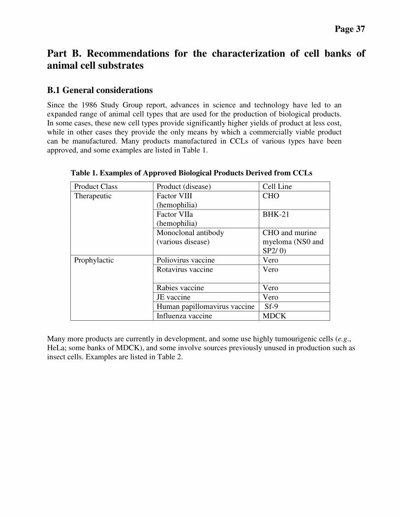
Page 37
Part B. Recommendations for the characterization of cell banks of
animal cell substrates
B.1 General considerations
Since the 1986 Study Group report, advances in science and technology have led to an
expanded range of animal cell types that are used for the production of biological products.
In some cases, these new cell types provide significantly higher yields of product at less cost,
while in other cases they provide the only means by which a commercially viable product
can be manufactured. Many products manufactured in CCLs of various types have been
approved, and some examples are listed in Table 1.
Table 1. Examples of Approved Biological Products Derived from CCLs
Product Class Product (disease) Cell Line
Factor VIII
(hemophilia)
CHO
Factor VIIa
(hemophilia)
BHK-21
Therapeutic
Monoclonal antibody
(various disease)
CHO and murine
myeloma (NS0 and
SP2/ 0)
Poliovirus vaccine Vero
Rotavirus vaccine
Vero
Rabies vaccine Vero
JE vaccine Vero
Human papillomavirus vaccine Sf-9
Prophylactic
Influenza vaccine MDCK
Many more products are currently in development, and some use highly tumourigenic cells (e.g.,
HeLa; some banks of MDCK), and some involve sources previously unused in production such as
insect cells. Examples are listed in Table 2.
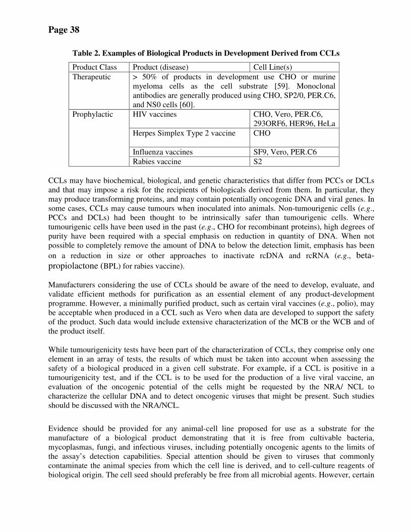
Page 38
Table 2. Examples of Biological Products in Development Derived from CCLs
Product Class Product (disease) Cell Line(s)
Therapeutic
> 50% of products in development use CHO or murine
myeloma cells as the cell substrate [ 59]. Monoclonal
antibodies are generally produced using CHO, SP2/0, PER.C6,
and NS0 cells [ 60].
HIV vaccines CHO, Vero, PER.C6,
293ORF6, HER96, HeLa
Herpes Simplex Type 2 vaccine CHO
Influenza vaccines SF9, Vero, PER.C6
Prophylactic
Rabies vaccine S2
CCLs may have biochemical, biological, and genetic characteristics that differ from PCCs or DCLs
and that may impose a risk for the recipients of biologicals derived from them. In particular, they
may produce transforming proteins, and may contain potentially oncogenic DNA and viral genes. In
some cases, CCLs may cause tumours when inoculated into animals. Non-tumourigenic cells (e.g.,
PCCs and DCLs) had been thought to be intrinsically safer than tumourigenic cells. Where
tumourigenic cells have been used in the past (e.g., CHO for recombinant proteins), high degrees of
purity have been required with a special emphasis on reduction in quantity of DNA. When not
possible to completely remove the amount of DNA to below the detection limit, emphasis has been
on a reduction in size or other approaches to inactivate rcDNA and rcRNA (e.g., beta-
propiolactone (BPL) for rabies vaccine).
Manufacturers considering the use of CCLs should be aware of the need to develop, evaluate, and
validate efficient methods for purification as an essential element of any product-development
programme. However, a minimally purified product, such as certain viral vaccines (e.g., polio), may
be acceptable when produced in a CCL such as Vero when data are developed to support the safety
of the product. Such data would include extensive characterization of the MCB or the WCB and of
the product itself.
While tumourigenicity tests have been part of the characterization of CCLs, they comprise only one
element in an array of tests, the results of which must be taken into account when assessing the
safety of a biological produced in a given cell substrate. For example, if a CCL is positive in a
tumourigenicity test, and if the CCL is to be used for the production of a live viral vaccine, an
evaluation of the oncogenic potential of the cells might be requested by the NRA/ NCL to
characterize the cellular DNA and to detect oncogenic viruses that might be present. Such studies
should be discussed with the NRA/NCL.
Evidence should be provided for any animal-cell line proposed for use as a substrate for the
manufacture of a biological product demonstrating that it is free from cultivable bacteria,
mycoplasmas, fungi, and infectious viruses, including potentially oncogenic agents to the limits of
the assay’s detection capabilities. Special attention should be given to viruses that commonly
contaminate the animal species from which the cell line is derived, and to cell-culture reagents of
biological origin. The cell seed should preferably be free from all microbial agents. However, certain
Page 39
CCLs may express endogenous retroviruses. Tests capable of detecting such agents should be carried
out on cells grown under cell-culture conditions that mimic those used during production, and the
levels of viral particles should be quantified. Viral contaminants in a MCB and WCB should be
shown to be inactivated and/or removed by the purification procedure used in production. The
validation of the purification procedure used is considered essential.
The characterization of any DCL, SCL or CCL to be used for the production of biologicals should
include: a) a history of the cell line (i.e., provenance) and a detailed description of the production of
the cell banks, including methods and reagents used during culture, PDL, storage conditions,
viability after thawing, and growth characteristics; b) the results of tests for infectious agents; c)
distinguishing features of the cells, such as biochemical, immunological, genetic, or cytogenetic
patterns, that allow them to be clearly distinguished from other cell lines; and d) the results of tests
for tumourigenicity, including data from the scientific literature. Additional consideration should be
given to products derived from cells that contain known viral sequences (e.g., Namalwa, HeLa, 293,
and PER.C6).
The recommendations that follow are intended as guidance for NRAs, NCLs, and manufacturers as
the minimum amount of data on the cell substrate that should be available when considering a new
biological product for approval. The amount of data that may be required at various stages of clinical
development of the product should be discussed and agreed with the NRA/NCL at each step of the
program.
B.2 Identity
Cell Banks should be authenticated by a cell-identification method approved by the NRA/NCL.
Wherever practicable, methods for identity testing should be used that give specific identification of
the cell line to confirm that no switching or major cross-contamination of cultures has arisen during
cell banking and production. A number of the commonly used identity testing methods are compared
in Table 3. In the case of human cells, genetic tests such as DNA profiling (e.g., Short Tandem
Repeat analysis, multiple Single Nucleotide Polymorphisms) will give a profile that is at least
specific to the individual from whom the cells were isolated. Another test that might be used for
human cells is HLA typing. Other tests that may be used but tend to be less specific include
isoenzyme analysis and karyology, which may be particularly useful where there are characteristic
marker chromosomes. However, where more specific genetic markers are available, they should be
considered. It is not unexpected that a small proportion of cell lines, particularly those which are
transformed, may show alterations to the expected identity profile. This has been observed in
isoenzyme analysis where in rare cases a particular cell line may show a consistently different profile
to that expected for the species of origin and is also a general issue relating to the effect of genetic
instability for molecular identity testing techniques. Such effects in standard technologies are rare
and may also arise with the implementation of new techniques. The implications of any unexpected
results should be discussed with the NRA/NCL. For recombinant-protein products, cell line identity
testing should also include tests for vector integrity, expression plasmid copy number, insertions,
deletions, number of integration sites, the percentage of host cells retaining the expression system,
verification of protein-coding sequences, and protein-production levels.
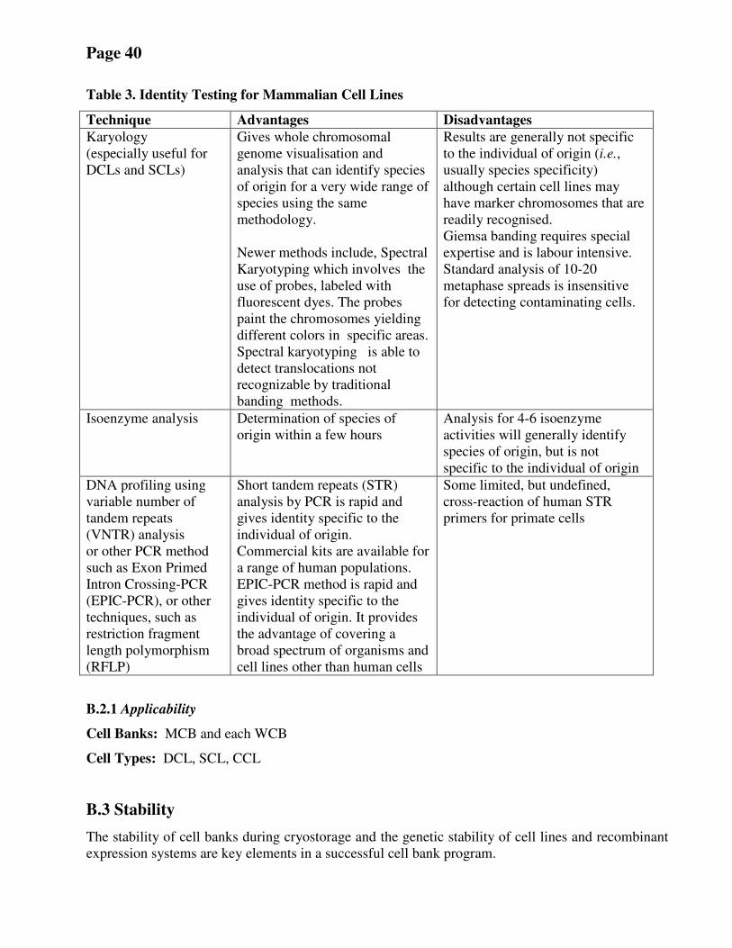
Page 40
Table 3. Identity Testing for Mammalian Cell Lines
Technique Advantages Disadvantages
Karyology
(especially useful for
DCLs and SCLs)
Gives whole chromosomal
genome visualisation and
analysis that can identify species
of origin for a very wide range of
species using the same
methodology.
Newer methods include, Spectral
Karyotyping which involves the
use of probes, labeled with
fluorescent dyes. The probes
paint the chromosomes yielding
different colors in specific areas.
Spectral karyotyping is able to
detect translocations not
recognizable by traditional
banding methods.
Results are generally not specific
to the individual of origin (i.e.,
usually species specificity)
although certain cell lines may
have marker chromosomes that are
readily recognised.
Giemsa banding requires special
expertise and is labour intensive.
Standard analysis of 10-20
metaphase spreads is insensitive
for detecting contaminating cells.
Isoenzyme analysis Determination of species of
origin within a few hours
Analysis for 4-6 isoenzyme
activities will generally identify
species of origin, but is not
specific to the individual of origin
DNA profiling using
variable number of
tandem repeats
(VNTR) analysis
or other PCR method
such as Exon Primed
Intron Crossing-PCR
(EPIC-PCR), or other
techniques, such as
restriction fragment
length polymorphism
(RFLP)
Short tandem repeats (STR)
analysis by PCR is rapid and
gives identity specific to the
individual of origin.
Commercial kits are available for
a range of human populations.
EPIC-PCR method is rapid and
gives identity specific to the
individual of origin. It provides
the advantage of covering a
broad spectrum of organisms and
cell lines other than human cells
Some limited, but undefined,
cross-reaction of human STR
primers for primate cells
B.2.1 Applicability
Cell Banks: MCB and each WCB
Cell Types: DCL, SCL, CCL
B.3 Stability
The stability of cell banks during cryostorage and the genetic stability of cell lines and recombinant
expression systems are key elements in a successful cell bank program.
Page 41
B.3.1 Stability during cryostorage
Data should be generated to support the stability or suitability of the cell substrate and any
recombinant expression system or necessary cell phenotype during cultivation to or beyond the limit
of production, and to support the stability of the cryopreserved cell banks during storage. The latter
may be demonstrated by successful manufacture of WCBs or production lots. Periodic testing for
viability is not necessary if continuous-monitoring records for storage show no deviations out of
specification, and periodic production runs are successful. If banks are used less than once every 5
years, then it may be prudent to generate data confirming suitability for manufacturing on a schedule
that takes into account the storage condition once every 5 years.
B.3.1.1 Applicability
Cell Banks: MCB and WCB
Cell Types: DCL, SCL, CCL
B.3.2 Genetic Stability Any form of genetic instability could potentially affect the quality of the final product and it will be
important to know if the cells in culture are changing in a way that could affect the nature or safety of
the product. Any features of the cell lines that might affect quality should be discussed with the
NRA/NCL to ensure that tests used by the manufacturer to monitor genetic stability are adequate.
The specific tests will vary according to the nature of the product, but the aim is to show consistency
in the amount and characteristics of the product derived from cells within a few passages of the MCB
or WCB with those derived from an ECB or EOPC. For recombinant protein products, emphasis will
be on the protein sequence and post translational modifications.
For cell lines containing DNA expression constructs, the stability of these constructs between the
MCB/WCB and an ECB or EOPC should be determined. The copy number of the construct and, if
relevant, the sites of chromosomal insertion should be determined. The latter is accomplished by
sequencing into the cellular flanking regions, but methods like fluorescent in situ hybridization may
provide useful additional information, particularly where concatamers of the gene insert are present
at individual chromosomal loci. The sequence of the construct within the cells should be determined.
With conventional sequencing, a consensus sequence is obtained, but with massively parallel
sequencing, it is possible to determine the sequence of individual gene inserts or their transcripts.
Where proteins are derived from non-genetically modified cells, consistency in the yield and
properties of the protein should be evaluated together with the sequence of the mRNA encoding the
protein of interest.
Additional characterization of the cell-biological processes and responses during cultivation (for
instance using global or targeted gene expression, proteomic or metabolic profiles and other
phenotypic markers) might be useful in further developing a broad understanding of the cell substrate.
Appropriate methods should be applied to assure that cell age is correctly assessed in the event that
cell viability falls dramatically at any given step. Losses in viability are reflected in increased
cultivation times to reach defined levels of growth.
Page 42
The stability of cell function in terms of productivity within the production process also may need to
be evaluated. Other stability studies may be performed where bioreactor methods are employed,
especially where extended culture periods are involved.
B.3.2.1 Applicability
Cell Banks: MCB taken to EOPC/ECB
Cell Types: DCL, SCL, CCL
B.4 Sterility (see B.11.3.1)
B.5 Viability High level of viability of cryopreserved cells is important for efficient and reliable production.
Typically, thawed cells should have viability levels in excess of 80%, though this is not always
achieved and may depend on the cell line. Lower viabilities may still result in suitable growth
recovery and in acceptable product qualities. In such cases, the data should be discussed with the
NRA/NCL. A range of viability tests are available that measure different attributes of cell function
(membrane integrity, metabolic activity, DNA replication). Under certain circumstances, such as
pre-apoptotic cells excluding trypan blue, viability assays may give misleading results, and it is
important to be aware of the exact information that a particular viability assay provides. Therefore, it
is important to evaluate growth recovery of cryopreserved cells upon thawing.
For certain cell cultures such as hybridomas, where a membrane-integrity
test is used, additional cell markers such as indicators of apoptosis should
be studied in order to avoid significantly overestimating viability.
A suitable viability test should be selected for the cell substrate in question and typical test values
established for cultures considered to be acceptable (see B.6, B.7). It may also be necessary to select
alternative viability assays that are better suited to providing in-process viability data required during
production, e.g., lactate dehydrogenase levels in bioreactor systems.
B.5.1 Applicability
Cell Banks: MCB and WCB
Cell Types: DCL, SCL, CCL
B.6 Growth Characteristics For the development of production processes, the growth characteristics of the production-cell line
should be well understood to ensure consistency of production. Changes in these characteristics
could indicate any one of a range of events. Accordingly, data on growth characteristics, such as
viability, morphology, cell-doubling times, cloning and/or plating efficiency, if applicable, should be
developed. For certain cell substrates, it may be appropriate to apply such tests in homogeneity
testing (see B.7). Experiments to demonstrate homogeneity and growth characteristics may be
combined, although the analysis should be carried out separately.
B.6.1 Applicability
Cell Banks: MCB and WCB
Page 43
Cell Types: DCL, SCL, CCL
B.7 Homogeneity Testing Each cell culture derived from a container of the WCB should perform in the same way (i.e., within
acceptable limits and provide the same number of viable cells of the same growth characteristics). In
order to assure this, it is important to recover a proportion of containers from each cell bank and
check their characteristics, as indicated in B.6. The number of containers tested should be discussed
with the NRA/NCL and be broadly in line with those normally sampled to establish product
consistency. Recovery of a sufficient percentage of vials representative of the beginning, middle and
end of the aliquotting process should be demonstrated to give confidence in the production process
based on the use of that cell bank. Ultimately, stability and integrity of cryopreserved vials are
demonstrated when the vials are thawed for production and demonstrated to produce the intended
product at scale. Instead of testing a portion of containers at different stages of the banking process,
an alternative strategy to ensure the homogeneity of the banks can be used based on the validation of
the process method for filling and freezing. Assessment of growth characteristics (B.6) and
homogeneity testing are commonly combined experimentally; however, the analysis and
interpretation of each should be distinct. It may be appropriate to also test homogeneity of the MCB
to assure future WCBs are consistent with the first WCB.
B.7.1 Applicability
Cell Banks: MCB, WCB
Cell Types: DCL, SCL, CCL
B.8 Tumourigenicity B.8.1 General considerations Several in vitro test systems, such as cell growth in soft agar [ 61] and muscle organ culture [ 62],
have been explored as alternatives to in vivo tests for tumourigenicity; however, correlations with in
vivo tests have been imperfect or the alternative tests have been technically difficult to perform.
Therefore, in vivo tests remain the standard for assessing tumourigenicity.
Although WHO Requirements [ 1] have described acceptable approaches to tumourigenicity testing,
a number of important aspects of such testing were not addressed. Therefore, a model protocol has
been developed and is appended to this document. The major points included in the model protocol
are listed below along with comments on each specific item.
A new diploid cell line (i.e., other than WI-38, MRC-5, and FRhL-2) should be tested for
tumourigenicity as part of the characterization of the cell line, but should not be required on a routine
basis.
The tumourigenicity tests currently available are in mammalian species whose body temperatures
and other physiologic factors are different from those of avian and insect species. Therefore, when
the test is performed on avian or insect cells, the validity of the data is open to question unless a
tumourigenic cell line of the species being tested is included as a positive control. The NRA/NCL
may accept the results of an in vitro test such as growth in soft agar as a substitute for the in vivo test
for avian and insect cell lines. However, as mentioned above, correlations of in vitro tests with in
vivo tests are imperfect. This should be discussed with the NRA/NCL.
Page 44
Many CCLs (e.g., BHK-21, CHO, HeLa) are classified as tumourigenic because they possess the
capacity to form tumors in immunosuppressed animals such as rodents. Some CCLs become
tumourigenic at high PDLs (e.g., Vero), even though they do not possess this capacity at lower PDLs
at which vaccine manufacture occurs. A critical feature regarding the pluripotency of
embryonic SCLs, even though they display a diploid karyotype, is that they form tumours in
immunocompromised mice.
The expression of a tumourigenic phenotype can be quite variable from one CCL to another, and
even within different sub-lines of the same CCL. This range of variability, from non-tumourigenic,
to weakly tumourigenic, to highly tumourigenic, has been viewed by some as indicating different
degrees of risk when they are used as substrates for the manufacture of biological products [ 10, 11].
If the CCL has already been demonstrated to be tumourigenic (e.g., BHK-21, CHO, HEK293, Cl27),
or if the class of cells to which it belongs is tumourigenic (e.g., hybridomas, SCLs), it may not be
necessary to perform additional tumourigenicity tests on cells used for the manufacture of
therapeutic products. Such cell lines may be used as cell substrates for the production of biological
products if the NRA/NCL has determined, based on characterization data as well as manufacturing
data, that issues of purity, safety, and consistency have been addressed. A new cell line (DCL, SCL,
or CCL) should be presumed to be tumourigenic unless data demonstrate that it is not. If a
manufacturer proposes to characterize the cell line as non-tumourigenic, the following tests should
be undertaken.
Cells from the MCB or WCB propagated to the proposed in vitro cell age used for production or
beyond should be examined for tumourigenicity in a test approved by the NRA/NCL. The test
should involve a comparison between the cell line and a suitable positive reference preparation (e.g.,
HeLa cells) and a standardized procedure for evaluating results.
B.8.2 Type of test animals A variety of animal systems have been used to assess the tumourigenic potential of cell lines. Table
4 lists several examples of such tests along with advantages and disadvantages of each. Because
assessing the tumourigenic phenotype of a cell substrate requires the inoculation of xenogeneic or
allogeneic cells, the test animal should be rendered deficient in cytotoxic T-lymphocyte (CTL)
activity. This can be accomplished either by the use of rodents that are genetically
immunocompromised (e.g., nude mice, severe combined immunodeficiency (SCID) mice) or by
inactivating the T-cell function with anti-thymocyte globulin (ATG), anti-thymocyte serum (ATS),
or anti-lymphocyte serum (ALS). The use of animals with additional defects in NK-cell function,
such as, the SCID-NOD mouse, the SCID-NOD-gamma mouse, and the CD3 epsilon mouse, has not
yet been explored for cell-substrate evaluation, but they might offer some advantages. In addition to
these systems, several other in vivo systems, such as the hamster cheek pouch model and ATG-
treated non-human primates (NHPs), have been used in the past, but rarely at present.
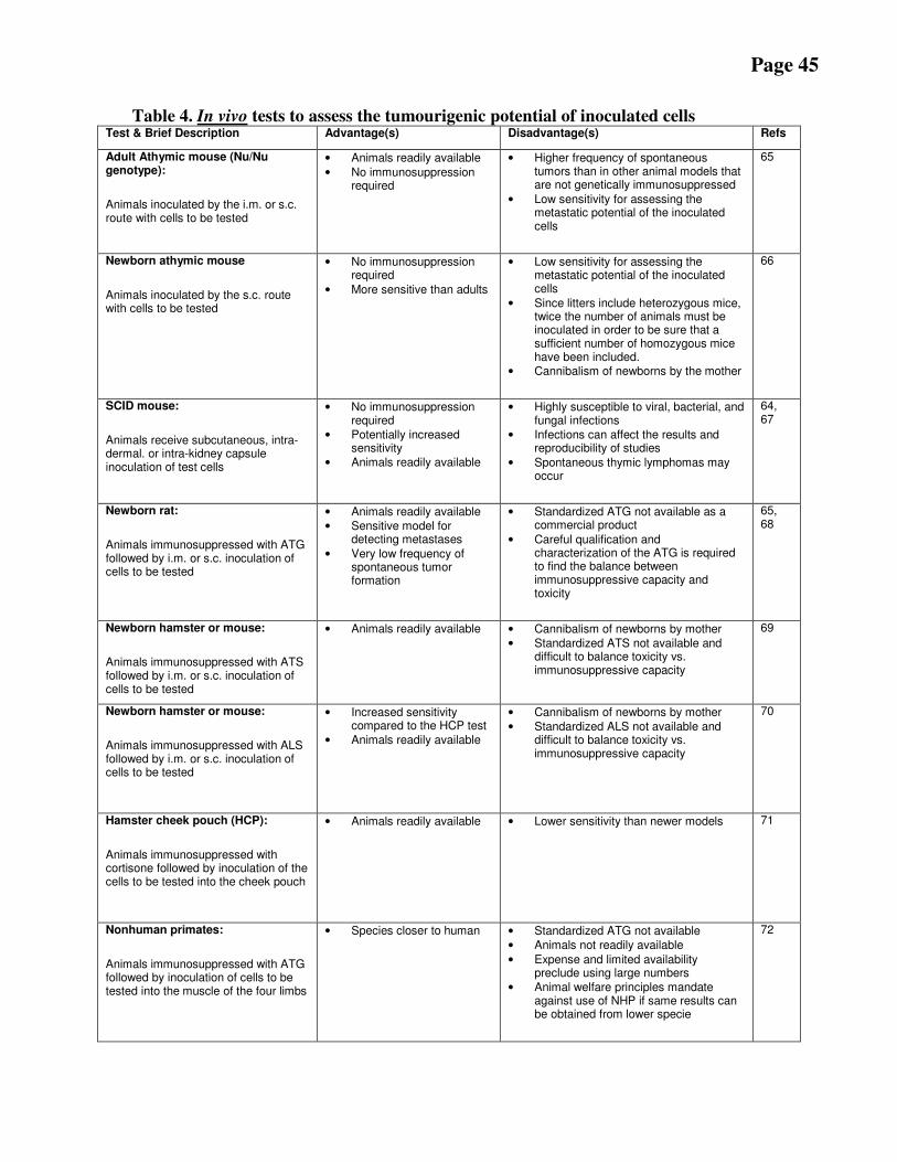
Page 45
Table 4. In vivo tests to assess the tumourigenic potential of inoculated cells Test & Brief Description Advantage(s) Disadvantage(s) Refs
Adult Athymic mouse (Nu/Nu genotype):
Animals inoculated by the i.m. or s.c. route with cells to be tested
• Animals readily available
• No immunosuppression required
• Higher frequency of spontaneous tumors than in other animal models that are not genetically immunosuppressed
• Low sensitivity for assessing the metastatic potential of the inoculated cells
65
Newborn athymic mouse
Animals inoculated by the s.c. route with cells to be tested
• No immunosuppression required
• More sensitive than adults
• Low sensitivity for assessing the metastatic potential of the inoculated cells
• Since litters include heterozygous mice, twice the number of animals must be inoculated in order to be sure that a sufficient number of homozygous mice have been included.
• Cannibalism of newborns by the mother
66
SCID mouse:
Animals receive subcutaneous, intra-dermal. or intra-kidney capsule inoculation of test cells
• No immunosuppression required
• Potentially increased sensitivity
• Animals readily available
• Highly susceptible to viral, bacterial, and fungal infections
• Infections can affect the results and reproducibility of studies
• Spontaneous thymic lymphomas may occur
64, 67
Newborn rat:
Animals immunosuppressed with ATG followed by i.m. or s.c. inoculation of cells to be tested
• Animals readily available
• Sensitive model for detecting metastases
• Very low frequency of spontaneous tumor formation
• Standardized ATG not available as a commercial product
• Careful qualification and characterization of the ATG is required to find the balance between immunosuppressive capacity and toxicity
65, 68
Newborn hamster or mouse:
Animals immunosuppressed with ATS followed by i.m. or s.c. inoculation of cells to be tested
• Animals readily available
• Cannibalism of newborns by mother
• Standardized ATS not available and difficult to balance toxicity vs. immunosuppressive capacity
69
Newborn hamster or mouse:
Animals immunosuppressed with ALS followed by i.m. or s.c. inoculation of cells to be tested
• Increased sensitivity compared to the HCP test
• Animals readily available
• Cannibalism of newborns by mother
• Standardized ALS not available and difficult to balance toxicity vs. immunosuppressive capacity
70
Hamster cheek pouch (HCP):
Animals immunosuppressed with cortisone followed by inoculation of the cells to be tested into the cheek pouch
• Animals readily available
• Lower sensitivity than newer models 71
Nonhuman primates:
Animals immunosuppressed with ATG followed by inoculation of cells to be tested into the muscle of the four limbs
• Species closer to human
• Standardized ATG not available
• Animals not readily available
• Expense and limited availability preclude using large numbers
• Animal welfare principles mandate against use of NHP if same results can be obtained from lower specie
72
Page 46
Although all of the animal models listed in Table 4 have been used to assess the tumourigenicity of
cells, several sensitivity parameters from studies using positive-control cells should be considered
when attempting to compare the various in vivo tumourigenicity models: i) frequency of tumour
formation; ii) time to appearance of tumors; iii) size of tumours; iv) lowest number of inoculated
tumour cells that result in tumour formation; and v) metastatic tumor formation. Factors i, ii, iii, and
v usually depend on the number of cells inoculated (i.e., they are dose-dependent). In addition, the
rate of spontaneous tumour formation should be considered. Although comparisons of two or more
assays have been reported in the literature [ 63, 69, 73] none of them takes into account all of these
factors, nor do they use the same tumourigenic cell lines. Thus, it is not possible to draw definitive
conclusions on the relative sensitivity of the various tumourigenicity assays. Nevertheless, the
following points appear to be generally accepted: i) the ATS-treated newborn rat and the ATG-
treated nonhuman primate systems are the most sensitive to assess the metastatic potential of
inoculated cells; ii) ATS and ATG provide better immunosuppression than ALS; iii) the nude mouse
has a more well-defined level of immunosuppression than those that depend on ALS, ATS, or ATG,
and inter-laboratory comparisons of nude mouse data are more likely to yield valid conclusions.
The overall experience during the past 30 years, and taking into consideration the points mentioned
above, has led to the conclusion that the athymic nude mouse is an appropriate test system for
determining the tumourigenic potential of cells proposed for use in the production of biologicals.
The major advantages of the athymic nude mouse system are that it is easier to establish and
standardize and is generally available, while the newborn rat system is more sensitive for assessing
the metastatic potential of tumourigenic cells. In some cases, it might be preferable to use newborn
athymic nude mice, as these animals are more sensitive than adults for the detection of weakly
tumourigenic cells [ 64]. A tumourigenicity testing protocol using athymic nude mice is provided as
Appendix 2. The animal system selected should be approved by the NRA/NCL.
B.8.3 Point in the life history of the cells at which they should be tested
Investigation of tumourigenicity should form part of the early evaluation of a new cell substrate for
use in production. Cells from the MCB or WCB, propagated to the proposed in vitro cell age used
for production or beyond should be examined for tumourigenicity. The extra population doublings
(e.g., 3 to 10) ensure that the results of the tumourigenicity test can be used in the assessment of
overall safety of the product even under the assumption of a "worst case" situation and therefore
provides a safety buffer.
B.8.4 Use of control cells
The tumourigenicity test should include a comparison between the CCL and a positive control
reference preparation such as HeLa cells from a reliable source. This source is preferred in order to
standardize the test among laboratories, so that the cumulative experience over time can be assessed
and made available to NRAs/NCLs and manufacturers to assist them in the interpretation of data.
However, other sources for establishing positive-control cells may be acceptable. The purpose of the
positive control is to assure that an individual test is valid by demonstrating that the animal model
has the capacity to develop tumors from inoculated cells (i.e., a negative result is unlikely to be due
to a problem with the in vivo model). If the positive-control cells fail to develop tumours at the
expected frequency, then this could be indicative of problems with the animals or at the testing
facility, such as infections, which can reduce the efficiency of tumour development.
Page 47
When the cell substrate has been adapted to growth in serum-free medium, which might contain
growth factors and other components that could influence growth as well as detection of a
tumourigenic phenotype, consideration should be given to processing the positive-control cells in the
same medium. Whenever possible, both the test article and the positive-control cells should be
resuspended in the same medium, such as phosphate-buffered saline (PBS) for inoculation.
In designing a tumourigenicity protocol, it is important to recognize that tumors arise spontaneously
in nude mice and that the incidence of such tumors increases with the age of the mice. Therefore,
databases (both published data and the unpublished records/data of the animal production facility
that supplied the test animals) of rates of spontaneous neoplastic diseases in nude mice should be
taken into account during the assessment of the results of a tumourigenicity test. Generally, negative
controls are not recommended because the rates of spontaneous neoplastic disease in nude mice are
low, and small numbers of negative control animals are unlikely to provide meaningful data.
However, if negative control cells such as WI-38, MRC-5, or FRhl-2 are included, clear justification
for including them should be provided. For example, if serum-free medium is used to grow the cell
substrate, it is conceivable that growth factors may influence the appearance of spontaneous
tumours; therefore, negative-control cells suspended in the same medium may be needed to interpret
the test results.
B.8.5 Number of test animals To determine whether the cells being characterized have the capacity to form tumors in animals, the
cells being tested, the reference positive-control cells, and if any, the reference negative control cells
should be injected into separate groups of 10 animals each. In a valid test, progressively growing
tumors should be produced in at least 9 of 10 animals injected with the positive reference cells.
B.8.6 Number of inoculated cells Each animal should be inoculated intramuscularly or subcutaneously [ 74] with a minimum of 10
7
viable cells. If there is no evidence of a progressively growing nodule at the end of the observation
period, the cell line may be considered to be non-tumourigenic. If the cell line is found to be
tumourigenic, the NRA/NCL might request additional studies to be done to determine the level of
tumourigenicity. This can be done with dose-response studies, where doses of 107, 10
5, 10
3 and 10
1
are inoculated, and the data can be expressed as tumor-producing dose at the 50% endpoint (TPD50
value) [ 75].
B.8.7 Observation period Animals are examined weekly by observation and palpation for evidence of nodule formation at the
site of injection. The minimum observation period depends on the test system selected. In the case of
the nude mouse, a minimum of 4 months is recommended. A shorter period is recommended for the
ATS-treated newborn rat, because the immunosuppressive effect of the ATS declines after the final
injection at two weeks.
B.8.8 Assessment of the inoculation site over time (progressive or regressive growth) If nodules appear, they are measured in two perpendicular dimensions, the measurements being
recorded weekly to determine whether the nodule grows progressively, remains stable, or decreases
in size over time. Animals bearing nodules that are progressing should be sacrificed before the end
of the study if the tumor reaches the limit set by the relevant authorities for the humane treatment of
Page 48
animals. Animals bearing nodules that appear to be regressing should not be sacrificed until the end
of the observation period. Cell lines that produce nodules that fail to grow progressively are not
considered to be tumourigenic. If a nodule persists during the observation period and it retains the
histopathological characteristics of a tumor, this should be investigated further and discussed with
the NRA/NCL.
B.8.9 Final Assessment of the inoculation site
At the end of the observation period, all animals, including the reference group(s), are euthanized
and examined for gross and microscopic evidence of the growth of inoculated cells at the site of
injection and in other sites.
B.8.10 Evaluation of animals for metastases Animals are examined for microscopic evidence of metastatic lesions in sites such as the liver, heart,
lungs, spleen, and regional lymph nodes.
B.8.11 Assessment of metastases (if any) Any metastatic lesions are examined further to establish their relationship to the primary tumor at the
injection site. If what appears to be a metastatic tumor differs histopathologically from the primary
tumor, it is necessary to consider the possibility that this tumor either developed spontaneously or
that it was induced by one or more of the components of the cell substrate, such as an oncogenic
virus. This may require further testing of the tumor itself or the tumourigenicity assay may need to
be repeated. In such cases, appropriate follow-up studies should be discussed and agreed with the
NRA/NCL (also see B.9 Oncogenicity).
B.8.12 Interpretation of results A CCL is considered to be tumourigenic if at least 2 of 10 animals develop tumors at the site of
inoculation within the observation period. However, the reported rate of spontaneous neoplastic
diseases in the test animals should be taken into account during the assessment of the results. In
addition, the histopathology of the tumors must be consistent with the inoculated cells, and a
genotypic marker should show that the tumor is not of nude mouse origin.
If only one of 10 animals develops a tumor, further investigation is appropriate to determine, for
example, if the tumor originated from the cell-substrate inoculum or the host animal and whether
there are any viral or inoculated cell DNA sequences present. The NRA/NCL should be consulted in
this regard.
The dose-response of the CCL may be studied in a titration of the inoculum as part of the
characterization of the CCL. The need for such data will depend upon many factors specific to a
given CCL and the product being developed. The NRA/NCL should be consulted in this regard.
B.8.13 Applicability Cell Banks: Representative EOPC or ECB from the MCB or first WBC
Cell Types: DCL, SCL, CCL
B.9 Oncogenicity B.9.1 Tests for oncogenicity
Page 49
While tumourigenicity is the property of cells to form tumors when inoculated into susceptible
animals, oncogenicity is the property of an acellular agent to induce cells of an animal to become
tumor cells. As such, tumors that arise in a tumourigenicity assay contain cells derived from the
inoculated cells, while tumors that arise in an oncogenicity assay are derived from the host.
Oncogenic activity from cell substrates could be due to either the cell substrate DNA (and perhaps
other cellular components) or an oncogenic viral agent present in the cells. Although there might be
a perception that the cellular DNA from highly tumourigenic cells would have more oncogenic
activity than the DNA of weakly or non-tumourigenic cells, at this time, it is not known if there is a
relationship between the tumourigenicity of a cell and the oncogenicity of its DNA. Nevertheless,
the NRA/NCL might require oncogenicity testing of the DNA and cell lysate from a new cell line
(i.e., other than those such as CHO, NS0, Sp2/0, and low passage Vero, for which there is
considerable experience) that is tumourigenic in animal model systems (see below) because of the
perception that a vaccine manufactured in such a cell line poses a neoplastic risk to vaccine
recipients.
The major complication in assaying cellular DNA in animals arises from the size of the mammalian
genome. Because the mammalian haploid genome is approximately 3 x 10⁹ base pairs (bp) whereas
the size of a typical oncogene could be 3-30 x 103 bp, the concentration of an oncogene in cellular
DNA expression systems would be about 105 to 10
6 fold less concentrated than a plasmid containing
the same oncogene. As a consequence, if 1 µg of an oncogene expression plasmid induces a tumor in
an experimental animal model, the amount of cellular DNA that would contain a similar amount of
the same oncogene is 105 µg to 10
6 µg (i.e., 100 mg to 1 g). To date, three studies have indicated that
between 1 and10 µg of expression plasmids for cellular oncogenes can be oncogenic in mice ( 33, 42,
43). Therefore, more sensitive in vivo assays need to be developed before the testing of the
oncogenic activity of cellular DNA becomes practicable. Recent results suggest that the sensitivity
of the assay can be increased by several orders of magnitude with the use of certain immune-
compromised strains of mice prone to develop tumours after inoculation with oncogenes. Thus, it
may be possible to assess the oncogenic activity of cellular DNA in the future. However, at present
there is no standardized in vivo oncogenicity test for cellular DNA. An example protocol is
nonetheless provided in Appendix 3.
Several in vitro systems, such as scoring the neoplastic transformation of NIH 3T3 cells in a focus-
forming assay following transfection of oncogenic DNA [ 75, 77, 78] have been used to assess
oncogenicity, but it is not clear how these assays reflect the oncogenic activity of DNA in vivo, since
they predominantly detect the oncogenic activity of activated ras-family members, and thus it is
unclear how these assays can assist in estimating risk associated with the DNA or cell lysate from a
cell substrate.
Based on experience with DCLs WI-38, MRC-5, and FRhL-2, testing of new MCBs of these cell
lines for oncogenicity is not recommended. Other DCLs for which there is substantial experience
also may not need to be tested. The NRA/NCL should be consulted in this regard. As stated in
Section B.8.1, a new CCL should be presumed to be tumourigenic unless data demonstrate that it is
not. If a manufacturer demonstrates that a new CCL is non-tumourigenic, oncogenicity testing on
cell DNA and cell lysates might not be required by the NRA/NCL.
Page 50
When appropriate, and particularly for vaccines, cell DNA and cell lysates should be examined for
oncogenicity in a test approved by the NRA/NCL. An oncogenicity testing protocol is provided as
Appendix 3.
B.9.2 Applicability Cell Banks: MCB or first WCB taken to representative EOPC or ECB
Cell Types: CCL, SCL (recommended when tumourigenic cells are used in vaccine production)
B.10 Cytogenetics B.10.1 Characterization Chromosomal characterization and monitoring were introduced in the 1960s to support the safety
and acceptability of human DCLs as substrates for vaccine production. Human DCLs differ from
CCLs by retaining the characteristics of normal cells, including the normal human diploid karyotype.
A significant quantity of data have been accumulated since then, and this has led to the conclusion
that less extensive cytogenetic characterization is appropriate because of the demonstrated
karyotypic stability of human DCLs used in vaccine production [ 79]. Thus, the use of karyology as a
lot-by-lot quality-control test is unnecessary for well-characterized and unmodified human DCLs
(e.g., WI-38, MRC-5) and for FRhL-2.
Cytogenetic data may be useful for the characterization of CCLs, especially when marker
chromosome(s) are identified. Such data might be helpful in assessing the genetic stability of the cell
line as it is expanded from the MCB to the WCB and finally to production cultures (see B.3). The
following recommendations are appropriate for the characterization of DCL and CCL cell banks.
Cytogenetic recharacterization of DCLs (e.g., WI-38, MRC-5, and FRhL-2) should not be required,
unless the cells have been genetically modified or the culture conditions have been changed
significantly, since such data are already available ( 18, 19, 20). However, for each WCB generated,
manufacturers should confirm once that the cells grown in the manner to be used in production are
diploid and have the expected lifespan.
For the determination of the general character of a new or previously uncharacterized DCL, samples
from the MCB should be examined at approximately four equally spaced intervals during serial
cultivation from the MCB through to the proposed in vitro cell age used for production or beyond.
The testing intervals should be agreed upon with the NRA. Each sample should consist of a
minimum of 100 cells in metaphase and should be examined for exact counts of chromosomes, as
well as for breaks and other structural abnormalities. Giemsa-banded karyotypes of an additional five metaphase cells in each of
the four samples may provide additional useful information. ISCN [ 80] 400
band is the minimum acceptable level of Giemsa-banding analysis for
human cells.
Stained slide preparations of the chromosomal characterization of the cells (i.e., DCL, CCL), or
photographs of these, should be maintained permanently as part of the cell-line record. Further
recommendations have been proposed for SCLs [ 53].
B.10.2 Applicability
Page 51
Cell Banks: MCB, ECB or representative EOPC
Cell Types: DCL, SCL, CCL (as a test for genetic stability, when appropriate),
B.11 Microbial agents B.11.1 General considerations While many biological production systems require human or animal-cell substrates, such cells are
subject to contamination with and have the capacity to propagate extraneous, inadvertent, or so-
called adventitious organisms, such as mycoplasma and viral agents. In addition, animal cells
contain endogenous agents such as retroviruses that also may be of concern. Testing for both
endogenous (e.g., retroviruses) and adventitious agents (e.g., mycoplasmas) is described in the
succeeding sections. In general, cell substrates contaminated with microbial agents are not suitable
for the production of biological products. However, there are exceptions to this general rule. For
example, the CHO and other rodent cell lines that are used for the production of highly purified
recombinant proteins express endogenous retroviral particles. Risk versus benefit must be considered
when determining the suitability of a cell substrate for the production of a specific product. Further,
risk-mitigation strategies during production, including purification (removal) and inactivation by
physical, enzymatic, and/or chemical means, should be implemented whenever appropriate and
feasible. Even though a cell substrate might be unacceptable for some products, such as for a live
viral vaccine subjected to neither significant purification nor inactivation, that same cell substrate
might be an acceptable choice for a different type of product, such as a highly purified recombinant
protein or monoclonal antibody for which risk mitigation has been achieved by significant and
validated viral clearance in the production process.
A strategy for testing cell banks for microbial agents should be developed. One strategy is to
perform exhaustive testing at the MCB level and to perform more limited testing on the WCB
derived from the MCB. This more limited testing would be selected on the basis of those agents that
could potentially be introduced during the production of the WCB from the MCB. Testing would not
need to be replicated for agents that could only have been present prior to the production of the MCB
(e.g., an endogenous retrovirus, or BVDV from serum used for developing the cell seed or in the
legacy of establishing the cell line).
However, if the number of vials of a MCB is limited, an alternative strategy would be to conduct the
more exhaustive testing on the first WCB made from that MCB, and to limit testing on the MCB
itself. An advantage to the strategy of performing more exhaustive testing on the first WCB is that it
provides a greater opportunity for amplification of any agents that may have been introduced earlier
and through to production of the WCBs. There are advantages and disadvantages to more extensive
testing of the MCB or the WCB, and consideration should be given to what is more appropriate for
the particular product(s) to be manufactured using a given cell bank. Consultation with the
NRA/NCL should be considered prior to implementation to determine whether a proposed testing
strategy is acceptable.
EOPC/ECB should be characterized once for each commercial production process. Testing of the
ECB serves as further characterization of the MCB or WCB that was exhaustively tested and permits
additional time/passages for amplification of low-level contaminants or reactivation of viral
contaminants that may have been missed in the testing of the upstream bank.

Page 52
B.11.2 Viruses Manifestations of viral infections in cell cultures vary widely among the broad array of virus
families that are potential contaminants; thus, the methods used to detect them vary. Lytic infections
frequently are detected by the CPE they cause. However, in some cases such as non-cytopathic
BVDV, no CPE is observed. Viruses also may be present latently (e.g., herpesviruses) or
endogenously (in the germline, e.g., retroviral proviruses). Such inapparent infections might require
specific techniques designed to reveal their presence, such as molecular and immunological
methods, and electron microscopy. For new cell substrates, induction of a detectable infection by
exposing the cells to special conditions (e.g., chemical induction; heat shock) may be required, and
special detection techniques like transcriptome sequencing or degenerate primer PCR may have
utility.
The strategy developed to test cell substrates for viruses should take into consideration the families
of viruses and specific viruses that may be present in the cell substrate. Consideration should be
given to the species and tissue source from which the cell substrate originated, and to the original
donor's medical history in the case of human-derived cell substrates or to the pathogen status of
donor animals in the case of animal-derived cell substrates. In addition, consideration should be
given to viruses that could contaminate the cell substrate from the donors or from animal- or human-
derived raw materials used in the establishment and passage history (legacy) of the cell substrate
prior to and during cell banking or production (e.g., serum, trypsin, animal- or human-derived
medium components, antibodies used for selection, or animal species through which the cell
substrate may have been propagated), as well as laboratory contamination from operators or other
cell cultures.
Tests should be undertaken to detect, and where possible identify, any endogenous or exogenous
agents that may be present in the cells. Attention should be given to tests for agents known to cause
an inapparent infection in the species from which the cells were derived, making it more difficult to
detect (e.g., simian virus 40 in rhesus monkeys).
Primary cells are obtained directly from the tissues of healthy animals and are more likely to contain
adventitious agents than banked, well-characterized cells. In addition, recent vaccination of source
animals should be considered, as they may be exposed to live vaccines. This risk with primary cells
can be mitigated by rigorous qualification of source animals and the primary cells themselves. When
feasible, animals from which primary cultures are established should be from genetically closed
flocks, herds, or colonies monitored for freedom from pathogens of specific concern. Such animals
are known as specific-pathogen-free (SPF). The term "closed" refers to the maintenance of a group
(flock, herd, or colony) free from introduction of new animals (new genetic material that could
introduce, e.g., new retroviral proviruses). Many live viral vaccines are commonly produced in
primary cells and undergo little purification during production. In such cases, and when feasible, the
use of SPF animals is highly recommended. Documentation of the status of the source animals
should be provided to the NRA/NCL. Animals that are not from closed flocks, herds, or colonies
should be quarantined and thoroughly evaluated for a period sufficient to detect signs of disease or
infection (e.g., monkeys are generally quarantined for 6 or more weeks [ 81]). Such animals also
should be screened serologically for appropriate adventitious agents to determine their suitability as
a source for the primary-cell substrate. Animal-husbandry practices should be documented. Even so,
viral contamination of the cells may not be excluded from all cultures. For example, contamination
Page 53
of primary monkey kidney cells with foamy virus or simian cytomegalovirus is common in the
absence of specific concerted efforts to prevent these contaminations.
For primary-cell cultures, the principles and procedures outlined in Part C, Requirements for
Poliomyelitis Vaccine (Oral) [ 81], together with those in section A.4 of Requirements for Measles,
Mumps and Rubella Vaccines and Combined Vaccine (Live) [ 82] may be followed.
The production of viral vaccines, such as those against smallpox or rabies, originally required the
use of living animals, and the great range of possible viral contaminants only became apparent as
cell-culture methods were developed. For example, human enteroviruses were not recognised until
the development of monkey kidney cell cultures, in which they could produce cytopathic effects,
because the disease produced in humans is either relatively mild or in some cases non-existent. It
was also clear that viruses could be detected in some systems but not others; for instance, the
polyomavirus SV40 does not produce a cytopathic effect in cultures from rhesus monkey kidney
cells (in which much of the early polio vaccines were produced) derived from SV40-infected
monkeys but will do so in cultures from cynomolgous or African green monkey kidney cells. The
suspicion was therefore that there were many viruses in the culture systems of the time and that they
were detected only if the assays were appropriate. This remains an accurate view and has lead to a
range of different approaches to try and detect all contaminants.
Coxsackie viruses are named after the town in New York where they were first identified and were
historically detected by their effects in mice. Coxsackie B viruses produce clinical signs and death in
adult mice, while Coxsackie A viruses will affect only suckling mice. For many years, tissue-culture
methods were a less reliable method of detection of Coxsackie A viruses than suckling mice, and the
continued use of these animals in cell bank characterisation reflects this.
In the 1940s, embryonated chicken eggs were a popular substrate for the growth and assay of viruses
such as influenza, measles, mumps, yellow fever and vaccinia. They therefore appear to have a wide
range of susceptibility. Simian viruses such as SV5 or viruses such as Sendai virus also grow well in
them. As many are paramyxoviruses with haemagglutinating (HA) activity, the egg-based assays
include tests for HA activity as well as death of the embryos.
A range of tissue-culture cells is also used, typically including one human, one of the same species
as the production cell, and one other (often, monkey origin). The hope is that the range will catch
viruses not detected by other means, although in practice it is wise to assume that there is no such
thing as a generic detection method; for example, cells from an inappropriate monkey species would
not necessarily detect SV40 whereas cells from other species will (e.g., Vero cells). In certain
circumstances, where a virus is of particular concern, specific tests have been applied. For example,
herpes B virus is a common infection of monkeys in the absence of precautions such as quarantine
and clinical evaluation of the donor animals, and has very serious effects on infected humans. While
herpes B virus was routinely detected by the use of primary rabbit kidney cell cultures, established
rabbit cell lines are now acceptable for this purpose. Another example is Marburg virus, which in the
1970s caused a number of deaths in workers who handled monkeys that were to be used in a
vaccine-production facility. The incident might have been avoided had the animals been adequately
quarantined. A specific test in guinea pigs was introduced and maintained for a number of years to
ensure the absence of the agent.
Page 54
There is a disparate range of tests that have been or are still used with the aim of detecting any
significant contaminant that may be present in cell cultures. Some, such as the rabbit kidney cell test,
are very specific in intent while others may be expected to be more generic. In general, however, it is
wise to assume that an assay will never be all encompassing whether based on historical virological
approaches or more current methodologies. Thus, the consequences of deleting tests on the grounds
of redundancy must be very carefully evaluated before any action is taken. On the other hand, it is
difficult to justify maintenance of a test if it only detects viruses also detected by other methods of
equivalent sensitivity, comparable ease of use, and cost. Each of these considerations should be
given when developing an appropriate testing strategy for the given cell bank. Policies to minimize
the use of animals in safety testing should also be considered, but must be considered in balance with
the utility and necessity (sensitivity and ability to detect particular adventitious agents not readily
detected by other means) of the test in which they are used.
B.11.2.1 Tests in animals and eggs
The cells of the MCB and WCB are unsuitable for production if any of the animal or egg tests shows
evidence of the presence of any viral agent attributable to the cell banks.
Generally, MCBs are thoroughly characterized by the methods listed below. WCBs may be
characterized by an abbreviated strategy, when appropriate. However, an alternative strategy to this
general rule may be used, as discussed in section B.11.2 above. These tests may be performed
directly on cells or supernatant fluids or cell lysates from the bank itself or on cells or supernatant
fluids or cell lysates from passaged cells from the bank that have been passaged to the proposed in
vitro cell age for production or beyond.
In some countries, policies exist to minimize the use of animals in safety
testing.
B.11.2.1.1 Adult mice
The original purpose of this test was for the detection of lymphocytic choriomeningitis virus
(LCMV). The test in adult mice for pathogenic viruses includes inoculation by the intraperitoneal
route (0.5 mL) with cells and culture fluids from the MCB or WCB, where at least l07 viable cells or
the equivalent cell lysate are divided equally among at least ten adult mice weighing 15-20 g.
In some countries, the adult mice are also inoculated by the intracerebral
route (0.03 mL).
In some countries at least 20 mice are required for each test.
The animals are observed for at least 4 weeks. Any animals that are sick or show any abnormality
are investigated to establish the cause. Animals that do not survive the observation period should be
examined for gross pathology and histopathology in order to determine a cause of death, and if a
viral infection is indicated, efforts should be undertaken to identify the virus. Viral identification
may involve culture and/or molecular methods. Further, each mouse that dies after the first 24 hours
of the test, or is sacrificed because of illness, should be necropsied and examined for evidence of
viral infection by subinoculation of appropriate tissue into at least five additional mice which should
be observed for 21 days. The test is not valid if more than 20% of the animals in either or both of the
Page 55
test and negative control groups (if used) become sick for non-specific reasons and do not survive
the observation period.
In some countries, the adult mice are observed for 21 days.
If the cell substrate is of rodent origin, at least 106 viable cells or the equivalent cell lysate are
injected intracerebrally into each of ten susceptible adult mice to test for the presence of LCMV.
In some countries, after the observation period, the animals are challenged
with live LCMV to reveal the development of immunity against non-
pathogenic LCMV contaminants resulting in otherwise unapparent
infection.
B.11.2.1.1.1 Applicability
Cell Banks: MCB, WCB, or ECB or representative EOPC
Cell Types: PCC, DCL, SCL, CCL
B.11.2.1.2 Suckling mice
The original purpose of this test was for the detection of Coxsackie viruses. The test in suckling
mice for pathogenic viruses includes inoculation by the intraperitoneal route (0.1 mL) with cells and
culture fluids from the MCB or WCB; at least l07 viable cells or the equivalent cell lysate are divided
equally between two litters of suckling mice, comprising a total of at least ten animals less than 24-
hours old.
In some countries, the suckling mice are also inoculated by the intracerebral
route (0.01 mL).
In some countries, 20 suckling mice are inoculated.
The animals are observed for at least 4 weeks. Any animals that are sick or show any abnormality
are investigated to establish the cause. Animals that do not survive the observation period should be
examined for gross pathology and histopathology in order to determine a cause of death, and if a
viral infection is indicated, efforts should be undertaken to identify the virus, where this is
practicable. Viral identification may involve culture and/or molecular methods. Further, such
examination of viral infection should include subinoculation of appropriate tissue suspensions into
an additional group of at least five suckling mice by intracerebral and intraperitoneal routes and
observation daily for 14 days. In the case of suckling mice, it is often observed that those that perish
are cannibalized by their mother and this renders determining a cause of death impossible (when
they are fully cannibalized and no remains can be recovered). The test is not valid if more than 20%
of the animals in either or both of the test group and negative control group (if used) do not survive
the observation period.
In some countries, the suckling mice may be observed for a period of 14
days followed by a sub-passage involving a blind passage (via
intraperitoneal and intracerebral inoculation into at least 5 additional mice)
of a single pool of the emulsified tissue (minus skin and viscera) of all mice
surviving the original 14-day test.
B.11.2.1.2.1 Applicability
Page 56
Cell Banks: MCB, WCB or ECB or representative EOPC
Cell Types: PCC, DCL, SCL, CCL
B.11.2.1.3 Guinea pigs
The original purpose of this test was for the detection of LCMV and Mycobacterium tuberculosis.
When it is necessary to detect Mycobacterium species, a test in guinea pigs is performed and
includes inoculation by the intraperitoneal route (5 mL) with cells and culture fluids from the MCB
or WCB, where at least l07 viable cells or the equivalent cell lysate are divided equally among the
animals.
In some countries, tests in five guinea-pigs weighing 350-450 g are also
inoculated by the intracerebral route (0.1 mL) and observed for 42 days to
reveal Mycobacterium tuberculosis and other species.
The animals are observed for at least 6 weeks. Any animals that are sick or show any abnormality
are investigated to establish the cause. Animals that do not survive the observation period should be
examined for gross pathology and histopathology in order to determine a cause of death, and if a
viral infection is indicated, efforts should be undertaken to identify the virus. Viral identification
may involve culture and/or molecular methods. The test is not valid if more than 20% of the animals
in either or both of the test and negative control (if used) groups do not survive the observation
period.
The test in guinea-pigs for the presence of Mycobacterium may be replaced by an alternative in vitro
method such as culture, or shortened culture with a PCR endpoint (also see B.11.3).
B.11.2.1.3.1 Applicability
Cell Banks: MCB, WCB or ECB or representative EOPC
Cell Types: PCC, DCL, SCL, CCL (the latter three are dependent on legacy and current use of
media components of animal origin that could result in contamination with Mycobacterial species)
B.11.2.1.4 Rabbits
The original purpose of this test was for the detection of Herpes B virus. When it is necessary to
detect simian herpes B virus, the test in rabbits for pathogenic viruses is performed and includes the
inoculation by the intradermal (1 mL) and subcutaneous (>2 mL) routes with cells and culture fluids
from the MCB or WCB, where at least l07 viable cells or the equivalent cell lysate are divided
equally among the animals.
In some countries, tests in five rabbits weighing 1.5-2.5 kg are inoculated
by the subcutaneous route with either 2 mL or between 9 and 19 mL.
Consultation with the NRA/NCL regarding acceptable methods should be
considered.
The animals are observed for at least 4 weeks. Any animals that are sick or show any abnormality
are investigated to establish the cause. Animals that do not survive the observation period should be
examined for gross pathology and histopathology in order to determine a cause of death, and if a
viral infection is indicated, efforts should be undertaken to identify the virus. Viral identification
may involve culture and/or molecular methods. The test is not valid if more than 20% of the animals
in either the test or the negative control (if used) groups do not survive the observation period.
Page 57
The test in rabbits for the presence of herpes B virus is intended for primary simian cultures, and
may be replaced by a test in rabbit kidney cell cultures.
B.11.2.1.4.1 Applicability
Cell Banks: MCB, WCB or ECB or representative EOPC
Cell Types: PCC, DCL, SCL, CCL
B.11.2.1.5 Embryonated chicken eggs
At least 106 viable cells or the equivalent cell lysate, along with culture fluids, from the MCB or
WCB of avian origin, propagated to the proposed in vitro cell age for production or beyond are
injected into the allantoic cavity of each of at least ten embryonated hens' eggs, and into the yolk sac
of each of at least another ten embryonated hens' eggs. The eggs are examined after not fewer than 5
days of incubation. The allantoic fluids of the eggs are tested with red cells from guinea pig and
chickens (or other avian species) for the presence of haemagglutinins. The test is not valid if more
than 20% of the embryonated hens' eggs in either or both of the test group and negative control
group (if used) are discarded for non-specific reasons.
In some countries, the NRA/NCL also requires that other types of red cells,
including cells from humans (blood group IV O) or monkeys should be used
in addition to guinea-pig and chicken (or other avian species) cells. In all
tests, readings should be taken after incubation for 30 minutes at 0-4°C, and
again after a further incubation for 30 minutes at 20-25°C. For the test with
monkey red cells, readings also should be taken after a final incubation for
30 minutes at 34-37°C.
In some countries, inoculation by the amniotic route is used.
In some countries, following incubation, allantoic fluids or a 10%
suspension of yolk sacs, as appropriate, should be harvested, pooled, and
blind passaged into an additional group of eggs.
Usually, the eggs used for the yolk sac test should be 5-7 days old. The eggs used for the allantoic
cavity test should be 9-11 days old.
Alternative ages for the embryonated chicken eggs and alternative
incubation periods are acceptable if they have been determined to be
equivalent or better at detecting the presence in the test samples of the
adventitious agents this test is capable of detecting when performed as
above.
Embryos that do not survive the observation period should be examined for gross pathology in order
to determine a cause of death, and if a viral infection is indicated, efforts should be undertaken to
identify the virus. Viral identification may involve culture and/or molecular methods.
B.11.2.1.5.1 Applicability
Cell Banks: MCB of avian origin, WCB of avian origin or ECB or representative EOPC
Cell Types: avian PCC, DCL, SCL, CCL (also recommended for novel cell substrates)
B.11.2.1.6 Antibody-production tests
Page 58
Rodent cell lines are tested for species-specific viruses using mouse, rat and hamster antibody
production tests, as appropriate. In vivo testing for lymphocytic choriomeningitis virus, including a
challenge for non-lethal strains, is performed for such cell lines, as described in Section B.11.2.1.1.
Avian cell lines may also be tested using a chick antibody-production test, e.g., to detect chicken
anemia virus. Further, if the cell substrate (even if not of rodent origin) has been exposed to rodent-
origin materials, e.g., selection using a monoclonal antibody, testing should be considered for the
relevant-species viruses using an antibody-production test [ 83, 84].
In some countries, consideration is being given to use of
nucleic acid testing (NAT) in place of the in vivo antibody-
production testing. In these cases, data should be provided to
the NRA/NCL that justifies this practice.
B.11.2.1.6.1 Applicability
Cell Banks: MCB, WCB, or ECB or representative EOPC
Cell Types: DCL, SCL, CCL (recommended primarily for cells of rodent origin)
B.11.2.2 Tests in cell culture
Tests in cell culture are capable of detecting a broad array of viral families. Readouts include
monitoring the cultures periodically for CPE and tests for haemadsorbing and haemagglutinating
viruses, which are conducted at the end of the culture period. In addition to the indicator cells
described below, it may be appropriate to expand the different types of indicator cells used (beyond
2 or 3) to enable the detection of viruses with differing host requirements. Decisions about which
cell lines to use as indicator cells should be guided by the species and legacy of the production cell
substrate taking into the consideration the types of viruses to which the cell substrate could
potentially have been exposed and thus the viruses one would like to detect by this assay method.
The cell substrate is unsuitable for production if any of the indicator cell cultures shows evidence of
the presence of any viral agent attributable to the tested cell substrate.
B.11.2.2.1 Applicability
Cell Banks: MCB, WCB or ECB or representative EOPC
Cell Types: PCC, DCL, SCL, CCL
B.11.2.2.2 Indicator cells
Live cells or cell lysate, each with spent culture fluids of the MCB or WCB are inoculated onto
monolayer cultures or cultivated with monolayer cultures of the cell types listed below, as
appropriate.
A lysate of the cells may be prepared by a method that avoids
virus disruption while allowing maximal virus release (e.g.,
typically three freeze/thaw cycles followed by low-speed
centrifugation). If cells, lysate, or spent culture fluids are to be
stored prior to testing, then they should be stored at <-70°C.
• Cultures (primary cells or CCL) of the same species and tissue type as that used for
production.
Page 59
• Cultures of a human DCL. The original purpose of this test, utilizing primary human cells,
was the detection of measles virus. But, where the cell substrate is of human origin, a simian
kidney cell line should be used as the second indicator cell line. The original purpose of the
use of this cell type was the detection of simian viruses.
In some countries, cultures of another (third) cell line from a different
species are required.
In many circumstances, more than two cell lines may be necessary to cover
the range of potential viral contaminants and typically, a third cell line
would be used that is of simian origin, if the cell substrate is not of simian
origin.
For new cell substrates, additional cell lines to detect viruses known to be potentially harmful to
humans could be considered (e.g., for insect cell lines, if the cells selected for the above mentioned
tests are not known to be permissive to insect viruses, an additional detector cell line should be
included in the testing).
The cell bank sample to be tested is diluted as little as possible. At least 107 cells, or equivalent cell
lysate, and spent culture fluids are inoculated onto each of the indicator cell types. The resulting co-
cultivated or inoculated cell cultures are observed for evidence of viruses by cytopathic effect for at
least 2 weeks. If the cell line is known to be capable of supporting the growth of human or simian
cytomegalovirus, HDC cultures are observed for at least 4 weeks. Extended (4-weeks) cell culture
for the purposes of detecting human or simian cytomegalovirus can be replaced by the use of NAAT
to detect cytomegalovirus nucleic acid.
In some countries, a passage onto fresh cultures for an additional 2 weeks is
recommended for all indicator cultures. In some cases, it may be difficult to
keep the cell cultures healthy for 2 weeks without subculturing. In those
cases, it may be necessary to feed the cultures with fresh medium or to
subculture after two weeks onto fresh cultures in order to be able to detect
viral agents.
At the end of the observation period, samples of each of the co-cultivated or inoculated cell culture
systems are tested for haemadsorbing and/or haemagglutinating viruses, as described in section
B.11.2.1. 5.
B.11.2.2.3 Additional considerations to the tests in cell culture for insect viruses
Many insect cell lines carry persistent viral infections that do not routinely produce a noticeable CPE
(e.g., some clones of the Hi-5 cell line are persistently infected with an insect nodavirus). However,
the viruses may be induced to replicate by stressing the cells using a variety of techniques such as
increased/reduced culture temperature (above or below that routinely used for production), heat-
shock for a short period, super-infection with other insect viruses, or chemical inducers. Therefore,
the probability of detecting such low-level persistent infections may be increased by stressing the
cells prior to analysis.
Intact cells and cell lysates from a passage level at or beyond that equivalent to the EOPC is co-
cultivated with indicator cells from at least three different species of insect in addition to the
Page 60
indicator cells as noted in section B.11.2.2.2. Cell lines should be selected on the following basis:
one of the lines has been demonstrated to be permissive for the growth of human arboviruses, one
has been shown to be permissive for the growth of a range of insect viruses, and the third has been
derived from a species that is closely related to the host from which the MCB is derived (or another
line from the same species). Duplicate cultures of indicator cells are typically incubated at two
temperatures, such as 37 ± 1°C and a lower temperature, such as 28 ± 1°C, observed for a period of
14 days, and examined for possible morphological changes. The cell-culture fluids from the end of
the test period are tested for haemagglutinating viruses, or the intact cells from the end of the test
period are tested for haemadsorbing viruses. The cells comply with the test if no evidence of any
viral agent is found.
Several mosquito cell lines are available that are permissive for the growth of some human
arboviruses and could be considered for these tests. Alternatively, BHK-21 cells could be considered
for this purpose. The most permissive insect cell lines characterised to date have been derived from
embryonic Drosophila tissues. While the mosquito and Drosophila cell lines may be suitable for
some aspects of the testing, it should be remembered that many insect cell lines are persistently
infected with insect viruses that usually produce no obvious CPE. In addition, many insect cells may
be infected with mammalian viruses, such as BVDV, that are known to replicate in insect cells.
Demonstrating that the indicator cell lines are themselves free from adventitious agents is an
important pre-requisite to their use in the testing outlined above. Consideration should also be given
to risk-mitigation strategies as discussed above for highly purified products for which viral clearance
can be achieved and validated.
B.11.2.3 Transmission electron microscopy
At least 200 cells from the MCB or WCB, and ECB are examined by transmission electron
microscopy (TEM) for evidence of contamination with microbial agents. Methods include negative
staining and thin section. A discussion of these methods is provided by Bierley et al. [ 85]. It may be
appropriate to examine more cells in some cases, such as discussed below for insect cell lines, and
the NRA/NCL should be consulted in this regard. Any unusual or equivocal observations that might
be of microbiological significance should be noted and discussed with the NRA/NCL.
TEM can detect viral particles in a cell substrate, including certain endogenous retroviruses. While
TEM is fairly insensitive (generally detecting gross contamination, but not necessarily low-level
contamination), it is a generic assay that can detect microbial agents of many types.
B.11.2.3.1 Applicability
Cell Banks: MCB, WCB, or ECB or representative EOPC
Cell Types: DCL, SCL, CCL
B.11.2.3.2 Additional considerations to TEM for insect cells
For MCBs and WCBs derived from insect cells, the general screening test outlined above applies. In
addition, cell lines should be subjected to stress conditions, such as described in section B.11.2.2.3,
prior to examination by transmission electron microscopy. Further, increasing the number of cells
examined may also improve the probability of detecting an agent (e.g., errantiviruses and
hemiviruses). The maintenance temperatures and treatments used should be agreed with the
NRA/NCL along with the number of sectioned cells to be examined.
Page 61
B.11.2.4 Tests for retroviruses
All vertebrate and insect cells that have been analysed possess endogenous, genetically acquired
retroviral sequences integrated into chromosomal DNA in the form of proviruses. These sequences
may be expressed, or be induced, as mRNA. In some cases, the mRNA is translated into viral protein
and virus particles (virions) are produced. In many cases, these virions are defective for replication
(e.g., avian endogenous retrovirus EAV, Chinese hamster ovary cell line gamma-retrovirus [ 86])
whereas in others (e.g., X-MuLV) the retroviruses may be capable of infecting cells of other species
including human cells.
Consideration should also be given to the possibility that cell banks may be infected with non-
genetically acquired retroviruses (exogenous retroviruses), either because the donor animal was
infected or through laboratory contamination.
It should be noted that infection by retroviruses is not necessarily associated with any cytopathic
effect on the cells and therefore it may require screening assays, like the Product-Enhanced RT
(PERT) assay for reverse transcriptase or TEM to reveal their presence.
The cells of the MCB or WCB are unsuitable for production if the tests for infectious retroviruses, if
required, show evidence of the presence of any viral agent attributable to the substrate that cannot be
demonstrated to be cleared during processing. Generally, the downstream manufacturing process for
products (e.g., monoclonal antibodies) made in cell substrates that produce retroviral particles (e.g.,
CHO cells) or infectious endogenous retrovirus (i.e., NS0, SP2/0 cells) is validated to provide
adequate viral clearance [ 14]. The margin of viral clearance required should be agreed with the
NRA/NCL.
Chick embryo fibroblasts (CEF) contain defective retroviral elements that frequently produce
defective particles with reverse transcriptase activity. This has been the subject of many studies and
WHO consultations because they are used for live viral vaccine production. If evidence is presented
that the donor flock is free of infectious retroviruses and there is no evidence that the cultures are
contaminated with infectious retroviruses, then the cultures can be considered acceptable with
respect to retrovirus tests.
Rodent cell lines express endogenous retroviruses, and thus infectivity tests should be performed to
determine whether these endogenous retroviral particles are infectious.
Cell lines such as CHO, BHK-21, NS0, and Sp2/0 have frequently been used as substrates for drug
production with no reported safety problems related to virus contamination of the products and may
be classified as "well-characterized" because the endogenous retrovirus particles have been studied
extensively. Furthermore, the total number of retrovirus-like particles present in the harvest is
evaluated quantitatively (TEM or QPCR) on a representative number of lots and retrovirus clearance
is demonstrated with significant safety factors. Thus, in these situations testing for infectious
retrovirus may be reduced; e.g., test one lot then discontinue testing, but repeat when there is a
significant change in the cell-culture process such as a change in scale. Sponsors are encouraged to
consult with the NRA.
Page 62
B.11.2.4.1 Applicability
MCBs and cells that have been propagated to the proposed in vitro cell age for production or
beyond. Alternatively, this testing could be performed on WCBs.
Cell Banks: MCB or WCB, and ECB or representative EOPC
Cell Types: DCL, SCL, CCL
B.11.2.4.2 Reverse Transcriptase (RT) Assay
Test samples from the MCBs or WCBs propagated to the proposed in vitro cell age for production or
beyond are examined for the presence of retroviruses.
Culture supernatants are tested by a highly sensitive, quantitative, polymerase chain reaction (PCR)-
based RT assay or PERT assay [ 87, 88, 89, 90].
RTase activity is not specific to retroviruses and may derive from other sources, such as retrovirus-
like elements that do not encode a complete or infectious genome [ 91, 92, 93, 94, 95] or cellular DNA-
dependent DNA polymerases [ 96, 97]. Attempts to reduce the PERT activity associated with cellular
DNA-dependent DNA polymerases have been reported [ 97, 98, 99], although no treatment can
eliminate all activity, and thus the results of such highly sensitive assays need to be interpreted with
caution. Use of appropriate controls in the assay assist in this regard. Since RT activity can be
associated with the presence of defective retrovirus-like particles and since polymerases other than
reverse transcriptase can result in apparent RT activity, a positive result in an RT assay is not
conclusive evidence of the presence of infective retrovirus. Positive results may require further
investigation, e.g., carrying out infectivity assays (see Section B.9.1.4.4). Also, it may be useful to
utilize the conventional RT assay in this investigation to determine whether the RT activity is Mg++
or Mn++ dependent. Such testing should be agreed in advance with the NRA/NCL.
CEFs and other cells of avian origin are known to express retroviral elements. With such cells, the
appropriateness of this test should be discussed with the NRA/NCL. For example, it may be
appropriate to direct testing strategies at the detection of infectious avian retroviruses, such as avian
leukosis viruses and reticuloendotheliosis virus, including serological screening of flocks that are the
source of the CEFs. Additionally, it is known that insect cells have retroviral elements that are
detected by a PERT assay, and so they too can test positive by this assay.
B.11.2.4.3 PCR or other specific in vitro tests for retroviruses
When the PERT test gives unclear results, or when it is unavailable, it may be appropriate to screen
the cell substrate for species-specific retroviruses by molecular methods, such as the PCR,
immunofluorescence, ELISA, or other virus-specific detection methods. Molecular methods, such as
PCR, also may be used for quantification of retrovirus like particles in the production harvests
provided that the method is validated accordingly. Consultation with the NRA/NCL regarding the
acceptability of this approach is recommended.
B.11.2.4.4 Infectivity test for retroviruses
When the test sample is found to have RT activity, it might be necessary to carry out infectivity
assays to assess whether the activity is associated with replicating virus.
Page 63
Because rodent cells generally express endogenous retroviruses, the infectivity and in vitro host
range of such retroviruses should be assessed. Test samples from the MCB or WCB, propagated to
the proposed in vitro cell age for production or beyond should be examined for the presence of
retroviruses by infectivity assays. Cells to be used for these assays should be able to support the
replication of a broad range of viruses; this might require using cells of various species and cell
types. The testing strategy should be agreed with the NRA/NCL.
It is often possible to increase the sensitivity of assays by first inoculating the test material onto cell
cultures that can support retroviral growth in order to amplify any retrovirus contaminant that may
be present at low concentrations. For non-murine retroviruses, test cell lines should be selected for
their capacity to support the growth of a broad range of retroviruses, including viruses of human and
non-human primate origin [ 100, 101].
For murine retroviruses, it is important to assess whether the cells release infectious retroviruses and,
if so, to determine the host range of those viruses. The testing for murine retroviruses can be
complex, and the NRA/NCL should be consulted for guidance. Murine and other rodent cell lines
(CHO, NS0, SP2/0) or hybrid cell lines containing a rodent component should be assumed to be
inherently capable of producing infectious retroviruses or non-infectious retrovirus-like particles. In
such cases, the clearance (removal and/or inactivation) of such retroviruses during the manufacturing
process should be quantified and provide a level of clearance acceptable to the NRA/NCL.
Any testing proposed by the manufacturer should be agreed with the NRA/NCL.
B.11.2.5 Tests for particular viruses not readily detected by the tests described in sections B.11.2.1,
B.11.2.2, B.11.2.3, and B.11.2.4 and their sub-sections
Some viruses, such as hepatitis B or C viruses or human papillomaviruses, cannot be detected
readily by any of the methods described above because they are not known to grow readily in cell
culture or are restricted to human host range. Some animal viruses, e.g., bovine polyomavirus and
porcine circoviruses, are not readily detected by the routine tests previously described. In such
circumstances, it may be necessary to include specific assays for such viruses. While broad general
tests are preferable for detecting unknown contaminants, some selected viruses may be screened by
using specific assays, such as molecular techniques (e.g., nucleic acid amplification). Antibody-
based techniques might also be employed, such as immunofluorescence assays.
Generally, once the MCB, WCB, or ECB has been demonstrated to be free of selected viruses, it
might not be necessary to test the cells at later stages (e.g., at the production level) if such viruses
could not be introduced readily during culture.
Human cell lines should be screened using appropriate in vitro techniques for specific viruses that
are the cause of significant morbidity, for those viruses that might establish latent or persistent
infections and for viruses that may be difficult or impossible to detect by the techniques described in
sections B.11.2.1, B.11.2.2, B.11.2.3, and B.11.2.4 and their sub-sections. Selection of the viruses to
be screened should take into account the tissue source and medical history of the donor, if available,
from whom the cell line was derived.
Page 64
Under circumstances in which the cell origin or medical history of the donor, if available, would
suggest their presence, it may be appropriate to perform specific testing for the presence of human
herpesviruses, human retroviruses, human papillomaviruses, human hepatitis viruses, human
polyomaviruses, or difficult-to-culture types of human adenoviruses.
Consideration should be given to screening insect cell lines for specific viruses that have been
reported to contaminate that particular cell line (e.g., nodaviruses) or viruses that may be present
persistently in insect cell lines and that are known to be infectious for humans.
B.11.2.5.1 Applicability
The NRA/NCL should be consulted in regard to the specific pathogens or selected viruses that
should be included in the testing strategy as these will be directed on a case-by-case basis depending
on the species and origin of the cell and the medical history of the donor, if available.
Cell Banks: MCB, WCB, or ECB or representative EOPC
Cell Types: PCC (as needed), DCL, SCL, CCL
B.11.2.5.2 Nucleic acid detection methods
Tests for selected viruses usually are performed using nucleic acid amplification and detection
methods. PCR can be performed directly on DNA extracted from the cells or on cell lysates or
supernatant fluids by DNA amplification, or on RNA by reverse transcription followed by DNA
amplification (RT-PCR). In this manner, both DNA and RNA viruses can be detected, as can the
proviral DNA of retroviruses. PCR primers can be directed against variable regions of viral nucleic
acids in order to ensure detection of a specific virus or viral strain, or against conserved regions of
viral sequences shared amongst strains or within a family in order to increase the opportunity of
detecting multiple related viruses. Standard PCR analysis can be coupled with hybridization methods
to increase its versatility, sensitivity, and specificity. For example, the use of probes to various
regions of the amplicon might be useful in identifying the virus strain or family. However, PCR
methods have the limitation that viral genes might not be sufficiently conserved among all members
of any particular viral family to be detected even when conserved regions are selected.
New, sensitive, molecular methods, with broad detection capabilities are being developed. These are
not yet in routine use, but as they become widely available and validated, they will play an
increasing role in the evaluation of cell substrates. The sensitivity of these methods as well as their
breadth of detection should be considered when evaluating their applicability. One of the advantages
of some of these new methods is that they have the potential to discover new viruses. These new
approaches involve either degenerate PCR for whole virus families or random-priming methods,
which do not depend on a known sequence. Analysis of the resulting amplicons has employed
sequencing, hybridization to oligonucleotide arrays, and mass spectrometry [ 102, 103, 104]. The
new generation of massively parallel (deep) sequencing (MPS) methods may have particular utility.
They can be applied to detect virions after nuclease treatment to remove cellular DNA and
unencapsidated genomes. Used in this mode, MPS has been used to discover new viruses in serum
and other tissues and has revealed the contamination of human vaccines by porcine circovirus [ 102,
105, 106, 107, 108, 109]. MPS can also be employed to screen cell substrates for both latent and
lytic viruses by sequencing the transcriptome. In this mode, enormous quantities of data are
generated, and robust bioinformatic methods are required to detect viral sequences by either positive
selection against viral databases or negative selection to remove cellular sequences [ 102, 109, 110].
Page 65
Care is required to exclude false “hits” to viruses due to recognition of transduced cellular sequences
present in some viral genomes or due to viral genes like virokines that have a close homology to
cellular genes.[ 102, 104, 110]
It is probable that application of methods of this type will be expected or required by regulatory
agencies in future. At present they have not been evaluated for sensitivity and specificity and should
be thought of as powerful investigational tools that can reveal issues to be explored by more
established methods.
B.11.3 Bacteria, fungi, mollicutes, and mycobacteria The most common contaminants of cell culture are non-viral. These can be introduced easily from
the environment, materials, personnel, etc. Furthermore, many such organisms multiply rapidly and
can be pathogenic for humans. It is also important in risk evaluation for the manufacturer to bear in
mind that standard compendial tests for "sterility" are intended to give an indication of the
effectiveness of aseptic processing to prevent general bacterial or fungal contamination and are not
capable of isolating all potential bacterial and fungal contaminants. The manufacturer should consult
with the NRA/NCL regarding any particular materials or environments where there may be an
elevated hazard of contamination with particular types of fastidious organisms.
Biological starting materials, like cell substrates, should be characterized to ensure that they are free
from adventitious infectious organisms such as bacteria, fungi, cultivable and non-cultivable
mycoplasmas, spiroplasma (in the case of insect cells or cells exposed to plant-derived materials),
and mycobacteria. For a substance to be considered free of such contaminants, the assays should
demonstrate, at a predefined level of sensitivity, that a certain quantity of the substance does not
contain detectable levels of that contaminant. Testing should be conducted in an aseptic
environment under appropriate clean room conditions to avoid false-positive results. Testing should
include a plan to account for the need for repeat testing to deal with potentially false-positive results
and a prequalification plan for reagents used in the tests.
Mycobacterial testing might be applied to cell-bank characterization if the cells are susceptible to
infection with Mycobacterium tuberculosis or other species. Such testing also should be performed
on primary-cell cultures. It may be necessary to lyse the host cells in order to detect Mycobacteria,
because some strains may be primarily intracellular.
Detection of mycoplasma/spiroplasma may require different growth conditions from methods used
for mammalian cells - although at least one, Spiroplasma, can be cultivated at 30ºC. Positive controls
for these tests (particularly for Spiroplasmas) are an issue that needs to be resolved. Spiroplasma
have been reported as infectious agents in a number of insect species and insect cell lines, and in
addition have been reported to cause pathogenic effects in mammals.
B.11.3.1 Bacterial and Fungal Sterility
Tests are performed as specified in Part A, section 5.2 [ 111] of the Requirements for Biological
Substances No. 6 (General Requirements for the Sterility of Biological Substances), by a method
approved by the NRA/NCL. Additional information can be found in national pharmacopoeias and
ICH documents [ 10, 112, 113, 114]. For the MCB and WCB, the test is carried out using for each
medium 10 mL of supernatant fluid from cell cultures. In addition, the test is carried out on at least
Page 66
1% of the filled containers (i.e., cyropreservation vials) with a minimum of two containers. For
supernatant fluid, the recommended method is the membrane filtration method. For cell bank vial
testing, it may be necessary to use the direct inoculation method. Bacteriostasis and fungistasis
should be excluded.
B.11.3.1.1 Applicability
Cell Banks: MCB, and each WCB
Cell Types: PCC, DCL, SCL, CCL
B.11.3.2 Mollicutes
Mollicutes are distinguished by an absence of a cell wall and includes mycoplasmas, acholeplasmas,
spiroplasmas, and others. They are parasites of various animals and plants, living on or in the host's
cells. They are also a frequent contaminant of cell cultures. In addition to their potential
pathogenicity, mycoplasmas compete for nutrients, induce chromosomal abnormalities, interrupt
metabolism and inhibit cell fusion of host cells. M. pneumoniae is pathogenic for humans, although
there are no reported cases of human infections with this organism arising from exposure to cell
cultures or cell-derived products. In any case, cell banks should be demonstrated to be free of such
contamination in order to be suitable for the production of biologicals.
B.11.3.2.1 Mycoplasma and acholesplasma
Tests for mycoplasmas are performed as specified in Part A, sections 5.2 and 5.3 of the
Requirements for Biological Substances No. 6 (General Requirements for the Sterility of Biological
Substances) [ 115], or by a method approved by the NRA/NCL. Both the culture method and the
indicator cell-culture method should be used. NAAT alone, in combination with cell culture, or with
an appropriate detection method, might be used as an alternative to one or both of the other methods
after suitable validation and discussion with the NRA/NCL. In this case, a comparability study
should be carried out. The comparability study should include a comparison of the respective
detection limits of the alternative method and official methods. Specificity (mycoplasma panel
detected, potential false-positive results due to cross-reaction to other bacteria) should also be
considered. More details are available in the European Pharmacopoeia chapter 2.6.7 [ 116]. One or
more containers of the MCB and each WCB are used for the test.
B.11.3.2.1.1 Applicability
Cell Banks: MCB and each WCB
Cell Types: PCC, DCL, SCL, CCL
B.11.3.2.2 Spiroplasma and others
Other mollicutes such as Spiroplasma may be introduced in cell substrates through contamination of
raw materials (Peptons) or due to the nature and permissivity of the cells (e.g., insect cells).
According to the cell bank manufacturing process, e.g., if the raw material exposure is at the level of
MCB or before, it may be appropriate to test the MCB only. If further exposure is possible, then
testing of WCB might be necessary too.
Detection of such mollicutes may require adapted culture conditions (medium and/or temperature)
depending on the strain to be detected. To guarantee a broad detection of such mollicutes, it is
Page 67
helpful to use NAAT after suitable validation with an appropriate model (e.g., Spiroplasma citri or
other strain according to the cell origin).
B.11.3.2.2.1 Applicability
Cell Banks: MCB, WCB (recommended for insect cells)
Cell Types: DCL, SCL, CCL (recommended for insect cell substrates and when raw materials of
plant origin are used during the cell bank preparation or production process).
B.11.3.3 Mycobacteria
The test for mycobacteria is performed as described below or by a method approved by the
NRA/NCL.
Inoculate 0.2 mL of the sample in triplicate onto each of 2 suitable solid media (such as Löwenstein-
Jensen medium and Middlebrook 7H10 medium). Inoculate 0.5 mL in triplicate into a suitable liquid
medium at 37°C for 56 days.
In some countries, the incubation period is 42 days.
An appropriate positive control test should be conducted simultaneously with the sample under
evaluation, and the test should be shown to be capable of detecting the growth of small amounts of
mycobacteria. In addition, the fertility of the medium in the presence of the preparation to be
examined should be established by a spiking inoculation of a suitable strain of a Mycobacterium sp.,
such as BCG. If at the end of the incubation time no growth of mycobacteria occurs in any of the test
media and the positive control and spiked control show appropriate growth, the preparation complies
with the test.
Nucleic acid amplification techniques might be used as an alternative to this culture method,
provided that such an assay is shown to be comparable to the compendial culture method. An
appropriate comparability study should be carried out that includes a comparison of the respective
detection limits of the alternative method and culture method. Specificity (mycobacteria panel
detected, potential false-positive results due to cross-reaction to other bacteria) should also be
considered. An in vivo method, as described in the test in guinea pigs, also may be used
(B.11.2.1.3).
B.11.3.3.1 Applicability
Cell Banks: MCB or WCB
Cell Types: PCC, DCL, SCL, CCL
B.11.4 Transmissible Spongiform Encephalopathies TSEs are a group of slowly developing fatal neurological diseases affecting the brain of animals and
humans. The accepted view at present is that they are caused by non-conventional infectious agents
known as prions (PrPtse
), which are made up of a normal host protein (PrP) in an abnormal
conformation. TSEs include BSE of cattle, scrapie of sheep, CJD and its variant form (vCJD), GSS
and FFI in humans, CWD in elk and deer, and transmissible mink encephalopathy (TME) [ 117, 118].
Normal PrP (PrPc) protein may be expressed on cell surfaces, but in vivo this protein can mis-fold
and become the abnormal disease-causing type PrPtse
, which is able to catalyse the conversion of
Page 68
PrPc protein into the abnormal conformation. PrPtse
is relatively resistant to common proteolytic
enzymes, such as proteinase K, compared with PrPc.
Bovine spongiform encephalopathy (BSE) was first described in the United Kingdom in 1984, and
the numbers of clinical cases there reached a peak in 1992-93. Other countries were also affected.
Currently, the number of new infections detected annually is low [ 119]. However, BSE remains a
particular concern because cases still occur, albeit at a low rate, and there is a legacy arising from the
prolonged incubation period of the disease, the life expectancy of cell banks, and the complexity of
the processes by which they are established.
BSE in cattle has been transmitted to humans in the form of vCJD. Approximately 200 individuals
have been affected either directly through exposure to BSE-infected material or through secondary
transmission by non-leukocyte-depleted red blood cells. Classical CJD has also been transmitted by
medical procedures, including administration of cadaveric growth hormone [ 120], corneal transplant,
and the use of dura mater and vCJD may be transmissible by the same routes. vCJD has also been
transmitted by human blood products. Although there is no evidence of vCJD transmission by
plasma products, public health precautions have been implemented to minimize the possible risk of
onward vCJD transmission by plasma products [ 121]. Cattle-derived proteins, including serum, have
often been used in the growth of cells in culture and the production of biological products, including
vaccines and recombinant products. Therefore, it is important to ensure that any ruminant-derived
material used in biopharmaceutical manufacture is free of the agents that cause TSE. Moreover, as
there is a possible but unquantifiable risk that cells can become infected by the agents of TSE, it is
important that possibly contaminated ruminant material has been excluded from the start of the
development of any cell line used. When there is insufficient traceability in the legacy of a cell line,
a risk assessment should be undertaken to aid in decision-making about the suitability of the cell line
for the intended use. There is currently no practical validated test that can be used for biological
products or cell-line testing for the agents of TSE other than infection of susceptible species, where
the experiments are very difficult because of the length of the incubation. More usable tests such as
protein misfolding cyclic amplification (PMCA), which is analogous to PCR for nucleic acids, and
epitope protection assays [ 121] are under investigation, but their performance characteristics when
used to detect TSE agents in biological products or cell lines have not been defined. Strategies for
minimising risk have therefore focused so far on sourcing materials from countries believed to be at
very low risk of infection and on substituting animal-derived materials with non-animal-derived
materials.
B.11.4.1 Infectivity categories of tissues
Ruminant tissues are categorized by the World Health Organization and other scientific bodies such
as the European Medicines Agency into three Categories (Category A - High infectivity, Category B
- Lower infectivity, and Category C - no detectable infectivity) [ 55, 123]. Category A includes brain
and Category C includes materials such as testes and bile. Assays of improved sensitivity have
shown infectivity in tissues, such as muscle, previously thought to be free of infectious agents, and
the implication is that while certain tissues contain large amounts of infectivity, many other tissues
may contain low levels that are difficult to detect [ 55, 123, 124, 125].
B.11.4.2 Control measures, sourcing and traceability
Page 69
Where effective alternatives to ruminant-derived material are available, they should be used in cell
culture/manufacturing procedures. Examples include: 1) cell culture medium free of animal material,
2) polysorbate and magnesium stearate of plant origin, 3) enzymes, such as rennet, of microbial
origin (used in lactose production), and 4) recombinant insulin and synthetic amino acids. It should
be noted, however, that recombinant materials may themselves be exposed to animal materials, so
this potential should be considered when choosing recombinant materials as alternatives. However, it
is not always possible to use ingredients free of animal materials, and raw materials of non-ruminant
origin. For example, foetal bovine serum might have to be used in the development of cell lines or
for fermentation. Under these circumstances, the raw materials should be sourced from countries
classified by the World Organisation for Animal Health (OIE) as negligible BSE risk or GBR I, as
classified by the European Food Safety Authority (EFSA). Raw materials of Category C may be
sourced from countries that are classified as controlled risk provided there is assurance that no cross-
contamination with materials of Category A or B could have occurred during collection and
processing, with the caveat that while they have undetectable levels of infectivity, it could
conceivably be present. Manufacturers should maintain records, so that the finished product from
any batch is traceable to the origin of any ruminant ingredient used in its manufacture that might
pose a risk of exposure to a TSE agent, and each ruminant ingredient is traceable to the finished
product. This includes ingredients used to develop and produce the MCB and WCB, and as far as is
possible, to the derivation of the cell line itself. This traceability in both directions is important for
appropriate regulatory action if new scientific research indicates that there is a TSE infectivity risk in
the materials used, or if there is an association of the products’ use with vCJD. Category A and B
ruminant materials, originating from BSE-enzootic countries, should not be used in biologicals
production under any circumstances. Because new BSE cases continue to occur despite feed bans,
because suitable tests for TSE agents in raw-materials are not available, and because developments
in scientific research indicate the presence of pathological prions in Category C materials, the best
approach to TSE safety is not to use animal-derived protein. The next best approach is to source raw
materials from countries classified as free of BSE, bearing in mind that cases may be detected in
future.
B.11.4.3 Tests
Currently, there are no suitable screening tests available for TSE agents in raw materials of
human/ruminant origin similar to serological or PCR assays for the screening for viral agents. Newer
tests are being developed to screen for the presence of TSE agents in blood (such as PMCA, epitope-
protection assay, etc.). Such tests, once validated, could eventually become suitable for the screening
of raw materials and cell banks.
Approximately 15% of human TSEs are associated with inherited mutations in the PrP gene. These
familial, but transmissible, TSEs are associated with around 30 known pathogenic mutations or with
insertions and deletions in the octapeptide-repeat region of PrP [ 126]. The PrP gene of new human
cell substrates should be sequenced to exclude the presence of these genetic changes.
B.12 Summary of tests for the evaluation and characterization of animal-cell
substrates The following sections provide an overview of tests that are recommended for the evaluation and
characterization of animal-cell substrates proposed for use in the production of biological products.
Not all of the tests are appropriate for all animal-cell substrates, but each of them should be
Page 70
considered and a determination made as to its applicability for a given cell substrate in the context of
its use to manufacture a specific product. In addition, the point(s) at which a test should be applied
needs to be rationalized. The overall testing strategy should provide assurance that risks have been
mitigated to reasonable levels for the product and its intended use. The testing strategy should be
agreed with the NRA/NCL.
B.12.1 Cell Seed
The cell seed generally is derived from a cell or tissue source of interest because of its potential
utility in the development of a biological product. In some cases, the cell may be expected to serve
as the substrate for the production of multiple products. Reference Cell Banks (see also A.5.3) would
be considered cell seeds. The cell seed is usually of limited quantity so that extensive testing is not
feasible. Some of the seed is therefore used to produce a supply of cells in a quantity that allows
more extensive testing as well as providing a long-term source of cells for use in manufacturing.
This secondary cell source is usually termed the master cell bank (MCB). However, the cell seed
also may be used to produce additional low-passage material that can be banked (pre-MCB) and
used to generate MCBs that then are characterized as described in this document.
Tests on the cell seed can be done at any point before the establishment of the MCB. Usually, those
tests are limited to information essential to make the decision to commit resources to the preparation
of a MCB. Such tests typically include: viability, morphology, identity (e.g., karyotype,
isoenzymes), and sterility (e.g., bacterial, fungal, mycoplasma). These data serve as important
background information, but they cannot substitute for the full characterization of the MCB.
B.12.2 Master Cell Bank (MCB) and Working Cell Bank (WCB) The MCB generally will be developed to generate a sufficient quantity of cells to supply enough
vials of cells to produce many WCBs over an extended period (usually years). MCBs typically
contain at least 200 vials and often 1000 or more. There also should be a sufficient number of vials
in the WCB to provide material for the characterization of the cell line.
Some tests on the WCB are conducted on cells recovered directly from the bank itself; but other tests
will be conducted on cells that have been propagated to a passage at or beyond the level that will be
used for production. In addition, some tests may be appropriate to use as in-process control (IPC)
tests. In such cases, they should be identified and described in the recommendations applicable to
specific products.
Page 71
Authors The scientific basis for the revision of the Requirements published in WHO TRS 878 was developed
at the meetings of the WHO Study Group on cell substrates in 2006 and 2007 attended by the
following people:
Dr Peter Christian, NIBSC, Potters Bar, UK; Dr M. Deschamps, GSK, Rixensart, Belgium; Dr
Rajeev M. Dhere, Serum Institute of India, Pune, India; Dr Clarice Hutchens, Pfizer, St. Louis,
Missouri, USA; Dr José Lebron, Merck Research Labs., West Point, USA; Dr Ivana Knezevic,
Quality and Safety and Standards, IVB/FCH/WHO, Dr Scott Lambert, Quality and Safety and
Standards, IVB/FCH/WHO; Dr Andrew Lewis, CBER, Bethesda, USA; Dr Laurent Mallet, Sanofi
Pasteur, Marcy L'Etoile, France; Dr P. Nandapalan, TGA, Woden, Australia; Dr Keith Peden,
CBER, Bethesda, USA; Dr John Petricciani, Consultant, Palm Springs, USA; Dr Rebecca Sheets,
NIH/NIAID, Bethesda, USA; Dr Jinho Shin, Quality and Safety and Standards, IVB/FCH/WHO; Dr
Yeowon Sohn, KFDA, Seoul, Republic of Korea; Dr Glyn Stacey, NIBSC, Potters Bar, UK; Mrs
C.A.M. van der Velden, Consultant, Groenekan, The Netherlands; Dr Ralph Wagner, Paul-Ehrlich
Institute, Langen, Germany; Dr Omala Wimalaratne, Medical Research Institute, Colombo, Sri
Lanka; and Dr David Wood, Coordinator, Quality and Safety and Standards, IVB/FCH/WHO.
Since then, several draft recommendations were prepared by the drafting group Dr John Petricciani,
Consultant, Palm Springs, USA; Dr Rebecca Sheets, NIH/NIAID, Bethesda, USA; Dr Glyn Stacey,
NIBSC, Potters Bar, UK; and Dr Ivana Knezevic, Quality and Safety and Standards, IVB/ FCH/
WHO, and were reviewed by the WHO Study Group on cell substrates in 2008.
Following the meeting of the Study Group in April 2009, Bethesda, USA, draft recommendations
were revised taking into account information on the current manufacturing and regulatory practice
provided at the meeting attended by the following participants:
Dr K.S. Ahn, KFDA, Seoul, Republic of Korea; Dr J.H. Blusch, Novartis, Basel, Switzerland; Dr da
Silva Guedes Jr., Bio-Manguinhos/Fiocruz, Rio de Janeiro, Brazil; Dr Michel Deschamps, GSK,
Wavre, Belgiuim; Dr Guanmu Dong, NICPBP, Beijing, People's Republic of China; Dr B. Gauvin,
Amgen Inc., Thousand Oaks, USA; Dr H. Kavermann, Roch Diagnostics GmbH, Penzeberg,
Germany; Dr Katherine King, FDA, Bethesda, USA; Dr Ivana Knezevic, Quality and Safety and
Standards, IVB/ FCH/ WHO; Dr Andrew Lewis, CBER, Bethesda, USA; Dr Laurent Mallet, Sanofi
Pasteur, Marcy L'Etoile, France; Dr Philip Minor, NIBSC, Potters Bar, UK; Dr P. Nandapalan, TGA,
Woden, Australia; Dr David Onions, Invitrogen Corporation, Carlsbad, USA; Dr Keith Peden,
CBER, Bethesda, USA; Dr John Petricciani, Consultant, Palm Springs, USA; Ms E. Ika Prawahju,
NADFC, Jakarta Pusat, Indonesia; Dr Rebecca Sheets, NIH/NIAID, Bethesda, USA; Dr Glyn
Stacey, NIBSC, Potters Bar, UK; Dr Ralph Wagner; Paul-Ehrlich Institute, Langen, Germany; and
Dr David Wood, Coordinator, Quality and Safety and Standards, IVB/FCH/WHO.
Based on the comments received from a broad range of regulators, manufacturers of vaccines and
other biologicals and other relevant experts in 2009, draft recommendations were updated by the
drafting group and posted on the WHO biologicals web site for public consultation from 4 to 31 May
2010.

Page 72
The WHO/BS/10.2132 document was prepared by the drafting group at its meeting held on 1-3 June
2010 at WHO, Geneva, taking into account comments received from the reviewers as well as from
the following meeting participants:
Dr Christoph Conrad, Quality and Safety and Standards, IVB/ FCH/ WHO; Dr HyeNa Kang,
Quality and Safety and Standards, IVB/ FCH/ WHO; Dr Ivana Knezevic, Quality and Safety and
Standards, IVB/ FCH/ WHO; Dr John Petricciani, Consultant, Palm Springs, USA; Dr Rebecca
Sheets, NIH/NIAID, Bethesda, USA; Dr Philip Minor, NIBSC, Potters Bar, UK; Dr David Onions,
Invitrogen Corporation, Carlsbad, USA; Dr Keith Peden, CBER, Bethesda, USA; Dr JinHo Shin,
Quality and Safety and Standards, IVB/ FCH/ WHO; and Dr Glyn Stacey, NIBSC, Potters Bar, UK.
Further changes were made to WHO/BS/10.2132 by the Expert Committee on Biological
Standardization, resulting in the present document.
Page 73
Acknowledgements Acknowledgements are due to the following experts for their useful comments during several rounds
of the draft recommendations:
Dr. Rajeev M. Dhere, Mr. Subhash G. Bankar, and Mr. Vivek B. Vaidya, Serum Institute of India,
India; Dr. Guanmu Dong, National Institute for the Control of Pharmaceutical and Biological
Products (NICPBP) China; Elizabeth Ika Prawahju, National Agency of Drug and Food (NADFC),
Indonesia; Dr Nagendram (Palan) Nandapalan BVSc, Therapeutic Goods Administration (TGA),
Austria; International Federation of Pharmaceutical Manufacturers and Associations (Gay Gauvin,
Amgen; Ray Field, Joe Kutza, Jonathan Liu, and David Lindsay, AstraZeneca / MedImmune;
Kimberly Duffy, Paul Duncan, Lisa Plitnick, Lisette Vromans, and Jayanthi Wolf, Merck; Jürgen
Hubert Blusch, Novartis; Ronald W Fedechko, and Stefanie Pluschkell, Pfizer; Holger Kavermann,
Roche; Lorenz Scheppler, Crucell; Frédéric Mortiaux, GSK Biologicals; Benedicte Mouterde, and
Mallet Laurent, Sanofi Pasteur; Ryoko Krause, IFPMA); Jim Robertson, NIBSC, UK; Parenteral
Drug Association (Anthony Cundell, Schering-Plough; Jens-Peter Gregersen, Novartis; Leonard
Hayflick, University of California San Francisco; Linda Hendrick, Centocor; Arifa Khan, FDA
Center for Biologics Evaluation and Research; Kathryn King, FDA Center for Drug Evaluation and
Research (Committee Chair); Robert Kozak, Bayer; Zhong Liu, Schering-Plough; Barbara Potts,
Consultant; Michael Ruffing, Boehringer-Ingelheim; Sally Seaver, Seaver Associates LLC; Glyn
Stacey, National Institute for Biological Standards and Control; Dominick Vacante, Centocor;
Hannelore Willkommen, Regulatory Affairs and Biological Safety Consulting; Martin Wisher,
BioReliance; Ruth Wolff, Biologics Consulting Group); Koen Brusselmans, and Geneviève
Waeterloos, Scientific Institute of Public Health, Belgium, Jens-Peter Gregersen, Novartis Vaccines
and Diagnostics, Germany; Barbara Potts, Nereus Pharmaceuticals, USA.
Page 74
References 1. Requirements for the use of animal cells as in vitro substrates for the production of
biologicals.In: WHO Expert Committee on Biological Standardization, 47th Report, World
Health Organization, 1998. Annex 1. (WHO Technical Report Series, No. 878).
2. Hilleman, MR (1968). Cells, vaccines, and the pursuit of precedent. Nat Cancer Inst Mono
29:463-470.
3. Hayflick L, Plotkin S, Stevenson R. History of the acceptance of human diploid cell strains
as substrates for human virus vaccine production. Developments in biological
standardization, 1987,68:9-17.
4. (1959) Requirements for Poliomyelitis Vaccine (Inactivated). In Requirements for Biological
Substances: 1. General Requirements for Manufacturing Establishments and Control
Laboratories; 2. Requirements for Poliomyelitis Vaccine (Inactivated). Report of a Study
Group. In WHO Technical Report Series No. 178 World Health Organization, Geneva.
5. Requirements for Poliomyelitis Vaccine (Inactivated). In Requirements for Biological
Substances. Manufacturing and Control Laboratories. Report of a WHO Expert Group. In
WHO Technical Report Series No. 323, 1966, World Health Organization, Geneva.
6. L Hayflick. History of the development of human diploid cell strains () in Proceedings,
symposium on the characterization and uses of human diploid cell strains, 1963, 37- 54,
Opatija
7. Hayflick, L. (2001) A brief history of cell substrates used for the preparation of human
biologicals. Dev Biol 106, 5-23.
8. Proc Symp on Human Diploid Cell. Suggested methods for the management and testing of
diploid cell culture used for virus vaccine production. Yugoslav Academy of Sciences and
Art, Zagreb. P 213-216, 1970.
9. (1972) Requirements for Poliomyelitis Vaccine (Oral). In Requirements for Biological
Substances No.7. In WHO Technical Report Series No. 486, World Health Organization,
Geneva.
10. International Conference on Harmonization, Q5D, Derivation and Characterization of Cell
Substrates Used for Production of Biotechnological/Biological Products, 1997,
http://www.ich.org/LOB/media/MEDIA429.pdf.
11. CBER Guidance for Industry, Characterization and Qualification of Cell Substrates and
Other Biological Starting Materials Used in the Production of Viral Vaccines for the
Pervention of Infectious Diseases, 2010.
http://www.fda.gov/downloads/BiologicsBloodVaccines/GuidanceComplianceRegulatoryInf
ormation/Guidances/Vaccines/UCM202439.pdf
12. Report of a WHO Study Group on Biologicals, Acceptability of cell substrates for production
of biologicals. WHO Technical Report Series 747, 1987.
13. Requirements for continuous cell lines used for biologicals production. In: WHO Expert
Committee on Biological Standardization. Thirty-sixth Report. Geneva, World Health
Organization, 1987, Annex 3 (WHO Technical Report Series, No. 745)
14. International Conference on Harmonization, Q5A, Viral Safety Evaluation of Biotechnology
Products Derived from Cell Lines of Human or Animal Origin,
http://www.ich.org/LOB/media/MEDIA425.pdf
15. International Conference on Harmonization, Q5B, Quality of Biotechnological Products:
Analysis of the Expression Construct in Cells Used for Production of r-DNA Derived Protein
Products, http://www.ich.org/LOB/media/MEDIA426.pdf
Page 75
16. Schaeffer WI. Terminology associated with cell, tissue, and organ culture, molecular biology,
and molecular genetics. Tissue Culture Association Terminology Committee. In Vitro Cell
Dev Biol. 1990 Jan;26(1):97-101.
17. Laboratory animals in vaccine production and control: replacement, reduction and
refinement. CFM Hendriksen Ed., Kluwer Academic Publishers, 1988, Dordrecht.
18. Jacobs, J.P. (1976) Updated results on the karyology of the WI-38, MRC-5 and MRC-9 cell
strains. Dev Biol Stand 37, 155-156.
19. Jacobs, J.P., Magrath, D.I., Garrett, A.J., Schild, G.C. (1981) Guidelines for the acceptability,
management and testing of serially propagated human diploid cells for the production of live
virus vaccines for use in man. J Biol Stand 9, 331-342].
20. Petricciani, JC, Huang, CC, BA Rubin, DP Yang, LC Minecci, Z Kadanka, EM Earley,
Karyology standards for rhesus diploid cell line DBS-FRhL-2. J Biol Stand, 1976, 43-49.
21. Schollmayer, E., Schafer, D., Frisch, B., Schleiermacher, E. (1981) High resolution analysis
and differential condensation in RBA-banded human chromosomes. Hum Genet 59, 187-193.
22. Rønne, M. (1989) Chromosome preparation and high resolution banding techniques. A
review. J Dairy Sci 72, 1363-1377.
23. Healy LE, Ludwig TE, Choo A. International banking: checks, deposits, and withdrawals.
Cell Stem Cell. 2008 Apr 10;2(4):305-6.
24. Laflamme MA, Chen KY, Naumova AV, Muskheli V, Fugate JA, Dupras SK, Reinecke H,
Xu C, Hassanipour M, Police S, O'Sullivan C, Collins L, Chen Y, Minami E, Gill EA, Ueno
S, Yuan C, Gold J, Murry CE. Cardiomyocytes derived from human embryonic stem cells in
pro-survival factors enhance function of infarcted rat hearts. Nat Biotechnol. 2007
Sep;25(9):1015-24. Epub 2007 Aug 26.
25. Fleckenstein B, Daniel MD, Hunt RD. Tumor inductions with DNA of oncogenic primate
herpesviruses. Nature, 1978, 274:57-9.
26. Petricciani JC, Regan PJ. Risk of neoplastic transformation from cellular DNA: calculations
using the oncogene model. Developments in biological standardization, 1986, 68:43-49.
27. Temin HM. Overview of biological effects of addition of DNA molecules to cells. Journal of
Medical Virology, 1990, 31: 13-17.
28. Wierenga DE, Cogan J, Petriccaini JC. Administration of tumor cell chromatin to
immunosuppressed and non-immunosuppressed primates. Biologicals, 1995,23:221-24.
29. Petricciani JC, Horaud FN. DNA, dragons, and sanity. Biologicals, 1995, 23:233-238.
30. Nichols WW et al, Potential DNA vaccine integration into host cell genome. Annals of the
New York Academy of Science, 1995, 772:30-38.
31. Kurth R. Risk potential of the chromosomal insertion of foreign DNA. Annals of the New
York Academy of Sciences, 1995,772:140-150.
32. Coffin JM. Molecular mechanisms of nucleic acid integration. Journal of medical virology,
1990, 31:43-49.
33. Sheng L, Cai, F, Zhu, Y, Pal, A, Athanasiou, M, Orrison, B, Blair, DG, Hughes, SH, Coffin,
JM, Lewis, AM, Peden, K (2008) Oncogenicity of DNA in vivo: Tumor induction with
expression plasmids for activated H-ras and c-myc. Biologicals 36, 184-197.
34. Garnick, R.L. (1996) Experience with viral contamination in cell culture. Dev Biol Stand 88,
49-56.
35. Contamination of the CHO cells by orbiviruses: Burstyn DG, Dev Biol Stand. 1996;88:199-
203.
Page 76
36. Contamination of genetically engineered Chinese hamster ovary cells.: Rabenau H, Ohlinger
V, Anderson J, Selb B, Cinatl J, Wolf W, Frost J, Mellor P, Doerr HW., Biologicals. 1993
Sep;21(3):207-14.
37. Sheng-Fowler L, Lewis AM Jr, Peden K. Quantitative determination of the infectivity of the
proviral DNA of a retrovirus in vitro: Evaluation of methods for DNA inactivation.
Biologicals. 2009 Aug;37(4):259-69.
38. Lewis, AM, Jr., Krause, P, Peden, K (2001), A defined-risks approach to the regulatory
assessment of the use of tumourigenic cells as substrates for viral vaccine manufacture. Dev
Biol 106, 513-535.
39. Krause, P.R., Lewis, A.M., Jr. (1998) Safety of viral DNA in biological products. Biologicals
26, 317-320.
40. Li Sheng-Fowler, Andrew M. Lewis Jr., Keith Peden. Issues associated with residual cell-
substrate DNA in viral vaccines. Biologicals 37 (2009) 190-195
41. Ledwith, B.J., Manam, S., Troilo, P.J., Barnum, A.B., Pauley, C.J., Griffiths, I.T., Harper,
L.B., Beare, C.M., Bagdon, W.J., Nichols, W.W. (2000) Plasmid DNA Vaccines:
Investigation of Integration into Host Cellular DNA following Intramuscular Injection in
Mice. Intervirology 43, 258-272.
42. Li Sheng-Fowler, Fang Cai, Haiqing Fu, Yong Zhu, Brian Orrison1, Gideon Foseh, Don G.
Blair, Stephen H. Hughes, John M. Coffin, Andrew M. Lewis Jr and Keith Peden. Int. J. Biol.
Sci. 2010; 6(2):151-162
43. Burns, P.A., Jack, A., Neilson, F., Haddow, S., Balmain, A. (1991). Transformation of mouse
skin endothelial cells in vivo by direct application of plasmid DNA encoding the human T24
H-ras oncogene. Oncogene 6, 1973-1978.
44. Meng, F., Henson, R., Wehbe-Janek, H., Ghoshal, K., Jacob, S.T., Patel, T. (2007)
MicroRNA-21 regulates expression of the PTEN tumor suppressor gene in human
hepatocellular cancer. Gastroenterology 133, 647-658.
45. Ma, L., Teruya-Feldstein, J., Weinberg, R.A. (2007) Tumour invasion and metastasis
initiated by microRNA-10b in breast cancer. Nature 449, 682-688.
46. He, L., Thomson, J.M., Hemann, M.T., Hernando-Monge, E., Mu, D., Goodson, S., Powers,
S., Cordon-Cardo, C., Lowe, S.W., Hannon, G.J., Hammond, S.M. (2005) A microRNA
polycistron as a potential human oncogene. Nature 435, 828-833.
47. Hayashita, Y., Osada, H., Tatematsu, Y., Yamada, H., Yanagisawa, K., Tomida, S., Yatabe,
Y., Kawahara, K., Sekido, Y., Takahashi, T. (2005) A polycistronic microRNA cluster, miR-
17-92, is overexpressed in human lung cancers and enhances cell proliferation. Cancer Res
65, 9628-9632.
48. Israel, M.A., Chan, H.W., Hourihan, S.L., Rowe, W.P., Martin, M.A. (1979) Biological
activity of polyoma viral DNA in mice and hamsters. J Virol 29, 990-996.
49. Peden, K., Sheng, L., Pal, A., Lewis, A. (2006) Biological activity of residual cell-substrate
DNA. Dev Biol (Basel) 123, 45-53.
50. Perrin, P., Morgeaux, S. (1995) Inactivation of DNA by beta-propiolactone. Biologicals 23,
207-211.
51. Lebron, J.A., Troilol, P.J., Pacchione, S., Griffiths, T.G., Harper, L.B., Mixson, L.A.,
Jackson, B.E., Michna, L., Barnum, A.B., Denisova, L., Johnson, C.N., Maurer, K.L.,
Morgan-Hoffman, S., Niu, Z., Roden, D.F., Wang, Z., Wolf, J.J., Hamilton, T.R., Laux,
K.M., Soper, K.A., Ledwith, B.J. (2006) Adaptation of the WHO guideline for residual DNA
Page 77
in parenteral vaccines produced on continuous cell lines to a limit for oral vaccines. Dev Biol
(Basel) 123, 35-44.
52. Coecke, S., Balls, M., Bowe, G., Davis, J., Gstraunthaler, G., Hartung, T., Hay, R., Merten,
O-W., Price, A., Shechtman, L., Stacey, G.N. and Stokes, W., (2005), Guidance on Good
Cell Culture Practice. A report of the second ECVAM Task Force on Good Cell Culture
Practice, ATLA, 33: 1-27
53. Consensus Guidance for Banking and Supply of Human Embryonic Stem Cell Lines for
Research Purposes, J Stem Cell Reviews and Reports, 2009 5 301-314.
54. Stacey (2007) Risk Assessment of cell Culture processes. Medicines from Animal Cells, Eds
Stacey GN and Davis JM, J Wiley & sons, Chichester, 27 April 2007, ISBN 9780470850947
55. WHO guidelines on tissue infectivity distribution in transmissible spongiform
encephalopathies. WHO 2006.
http://www.who.int/bloodproducts/tse/WHO%20TSE%20Guidelines%20FINAL-
22%20JuneupdatedNL.pdf
56. Plavsic ZM, Bolin S. Resistance of porcine circovirus to gamma irradiation. BioPharm, 2001.
57. Requirements for the collection, processing and quality control of blood, blood components
and plasma derivatives. In: WHO Expert Committee on Biological Standardization. Forty-
third report. Geneva, World Health Organization, 1994, Annex 2 (WHO Technical Report
Series, No. 840).
58. G N Stacey, J R Masters, Cryopreservation and banking of mammalian cell lines. Nature
protocols 2008;3(12):1981-9.
59. Chu L, Robinson DK. Industrial choices for protein production by large-scale cell culture.
Current Opinion in Biotechnology 2001, 12:180-187.
60. Ho Y, Varley J, Mantalaris A. Development and analysis of a mathematical model for
Antibody-producing GS-NSO cells under normal and hyperosmotic culture conditions.
Biotechnol Progr 2006, 22, 1560-1569.
61. McPherson I, Montagnier I. Agar suspension culture for the selective assay of cells
transformed by polyoma virus. Virology 23:291-294;1964.
62. Petricciani JC, Levenbook I, Locke, R. Human muscle: a model for the study of human
neoplasia. Investigational New Drugs 1:297-302;1983.
63. Furesz J, Farok A, Contreras G, Becker B. Tumorigencity testing of various cell substrates
for production of biologicals. Develop Biol Stand 70:233-243;1989.
64. Manohar M, Orrisoam B, Peden K, Lewis AM. Assessing the tumourigenic phenotype of
Vero cells in adult and newborn nude mice. Biologicals 36:65-72;2008.
65. Giovanella BC, Yim SO, Stehlin JC, Williams LJ. Development of invasive tumors in nude
mice after injection of cultured human melanoma cell. J Natl Cancer Inst 48:15311-
1534;1972.
66. Manu Manohar1, Brian Orrison, Keith Peden, Andrew M. Lewis Jr. Assessing the
tumourigenic phenotype of VERO cells in adult and newborn nude mice. Biologicals 36
(2008) 65-72
67. Hentze H, Soong PL, Wang ST, Phillips BW, Putti TC, Dunn NR. Teratoma formation by
human embryonic stem cells: Evaluation of essential parameters for future safety studies.
Stem Cell Res. 2009 Feb 12.
68. van Steenis G, van Wezel AL. Use of ATG-treated newborn rat for in vivo tumourigenicity
testing of cell substrates. Dev Biol Stand 50:37-46;1982.
Page 78
69. Noguchi PD, Johnson JB, Petricciani JC. Comparison of the nude mouse and
immunosuppressed newborn hamsters a quantitative tumourigenicity test for human cell
lines. IRCS Medical Science: biomedical tech: cancer: cell and membrane biology:
experimental animals: immunology and allergy: Pathology, 7:302-4; 1979.
70. Wallace R, Vasington PJ, Petricciani JC. Heterotransplantation of cultured cell lines in
newborn hamsters treated with antilymphocyte serum. Nature 230:454-455;1971.
71. Foley GE, Handler AH, Adams RA, Craig, JM. Assessment of potential malignancy of
cultured cells: further observations on the differentiation of Normal" and "Neoplastic" cells
maintained in vitro by heterotransplantation in Syrian hamsters. Natl Cancer Inst Monograph
7:173-204;1962.
72. Petricciani JC, Martin DP. Use of nonhuman primates for assaying tumourigenicity of viral
vaccine cell substrates. Transplant Proc 6:189-192;1974.
73. Levenbook I, Petricciani JC, Qi Y, Elisberg BL, Rogers L, Jackson LB, Wierenga DE,
Webster BA. Tumourigenicity testing of primate cell lines in nude mice, muscle organ
culture and for colony formation in soft agarose. J Biol Standard 13:135-141;1985.
74. Levenbook I, Smith PL, Petricciani JC. A comparison of three routes of inoculation for
testing tumourigenicty of cell lines in nude mice. J Biol Stand 9:75-80, 1981.
75. Andrew M. Lewis Jr, David W. Alling, Steven M. Banks, Silvia Soddu, James L. Cook.
Evaluating virus-transformed cell tumourigenicity. Journal of Virological Methods 79 (1999)
41–50
76. Pulciani, S., Santos, E., Lauver, A.V., Long, L.K., Barbacid, M. (1982) Transforming genes
in human tumors. J Cell Biochem 20, 51-61.
77. Murray, M.J., Shilo, B.Z., Shih, C., Cowing, D., Hsu, H.W., Weinberg, R.A. (1981) Three
different human tumor cell lines contain different oncogenes. Cell 25, 355-361.
78. Shih, C., Padhy, L.C., Murray, M., Weinberg, R.A. (1981) Transforming genes of
carcinomas and neuroblastomas introduced into mouse fibroblasts. Nature 290, 261-264.
79. Wood DJ, Minor PD. (1990) Meeting report: Use of human diploid cells in vaccine
production. Biologicals 18:143-146.
80. An international system for human cytogenetic nomenclature (2005) Shaffer LG and
Tommerup N. Karger, editors, 2005.
81. Annex 1. Recommendations for the production and control of poliomyelitis vaccine
(oral)(Addendum 2000). In World Health Organization Technical Report Series, Volume
910, World Health Organization, Geneva.
82. (1994) Requirements for measles, mumps and rubella vaccines and combined vaccine (Live)
(Requirements for Biological Substances No. 47. 1992) In: WHO Expert Committee on
Biological Standardization. Forty-fourth Report. In WHO Technical Report Series.
83. Nicklas W., Kraft V., Meyer, B. (1993) Contamination of transplantable tumors, cell lines,
and monoclonal antibodies with rodent viruses. Laboratory Animal Science 43 (4), 296-300.
84. Collins, M.J., Parker, J. C. (1972) Murine virus contaminants of leukemia viruses and
transplantable tumors. Journal of the National Cancer Institute 49 (4), 1139-1143.
85. Bierley, S.T., Raineri, R., Poiley, J.A., Morgan, E.M. (1996) A comparison of methods for
the estimation of retroviral burden. Dev Biol Stand 88, 163-165.
86. Goff SP, Chapter 55: Retroviridae: The Retroviruses and their Replication, Fields Virology,
5th edition edited by Knipe DM, Howley PM, Griffin DE, Martin MA, Lamb RA, Lippincott,
Williams, & Wilkins, Baltimore, MD, 2007, p. 2001
Page 79
87. Arnold, B.A., Hepler, R.W., Keller, P.M. (1998) One-step fluorescent probe product-
enhanced reverse transcriptase assay. Biotechniques 25, 98-106.
88. Maudru, T., Peden, K.W. (1998) Adaptation of the fluorogenic 5'-nuclease chemistry to a
PCR-based reverse transcriptase assay. Biotechniques 25, 972-975.
89. Lovatt, A., Black, J., Galbraith, D., Doherty, I., Moran, M.W., Shepherd, A.J., Griffen, A.,
Bailey, A., Wilson, N., Smith, K.T. (1999) High throughput detection of retrovirus-
associated reverse transcriptase using an improved fluorescent product enhanced reverse
transcriptase assay and its comparison to conventional detection methods. J Virol Methods
82, 185-200.
90. Sears, J.F., Khan, A.S. (2003) Single-tube fluorescent product-enhanced reverse transcriptase
assay with Ampliwax (STF-PERT) for retrovirus quantitation. J Virol Methods 108, 139-142.
91. Pyra, H., Böni, J., Schüpbach, J. (1994) Ultrasensitive retrovirus detection by a reverse
transcriptase assay based on product enhancement. Proc Natl Acad Sci U S A 91, 1544-1548.
92. Böni, J., Pyra, H., Schüpbach, J. (1996) Sensitive detection and quantification of particle-
associated reverse transcriptase in plasma of HIV-1-infected individuals by the product-
enhanced reverse transcriptase (PERT) assay. J Med Virol 49, 23-28.
93. Böni, J., Stalder, J., Reigel, F., Schüpbach, J. (1996) Detection of reverse transcriptase
activity in live attenuated virus vaccines. Clin Diagn Virol 5, 43-53.
94. Weissmahr, R.N., Schüpbach, J., Böni, J. (1997) Reverse transcriptase activity in chicken
embryo fibroblast culture supernatants is associated with particles containing endogenous
avian retrovirus EAV-0 RNA. J Virol 71, 3005-3012.
95. Khan, A.S., Maudru, T., Thompson, A., Muller, J., Sears, J.F., Peden, K.W.C. (1998) The
reverse transcriptase activity in cell-free medium of chicken embryo fibroblast cultures is not
associated with a replication-competent retrovirus. J Clin Virol 11, 7-18.
96. Maudru, T., Peden, K. (1997) Elimination of background signals in a modified polymerase
chain reaction-based reverse transcriptase assay. J Virol Methods 66, 247-261.
97. Lugert, R., König, H., Kurth, R., Tönjes, R.R. (1996) Specific suppression of false-positive
signals in the product-enhanced reverse transcriptase assay. Biotechniques 20, 210-217.
98. Voisset, C., Tönjes, R.R., Breyton, P., Mandrand, B., Paranhos-Baccalà, G. (2001) Specific
detection of RT activity in culture supernantants of retrovirus-producing cells, using
synthetic DNA as competitor in polymerase enhanced reverse transcriptase assay. J Virol
Methods 94, 187-193.
99. Fan, X.Y., Lu, G.Z., Wu, L.N., Chen, J.H., Xu, W.Q., Zhao, C.N., Guo, S.Q. (2006) A
modified single-tube one-step product-enhanced reverse transcriptase (mSTOS-PERT) assay
with heparin as DNA polymerase inhibitor for specific detection of RTase activity. J Clin
Virol 37, 305-312.
100. Peebles, P.T. (1975) An in vitro focus-induction assay for xenotropic murine leukemia virus,
feline leukemia virus C, and the feline--primate viruses RD-114/CCC/M-7. Virology 67,
288-291.
101. Sommerfelt, M.A., Weiss, R.A. (1990) Receptor interference groups of 20 retroviruses
plating on human cells. Virology 176, 58-69.
102. Onions D, Kolman J Massively parallel sequencing, a new method for detecting adventitious
agents. Biologicals. 2010 May;38(3):377-80. Epub 2010 Mar 24
103. Wang D, Urisman A, Liu YT, Springer M, Ksiazek TG, Erdman DD, Mardis ER,
Hickenbotham M, Magrini V, Eldred J, Latreille JP, Wilson RK, Ganem D, DeRisi JL. PLoS
Page 80
Biol. 2003 Nov;1(2):E2. Epub 2003 Nov 17. Viral discovery and sequence recovery using
DNA microarrays.
104. Ecker, D.J., Sampath, R., Massire, C., Blyn, L.B., Hall, T.A., Eshoo, M.W., Hofstadler, S.A.
(2008) Ibis T5000: a universal biosensor approach for microbiology. Nat Rev Microbiol 6,
553-558.
105. Allander T, Tammi MT, Eriksson M, Bjerkner A, Tiveljung-Lindell A, Andersson B.
Cloning of a human parvovirus by molecular screening of respiratory tract samples. Proc
Natl Acad Sci U S A 2005;102(43):15712.
106. Gaynor AM, Nissen MD, Whiley DM, Mackay IM, Lambert SB, Wu G, et al. Identification
of a novel polyomavirus from patients with acute respiratory tract infections. PLoS Pathog
2007;3(5):e64.
107. Allander T, Emerson SU, Engle RE, Purcell RH, Bukh J. A virus discovery method
incorporating DNase treatment and its application to the identification of two bovine
parvovirus species. Proc Natl Acad Sci U S A 2001;98(20):11609–14.
108. Allander T, Andreasson K, Gupta S, Bjerkner A, Bogdanovic G, Persson MA, et al.
Identification of a third human polyomavirus. J Virol 2007;81(8): 4130–6.
109. Victoria JG, Wang C, Jones MS, Jaing C, McLoughlin K, Gardner S, Delwart EL. Viral
nucleic acids in live-attenuated vaccines: detection of minority variants and an adventitious
virus. J Virol. 2010 Jun;84(12):6033-40. Epub 2010 Apr 7.
110. Sampath, R., Russell, K.L., Massire, C., Eshoo, M.W., Harpin, V., Blyn, L.B., Melton, R.,
Ivy, C., Pennella, T., Li, F., Levene, H., Hall, T.A., Libby, B., Fan, N., Walcott, D.J.,
Ranken, R., Pear, M., Schink, A., Gutierrez, J., Drader, J., Moore, D., Metzgar, D.,
Addington, L., Rothman, R., Gaydos, C.A., Yang, S., St George, K., Fuschino, M.E., Dean,
A.B., Stallknecht, D.E., Goekjian, G., Yingst, S., Monteville, M., Saad, M.D., Whitehouse,
C.A., Baldwin, C., Rudnick, K.H., Hofstadler, S.A., Lemon, S.M., Ecker, D.J. (2007) Global
surveillance of emerging Influenza virus genotypes by mass spectrometry. PLoS ONE 2,
e489.
111. General Requirements for the Sterility of Biological Substances (Requirements for Biological
Substances No. 6 revised 1973) In: WHO Expert Committee on Biological Standardization.
Twenty-fifth Report. Annex 4. In WHO Technical Report Series No. 530 World Health
Organization, Geneva.
112. Sterility test. 21CFR 610.12
113. 71. Sterility testing. United States Pharmacopeia
114. 2.6.1. Sterility. European Pharmacopoeia 6.0
115. General Requirements for the Sterility of Biological Substances (Requirements for Biological
Substances No. 6 revised 1973) In: WHO Expert Committee on Biological Standardization.
Forty-sixth Report. 1998 Annex 3. In WHO Technical Report Series No. 872 World Health
Organization, Geneva.
116. European Pharmacopoeia 6th Edition, 2007.
117. Kovacs, G.G., Budka, H. (2008) Prion diseases: from protein to cell pathology. Am J Pathol
172, 555-565.
118. Ryou, C. (2007) Prions and prion diseases: fundamentals and mechanistic details. J
Microbiol Biotechnol 17, 1059-1070.
119. Annual incidence rate of bovine spongiform encephalopathy (BSE) in OIE Member
Countries that have reported cases, excluding the United Kingdom. World Organisation for
Animal Health. http://www.oie.int/eng/info/en_esbincidence.htm.
Page 81
120. Preece M.A. 1991 Creutzfeldt-Jakob disease following treatment with human pituitary
hormones. Clinical Endocrinology 34: 527-529.
121. C. M. Millar, N. Connor, G. Dolan, C. A. Lee, M. Makris, J. Wilde, M. Winter, J. W.
Ironside, N. Gill, and F. G. H. Hill. Risk reduction strategies for variant Creutzfeldt–Jakob
disease transmission by UK plasma products and their impact on patients with inherited
bleeding disorders. Haemophilia, 2010, 16(2): 305-15.
122. Minor P and Brown P (2006) Diagnostic Tests for Antemortem Screening of Creutzfeldt-
Jakob Disease. 5: 119-148. In Creutzfeldt-Jakob Disease: Managing the Risk of
Transmission by Blood, Plasma, and Tissues, Ed M.L Turner, AABB, Bethesda, MD.
123. WHO tables on guidelines on tissue infectivity distribution in transmissible spongiform
encephalopathies. WHO 2010.
http://www.who.int/bloodproducts/tablestissueinfectivity.pdf
124. Updated opinion on TSE infectivity distribution in ruminant tissues. European Commission,
2002. http://ec.europa.eu/food/fs/sc/ssc/out296_en.pdf.
125. Note for guidance on minimizing the risk of transmitting animal spongiform encephalopathy
agents via juman and veterinary medicinal products adopted by the Committee for
proprietary medicinal products and by the Committee for veterinary medicinal products.
Official Journal of the Duropean Union, 2004.
http://www.ema.europa.eu/pdfs/human/bwp/TSE%20NFG%20410-rev2.pdf.
126. McKintosh E, Tabrizi SJ, Collinge J. Prion diseases. J Neurovirol. 2003 Apr;9(2):183-93.
Page 82
Appendix 1
Tests for Bovine Viruses in Serum Used to Produce Cell Banks
Serum should be tested for adventitious agents, such as bacteria, fungi, mycoplasmas, and viruses,
prior to use in the production of MCBs and WCBs. In addition, consideration should be given to
risk-mitigation strategies, such as inactivation by heat or irradiation, to ensure that adventitious
agents that were not detected in the manufacture and quality control of the serum would be
inactivated to a degree acceptable to the NRA/NCL. If irradiation or other inactivation (e.g., heat
sterilization) methods are used in the manufacture of the serum, the tests for adventitious agents
should be performed prior to inactivation to enhance the opportunity for detecting the contamination.
If evidence of viral contamination is found in any of the tests, the serum is acceptable only if the
virus is identified and shown to be present in an amount that has been shown in a validation study to
be effectively inactivated. For serum that is not to be subjected to a virus inactivation / removal
procedure, if evidence of viral contamination is found in any tests, generally, the serum would not be
acceptable. If the manufacturer chooses to use serum that has not been inactivated, thorough testing
of the serum for adventitious agents using current best practices should be undertaken. If any
viruses are identified in the serum, the cell banks made in this manner should be shown to be free of
the identified virus(es).
If irradiation is used, it is important to ensure that a reproducible dose is delivered to
all batches and the component units of each batch. The irradiation dose must be low
enough so that the biological properties of the reagents are retained while being high
enough to reduce virological risk. Therefore, irradiation delivered at such a dose may
not be a sterilizing dose.
Factors to be considered in testing serum
Bovine serum can be contaminated by a wide range of viruses. Manufacturers typically produce very
large pools of serum involving samples from up to a thousand animals. Consequently, many serum
batches contain detectable, genomic sequences of viruses like bovine viral diarrhoea virus (BVDV)
and bovine polyomavirus (BPyV) [1] although, this might represent contamination of the pool by
one viraemic animal.
Other viruses are sporadic contaminants and may be regionally restricted like Cache Valley virus,
bluetongue virus and epizootic haemorrhagic disease virus. In some cases, contamination has only
been reported on a few occasions, as in the case of calicivirus 2117 [2].
Application of new methods, like massively parallel sequencing, has revealed new viruses, like
parvoviruses, some of which are frequent and high level contaminants of serum [3, 4]. The
importance and potential pathogenicity of these viruses requires further investigation.
An important factor in infectivity assays is that virions might be neutralised by antibody in the serum
pool. It is advisable to set limits for the level of BVDV neutralising antibody in serum pools as this
may mask the presence of potentially infectious virus.
Page 83
There should be an awareness of the statistical limits of screening assays to detect viruses in large
serum pools. For example, in an infection of a fermenter by Cache Valley virus it was estimated that
less than 10 viruses per litre were present in the serum and, at this low level, the virus escaped
detection by conventional screening methods [5].
General screening assay for infectious viruses
A general screening assay typically involves culturing indicator cells over 21 days with test serum
at 15% in the medium. At least 2 sub-passages of the cells should be undertaken, usually at days 7
and 14. Detection of virus infection involves regular examination for the development of a
cytopathic effect, haemadsorption assays, and immunofluorescence (or other appropriate
immunological detection method) for specific viruses. Immunofluorescence (IF) is particularly
important for the detection of BVDV as many isolates arenon-cytopathic. At the end of the assay,
cytological staining (e.g., with Giemsa stain) is used to reveal viral inclusions and other cytopathic
effects not detected during the direct observation of the live cells.
Indicator cells should be selected that are permissive for a wide range of bovine viruses. Madin-
Darby Bovine kidney Cells (MDBK) or, Bovine Turbinate cells, are often used and it is also of value
to include additional cells like Vero cells.
The assay should be capable of detecting: bovine viral diarrhea virus (BVDV), bovine parainfluenza
type 3 Virus (BPIV3), bovine parvovirus 1 (BPV), rabies virus (RV), reovirus 3 (REO3), infectious
bovine rhinotracheitis Virus (IBR), bovine respiratory syncytial virus (BRSV), blue tongue virus
(BTV), bovine adenovirus 5 (BAV5), and vesicular stomatitis virus (VSV). Separate, positive
control bottles, of indicator cells should be infected with each of the viruses above, except rabies
virus. In the case of rabies virus, slides of fixed, infected, cells should be used as a positive control
for the immunofluorescent assay. Uninfected negative control cells should also be established.
A typical assay involves the use of 75cm2 bottles containing the indicator cells and a total of ∼250
ml of test serum, allowing for serum used during re-feeding the cells after passage.
Procedure
Assay set up
Initially, negative control bottles and test article bottles are established. The test article bottles are
inoculated and maintained with the test serum at 15% in the medium. The negative control bottles
are mock infected with serum known to be free of detectable viruses. Passage of the cells is usually
required on day 7.
Cells for the positive control are prepared from the negative control bottles on day 13 or 14 or when
the cells are ≥70% confluent. The cells are subcultured into 25 cm2 flasks (for IF) and 6-well plates
[for haemadsorption (HAd) and cytological staining (CS)].
The following day, the remaining negative control and test article cells are subcultured to
75 cm2 flasks for IF and to 6-well plates for haemadsorption (HAd) and cytological staining
(CS).
Page 84
Infection with positive controls
Coincident with the final subculture of test article and negative control cultures, flasks of
bovine turbinate cells (BT) are inoculated with the IF positive control viruses BVDV, BAV5,
BPV, BTV, BRSV, IBR, and BPIV-3. Plates of BT cells are inoculated with BPIV-3, the positive
control for haemadsorption, and with cytopathic BVDV, the positive control for cytological
staining. Likewise, Vero flasks are inoculated with REO3, the IF positive control, and plates are
inoculated with BPIV-3, the haemadsorption and cytological staining positive control. All IF
positive control viruses should be inoculated at 100-300 TCID50.
Analysis
After a minimum of 21 days post-inoculation, and at least 7 days after the last subculture, (but
earlier if CPE is observed), negative control and test article cultures are assayed for HAd and
fixed for IF and CS.
Cells from the positive control flasks are transferred to multi-well slides and fixed for IF when
CPE involving ≥10% of the monolayer is observed and stored at ≤ -60°C. Cells in the positive
control 6-well plates are assayed for HAd and CS 7 days after inoculation or when CPE is
apparent. HAd involves testing at least one 6 well plate with chicken and guinea pig
erythrocytes at 2-8oC and at 20-25oC.
NAAT assays for viruses
Nucleic acid amplification technologies like PCR have utility in screening serum for sporadic
contaminants and for those viruses where infectivity assays are not available. Nucleic acid
extractions should be from a significant volume (e.g., 25 to 50ml) and the statistical limits for
detection in the serum pool should be calculated. The presence of genomic sequences does not
necessarily indicate the presence of infectious virus, although encapsidated genomes can be
identified by treatment of the sample with nucleases prior to amplification. Some virus inactivating
or removal processes can be evaluated using NAAT, by determining if intact, full-length,
amplifiable, genomes are present before and after treatment.
Specific in vitro infectivity assays
Bovine polyomavirus is an important contaminant because it is able to infect primate cells [6],
belongs toan oncogenic family of viruses and expresses a T-antigen that can transform primary
cells into tumour cells [7]. Furthermore, there is serological evidence of zoonotic infection [8].
Infectious virus is not easily detected in conventional assays, a long period of culture and a NAAT
endpoint or, immunological endpoint like immunofluorescence, should be used.
Other viruses are not easily detected in standard infectivity assays. For instance calicivirus 2117
appears to be more permissive for replication in CHO cells than standard bovine cell lines used in in
vitro infectivity assays. Similarly, while general screening methods will detect certain bovine
adenoviruses, herpesviruses and parvoviruses, not all bovine viruses belonging to these families are
detected.
Page 85
References
1. Schuurman R, van Steenis B, van Strien A, van der Noordaa J, Sol C. Frequent detection of
bovine polyomavirus in commercial batches of calf serum by using the polymerase chain
reaction. J Gen Virol. 1991 Nov;72 ( Pt 11):2739-45.
2. Oehmig A, Büttner M, Weiland F, Werz W, Bergemann K, Pfaff E. Identification of a
calicivirus isolate of unknown origin.J Gen Virol. 2003 Oct;84(Pt 10):2837-45.
3. Allander T, Emerson SU, Engle RE, Purcell RH, Bukh J. A virus discovery method
incorporating DNase treatment and its application to the identification of two bovine
parvovirus species. Proc Natl Acad Sci U S A. 2001 Sep 25;98(20):11609-14. Epub 2001
Sep 18.
4. Onions D, Kolman J. Massively parallel sequencing, a new method for detecting adventitious
agents. Biologicals. 2010 May;38(3):377-80. Epub 2010 Mar 24.
5. Nims RW, Dusing SK, Hsieh W-T, Lovatt A, Reid G, Onions D, Milne EW. Detection of
Cache Valley Virus in Biologics Manufactured in CHO Cells. BioPharm International. Vol.
21, Issue 10 (2008).
6. Richmond JE, Parry JV, Gardner SD Characterisation of a polyomavirus in two foetal rhesus
monkey kidney cell lines used for the growth of hepatitis A virus. Arch Virol. 1984;80(2-
3):131-46.
7. Schuurman R, van Strien A, van Steenis B, van der Noordaa J, Sol C. Bovine polyomavirus,
a cell-transforming virus with tumourigenic potential. J Gen Virol. 1992 Nov;73 ( Pt
11):2871-8.
8. Parry JV, Gardner SD. Human exposure to bovine polyomavirus: a zoonosis? Arch Virol.
1986;87(3-4):287-96
Page 86
Appendix 2
Tumourigenicity Protocol Using Athymic Nude Mice to Assess Mammalian Cells
During the characterization of a MCB (or WCB), the cells should be examined for tumourigenicity
in a test approved by the national regulatory authority (NRA) or the national control authority
(NCA).
The following model protocol is provided to assist manufacturers and NRAs/NCLs in standardizing
the tumourigenicity testing procedure so that the interpretation and comparability of data among
various laboratories and regulatory authorities can be facilitated.
1. Test Animals
The test article cell line and the control cells are each injected into separate groups of 10
athymic mice (Nu/Nu genotype) 4-7 weeks old. Because male athymic mice often display aggressive traits against each
other when housed together, loss of some mice during the observation
period occurs often. Therefore, the use of only female mice should be
considered.
2. Test article cells
Cells from the MCB or MWCB that have been propagated to at least 3 population doublings
beyond the limit for production are examined for tumourigenicity.
3. Control cells
a. Positive control cells HeLa cells from the WHO cell bank are recommended as the positive control
reference preparation. Portions of that bank are stored at the ATCC (USA) and
NIBSC (UK). Other cells may be acceptable to the NRA/NCL if HeLa cells from the
WHO cell bank are not available.
b. Negative control cells Negative control cells are not required. Databases (both published data and the
unpublished records/data of the animal production facility that supplied the test
animals) of rates of spontaneous neoplastic diseases in nude mice may be taken into
account during the assessment of the results of a tumourigenicity test.
If negative control cells are included, clear justification must be provided. In
particular, the number of animals used must provide meaningful data, and the
rationale for generating additional data must be persuasive to the NRA/NCL in the
context of animal welfare regulations.
4. Validity In a valid test, progressively growing tumors should be produced in at least 9 of 10 animals
injected with the positive control reference cells. At least 90% of the inoculated control and
cells and test cells must be viable for the test to be valid.
Page 87
5. Inoculum The inoculum for each animal is 10
7 viable cells (except as described in 11.b), suspended in a
volume of 0.1 mL of PBS. Cell culture media without serum has been used in the past to suspend the
cell inoculum. However, many current media are serum-free and contain
one or more growth factors that may affect the result of the tumourigenicity
assay. Therefore careful consideration should be given to the choice of the
liquid into which the cells are suspended.
6. Injection route and site The injection of cells may be by either the intramuscular (IM) or subcutaneous (SC) route. If
the IM route is selected, the cells should be injected into the thigh of one leg. If the SC route
is selected, the cells should be injected into the supracavicular region of the trunk. Based on published studies, the intracerebral route may be more appropriate
in some cases. For example, lymphoblastoid cells have been shown to
proliferate best when inoculated by the intracerebral route.
7. Observation period All animals are examined weekly by observation and palpation for a minimum of 16 weeks
(i.e., 4 months) for evidence of nodule formation at the site of injection when the route of
inoculation is IM or SC. Examinations need not be more frequent than twice a week for the
first 3 to 6 weeks, and once a week thereafter. In some countries, the observation period is 4 to 7 months, depending on the
level of concern associated with the specific cell substrate in the context of
the product being developed. Whether a longer observation period is needed
should be agreed with the NRA/NCL. Also see 8 below.
8. Assessment of the inoculation site over time If a nodule appears, it is measured in two perpendicular dimensions, the measurements being
recorded weekly to determine whether the nodule grows progressively, remains stable, or
decreases in size over time. Animals bearing nodules that appear to be regressing should not
be sacrificed until the end of the observation period. Cell lines that produce nodules that fail
to grow progressively are not considered to be tumourigenic. If a nodule that fails to grow progressively, but persists during the
observation period and it retains the histopathological morphology of a
neoplasm, this should be discussed with the NRA/NCL to determine
whether additional testing will be required. Such testing could include
extending the observation period or switching to a newborn nude mouse,
ATS-treated newborn rat, or other in vivo model to assess the
tumourigenicity of the cell substrate.
If the cells that are injected fail to form tumors or to persist during the 4-
month observation period, it might be necessary to extend the observation
period or switch to a newborn nude mouse, ATS-treated newborn rat, or
other in vivo model to assess the tumourigenicity of the cell substrate. This
will depend on the level of concern associated with the specific cell
substrate in the context of the product being developed. Whether such
additional testing is needed should be agreed with the NRA/NCL.
9. Final Assessment of the inoculation site and other sites
Page 88
At the end of the observation period, or at an earlier time if required due to the death of an
animal or other justifiable circumstances, all animals, including the reference group(s), are
sacrificed and examined for gross and microscopic evidence of the proliferation of inoculated
cells at the site of injection and in other sites such as the heart, lungs, liver, spleen, kidneys,
brain, and regional lymph nodes since some CCLs may give rise to tumors at distant sites
without evidence of tumor at the injection site. The tissues are fixed in 10% formol saline
and sections are stained with hematoxylin and eosin for histological examination to
determine if there is evidence of tumor formation and metastases by the inoculated cells.
10. Assessment of metastases (if any) Any metastatic lesions are examined further to establish their relationship to the primary
tumor. If what appears to be a metastasis to a distant site differs histopathologically from the
primary tumor, consideration should be given to the possibility that the tumor either
developed spontaneously, or that it was induced by one or more of the components of the cell
substrate such as an oncogenic virus. If the histopathology or genotype of any tumors that develop are
inconsistent with the inoculated cell type, or are of a histopathological type
that has not been recognized as occurring spontaneously in the test species,
additional tests should be undertaken to determine whether such tumors are
actually spontaneous or are induced by elements within the cell substrate
itself such as oncogenic viruses or oncogenic DNA sequences. In such
cases, appropriate follow-up studies should be discussed and agreed with
the NRA/NCL.
11. Interpretation of results
a. The test in nude mice is considered positive if at least 2 of 10 animals inoculated with
the test article cells develop tumors that meet all of the following two criteria:
i. Tumors appear at the site of inoculation or at a metastatic site
ii. Histologic or genotypic examination reveals that the nature of the cells
constituting the tumors is consistent with that of the inoculated cells In the past, chromosomal markers have been useful to demonstrate that the
tumor cells are of the same species as that from which the inoculated cells
were derived. However, the use of cytogenetics for this purpose has largely
been replaced by genetic and antigenic markers.
b. If only one of 10 animals develops a tumor that meets the three criteria in 11.a, the
cell line should be considered possibly tumourigenic and examined further. Such
testing could include one or more of the following: a) repeating the test in an
additional 10 nude mice, b) extending the observation period, c) increasing the size of
the inoculum, or d) switching to the newborn nude mouse model, the ATS-treated
newborn rat model, or other in vivo model. In such cases, appropriate follow-up
investigations should be discussed and agreed with the NRA/NCL. For example, it
may be appropriate to determine if the tumor is of nude mouse origin and whether
there are any viral or inoculated cell DNA sequences present. Assessment of dose-response may provide additional information on the
characteristics of the CCL. If such studies are undertaken, the design should
be based on the in vivo titration of the inoculum in groups of 10 animals per
dose level. For example, if 10 of 10 animals develop tumors with an

Page 89
inoculum of 107 cells, the titration could be done with 10
5, 10
3, and 10
1 cells
in groups of 10 animals each.
Page 90
Appendix 3
Oncogenicity Protocol for the Evaluation of Cellular DNA and Cell Lysates
When appropriate, and particularly for vaccines, cell DNA and cell lysates from tumorigenic cell
substrates should be examined for oncogenicity in a test approved by the NRA/NCL.
In some countries, the following testing strategy is used.
1. Type of test animals
Newborn (i.e., <3 days old) nude mice, newborn hamsters, and newborn rats have been used
to assess the oncogenic potential of cell lines. At this stage, it is not possible to draw
definitive conclusions on the relative sensitivity of the three animal assays for oncogenicity,
and testing is recommended in each of them. When data on the ability of these models to
detect oncogenic activity are obtained, this recommendation may change.
2. Point in the life history of the cells at which they should be tested
Cells from the MCB or WCB, propagated to the proposed in vitro cell age for production or
beyond, should be examined for oncogenicity. Three extra population doublings ensure that
the results of the oncogenicity test can be used in the assessment of overall safety of the
product even under the assumption of a "worst case" situation and therefore provide a safety
buffer.
3. Use of controls
The purpose of the positive control is to assure that an individual test is valid by
demonstrating that the animal model has the capacity to develop tumors from inoculated cell
components (i.e., a negative result is unlikely to be due to a problem with the in vivo test).
While an appropriate positive control for cell-lysate oncogenicity assay is not clear, the
recent description of an oncogene-expression plasmid for activated H-ras and c-myc has been
shown to induce tumours in animals [ 1]. As the test with cell lysates is designed primarily to
detect oncogenic viruses rather than oncogenes, the use of DNA as a positive control might
not be suitable, both because of the nature of the assay and because DNA might not be stable
in a cellular lysate.
Whether a negative control arm, such as PBS, is included should be discussed with the
NRA/NCL. An advantage of including a negative-control arm is that the frequency of tumour
induction with lysates is expected to be low and may approximate the spontaneous tumour
frequency in the indicator rodent, providing an important comparison to the test article arm.
4. Number of test animals
While the number of animals in a tumourigenicity test can be 10 per group, the number in an
oncogenicity test should be larger due to the lower expected tumour incidence. The number
per group should be discussed with the NRA/NCL.
5. Inoculation of test material
Page 91
a. Cell lysate. A lysate of the cells should be prepared by a method that avoids virus
disruption while allowing maximum virus release and ensuring all cells are lysed
(e.g., three freeze/thaw cycles followed by low-speed centrifugation). Each animal
should be inoculated subcutaneously above the scapula with a lysate obtained from
107 cells. Before inoculation, it should be determined that no viable cells are present,
as development of tumours from cells would invalidate the test. The cell lysate is
suspended in PBS and inoculated in a volume of 50-100 µL into newborn nude mice,
newborn hamsters, and newborn rats [ 2]. If there is no evidence of a progressively
growing tumour at the site of inoculation or at distant sites at the end of the
observation period, the cell line may be considered not to possess oncogenic activity.
If tumours are observed in this assay, the species of origin will need to be confirmed.
The species of tumours that arise in a tumourigenicity assay will be that of the cell
substrate, while the species of tumours that arise in an oncogenicity assay is that of
the host (e.g., rodent). If the cells were not lysed properly, it may be that the tumours
that arose were from the species of the cell substrate.
b. DNA. Total cellular DNA isolated from the cell substrate should be inoculated
subcutaneously above the scapulae in PBS into newborn nude mice, newborn
hamsters, and newborn rats. The amount of DNA inoculated should be ≥100 µg in 50
– 100 µL. Because of the concentrations necessary to achieve ≥100 µg of DNA, it
might be necessary to shear the DNA; this can be done by sonication or by several
passes in a needle and syringe. A positive-control plasmid with the test article DNA
should be inoculated into a few mice to confirm that the cellular DNA is not
inhibitory and that the animals are susceptible to tumour induction by DNA.
6. Observation period
Animals are examined weekly by observation and palpation for evidence of nodule formation
at the site of injection. The observation period should be at least 4 months.
7. Assessment of the inoculation site over time (progressive or regressive growth)
If one or more nodules appear, they are measured in two perpendicular dimensions, the
measurements being recorded weekly to determine whether the nodule grows progressively,
remains stable, or decreases in size over time. Animals bearing nodules that are progressing
should be sacrificed when the nodule reaches a size of approximately 2 cm in diameter,
unless a lower limit has been established by authorities for the humane treatment of animals.
8. Final Assessment of the inoculation site
At the end of the observation period, all animals, including the reference group(s), are
sacrificed and examined for gross and microscopic evidence of tumour formation at the site
of injection and in other sites. Any tumour that is identified is divided into three equal parts:
a) fixed in formalin for histopathology; b) used to establish a cell line, when possible; and c)
frozen for subsequent molecular analysis.
9. Evaluation of animals for metastases
Animals are examined for microscopic evidence of metastatic lesions in sites such as the
liver, heart, lungs, spleen, and regional lymph nodes.
Page 92
10. Assessment of metastases (if any)
All tumours are examined to establish their relationship to the primary tumour at the site of
inoculation. If what appears to be a metastatic tumour differs histopathologically from the
primary tumour, it is necessary to consider the possibility that this tumour developed
spontaneously. This may require further testing of the tumour itself. In such cases,
appropriate follow-up studies should be discussed and agreed with the NRA/NCL.
11. Interpretation of results
If tumours arise in the cell lysate or DNA assay, then these could be induced by an oncogenic
virus or oncogenic DNA. Because of the implications for the use of a cell substrate that
contains an oncogenic agent or an oncogenic activity for a biological, the NRA/NCL should
be consulted to consider additional experiments to identify the oncogenic agent/activity and
to determine the suitability of the use of the CCL.
12. Applicability
Cell Banks: MCB or WCB taken beyond EOPC level/ECB
Cell Types: CCL, SCL (recommended when tumourigenic cells are used in vaccine
production)
References
1. Li Sheng-Fowler, Fang Cai, Haiqing Fu, Yong Zhu, Brian Orrison1, Gideon Foseh, Don G. Blair,
Stephen H. Hughes, John M. Coffin, Andrew M. Lewis Jr and Keith Peden. Int. J. Biol. Sci. 2010;
6(2):151-162
2. Tatalick, L.M., Gerard, C.J., Takeya, R., Price, D.N., Thorne, B.A., Wyatt, L.M. and Anklesaria,
P. (2005) Safety characterization of HeLa-based cell substrates used in the manufacture of a
recombinant adenoassociated virus-HIV vaccine. Vaccine., 23: 2628-2638.
Page 93
List of Abbreviations
ALS anti-lymphocyte serum
ATG anti-thymocyte globulin
ATS anti-thymocyte serum
BHK-21 baby hamster kidney
bp base pairs
BPL beta-propiolactone
BSE bovine spongiform encephalopathy
BVDV bovine viral diarrhoea virus
CCLs continuous cell lines
CEF chick embryo fibroblasts
CHO Chinese hamster ovary
CJD Creutzfeldt–Jakob disease
CPE cytopathic effect
CTL cytotoxic T-lymphocyte
CWD chronic wasting disease
DCLs diploid cell lines
EBV Epstein-Barr virus
ECB Extended Cell Bank
EFSA European Food Safety Authority
EMA European Medicines Agency
EOP End of production
EOPC End of production cells
EPIC-PCR Exon Primed Intron Crossing-PCR
FFI Fatal Familial Insomnia
GMPs good manufacturing practices
GSS Gerstmann-Sträussler-Scheinker syndrome
HA haemagglutinating
HCP hamster cheek pouch
HDCs human diploid cells
HEK human embryonic kidney
HERVs human endogenous retroviruses
HLA human leukocyte antigen
ICH International Conference on Harmonization
IFA immunofluorescence assays
IFN interferon
IM intramuscular
IPC in process control
LCMV lymphocytic choriomeningitis virus
MAbs monoclonal antibodies
MCB master cell bank
miRNA microRNA
MOI multiplicity of infection
NAAT nucleic acid amplification techniques
NCLs National Control Laboratories
Page 94
NRAs National Regulatory Authorities
OIE World Organisation for Animal Health
PCCs primary cell cultures
PCR polymerase chain reaction
PDL population doubling level
PERT product enhanced reverse transcriptase
PMCA protein misfolding cyclic amplification
RCBs Reference Cell Banks
rcDNA residual cellular DNA
rDNA recombinant DNA
RFLP restriction fragment length polymorphism
RT reverse transcriptase
s.c. subcutaneous
SCs stem cells
SCID severe combined immunodeficiency
SCLs stem cell lines
SG Study Group
SIVB Society for in Vitro Biology
SOP standard operating procedure
SPF specific-pathogen-free
STR short tandem repeats
SV40 simian virus 40
TEM transmission electron microscopy
TME transmissible mink encephalopathy
TPD50 tumor-producing dose at the 50% endpoint
TSEs transmissible spongiform encephalopathies
TTV transfusion-transmitted virus
VNTR variable number of tandem repeats
WCB working cell bank