Embed Size (px)
Citation preview
Engineering the D-amino acid oxidase fromTrigonopsis variabilistofacilitate its overproduction inEscherichia coliand its downstream
processing by tailor-made metal chelate supports
Jorge Alonsoa, Jose´ L. Barredob, Pilar Armisenc, Bruno Dıezb, Francisco Saltob,JoseM. Guisand, Jose´ L. Garcıaa,*, Estrella Corte´sa
aDepartment of Molecular Microbiology, Centro de Investigaciones Biolo´gicas (CSIC), Madrid, SpainbLaboratory of Genetic Engineering, Antibio´ticos S.A., Leo´n, Spain
cHispanagar S.A., Burgos, SpaindDepartment of Biocatalysis, Instituto de Cata´lisis y Petroleoquı´mica, CSIC, Madrid, Spain
Received 10 August 1998; received in revised form 29 December 1998; accepted 17 January 1999
Abstract
The DAO1 gene of the yeastTrigonopsis variabilisencoding a D-amino acid oxidase (DAAO) has been cloned, sequenced, andoverexpressed inEscherichia colionce the intron, which interrupts the reading frame, was eliminated by polymerase chain reactionmutagenesis. Moreover, to facilitate the purification of DAAO, a fully active tagged enzyme was constructed by engineering a six histidinetail in the N-terminal region of the protein. Unexpectedly, the resulting His-DAAO could not be purified by metal-chelate chromatographyby using supports containing copper or zinc since the adsorption process inactivates the enzyme that was so strongly bound to these supportsthat it could be eluted only after boiling the matrix with 4% sodium dodecyl sulfate. However, we were able to purify the enzyme from acrude extract in a single step by using a tailor-made metal chelate support containing a very low density of cobalt ligands. Interestingly, theenzyme bound to this support remained active, opening a new scenario to investigate the design of an industrial process based on itsimmobilization in this support. This is the first time that this important industrial enzyme has been successfully modified by proteinengineering to facilitate its downstream processing. Therefore, our biotechnological approach not only provides the tools to develop moreefficient industrial processes, such as the production of 7-amino cephalosporanic acid from cephalosporin C, by using a highly active DAAOpreparation, free from undesirable contaminant enzymatic activities, but also illustrates the importance of a careful design of the metalchelate support for optimizing the purification of His-tagged proteins. © 1999 Elsevier Science Inc. All rights reserved.
Keywords:D-Amino acid oxidase;Trigonopsis variabilis; Escherichia coli; Metal-chelate chromatography
1. Introduction
D-Amino acid oxidase (DAAO, EC 1.4.3.3) is a flavo-protein that catalyzes the oxidation of D-amino acids to thecorresponding 2-oxoacids and ammonia, with the simulta-neous reduction of molecular oxygen to hydrogen peroxide.DAAO has been used for the production of 7-amino cepha-losporanic acid (7ACA) from cephalosporin C in a processinvolving two steps [1,2]. The first one involves the con-version by DAAO of cephalosporin C to 7-b-(5-carboxy-5-oxopentanamido)-cephalosporanic acid (keto-AD-7ACA),
which reacts spontaneously with the hydrogen peroxideproduced in this reaction to render 7-b-(4-carboxybutana-mido)-cephalosporanic acid (GL-7ACA). In the secondstep, GL-7ACA is further hydrolyzed to 7ACA by a glutarylacylase [1,3]. This intermediate has a great industrial inter-est for the preparation of semisynthetic cephalosporins [4],and it has been produced traditionally by complex chemicalmethods. Therefore, the development of efficient enzymaticprocedures is an attractive goal for the pharmaceutical com-panies.
Several DAAOs have been characterized in a wide va-riety of organisms, from microorganisms to vertebrates[5–7] and someDAO genes have been cloned and se-quenced [8–15]. The amino acid sequence of the DAAOfrom Trigonopsis variabiliswas first described in a com-
* Corresponding author. Tel.:134-1-5611-800; fax:134-1-562-7518.E-mail address:[email protected] (J.L. Garcı´a)
Enzyme and Microbial Technology 25 (1999) 88–95
0141-0229/99/$ – see front matter © 1999 Elsevier Science Inc. All rights reserved.PII: S0141-0229(99)00019-8
parative study [10] using the sequence included in a Japa-nese patent [16]. Later, the cloning and sequencing of theDAO1gene fromT. variabiliswas illustrated in a Europeanpatent [17] that describes the successfully expression of theDAO1 gene in a mutant strain ofT. variabilis with lowesterase activity. Nevertheless, since until very recently, itscDNA nucleotide sequence was not registered in the Gen-Bank (accession no. I14802) [17], it was mandatory to cloneand sequence theDAO1 gene again to study its expressionin heterologous hosts. Thus, the expression of theDAO1gene inSaccharomyces cerevisiaeandKluyveromyces lactis[18] has been recently reported. In our case, we decided toinvestigate the possibility of overproducing this enzyme ina prokaryotic organism such asEscherichia coli, as well asto study the utility of an engineered form of the protein thatcould be easily and efficiently purified. This approachopened the possibility of exploring two issues of greatbiotechnological significance and industrial application, i.e.development of DAAO biocatalysts with higher activity andfree of contaminant enzymes that could be useful for im-proving the production of cephalosporins, and enhancementof the selectivity of metal-affinity separations of recombi-nant proteins by designing tailor-made chelate supports.
2. Materials and methods
2.1. Strains, vectors, and culture media
T. variabilis ATCC 20931 was used as source of theDNA to clone the genomicDAO1 gene. This strain wascultured in the conditions previously described [6]. TheE.coli strains used were: TG1 (Amersham), NM538 (Pro-mega), NM539 (Promega), and XL1-Blue (Stratagene).This last strain was used to prepare single-stranded DNAfrom the plasmid pBluescript I KS(1) (Stratagene). PhageM13K07 [19] was used as helper for single-stranded DNApreparations. Plasmids pKK233.2 (Pharmacia) and pBCKS(1) (Stratagene) were used to overexpress theDAO1gene.
2.2. DNA and RNA manipulations
Total DNA of T. variabilis prepared as previously de-scribed [20] was partially digested withSau3AI, and thefragments of about 20 kb were isolated by using a sucrosegradient (10–40%). These fragments were ligated to puri-fied l-GEM12 BamHI-arms, and the ligation mixture waspackaged by using the Packagene System (Promega). Thegenomic library contained about 200 000 p.f.u. with 85% ofrecombinant clones. The complete genomic library wastransferred to nitrocellulose paper (BA85 0.45mm, Schlei-cher and Schuell) and screened by hybridization by usingstandard methods [21]. Southern blotting was carried out asdescribed elsewhere [21]. The oligonucleotides OA (59-TCTTGTCCTCGACACC-39) and OB (59-GACGTGATTG
TCAACTG-39) were 59-end labeled with T4 polynucleotidekinase and [g-[32P]ATP (7000 Ci/mmol) (ICN) by follow-ing standard procedures [21]. Both strands of DNA frag-ments were sequenced with [a-35S]dATP (600 Ci/mmol)(Amersham) by the dideoxynucleotide method [22] by us-ing pBluescript I KS(1) as vector and the T7 Sequenaseversion 2.0 DNA sequencing kit (USB). Total RNA wasobtained by the phenol/sodium dodecyl sulfate (SDS)method as described [23]. The purified RNA was run inagarose-(1.2%)-formaldehyde gel, blotted onto a nitrocellu-lose filter (BA85, 0.45mm, Schleicher and Schuell) andhybridized by standard methods [21]. Polymerase chainreaction (PCR) was performed by using Taq polymerase(Perkin–Elmer) in a Gene-ATAQ equipment (Pharmacia).PCR fragments were purified by usingb-agarase (Biolabs).The oligonucleotides DAO1 (59-CATGCCATGGCTAAAATCGTTGTTATTGGGGCCGGTGTTGCCGGTTTAAC-39) and DAO2 (59-CCCAAGCTTCTAAAGGTTTGGACGAG-39) were used to amplify theDAO1 gene. Thereaction was carried out for seven cycles under the follow-ing conditions: 1 min at 98°C, 2 min at 50°C, and 2.5 minat 72°C, followed by 30 cycles under the following condi-tions: 1 min at 95°C, 2 min at 50°C, and 2.5 min at 72°C.The oligonucleotides HIS1 (59-CATCATCACCACCATCACTT-39) and HIS2 (59-AAGTGATGGTGGTGATGATG-39) were used to create the polyhistidine tag.
2.3. Enzymatic assays and protein analyses
DAAO activity was assayed after the increase in opticaldensity at 252 nm by using 25 mM D-phenylglycine assubstrate [8]. Incubation was carried out at 25°C in 50 mMphosphate buffer, pH 8.0, containing 1mM flavin-adeninedinucleotide (FAD). One unit of the enzyme activity wasdefined as the amount of enzyme transforming 1 nmol ofsubstrate per min. The protein concentration was deter-mined by the method of Bradford [24]. Electrophoresis wascarried out by using 12% polyacrylamide gels as previouslydescribed [25], and gels were stained with Coomassie blue.N-terminal amino acid analysis was performed by using thesequencing facilities of the Centro de Investigaciones Bio-logicas.
2.4. Preparation of metal chelate supports
Supports with high density of epoxide groups (contain-ing 30mmol epoxide groups/ml gel and called EPI-30) wereprepared as follows: Agarose 6BCL (7 g) was washedthoroughly with distilled water. The moist gel was sus-pended in 30 ml of 0.8 N NaOH containing 340 mg ofNaBH4, 11.4 ml of acetone, and 5.7 ml of epichlorohydrin.Two additions of 5.7 ml of epichlorohydrin were performedafter 2 and 4 h of reaction. The suspension was stirred for8 h at 25°C and washed thoroughly with distilled water. Toprepare supports with low density of epoxide groups (con-taining 5mmol epoxide groups/ml gel and called EPI-5) the
89J. Alonso et al. / Enzyme and Microbial Technology 25 (1999) 88–95
concentration of NaOH was decreased to 0.2 N, the reactiontime was 4 h, and acetone was substituted by 1,2-dime-thoxyethane. In this case, epichlorohydrin (5.7 ml) wasadded only at the beginning of the reaction. The preparationof metal chelate supports was made according to a modifi-cation of the method previously described [26,27]. Epoxide-agarose (10 ml) was suspended in 10 ml of 0.1 M Na2CO3
buffer, containing 0.9 g of iminodiacetic acid (IDA) andadjusted to pH 11.0 with NaOH. The flask was gently stirredat 25°C during 12 h. Then IDA-supports were washed withdistilled water. The gel was then incubated in distilled watercontaining 5 mg/ml CuSO4 or ZnCl2 or in 50 mM Na-phosphate buffer, pH 6.0, containing 1.0 M NaCl plus 5mg/ml CoCl2 [28]. Finally, the support was washed thor-oughly with distilled water.
2.5. Computer analysis
Computer analyses were performed with the programpackages DNAstar and PCGENE. Nucleotide and proteinsequence similarity searches were done with the BLAST,BLASTN, and BLASTX programs via the National Centerfor Biotechnology Information server. Pairwise and multi-ple-protein sequence alignments were done with the ALIGNand CLUSTAL W programs, respectively, with the BaylorCollege of Medicine Human Genome Center server.
2.6. Accession number
The sequence reported in this paper has been submittedto the GenBank/EMBL databank under accession no.Z80895.
3. Results and discussion
3.1. Cloning and sequencing of theDAO1 gene fromT. variabilis
When this research began, theDAO1 gene ofT. varia-bilis has not been freely available and we did not know itsnucleotide sequence, so we had to develop a strategy toclone it in order to study its expression inE. coli. In thissense, two hybridization probes—OA and OB—were de-signed according to conserved regions found in differentDAAOs [10]. Then, aSau3AI genomic library ofT. varia-bilis was constructed inl-GEM12, and 60 000 p.f.u. wereplated and hybridized as described above. Assuming a ge-nome size forT. variabilis of 13 000 kb and an averageinsert size of 17 kb, the probability that any particular genewas present in this library was 99.99% [21]. A total of 63phages that gave strong positive hybridization signals withboth probes were isolated, but only nine phages were puri-fied by two additional hybridization rounds.
To determine the sequence of the putativeDAO1gene, a
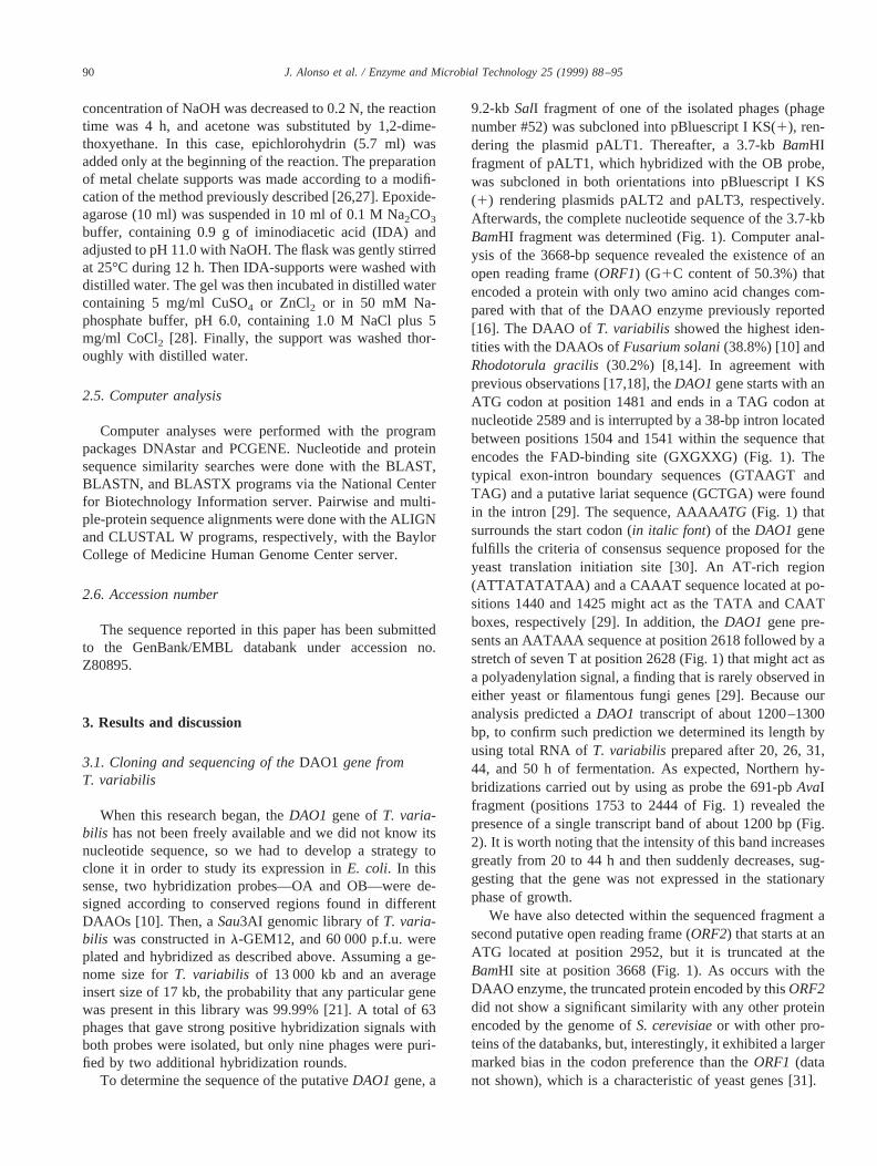
9.2-kb SalI fragment of one of the isolated phages (phagenumber #52) was subcloned into pBluescript I KS(1), ren-dering the plasmid pALT1. Thereafter, a 3.7-kbBamHIfragment of pALT1, which hybridized with the OB probe,was subcloned in both orientations into pBluescript I KS(1) rendering plasmids pALT2 and pALT3, respectively.Afterwards, the complete nucleotide sequence of the 3.7-kbBamHI fragment was determined (Fig. 1). Computer anal-ysis of the 3668-bp sequence revealed the existence of anopen reading frame (ORF1) (G1C content of 50.3%) thatencoded a protein with only two amino acid changes com-pared with that of the DAAO enzyme previously reported[16]. The DAAO of T. variabilis showed the highest iden-tities with the DAAOs ofFusarium solani(38.8%) [10] andRhodotorula gracilis(30.2%) [8,14]. In agreement withprevious observations [17,18], theDAO1gene starts with anATG codon at position 1481 and ends in a TAG codon atnucleotide 2589 and is interrupted by a 38-bp intron locatedbetween positions 1504 and 1541 within the sequence thatencodes the FAD-binding site (GXGXXG) (Fig. 1). Thetypical exon-intron boundary sequences (GTAAGT andTAG) and a putative lariat sequence (GCTGA) were foundin the intron [29]. The sequence, AAAAATG (Fig. 1) thatsurrounds the start codon (in italic font) of the DAO1 genefulfills the criteria of consensus sequence proposed for theyeast translation initiation site [30]. An AT-rich region(ATTATATATAA) and a CAAAT sequence located at po-sitions 1440 and 1425 might act as the TATA and CAATboxes, respectively [29]. In addition, theDAO1 gene pre-sents an AATAAA sequence at position 2618 followed by astretch of seven T at position 2628 (Fig. 1) that might act asa polyadenylation signal, a finding that is rarely observed ineither yeast or filamentous fungi genes [29]. Because ouranalysis predicted aDAO1 transcript of about 1200–1300bp, to confirm such prediction we determined its length byusing total RNA ofT. variabilis prepared after 20, 26, 31,44, and 50 h of fermentation. As expected, Northern hy-bridizations carried out by using as probe the 691-pbAvaIfragment (positions 1753 to 2444 of Fig. 1) revealed thepresence of a single transcript band of about 1200 bp (Fig.2). It is worth noting that the intensity of this band increasesgreatly from 20 to 44 h and then suddenly decreases, sug-gesting that the gene was not expressed in the stationaryphase of growth.
We have also detected within the sequenced fragment asecond putative open reading frame (ORF2) that starts at anATG located at position 2952, but it is truncated at theBamHI site at position 3668 (Fig. 1). As occurs with theDAAO enzyme, the truncated protein encoded by thisORF2did not show a significant similarity with any other proteinencoded by the genome ofS. cerevisiaeor with other pro-teins of the databanks, but, interestingly, it exhibited a largermarked bias in the codon preference than theORF1 (datanot shown), which is a characteristic of yeast genes [31].
90 J. Alonso et al. / Enzyme and Microbial Technology 25 (1999) 88–95
3.2. Overproduction of DAAO in E. coli
Because the main objective of this work was to investi-gate the expression of theDAO1 gene inE. coli, we first
eliminated the intron by using a PCR mutagenesis approach.Primers DAO1 and DAO2 were used to amplify theDAO1gene contained in plasmid pALT2. Primer DAO1 overlapsthe two ends of the intron and exactly reconstructs the
Fig. 1. Nucleotide sequence of the clonedBamHI fragment containing theDAO1gene ofT. variabilis. (A) The sequence of one DNA strand and the deducedamino acid sequence is shown in one letter code. The intron-exon (E1 I1-I1 E2) boundaries and the lariat are shown in boldface letters. The putative CAATand TATA boxes and the nucleotide sequence surrounding the start codon are underlined. The putative polyadenylation signals are double underlined.(B)N-terminal sequences of the DAAO reconstructed by PCR and after insertion of the histidine tag. The nucleotides corresponding to theNcoI site are indicatedin boldface letters. The newNsiI restriction site generated by the fusion of the six-histidine linker is also shown.
91J. Alonso et al. / Enzyme and Microbial Technology 25 (1999) 88–95

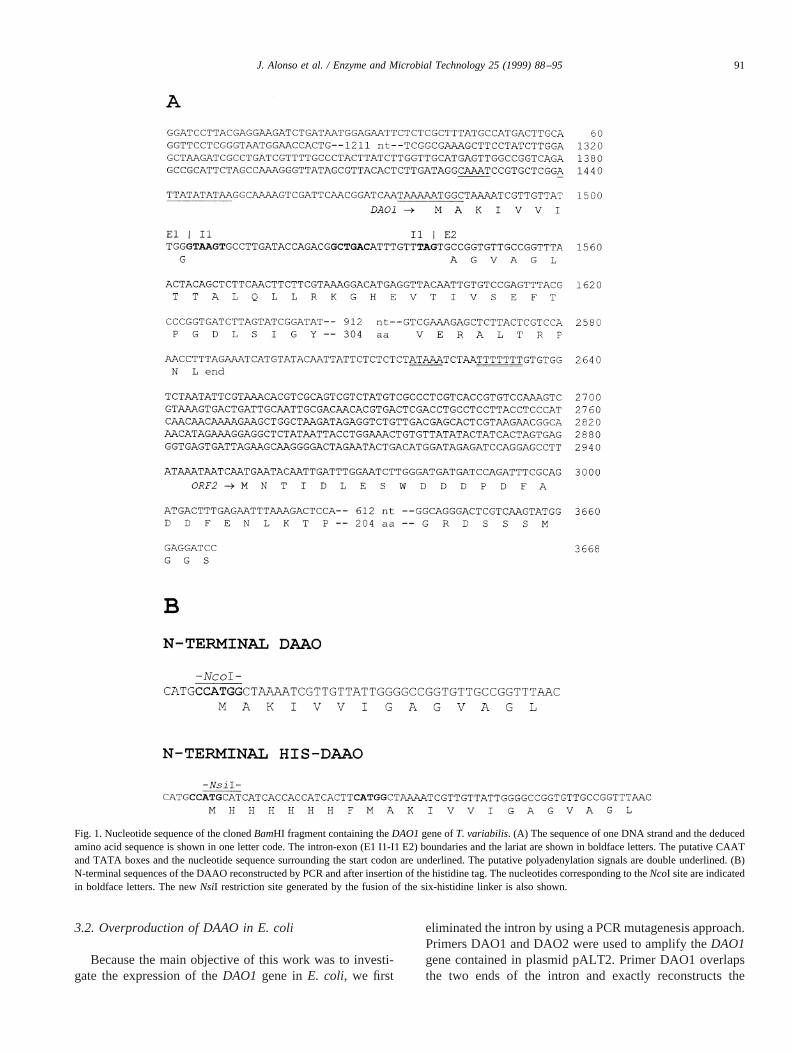
coding sequence ofDAO1 (Fig. 1). The PCR-amplifiedfragment of about 1 110 bp was cloned according to theprocedure described in Fig. 3, and the resulting phageM13DAO was sequenced to ascertain that the intron hasbeen eliminated. Then, theNcoI andHindIII sites created inthe DAO1 and DAO2 primers, respectively, were used toclone theDAO1 gene downstream of thetrc promoter ofplasmid pKK233.2. When the resulting plasmid pKDAO3(Fig. 3) was transformed intoE. coli TG1, we observed thatthe recombinant cells, cultured in LB medium, produced ahigh level of DAAO that was mainly recovered in thesoluble fraction (Fig. 4). Because plasmid pKDAO3 pro-duces ab-lactamase activity, which obligates to purify theDAAO enzyme to avoid the hydrolysis of theb-lactamnucleus during the industrial production of 7ACA, theDAO1 gene was subcloned into the vector pBC KS(1)carrying a chloramphenicol resistance gene. When the re-sulting plasmid pBDAO24 (Fig. 3) was transformed intoE.coli TG1, we observed that the recombinant cells also pro-duced a high level of soluble DAAO, comparable to thatsynthesized by plasmid pKDAO3 (data not shown). There-fore, in this case, theE. coli crude extract could be used asa source of DAAO without further purification.
We observed that the DAAO produced in the recombi-nant E. coli cells was FAD dependent, i.e. the enzymecontained in the cell extracts only reached its full activityafter the addition of 1mM FAD to the assay. Thus, some
extracts prepared fromE. coli TG1 (pKDAO3) cells grownunder a low aeration rate at 25°C showed a DAAO activityof 150 U/mg in the presence of FAD but only 15 U/mg inits absence (Table 1). This result indicated that, in suchculture conditions, about 90% of DAAO was produced in itsapoenzyme form. In this sense, we have observed that thetotal amount of DAAO produced in the recombinant cells,as well as the percentage of apoenzyme contained in the cellextracts, clearly depend of the culture conditions (Table 1).
Fig. 2. Transcriptional analysis of theDAO1 gene. Northern blot analysiswas carried out by using total RNA fromT. variabilis that was extractedafter 20 h (lane 1), 26 h (lane 2), 31 h (lane 3), 44 h (lane 4), and 50 h (lane5) of growth. The filter was probed with the labeled 691-pbAvaI fragment(positions 1753 to 2444 of Fig. 1). Sizes of the 28S and 18S rRNAs areindicated.
Fig. 3. Subcloning of theDAO1 gene. Plasmids are drawn in circles; therelevant elements and restriction sites are indicated. The directions oftranscription of the genes are indicated by arrows.DAO1* gene representsthe cloned genomic gene.DAO1gene corresponds to the gene without theintron. H-DAO1 gene represents the gene encoding the HIS-DAAO. B,BamHI; E, EcoRI; H, HindIII; N, NcoI; P, PstI; ApR, ampicillin resistance;CmR, chloramphenicol resistance; andtrcP, trc promoter. DAO1, DAO2,HIS1, and HIS2 represent the oligonucleotides used as primers or linkers.
92 J. Alonso et al. / Enzyme and Microbial Technology 25 (1999) 88–95
We found that a decrease in the aeration rate of the cultureincreased the DAAO activity and reduced the percentage ofapoenzyme form. The high content in the apoenzyme formin the crude extracts was surprising and might indicateeither that E. coli is unable to synthesize all the FADrequired to activate the high amount of enzyme synthesizedin the recombinant cells, or that the intracellular environ-ment of E. coli did not facilitate the formation of the ho-loenzyme form. On the other hand, it is important to con-sider that an active DAAO enzyme would presumablyreduce the internal pool of D-alanine needed for cell-wallsynthesis, producing the deleterious effect that we haveobserved in some clones. Hence, we cannot rule out thepossibility that stable clones might have reduced the en-zyme toxicity impairing the FAD synthesis. Interestingly, asimilar result has been observed when the DAAO fromR.gracilis was overproduced inE. coli [8].
3.3. Engineering the DAAO enzyme to facilitate itsdownstream processing
It is well known that through the fusion of a polyhistidinetag, proteins can be purified in a single step by using
metal-affinity chromatography [32], but although this tech-nique has been successful in many cases, the fusion of thistag to the DAAO enzyme presented some uncertainties. Forinstance, no fusions have been constructed with otherDAAO enzymes, and the metals have been described to actas strong DAAO inhibitors [33]. Nevertheless, in spite ofthese hypothetical problems, we decided to investigate theuse of a six-histidine tag to improve the DAAO purification.Hence, a chimeric protein was constructed according to thescheme depicted in Fig. 3. To insert the poly histidine tag inthe N-terminus of DAAO, we used theNcoI site present inplasmid pKDAO3, which overlaps the ATG codon of theDAO1 gene. Hence, theNcoI-digested plasmid pKDAO3was treated with the Klenow fragment ofE. coli DNApolymerase and ligated to the complementary oligonucleo-tides HIS1 and HIS2. The HIS1-HIS2 linker provides anadditional codon for Phe, which has been described as ametal-binding enhancer (Fig. 1) [32]. The ligation mixturewas used to transformE. coli TG1. Positive clones werescreened by colony hybridization by using the oligonucle-otide HIS1 as probe. Although the poly histidine tag can becloned in both orientations, the two types of resulting plas-mids can be distinguished because only the correct onecontains a newNsiI site (Fig. 1) and should produce anactive DAAO. Thus, the HIS1 positive clones werescreened for the presence of thisNsiI site and for theproduction of DAAO activity. One of the selected plasmids,named pKDAOHIS, was sequenced to confirm that thesix-histidine tag had been precisely inserted (data notshown). Interestingly, the crude extracts ofE. coli TG1(pKDAOHIS) showed a DAAO activity of about 300 U/mg,which correlates with the presence of an intense proteinband in SDS-PAGE (Fig. 4) similar to that obtained in thecell extracts ofE. coli TG1 (pKDAO3). As expected, thechimeric protein showed a slightly higher molecular weightthan the native DAAO (Fig. 4). These results demonstratedthat the six-histidine-tagged DAAO (hereafter HIS-DAAO)retained the activity but did not preclude that the proteincould be purified by metal-affinity chromatography.
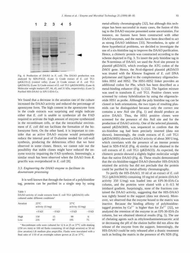
To purify the HIS-DAAO, 10 ml of an extract ofE. coliTG1 (pKDAOHIS) containing 10 mg/ml of protein (DAAOactivity 350 U/mg) was loaded into an EPI-30-IDA-Cucolumn, and the proteins were eluted with a 0–0.5 Mimidazol gradient. Surprisingly, none of the fractions con-tained the DAAO activity, suggesting that the HIS-DAAOwas tightly bound to the support (data not shown). More-over, we observed that the enzyme bound to the matrix wasinactive. Because the binding affinity of polyhistidine-tagged proteins by Cu21 is higher than for Zn21 [32], weanalyzed the retention of the enzyme in an EPI-30-IDA-Zncolumn, but we obtained identical results (Fig. 5). The useof chelating agents such as ethylenediaminetetraacetic acidor decreasing the pH of the elution buffer did not allow therelease of the enzyme from the support. Interestingly, theHIS-DAAO could be only released after a drastic treatmentsuch as boiling the matrix in the presence of 4% SDS (Fig.
Fig. 4. Production of DAAO inE. coli. The DAAO production wasanalyzed by SDS-PAGE. (Lane 1) Crude extract ofE. coli TG1(pKK233.2) (control cells). (Lane 2) Crude extract ofE. coli TG1(pKDAO3). (Lane 3) Crude extract ofE. coli TG1 (pKDAOHIS). (Lane 4)Molecular weight markers (97, 66, 45, and 31 kDa, respectively). (Lane 5)Purified HIS-DAAO in EPI-5-IDA-Co.
Table 1DAAO activity of crude extracts fromE. coli TG1 (pKDAO3) cellscultured under different conditionsa
Aeration 25°Cactivity (U/mg)
37°Cactivity (U/mg)
1FAD 2FAD 1FAD 2FAD
High 150 15 99 6Low 235 97 195 75
a Recombinant cells were cultured for 12 h at 25 or 37°C with shaking(250 rev./min) in 100 ml flasks containing 10 ml (high aeration) or 50 ml(low aeration) LB medium plus ampicillin. Flasks were inoculated with afinal ratio of 1/10 of an overnight culture grown at 25 or 37°C.
93J. Alonso et al. / Enzyme and Microbial Technology 25 (1999) 88–95
5). Because the protein eluted by the SDS treatment wasinactive, we determined that it corresponded to HIS-DAAOby analyzing its N-terminal sequence (data not shown). Toovercome these problems, we also analyzed the retention ofthe enzyme in supports with a lower density of zinc chelates(EPI-5-IDA-Zn), but the protein was still strongly adsorbedon the chelate support (data not shown). These findingssuggested that the HIS-DAAO was bound to the support bya mechanism that involved the formation of additional in-teractions. In this sense, because Cu21 and Zn21 have beendescribed as strong inhibitors of DAAO through their inter-action with the FAD-binding site [33], and, in this particularcase, the FAD-binding site is very close to the six-histidinetag, one might assume that such interaction could contributeto increase the binding affinity of the HIS-DAAO.
To overcome the retention and inactivation problems, weinvestigated the behavior of HIS-DAAO in an EPI-5-IDA-Co matrix that, in theory, should provide a supportwith a lower affinity. Interestingly, by using this support, wewere able to obtain an active HIS-DAAO after washing thecontaminant proteins with 20 mM phosphate buffer, pH 8.0,containing 0.2 M NaCl and 10% glycerol and eluting theenzyme with the same buffer containing 10% glycerol and10 mM imidazol (Fig. 4). This procedure allowed the puri-fication of HIS-DAAO in a single step and provided anenzyme with a very high specific activity (9,000 U/mg)(Table 2). Interestingly, we observed that the enzyme boundto this support retained its activity.
Summarizing, the results described here demonstrate thatan important industrial enzyme as the DAAO fromT. varia-
bilis can be overproduced inE. coli, supporting the hypoth-esis that this expression system may constitute an alterna-tive source for the production of DAAO at an industriallevel. Moreover, we have shown that this enzyme can bemodified at its N-terminus with a six-histidine tag to facil-itate its purification by metal-affinity chromatography with-out losing its activity. The attempts to purify the enzyme byusing this approach have allowed us to illustrate the impor-tance of a careful design of the metal chelate support foroptimizing the downstream processing of the tagged pro-tein, and have opened an interesting scenario to investigatethe possibility of using these supports for immobilizing theDAAO as a novel procedure for the production of 7ACA.This simple and economic method for purifying DAAO is amust for increasing the specific activity of the DAAO-immobilized supports and improving the transformationprocess. In addition, its purification contributes to overcom-ing the problem caused by the presence of catalase in theDAAO preparations that destroys the hydrogen peroxiderequired to convert the keto-AD-7ACA to the desired GL-7ACA. The exogenous hydrogen peroxide added to com-pensate for such loss induces a partial deactivation ofDAAO that reduces the efficiency and the half-life of thebiocatalyst.
Acknowledgments
The art work of A. Hurtado and the technical assistanceof E. Cano and M. Carrasco are gratefully acknowledged.This work was supported by a grant from Antibio´ticos S.A.
References
[1] Croux C, Costa J, Barredo JL, Salto F. A process for the enzymaticpreparation of 7-aminocephalosporanic acid. European Patent Appli-cation 0 469 919 A2, 1991.
[2] Isogai T, Fukagawa M, Aramori L, Iwami M, Kojo H, Ono T, UedaY, Kohsaka M, Imanaka H. Construction of a 7-aminocephalospo-ranic acid (7ACA) biosynthetic operon and direct production of7ACA in Acremonium chrysogenum.Biotechnology (NY) 1991;9:188–91.
[3] Matsuda A, Toma K, Komatsu KL. Nucleotide sequences of thegenes for two distinct cephalosporin acylases from aPseudomonasstrain. J Bacteriol 1987;169:5821–6.
[4] Bunnel CA, Luke WD, Perry FM. Industrial manufacture of cepha-losporins. In: Queener SF, Webber JA, Queener SW, editors. Beta-
Fig. 5. Retention of DAAO in EPI-30-IDA-Zn. A crude extract ofE. coliTG1 (pKDAOHIS) was loaded into an EPI-30-IDA-Zn column, and theproteins eluted from the column were analyzed by SDS-PAGE. (Lane 1)molecular weight markers (see Fig. 4). (Lane 2) Crude extract. (Lane 3)Proteins that were not retained in the matrix. (Lane 4) Proteins eluted with10 mM phosphate buffer, pH 7.0, containing 0.2 M NaCl and 30 mMimidazol. (Lane 5) Proteins eluted with 10 mM phosphate buffer, pH 7.0,containing 0.2 M NaCl and 200 mM imidazol. (Lane 6) Proteins elutedwith 20 mM phosphate buffer, pH. 7.2, containing 0.5 M NaCl and 50 mMEDTA. (Lane 7) Proteins eluted with 0.1 M citrate buffer, pH 2. (Lane 8)Proteins eluted after boiling the support for 5 min in the presence of 4%SDS.
Table 2Summary of purification of HIS-DAAO fromE. coli TG1 (pKDAOHIS)in an EPI-5-IDA-Co matrix
Sample Totalprotein(mg)
Totalactivity(U)
Sp.activity(U/mg)
Purificationfold
Recovery(%)
Crude extract 100 35 000 350 1 10010 mM imidazol 3 27 000 9000 26 77
94 J. Alonso et al. / Enzyme and Microbial Technology 25 (1999) 88–95
Lactam antibiotics for clinical use. New York: Marcel Dekker, 1986,p. 255–84.
[5] Pilone SM, Vanoni MA, Curti B. D-amino acid oxidase activity in theyeastRhodotorula gracilis.FEMS Microbiol Lett 1982;15:27–31.
[6] Szwajcer E, Mosbach K. Isolation and partial characterization of aD-amino acid oxidase active against cephalosporin C from the yeastTrigonopsis variabilis.Biotechnol Lett 1985;7:1–7.
[7] Zwart KB, Overmars EH, Harder W. The role of peroxisomes in themetabolism of D-alanine in the yeastCandida utilis.FEMS MicrobiolLett 1983;19:225–31.
[8] Alonso J, Barredo JL, Dı´ez B, Mellado E, Salto F, Garcı´a JL, Corte´sE. D-Amino-acid oxidase gene fromRhodotorula gracilis (Rhodo-sporidium toruloides) ATCC 26217. Microbiology 1998;144:1095–101.
[9] Fukui K, Watanabe F, Shibata T, Miyake Y. Molecular cloning andsequence analysis of cDNAs encoding porcine kidney D-amino acidoxidase. Biochemistry 1987;26:3612–8.
[10] Isogai T, Ono H, Ishitani Y, Kojo H, Ueda Y, Kohsaka M. Structureand expression of cDNA for D-amino acid oxidase active againstcephalosporin C fromFusarium solani.J Biochem 1990;108:1063–9.
[11] Jacobs P, Brockly F, Massaer M, et al. Porcine D-amino acid oxidase:determination of the mRNA nucleotide sequence by the characteriza-tion of genomic and cDNA clones. Gene 1987;59:55–61.
[12] Momoi, K, Fukui K, Watanabe F, Miyake Y. Molecular cloning andsequence analysis of cDNA encoding human kidney D-amino acidoxidase. FEBS Lett 1988;238:180–4.
[13] Momoi K, Fukui K, Tada M, Miyake Y. Gene expression of D-aminoacid oxidase in rabbit kidney. J Biochem 1990;108:406–13.
[14] Pollegioni L, Molla G, Campaner S, Martegani E, Pilone MS. Clon-ing, sequencing, and expression inE. coli of a D-amino acid oxidasecDNA from Rhodotorula gracilis, active on cephalosporin C. J Bio-technol 1997;58:115–23.
[15] Tada, M, Fukui K, Momoi K, Miyake Y. Cloning and expression ofa cDNA encoding mouse kidney D-amino acid oxidase. Gene 1990;90:293–7.
[16] Komatsu K, Matsuda A, Sugiura K. DNA fragment encoding D-amino acid oxidase. Useful enzyme for catalytic oxidative deamina-tion of D-amino acids. Jpn Koka Tokkyo Koho JP 62 262 994, 1987.
[17] Furuya K, Matsuda A. A transformation capable of producing D-amino acid oxidase. European Patent Application 0 583 817 A2,1993.
[18] Gonzalez FJ, Montes J, Martı´n F, Lopez MC, Fermin˜an E, Catala´n J,Galan MA, Dominguez A. Molecular cloning ofTvDAO1, a geneencoding a D-amino acid oxidase fromTrigonopsis variabilisand its
expression inSaccharomyces cerevisiaeand Kluyveromyces lactis.Yeast 1997;13:1399–1408.
[19] Mead DA, Kemper B. Chimeric single-stranded DNA phage-plasmidcloning vectors. In: Rodrı´guez RL, Denhardt DT, editors. Vectors: ASurvey of Molecular Cloning Vectors and Their Uses. Boston: But-terworth, 1987, pp. 85–102.
[20] Specht CA, DiRusso CC, Novotny CP, Ullrich RC. A method forextracting high-molecular weight deoxyribonucleic acid from fungi.Anal Biochem 1982;119:158–63.
[21] Sambrook J, Fritsch EF, Maniatis T. Molecular cloning: a laboratorymanual. Cold Spring Harbor, NY: Cold Spring Harbor LaboratoryPress, 1989.
[22] Sanger F, Nicklen S, Coulson AR. DNA sequencing with chain-terminating inhibitors. Proc Natl Acad Sci USA 1977;74:5463–7.
[23] Ausubel FM, Brent R, Kingston RE, et al. Current Protocols inMolecular Biology. New York: Wiley, 1987.
[24] Bradford MM. A rapid and sensitive method for the quantification ofmicrogram quantities of protein utilizing the principle of protein-daybinding. Anal Biochem 1976;72:248–54.
[25] Laemmli UK. Cleavage of structural proteins during the assembly ofthe head of bacteriophage T4. Nature (London) 1970;227:680–5.
[26] Hubert H, Porath J. Metal chelate affinity chromatography. I. Influ-ence of various parameters on the retention of nucleotides and relatedcompounds. J Chromatogr 1980;198:247–55.
[27] Sundber L, Porath J. Preparation of adsorbents for biospecific affinitychromatography. I. Attachment of group containing ligands to insol-uble polymers by means of bifunctional oxiranes. J Chromatogr1974;90:87–98.
[28] Hemdan ES, Zhao Y-J, Sulkowski E, Porath J. Surface topography ofhistidine residues: A facile probe by immobilized metal ion affinitychromatography. Proc Nat Acad Sci USA 1989;86:1811–5.
[29] Ballance DJ. Sequences important for gene expression in filamentousfungi. Yeast 1986;2:229–36.
[30] Caverner DR, Ray SC. Eukaryotic start and stop translation sites.Nucleic Acids Res 1991;19:3185–92.
[31] Bennetzen JL, Hall BD. Codon selection in yeast. J Biol Chem1982;257:3026–31.
[32] Arnold FH. Metal-affinity separations: a new dimension in proteinprocessing. Biotechnology (NY) 1991;4:151–6.
[33] Schrader T, Andreesen JR. Evidence for the functional importance ofCys298 in D-amino acid oxidase fromTrigonopsis variabilis.EurJ Biochem 1993;218:735–4.
95J. Alonso et al. / Enzyme and Microbial Technology 25 (1999) 88–95


![DEO PRODUCT A4 COBALT CHELATE€¦ · product composition components cas-no. concentration [%] cobalt chelate (14%) 15137-09-4](https://img.pdfslide.us/doc/110x75/5e9181400f844c648e218a22/deo-product-a4-cobalt-product-composition-components-cas-no-concentration-cobalt.jpg)