Embed Size (px)
Citation preview

Energy Metabolism and Uranium (VI) Reduction by Desulfovibrio
A Dissertation
presented to
the Faculty of the Graduate School
University of Missouri-Columbia
In Partial Fulfillment
of the Requirements of the Degree
Doctor of Philosophy
by
Rayford B. Payne
Dr. Judy D. Wall, Dissertation Supervisor
MAY 2005


Acknowledgements
I thank my committee members Drs. John Canon, David Emerich, Mark
McIntosh, Joe Polacco, and Arnold Smith for their helpful discussions. I thank Dr. Judy
Wall for being a terrific scientist, advisor, and friend. Her limitless energy and
enthusiasm have inspired me throughout my career.
Many members of the Wall Lab, past and present, contributed to both the research
presented here and my development as a scientist. I thank senior lab members Barbara
Giles, Dr. Laurie Casalot, and Dr. Joe Ringbauer for helping me learn the ropes when I
started in Judy’s lab. I thank fellow graduate student Christopher Hemme (the Wall Lab
Computer Guy!) for his patience and for always taking time from his research project to
assist me with computer analysis. I thank Darren Gentry, Suzanne Miller, Tessa Rivere,
Kelly Titkemeier, and Drs. Kelly Bender, Lise Larsen, Leena Pattarkine, Jeffery Terry,
and “Big Bad” Bill Yen for their assistance with experiments presented within.
This work was supported in part by the Natural and Accelerated Bioremediation
Research Program and the Basic Energy Research Program of the U.S. Department of
Energy through grants DE-FG02-97ER62495 and DE-FG02-87ER13713, respectively;
the Missouri Agricultural Experiment Station; and the University of Missouri-Columbia
Life Sciences Program.
ii

Energy Metabolism and Uranium (VI) Reduction by Desulfovibrio
Rayford B. Payne
Dr. Judy D. Wall, Dissertation Supervisor
Abstract
Sulfate reducing bacteria (SRB) of the genus Desulfovibrio can reduce uranium
(VI) to uranium (IV) enzymatically. The reduction of U(VI) to U(IV) alters the solubility
state of the uranium ion from a soluble to an insoluble, and therefore less biologically
available, species. Because SRB are commonly found in uranium contaminated
groundwater and soil, it is theoretically possible that we could use them to bioremediate
uranium contaminated environments. However, before we attempt to manipulate the
system, we must first understand the SRB genes and enzymes involved in uranium
reduction and energy metabolism. Previous in vitro work by Lovley and coworkers
suggested that the biochemical pathway for U(VI) reduction by Desulfovibrio was
hydrogenase-to cytochrome c3-to U(VI). In this pathway, cytochrome c3 was suggested
to be the sole U(VI) reductase. First, we tested this model in vivo with strains carrying
mutations in the dominant Fe-hydrogenase or cytochrome c3. We determined that the
Lovley model is the primary pathway for U(VI) reduction in vivo when hydrogen gas is
the electron donor; however, alternate pathways utilizing lactate or pyruvate for U(VI)
reduction exist. In addition, at least one other cellular protein must be capable of acting
as a U(VI) reductase in cytochrome c3-lacking cells. Second, we grew Desulfovibrio
desulfuricans G20 in the presence of a non-lethal concentration of uranium in order to
iii

understand some of the effects of uranium on its physiology. By doing so, we showed
that G20 cells grown in the presence of uranium are impaired for U(VI) reduction. While
exploring this observation, we found that the electron carrier protein cytochrome c3
tightly adsorbs to insoluble uranium (IV) oxide, as well as copper oxide and iron oxide.
Finally, we observed that sodium ions play an important role in energy metabolism and
antibiotic resistance of Desulfovibrio when grown on lactate sulfate medium. We
speculate that Desulfovibrio is capable of coupling lactate, but not pyruvate, oxidation
and subsequent electron transport to the generation of a transmembrane sodium gradient.
We propose that Desulfovibrio uses this “sodium circuit” to complement the proton
motive force, for growth and for the efflux of some toxic compounds.
iv

List of Tables
Table 2.1. Plasmids and strains used in this study………………………………19
Table 2.2. Primers used in this study……………………………………………..31
Table 3.1. U(VI) reduction by a cytochrome c3 mutant of
D. desulfuricans G20……………………………………………………………………41
Table 3.2. Nitrite reduction by a cytochrome c3 mutant of
D. desulfuricans G20…………………………………………………………………....51
Table 4.1. U(VI) reduction by a Fe-hydrogenase mutant of D. vulgaris
Hildenborough………………………………………………………………………….72
Table 5.1. β-galactosidase activity of cells grown in the presence of uranium....88
Table 6.1. Protein yield of D. desulfuricans G20 cells grown in the presence of
CCCP and monensin………………………………………………………………….106
Table 6.2. MIC of inhibitors for D. desulfuricans G20 grown with
varying NaCl…………………………………………………………………………..113
Table 7.1. Expression of Genes Encoding Electron Carrier Proteins in CycA..125
v

List of Figures
Figure 1.1. Electron micrographs of Desulfovibrio strains…………………………2
Figure 1.2. Lovley Model for U(VI) reduction by Desulfovibrio vulgaris…...…….6
Figure 1.3. Chemiosmotic Hypothesis for energy metabolism …………………...10
Figure 1.4. Hydrogen Cycling Model for energy metabolism in Desulfovibrio….11
Figure 1.5. U(VI) reduction by Desulfovibrio desulfuricans G20…………………14
Figure 2.1. U(VI) standard curve…………………………………………………...28
Figure 3.1. U(VI) reduction by a cytochrome c3 mutant of
D. desulfuricans G20…………………………………………………………………....40
Figure 3.2. Cytochrome c3 expression in iron-limited cells of
D. desulfuricans G20…………………………………………………………………....43
Figure 3.3. U(VI) reduction by iron-limited cells of D. desulfuricans G20……....44
Figure 3.4. The nitrite reductase operon of Desulfovibrio strains……………..…48
Figure 3.5. Nitrite reductase expression in a cytochrome c3 mutant ………….…49
Figure 3.6. Nitrite reduction by a cytochrome c3 mutant ………………………...52
Figure 3.7. Hydrogenase assay of a cytochrome c3 mutant……………………….56
Figure 3.8. Modified Lovley Model for U(VI) reduction in cyt c3-lacking cells…59
Figure 3.9. Altered electron flux in a cytochrome c3 mutant……………………..64
Figure 4.1. Growth of a Fe-hydrogenase mutant of Desulfovibrio vulgaris……...70
Figure 4.2. Modified Lovley Model for U(VI) reduction in a Fe-hydrogenase
mutant of D. vulgaris…………………………………………………………………...78
Figure 5.1. U(VI) reduction by D. desulfuricans G20 previously exposed to
uranium……………………………………………………………………………….....82
vi

Figure 5.2. Cytochrome c3 is not recovered in the periplasm of cells grown in the
presence of uranium………………………………………………………………..….85
Figure 5.3. Adsorption of cytochrome c3 to uraninite…………………………….91
Figure 5.4. Cytochrome c3 recovery from cells grown in the presence of
uranium………………………………………………………………………………....93
Figure 6.1 The Na+-Translocating NADH Quinone Oxidoreductase (NQR)
operon of SRB…………………………………………………………………………101
Figure 6.2. Expression of the NQR complex operon measured by RT-PCR…...102
Figure 6.3. Growth of D. desulfuricans G20 under low proton- and sodium-
motive force conditions……………………………………………………………..…104
Figure 6.4. Expression of the NQR complex operon at pH 9, and by a cytochrome
c3 mutant of D. desulfuricans G20…………………………………………………....108
Figure 6.5. The Na+-dependent multidrug exporters (NorM) of the SRB...……110
Figure 6.6. D. desulfuricans G20 is more sensitive to kanamycin in the absence of
the sodium motive force……………………………………………………………….112
Figure 6.7. The “Sodium Circuit” of the SRB…………………………………....115
Figure 7.1. Overall Model for the Electron Transport Chain in Desulfovibrio
from this dissertation………………………………………………………………….126
vii

Table of Contents
Acknowledgements……………………………………………………………………....ii
Abstract………………………………………………………………………………….iii
List of Tables…………………………………………………………………………..…v
List of Figures…………………………………………………………………………....vi
Table of Contents……………………..……………………………………….……….viii 1. Introduction 1.1. The Sulfate-Reducing Bacteria…………………………………………………….1
1.2. The Lovley model for uranium (VI) reduction…………………………………...4
1.3. Energy Metabolism in Desulfovibrio………………………………………………9
1.4. Aim of this dissertation……………………………………………………………16
2. Experimental procedures 22 2.1. Growth of Desulfovibrio…………………………………………………………..22
2.1.1. Growth of Desulfovibrio desulfuricans G20
2.1.2. Growth of D. desulfuricans G20 in iron-limiting medium
2.1.3. Growth of a Fe-hydrogenase mutant of Desulfovibrio vulgaris
2.1.4. Growth of D. desulfuricans G20 in the presence of 1 mM uranium
2.1.5. Growth of D. desulfuricans G20 under low sodium- and proton-motive force
conditions
2.1.6. Antibiotic resistance of D. desulfuricans G20 under low sodium- and proton-
motive force conditions
2.2. Western analysis for cytochrome c3…………………………………………...…27
2.2.1. Protein extraction from Desulfovibrio grown on LS medium
viii

2.2.2. Western analysis for cytochrome c3.
2.2.3. Adsorption of cytochrome c3 to uraninite
2.2.4. Separation of cells and uraninite by ultracentrifugation
2.3. Uranium (VI) reduction assay……………………………………………………30
2.4. Northern analysis and RT-PCR……………………………………………….…34
2.5. Nitrite reduction assay…………………………………………………………….27
2.6. Hydrogenase assay………………………………………………………………...37 2.7. β-galactosidase assay……………………………………………………………...48
3. Uranium reduction by a cytochrome c3 mutant of Desulfovibrio 40 3.1. Introduction………………………………………………………………………..41
3.2. Results……………………………………………………………………………...43
3.2.1. Uranium (VI) reduction by a cytochrome c3 mutant
3.2.2. Fe-requirement for the synthesis of cytochrome c3
3.2.3. Uranium (VI) reduction by cytochrome c3 -lacking cells
3.2.4. Nitrite reduction by a cytochrome c3 mutant
3.2.5. Hydrogenase activity by a cytochrome c3 mutant
3.3. Discussion………………………………………………………………………….67
4. Uranium (VI) reduction by a hydrogenase mutant of Desulfovibrio 77 4.1. Introduction………………………………………………………………………..78
4.2. Results…………………………………………………………………………...…80
4.2.1. Growth of Desulfovibrio vulgaris
4.2.2. U(VI) reduction by a Fe-hydrogenase mutant
4.2.3. U(VI) reduction in the presence of inhibitors
ix

4.3. Discussion………………………………………………………………………….85
5. Interaction between cytochrome c3 and uranium 92 5.1. Introduction……………………………………………………………………..…93
5.2. Results…………………………………………………………………………...…93
5.2.1. Uranium (VI) reduction by cells grown in the presence of uranium
5.2.2. Cytochrome c3 recovery from cells grown with uranium
5.2.3. Effect of uranium on transcription/translation of cytochrome c3.
5.2.4. Adsorption of cytochrome c3 to uraninite
5.2.5. Cytochrome c3 recovery from cells grown on LSU medium after separation
of cells and uraninite by ultracentrifugation
5.3. Discussion………………………………………………………………………...111
6. The role of the sodium motive force in Desulfovibrio 113 6.1. Introduction……………………………………………………………………....114
6.2. Results…………………………………………………………………………….116
6.2.1. The Na+-Translocating NADH Quinone Oxidoreductase (NQR Complex)
of the Sulfate-Reducing Bacteria
6.2.2. Expression of the NQR complex operon
6.2.3. The Na+/multidrug exporters of the Sulfate-Reducing Bacteria
6.2.4. The role of the sodium motive force during drug resistance
6.3. Discussion………………………………………………………………………...135
7. Overall conclusions of this dissertation 143
8. References 152
Vita…………………………………………………………………………..…………141
x

1. Introduction
1.1. The Sulfate-Reducing Bacteria
The sulfate reducing bacteria (SRB) are a diverse group of microorganisms that
are characterized by the ability to reduce sulfate enzymatically as the sole electron
acceptor for growth (Postgate, 1979). The process of reducing sulfate as the terminal
electron acceptor for anaerobic growth with the production of sulfide is termed
dissimilatory sulfate reduction. This contrasts with assimilatory sulfate reduction,
whereby sulfate is reduced enzymatically first to sulfide, incorporated into cysteine, and
then finally used to make proteins (Kredich, 1996). Illustrative of their diversity, SRB
include both Gram-negative and Gram-positive Bacteria and Archaea (Stetter, 1988).
Most, but not all, strains of SRB can utilize hydrogen gas as the sole electron donor for
growth by sulfate reduction (Postgate, 1979). Although typically thought of as strict
anaerobes, meaning exposure to oxygen is lethal, SRB show various levels of tolerance to
oxygen. In fact, members of the genus Desulfovibrio have even been shown to reduce
molecular oxygen, perhaps as a defense mechanism (Fournier et al., 2003; van Niel and
Gottschal, 1998). There is evidence that some strains of SRB can even couple the
reduction of molecular oxygen to proton translocation and ATP synthesis (Cypionka,
2000; Johnson et al., 1997; Lemos et al., 2001).
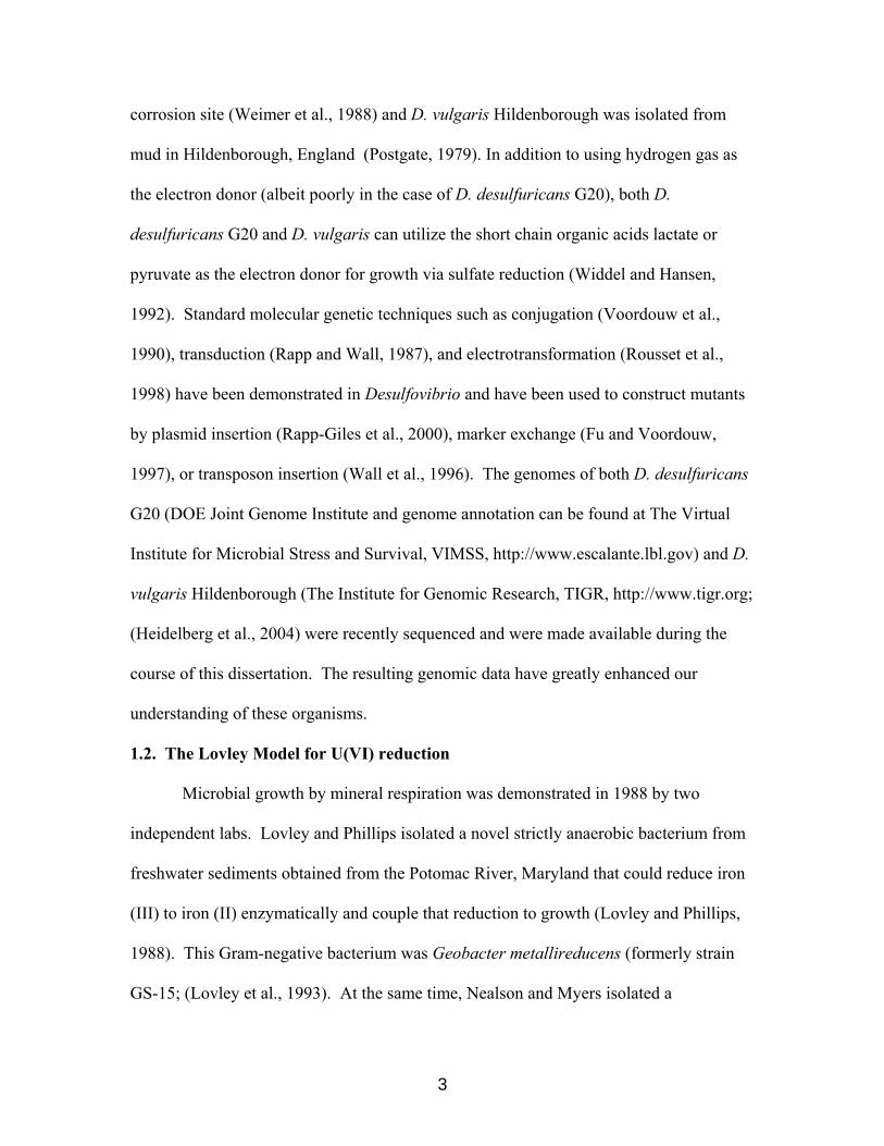
Strains of the genus Desulfovibrio are among the better-characterized SRB due in
part to their relative ease of cultivation in the laboratory. Desulfovibrio are Gram-
negative, slightly curved rods, typically about 1 µm in length (Fig. 1.1). Representative
strains used in this study were Desulfovibrio desulfuricans strain G20 and Desulfovibrio
vulgaris strain Hildenborough. D. desulfuricans G20 was isolated from an oil well
1

Figure 1.1. Electron micrograph of Desulfovibrio desulfuricans G20.
Transmission electron micrograph of D. desulfuricans G20. Cells were grown to early
stationary phase on medium containing 60 mM lactate as carbon and reductant source
and 50 mM sulfate as terminal electron acceptor. The location of the cytoplasm,
periplasm, inner membrane (IM), and outer membrane (OM) are indicated with arrows.
2
corrosion site (Weimer et al., 1988) and D. vulgaris Hildenborough was isolated from
mud in Hildenborough, England (Postgate, 1979). In addition to using hydrogen gas as
the electron donor (albeit poorly in the case of D. desulfuricans G20), both D.
desulfuricans G20 and D. vulgaris can utilize the short chain organic acids lactate or
pyruvate as the electron donor for growth via sulfate reduction (Widdel and Hansen,
1992). Standard molecular genetic techniques such as conjugation (Voordouw et al.,
1990), transduction (Rapp and Wall, 1987), and electrotransformation (Rousset et al.,
1998) have been demonstrated in Desulfovibrio and have been used to construct mutants
by plasmid insertion (Rapp-Giles et al., 2000), marker exchange (Fu and Voordouw,
1997), or transposon insertion (Wall et al., 1996). The genomes of both D. desulfuricans
G20 (DOE Joint Genome Institute and genome annotation can be found at The Virtual
Institute for Microbial Stress and Survival, VIMSS, http://www.escalante.lbl.gov) and D.
vulgaris Hildenborough (The Institute for Genomic Research, TIGR, http://www.tigr.org;
(Heidelberg et al., 2004) were recently sequenced and were made available during the
course of this dissertation. The resulting genomic data have greatly enhanced our
understanding of these organisms.
1.2. The Lovley Model for U(VI) reduction
Microbial growth by mineral respiration was demonstrated in 1988 by two
independent labs. Lovley and Phillips isolated a novel strictly anaerobic bacterium from
freshwater sediments obtained from the Potomac River, Maryland that could reduce iron
(III) to iron (II) enzymatically and couple that reduction to growth (Lovley and Phillips,
1988). This Gram-negative bacterium was Geobacter metallireducens (formerly strain
GS-15; (Lovley et al., 1993). At the same time, Nealson and Myers isolated a
3
facultatively anaerobic bacterium from freshwater sediments obtained from Lake Oneida,
New York, that could utilize Fe(III) or manganese (IV), as well as oxygen, nitrate, or
chromate (VI) as the terminal electron acceptor (Myers and Nealson, 1988). This Gram-
negative bacterium was Shewanella oneidensis (formerly Shewanella putrefaciens and
Alteromonas putrefaciens (Venkateswaran et al., 1999), The reduction of Fe(III) to Fe(II)
and Mn(IV) to Mn(II) by Geobacter or Shewanella strains was therefore truly a form of
anaerobic respiration using a mineral as the terminal electron acceptor. Geobacter and
Shewanella strains are ubiquitous and have been isolated from both freshwater and
marine sediments, and terrestrial subsurface environments. They, along with the SRB,
represent a significant proportion of the biological activity in those environments
(Caccavo et al., 1992; Coates et al., 1998; Coleman et al., 1993; Nealson and Saffarini,
1994).
Geologic uranium (IV) deposits are often found in Fe(III)- or sulfide-rich rocks
(Lovley et al., 1991). Historically, it was supposed that U(VI) was reduced and deposited
as U(IV) in uranium rocks by geochemical means, not by the direct action of microbes.
Lovley and coworkers reasoned that since Fe(III) and U(IV) are often found together in
uranium deposits, dissimilatory Fe(III) reducing microorganisms might also reduce U(VI)
enzymatically and couple this reduction to growth. This was the result observed with
whole cells of G. metallireducens and S. oneidensis in an in vitro system (Lovley et al.,
1991). Using the same reasoning, Lovley speculated that SRB might also enzymatically
reduce U(VI)and tested SRB of the genus Desulfovibrio for their ability to reduce U(VI).
They found that Desulfovibrio desulfuricans strain Essex (ATCC 29577) could reduce
this radionuclide; however, the reduction did not support growth under the conditions
4

used (Lovley and Phillips, 1992). After additional biochemical experiments, Lovley et al.
proposed a metabolic pathway for U(VI) reduction by Desulfovibrio (Fig. 1.2) (Lovley et
al., 1993). Using partially purified cell extracts containing hydrogenase and cytochrome
c3 of D. vulgaris, they coupled hydrogen oxidation to the reduction of U(VI) in vitro.
When they removed either hydrogenase or cytochrome c3 from the in vitro system,
reduction did not occur. When hydrogenase or the cytochrome c3 was added back to the
system, reduction was restored (Lovley et al., 1993). Based upon these observations,
they proposed that cytochrome c3 acts as the U(VI) reductase (Fig. 1.2).
5

Figure 1.2. Lovley Model of uranium (VI) reduction for Desulfovibrio vulgaris.
Each arrow indicates electron flow and implies a single step process.
U(VI) (ox)
2e- 2e-2e-
U(IV) (red)
H2 Cytochrome c3 Hydrogenase
6

1.3. Energy Metabolism in Desulfovibrio
It has long been known that most strains of Desulfovibrio can grow with hydrogen
gas as the sole electron donor (Postgate, 1979) that is suggested to be oxidized in the
periplasm primarily by the periplasmic Fe-hydrogenase (Pohorelic et al., 2002).
Biochemical experiments demonstrated that the Fe- or NiFe-hydrogenase can directly
interact with and reduce the high molecular mass cytochrome c complex (HMC), a
transmembrane complex; however, the addition of catalytic amounts of cytochrome c3
increased the rate of reduction of HMC by two orders of magnitude (Pereira et al., 1998).
Thus it has been proposed that electrons liberated from hydrogen oxidation are first
transferred to the dominant periplasmic cytochrome c3, then passed through the
cytoplasmic membrane via HMC where they subsequently are used to reduce sulfate to
sulfide. Analysis of D. vulgaris and D. desulfuricans mutants of these proteins suggests
that this pathway of electron flow from hydrogen to sulfate is likely true; however, other
proteins can compensate for each of the single mutants (Dolla et al., 2000; Pohorelic et
al., 2002 ; Rapp-Giles et al., 2000). Protons generated from the periplasmic oxidation of
hydrogen are then available for the synthesis of ATP via a classical membrane bound
F1F0 ATPase.
The mechanism for generation of a transmembrane proton gradient in
Desulfovibrio when organic compounds like lactate or pyruvate are the electron donors
and sulfate is the terminal electron acceptor has been intensely debated. The classical
“Mitchell Hypothesis” for chemiosmotic ATP synthesis by Desulfovibrio envisions
electrons generated from lactate or pyruvate oxidation being carried through membrane
bound electron carrier components and eventually used to reduce sulfate to sulfide.
7

Electron flux through the electron transport chain is coupled to the translocation of
cytoplasmic protons into the periplasm (Mitchell and Moyle, 1965). The consequent
separation of ions (more protons in the periplasm) and charge (more positive in the
periplasm) across the cytoplasmic membrane creates an energy gradient that is utilized to
synthesize ATP via a membrane bound ATPase (Fig. 1.3). The energy potential of the
transmembrane proton gradient can also be used for flagella rotation, substrate transport,
and other processes in addition to ATP synthesis (for a recent review of the chemiosmotic
hypothesis, see Harold, 2001). Researchers have reported classical proton translocation
coupled to electron transport after the addition of hydrogen as the electron donor to
resting cell suspensions of Desulfovibrio (Barton et al., 1983; Fitz and Cypionka, 1989).
The evidence for classical proton translocation by Desulfovibrio strains is as follows.
When 1 mole of hydrogen was added to resting cells supplied with an excess of electron
acceptor, up to 6 moles of protons were released into the periplasm. Since the oxidation
of 1 mole of hydrogen yields 2 moles of protons, the additional protons (up to 4 moles)
released must come from cytoplasmic protons translocated into the periplasm via electron
transport.
Many researchers have observed that Desulfovibrio strains generate, then
consume, hydrogen gas when grown with an organic compound such as lactate as the
sole electron donor (Tsuji and Yagi, 1980). Various models have been put forward to
explain this hydrogen transient. One model proposes that the hydrogen burst is used to
control the redox state of various intracellular electron carriers (Lupton et al., 1984).
That is, the hydrogen burst is used to release excess electrons generated by the initial
oxidation of organic compounds. In 1981, Odom and Peck proposed another
8

explanation: that Desulfovibrio strains produce and consume hydrogen as the primary
means to generate a transmembrane proton gradient rather than the classical Mitchell
model of electron transport driven proton translocation (Odom and Peck, 1981). The
model proposed by Odom and Peck is commonly referred to as the “Hydrogen Cycling”
model, and it has been quite controversial (Fig. 1.4). The periplasmic Fe-hydrogenase
and cytochrome c3 in the hydrogen cycling model have been proposed to re-oxidize
hydrogen that is generated in the cytoplasm and then diffuses across the cytoplasmic
membrane. Evidence has accumulated both for and against the hydrogen cycling model
as a general means for generation of a transmembrane proton gradient. As might be
expected if hydrogen gas is an intermediary in metabolism, a high concentration of
external hydrogen gas inhibited organic substrate consumption when D. vulgaris strain
Madison was cultured on pyruvate alone (Lupton et al., 1984). However, high external
hydrogen concentrations did not inhibit growth of the same organism when cultured on
lactate sulfate or pyruvate sulfate (Lupton et al., 1984). Curiously, a mutant of D.
desulfuricans strain 27774 was isolated that was incapable of growth on lactate with
sulfate under a hydrogen atmosphere or on hydrogen with sulfate; however, under an
argon atmosphere, the mutant was still capable of growth on lactate with sulfate (Odom
and Wall, 1987). Analysis of this mutant suggested that hydrogen was not an obligate
intermediate during the oxidation of lactate in the wild-type strain 27774.
9

Figure 1.3. Chemiosmotic Hypothesis for energy metabolism.
Electrons liberated by the oxidation of reduced carbon compounds travel through a
membrane bound electron transport chain driven by the different redox potential of the
components and eventually are used to reduce a terminal electron acceptor (TEA), for
example sulfate in the case of Desulfovibrio. The movement of electrons drives protons
across the membrane into the periplasm. The separation of ions (more protons in the
periplasm) and charge (a more positive charge in the periplasm) creates an energy
gradient that can be used to synthesized ATP via the membrane bound ATPase, as well as
rotate flagella, transport substrates, etc.
Periplasm
Cytoplasm
H+ H+
H+ H+
H+ H+
H+ H+ H+
e- Transport +
-
[CHO] Oxidation
e- e
-H+- +
H+TEA - +
- H++ ATP
ADP
H+
10

Figure 1.4. Hydrogen Cycling model for energy metabolism in Desulfovibrio (Odom
and Peck, 1981). Electrons and protons liberated by the oxidation of reduced carbon
compounds are the substrate of a cytoplasmic hydrogenase which generates hydrogen
gas. Hydrogen gas diffuses across the cytoplasmic membrane and is oxidized by a
periplasmic hydrogenase. Electrons are passed to cytochrome c3 and then through a
transmembrane complex like the high molecular mass cytochrome c complex (HMC).
Once across the membrane, they are used to reduce sulfate to sulfide. The separation of
protons and charge across the membrane is used to synthesize ATP through a membrane
bound ATPase. Arrows do not necessarily imply a single step process.
Periplasm Cytoplasm
Lactate
Cyt c3
Pyruvate + 2H+
+ 2e-
Hydrogenase
H2 H2 2H+ + 2e
-
e-
Hydrogenase
H2
H+
e-
ATP
ADP Acetate + 2H+
+ 2e-
HMC
ATPase
11

The question remains: does hydrogen cycling as envisioned by Odom and Peck
actually function in any strain of Desulfovibrio? Hydrogen cycling as a means to
generate a proton gradient is not mutually exclusive with electron transport chain coupled
proton translocation. Bacteria are frequently over designed and Desulfovibrio are clever
organisms. Recent studies by Noguera and coworkers suggested that D. vulgaris can use
both electron transport and hydrogen cycling to establish a periplasmic proton gradient
from the oxidation of lactate or pyruvate (Noguera et al., 1998). This could benefit the
bacterium in several ways. Rapid hydrogen oxidation would allow the bacterium to reuse
hydrogen that may be unavoidably released during the oxidation of organic compounds.
Alternately it might allow the SRB to out compete other bacteria for trace amounts of H2
in the environment. This second advantage is supported by the observation that when
sulfate is present in the environment, hydrogen-oxidizing SRB typically out compete
hydrogen-oxidizing methanogenic bacteria (Kristjannson et al., 1982; Lovley and
Goodwin, 1988; Robinson and Tiedje, 1984).
1.4. Aim of this dissertation
Our nation has used nuclear energy for over 50 years, and as a result our country
is faced with cleaning or decontaminating a great deal of hazardous and radioactive
waste. The U.S. Department of Energy is the steward of more than 120 sites, which
contain approximately 475 billion gallons of contaminated water and 75 million cubic
meters of contaminated soil (U.S. Department of Energy, http://www.lbl.gov/NABIR/).
SRB have been shown to reduce radionuclides like uranium (VI) and technetium
(VII) in situ in contaminated waters and soils and this reduction can be stimulated by the
addition of nutrients such as lactate to the contaminated environment (Abdelouas et al.,
12

2000; Abdelouas et al., 2002). Promisingly, the reduced species U(IV) and Tc(IV) are
less soluble in water and precipitate out of aqueous environments as minerals (Fig. 1.5).
Although the reduction of these radioactive metals does not alter their radioactivity, the
reduced species are less environmentally mobile and therefore less biologically available.
Before we can use SRB to decontaminate uranium-polluted environments we
must first understand the SRB genes and enzymes involved in energy metabolism in
general, and U(VI) reduction in particular. The idea of creating a “super bug” that is very
efficient at reducing U(VI) enzymatically is somewhat appealing; however, the release of
genetically modified organisms into the environment is problematic due to both rightful
public concern, and to the observation that genetically modified laboratory organisms are
typically at a disadvantage in the environment compared to their wild-type counterparts.
Even though it is not technologically feasible to genetically engineer such a “super bug”
at this time, understanding the pathway and regulation of electron flow from electron
donor to U(VI) will allow us to make predictions on which sites are amenable to
bioremediation by SRB, and how to increase the efficiency of the natural bioremediation,
for example by nutrient addition.
13

Figure 1.5. U(VI) reduction by Desulfovibrio desulfuricans G20.
Pictured are test tubes containing 5 ml of 30 mM sodium bicarbonate buffer, pH 7.0
supplemented with 10 mM lactate as the electron donor (as described in section 2.3).
Test tube 1 contains cells (ca. 1 mg cell protein) heat-killed by boiling for 15 min; Test
tube 2 contains living cells (ca. 1 mg cell protein). Test tubes were incubated
anaerobically at 31ºC for 16 hours. No U(VI) reduction was seen in the absence of
electron donor (data not shown). U(IV) precipitates from solution as the black mineral
uraninite (UO2) in the presence of living cells and an electron donor.
21
14
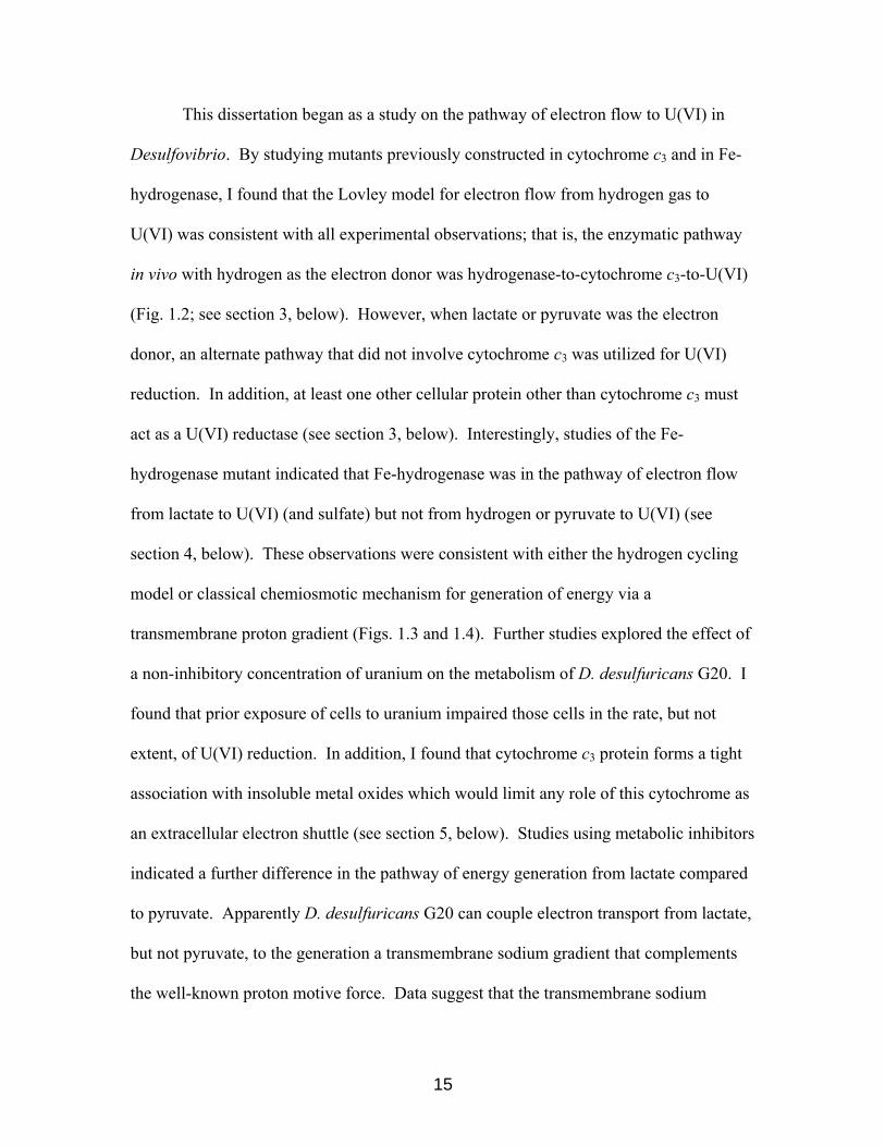
This dissertation began as a study on the pathway of electron flow to U(VI) in
Desulfovibrio. By studying mutants previously constructed in cytochrome c3 and in Fe-
hydrogenase, I found that the Lovley model for electron flow from hydrogen gas to
U(VI) was consistent with all experimental observations; that is, the enzymatic pathway
in vivo with hydrogen as the electron donor was hydrogenase-to-cytochrome c3-to-U(VI)
(Fig. 1.2; see section 3, below). However, when lactate or pyruvate was the electron
donor, an alternate pathway that did not involve cytochrome c3 was utilized for U(VI)
reduction. In addition, at least one other cellular protein other than cytochrome c3 must
act as a U(VI) reductase (see section 3, below). Interestingly, studies of the Fe-
hydrogenase mutant indicated that Fe-hydrogenase was in the pathway of electron flow
from lactate to U(VI) (and sulfate) but not from hydrogen or pyruvate to U(VI) (see
section 4, below). These observations were consistent with either the hydrogen cycling
model or classical chemiosmotic mechanism for generation of energy via a
transmembrane proton gradient (Figs. 1.3 and 1.4). Further studies explored the effect of
a non-inhibitory concentration of uranium on the metabolism of D. desulfuricans G20. I
found that prior exposure of cells to uranium impaired those cells in the rate, but not
extent, of U(VI) reduction. In addition, I found that cytochrome c3 protein forms a tight
association with insoluble metal oxides which would limit any role of this cytochrome as
an extracellular electron shuttle (see section 5, below). Studies using metabolic inhibitors
indicated a further difference in the pathway of energy generation from lactate compared
to pyruvate. Apparently D. desulfuricans G20 can couple electron transport from lactate,
but not pyruvate, to the generation a transmembrane sodium gradient that complements
the well-known proton motive force. Data suggest that the transmembrane sodium
15

gradient is used for both growth during lactate supported sulfate respiration and for drug
resistance by the export of some toxic compounds (see section 6, below). Not
surprisingly, Desulfovibrio has multiple pathways for energy generation. In
Desulfovibrio, it appears that both a hydrogen cycling mechanism and a classical electron
transport chain coupled mechanism are used to generate a transmembrane proton or
sodium ion gradient.
16

2. Experimental Procedures
2.1. Growth of Desulfovibrio
2.1.1. Growth of Desulfovibrio desulfuricans G20
D. desulfuricans strain G20 is a spontaneously nalidixic acid-resistant derivative
of the parent strain G100A that is also cured of the endogenous cryptic plasmid pBG1
(Wall et al., 1993; Table 2.1). The cytochrome c3 mutant, I2, was created by insertion of
a plasmid containing the kanamycin resistance cassette into the single chromosomal copy
of the monocistronic cycA operon of the parent strain G20 (Wall et al., 1993; Table 2.1).
Cells were grown anaerobically in medium (LS) containing lactate (60 mM) as
the primary electron donor and carbon source with sodium sulfate (50 mM) as the
terminal electron acceptor (Rapp and Wall, 1987). Kanamycin (175 µg/ml) was added to
all media used to grow I2 to select for maintenance of the inserted plasmid.
Growth curves were started by the subculture of early stationary phase cells
(about 16 hour old cultures with an OD600nm of about 0.9) 1:20 into fresh growth medium.
Growth was at 37 °C in 13 × 100 mm test tubes containing 3 ml of medium and a
headspace of 4 ml that were sealed with black rubber stoppers. The headspace was the
atmosphere from the anaerobic chamber (Coy Laboratory Products, www.coylab.com)
that was predominantly 90:10 N2:H2.
I2 cultures were always started from freezer stocks and were subcultured no more
than three times. Since suppressors that restore cytochrome c3 have been shown to
accumulate in I2 cultures after an extended time in stationary phase, I2 cultures were
monitored by Western analysis (see below) to ensure that the majority of the cells were
not expressing cytochrome c3 (data not shown) (Rapp-Giles et al., 2000).
17

2.1.2. Growth of Desulfovibrio desulfuricans G20 in iron-limiting medium
In experiments testing the requirement of iron for the synthesis of cytochrome c3,
iron salts were omitted from the trace mineral solution used to make LS medium. Yeast
extract was also omitted. Growth in this modified medium has been shown to be limited
by available iron (Postgate, 1956).
2.1.3. Growth of Desulfovibrio vulgaris
D. vulgaris strain Hildenborough wild-type and the Fe-hydrogenase mutant were
a generous gift of the Voordouw laboratory (University of Calgary, Alberta, Canada).
The Fe-hydrogenase mutant was created by gene replacement of the operon coding for
the Fe-hydrogenase, hydAB, with the chloramphenicol resistance cassette (Pohorelic et
al., 2002; Table 2.1). Wild-type and mutant strains were grown in LS medium.
Chloramphenicol (10 µg/ml) was added to media used to grow the Fe-hydrogenase
mutant.
2.1.4. Growth of D. desulfuricans G20 in the presence of 1 mM uranium
One mM uranyl acetate was added from a sterile stock solution to autoclaved LS
medium to make LSU medium. Growth of G20 in LSU medium was identical to that in
LS medium as measured by OD600nm or by protein yield (Payne et al., 2002).
18
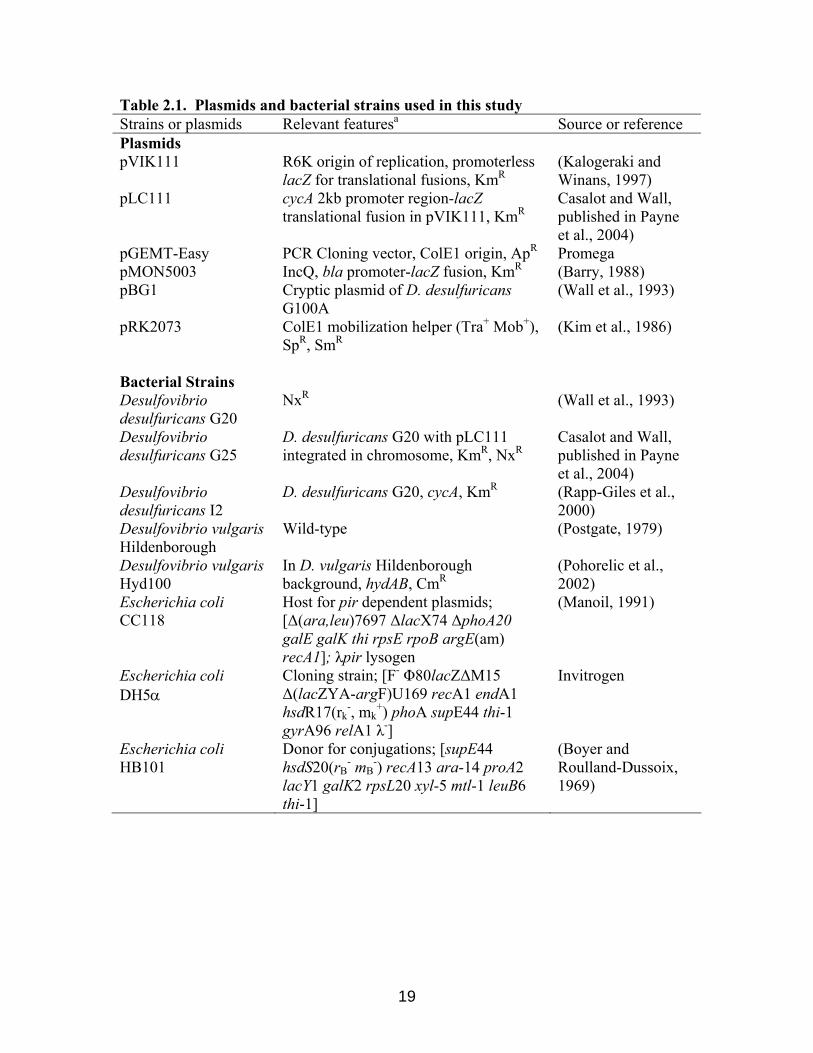
Table 2.1. Plasmids and bacterial strains used in this study Strains or plasmids Relevant featuresa Source or reference Plasmids pVIK111 R6K origin of replication, promoterless
lacZ for translational fusions, KmR(Kalogeraki and Winans, 1997)
pLC111 cycA 2kb promoter region-lacZ translational fusion in pVIK111, KmR
Casalot and Wall, published in Payne et al., 2004)
pGEMT-Easy PCR Cloning vector, ColE1 origin, ApR Promega pMON5003 IncQ, bla promoter-lacZ fusion, KmR (Barry, 1988) pBG1 Cryptic plasmid of D. desulfuricans
G100A (Wall et al., 1993)
pRK2073 ColE1 mobilization helper (Tra+ Mob+), SpR, SmR
(Kim et al., 1986)
Bacterial Strains Desulfovibrio desulfuricans G20
NxR (Wall et al., 1993)
Desulfovibrio desulfuricans G25
D. desulfuricans G20 with pLC111 integrated in chromosome, KmR, NxR
Casalot and Wall, published in Payne et al., 2004)
Desulfovibrio desulfuricans I2
D. desulfuricans G20, cycA, KmR (Rapp-Giles et al., 2000)
Desulfovibrio vulgaris Hildenborough
Wild-type (Postgate, 1979)
Desulfovibrio vulgaris Hyd100
In D. vulgaris Hildenborough background, hydAB, CmR
(Pohorelic et al., 2002)
Escherichia coli CC118
Host for pir dependent plasmids; [∆(ara,leu)7697 ∆lacX74 ∆phoA20 galE galK thi rpsE rpoB argE(am) recA1]; λpir lysogen
(Manoil, 1991)
Escherichia coli DH5α
Cloning strain; [F- Φ80lacZ∆M15 ∆(lacZYA-argF)U169 recA1 endA1 hsdR17(rk
-, mk+) phoA supE44 thi-1
gyrA96 relA1 λ-]
Invitrogen
Escherichia coli HB101
Donor for conjugations; [supE44 hsdS20(rB
- mB-) recA13 ara-14 proA2
lacY1 galK2 rpsL20 xyl-5 mtl-1 leuB6 thi-1]
(Boyer and Roulland-Dussoix, 1969)
19

2.1.5. Growth of D. desulfuricans G20 under low sodium- or low proton-motive
force conditions
Standard LS medium contains approximately 160 mM sodium, primarily from the
addition of sodium lactate and sodium sulfate (Rapp and Wall, 1987). To remove sodium
from the LS medium, the following modifications were made. Lactate was added from
concentrated lactic acid (Fisher Scientific, www.fishersci.com) and sulfate was added
from a 0.5 M potassium sulfate stock solution. Yeast extract was omitted, as was sodium
nitrilotriacetic acid used as a chelator in the trace element solution. The sodium salts of
molybdate, selenate, and tungstate were also omitted from the trace mineral solution.
The 0.5 M HEPES stock solution used in LS was adjusted with dry KOH to pH 7.0. The
pH of the medium was adjusted with dry KOH to 7.2 before the medium was sterilized
by autoclaving. Sterile cysteine•HCl was added (1.5 mM final concentration) to reduce
the medium after autoclaving. Sodium carbonate was omitted. When necessary, the pH
of the medium was readjusted after being reduced with sterile 1 N KOH or 1 N HCl to
obtain the desired pH (7.2 or 9.0). In spite of efforts to remove sodium ions, inductively
coupled plasma-mass spectrometry (ICP) analysis of the medium (Agricultural
Experiment Station Laboratories of the University of Missouri-Columbia) revealed that it
contained about 0.7 mM sodium still, likely due to sodium leaching from glassware or as
a contaminant introduced with other minerals. Medium prepared in this fashion was
termed LS0 medium (for LS + 0 mM sodium medium). To prepare media with different
NaCl concentrations, NaCl was added before autoclaving.
Pyruvate sulfate (PS) medium was prepared as for LS medium, except lactate was
omitted and pyruvate was added from a sterile 0.5 M sodium pyruvate stock solution to
20

autoclaved medium to reach a final concentration of 50 mM. The final concentration of
sodium in PS medium was approximately 160 mM. The addition of inhibitors (see
below) sometimes caused LS and PS media to have a yellowish color that prevented an
accurate OD600nm reading. Therefore OD620nm was used where indicated to follow cell
growth. High concentrations of inhibitors (monensin, ethidium bromide, or methylene
blue, in particular) sometimes formed a precipitate in uninoculated media that prevented
an accurate OD620nm reading. In this case, both protein measurements (Bradford, 1976)
and cell counts were used to follow cell growth.
2.1.6. Antibiotic resistance of D. desulfuricans G20 under low sodium- or low
proton-motive force conditions
Carbonyl cyanide m-chlorophenyl hydrazone (CCCP) and sodium monensin
(Fisher Scientific) were made fresh weekly in isopropanol as 20 mM stock solutions. As
a control, isopropanol alone was added to medium to reach the same final concentration
as would be present with CCCP or monensin (typically 0.5 to 1.0% vol/vol). The
addition of isopropanol at these concentrations had no noticeable effect on the growth of
D. desulfuricans G20 (data not shown). Kanamycin, ethidium bromide, methylene blue,
and nalidixic acid were made as stock solutions in water (25 mg/ml). The pH of nalidixic
acid stock solutions was adjusted with KOH to about pH 7.0. All stock solutions were
filter sterilized and kept at -20C.
Cells to be tested for growth in medium containing low sodium were harvested
from complete LS medium by centrifugation at 7,000 × g for 5 min and then subcultured
into LS0 medium three times sequentially before growth data were collected.
21

2.2. Western analysis for cytochrome c3
2.2.1. Protein extraction from Desulfovibrio grown on LS medium
Proteins were extracted from cells by a pH shift (van der Westen et al., 1978).
Briefly, D. desulfuricans cells were grown on LS medium to early stationary phase (OD
600nm of about 0.9) and 50-ml samples were harvested by centrifugation at 7000 × g for 5
min. The cell pellet was resuspended in 0.5 ml of buffer containing 50 mM EDTA and
50 mM Tris-HCl (pH 9.0, TE) and the suspension stirred on ice for 30 min to release cell
proteins. Whole cells, insoluble precipitates, and debris were removed from the soluble
extract by a second centrifugation at 7000 × g for 5 min at room temperature and
supernatants were retained for Western analysis where indicated.
While original studies suggested this procedure allowed fractionation of
periplasmic proteins of E. coli (Nossal and Heppel, 1966), recent analyses of released
proteins showed that both periplasmic and cytoplasmic proteins were extracted (Vazquez-
Laslop et al., 2001). The majority of both periplasmic and cytoplasmic proteins
recovered were that subset that passed through a 100-kDa-cutoff membrane filter
(Vazquez-Laslop et al., 2001). Vazquez-Laslop and coworkers suggested that during the
osmotic shock procedure, cellular proteins (periplasmic and cytoplasmic) are strained
through the extracellular peptidoglycan mesh. The peptidoglycan layer therefore acts to
retain proteins or complexes of proteins that are greater than approximately 100 kDa.
The high pH TE wash procedure has been used to characterize the proteome of D.
vulgaris (Fournier et al., 2003; Haveman et al., 2003). We have observed that the high
pH TE wash procedure releases about 10% of the total whole cell protein of D.
desulfuricans G20; about 90% of the whole cell protein remains in the insoluble pellet as
22

measured by Bradford analysis using bovine serum albumin (Fisher Scientific) as a
standard (Bradford, 1976; Payne and Wall, unpublished).
2.2.2. Western analysis for cytochrome c3.
The separated protein fractions were mixed with an approximately equal volume
of loading buffer for SDS PAGE (8 M urea, 4% wt/vol SDS, 4% vol/vol β-
mercaptoethanol, 2% wt/vol bromophenol blue), heated for 30 min in a 65°C water bath,
and then loaded onto a denaturing polyacrylamide gel (12.5% acrylamide in the
separation phase and 3% acrylamide in the stacking phase). Polyacrylamide was 29.2%
(w/v) acrylamide and 0.8% (w/v) bis-acrylamide in dH2O. Running buffer was Tris (3.3
g/l), glycine (14.4 g/l), SDS (1 g/l). Gels were transferred to 0.45 micron nitrocellulose
membrane (Micron Separations Inc., Westborough, MA) and analyzed for cytochrome c3
by Western analysis using a polyclonal antibody generated against purified D.
desulfuricans G20 cytochrome c3 as the primary antibody and anti-rabbit IgG (alkaline
phosphatase-conjugated) as the secondary antibody (Sigma,
http://www.sigmaladrich.com) (Rapp-Giles et al., 2000). Bands were quantitated with
Kodak 1D version 3.6 (http://www.kodak.com).
2.2.3. Adsorption of cytochrome c3 to uraninite
D. desulfuricans G20 cytochrome c3 was purified by the procedure of Pattarkine
and Wall (unpublished). Briefly, the soluble fraction from the high pH TE wash was
loaded onto a hydroxyapatite column (Biorad, www.bio-rad.com) equilibrated with 10
mM Tris-HCl buffer, pH 7.6. The column was washed with 10 mM Tris-HCl, pH 7.6,
and then the pink cytochrome c3 band was eluted from the column by a 0.2-0.5 M
gradient of phosphate buffer, pH 7.6. Purified D. desulfuricans G20 cytochrome c3 was
23

tested for its ability to adsorb to the insoluble precipitates of the metals uranium, copper,
and iron. Samples of the insoluble oxides, 13.5 mg UO2 (uranium (IV) oxide, Strem
Chemicals, Newburyport, MA), 4 mg CuO (cupric oxide, Fisher Scientific), and 8 mg
Fe2O3 (ferric oxide, Fisher Scientific), were suspended in 50 ml of deionized water. The
final concentration of each metal oxide suspension was 1 mM, if the metal oxide would
have been soluble. The metal oxides were pelleted by centrifugation at 7000 × g for 5
min at room temperature and the supernatant was decanted. To duplicate the high pH TE
wash extraction procedure, the metal oxide pellet (about 0.5 ml) was resuspended in 0.5
ml of TE buffer and purified cytochrome c3 was then added to reach a final concentration
of 5 ng/µl. The metal oxide-TE suspension containing cytochrome c3 was then stirred on
ice for 30 min, then centrifuged a second time at 7000 × g for 5 min to separate the
insoluble metal oxide from the solution. The supernatant was diluted 1:1 in loading
buffer, heated for 30 min at 65°C and subjected to Western analysis for cytochrome c3. A
portion of the metal oxide precipitate was estimated by volume in the microfuge tube and
an equal volume of loading buffer was added and processed as for the supernatant.
Another portion of metal oxide precipitate was washed by resuspending in an equal
volume of 2 M NaCl followed by centrifugation at 7000 × g for 5 min at room
temperature. The supernatant from the NaCl wash was analyzed as above for release of
cytochrome c3 by Western analysis.
2.2.4. Separation of cells and uraninite by ultracentrifugation
To examine the effect of uraninite on cytochrome c3 recovery during the high pH
TE wash procedure, cells grown on LSU medium were separated from the mineral
uraninite by sedimentation through a step gradient of sucrose before cytochrome c3 was
24

released by TE extraction. For the gradient separation, a 50-ml early stationary phase
(OD 600nm of about 0.9) culture of D. desulfuricans G20 grown on LSU medium was
harvested by centrifugation at 7000 × g for 5 min at room temperature. The pellet,
containing both unextracted cells and uraninite, was then layered onto a 36 ml sucrose
gradient of three steps, 65, 85, and 100% (wt/vol) sucrose in H2O. The gradient was
subjected to centrifugation at 72,000 × g for 30 min at room temperature. Cells banded at
the 85-100% interface and uraninite pelleted to the bottom of the tube. The separation of
uraninite and cells was visually confirmed by light microscopy (data not shown). The
separated uraninite and cells were resuspended in equal volumes of TE buffer (about 0.5
ml each). Cells and uraninite precipitates were both subjected to extraction as described
above in section 2.2. The final volumes of the uraninite fraction and the soluble protein
fraction were equalized by addition of TE buffer, if necessary, so that both were
concentrated ca. 50-fold. The presence of cytochrome c3 was assayed in both the soluble
protein fraction and the uraninite fraction by Western analysis as above.
2.3. Uranium (VI) reduction assay
Early exponential and late exponential phase (OD600 nm of about 0.3 and 0.9,
respectively, when grown on complete LS medium) cultures of the wild-type and the
cytochrome c3 mutant of D. desulfuricans G20 were used for the U(VI) reduction assay.
Mid-exponential phase cells of the wild-type and the Fe-hydrogenase mutant of D.
vulgaris (OD600nm of about 0.5 and about 0.3, respectively, when grown on complete LS
medium) were used for the U(VI) reduction assay. Cells were harvested by
centrifugation at 6000 x g for 10 min and washed once in an equal volume anaerobic
sodium bicarbonate buffer. This buffer, 2.5 g NaHCO3 per liter, was always made fresh
25

the day before use, boiled under CO2 for 20 min to degas, and taken into the anaerobic
chamber (atmosphere of N2:H2, 95:5) while still warm. The day of the assay, the pH of
the buffer was adjusted to 7.0-7.2 with 5 M HCl. The washed cell pellet was resuspended
in 1 ml of this buffer inside of the anaerobic chamber. To initiate the assay, a sample of
the culture equivalent to 0.5-1.0 mg total cell protein was transferred to a tube containing
5 ml of an assay solution (1 mM uranyl acetate in anaerobic sodium bicarbonate buffer
plus 10 mM Na pyruvate or 10 mM Na lactate as the electron donor). For experiments
investigating H2 as the electron donor, alternate electron donors were omitted from the
medium, the headspace (~12 ml) of a Hungate tube (Bellco, Vineland, NJ) was replaced
with 100% H2, and the tubes were incubated horizontally to maximize the surface area
for gas exchange. All assays were incubated and sampled in the anaerobic chambers that
were maintained at 31°C. During the 24 h assays, the pH of the assay buffer increased
less than 0.4 pH units.
U(VI) reduction was followed by the disappearance of U(VI) from the assay
solution with a Kinetic Phosphorescence Analyzer (KPA; KPA-10, Chemchek
Instruments, Richland, Washington). Samples of 100 µl were removed at the indicated
times, appropriately diluted with anaerobic H2O, and then transferred from the anaerobic
chambers in chilled microfuge tubes. The samples were mixed with Uraplex™
complexant and the U(VI) concentration determined according to the manufacturer’s
directions (Chemchek Instruments), essentially by phosphorescence following excitation
by a pulsed nitrogen dye laser with comparison of the response to a standard curve (Fig.
2.1). U(VI) at 1mM is 238 mg/l, therefore, excellent sensitivity in the µM range is
achieved with the KPA. Since spontaneous reoxidation of U(IV) to U(VI) occurs in
26

aerobic conditions, tests were made to determine whether reoxidation was occurring
during the dilution and reading of samples. None was detected in diluted samples left for
over two hours, although reoxidation did occur after standing overnight.
27

Figure 2.1. Standard curve of U(VI).
A representative standard curve of uranium (VI) acetate as measured by the Kinetic
Phosphorescence Analyzer (Chemchek Instruments). Data points represent one
measurement.
0
10000
20000
30000
40000
50000
60000
70000
0 200 400 600 800 1000
U(VI) (µg/l)
Phos
phor
esce
nce
(Arb
itrar
y un
its)
28

Hydrogenase inhibitor studies. For studies measuring U(VI) reduction in the
presence of copper or nitrite added as hydrogenase inhibitors, cells were pre-incubated in
assay buffer lacking uranyl acetate but containing 1 mM NaNO2 (Berlier et al., 1987) or
0.5 mM CuCl2 (Fitz and Cypionka, 1989) for 10 min. The U(VI) reduction assay (using
cells exposed to inhibitor) was started by the addition of sterile U(VI) acetate to a final
concentration of 1 mM.
2.4. Northern analysis and RT-PCR
Cultures of 30 ml of D. desulfuricans G20 were grown to early exponential phase
(an OD620nm of about 0.3 when grown on LS medium), then harvested by centrifugation
for 5 min at 4,000 × g and the spent medium decanted. Cell pellets were immediately
resuspended in 1 ml of RNAWiz (Ambion, www.Ambion.com), and total RNA was
extracted according to the manufacturer’s directions.
For Northern analysis, approximately 5 µg of the extracted RNA was mixed with
one-half volume of formamide and ethidium bromide-containing RNA loading buffer
(Sigmal, http://www.sigma-aldrich.com), then loaded onto a denaturing gel [1.2%
(wt/vol) agarose/1% (vol/vol) formaldehyde]. Molecular weight standards were RNA
Markers (R-4268, Sigma) and were used according to the manufacturer’s directions. The
gel was subjected to a field strength of 75 volts over 10 cm for ca.1 hour. The gel was
documented with a UV transilluminator and then blotted to a 0.45-micron pore sized
Magnacharge nylon membrane (Micron Separations Inc.). Probes for Northern analysis
were PCR products labeled with [α-32P]dCTP (10 mCi/ml, 3000Ci/mmol; Perkin Elmer,
http://las.perkinelmer.com) using the Prime It II kit (Stratagene, www.stratagene.com)
according to the manufacturer’s directions. The probe for the nitrite reductase was the
29

360-bp internal fragment of the nitrite reductase small subunit generated using primers
nir_left and nir_right (Table 2.2). The probe for the NQR complex operon was the 560-
bp internal fragment of the decaheme cytochrome c subunit generated using primers
10c_left and 10c_right. The probe for cycA was the 377-bp internal fragment of the
monocistronic cycA gene using primers c3_left and c3_right (Rapp-Giles et al., 2000).
Membranes were pre-hybridized and hybridized using Ultarhyb (Ambion), and washed as
described by Ambion. Approximately 0.02 µCi of 32P-labelled probe was used to probe
each Northern blot.
30

Table 2.2. Primers used in this study.
Gene amplified encodes Primer Name Sequence 5’ to 3’ Reference
nir_left GCGGACTGTATGGACTGTCA This work Nitrite Reductase small subunit (nfrH) nir_right TACCGGTATTGAGGCCTGTC This work
10c_left AGCCCATGCGTATTTTGTTC This work Decaheme cyt c of Na+-translocating NADH-Quinone Oxidoreductase (NQR)
10c_right CAGGGTTCCGTTTTTCAGAG This work
nqr_left TACTCCGGAATGTGTGGTCA This work 1st subunit of NQR
nqr_right GCGGTGTCATCTTTGCCTAT This work
c3_left GAAGGAGGTATCACAGTTATGAGGA Rapp-Giles et al., 2000 Tetraheme cyt c
(cycA) c3_right AGTTCCTTTTTCAGGTCCTTGTC Rapp-Giles et al., 2000
31

2.5. Nitrite reduction assay
Cells were grown and harvested as described for the U(VI) reduction assay in
section 2.3. The buffer was anaerobic (2.5 g/l) NaHCO3 buffer, pH 7.0-7.2, and electron
donors tested were hydrogen, lactate, and pyruvate as described in section 2.3. In
experiments measuring nitrite reduction alone, uranyl acetate was omitted from the assay
buffer and NaNO2 was added at a final concentration of 1 mM from a sterile 100 mM
stock solution. For experiments measuring simultaneous reduction of nitrite and U(VI),
U(VI) (as uranyl acetate) was also included in the assay buffer at a final concentration of
1 mM.
Nitrite was measured spectrophotometrically (Hamilton, 1976). Briefly, 100-µl
subsamples were obtained from the nitrite reduction assay and diluted with 900 µl of
anoxic H20 so that the nitrite concentration was within the range of the standard curve.
Then, 100-300 µl of the diluted subsample was added to a 13×100 mm screw-capped test
tube containing a volume of assay mix (0.017 mg/ml N-(1-naphthyl)ethylenediamine
dihydrochloride, 0.17 mg/ml sulfanilic acid, and 0.00083% (wt/vol) phosphoric acid) to
make the final volume 3.0 ml. Under acidic conditions, nitrite quantitatively converts the
sulfanilic acid to a salt, which then couples to N-(1-naphthyl)ethylenediamine
dihydrochloride to form an azo dye (Hamilton, 1976). The azo dye is quantified
spectrophotometrically based on its absorbance at 548 nm following comparison to a
standard curve of known nitrite concentrations.
2.6. Hydrogenase assay
Cells were grown and harvested as described for the U(VI) reduction assay except
that anaerobic Tris buffer (50 mM, pH 8.5) was used in place of sodium bicarbonate
32

buffer. Tris buffer was made anaerobic by boiling under nitrogen for 20 min and then
taken immediately into the anaerobic chamber. Sodium dithionite (Fisher) was added as
dry powder to the Tris buffer to reach a final concentration of 3 µM.
Hydrogenase activity by whole cells was quantitated as follows. A 5-ml volume
of Tris buffer amended with sodium dithionite and 1 mM methyl viologen were added to
Hungate tubes (Bellco, www.bellcoglass.com) that were then sealed with stoppers. The
tubes were removed from the anaerobic chamber and the headspace (approximately 12
ml) was exchanged with hydrogen gas. The assay was started by the addition of 10 µg of
whole cell protein, as determined by Bradford (Bradford, 1976), to each Hungate tube.
Hydrogenase activity was followed by the reduction of methyl viologen (as increasing
absorbance at 602 nm) and comparison to a standard curve of known reduced methyl
viologen concentrations. Negative controls with no added hydrogen showed no methyl
viologen reduction during the time frame of this assay.
2.7. β-galactosidase assay
Gene-fusion plasmids were derived from pVIK111 (Table 2.1), a plasmid
allowing translational fusions with lacZ as the reporter gene (Kalogeraki and Winans,
1997). An approximately 2-kb fragment was amplified by PCR that contained the
deduced start codon of the monocistronic cytochrome c3 gene (cycA) gene at one end, a
SalI site for cloning in-frame, and the upstream region of the gene. After cloning the
uncut PCR product into pGEMT-Easy (Promega, Madison, WI) and confirming that the
correct sequence had been obtained, an approximately 2-kb KpnI/SalI fragment upstream
of the cycA coding sequence containing the putative cycA promoter was cloned in frame
into pVIK111 creating pLC111 (Casalot and Wall, Table 2.1). β-galactosidase activity
33

assays with o-nitrophenol-β-D-galactoside as substrate were performed as described by
Miller (1972). Light scattering cell debris was removed by centrifugation. The specific
activity (SA) was calculated using the following formula with an optical density at 420
nm of 0.0045 for 1 nmole/ml of o-nitrophenol in a 10 mm light path:
OD420nm
SA(U/mg protein)= 0.0045 • Time (min) • Protein (mg/ml) • Volume (ml)
34

3. Uranium reduction by a cytochrome c3 mutant of Desulfovibrio
Rayford B. Payne, Darren M. Gentry, Barbara J. Rapp-Giles, Laurence Casalot,
and Judy D. Wall
Biochemistry Department, University of Missouri-Columbia, Columbia, MO 65211
Text, Table 3.1, and Figure 3.1 were modified and used with permission from
Applied and Environmental Microbiology 2002, 68(6):3129-32.
35

3.1. Introduction
The Lovley model posited that the pathway of electron flow from hydrogen to
U(VI) in Desulfovibrio was hydrogen-to-hydrogenase-to-cytochrome c3-to-U(VI) (Fig.
1.2). To determine if cytochrome c3 was essential for U(VI) reduction, a cytochrome c3
mutant of D. desulfuricans strain G20, named I2, was assayed for its ability to reduce
U(VI) enzymatically. In addition, wild-type cells, severely reduced in cytochrome c3
content due to growth in iron-limiting medium, were tested for their ability to reduce
U(VI). Cells lacking cytochrome c3, either due to plasmid interruption of the cycA gene
or due to iron starvation, were severely impaired in U(VI) reduction when hydrogen was
the electron donor. When the organic acid lactate or pyruvate was the electron donor, the
impairment was less severe. These data suggest that cytochrome c3 is the primary U(VI)
reductase when hydrogen is the electron donor, and that other pathways for U(VI)
reduction independent of cytochrome c3 function when lactate or pyruvate are the
electron donors.
Our experiments with cells lacking cytochrome c3 indicated that at least one
protein other than cytochrome c3 could function as a U(VI) reductase. Two possible
alternate U(VI) reductases were suggested by work from other labs: (1) the c-type
cytochrome nitrite reductase; and (2) hydrogenase. Previous mutagenesis work by Wade
and DiChristina suggested that the c-type cytochrome nitrite reductase of Shewanella
oneidensis could act as a U(VI) reductase (Wade and DiChristina, 2000). The first
candidate for an alternate uranium reductase in the I2 mutant that we tested was the c-
type cytochrome nitrite reductase. The completed genome sequence of D. desulfuricans
G20 revealed a possible candidate for a c-type cytochrome nitrite reductase, and G20
36

cells were shown to reduce nitrite enzymatically. To determine if the nitrite reductase of
D. desulfuricans G20 could act as a U(VI) reductase, simultaneous reduction kinetics of
U(VI) and nitrite were measured in resting cells. At the concentrations tested, U(VI)
reduction and nitrite reduction proceeded independently of each other. We interpreted
this result to mean that the nitrite reductase of D. desulfuricans G20 was unlikely to
function as a significant U(VI) reductase.
The second candidate for an alternate uranium reductase in I2 mutant that we
tested was hydrogenase. The Fe-hydrogenase of D. vulgaris was reported to act as a
chromium reductase in vitro, reducing Cr(VI) to Cr(III) (Chardin et al., 2003), and the
NiFe-hydrogenase of Desulfovibrio fructosovorans was implicated as a Tc(VII) reductase
by assaying a mutant in the dominant NiFe-hydrogenase for its ability to reduce Tc(VII)
(De Luca et al., 2001). Although neither lab tested U(VI) reduction in their systems,
these data suggest that hydrogenase, in addition to cytochrome c3, may act as a reductase
for metal oxoanions in Desulfovibrio. Since the uranyl ion forms a negatively charged
complex with carbonate (Ganesh et al., 1997), it is feasible that the hydrogenases might
be functional in reducing it. If the hydrogenase activity of I2 were repressed because of
the lack of cytochrome c3, then the inability of I2 cells to reduce U(VI) with hydrogen as
the electron donor could be due to an impairment of hydrogenase activity and not due to
the lack of cytochrome c3 per se.
To test the possibility that hydrogenase were impaired in the I2 mutant, we
assayed whole cell hydrogenase activity. We found that the I2 mutant had more
hydrogenase activity than wild-type G20, and we interpret this result to mean that the
hydrogenase of D. desulfuricans is unlikely to act as a U(VI) reductase. Further, the role
37

of hydrogenase in U(VI) reduction by a Fe-hydrogenase mutant of D. vulgaris was also
explored and the results are shown in section 4.
3.2. Results
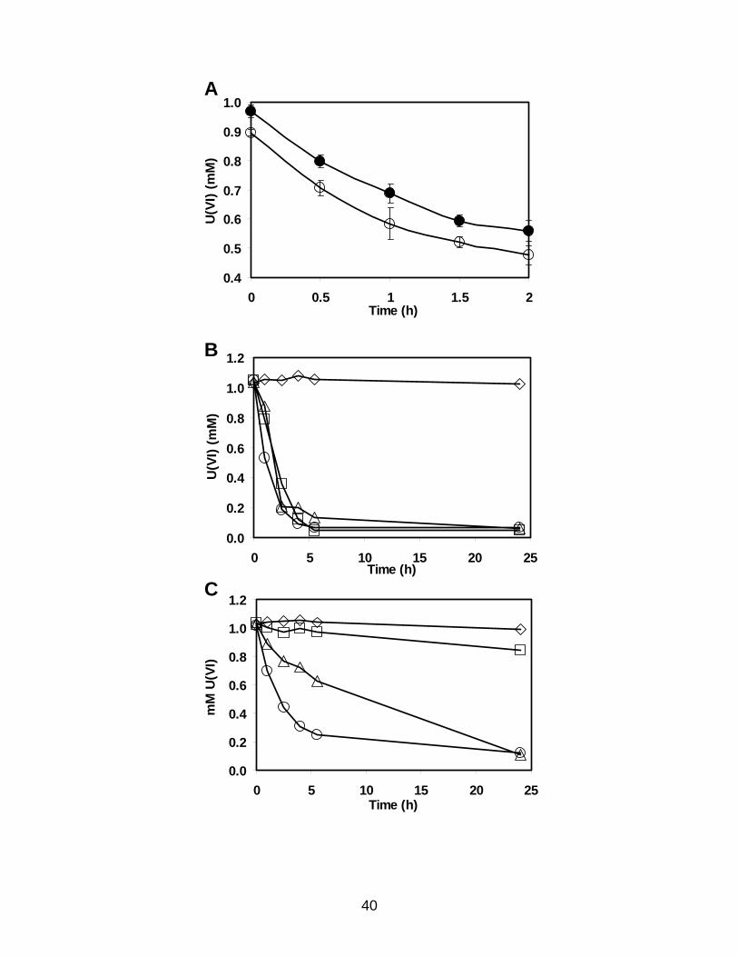
3.2.1. Uranium(VI) reduction by cytochrome c3 mutant
U(VI) reduction assays of wild-type D. desulfuricans G20 showed no difference
in the specific rate of reduction when cultures were harvested at early exponential phase
as compared to early stationary phase (Fig. 3.1A), suggesting there was no major
difference in U(VI) reduction capacity with growth phase. Therefore early stationary
phase cells were selected for assays to provide cell material. The mutant I2 lacking
cytochrome c3 reduced U(VI) with hydrogen as the electron donor poorly. However, I2
was still capable of reducing U(VI) at about 10% of the rate of the wild-type with
hydrogen as the electron donor (Fig. 3.1 B,C). I2 was still capable of reducing U(VI) at a
rate about 50% that of wild-type using lactate as the electron donor, and at a rate of about
33% of wild-type using pyruvate as the electron donor (Fig. 3.1 B,C and Table 3.1).
38

Figure 3.1. U(VI) reduction by a cytochrome c3 mutant of D. desulfuricans G20.
U(VI) reduction by D. desulfuricans strain G20 (A,B) or by the cytochrome c3 mutant I2
(C). All samples have 1 mM uranyl acetate and 200 µg whole cell protein/ml. (A) 10
mM sodium lactate is included as the electron donor. (○) cells harvested at early
exponential phase, (●) cells harvested at late exponential phase. Data points are the
average of three measurements and the figure is representative of four independent trials.
(B,C) cells are harvested at late exponential phase. 10 mM sodium lactate (○), 10 mM
sodium pyruvate (∆), 1 atm hydrogen gas (□), or no electron donor (◊) was added. Each
point is the average of three or more U(VI) measurements from two different
experiments. The figure is representative of four trials of no reductant and hydrogen as
the electron donor, and a minimum of five trials each of lactate or pyruvate as the
electron donor.
39
0.4
0.5
0.6
0.7
0.8
0.9
1.0
0 0.5 1 1.5 2Time (h)
U(V
I) (m
M)
A
0.0
0.2
0.4
0.6
0.8
1.0
1.2
0 5 10 15 20 25Time (h)
U(V
I) (m
M)
B
0.0
0.2
0.4
0.6
0.8
1.0
1.2
0 5 10 15 20 25Time (h)
mM
U(V
I)
C
40

Table 3.1. U(VI) reduction by a cytochrome c3 mutant of D. desulfuricans G20a
Electron Donor Strain (relevant genotype) Lactateb Pyruvatec Hydrogend
G20 (wild type) 2.2 ± 0.3 2.2 ± 0.3 1.5 ± 0.2
I2 (cycA mutant) 1.0 ± 0.2 0.6 ± 0.2 0.2 ± 0.1 aRates are from assays with whole cells, 200 µg cell protein/ml, determined from the first 2.5 h of the assay; µmol U(VI) reduced x mg cell protein-1 x h-1. No U(VI) reduction rate was measured in the absence of electron donor. bLactate, 10 mM; averages of seven or more determinations. cPyruvate, 10 mM; averages of five or more determinations. dHydrogen, 1 atm; averages of four determinations.
41

3.2.2. Fe-requirement for the synthesis of cytochrome c3
Western analysis of periplasmic extracts showed that wild-type D. desulfuricans
G20 cells grown in complete LS medium expressed abundant cytochrome c3. As
expected, the I2 mutant did not contain measurable cytochrome c3. Wild-type cells
grown in LS medium lacking iron have no more than 10% of the normal levels of
cytochrome c3 as wild-type cells grown in medium with excess iron (Fig. 3.2).
3.2.3. Uranium (VI) reduction by iron-limited cells
Wild-type cells grown in complete LS medium, and therefore expressing
abundant cytochrome c3, reduce U(VI) with lactate, pyruvate, or hydrogen gas as the
electron donor at about the same rate and extent for all three electron donors (about 2
µmol U(VI)/mg cell protein/hour; Table 3.1). Cells lacking wild-type levels of
cytochrome c3 due to growth in iron-limiting medium reduced U(VI) poorly with all
electron donors tested (Fig. 3.3). Typically, no increase in the rate or the amount of
U(VI) reduction was detected in cells grown in iron-limiting medium when electron
donor was included in the assay as compared to the rate seen in the absence of exogenous
electron donor (Fig. 3.3). We interpret the endogenous rate of U(VI) reduction seen with
no added external electron donor as being due to the oxidation of an internal storage
reserve of reductant, like polyglucose. Desulfovibrio gigas has been shown to store
reductant as polyglucose (Santos et al., 1993)
42

Figure 3.2. Cytochrome c3 expression in iron-limited cells of D. desulfuricans G20.
Western analysis showing cycA mutant cells and wild-type cells limited for iron do not
make cytochrome c3. Lane M: Molecular mass markers, 14 kDa is indicated; lane 1: 10
µg high pH TE wash protein fraction from wild-type cells grown in complete medium;
lane 2: 10 µg high pH TE wash protein fraction from wild-type cells grown in iron
limiting medium; lane 3: 10 µg high pH TE wash protein fraction from cycA cells grown
in complete medium; lane 4: 25 ng of pure cytochrome c3.
21 43M
14 kDa
43

Figure 3.3. U(VI) reduction by iron-limited cells of D. desulfuricans G20.
U(VI) reduction by D. desulfuricans strain G20 grown in complete medium with iron (A)
or grown in iron-limiting medium (B). All samples have 1 mM uranyl acetate and 200 µg
whole cell protein/ml. 10 mM sodium lactate (○), 10 mM sodium pyruvate (∆), 1 atm
hydrogen gas (□), or no electron donor (◊) was added. Each point is the average of three
U(VI) determinations, in most cases the error bars are within the symbols. The figure is
representative of three trials.
A
0.0
0.2
0.4
0.6
0.8
1.0
1.2
0 5 10 15 20 25Time (h)
mM
U (V
I)
B
0.0
0.2
0.4
0.6
0.8
1.0
1.2
0 5 10 15 20 25Time (h)
mM
U (V
I)
44

3.2.4. Nitrite reduction by a cytochrome c3 mutant
DiChristina and coworkers screened a library of chemically mutagenized
Shewanella oneidensis and found a mutant incapable of growth using U(VI) or nitrite as
the sole electron acceptor, but this mutant was still capable of aerobic growth or
anaerobic growth using Fe(III) or Mn(IV) (Wade and DiChristina, 2000). They
interpreted this observation to mean that in Shewanella, U(VI) reduction and nitrite
respiration shared a common component that the other forms of respiration did not.
Furthermore, they hypothesized that the component shared between U(VI) and nitrite
respiration was the c-type cytochrome, nitrite reductase. Some strains of Desulfovibrio,
such as D. desulfuricans strain 27774, can utilize nitrate or nitrite as the terminal electron
acceptor for respiration (Almeida et al., 2003). In these strains, the periplasmic c-type
cytochrome, nitrite reductase, is the terminal enzyme in the respiratory pathway of nitrite
respiration (Liu and Peck, 1981). Nitrite reductase has been purified from D.
desulfuricans strain 27774 and found to be a heterodimer that is membrane-bound with
the active site periplasmic-facing (Almeida et al., 2003). Each subunit is a multiheme c-
type cytochrome and the two subunits are encoded in an operon nfrHA. The first gene,
nfrH, encodes a tetraheme c-type cytochrome and is an integral membrane protein with a
predicted transmembrane helix motif composed of the first 33 amino acids, using the
transmembrane helix prediction programs TMHMM (Almeida et al., 2003). NfrH is a
member of the NapC/NirT family of oxidoreductases, whose members in other
proteobacteria accept electrons from the quinone pool of the electron transport chain and
transfer those electrons to the catalytic subunit encoded by the gene nfrA (Almeida et al.,
2003; Simon et al., 2000). The second subunit, NfrA, is a pentaheme c-type cytochrome.
45
Interestingly, one of the heme-binding sites is comprised of the amino acids CXXCK
rather than the typical CXXCH motif (Cunha et al., 2003). The second subunit contains
no transmembrane domains and is more loosely associated with the membrane. NfrA is
the catalytic subunit, reducing nitrite to ammonia, a six electron transfer event (Almeida
et al., 2003; Simon et al., 2000).
Although D. desulfuricans G20 and D. vulgaris cannot grow by nitrate or nitrite
respiration (data not shown; Postgate, 1979), sequence analysis of the completed
genomes of D. desulfuricans G20 and D. vulgaris revealed an operon similar to the
biochemically characterized nitrite reductase of D. desulfuricans 27774 (Fig. 3.4). The
intergenic region between the nfrH and nfrA genes of G20 is 100 bp and operon structure
is a possibility (Oak Ridge National Labs, http://genome.ornl.gov/microbial/ddes/.html).
The nfrHA orthologs found in D. vulgaris Hildenborough overlap by 76 bp supporting
operon structure (VIMSS, http://www.escalante.lbl.gov). The first subunit encoded in the
predicted D. desulfuricans G20 and D. vulgaris nitrite reductase operon (nfrH) has the
characteristic tetraheme c-type cytochrome heme-binding motif. In addition, both the D.
desulfuricans G20 and D. vulgaris NfrH proteins have the predicted transmembrane
motif characteristic of an integral membrane protein of the NapC/NirT family (VIMSS,
http://www.escalante.lbl.gov; TMHMM, www.cbs.dtu.dk/services/TMHMM-2.0.html).
Interestingly, the second subunit of the predicted D. desulfuricans G20 nitrite reductase
operon, NfrA, has an octaheme c-type cytochrome binding motif. All eight of the heme-
binding motifs of the D. desulfuricans G20 NfrA are of the CXXCH type. Preliminary
computer annotation by VIMSS identifies the D. vulgaris operon as a nitrite reductase,
while preliminary computer annotation by VIMSS identifies the D. desulfuricans G20
46

operon as a member of the NapC/NirT family of reductases, possibly a trimethylamine or
hydroxylamine reductase (Simon et al., 2000). The true nitrite reductase from D.
desulfuricans 27774 was also shown to have hydroxylamine reductase activity in addition
to nitrite reductase activity (Liu and Peck, 1981). This circumstantial evidence suggested
that these two genes of D. desulfuricans G20 encode nitrite reductase; however, the
assignment is still tentative.
Northern analysis indicated that the putative D. desulfuricans G20 nitrite
reductase operon was expressed in wild-type G20 and in the cytochrome c3 mutant I2.
Interestingly, the expression of the nitrite reductase was at least 8-fold higher in the wild-
type than in the cytochrome c3 mutant (Fig. 3.5). In addition, the ca. 2.5-kb transcript
observed when using a probe from the internal region of the nfrH gene suggested that the
putative nfrH and nfrA genes were cotranscribed on a single transcript.
47

Figure 3.4. The nitrite reductase operon of Desulfovibrio strains.
Diagramatic representation of the gene order of the putative nitrite reductase operons of
the SRB. VIMSS (http://www.eslcante.lbl.gov) orf numbers of each putative gene are
shown for D. vulgaris and D. desulfuricans G20. The predicted number of heme-binding
motifs deduced from the translated protein is shown above each gene. The percent
identity/percent similarity of each putative protein as compared to the nitrite reductase
translated from the corresponding gene of D. desulfuricans 27774 is indicated beneath
each. The genomic regions surrounding the putative nitrite reductase operons from D.
desulfuricans G20 and D. vulgaris share no synteny.
48

Figure 3.5. Nitrite reductase expression in a cytochrome c3 mutant.
Lane 1: G20 RNA. Lane 2: I2 RNA. Panel A: Northern blot probed with the internal
fragment of the small subunit of the G20 nitrite reductase. The Arrow indicates the
position of the 3-kb marker on the gel. Panel B: Gel showing the 23S and 16S rRNA. 5
µg total RNA added per lane in gels shown in both panels. These results are
representative of RNA from two independent experiments.
A
B49

In experiments measuring nitrite reduction, both wild-type G20 and cytochrome
c3 mutant I2 reduced nitrite. Interestingly, when pyruvate was the electron donor, I2
reduced nitrite about twice as fast as G20 (Table 3.2, Fig. 3.6 A). Hydrogen served as a
poor electron donor for nitrite reduction in G20. The rate of hydrogen-supported nitrite
reduction by G20 was higher than the rate of nitrite reduction when no electron donor
was added (P=0.039, Table 3.2). Apparently hydrogen gas did not support nitrite
reduction by I2, as the rate when hydrogen was the electron donor was not faster then the
rate when no electron donor was added (P=0.24, Table 3.2).
Viamajala and coworkers tested the nitrite reductase of Shewanella for its ability
to reduce Cr(VI) (Viamajala et al., 2002). They observed that Cr(VI) inhibited nitrite
reduction, and nitrite inhibited Cr(VI) reduction in Shewanella when the two compounds
were added in equal molar amounts (0.1-0.5 mM) (Viamajala et al., 2002). Since
electron donor was in excess in their system, they interpreted this result to mean that the
inhibition was due to competition at the site of the common reductase. In our
experiments measuring the simultaneous reduction of 1 mM nitrite and 1 mM U(VI),
uranyl acetate failed to inhibit nitrite reduction when 10 mM lactate or 10 mM pyruvate
was the electron donor in either G20 or I2 (Fig. 3.6 A, data not shown). Similarly, nitrite
failed to inhibit U(VI) reduction by either G20 or I2 when lactate or pyruvate were the
electron donor (Fig. 3.6 B,C). Nitrite also failed to inhibit U(VI) reduction in G20 when
hydrogen was the electron donor (Fig. 3.6 D). Curiously, 1 mM nitrite did inhibit the
residual rate of U(VI) reduction measured in I2 (Fig 3.1, Table 3.1) when hydrogen was
the electron donor. (Fig. 3.6 D).
50

Table 3.2. Nitrite reduction by D. desulfuricans G20 wild-type and the cytochrome c3 mutant I2 a
Nitrite reduction ratea (avg + SD) with electron donorbStrain
(relevant genotype) Lactate Pyruvate Hydrogen None
G20 (wild-type) 0.52 ± 0.13 1.0 ± 0.36 0.15 ± 0.06 0.07
I2 (cycA mutant) 0.64 ± 0.04 2.0 ± 0.6 0.06 ± 0.02 0.07 aRate units are µmol NO2 reduced/mg cell protein/h. Rates are from assays with whole cells, 100-200 µg cell protein/ml, determined from the first 2.5 h of the assay when lactate or pyruvate were the electron donors, or from the first 20 h of the assay when hydrogen gas or no electron donor was added. All rates are the average of four independent determinations except control lacking added electron donor which was the average of two independent determinations. The endogenous rate of nitrite reduction is not subtracted. b Lactate, 10 mM; pyruvate, 10 mM; hydrogen, 1 atm.
51

Figure 3.6. Nitrite reduction by a cytochrome c3 mutant.
Panel A: Nitrite reduction by G20 (squares) and I2 (circles) in the presence or absence of
1 mM uranyl (VI) acetate. G20 without (□) and with (■) 1 mM U(VI), and I2 without (○)
and with (●) 1 mM U(VI). 10 mM sodium pyruvate added as the electron donor.
Panel B-D: U(VI) reduction by G20 (squares) and I2 (circles) in the presence or absence
of 1 mM sodium nitrite. G20 without (□) and with (■) 1 mM NO2, and I2 without (○)
and with (●) 1 mM NO2.
Panel B: 10 mM sodium lactate added as the electron donor;
Panel C: 10 mM sodium pyruvate added as the electron donor;
Panel D: 10 mM sodium pyruvate added as the electron donor.
Data points are the average of at least two independent measurements, and graphs are
representative of at least two independent experiments.
52

0
0.2
0.4
0.6
0.8
1
1.2
0 5 10 15 20 25Time (h)
Nitr
ite (m
M)
A
0.0
0.2
0.4
0.6
0.8
1.0
1.2
1.4
0 5 10 15 20 25Time (h)
U(V
I) (m
M)
B
0.0
0.2
0.4
0.6
0.8
1.0
1.2
1.4
0 5 10 15 20 25Time (h)
U(V
I) (m
M)
C
53

D
0.0
0.2
0.4
0.6
0.8
1.0
1.2
1.4
0 5 10Tim
U(V
I) (m
M)
54
15 20 25e (h)

3.2.5. Hydrogenase activity in the cytochrome c3 mutant of D. desulfuricans.
Hydrogenase has been reported to reduce Cr(VI) in vitro (Chardin et al., 2003)
and to reduce Tc(VII) in vivo (De Luca et al., 2001), although the ability of hydrogenase
to reduce U(VI) to U(IV) was not tested in these two systems. The cytochrome c3 mutant
of D. desulfuricans G20 was severely impaired for sulfate respiration (Rapp-Giles et al.,
2000), and for U(VI) reduction (Fig. 3.1) using hydrogen as the electron donor. We
tested the possibility that these two observations were due to repression of hydrogenase
activity in the cytochrome c3 mutant rather than the lack of the protein cytochrome c3 per
se. Hydrogenase activity was measured in resting cells of both wild-type G20 and the
cytochrome c3 mutant, I2 grown on LS medium. When activity was measured in the
direction of hydrogen oxidation with methyl viologen as the electron acceptor, I2 cells
reduced methyl viologen about twice as fast as G20 (Fig 3.7). Negative controls with no
added hydrogen showed no reduction of methyl viologen during the time frame of this
assay (30 min), although negative controls with no electron donor showed a similar
extent of methyl viologen reduction after 16 hours as when hydrogen was included (data
not shown). Therefore, the cycA mutant did not downregulate hydrogenase.
55

Figure 3.7. Hydrogenase assay of a cytochrome c3 mutant of D. desulfuricans.
Hydrogenase activity as measured by methyl viologen reduction by whole cells of
wild-type G20 (squares) and the cytochrome c3 mutant I2 (circles). G20 cells with (■)
and without (□) hydrogen as the electron donor; I2 cells with (●) and without (○)
hydrogen as the electron donor. Each tube contains 2.5 µg/ml whole cell protein. Each
point is one measurement. This figure is representative of 3 independent trials.
0
0.5
1
1.5
2
2.5
3
0 5 10 15 20
Time (min)
O.D
. 602
nm
56

3.3. Discussion
Both the mutant lacking cytochrome c3 and wild-type cells lacking cytochrome c3
due to growth in iron-limiting medium reduced U(VI) poorly with hydrogen as the
electron donor (Table 3.1, Fig. 3.1). However, some reduction of U(VI) with hydrogen
was consistently seen during assays of I2. This likely resulted from a bypass of
cytochrome c3 (for example through reduction of U(VI) directly by the Fe-hydrogenase,
see below) rather than from the accumulation of small numbers of suppressors that restore
cytochrome c3 since Western analysis of a portion of the I2 cells used in the U(VI)
reduction assay did not detect cytochrome c3 (data not shown). This observation supports
the earlier report that cytochrome c3 was necessary for U(VI) reduction by extracts of D.
vulgaris when hydrogen was the source of electrons (Lovley et al., 1993). Surprisingly, I2
was still capable of reducing U(VI) with lactate as the electron donor at a rate about one-
half that of the wild-type and of reducing U(VI) with pyruvate as the electron donor at a
rate of about one-third that of the wild-type (Table 3.1, Fig. 3.1). However, wild-type
cells, containing low amounts of cytochrome c3 when grown in iron-limiting medium
reduced U(VI) poorly, if at all, with lactate or pyruvate as the electron donor (Fig. 3.1).
Since iron is required for the synthesis of many cofactors needed by electron transport
proteins such as Fe-S clusters and hemes, growth in iron-limiting medium may cause a
general repression of other electron transport proteins in addition to cytochrome c3.
Previous experiments using sulfate as the electron acceptor showed that I2 grew at the
same rate and to 85% of the extent of the wild-type with lactate as the electron donor but
was unable to grow with pyruvate as the sole electron donor (Rapp-Giles et al., 2000).
We inferred that cytochrome c3 is apparently involved in the transfer of electrons from
57

pyruvate to sulfate to support growth but is not the sole carrier for electrons from lactate
to sulfate. We also expected that the cytochrome c3 mutant might be unable to reduce
U(VI) with pyruvate. However, the linkage between growth and U(VI) reduction with
pyruvate as the electron donor was not absolute suggesting multiple pathways for
electrons to reach toxic metals.
Clearly, electron flow independent of cytochrome c3 provides reductant for U(VI)
in D. desulfuricans strain G20 when organic acids are oxidized. We suggest that
essentially all of the electrons from hydrogen must go through the cytochrome c3
pathway for U(VI) reduction, but only some of the electrons from lactate or pyruvate go
through the cytochrome c3 pathway for U(VI) reduction (Fig. 3.8). This is consistent with
the hydrogen cycling model of electron flow for some of the electrons from organic acids
coupled with alternative pathways for others.
The periplasmic nitrite reductase of Shewanella (a facultative anaerobe capable of
growing by nitrate, Fe(III), Cr(VI), or U(VI) respiration (Myers and Nealson, 1988;
Lovley et al., 1991) has been suggested to be a carrier in the pathway for U(VI) reduction
in that organism (Wade and DiChristina, 2000). In addition, the nitrite reductase of
Shewanella has been proposed to also act as a Cr(VI) reductase (Viamajala et al., 2002).
Some strains of SRB (like D. desulfuricans strain 27774) can grow by nitrate respiration
(Mitchell et al., 1986) and, therefore, must have a bona fide nitrite reductase. Although
D. desulfuricans G20 and D. vulgaris are incapable of this respiration (data not shown),
the presence of an operon encoding a possible periplasmic nitrite reductase is present in
the D. desulfuricans G20 and D. vulgaris genome sequences (VIMSS,
http://www.escalante.lbl.gov; Fig 3.4). The function of nitrite reductase in SRB
58

Figure 3.8. Modified Lovley Model for U(VI) reduction in a cytochrome c3-lacking
cells.
Arrows indicate electron flow and do not necessarily imply a single step in vivo.
Although essentially all of the electrons from hydrogen are transferred to U(VI) via
cytochrome c3, some of the electrons from lactate or pyruvate can bypass cytochrome c3.
H Cytochrome U(VI)
U(IV)Lactate Pyruvate
Hydrogenase
UnidentifiedElectronCarrier(s)
red
ox3 2e-
2e-
2e-
2e-2e-
2e-c2
59

which are unable to grow by nitrate respiration has been suggested to be for the
detoxification of nitrite produced by neighboring sulfate-oxidizing nitrate-reducing
bacteria such as Thiomicrospira strains (Greene et al., 2003). We hypothesized that the
putative D. desulfuricans G20 nitrite reductase (a c-type cytochrome) might act as an
alternate U(VI) reductase in wild-type G20 or in the cytochrome c3 mutant cells. The
putative nitrite reductase of G20 was shown to be expressed by Northern analysis when
cells are grown in LS medium in the absence of nitrite, and whole cells were shown to
have nitrite reducing activity (Fig. 3.5 and Fig. 3.6).
Viamajala and coworkers observed that Cr(VI) inhibited nitrite reduction, and
nitrite inhibited Cr(VI) reduction in Shewanella when the two compounds were added in
equal molar amounts (0.1-0.5 mM) (Viamajala et al., 2002). Because of this result, it
was suggested that the nitrite reductase of Shewanella was capable of acting as a Cr(VI)
reductase (Viamajala et al., 2002). We reasoned that if the nitrite reductase of the wild-
type or cytochrome c3 mutant of D. desulfuricans G20 acted as a U(VI) reductase, then
the addition of nitrite should inhibit U(VI) reduction, and the addition of uranium should
inhibit nitrite reduction. The addition of a sub-inhibitory concentration of nitrite (1mM)
did inhibit U(VI) reduction by the I2 mutant, but not wild-type G20 cells when hydrogen
was the electron donor (Fig. 3.6). The interpretation of this observation is somewhat
complicated by the fact that nitrite has been shown to inhibit the Fe-hydrogenase of D.
vulgaris (Berlier et al., 1987). However, nitrite and U(VI) reduction proceeded
independently of each other in both G20 and the I2 mutant under the conditions of these
assays when lactate or pyruvate were the electron donors and the electron acceptors were
in equal molar concentrations (1 mM; Fig. 3.6). In particular, since nitrite and U(VI)
60

reduction proceeded independently of each other in cells lacking cytochrome c3, we
interpret this result to mean that the nitrite reductase is unlikely to act as a U(VI)
reductase.
Interestingly, the I2 mutant lacking cytochrome c3 reduced nitrite about twice as
fast as the wild-type G20 when pyruvate was the electron donor (Fig. 3.6). This
suggested to us that either nitrite reductase is up-regulated in I2, or that the blockage of
normal electron flux in the cytochrome c3 mutant caused more electron flow from
pyruvate through the nitrite reductase. Northern analysis showed that the transcription of
the putative nitrite reductase of I2 is not up-regulated, but is about 8-fold down-regulated
in the cytochrome c3 mutant (Fig. 3.5). However, it must be pointed out that cultures
used for Northern analysis and cultures used for nitrite reduction assays were not
identical. Northern analysis was performed on RNA isolated from early- to mid-
exponential phase cultures, as older cultures yielded degraded RNA (data not shown),
while the nitrite reduction assays were performed on early stationary phase cultures.
It is possible that the absence of cytochrome c3 in I2 causes an alteration in the
electron flow in this organism so that more electrons are available to the nitrite reductase
(Fig. 3.9) and that the additional electron flow accounts for the mutant’s faster rate of
nitrite reduction. We speculate that the build-up of electrons in the electron transport
chain of I2 might cause the mutant to downregulate the components of its electron
transport chain that are partnered with cytochrome c3 in the wild-type. The redox state of
the quinone pool has been shown to regulate the expression of electron carrier proteins in
the facultative anaerobe Paracoccus denitrificans (Otten et al., 2001) and the phototroph
Rhodobacter sphaeroides (Oh and Kaplan, 2000). Northern analyses (see sections 6 and
61
7, below) indicate that other membrane-bound c-type cytochromes are down-regulated in
the cytochrome c3 mutant. A similar phenomenon has been observed for the Fe-
hydrogenase mutant of D. vulgaris (Haveman et al., 2003; Pohorelic et al., 2002). The
gene encoding a cytoplasmic alcohol dehydrogenase is highly expressed in wild-type D.
vulgaris when cells are grown on media in which ethanol, lactate, pyruvate, formate, or
hydrogen provide energy for sulfate reduction (Haveman et al., 2003). The authors
interpreted this to suggest that electron flow in the wild-type from lactate or pyruvate, as
well as formate or hydrogen using acetate as the carbon source, requires alcohol
dehydrogenase (Haveman et al., 2003). In the Fe-hydrogenase mutant of D. vulgaris, the
alcohol dehydrogenase was at least 10-fold down-regulated (Haveman et al., 2003).
Alternatively, it is also possible that the proteins encoded by the putative G20
nitrite reductase operon may not be responsible for the measured nitrite reductase
activity. This would also account for the paradoxical observation between the nitrite
reductase activity and nitrite reductase expression in the cytochrome c3 mutant. A mutant
constructed in the putative G20 nitrite reductase, or biochemical analysis on the purified
NfrHA complex from G20 would help to answer these questions.
The final U(VI) reductase candidate explored was hydrogenase. The NiFe-
hydrogenase of Desulfovibrio fructosovorans has been reported to act as a technetium
(VII) reductase by DeLuca and coworkers (De Luca et al., 2001), and the Fe-hydrogenase
of D. vulgaris has been shown to act as a chromium (VI) reductase (Chardin et al.,
2003). These data suggest that low redox-potential Fe-S clusters (non-heme Fe
cofactors) may be sufficient for metal reduction in vivo (Chardin et al., 2003). We think
it is unlikely that the hydrogenase of wild-type G20 or the mutant I2 acts as a U(VI)
62

reductase, since the I2 mutant has at least as much, if not more, hydrogenase activity than
the wild-type, yet reduces U(VI) poorly with hydrogen as the electron donor. However,
it is possible that the Fe-hydrogenase shown to be responsible for greater than 90% of the
periplasmic hydrogen oxidation activity of D. vulgaris (Pohorelic et al., 2002) acts as a
U(VI) reductase, but with a much slower rate than cytochrome c3 (about 10% the rate of
cytochrome c3). This would account for the small rate of U(VI) reduction seen in I2
when hydrogen is the electron donor. The role of hydrogenase will be further tested in
the next chapter by assaying a Fe-hydrogenase mutant of the related species D. vulgaris
for U(VI) reduction.
63

Figure 3.9. Altered electron flux in a cytochrome c3 mutant.
Electron flow from lactate and pyruvate oxidation in the wild-type (Top panel) and in the
cycA mutant (Bottom panel). Cyt c3 represents the dominant periplasmic c-type
cytochrome encoded by cycA. Qpool represents the quinone pool. NiR is the putative
nitrite reductase. Hmc represents the transmembrane high molecular mass cytochrome c
complex which is proposed to transfer electrons to the cytoplasmic enzymes responsible
for sulfate reduction (Dolla et al., 2000). In the cycA mutant, the absence of cyt c3 causes
a blockage of electron flow from the quinone pool. Those electron transport proteins
linked to the quinone pool are “over-reduced” with electrons. Red arrows indicate
electron flow and do not necessarily imply a single step in vivo.
64

Cytoplasm
Cyt c3
NiR
Lactate Pyruvate
Qpool
NiR
HMCQpool
Periplasm
Cytoplasm
NiR
Lactate Pyruvate
Qpool
NiR
HMCQpool
Periplasm
65

4. Uranium(VI) reduction by a hydrogenase mutant of Desulfovibrio
vulgaris
Rayford B. Payne, Kelly R. Titkemeier, and Judy D. Wall
Biochemistry Department, University of Missouri-Columbia, Columbia, MO 65211
Part of the work described here was carried out by Kelly R. Titkemeier as part of a
semester project for a non-thesis Master’s degree in Geological Sciences completed
in May 2004.
66

4.1. Introduction
Three classes of hydrogenase have so far been identified in the periplasm of D.
desulfuricans G20 and D. vulgaris Hildenborough; each is named for the metal ion(s) at
the active site of the enzyme. The three classes are Fe-, NiFe-, and NiFeSe-hydrogenase
(Fauque et al., 1988; Lissolo et al., 1986; Pohorelic et al., 2002; Prickril et al., 1987).
The genome sequence of D. vulgaris suggests the existence of an additional NiFe-
hydrogenase isozyme and at least two cytoplasmic hydrogenases (Heidelberg et al.,
2004). The genome sequence of D. desulfuricans G20 suggests the existence of an
additional NiFe-hydrogenase isozyme, an additional Fe-hydrogenase isozyme, and one
cytoplasmic hydrogenase (VIMSS, http://www.escalante.lbl.gov). The existence of a
cytoplasmic hydrogenase to create hydrogen gas from protons and electrons generated by
the oxidation of organic acid, and the existence of a periplasmic hydrogenase to re-
capture cytoplasmically generated hydrogen, are essential for the generation of a proton
gradient across the cytoplasmic membrane by the mechanism illustrated in the hydrogen
cycling model (Odom and Peck, 1981; Fig. 1.4).
Since the 16S ribosomal RNA genes of D. desulfuricans G20 and D. vulgaris
Hildenborough are about 91% identical, and the cytochrome and hydrogenase content are
quite similar between the two organisms (VIMSS, http://www.escalante.lbl.gov), it seems
likely that many electron transport components used to reduce U(VI) will be shared. A
mutant in the Fe-hydrogenase encoding gene of D. vulgaris was constructed by
Voordouw and coworkers by marker exchange of the gene encoding the periplasmic Fe-
hydrogenase (hydAB) with a gene encoding chloramphenicol resistance (Pohorelic et al.,
2002). Whole, intact cells of the Fe-hydrogenase mutant of D. vulgaris have about 50%
67

of the hydrogen uptake activity of the wild-type D. vulgaris and have no detectable
periplasmic hydrogen uptake activity compared to the wild-type D. vulgaris when
measured by cyclic voltammetry (Pohorelic et al., 2002). These data suggest that the Fe-
hydrogenase is responsible for essentially all of the periplasmic hydrogen uptake activity
in the wild-type D. vulgaris, and that the contribution of the NiFe- and NiFeSe-
hydrogenases to periplasmic hydrogen uptake is negligible.
In previous experiments, a mutant in the related strain D. desulfuricans G20
lacking cytochrome c3 was shown to be impaired about 50% compared to the wild-type in
U(VI) reduction when lactate or pyruvate were the electron donors. The cytochrome c3
mutant was more severely impaired, about 90%, in U(VI) reduction when hydrogen gas
was the electron donor (Payne et al., 2002). In many Desulfovibrio strains, this
cytochrome has been proposed to act in series with the hydrogenases. Thus we would
predict that a mutation decreased in hydrogenase activity might also be impaired in U(VI)
reduction with hydrogen as the source of reductant. To determine the role played by the
Fe-hydrogenase in U(VI) reduction, we assayed the Fe-hydrogenase mutant of D.
vulgaris for its ability to reduce U(VI). We anticipated that the Fe-hydrogenase mutant
of D. vulgaris would have a similar phenotype as the cytochrome c3 mutant of D.
desulfuricans G20. In other words, the Fe-hydrogenase mutant of D. vulgaris was
anticipated to be about 90% impaired when hydrogen gas was the electron donor, and
about 50% when lactate or pyruvate were the electron donors. This was not the result
observed. We found that the Fe-hydrogenase mutant reduced U(VI) at the same rate as
wild-type cells when hydrogen gas or pyruvate were the electron donors. However, when
lactate was the electron donor the Fe-hydrogenase mutant was about 50% impaired in
68

U(VI) reduction. This suggests that the periplasmic Fe-hydrogenase is involved in the
flow of electrons to U(VI) when lactate, but not hydrogen or pyruvate, is the electron
donor.
4.2. Results
4.2.1. Growth of Desulfovibrio vulgaris
A representative growth curve of wild-type D. vulgaris and the Fe-hydrogenase
mutant of D. vulgaris on LS medium is shown (Fig. 4.1). The Fe-hydrogenase mutant
consistently grew at about a 30-50% slower rate, and reached ca. a 50% lower final
protein yield (data not shown) than the wild-type when lactate was the electron donor.
No attempts to grow D. vulgaris on PS or hydrogen plus sulfate were made during the
course of this study.
4.2.2. U(VI) reduction by a Fe-hydrogenase mutant of Desulfovibrio vulgaris
Comparative U(VI) reduction rates are shown in Table 4.1 for wild-type and the
Fe-hydrogenase mutant. These rates were corrected for those observed when no external
source of reductant was added to cultures. In these cultures that endogenous rate was
about one-half that of pyruvate supported U(VI) reduction activity. As expected, wild-
type cells expressing the Fe-hydrogenase reduced U(VI) with lactate, pyruvate, or
hydrogen gas as the electron donor. Unlike the result for D. desulfuricans wild-type
(Table 3.1), the rates of reduction were about twice as fast for lactate or hydrogen as for
pyruvate (Table 4.1). The Fe-hydrogenase mutant reduced U(VI) at the same rate as
wild-type when hydrogen gas or pyruvate was the electron donor, but was impaired about
50% in U(VI) reduction when lactate supplied the electrons (Table 4.1).
69

Figure 4.1. Growth of a Fe-hydrogenase mutant of Desulfovibrio vulgaris.
Growth of wild-type (□) and Fe-hydrogenase mutant (○) of D. vulgaris on LS medium as
measured by optical density at 600nm (Titkemeier and Wall, 2004). Data points represent
one measurement and this figure is representative of five independent trials.
0.1
1.0
0 5 10 15 20 25 30 35 40 45 50Time (h)
O.D
. 600
nm
70

4.2.3. U(VI) reduction by Desulfovibrio vulgaris in the presence of inhibitors
To further explore the contribution of the various periplasmic hydrogenases (Fe-,
NiFe-, and NiFeSe-) to U(VI) reduction, we exposed resting cells of D. vulgaris to
various hydrogenase inhibitors, then tested those cells for U(VI) reduction. 0.5 mM Cu2+
has been shown to inhibit greater than 90% of the periplasmic hydrogen uptake activity
of whole cells of D. vulgaris (Cypionka and Dilling, 1986; Fitz and Cypionka, 1991), and
Desulfovibrio fructosovorans (De Luca et al., 2001). When wild-type D. vulgaris was
preincubated with 0.5 mM CuCl2, there was about a 90% inhibition of the U(VI)
reduction rate with all electron donors tested (Table 4.1). This suggests that periplasmic
hydrogenase activity or its associated activities that may be copper inhibited are essential
for U(VI) reduction when hydrogen or the organic acids lactate or pyruvate supply the
electrons.
Nitrite at 100 µM was reported to inhibit the Fe- and NiFeSe-hydrogenase, but
not the NiFe-hydrogenase in cell-free extracts of D. vulgaris (Berlier et al., 1987). The
concentration of an inhibitor needed to cause inhibition is likely to be greater in whole
cells than in cell-free extracts. Therefore a concentration of 1 mM NaNO2 was chosen to
test for hydrogenase inhibition in whole cells of D. vulgaris. One mM NaNO2 does not
inhibt the growth of D. desulfuricans G20 on LS medium (data not shown). When wild-
type D. vulgaris was preincubated with 1 mM NaNO2, there was about a 50% inhibition
of the U(VI) reduction rate using lactate or hydrogen as the electron donors tested (Table
4.1). However, no inhibition of U(VI) reduction by nitrite was observed when pyruvate
was the source of electrons (Table 4.1).
71

Table 4.1. U(VI) Reduction by a Fe-hydrogenase mutant of D. vulgaris Hildenborougha
Electron Donor
D. vulgaris strain (relevant genotype)
Inhibitor
Lactate Pyruvate Hydrogen
Wild-typeb None 4.2 ± 0.4 1.5 ± 0.6 4.4 ± 1.0
hydAB mutantb None 1.4 ± 0.5 1.5 ± 0.3 4.5 ± 0.8
Wild-typec 0.5 mM CuCl2 0.15 0.1 0.1
Wild-typed 1 mM NaNO2 2.0 ± 0.2 1.6 ± 0.2 1.6 ± 0.5 aRates are from assays with whole cells, 100µg cell protein/ml, determined from the first 2 h of the assay; µmol U(VI) reduced x mg cell protein-1 x h-1. bRates are four independent determinations. cRates are two independent determinations. dRates are three independent determinations.
72
4.3. Discussion
Lovley and coworkers reconstituted a pathway for U(VI) reduction in vitro using
hydrogen gas as the electron donor, purified hydrogenase, and purified cytochrome c3
(Lovley et al., 1993). We have a cytochrome c3 mutant in D. desulfuricans G20 and a
Fe-hydrogenase mutant in D. vulgaris Hildenborough. Because these strains share about
91% identity in 16S ribosomal RNA and have many physiological and biochemical
phenotypes (such as substrates utilized, incomplete oxidation of organic acids, antibiotic
resistances, etc.) in common, we assumed that the electron transport chain components in
the two strains would be comparable. This assumption is supported by both biochemical
(Aubert et al., 1998) and genome sequence analysis (VIMSS,
http://www.escalante.lbl.gov) of the cytochrome and hydrogenase content of the two
strains. A mutant in the dominant periplasmic cytochrome c3, constructed in the parent
strain D. desulfuricans G20, was impaired about 90% with hydrogen gas as the electron
donor for U(VI) reduction (Payne et al., 2002). This observation supported the Lovley
model derived from D. vulgaris Hildenborough for U(VI) reduction when hydrogen is the
electron donor. The Fe-hydrogenase constitutes the majority of the periplasmic
hydrogenase activity of Desulfovibrio strains, and a mutant of D. vulgaris Hildenborough
lacking that enzyme had no detectable periplasmic hydrogen uptake activity (Pohorelic et
al., 2002). When I tested that mutant for U(VI) reduction, the rate and extent were not
impaired with hydrogen gas as the electron donor. This result was consistent with those
from Elias and coworkers who also found that the Fe-hydrogenase mutant of D. vulgaris
was not impaired in hydrogen supported U(VI) reduction (Elias et al., 2004). Two
possible explanations for this observation are: (1) the electrons generated from hydrogen
73

oxidation by the Fe-hydrogenase are not in the pathway of U(VI) reduction in wild-type
D. vulgaris; therefore, the mutant is not impaired in this process; or (2) though the
electrons generated from hydrogen oxidation by the Fe-hydrogenase are in the pathway
of U(VI) reduction in wild-type D. vulgaris, in the mutant the additional hydrogenases
compensate for the absence of the Fe-isozyme. Although Pohorelic and coworkers
showed that the NiFe- or NiFeSe-hydrogenases apparently were responsible for only
trace amounts of periplasmic hydrogen oxidation activity with methyl viologen as the
electron acceptor (Pohorelic et al., 2002), these isozymes might be altered in activity or
abundance when the Fe-hydrogenase is removed by mutation. The expression of the
alternate hydrogenase isozymes was not followed (by Northern analysis or RT-PCR)
during the course of this dissertation, but explanation (2) is appealing as the NiFe- or
NiFeSe-hydrogenase have been shown to have a higher affinity for hydrogen than the Fe-
hydrogenase (Fauque et al., 1988). This possibility is supported by the observation that
Cu2+, an inhibitor of all periplasmic hydrogenases (Cypionka and Dilling, 1986; De Luca
et al., 2001), inhibits greater that 90% of U(VI) reduction by both the Fe-hydrogenase
mutant and the wild-type when hydrogen is the electron donor (Table 4.1).
My experiments showed that the cytochrome c3 mutant of D. desulfuricans G20
was impaired by about 66% in U(VI) reduction compared to the wild-type D.
desulfuricans G20 when pyruvate was the electron donor. Surprisingly, the rate of U(VI)
reduction by the Fe-hydrogenase mutant of D. vulgaris Hildenborough with pyruvate as
the electron donor was not impaired compared to the wild-type. At least one alternate
electron transfer pathway apparently exists that does not require the Fe-hydrogenase. It is
possible that the NiFe- or NiFeSe-hydrogenase are involved in this alternate pathway.
74

This model is supported by the observation that Cu2+ inhibits greater that 90% of U(VI)
reduction by both the Fe-hydrogenase mutant and the wild-type when pyruvate is the
electron donor (Table 4.1)
With lactate as the electron donor, however, I found there was a statistically
significant difference in the rates of U(VI) reduction by the wild-type and the Fe-
hydrogenase mutant of D. vulgaris Hildenborough. The Fe-hydrogenase mutant had
about 30% of the rate of the wild-type D. vulgaris (Table 4.1). Interestingly, the mutant
was also impaired for growth on sulfate when lactate was the electron donor (Figure 4.1).
However, Voordouw and coworkers did not report an impairment in the growth of the
Fe-hydrogenase mutant on lactate sulfate medium (Pohorelic et al., 2002). However, van
den Berg and coworkers constructed via antisense RNA a D. vulgaris Hildenborough
mutant that expressed about 30% of the Fe-hydrogenase activity (van den Berg et al.,
1991). They observed that their Fe-hydrogenase mutant was impaired by two- to three-
fold in growth rate, and about 20% in final yield when lactate was the electron donor (van
den Berg et al., 1991). Although we cannot directly resolve these conflicting data, we
interpret our observations to mean that the Fe-hydrogenase is involved in the flow of
electrons from lactate to both U(VI) and to sulfate. However, an alternate pathway for
electron transport from lactate to U(VI) or sulfate (that may involve the NiFe- or the
NiFeSe-hydrogenases) must be present in the wild-type or accessible in the mutant. This
model is supported by the observation that 1 mM nitrite, an inhibitor of the Fe- and NiFe-
hydrogenase (De Luca et al., 2001), inhibits lactate supported U(VI) reduction by in wild-
type cells by about 50% (Table 4.1), and by the observation that Cu2+ inhibits about 90%
75
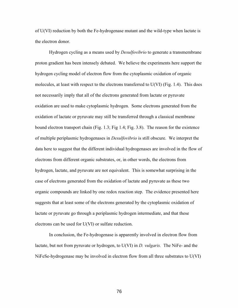
of U(VI) reduction by both the Fe-hydrogenase mutant and the wild-type when lactate is
the electron donor.
Hydrogen cycling as a means used by Desulfovibrio to generate a transmembrane
proton gradient has been intensely debated. We believe the experiments here support the
hydrogen cycling model of electron flow from the cytoplasmic oxidation of organic
molecules, at least with respect to the electrons transferred to U(VI) (Fig. 1.4). This does
not necessarily imply that all of the electrons generated from lactate or pyruvate
oxidation are used to make cytoplasmic hydrogen. Some electrons generated from the
oxidation of lactate or pyruvate may still be transferred through a classical membrane
bound electron transport chain (Fig. 1.3; Fig 1.4; Fig. 3.8). The reason for the existence
of multiple periplasmic hydrogenases in Desulfovibrio is still obscure. We interpret the
data here to suggest that the different individual hydrogenases are involved in the flow of
electrons from different organic substrates, or, in other words, the electrons from
hydrogen, lactate, and pyruvate are not equivalent. This is somewhat surprising in the
case of electrons generated from the oxidation of lactate and pyruvate as these two
organic compounds are linked by one redox reaction step. The evidence presented here
suggests that at least some of the electrons generated by the cytoplasmic oxidation of
lactate or pyruvate go through a periplasmic hydrogen intermediate, and that these
electrons can be used for U(VI) or sulfate reduction.
In conclusion, the Fe-hydrogenase is apparently involved in electron flow from
lactate, but not from pyruvate or hydrogen, to U(VI) in D. vulgaris. The NiFe- and the
NiFeSe-hydrogenase may be involved in electron flow from all three substrates to U(VI)
76

(Fig. 4.2). In addition, we observed that the Fe-hydrogenase appears to function in the
pathway from lactate to sulfate.
77

Figure 4.2. Modified Lovley Model for U(VI) reduction in a Fe-hydrogenase mutant
of D. vulgaris.
Arrows indicate electron flow and do not necessarily imply a single step in vivo. Some of
the electrons from the oxidation of lactate are eventually transferred by the Fe-
hydrogenase to U(VI), but some of the electrons bypass the Fe-hydrogenase pathway.
Apparently none of the electrons from the oxidation of hydrogen or pyruvate are
transported through the Fe-hydrogenase pathway. The NiFe- and NiFeSe-hydrogenases
also are likely involved in electron flow from all three substrates to U(VI).
Cyt c3
U(VI)
U(IV)
2e-
2H Pyruvate
2e- Fe-H2aseLactate 2e-2e-
2e- NiFe-H2ase 2e-
2NiFeSe-H ase
78

5. Interaction of Cytochrome c3 and Uranium
Rayford B. Payne1, Laurence Casalot1, Tessa Rivere1, Jeffrey H. Terry2, Lise
Larsen1, Barbara J. Giles1, Judy D. Wall1
1 Biochemistry Department, University of Missouri-Columbia, Columbia, MO 65211
2 Department of Biological, Chemical, and Physical Sciences, Illinois Institute of
Technology, Chicago, IL 60616, USA
Text, Tables 2.1 and 5.1, and Figures 5.1, 5.2, 5.3, and 5.4 were modified and used
with permission from Archives of Microbiology, 2004, 181(6):398-406.
79

5.1. Introduction
Since SRB are found in significant numbers at uranium-contaminated sites, their
response to low levels of toxic metals might indicate an interaction that could affect
U(VI) reduction. The hypothesis was tested that exposure to a subinhibitory
concentration of uranium might induce the cellular machinery of D. desulfuricans G20
responsible for the reduction of this metal. Prior exposure of D. desulfuricans G20 cells
to uranium actually decreased the ability of those cells to reduce U(VI). Interestingly,
growth of D. desulfuricans G20 in the presence of uranium did not affect the expression
of the periplasmic cytochrome c3; however, cytochrome c3 was no longer among the
soluble proteins from a high pH wash of those cells. Instead, periplasmic cytochrome c3
was found associated with precipitated uraninite. The adsorption of cytochrome c3 to
uraninite appeared to be non-specific since purified cytochrome c3 was shown to bind to
CuO (cupric oxide), Fe2O3 (ferric oxide), or commercially available U(IV) oxide in a
buffer system.
5.2. Results
5.2.1. Uranium(VI) reduction by cells grown in the presence of uranium
The possibility was tested that prior growth in the presence of a non-lethal
concentration of U(VI) would cause an increase of cellular machinery responsible for
U(VI) reduction, therefore such cells would reduce U(VI) at a faster rate than cells that
had not been previously exposed to U(VI). Cells were grown on LS medium amended
with a subinhibitory concentration of uranium (1 mM) (Payne et al., 2002) and tested for
their ability to reduce U(VI). These cells exhibited a slower rate of U(VI) reduction than
cells grown in the absence of uranium when the organic acids lactate or pyruvate were
80

the electron donors in the assay (Fig. 5.1). With hydrogen as the electron donor, the rate
of U(VI) reduction was not impaired by growth in the presence of uranium (Fig. 5.1).
81
Figure 5.1. U(VI) reduction by D. desulfuricans G20 previously exposed to
uranium.
U(VI) reduction kinetics of D. desulfuricans G20 grown in the absence (open symbols) or
in the presence (closed symbols) of 1 mM uranyl acetate. All samples have 1 mM uranyl
acetate and 100 µg whole cell protein/ml. Symbols are the average of two data points,
and the graphs are representative of 4 independent trials with each electron donor. (A) 10
mM sodium lactate (□, ■), or 10 mM sodium pyruvate (∆, ▲) was the electron donor for
U(VI) reduction. (B) 1 atm hydrogen gas (○, ●) was added as the electron donor for
U(VI) reduction.
0.0
0.2
0.4
0.6
0.8
1.0
0 5 10 15 20 25Time (h)
U(V
I) (m
M)
0.0
0.2
0.4
0.6
0.8
1.0
0 5 10 15 20 25Time (h)
U(V
I) (m
M)
B
A
82

5.2.2. Cytochrome c3 recovery from cells grown with uranium
The cytochrome c3 of D. vulgaris Hildenborough participates in the electron
transfer to uranium in a partially purified cell extract system (Lovley et al., 1993), and a
cytochrome c3 mutant of D. desulfuricans G20 is impaired in uranium reduction (Payne et
al., 2002). Since these data support the involvement of cytochrome c3 in U(VI)
reduction, it was speculated that the impairment observed in U(VI) reduction after
exposure of cells to the subinhibitory concentration of uranium might have resulted from
an alteration in cytochrome c3 content . Immunoblot analysis with antibodies to purified
cytochrome c3 was performed on protein extracts from cells grown in the presence of 1
mM uranium to test this possibility. Cytochrome c3 was not detected in the soluble
extracts of cells grown on LSU medium, in contrast to control cultures grown without
exposure to uranium (Fig. 5.2). Interestingly, cytochrome c3 was detected in the insoluble
fraction (containing both whole cells and uraninite) remaining after the soluble protein
was harvested from D. desulfuricans G20 cells (Fig. 5.2). The cytochrome c3 recovered
from the insoluble fraction of LSU grown cells was roughly equivalent to the amount of
cytochrome c3 recovered from the insoluble fraction from LS grown cells. The tetraheme
cytochrome c3 of D. vulgaris Hildenborough has been found both in the periplasm and
associated with the cytoplasmic membrane (Valente et al., 2001). Thus significant
cytochrome c3 should be recovered in the insoluble cell material of D. desulfuricans G20
after the periplasmic contents of cells were harvested by a pH shift. This was the result
observed. The inability to detect cytochrome c3 in the soluble extracts obtained from
LSU grown cells was due to the quantitative adsorption of the protein to the mineral
uraninite, as shown in following sections. If in fact cytochrome c3 adsorbs to uraninite,
83
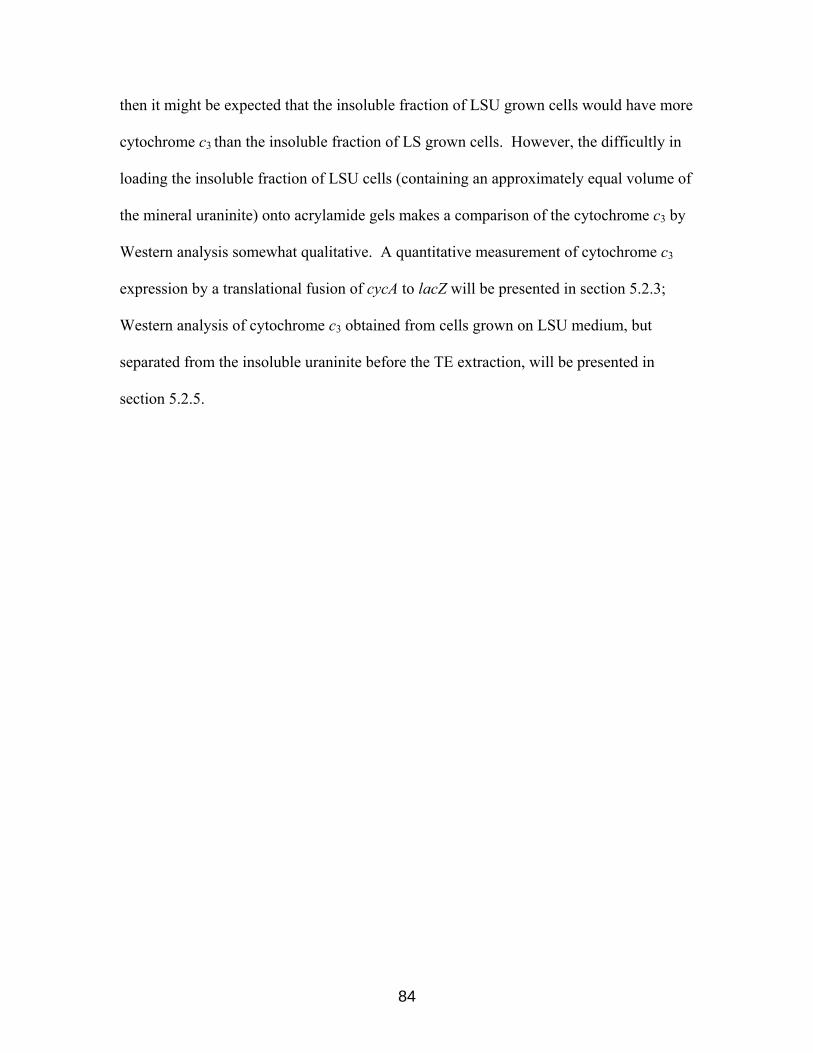
then it might be expected that the insoluble fraction of LSU grown cells would have more
cytochrome c3 than the insoluble fraction of LS grown cells. However, the difficultly in
loading the insoluble fraction of LSU cells (containing an approximately equal volume of
the mineral uraninite) onto acrylamide gels makes a comparison of the cytochrome c3 by
Western analysis somewhat qualitative. A quantitative measurement of cytochrome c3
expression by a translational fusion of cycA to lacZ will be presented in section 5.2.3;
Western analysis of cytochrome c3 obtained from cells grown on LSU medium, but
separated from the insoluble uraninite before the TE extraction, will be presented in
section 5.2.5.
84

Figure 5.2. Cytochrome c3 is not recovered in the periplasm of cells grown in the
presence of uranium.
Immunoblot comparison of the high pH TE wash extracted protein fractions from D.
desulfuricans G20 cultures grown in the absence or in the presence of 1 mM uranyl
acetate. Lane M, molecular mass markers; lane 1, 20 µg pH extracted protein from cells
grown on LS medium; lane 2, 20 µg pH extracted protein from cells grown on LSU
medium; lane 3, 30 µg insoluble fraction protein from cells grown on LS medium; lane 4,
30 µg insoluble fraction protein from cells grown on LSU medium. The arrow shows the
migration of purified cytochrome c3 on the gel.
85

5.2.3. Effect of uranium on transcription/translation of cytochrome c3.
To test the possibility that uranium might have also negatively influenced the
expression of the gene encoding cytochrome c3, the transcription of cycA was monitored
by reverse transcription-polymerase chain reaction (RT-PCR) and Northern analysis.
Qualitatively both techniques revealed that transcripts for cycA were still present in D.
desulfuricans G20 cells cultured in the presence of uranium (data not shown). Further
proof that uranyl acetate did not quantitatively alter the level of transcription came from
studies with a protein fusion of the promoter region and the start codon of cycA with the
lacZ gene creating the reporter vector pLC111 (Casalot and Wall, published in Payne et
al., 2004). The upstream region of cycA was confirmed to act as a strong promoter in E.
coli harboring pLC111 (Table 5.1). A single copy of this fusion construct was then
integrated into the chromosome of D. desulfuricans G20 at the site of the wild-type cycA
gene creating D. desulfuricans G25. The cycA gene had previously been shown to be in a
monocistronic operon (Rapp-Giles et al., 2000), therefore polar effects of reporter
plasmid integration were not an issue. As can be seen in Table 5.1, there was no
detectable difference in β-galactosidase activity when D. desulfuricans G25 cells
(harboring the integrated cycA-lacZ translation fusion) were grown in LS or LSU
medium. As a control, for β-galactosidase production in the sulfate-reducing bacterium,
D. desulfuricans G20 cells harboring the plasmid pMON5003, with a constitutive lacZ
under the control of the bla promoter, were assayed. There was also no detectable
difference in β-galactosidase activity when D. desulfuricans G20 (pMON5003) cells
were grown in LS or LSU medium (Table 5.1). The observation that the expression of
cytochrome c3, which is clearly involved in U(VI) reduction, is not detectably regulated
86

by a subinhibitory concentration of uranium suggests that strategies designed to reveal
genes involved in toxic metal reduction by altered gene transcription will clearly not
reveal all genes important to the process.
87
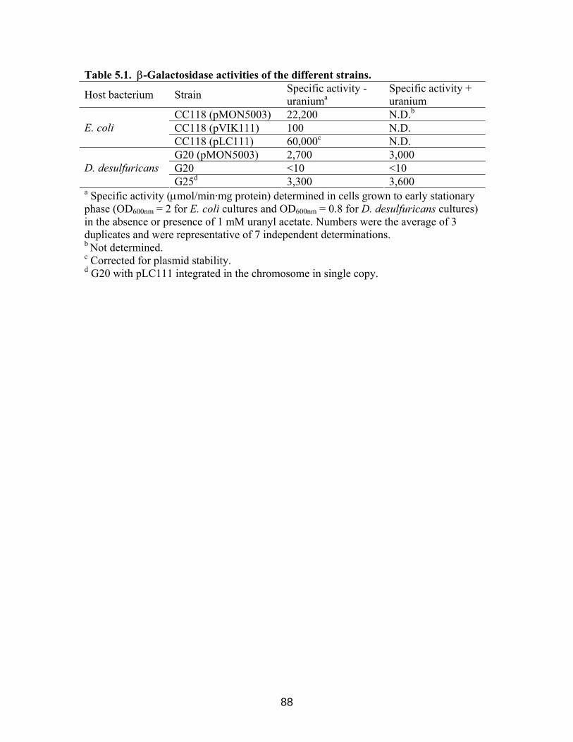
Table 5.1. β-Galactosidase activities of the different strains.
Host bacterium Strain Specific activity - uraniuma
Specific activity + uranium
CC118 (pMON5003) 22,200 N.D.b CC118 (pVIK111) 100 N.D. E. coli CC118 (pLC111) 60,000c N.D. G20 (pMON5003) 2,700 3,000 G20 <10 <10 D. desulfuricans G25d 3,300 3,600
a Specific activity (µmol/min·mg protein) determined in cells grown to early stationary phase (OD600nm = 2 for E. coli cultures and OD600nm = 0.8 for D. desulfuricans cultures) in the absence or presence of 1 mM uranyl acetate. Numbers were the average of 3 duplicates and were representative of 7 independent determinations. b Not determined. c Corrected for plasmid stability. d G20 with pLC111 integrated in the chromosome in single copy.
88
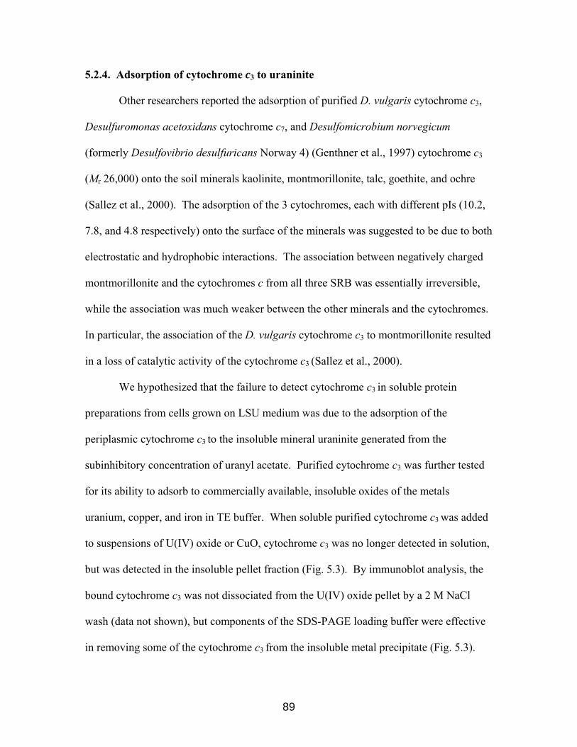
5.2.4. Adsorption of cytochrome c3 to uraninite
Other researchers reported the adsorption of purified D. vulgaris cytochrome c3,
Desulfuromonas acetoxidans cytochrome c7, and Desulfomicrobium norvegicum
(formerly Desulfovibrio desulfuricans Norway 4) (Genthner et al., 1997) cytochrome c3
(Mr 26,000) onto the soil minerals kaolinite, montmorillonite, talc, goethite, and ochre
(Sallez et al., 2000). The adsorption of the 3 cytochromes, each with different pIs (10.2,
7.8, and 4.8 respectively) onto the surface of the minerals was suggested to be due to both
electrostatic and hydrophobic interactions. The association between negatively charged
montmorillonite and the cytochromes c from all three SRB was essentially irreversible,
while the association was much weaker between the other minerals and the cytochromes.
In particular, the association of the D. vulgaris cytochrome c3 to montmorillonite resulted
in a loss of catalytic activity of the cytochrome c3 (Sallez et al., 2000).
We hypothesized that the failure to detect cytochrome c3 in soluble protein
preparations from cells grown on LSU medium was due to the adsorption of the
periplasmic cytochrome c3 to the insoluble mineral uraninite generated from the
subinhibitory concentration of uranyl acetate. Purified cytochrome c3 was further tested
for its ability to adsorb to commercially available, insoluble oxides of the metals
uranium, copper, and iron in TE buffer. When soluble purified cytochrome c3 was added
to suspensions of U(IV) oxide or CuO, cytochrome c3 was no longer detected in solution,
but was detected in the insoluble pellet fraction (Fig. 5.3). By immunoblot analysis, the
bound cytochrome c3 was not dissociated from the U(IV) oxide pellet by a 2 M NaCl
wash (data not shown), but components of the SDS-PAGE loading buffer were effective
in removing some of the cytochrome c3 from the insoluble metal precipitate (Fig. 5.3).
89

On occasion the purified cytochrome c3 showed multiple bands of higher than expected
molecular weight, suggesting that the protein formed multimers when concentrated under
these conditions. When soluble purified cytochrome c3 was added to suspensions of
Fe2O3, cytochrome c3 was not detected in solution nor was it detected in the insoluble
pellet fraction (Fig. 5.3), possibly due to a very strong association of the cytochrome c3 to
the ferric oxide. Neither a 2 M NaCl wash (data not shown) nor the SDS-PAGE loading
buffer was effective in removing cytochrome c3 from the insoluble ferric oxide
precipitate.
90

Figure 5.3. Adsorption of cytochrome c3 to uraninite.
Immunoblot showing adsorption of D. desulfuricans G20 cytochrome c3 to uraninite and
Fe2O3. Lane M, molecular mass markers; lane 1, supernatant from the cytochrome c3-
UO2 suspension; lane 2, pellet from the cytochrome c3-UO2 suspension; lane 3,
supernatant from the cytochrome c3-Fe2O3 suspension; lane 4, pellet from the cytochrome
c3-Fe2O3 suspension; lane 5, 125 ng purified cytochrome c3 diluted in the same manner as
the cytochrome c3-metal oxide suspensions; Cytochrome c3 adsorption to CuO not
shown.
91

5.2.5. Cytochrome c3 recovery from cells grown on LSU medium after separation of
cells and uraninite by ultracentrifugation
Western analysis for cytochrome c3 was performed on 15 × concentrated spent
LSU medium after cells had been removed by centrifugation. The tetraheme cytochrome
c3, accounting for over 80% of the periplasmic c-type cytochromes (Aubert et al., 1998),
was not detected in spent LS or LSU medium (data not shown). However, since
cytochrome c3 was shown to associate with UO2 in a cell-free suspension (Fig. 5.3), any
cytochrome c3 released into the medium during growth would be predicted to adsorb to
the insoluble uraninite and therefore not be detected in solution in the spent medium.
After separation of LSU grown cells from uraninite by sedimentation in a sucrose
gradient, the recovered cells were subjected to a high pH TE wash to recover periplasmic
proteins. Greater than 50% of the cytochrome c3 was recovered in the soluble extract
from the LSU grown cells, supporting the interpretation that 1 mM uranium did not cause
a drastic disruption of the outer membrane of D. desulfuricans G20 (Fig. 5.4). Lack of
full recovery could be attributed to small losses of cytochrome c3 during the
ultracentrifugation and protein extraction steps. About 25% of the amount of cytochrome
c3 recovered by pH shift from LSU grown cells was found associated with the insoluble
uraninite pellet separated by the sucrose gradient. This association could have occurred
because of normal cell death and lysis or to osmotic changes occurring during the
separation in sucrose (Fig. 5.4).
92

Figure 5.4. Cytochrome c3 recovery from cells grown in the presence of uranium.
Cells were grown on LSU medium then separated from uraninite by sucrose gradient
sedimentation before soluble protein was harvested by a high pH TE wash. Lane M,
molecular mass markers; lane 1, 25 µl of the 50-fold concentrated soluble protein
obtained from LSU grown cells as described above; lane 2, 25 µl of the 50-fold
concentrated uraninite fraction obtained as described above.
93

5.3. Discussion
The initial observation that growth of D. desulfuricans G20 cells in the presence
of a subinhibitory concentration of uranium impaired U(VI) reduction when lactate or
pyruvate are the electron donors (Fig. 5.1) remains to be explained, especially since cells
grown on LSU medium showed no detectable decrease in rate or extent of growth as
determined by whole cell protein analysis (Payne et al., 2002). In addition, the
impairment of U(VI) reduction was apparently not due to down-regulation of
transcription/translation of cytochrome c3, or to destabilization of the outer membrane.
Uranium might non-specifically inhibit a number of enzymes needed for energy and
carbon metabolism resulting in a decrease in the reductive capacity for U(VI) when
electron donor is supplied in low concentrations (10 mM in the U(VI) reduction assay
compared to 60 mM in LS medium). The observation that cells previously exposed to
uranium are not impaired in hydrogen-supported U(VI) reduction (Fig. 5.1) suggests to us
that 1 mM uranium causes an impairment of carbon metabolism (or transport) rather than
hydrogen oxidation alone. Microarray studies of cells exposed to non-inhibitory uranium
concentrations may provide insight into this phenomenon.
The biological implications of the adsorption of cytochrome c3 to uraninite
and metal oxides. Dissimilatory metal-reducing bacteria use several strategies to access
extracellular metal terminal electron acceptors (TEA) in the environment (Hernandez and
Newman, 2001). Possible means include: 1) direct contact of an oxidized metal with an
outer membrane electron transfer component (Beliaev and Saffarini, 1998; Lower et al.
2001; Magnuson et al., 2000; Nevin and Lovley, 2000); 2) the release of siderophores
which complex and solubilize the metal TEA (Ledyard and Butler, 1997); or 3) the use of
94

extracellular electron shuttling molecules such as quinone-containing compounds or c-
type cytochromes to transfer electrons from the cellular electron transport chain to the
metal TEA (Nevin and Lovley, 2002; Newman and Kolter, 2000; Seeliger et al., 1998).
The extracellular electron shuttles may either be already present in the environment due
to decomposition of organic material or intentionally transported by cellular processes
into the environment. The use of extracellular c-type cytochromes in Fe(III) reduction
has been questioned (Lloyd et al., 1999). Our results here taken together with previous
work suggest that the periplasmic cytochrome c3 of D. desulfuricans G20 is unlikely to
function as an significant extracellular electron carrier to U(VI), since the tight
association of the protein with both clay minerals and metal oxides would preclude a
catalytic role for the protein.
95

6. The role of the sodium motive force in Desulfovibrio
Rayford B. Payne and Judy D. Wall Biochemistry Department, University of Missouri-Columbia, Columbia, MO 65211 Manuscript in preparation.
96
6.1. Introduction
Most bacteria couple electron transport and respiration to the formation of a
transmembrane proton motive force. The transmembrane proton motive force represents
a potential source of energy which can be used to generate ATP (by passage of
periplasmic protons down a concentration gradient through the membrane-bound F1F0
ATPase), to transport substrates, or to rotate flagella (Fig. 1.4; for a recent review see
Harold, 2001). The electrochemical potential of protons (∆µH+) is composed of the sum
of two separate components: (1) the pH gradient across the cytoplasmic membrane (∆pH,
typically the periplasm is more acidic than the cytoplasm); and (2) the membrane
potential (∆Ψ, typically the periplasm is more positive than the cytoplasm). Under
alkaline conditions, both neutrophilic and alkaliphilic bacteria are faced with the problem
of generating a proton motive force with an inverse pH gradient, i.e. the cytoplasm must
be maintained at a lower pH than the external environment and periplasm (Krulwich
1995; Padan et al., 1981). In fact, an external pH of greater than about 7.8 abolishes the
∆pH of Escherichia coli (Pan and Macnab, 1990). In the absence of a ∆pH, only the ∆Ψ
contributes to the overall membrane activation. A sufficiently large ∆Ψ, which can be
created by transport of other positively charged ions into the periplasm, can allow the
translocation of H+ ions against an unfavorable ∆pH (Harold, 2001).
Slightly halophilic marine bacteria of the genus Vibrio can grow under low proton
motive force conditions such as in high pH environments or in the presence of the
protonophore CCCP (Tokuda and Unemoto, 1981; Tokuda and Unemoto, 1983). These
bacteria are capable of using a transmembrane sodium motive force in addition to a
transmembrane proton motive force for growth under such conditions (Hayashi et al.,
97

2001; Tokuda and Unemoto 1982). In the Vibrio strains, the sodium motive force can be
generated directly (i.e. in a mechanism not dependent upon the proton motive force) by
the translocation of cytoplasmic sodium ions into the periplasm coupled to the oxidation
of energy substrates. The transmembrane complex responsible for sodium ion
translocation coupled to electron transport is the NADH-dependent Na+-Translocating
Quinone Oxidoreductase (NQR). This contribution to the membrane potential apparently
supports growth in low proton motive force conditions (Hayashi et al., 2001; Tokuda and
Unemoto 1982). Na+ ions in the periplasm can then be exchanged for protons to
reestablish a proton gradient; in addition, positively charged Na+ ions contribute to the
∆Ψ component of the membrane potential, thus allowing protons to be used for ATP
synthesis against an unfavorable ∆pH. Alternatively, Vibrio alginolyticus can use the
sodium motive force directly for substrate transport, for flagella rotation (Chernyak et al.,
1983; Unemoto et al., 1993), or for extrusion of toxic compounds via a Na+/multidrug
exporter (Morita et al., 1998). The completed genome sequences of the sulfate-reducing
bacteria Desulfovibrio desulfuricans G20, Desulfovibrio vulgaris Hildenborough and
Desulfotalea psychrophila, revealed homologs of many of the sodium-related genes of V.
alginolyticus and Vibrio parahaemolyticus. We found homologs of the NQR complex
and homologs of the well-characterized Na+ /multidrug exporters in all three sequenced
genomes even though only Dt. psychrophila has been characterized as a marine
bacterium (Rabus et al., 2004). This observation suggested to us the existence of a
“sodium circuit” in the SRB that may play a major role their bioenergetics.
We sought to explore the role of the “sodium circuit” in the SRB with the model
organism D. desulfuricans G20. Initial RT-PCR experiments showed that the D.
98
desulfuricans G20 nqr operon was expressed. We hypothesized that D. desulfuricans
G20 used the sodium motive force in a complementary manner to the well known proton
motive force. For example, we predicted that under some conditions D. desulfuricans
G20 could grow in the absence of a proton gradient. We found that D. desulfuricans G20
could grow in the absence of a proton gradient when lactate, but not pyruvate, was the
electron donor. We also reasoned that the resistance of D. desulfuricans G20 to some
antimicrobials was dependent upon the sodium motive force and have established that
this is true.
6.2. Results
6.2.1. The Na+-Translocating NADH Quinone Oxidoreductase (NQR Complex) of
the Sulfate-Reducing Bacteria
Sequence analysis of the completed genomes of the three sulfate-reducing
bacteria D. desulfuricans G20, D. vulgaris Hildenborough, and Dt. psychrophila,
revealed an operon similar to the well-characterized nqr operon of V. alginolyticus (Fig.
6.1). While the amino acid identities of the SRB nqr encoded proteins to those of V.
alginolyticus are relatively low, the conservation of similar amino acids is somewhat
higher (Fig. 6.1). More importantly, the synteny of the operons is identical. To our
knowledge, genes immediately downstream of nqr6 in V. alginolyticus have not been
characterized. A gene encoding a homolog of apbE of Salmonella is downstream of nqr6
in the completed genome sequences of the Vibrio cholerae El Tor N16961, Vibrio
vulnificus CMCP6, Vibrio vulnificus YJ016, and Vibrio parahaemolyticus RIMD
2210633 (TIGR, http://www.tigr.org). The presence of an apbE ortholog promoter distal
is a conserved feature of the SRB NQR complex operons as well (TIGR,
99

http://www.tigr.org; VIMSS, http://www.escalante.lbl.gov). It is possible that apbE is
the last gene in the V. alginolyticus nqr operon as well. In Salmonella, ApbE is a
periplasmic lipoprotein that plays a role in thiamine biosynthesis or Fe-S cluster
formation (Beck and Downs, 1998; Skovran and Downs, 2003). The Salmonella apbE
gene appears to be in a monocistronic operon, and genes located nearby are not similar to
those of the nqr operon. The role, if any, of AbpE in the function of the NQR complex is
unknown; however, several of the NQR subunit proteins have Fe-S clusters.
The nqr operons of the three SRB all have an interesting difference from the
Vibrio spp. nqr operon. In the Desulfovibrio strains, the promoter proximal gene of the
operon encodes a potential decaheme c-type cytochrome and in the same position in the
Dt. psychrophila operon is a gene encoding a FAD flavoprotein (Fig. 6.1). It is possible
that the decaheme c-type cytochrome (of the Desulfovibrio spp.) or the flavoprotein (of
Desulfotalea) is intimately involved in electron flow from the NQR complex to other
members of the electron transport chain. RT-PCR analysis shows that the putative nqr
operon is expressed when D. desulfuricans G20 cells are grown on LS medium (Fig. 6.2).
The expected products were obtained but quantitation was not possible from this
experiment.
100
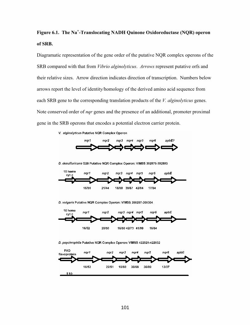
Figure 6.1. The Na+-Translocating NADH Quinone Oxidoreductase (NQR) operon
of SRB.
Diagramatic representation of the gene order of the putative NQR complex operons of the
SRB compared with that from Vibrio alginolyticus. Arrows represent putative orfs and
their relative sizes. Arrow direction indicates direction of transcription. Numbers below
arrows report the level of identity/homology of the derived amino acid sequence from
each SRB gene to the corresponding translation products of the V. alginolyticus genes.
Note conserved order of nqr genes and the presence of an additional, promoter proximal
gene in the SRB operons that encodes a potential electron carrier protein.
101

Figure 6.2. Expression of the NQR complex operon measured by RT-PCR.
RT-PCR analysis of nqr operon expression by D. desulfuricans G20 cells grown on LS
medium. Lanes 1, 2, and 3 have 50 ng of mRNA.
Lane: M, MW markers;
Lane1, test for DNA contamination with cycA primers, primers c3_left and
c3_right, no Reverse Transcriptase added;
Lane 2, test for decaheme cytochrome c gene expression, primers 10c_ left and 10
c_right with Reverse Transcriptase;
Lane 3, test for expression of gene encoding the transmembrane NADH oxidizing
subunit of NQR, primers nqr1_left and nqr1_right with Reverse Transcriptase.
M 1 2 3
102

Growth on LS but not PS requires either the proton- or sodium- motive
force. We tested D. desulfuricans G20 to determine its ability to grow in the presence of
the protonophore CCCP. Cells were grown on LS medium or PS medium with or
without CCCP added at 100 µM final concentration. No significant differences in growth
rates were observed with or without CCCP when lactate was the electron donor and
sodium was present (about 160 mM Na+ in standardly prepared LS or PS; Fig. 6.3A). In
addition, the same final optical density and protein concentration as cells grown in the
absence of CCCP were documented (Fig. 6.3A, Table 6.1). The addition of 100 µM
CCCP to PS medium completely abolished growth, even after cultures were incubated for
2 weeks (Fig. 6.3A, Table 6.1). The addition of 100 µM monensin, an electroneutral
antiporter of sodium, to LS medium did not noticeably impair the growth rate or final
yield of D. desulfuricans G20 when measured by OD or protein (Fig. 6.3B, Table 6.1).
The addition of both 100 µM CCCP and 100 µM monensin together resulted in the
decrease of both the growth rate and the final yield in LS medium (Fig. 6.3A,B, Table
6.1).
103

Figure 6.3. Growth of D. desulfuricans G20 under low proton- and sodium-motive
force conditions.
The effect of a protonophore and a sodium electroneutral antiporter on the growth of D.
desulfuricans G20. All media contained approximately 160 mM sodium.
Panel A. The effect of CCCP on the growth of G20 on LS or PS.
(□): LS growth medium.
(○): PS growth medium. Filled symbols had 100 µM CCCP added.
Panel B. The effect of CCCP and monensin on the growth of G20 on LS.
(□): LS growth medium.
(■): 100 µM CCCP added.
(♦): 100 µM monensin added.
(▲): 100 µM CCCP and 100 µM monensin added together.
All data points are the average of two independent cultures. The figure is representative
of four independent experiments.
104

A
0.01
0.10
1.00
0 5 10 15 20 25Time (h)
O.D
. 620
nm
B0.01
0.10
1.00
0 5 10 15 20 25
Time (h)
O.D
. 620
nm
105

Table 6.1. Protein yield of D. desulfuricans G20 cells grown in the presence of CCCP and monensin. Growth Mediuma Inhibitorb Final Protein Yield (µg/ml) c Lactate Sulfate None 206 ± 25d LS CCCP 206 ± 7d LS monensin 178 ± 4d LS CCCP + monensin 7 ± 5d Pyruvate Sulfate (PS) None 79e PS CCCP 4e aLS, 60 mM (D,L)-lactate and 50 mM Na2SO4; PS, 60 mM Na pyruvate and 50 mM Na2SO4. bCCCCP, carbonyl cyanide m-chlorophenyl hydrazone, is a protonophore; monensin, is a sodium ionophore. Each inhibitor was used at a final concentration of 100 µM. cProtein measurements after 44 hours of growth at 370C of whole cell protein by Bradford procedure. dThe average and standard deviation obtained from three independent cultures from two different experiments. eThe average of two different cultures from one experiment.
106

6.2.2. Expression of the NQR complex operon
We used Northern analysis to see if the D. desulfuricans G20 NQR complex
operon was differentially expressed under high pH or sodium concentrations. On
complete LS medium at pH 7.2, we observed that the addition of monensin or CCCP
alone did not affect the expression of the NQR. However, Northern analysis of RNA
obtained from cells grown on complete LS medium at pH 9.0 showed about a 10 fold
decrease of the expression of the NQR transcript (Fig. 6.4). Northern analysis was not
performed on cells grown in the absence of sodium as we were unable to obtain high
quality RNA from cells grown on LS0 medium during the course of this study.
Interestingly, Northern analysis of the cytochrome c3 mutant of D. desulfuricans showed
that this mutant had about a 7 fold decrease in the expression of the NQR transcript
compared to the wild-type when cultures were grown on complete LS medium (Fig. 6.4).
6.2.3. The Na+/multidrug exporters of the Sulfate-Reducing Bacteria
Sequence analysis of the completed genomes of the three SRB revealed homologs
to the Na+/multidrug exporter NorM of V. parahaemolyticus (VIMSS,
http://www.escalante.lbl.gov; Fig. 6.5). The identity at the amino acid level is somewhat
low. However, all of the putative SRB sodium dependent drug efflux pumps were
annotated as such by the VIMSS automatic annotation (http://www.escalante.lbl.gov),
primarily based upon the characteristic 12 alpha helical transmembrane domains present
in this family of proteins (TIGR, http://www.tigr.org). The NorM isozyme 3 from D.
desulfuricans G20 and the NorM isozyme 2 from D. vulgaris have in common an
unknown hypothetical protein at the promoter distal end of the operon (Fig. 6.5).
107

Figure 6.4. Expression of the NQR complex operon at pH 9, and by a cytochrome c3
mutant of D. desulfuricans G20.
Panels A: Northern Blots probed with the internal fragment of the D. desulfuricans G20
decaheme cytochrome c of the nqr operon. The band corresponds to approximately 9 kb
on the gel. Panels B: Gel showing the 23S and 16S rRNA stained with ethidium bromide.
Lanes 1-4: RNA from wild-type cells grown on LS medium showing that the putative nqr
operon is down-regulated at high pH. This Northern is representative of RNA from two
independent experiments. The Arrow corresponds to the position of the 9 kb marker on
the gel.
Lane 1: pH 7.2 medium.
Lane 2: pH 9.0 medium.
Lane 3: pH 7.2 medium + CCCP.
Lane 4: pH 7.2 medium + monensin.
Lanes 5-6: The putative nqr operon is down-regulated in the cytochrome c3 mutant of D.
desulfuricans G20.
Lane 5: RNA from wild-type D. desulfuricans G20.
Lane 6: RNA from the cytochrome c3 mutant of D. desulfuricans G20.
5 µg total RNA added per lane.
108

A
109
A
B B1
2 3 4 5 6
Figure 6.5. The Na+-dependent multidrug exporters (NorM) of the SRB.
Black arrows represent the NorM homologs of the sequenced sulfate-reducing bacteria.
Only the genetically and biochemically characterized NorM isozyme of V.
parahaemolyticus is shown, 9 other homologs of NorM in V. parahaemolyticus are
predicted based upon the sequence. Percent identity and percent similarity scores at the
amino acid level are shown beneath each gene. VIMSS orf numbers
(http://www.escalante.lbl.gov) are shown above each gene. The red arrows represent
orfs encoding unknown hypothetical proteins promoter distal to the NorM homologs.
V. parahaemolyticus NorM 1 isozyme
VIMSS 339991
100/100
D. desulfuricans G20 NorM orthologs Unknown
23/37
VIMSS 394445 VIMSS 394671 (dinF)
22/39 23/43
HypotheticalVIMSS 394252
D. vulgaris NorM orthologs Unknown
19/66
VIMSS 206656 HypotheticalVIMSS 208052
21/59
D. psychrophila NorM orthologs VIMSS 423518VIMSS 422225
20/5916/57
110

6.2.4. The role of the sodium motive force during drug resistance
The NorM of V. parahaemolyticus functions to pump some antimicrobial
compounds of the same general structure (like kanamycin) out of the cell. Typically the
compounds are relatively small (MW ca. 300-500), ring-shaped, and cationic or
hydrophilic. The activity of the V. parahaemolyticus NorM is dependent upon the
sodium motive force. D. desulfuricans G20 is somewhat resistant to many inhibitors, for
example kanamycin. We tested whether sodium was required for kanamycin resistance
in D. desulfuricans G20. When sodium was omitted from the LS medium, or when the
sodium ionophore monensin was added to LS medium containing sodium, D.
desulfuricans G20 was much more sensitive to kanamycin (Fig. 6.6, Table 6.2). The
resistance of D. desulfuricans G20 to methylene blue and ethidium bromide, as well as
kanamycin, was dependent on the sodium concentration in the growth medium. Over a
60 fold increase in sensitivity to kanamycin with low sodium concentrations was
observed (Table 6.2). Apparently nalidixic acid is not a substrate for any sodium driven
efflux pump because G20 cells were not more sensitive to nalidixic acid when sodium
was omitted from the LS medium or when LS medium contained both sodium and the
sodium ionophore monensin (Table 6.2).
111

Figure 6.6. D. desulfuricans G20 is more sensitive to kanamycin in the absence of
the sodium-motive force.
D. desulfuricans G20 is more sensitive to kanamycin in the absence of the sodium-motive
force. All cells are grown on LS0 medium, pH 7.2, modified as follows:
Open squares: 160 mM NaCl present
Closed squares: 160 mM NaCl + 120 µg kanamycin per ml.
Open circles: <1 mM NaCl.
Closed circles: <1 mM NaCl + 120 µg kanamycin per ml.
Closed triangles: 160 mM NaCl + 100 µM monensin + 120 µg kanamycin per ml.
0.01
0.10
1.00
0 5 10 15 20 25 30Time (h)
O.D
. 620
nm
112

Table 6.2. MICa of inhibitors for D. desulfuricans G20 when grown with different NaCl.
Medium Compositionb
Inhibitors 150 mM NaCl 150 mM NaCl
+ 100 µM Monensinc 0 mM NaCl
Kanamycin 480d 240d 7.5d Ethidium Bromide 60e 15e 15e Methylene Blue >800e >800e 100e Nalidixic Acid 800e 800e 800e aMinimum Inhibitory Concentration given as µg/ml of the compound that completely inhibited growth in liquid LS0 medium supplemented as indicated. bAll experiments were performed in LS0 medium supplemented as indicated. LS0 medium actually contained about 0.7 mM sodium as determined experimentally by ICP analysis by the Agricultural Experiment Station Laboratories, University of Missouri-Columbia. cMonensin is an electroneutral H+/Na+ exchange compound. dMIC of at least 3 independent experiments done in duplicate. eMIC of one experiment done in duplicate.
113

6.3. Discussion
The role of the proton- and sodium- motive force in Desulfovibrio. Sulfate-
reducing bacteria raise the pH of their environment as they grow due to volatilization of
sulfide, the end product of sulfate respiration. So-called neutrophilic SRB have been
shown to raise the pH of their local environment to pH 9.0 (Fitz and Cypionka, 1989;
Postgate, 1979). Since sulfate reduction can lead to alkanization of the media (and
periplasm) and a corresponding loss of the ∆pH, we reasoned that SRB might use the
alternate sodium motive force as a strategy to cope with low proton motive force
conditions generated by their metabolism. It is important to note that the existence of
classical electron transport driven proton translocation or “hydrogen cycling” as a
mechanism to generate a periplasmic proton motive force does not necessarily rule out
the use of other ion gradients by the sulfate-reducing bacteria. Cypionka and coworkers
have previously explored the role of the proton motive force (Fitz and Cypionka, 1991)
and the related sodium motive force (Kreke and Cypionka, 1994) in the SRB.
Interestingly, they suggested that the alkaliphilic sulfate-reducing bacterium
Desulfonatronovibrio hydrogenovorans coupled its electron transport chain to the
generation of a sodium gradient (Sydow et al., 2002). The genome sequence of all three
sequenced SRB indicated a homolog of the NQR complex, which in Vibrio alginolyticus
is responsible for the generation of a periplasmic sodium gradient at the expense of
electron transport chain. However, searches of the sequenced SRB genomes did not
reveal a Na+-ATPase in addition to the classical H+-ATPase.
114

Figure 6.7. The “Sodium Circuit” of the SRB.
The “Sodium Circuit” of D. desulfuricans G20. Red arrows represent proposed electron
flow from lactate which generates a transmembrane sodium gradient through the NQR
complex. NorM: Na+/multidrug exporter. LDH: lactate dehydrogenase. NQR: Na+-
translocating NADH Quinone Oxidoreductase. Cyt c3: cytochrome c3. HMC: high
molecular weight cytochrome transmembrane complex.
Periplasm
Na+
Lactate
Cytoplasm
Pyruvate SO42-
HMC
S2-
LDH
Cytc3
Kanamycin
NQR
Na+
NorM
115

The role of the NQR complex in energy metabolism of the sulfate-reducing
bacteria. The role of the putative NQR complex of the SRB is as yet unknown. The true
NQR of Vibrio couples electron transport to the translocation of cytoplasmic Na+ to the
periplasm. The NQR complex also shares homology with the RNF transmembrane
complex from Rhodobacter capsulatus (Rhodobacter Nitrogen Fixation) (Kumagai et al.,
1997). The RNF complex is essential for growth on medium that requires R. capsulatus
to fix nitrogen as the sole nitrogen source (Schmehl et al., 1993). Presumably, The RNF
complex is involved in electron transport to nitrogenase (Schmehl et al., 1993). The
NQR complex and the RNF complexes are members of a family of energy-coupling
NADH oxidoreductases (Kumagai et al., 1997). It is unlikely that the NQR complex acts
in nitrogen metabolism in Desulfovibrio and Desulfotalea since only D. vulgaris has
genes for nitrogen fixation.
The simple interpretation of the effect of ionophores may not be accurate. Thus,
it is also possible that CCCP and monensin are inhibiting some other cellular process
rather than disrupting the proton- or sodium- motive force, since we have not directly
measured H+ or Na+ ion translocation in this study. However, the inhibition of growth on
lactate when both ionophores are added together and the disruption of antibiotic
resistance by monensin, but not CCCP (Fig. 6.3, Table 6.1), suggest to us that the
ionophores are in fact acting as proposed. Biochemical experiments measuring
translocation of Na+ and H+ by whole cells or by purified NQR complex, as well as the
construction and testing of nqr mutants, will help to elucidate the role of this protein
complex in the SRB.
116

To the best of our knowledge, expression of the nqr operon in other organisms
has received little attention. Initially, investigators thought that the expression of the
NQR complex of V. alginolyticus was upregulated at alkaline pH. However, it was later
shown that expression of the NQR complex was not in fact upregulated at alkaline pH,
instead that the NQR complex had more activity at alkaline pH than at near-neutral pH
(Tokuda and Unemoto, 1981). The regulation of other genes encoding sodium-related
functions has been studied. For example, the E. coli H+/Na+ antiporter NhaA is regulated
by pH, Na+ and Li+ concentrations, and growth phase (Dover and Padan, 2001; Karpel et
al., 1991). We reasoned that if the putative NQR complex in D. desulfuricans G20 was
involved in the “sodium circuit”, then it might be differentially expressed under various
salt or pH conditions. While we were unable to obtain the RNA for analysis of low salt
effects on expression, RNA from cells grown at pH 9.0 in LS medium showed down-
regulation of the expression of the putative nqr operon. The addition of monensin or
CCCP alone had no effect on the expression of the putative nqr operon. The
responsiveness of the nqr operon to pH may support the suggestion that the NQR
complex has a role in the “sodium circuit” of the SRB, but it is the opposite of what
would be predicted if the expression of the NQR complex is needed at a higher level
when cells are grown under low proton motive force conditions (like high pH).
Earlier in the course of the dissertation research, we observed that the cytochrome
c3 mutant of D. desulfuricans G20 had a higher nitrite reductase activity than the wild-
type. Conversely, we observed that the expression of the putative nitrite reductase was
down-regulated in the cytochrome c3 mutant of D. desulfuricans G20 as compared to the
wild-type. Interestingly, we found that the nqr operon is also down-regulated in the
117

cytochrome c3 mutant of D. desulfuricans G20 as compared to the wild-type. Both the
putative nitrite reductase and the putative NQR complex share common features: (1) they
are transmembrane complexes; (2) they are predicted to be linked to electron transport at
the level of the quinone pool; and (3) one of the subunits of the complex is a periplasmic
facing c type cytochrome.
Growth of D. desulfuricans G20 on lactate sulfate at low sodium or at high
pH. We sought to test if D. desulfuricans G20, like V. alginolyticus, could grow in the
absence of a proton gradient. We found that pyruvate supported growth by D.
desulfuricans G20 was dependent upon the proton motive force and was inhibited by
CCCP. Surprisingly, growth on lactate by D. desulfuricans G20 was unaffected by
CCCP. This observation led us to speculate that the D. desulfuricans G20 NQR complex
might generate a transmembrane sodium motive force from the oxidation of lactate, but
not pyruvate, and that the sodium motive force would therefore be sufficient to allow
growth under low proton motive force conditions when lactate was the electron donor
(Fig. 6.7). The addition of CCCP and monensin (a sodium ionophore) together inhibited
growth when lactate was the electron donor suggesting that a proton- or sodium-motive
force was necessary for the cells to grow. While we cannot demonstrate an absolute
requirement for sodium ions per se during growth on LS medium, G20 cells in medium
with very low sodium (about 0.7 mM), became more sensitive to the sodium ionophore
monensin either because monensin is a substrate for the sodium-driven multidrug efflux
pump or because the cell ultimately needs a sodium gradient for growth (data not shown).
In our model, we speculate the uptake of lactate by cells is coupled to H+ or Na+
cotransport, while pyruvate may be taken up by H+ cotransport alone. Lactate was
118

suggested to be taken up by cotransport with either H+ or Na+ in Shewanella oneidensis
by inference from results with various inhibitors (Stenberg et al., 1984).
Role of the Na+/multidrug exporters in the sulfate-reducing bacteria. In 1998
a novel multidrug efflux pump, NorM, was characterized from V. parahaemolyticus.
NorM is genetically and functionally related to its homolog YdhE of E. coli (Morita et
al., 1998). These 2 proteins represented the first members of a new class of multidrug
efflux pumps, termed the MATE class (Multidrug And Toxic compound Extrusion). This
family of drug transporters is dependent upon the sodium motive force (Na+ ion antiport),
not the proton motive force (H+ antiport) or ATP hydrolysis, for the translocation of toxic
compounds (Morita et al., 2000). In Vibrio parahaemolyticus NorM functions to
transport the drugs norfloxacin and ciprofloxacin and also the unrelated antimicrobial
compounds ethidium, kanamycin, and streptomycin from the cell in a sodium motive
force-dependent manner (Morita et al., 1998). The three sequenced SRB each have
homologs to the characterized NorM Na+/multidrug efflux pumps of V.
parahaemolyticus. D. desulfuricans G20 apparently has three Na+/multidrug efflux pump
homologs. It has long been known that SRB are naturally resistant to many antimicrobial
compounds, in part because of their anaerobic metabolism, but also because of their
production of sulfide, which can inactivate many antimicrobials (Salleh et al., 1964). The
extreme resistance of SRB makes it a challenge to prevent or treat SRB-induced
corrosion.
We speculated that the resistance of the SRB to some antimicrobial compounds
was due at least in part to the efflux of the antimicrobial by a NorM isozyme and
therefore coupled to the sodium motive force (Fig. 6.7). Previous studies showed that the
119

NorM of Vibrio strains functioned in the efflux of the antimicrobial compounds
kanamycin, ethidium bromide, and methylene blue, among others (Morita et al., 1998).
Nalidixic acid was not a substrate of NorM from the tested Vibrio strains (Morita et al.,
1998). As predicted, the growth of D. desulfuricans G20 in the presence kanamycin,
ethidium bromide, and methylene blue was shown here to be dependent upon the sodium
motive force. The sodium gradient was reduced by omitting sodium salts from laboratory
growth media or by including a sodium ionophore like monensin in the growth medium.
When this was done, the sensitivity of the SRB to some antimicrobial compounds was
increased (Table 6.2). This observation has obvious implications for the control of SRB
in an industrial setting. If sodium ions could be removed in an economic fashion from
water before its use during industrial operations such as oil recovery or recirculating air-
conditioning systems, then a lower concentration of an industrial biocide might inhibit
SRB growth and corrosion. In addition, a biocide previously thought to be ineffective
against SRB corrosion might be effective in the absence of sodium ions. In the future,
development of new antimicrobials that target the sodium/drug exporter, or that disrupt
the periplasmic sodium gradient (like monensin), would be an exciting avenue of
research.
120

7. Overall conclusions of this dissertation
The classic compounds for growth of SRB in the laboratory are hydrogen, lactate,
and pyruvate, although the metabolic versatility of some strains of SRB is much greater
(Postgate, 1979). The mechanism by which sulfate-reducing bacteria generate a
transmembrane proton motive force has been intensely debated. When using carbon
compounds as electron donors, SRB have been shown to both produce and consume
molecular hydrogen (Hatchikian et al., 1976). Odom and Peck first proposed a
“hydrogen cycling” model for generation of a transmembrane proton gradient (Odom and
Peck, 1981). In that model, the oxidation of organic electron donors in the cytoplasm
was coupled to the generation of cytoplasmic hydrogen. Cytoplasmic hydrogen was then
suggested to diffuse across the cytoplasmic membrane and be oxidized by a periplasmic
hydrogenase, generating protons and allowing the recapture of electrons. Noguera and
coworkers proposed that both hydrogen cycling and classical electron transport driven
proton translocation functioned in D. vulgaris to generate a proton gradient (Noguera et
al., 1998).
Experiments here support the suggestion that classical electron transport driven
proton translocation functions in addition to hydrogen cycling to generate a
transmembrane proton gradient in Desulfovibrio (Figs. 1.3 and 1.4). Our studies began
by testing a single mutant in the predominant periplasmic cytochrome c3 for its ability to
reduce U(VI). The mutant reduced U(VI) at a rate of about 10% that of the wild-type
with hydrogen as the electron donor; when lactate or pyruvate were electron donors the
impairment was less severe. These observations were consistent with either hydrogen
cycling or classical electron transport. Growth and U(VI) reduction experiments using
121

the Fe-hydrogenase mutant suggested that electrons from lactate oxidation, but not
pyruvate oxidation, were transferred through the periplasmic Fe-hydrogenase. This
observation suggests that hydrogen cycling from lactate does exist. Many experiments
indicated that the electrons generated from oxidation of lactate are quite different from
those electrons generated from the oxidation of pyruvate (for example, the experiments
using the Fe-hydrogenase mutant). Measurements of nitrite reduction by the wild-type
and the cytochrome c3 mutant suggested that more electrons from pyruvate oxidation than
from lactate oxidation enter the electron transport chain (ETC) at the level of the putative
nitrite reductase (NiR). Inhibitor studies using the protonophore CCCP and the sodium
ionophore monensin were interpreted to mean that electrons from lactate oxidation, but
not from pyruvate oxidation, entered the ETC at the putative Na+-translocating NADH-
dependent Quinone Oxidoreductase (NQR). Both of these protein complexes, NiR and
NQR, are coupled to the quinone pool in other organisms (Hayashi et al., 2001; Simon et
al., 2000)
Mutant studies of the HMC carried out by others were interpreted to support a
role for HMC in electron transfer from the periplasm into the cytoplasm where sulfate
reduction could take place (Dolla et al., 2000). In vitro studies suggested that although
the HMC could accept electrons directly from the Fe-, NiFe-, or NiFeSe-hydrogenases, a
catalytic amount of cytochrome c3 increased the rate of electron transfer 100- to 1000-
fold (Pereira et al., 1998). Northern analysis (Table 7.1) and protein analysis (Giles and
Wall, unpublished) indicated that the high-molecular mass cytochrome c complex (HMC)
was upregulated in the cytochrome c3 mutant. Therefore it appears that the expression of
122

at least one electron carrier protein downstream of cytochrome c3 (in the ETC) was
upregulated in the cytochrome c3 mutant.
Previous expression studies showed that some of the electron carrier proteins
coupled to the Fe-hydrogenase were down-regulated in the Fe-hydrogenase mutant of D.
vulgaris (Haveman et al., 2003). We found that the NiR and the NQR complexes were
down-regulated (at least 8- and 7-fold, respectively) in the cytochrome c3 mutant of D.
desulfuricans G20 (Figs. 3.5 and 6.4, Table 7.1). In addition, we found that the
cytochrome c553 (c553), suggested by in vitro studies to act as an electron carrier between
lactate dehydrogenase (LDH) or formate dehydrogenase and cytochrome c3, was at least
10-fold down-regulated in the cytochrome c3 mutant (Table 7.1). Similarly, the operon
encoding the predicted acidic tetraheme c type cytochrome (c3 T-II) and its associated
transmembrane complex was observed to be downregulated (Table 7.1). Other
researchers have used gene expression profiling to model metabolic pathways. Often, but
not always, functionally related genes are coordinately expressed (Hughes et al., 2000;
Kabir and Shimizu, 2003; Thompson and Zhou, 2004). We have so far looked at the
expression of only a small sample of the electron carrier proteins in the cytochrome c3
mutant. The expression the HMC complex, which has been shown in vitro and in vivo to
be downstream of cytochrome c3 in the ETC (Dolla et al., 2000; Pereira et al., 1998), is
upregulated in the cytochrome c3 mutant compared to the wild type. In contrast, the
expression of four other electron carrier transmembrane complexes (acidic tetraheme c
type cytochrome, cytochrome c553, NiR, and NQR) is downregulated in the cytochrome
c3 mutant. Previous in vitro work suggested that the acidic tetraheme c type cytochrome
and the cytochrome c553 are electron donors to cytochrome c3 (Pereira et al., 1998;
123

Valente et al., 2001). Our work suggests that cytochrome c3 does not donate electrons to
the putative NiR. Based upon these observations along with the differential transcription
of these complexes, I hypothesize that the acidic tetraheme c type cytochrome,
cytochrome c553, NiR, and NQR transmembrane complexes are all upstream of
cytochrome c3 in the ETC of Desulfovibrio (Fig. 7.1).
124

Table 7.1. Expression of Genes Encoding Electron Carrier Proteins in CycA Genes Downregulated in CycA
Gene Change in CycAa VIMSSb Acidic cytochrome c3 operon 10×c 393426-393418 Cytochrome c553 8×d 394971 Nitrite Reductase operon 8×d 393132,393130 Na+-translocating NADH-Quinone Oxidoreductase (NQR) operon
7×e 392883-392876
Genes Upregulated in CycA
Gene Change in CycA a VIMSS High molecular mass cytochrome c (HMC) operon 4.5×f 392827-392831 aFold change (up- or down-regulated) as measured by Northern analysis in the CycA mutant as compared to the wild-type as quantitated by Kodak 1D version 3.6 (http://www.kodak.com). bOrf numbers of genes annotated by VIMSS (http://www.escalante.lbl.gov). cExpression from RNA obtained from 1 experiment. dAvg of expression from RNA obtained from 2 experiments. eAvg of expression from RNA obtained from 3 experiments. f Expression from RNA obtained from 1 experiment; 2-D gel analysis of proteins from late-log cells confirms Northern data (Giles and Wall, unpublished).
125

Figure 7.1. Overall Model for the Electron Transport Chain in Desulfovibrio from
this dissertation.
Expansion of the “classical model” for electron transport via a membrane bound
electron chain (ETC).
Top Panel: Electron flow in wild-type Desulfovibrio.
Cyt c3 represents cytochrome c3, which is envisioned in this model as the central
electron carrier protein in the ETC. Cytochrome c3 has two roles: (1) to accept electrons
from upstream proteins in the ETC (the left side of the figure); and (2) to donate electrons
to downstream proteins in the ETC (the right side of the figure). Proteins upstream of
cytochrome c3 are the cytochrome c553 (c553), the putative nitrite reductase (NiR), the
putative Na+-translocating NADH-Quinone Oxidoreductase (NQR), the Fe-hydrogenase
(Fe-H2ase), and acidic tetraheme c type cytochrome (C3 T-II). Note that electrons
generated from the oxidation of lactate and pyruvate are not equivalent, they enter the
ETC at different points. Apparently electrons from the oxidation of lactate, but not
pyruvate, are transferred through the NQR or the Fe-hydrogenase; however, more
electrons from the oxidation of pyruvate are transferred through the NiR than the
electrons from lactate. While not shown, pyruvate fermentation can yield ATP through
substrate level phosphorylation (Postgate, 1979).
Bottom Panel: Electron flow in the cytochrome c3 mutant.
Electron carrier proteins in the upstream part of the ETC (C3 T-II, c553, NiR, NQR, and
Fe-H2ase, in the dotted circle) donate electrons directly to downstream transmembrane
complexes (like HMC) for the subsequent reduction of sulfate. Possibly some or all of
126

the electron carrier proteins in the dotted circle are responsible for U(VI) reduction in
cycA mutant cells.
127

Lactate
Fe- H2ase
SO32-
Periplasm
Cytoplasm
Pyruvate
NQRc553 NiR
Cyt c3
H+ H2
Cyt c3
Cyt c3
H2
H+
U(IV)U(VI)
H2ase
SO42-
C3 HMC T-II
TMC
S2-
LDH
Pyruvate
Acetate
PFO
PeriplasmH+ H2
Lactate
SO32-
Cytoplasm
Pyruvate
NQRc553 NiR
U(VI)
H2ase
SO42-
HMC
S2-
C3 T-II
LDH
Pyruvate
Acetate
PFO
TMC
H+
H2
Fe- H2ase
U(IV)
128

The knowledge gained of the electron transport pathways of the SRB can allow us
to make predictions on bioremediation of uranium contaminated sites. For example, we
know that SRB can utilize lactate, but not pyruvate, as a carbon and energy source when
grown under low proton motive force conditions. Therefore we predict that we would
need to add lactate as an energy source to stimulate SRB U(IV) reduction in high pH (low
proton motive force) environments. The growth of SRB are not always desirable as they
have been shown to sour oil wells and corrode ferrous metals in industrial settings
(Hamilton, 1985). During oil recovery, water (often salt-containing sea water) is pumped
into the reservoir to allow additional oil recovery. The process of flooding is associated
with the growth of SRB blooms that hamper oil recovery. It is a great challenge for the
oil industry to control the growth of SRB (Saleh et al., 1964). We have learned that the
natural resistance of D. desulfuricans G20, which was isolated from an oil well corrosion
site (Weimer et al., 1988), to some antimicrobial compounds is linked to the presence of
the sodium motive force. The genome sequence of three sequenced Gram-negative SRB
all have homologs to the biochemically and genetically characterized sodium-driven
multidrug efflux pump (NorM) of V. parahaemolyticus. This observation may result in a
practical improvement in management of SRB industrial contamination. If sodium ions
could be removed from water in a cost effective fashion before oil well flooding, then it
may be easier to control the resulting SRB blooms. Alternatively, the development and
use of toxic compounds that inhibit the sodium motive force may allow for more control
of SRB in the industrial setting.
129

8. References
Abdelouas, A.M., Fattahi M., Grambow, B., Vichot, L., and Gautier, E. (2002). Precipitation of technetium by subsurface sulfate-reducing bacteria. Radiochimica Acta 90: 773-777.
Abdelouas, A., Lutze, W., Weiliang, G., Nuttall, E.H., Strietelmeier, B.A., and Travis,
B.J. (2000). Biological reduction of uranium in groundwater and subsurface soil. The Science of The Total Environment 250: 21-35.
Almeida, M.G., Macieira, S., Goncalves, L.L., Huber, R., Cunha, C.A., Romao, M.J.,
Costa, C., Lampreia, J., Moura, J.J.G., and Moura, I. (2003). The isolation and characterization of cytochrome c nitrite reductase subunits (NrfA and NrfH) from Desulfovibrio desulfuricans ATCC 27774: Re-evaluation of the spectroscopic data and redox properties. Eur J Biochem 270: 3904-3915.
Aubert, C., Leroy, G., Bianco, P., Forest, E., Bruschi, M., and Dolla, A. (1998).
Characterization of the cytochromes c from Desulfovibrio desulfuricans G201. Biochemical and Biophysical Research Communications 242: 213-218.
Barton, L.L., LeGall, J., Odom J.M., and Peck Jr., H.D. (1983). Energy coupling to
nitrite respiration in the sulfate-reducing bacterium Desulfovibrio gigas. J Bacteriol 153: 867-71.
Beck, B.J., and Downs, D.D. (1998). The apbE Gene Encodes a Lipoprotein Involved in
Thiamine Synthesis in Salmonella typhimurium. J Bacteriol 180: 885-891. Beliaev, A.S., and Saffarini, D.A. (1998). Shewanella putrefaciens mtrB Encodes an
Outer Membrane Protein Required for Fe(III) and Mn(IV) Reduction. J Bacteriol 180: 6292-6297.
Berlier, Y., Fauque, G.D., LeGall, J., Choi, E.S., Peck Jr., H.D., and Lespinat, P.A.
(1987). Inhibition studies of three classes of Desulfovibrio hydrogenase: application to the further characterization of the multiple hydrogenases found in Desulfovibrio vulgaris Hildenborough. Biochemical and Biophysical Research Communications 146: 147-153.
Bradford, M.M. (1976). A rapid and sensitive method for the quantitation of microgram
quantities of protein utilizing the principle of protein-dye binding. Anal Biochem 72: 248-254.
Caccavo Jr., F., Blakemore, R.P., and Lovley, D.R. (1992). A Hydrogen-Oxidizing,
Fe(III)-Reducing Microorganism from the Great Bay Estuary, New Hampshire. Appl Environ Microbiol 58: 3211-3216.
130

Chardin, B., Giudici-Orticoni, M.T., De Luca, G., Guigliarelli, B., and Bruschi, M. (2003). Hydrogenases in sulfate-reducing bacteria function as chromium reductase. Appl Microbiol Biotechnol 63: 315-21.
Chernyak, B.V., Dibrov, P.A., Glogolev, A.N., Sherman, M.Y., and Skulachev, V.P.
(1983). A novel type of energetics in a marine alkali-tolerant bacterium. µNa-driven motility and sodium cycle. FEBS Lett 164: 38-42.
Coates, J.D., Ellis, D.J., Blunt-Harris, E.L., Gaw, C.V., Roden, E.E., and Lovley, D.R.
(1998). Recovery of Humic-Reducing Bacteria from a Diversity of Environments. Appl Environ Microbiol 64: 1504-1509.
Coleman, M.L., Hedrick, D.B., Lovley, D.R., White, D.C., and Pye, K. (1993).
Reduction of Fe(III) in Sediments by Sulphate-Reducing Bacteria. Nature 361: 436-438.
Cunha, C. A., S. Macieira, et al. (2003). Cytochrome c Nitrite Reductase from
Desulfovibrio desulfuricans ATCC 27774. The relevance of the tow calcium sites in the structure of teh catalytic subunit (NfrA). J Biol Chem 278: 17455-17465.
Cypionka, H. (2000). Oxygen respiration by Desulfovibrio species. Annu Rev Microbiol
54: 827-848. Cypionka, H., and Dilling, W. (1986). Intracellular localization of the hydrogenase in
Desulfotomaculum orientis. FEMS Microbiol Lett 36: 257-260. De Luca, G., de Philip, P., Dermoun, Z., Rousset, M., and Vermeglio, A. (2001).
Reduction of Technetium(VII) by Desulfovibrio fructosovorans Is Mediated by the Nickel-Iron Hydrogenase. Appl Environ Microbiol 67: 4583-4587.
Dolla, A., Pohorelic, B.K.J., Voordouw, J.K., and Voordouw, G. (2000). Deletion of the
hmc operon of Desulfovibrio vulgaris subsp. vulgaris Hildenborough hampers hydrogen metabolism and low-redox-potential niche establishment. Archives of Microbiology 174: 143-151.
Dover, N. and Padan, E. (2001). Transcription of nhaA, the Main Na+/H+ Antiporter of
Escherichia coli, Is Regulated by Na+ and Growth Phase. J Bacteriol 183: 644-653.
Elias, D. A., Suflita, J.M., McInerney, M.J., and Krumholz, L.R. (2004). Periplasmic
Cytochrome c3 of Desulfovibrio vulgaris Is Directly Involved in H2-Mediated Metal but Not Sulfate Reduction. Appl Environ Microbiol 70: 413-420.
Fauque, G., Peck Jr., H.D., Moura, J.J., Huynh, B.H., Berlier, Y., DerVartanian, D.V.,
Teixeira, M., Przybyla, A.E., Lespinat, P.A., and Moura, I. (1988). The three
131

classes of hydrogenases from sulfate-reducing bacteria of the genus Desulfovibrio. FEMS Microbiol Rev 4: 299-344.
Fitz, R.M., and Cypionka, H. (1989). A study on electron transport-driven proton
translocation in Desulfovibrio desulfuricans. Archives of Microbiology 152: 369-376.
Fitz, R.M., and Cypionka, H. (1991). Generation of a proton gradient in Desulfovibrio
vulgaris. Archives of Microbiology 155: 444-448. Fournier, M., Zhang, Y., Wildschut, J.D., Dolla, A., Voordouw, J.K., Schriemer, D.C.,
and Voordouw, G. (2003). Function of Oxygen Resistance Proteins in the Anaerobic, Sulfate-Reducing Bacterium Desulfovibrio vulgaris Hildenborough. J Bacteriol 185: 71-79.
Fu, R., and Voordouw, G. (1997). Targeted gene-replacement mutagenesis of dcrA,
encoding an oxygen sensor of the sulfate-reducing bacterium Desulfovibrio vulgaris Hildenborough. Microbiology 143: 1815-1826.
Ganesh, R., Robinson, K.G., Reed, G.D., and Sayler, G.S. (1997). Reduction of
Hexavalent Uranium from Organic Complexes by Sulfate- and Iron-Reducing Bacteria. Appl Environ Microbiol 63: 4385-4391.
Genthner, B.R.S., Friedman, S.D., and Devereux, R. (1997). Reclassification of
Desulfovibrio desulfuricans Norway 4 as Desulfomicrobium norvegicum comb. nov. and confirmation of Desulfomicrobium escambiense (corrig., formerly "escambium") as a new species in the genus Desulfomicrobium. International Journal of Systematic Bacteriology 47: 889-892.
Greene, E.A., Hubert, C., Nemati, M., Jenneman, G.E., and Voordouw, G. (2003).
Nitrite reductase activity of sulphate-reducing bacteria prevents their inhibition by nitrate-reducing, sulphide-oxidizing bacteria. Environmental Microbiology 5: 607-617.
Hamilton, J.E. (1976). Collaborative study of the colorimetric determination of nitrate
and nitrite in cheese. J Assoc Off Anal Chem 59: 284-8. Hamilton, W.A. (1985). Sulphate-reducing bacteria and anaerobic corrosion. Annu Rev
Microbiol 39: 195-217. Harold, F.M. (2001). Gleanings of a chemiosmotic eye. Bioessays 23: 848-55. Haveman, S.A., Brunelle, V., Voordouw, J.K., Voordouw, G., Heidelberg, J.F., and
Rabus, R. (2003). Gene Expression Analysis of Energy Metabolism Mutants of Desulfovibrio vulgaris Hildenborough Indicates an Important Role for Alcohol Dehydrogenase. J Bacteriol 185: 4345-4353.
132

Hayashi, M., Nakayama, Y., and Unemoto, T. (2001). Recent progress in the Na+-translocating NADH-quinone reductase from the marine Vibrio alginolyticus. Biochimica et Biophysica Acta 1505: 37-44.
Heidelberg, J.F., et al. (2004). The genome sequence of the anaerobic, sulfate-reducing
bacterium Desulfovibrio vulgaris Hildenborough. Nature Biotechnol 22: 554-9. Hernandez, M.E., and Newman, D.K. (2001). Extracellular electron transfer. Cellular
and Molecular Life Sciences 58: 1562-1571. Hughes, T.R., Marton, M.J., Jones, A.R., Roberts, C.J., Stoughton, R., Armour, C.D.,
Bennett, H.A., Coffey, E., Dai, H., and He, Y.D. (2000). Functional Discovery via a Compendium of Expression Profiles. Cell 102: 109-126.
Johnson, M.S., Zhulin, I.B., Gapuzan, M.E., and Taylor, B.L. (1997). Oxygen-
dependent growth of the obligate anaerobe Desulfovibrio vulgaris Hildenborough. J Bacteriol 179: 5598-5601.
Kabir, M.M., and Shimizu, K. (2003). Gene expression patterns for metabolic pathway
in pgi knockout Escherichia coli with and without phb genes based on RT-PCR. Journal of Biotechnology 105: 11-31.
Kalogeraki, V.S., and Winans, S.C. (1997). Suicide plasmids containing promoterless
reporter genes can simultaneously disrupt and create fusions to target genes of diverse bacteria. Gene 188: 69-75.
Karpel, R., Alon, T., Glaser, G., Schuldiner, S., and Padan, E. (1991). Expression of a
sodium proton antiporter (NhaA) in Escherichia coli is induced by Na+ and Li+ ions. J Biol Chem 266: 21753-21759.
Kredich, N.M. (1996). Biosynthesis of Cysteine In "Escherichia coli and Salmonella
Cellular and Molecular Biology", Ed. in Chief, Neidhardt, F.C." 514-527. Kreke, B., and Cypionka, H. (1994). Role of sodium ions for sulfate transport and
energy metabolism in Desulfovibrio salexigens. Archives of Microbiology 161: 55-61.
Kristjannson, J.R., Schonheit, P., and Thauer, R.K. (1982). Different K2 values for
hydrogen of methanogenic bacteria and sulfate-reducing bacteria: An explanation for the apparent inhibition of methanogenesis by sulfate. Archives of Microbiology 131: 278-282.
Krulwich, T.A. (1995). Alkaliphiles: ‘basic' molecular problems of pH tolerance and
bioenergetics. Mol Microbiol 15: 403-10.
133

Kumagai, H., Fujiwara, T., Matsubara, H., and Seaki, K. (1997). Membrane localization, topology, and mutual stabilization of the rnfABC gene products in Rhodobacter capsulatus and implications for a new family of energy-coupling NADH oxidoreductases. Biochemistry 36: 5509-21.
Ledyard, K.M., and Butler, A. (1997). Structure of putrebactin, a new dihydroxamate
siderophore produced by Shewanella putrefaciens. Journal of Biological Inorganic Chemistry 2: 93-97.
Lemos, R.S., Gomes, C.M., Santana, M., LeGall, J., Xavier, A.V., and Teixeira, M.
(2001). The 'strict' anaerobe Desulfovibrio gigas contains a membrane-bound oxygen-reducing respiratory chain. FEBS Letters 496: 40-43.
Liu, M., and Peck Jr., H. (1981). The isolation of a hexaheme cytochrome from
Desulfovibrio desulfuricans and its identification as a new type of nitrite reductase. J Biol Chem 256: 13159-13164.
Lloyd, J.R., Blunt-Harris, E.L., Lovley, D.R. (1999). The Periplasmic 9.6-Kilodalton c-
Type Cytochrome of Geobacter sulfurreducens Is Not an Electron Shuttle to Fe(III). J Bacteriol 181: 7647-7649.
Lovley, D.R., Giovannoni, S.J., White, D.C., Champine, J.E., Phillips, E.J., Gorby, Y.A.,
and Goodwin, S. (1993). Geobacter metallireducens gen. nov. sp. nov., a microorganism capable of coupling the complete oxidation of organic compounds to the reduction of iron and other metals. Arch Microbiol 159: 336-44.
Lovley, D.R., and Goodwin,S. (1988). Hydrogen concentrations as an indicator of the
predominant terminal electron-accepting reactions in aquatic sediments. Geochimica et Cosmochimica Acta 52: 2993-3003.
Lovley, D.R., and Phillips, E.J.P. (1992). Reduction of uranium by Desulfovibrio
desulfuricans. Applied and Environmental Microbiology 58: 850-856. Lovley, D.R., and Phillips, E.J.P. (1988). Novel Mode of Microbial Energy Metabolism:
Organic Carbon Oxidation Coupled to Dissimilatory Reduction of Iron or Manganese. Appl Environ Microbiol 54: 1472-1480.
Lovley, D.R., Phillips, E.J.P., Gorby, Y.A., and Landa, E.R. (1991). Microbial reduction
of uranium. Nature 350: 413-416. Lovley, D.R., Widman, P.K., Woodward, J.C., and Phillips, E.J. (1993). Reduction of
uranium by cytochrome c3 of Desulfovibrio vulgaris. Appl Environ Microbiol 59: 3572-3576.
134

Lower, S.K., Hochella Jr., M.F., and Beveridge, T.J. (2001). Bacterial Recognition of Mineral Surfaces: Nanoscale Interactions Between Shewanella and alpha -FeOOH. Science 292: 1360-1363.
Lupton, F.S., Conrad, R., and Zeikus, J.G. (1984). Physiological function of hydrogen
metabolism during growth of sulfidogenic bacteria on organic substrates. J Bacteriol 159: 843-9.
Magnuson, T.S., Hodges-Myerson, A.L., and Lovley, D.R. (2000). Characterization of a
membrane-bound NADH-dependent Fe3+ reductase from the dissimilatory Fe3+-reducing bacterium Geobacter sulfurreducens. FEMS Microbiology Letters 185: 205-211.
Mitchell, G.J., Jones, J.G., and Cole, J.A. (1986). Distribution and regulation of nitrate
and nitrite reduction by Desulfovibrio and Desulfotomaculum species. Archives of Microbiology 144: 35-40.
Mitchell, P. and Moyle, J. (1965). Evidence discriminating between the chemical and
the chemiosmotic mechanisms of electron transport phosphorylation. Nature 208: 1205-6.
Morita, Y., Kataoka, A., Shiota, S., Mizushima, T., and Tsuchiya, T. (2000). NorM of
Vibrio parahaemolyticus is an Na+-driven multidrug efflux pump. J Bacteriol 182: 6694-7.
Morita, Y., Kodama, K., Shiota, S., Mine, T., Kataoka, A., Mizushima, T., and Tsuchiya,
T. (1998). NorM, a putative multidrug efflux protein, of Vibrio parahaemolyticus and its homolog in Escherichia coli. Antimicrob Agents Chemother 42: 1778-82.
Myers, C., and Nealson, K.H. (1988). Bacterial manganese reduction and growth with
manganese oxide as the sole electron acceptor. Science 240: 1319-1321. Nealson, K.H., and Saffarini, D. (1994). Iron and manganese in anaerobic respiration:
environmental significance, physiology, and regulation. Annu Rev Microbiol 48: 311-343.
Nevin, K.P., and Lovley, D.R. (2000). Lack of Production of Electron-Shuttling
Compounds or Solubilization of Fe(III) during Reduction of Insoluble Fe(III) Oxide by Geobacter metallireducens. Appl Environ Microbiol 66: 2248-2251.
Nevin, K.P., and Lovley, D.R. (2002). Mechanisms for Accessing Insoluble Fe(III)
Oxide during Dissimilatory Fe(III) Reduction by Geothrix fermentans. Appl Environ Microbiol 68: 2294-2299.
135

Newman, D.K., and Kolter, R. (2000). A role for excreted quinones in extracellular electron transfer. Nature 405: 94-97.
Noguera, D.R., Brusseau, G.A., Rittmann, B.E., and Stahl, D.A. (1998). A unified
model describing the role of hydrogen in the growth of Desulfovibrio vulgaris under different environmental conditions. Biotechnology and Bioengineering 59: 732-746.
Nossal, N.G., and Heppel, L.A. (1966). The release of enzymes by osmotic shock from
Escherichia coli. J Biol Chem 241: 3055-3062. Odom, J.M., and Peck Jr., H.D. (1981). Hydrogen cycling as a general mechanism for
energy coupling in the sulfate-reducing bacteria, Desulfovibrio. FEMS Microbiol Lett 12: 47-50.
Odom, J.M., and Wall, J.D. (1987). Properties of a hydrogen-inhibited mutant of
Desulfovibrio desulfuricans ATCC 27774. J Bacteriol 169: 1335-1337. Oh, J.I., and Kaplan, S. (2000). Redox signaling: globalization of gene expression.
Embo J 19: 4237-47. Otten, M.F., Stork, D.M., Reijnders, W.N., Westerhoff, H.V., and Van Spanning, R.J.
(2001). Regulation of expression of terminal oxidases in Paracoccus denitrificans. Eur J Biochem 268: 2486-2497.
Padan, E., Zilberstein, D., Schuldiner, S. (1981). pH homeostasis in bacteria. Biochim
Biophys Acta 650: 151-66. Pan, J.W., and Macnab, R.M. (1990). Steady-state measurements of Escherichia coli
sodium and proton potentials at alkaline pH support the hypothesis of electrogenic antiport. J Biol Chem 265: 9247-50.
Payne, R.B., Gentry, D.M. Rapp-Giles, B.J., Casalot, L., and Wall, J.D. (2002).
Uranium reduction by Desulfovibrio desulfuricans strain G20 and a cytochrome c3 mutant. Appl Environ Microbiol 68: 3129-3132.
Payne, R.B., Casalot, L., Rivere, T., Terry, J.H., Larsen L., Giles, B.J., and Wall, J.D.
(2004). Interaction between uranium and the cytochrome c3 of Desulfovibrio desulfuricans strain G20. Arch Microbiol 181: 398-406.
Pereira, I.A.C., Romao, C.V., Xavier, A.V., LeGall, J., and Teixeira, Miguel. (1998).
Electron transfer between hydrogenases and mono- and multiheme cytochromes in Desulfovibrio ssp. Journal of Biological Inorganic Chemistry 3: 494-498.
Pohorelic, B. K. J., Voordouw, J.K., Lojou, E., Dolla, A., Harder, J., and Voordouw, G.
(2002). Effects of Deletion of Genes Encoding Fe-Only Hydrogenase of
136

Desulfovibrio vulgaris Hildenborough on Hydrogen and Lactate Metabolism. J Bacteriol 184: 679-686.
Postgate, J.R. (1956). Iron and the synthesis of cytochrome c3. Journal of General Microbiology 15: 186-193.
Postgate, J.R. (1979). "The sulphate-reducing bacteria." Rapp, B.J., and Wall, J.D. (1987). Genetic transfer in Desulfovibrio desulfuricans.
PNAS 84: 9128-9130. Rapp-Giles, B.J., Casalot, L., English, R.S., Ringbauer Jr., J.A., Dolla, A., and Wall, J.D.
(2000). Cytochrome c3 mutants of Desulfovibrio desulfuricans. Appl Environ Microbiol 66: 671-677.
Robinson, J., and Tiedje, J. (1984). Competition between sulfate reducing and
methanogenic bacteria for H2 under resting conditions and growth conditions. Archives of Microbiology 137: 26-32.
Rousset, M., Casalot, L., Rapp-Giles, B.J., Dermoun, Z., de Phililp, P., Belaich, J.P., and
Wall, J.D. (1998). New Shuttle Vectors for the Introduction of Cloned DNA in Desulfovibrio. Plasmid 39: 114-122.
Saleh, A.M., Macpherson, R., and Miller, J.D.A. (1964). The effect of inhibitors on
sulphate reducing bacteria: a compilation. Journal of Applied Bacteriology 27: 281-293.
Sallez, Y., Bianco, P., and Lojou, E. (2000). Electrochemical behavior of c-type
cytochromes at clay-modified carbon electrodes: a model for the interaction between proteins and soils. Journal of Electroanalytical Chemistry 493: 37-49.
Santos, H., Fareleira, P., Xavier, A.V., Chen, L., Liu, M.Y., LeGall, J. (1993). Aerobic
metabolism of carbon reserves by the "obligate anaerobe" Desulfovibrio gigas. Biochem Biophys Res Commun 195: 551-557.
Schmehl, M., Jahn, A., Meyer zu Vilsendorf, A., Hennecke, S., Masepohl, B., Schuppler,
M., Marxer, M., Oelze, J., and Klipp, W. (1993). Identification of a new class of nitrogen fixation genes in Rhodobacter capsulatus: a putative membrane complex involved in electron transport to nitrogenase. Mol Gen Genet 241: 602-15.
Seeliger, S., Cord-Ruwisch, R., and Schink, B. (1998). A Periplasmic and Extracellular
c-Type Cytochrome of Geobacter sulfurreducens Acts as a Ferric Iron Reductase and as an Electron Carrier to Other Acceptors or to Partner Bacteria. J Bacteriol 180: 3686-3691.
Simon, J., Gross, R., Einsle, O., Kroneck, P.M.H., Kroger, A., and Klimmek, O. (2000).
A NapC/NirT-type cytochrome c (NrfH) is the mediator between the quinone
137

pool and the cytochrome c nitrite reductase of Wolinella succinogenes. Molecular Microbiology 35: 686-696.
Skovran, E., and Downs, D.M. (2003). Lack of the ApbC or ApbE Protein Results in a
Defect in Fe-S Cluster Metabolism in Salmonella enterica Serovar Typhimurium. J Bacteriol 185: 98-106.
Stenberg, E., Ringo, E., and Strom, A.R. (1984). Trimethylamine oxide respiration of Alteromonas putrefaciens NCMB 1735: Na+-stimulated anaerobic transport in cells and membrane vesicles. Appl Environ Microbiol 47: 1090-5.
Stetter, K.O. (1988). Archaeoglobus fulgidus gen. nov., sp. nov.: a new taxon of
extremely thermophilic Archaebacteria. Syst Appl Microbiol 10: 171-173. Sydow, U., Wohland, P., Wolke, I., and Cypionka, H. (2002). Bioenergetics of the
alkaliphilic sulfate-reducing bacterium Desulfonatronovibrio hydrogenovorans. Microbiology 148: 853-860.
Thompson, D.K., and Zhou, J. (2004). The Functional Genomics of Model Organisms:
Addressing Old Questions from a New Perspective In: Microbial Functional Genomics, Zhou, J., Thmopson, D.K., Xu, Y., and Tiedje, J. (Eds.). 325-375.
Tokuda, H., and Unemoto, T. (1981). A respiration-dependent primary sodium extrusion
system functioning at alkaline pH in the marine bacterium Vibrio alginolyticus. Biochem Biophys Res Commun 102: 265-71.
Tokuda, H., and Unemoto, T. (1982). Characterization of the respiration-dependent Na+
pump in the marine bacterium Vibrio alginolyticus. J Biol Chem 257: 10007-10014.
Tokuda, H., and Unemoto, T. (1983). Growth of a marine Vibrio alginolyticus and
moderately halophilic V. costicola becomes uncoupler resistant when the respiration-dependent Na+ pump functions. J Bacteriol 156: 636-43.
Tsuji, K. and T. Yagi (1980). "Significance of hydrogen burst from growing cultures of
Desulfovibrio vulgaris Miyazaki, and the role of hydrogenase, and cytochrome c3 in energy production system." Arch Microbiol 125: 35-42.
Unemoto, T., Akagawa, A., and Hayashi, M. (1993). Correlation between the
respiration-driven Na+ pump and Na+-dependent amino acid transport in moderatly halophilic bacteria. Journal of General Microbiology 139: 2779-2782.
Valente, F.M.A., Saraiva, L.M., LeGall, J., Xavier, A.V., Teixeira, M., and Pereira,
I.A.C. (2001). A membrane-bound cytochrome c3: A Type II cytochrome c3 from Desulfovibrio vulgaris hildenborough. ChemBioChem 2: 895-905.
138

van den Berg, W.A., van Dongen, W.M., and Veeger, C. (1991). Reduction of the amount of periplasmic hydrogenase in Desulfovibrio vulgaris (Hildenborough) with antisense RNA: direct evidence for an important role of this hydrogenase in lactate metabolism. J Bacteriol 173: 3688-3694.
van Niel, E.W.J., and Gottschal, J.C. (1998). Oxygen Consumption by Desulfovibrio
Strains with and without Polyglucose. Appl Environ Microbiol 64: 1034-1039. Vazquez-Laslop, N., Lee, H., Hu, R., and Neyfakh, A.A. (2001). Molecular sieve
mechanism of selective release of cytoplasmic proteins by osmotically shocked Escherichia coli. J Bacteriol 183 : 2399-2404.
Venkateswaran, K., et al. (1999). Polyphasic taxonomy of the genus Shewanella and
description of Shewanella oneidensis sp. nov. Int J Syst Bacteriol 49: 705-24. Viamajala, S., Peyton, B.M., Appel, W.A., and Petersen, J.N. (2002). Chromate/nitrite
interactions in Shewanella oneidensis MR-1: Evidence for multiple hexavalent chromium [Cr(VI)] reduction mechanisms dependent on physiological growth conditions. Biotechnology and Bioengineering 78: 770-778.
Voordouw, G., Pollock, W.B., Bruschi, M., Guerlesquin, F., Rapp-Giles, B.J., and Wall,
J.D. (1990). Functional expression of Desulfovibrio vulgaris Hildenborough cytochrome c3 in Desulfovibrio desulfuricans G200 after conjugational gene transfer from Escherichia coli. J Bacteriol 172: 6122-6.
Wade Jr., R. and DiChristina, T.J. (2000). Isolation of U(VI) reduction-deficient
mutants of Shewanella putrefaciens. FEMS Microbiology Letters 184: 143-148. Wall, J.D., Murnan, T., Argyle, J., English, R.S., and Rapp-Giles, B.J. (1996).
Transposon mutagenesis in Desulfovibrio desulfuricans: development of a random mutagenesis tool from Tn7. Appl Environ Microbiol 62: 3762-3767.
Wall, J.D., Rapp-Giles, B.J., and Rousset, M. (1993). Characterization of a small
plasmid from Desulfovibrio desulfuricans and its use for shuttle vector construction. J Bacteriol 175: 4121-8.
Weimer, P.J., van Kavelaar, M.J., Michel, C.B., and Ng, T.K. (1988). Effect of
phosphate on the corrosion of carbon steel and on the composition of corrosion products in two-stage continuous cultures of Desulfovibrio desulfuricans. Appl Environ Microbiol 54: 386-396.
Van der Westen, H.M., Mayhew, S.G., and Veeger. C. (1978). Separation of
hydrogenase from intact cells of Desulfovibrio vulgaris. FEBS Letters 86: 122-126.
139

Widdel, F., and Hansen, T.A. (1992). The dissimilatory sulfate- and sulfur-reducing bacteria. In: The prokaryotes (Eds): Balows, A., Trüper, H.G., Dworkin, M., Harder, W., Schleifer, K.H. New York, Springer-Verlag.
140

Vita Rayford Payne Jr. was born on December 13, 1974 to Rayford and Ann Payne in Seaford,
Delaware. In May of 1996, he received his Bachelor of Science degree in Biology from
Salisbury Univeristy, Maryland, graduating cum laude. Ray then taught high school
chemistry at Colonel Richardson High School in Federalsburg, Maryland before entering
graduate school. He was awarded the degree of Doctor of Philosophy in Molecular
Microbiology and Immunology in August 2004. He will continue his scientific training
as a postdoctoral fellow in the Biochemistry Department at the University of Missouri–
Columbia under the direction of Dr. Judy D. Wall.
My dissertation has resulted in the following papers:
• Payne, R.B., Gentry, D.M., Rapp-Giles, B.J., Casalot, L., and Wall, J.D.
(2002). Uranium reduction by Desulfovibrio desulfuricans strain G20 and a
cytochrome c3 mutant. Appl Environ Microbiol 68: 3129-3132.
• Payne, R.B., Casalot, L., Rivere, T., Terry, J.H., Larsen L., Giles, B.J., and
Wall, J.D. (2004). Interaction between uranium and the cytochrome c3 of
Desulfovibrio desulfuricans strain G20. Arch Microbiol 181: 398-406.
• Payne, R. B., Hemme, C. L., and Wall, J. D. (2004). A new frontier in
genomic research. World Pipelines 5: 53-55.
• Payne, R. B., and Wall, J. D. In preparation. The role of the sodium motive
force in the bioenergetics of Desulfovibrio desulfuricans G20.
141