Embed Size (px)
Citation preview
1
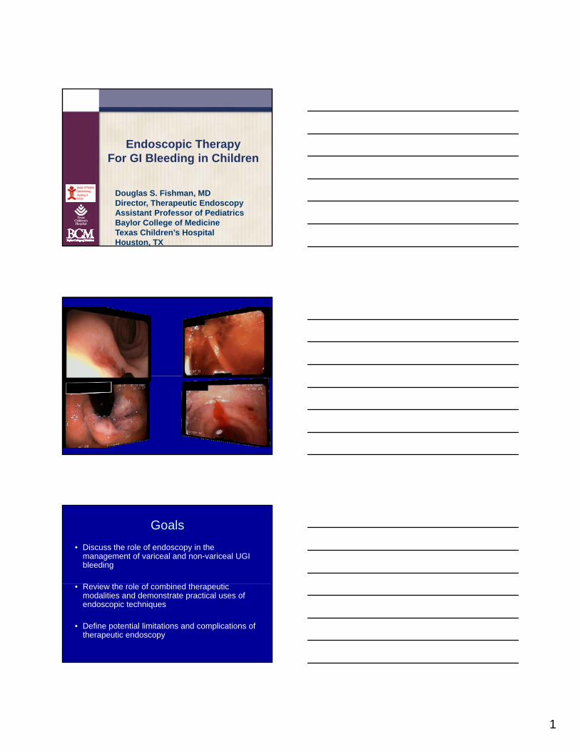
Endoscopic TherapyFor GI Bleeding in Children
Douglas S. Fishman, MDDirector, Therapeutic EndoscopyAssistant Professor of PediatricsBaylor College of MedicineTexas Children’s HospitalHouston, TX
Goals• Discuss the role of endoscopy in the
management of variceal and non-variceal UGI bleeding
R i th l f bi d th ti• Review the role of combined therapeutic modalities and demonstrate practical uses of endoscopic techniques
• Define potential limitations and complications of therapeutic endoscopy

2
Patient Assessment
• High risk stable vs. High risk unstable• Hemodynamics, measures of hemostasis• Where to do your endoscopy? (ED, ICU,
OR GI P d U it)OR, GI Procedure Unit)• When?• Co-morbidity (Cardiac, BMT)• Antibiotics
The Team
• Technicians• Nursing
– Endoscopy Unit– OR
• Pediatric Endoscopists– Fellows– Attendings– Endoscopy “Back-up”
• Surgical Staff• Adult Endoscopists
Equipment
• Bleeding kit (“tackle-box”)– VBL kit, sclerotherapy needles, multiple clips– Injectables (epi, sodium morrhuate)
I i ti• Irrigation • Suction• Endoscopes
– Scope size– Channel size– Duodensoscope

3
The techniques
–Injection therapy–Thermal coagulation
• MPEC • Argon Plasma
–Clip application–Variceal band ligation
Endoscopic Criteria
• Acute hemorrhage– Forrest I a (Spurting hemorrhage)– Forrest I b (Oozing hemorrhage)
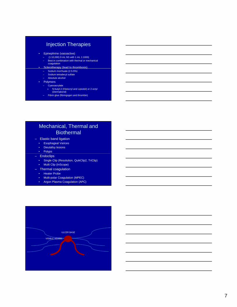
Si f t h h• Signs of recent hemorrhage– Forrest II a (Visible vessel)– Forrest II b (Adherent clot)– Forrest II c (Hematin on ulcer base)
• No signs of recent hemorrhage– Forrest III
Forrest 1a-”spurters”

4
Visible Vessel or Adherent Clot ?
A B
DC
Forrest Iic vs. Forrest III
Forrest III

5
Variceal Grading• I, II, III (IV)
– Small, medium, large– % obstruction of lumen
• Additional signs:Additional signs:– cherry red spots (petechiae of 1-2 mm on the
variceal surface)– red wale marks (fine capillaries on the
variceal surface, resembling whipping marks)• Gastroesophageal and Gastric Varices
– Sarin Classification
Sarin Classification of Gastric Varices Sarin et al. Hepatology. 1992

6
Benefits of Endoscopic Therapy• Endoscopic therapy better than no therapy
for risk of rebleeding and need for surgery :–– ACTIVE BLEEDING ACTIVE BLEEDING –– NonNon--bleeding visible vesselsbleeding visible vesselsNonNon bleeding visible vesselsbleeding visible vessels
• Epinephrine compared to other monotherapies or epinephrine + monotherapies: epinephrine alone was less effective (NNT=9, NNT=5)– Rebleeding or need for surgery
Laine and McQuaid, Clin Gastroenterol Hepatol 2009; 7: 33-49
Endoscopy Consenus Statement
• A finding of high-risk endoscopic stigmata (active bleeding or a visible vessel in an ulcer bed) is an indication for immediate endoscopic hemostatic therapyendoscopic hemostatic therapy
• Monotherapy, with injection or thermal coagulation, is an effective endoscopic hemostatic technique for high-risk stigmata; however, the combination is superior to either treatment alone.
Barkun et al. Annals of Internal Medicine 2003
Endoscopy Consensus Statement
• A finding of low-risk endoscopic stigmata (a clean-based ulcer or a nonprotuberant pigmented dot in an ulcer bed) is not an indication for endoscopic hemostaticindication for endoscopic hemostatic therapy
• A finding of a clot in an ulcer bed warrants targeted irrigation in an attempt at dislodgment, with appropriate treatment of the underlying lesion
Barkun et al. Annals of Internal Medicine 2003
7
Injection Therapies• Epinephrine (vasoactive)
– (1:10,000) 9 mL NS with 1 mL 1:1000)– Best in combination with thermal or mechanical
coagulation
• Sclerotherapy (lead to thrombosis)py ( )– Sodium morrhuate (2.5-5%) – Sodium tetradecyl sulfate– Absolute alcohol
• Polymers– Cyanoacrylate
» N-butyl-2 (Histocryl and Lipiodol) or 2-octyl (Dermabond)
– Fibrin glue (fibringogen and thrombin)
Mechanical, Thermal and Biothermal
– Elastic band ligation• Esophageal Varices• Dieulafoy lesions• Polyps
– Endoclips• Single Clip (Resolution, QuikClip2, TriClip)• Multi Clip (InScope)
– Thermal coagulation• Heater Probe• Multi-polar Coagulation (MPEC)• Argon Plasma Coagulation (APC)
ULCER BASE
VISIBLE VESSELVISIBLE VESSEL

8
• Prime needle outside• Identify lesion• Insert catheter• Leave space between
lesion and scope to extend needle
• Inject 0.5 mL until bleb formed
• Pull catheter back• Repeat in all
quadrants

9
Multi-polar Electrocautery (MPEC)
• Generates heat indirectly by passage of current through tissue
• Allows for coaptation• Leads to coagulation
and vessel contraction
When to use multipolar or heater probe
• Duodenal ulcer• Gastric ulcer• Mallory-Weiss Tear• Dielafoy lesions• Vascular malformations (GAVE, radiation-
induced)
Multi-polar Electrocautery (MPEC)
• Use 7 or 10 French catheter• Several available options• No grounding necessary but
requires electrosurgical unitrequires electrosurgical unit• Set power to 15 to 20W (less for
colon)• Apply pressure first• Depress foot pedal 2-4 seconds• Pull probe back gently and irrigate

10
MPEC
MPEC

11
MPEC Tips
• Due to various angulations, may need to bring catheter out prematurely (e.g antrum for duodenal ulcer)
• Catheters with combined sclerotherapy• Catheters with combined sclerotherapy needle may be difficult in retroflexion
• Larger vessels require larger catheter• Less optimal for coagulopathy
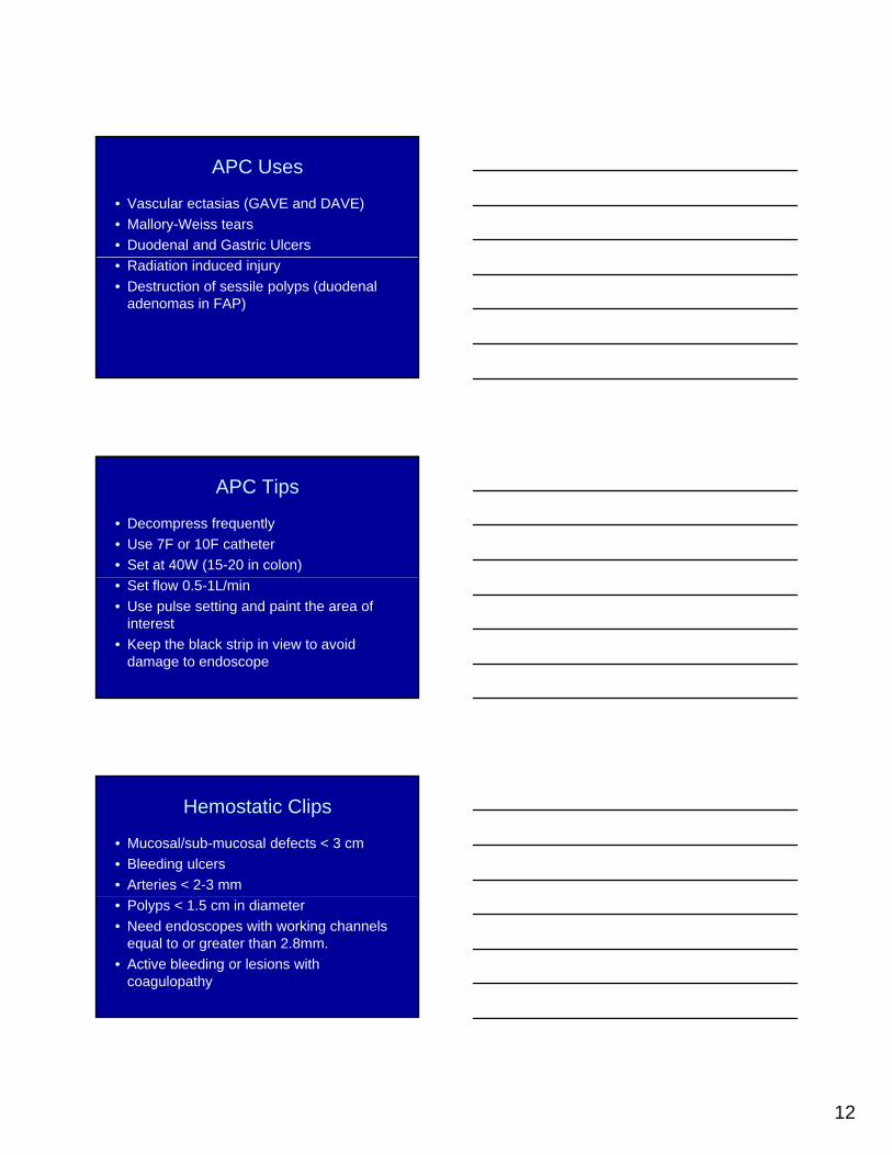
Argon Plasma Coagulation
• Non-contact thermal hemostasis• Tungsten electrode in probe ionizes argon
gasA b k t ti
Argon Plasma Coagulation
• Argon beam seeks nearest tissue• Limited depth of coagulation (2-3 mm) with
contact at surface
12
• Vascular ectasias (GAVE and DAVE)• Mallory-Weiss tears• Duodenal and Gastric Ulcers
APC Uses
• Radiation induced injury• Destruction of sessile polyps (duodenal
adenomas in FAP)
• Decompress frequently• Use 7F or 10F catheter• Set at 40W (15-20 in colon)
APC Tips
• Set flow 0.5-1L/min• Use pulse setting and paint the area of
interest• Keep the black strip in view to avoid
damage to endoscope
Hemostatic Clips
• Mucosal/sub-mucosal defects < 3 cm• Bleeding ulcers• Arteries < 2-3 mm• Polyps < 1.5 cm in diameter• Need endoscopes with working channels
equal to or greater than 2.8mm. • Active bleeding or lesions with
coagulopathy

13
When to use clips for hemostasis
• Duodenal ulcer• Gastric ulcer• Mallory-Weiss Tear• Early anastomotic bleeding• Post-polypectomy• Prophylaxis (pre-polypectomy, EMR) • Post-variceal banding
Hemostatic Clip Options
• Boston Scientific (Resolution Clip)• Olympus (Quick Clip2)• Wilson Cool (Tri-Clip)

14
Clip Tips
• Practice with assistant prior to “in vivo” use• May need to bring out in antrum and
assess opening, closing and angleM d t h d t t POP• May need two hands to create POP
• To release, assistant should open their hand, endoscopist should keep catheter steady and even pull back slightly
• Have both lengths available
Hemostatic Clip Tips• Be familiar with clips• Have multiple available• Work close• Inject with epinephrine if oozingj p p g• Head-on or tangential, don’t pinch the
vessel• Rotate clip• Push into mucosa and suction• Close
Hemostatic Clip Tips:Common Errors
• Deployment of clip too far from endoscope• Knock off clips already deployed• Premature closure of clip• Insufficient number of clips available• Failure to release clip

15
Variceal Band Ligation (VBL)
• Arrest bleeding and obliterate/eradicate the varix
• VBL is a the use of a rubber band when placed over a varix leads to thrombosisplaced over a varix, leads to thrombosis
Variceal Band Ligation
Wilson Cook4,6, 10 Shooter®
ConMedAuto-Band Ligator®
Boston ScientificSuper 7®
VBL use in children
• In adults, compared to sclerotherapy– Decreased mortality (45% vs 28%)– Decreased complications (22% vs 2%)
Less recurrent hemorrhage and fewer– Less recurrent hemorrhage and fewer sessions (NS)
• Majority of studies include patients with both intrahepatic and extrahepatic disease
• >90% variceal eradication in most seriesStiegmann GV et al. NEJM 1992; 326: 1527-1532McKiernan P et al. JPGN 2002; 34: 207-211Celinska-Cedro et al. J Pediat Surg; 38: 1008-11

16
VBL technique
• Identify varix of concern (map out remainder)
• Remove scope and attach ligation device• Remove scope and attach ligation device• Start low in the distal esophagus with high
risk lesions first
VBL technique• Angle scope so that varix can roll into
banding cap. All edges of the cap should surround the varix.

17
VBL technique• Apply suction and when varix engorges ¾
of cap obstructing endoscopic view, be ready to turn the banding device when there is a full “red out”
VBL technique
VBL Tips
• Re-intubation with ligation device can be difficult
• Major limitation is age (18 months)Mi i i t hi b d ith d• Minimize touching bands with endoscope after placement
• Have sclerotherapy equipment available

18
Complications of VBL
• Bleeding (early and late)• Infection
– SBE prophylaxis not recommendedA tibi ti f t bl di l– Antibiotics for acute bleeding only
• Perforation (rare)• Stricture (rare)
Training• Text/Journals
– Handbook of Gastroenterologic Procedures (Drossman)– JPGN, AJG, Gastrointestinal Endoscopy (GIE) and
Endoscopy• Video
DAVE j t– DAVE project– ASGE Training Library
• Computer Simulation (bleeding modules)• “Hands-On” Training
– NASPGHAN/ASGE courses– ASGE Center (Chicago, IL)
• Adult GI Collaboration (observation, preceptorship)
http://daveproject.org

19
Special ThanksGI Procedure Suite StaffTexas Children’s Hospital Endoscopy Team:
– Bryan Vartabedian– Anthony Olive– Bruno Chumpitazi– Kalpesh Thakkar– Mark Gilger– Isaac Raijman
Thank you!!
What should you do with a
CLOT?

20
What to do with adherent clots
• 56 patients at seven centers found to have fresh adherent clots with no active bleeding
• The clot was irrigated with 200 cc of forcibly injected waterinjected water.
• Randomized into treatment with injection and heater probe or medical management– Those randomized to endoscopic therapy had
the base of the adherent clot injected with 1/10,000 adrenaline in four quadrants with at least 1 cc in each quadrant.
To remove or not?
• The clot was removed and heater probe (30 J) a minimum of 3 coaptive pulses.
• Rebleeding rates were 34.3% (12/35) in the medical treatment arm vs 4 8% (1/21)the medical treatment arm vs 4.8% (1/21) in endoscopic group. (p<0.02).
• Endoscopic treatment with injection of the base of the clot, removal, and heater probe coagulation significantly reduces rebleeding rates.
Endoscopic Therapy vs. No Endoscopy for Treatment of Clots• No signficant benefit in further bleeding,
surgery or mortality• 2/5 favored endoscopy, 1/5 had n=5
patientspatients• Did not include rebleeding
Laine and McQuaid, Clin Gastroenterol Hepatol 2009; 7: 33-49

21
How to remove the clot
• Snare removal (like polyp)• Probe with biopsy forceps• Manipulate with endoscope• Suction