Embed Size (px)
Citation preview
17
eISSN 1303-5150 www.neuroquantology.com
NeuroQuantology | May 2019| Volume 17 | Issue 05 | Page 17-21| doi: 10.14704/nq.2019.17.05.2332Tacconi L., Endoscopic Approach Technique for Recurrent Lumbar Prolapsed Disc
Endoscopic Approach Technique for Recurrent Lumbar Prolapsed Disc
Leonello Tacconi1*, Francesco Signorelli2, Enrico Giordan1
Key Words: percutaneous discectomy, PELD, recurrent disc prolapse, transforaminal endoscopic discectomy, recurrent lumbar disc
DOI Number: 10.14704/nq.2019.17.05.2332 NeuroQuantology 2019; 17(05):17-21
Corresponding author: Leonello TacconiAddress: 1Neurosurgical Department, Azienda Sanitaria Universitaria Integrata di Trieste, Trieste, Friuli-Venezia Giulia, Italy; 2Neurosurgical Department, University “Aldo Moro” of Bari; Medical School, Bari, Italye-mail [email protected] (Leonello Tacconi); [email protected] (Francesco Signorelli); [email protected] (Enrico Giordan)Phone: +39 040 3994041Relevant conflicts of interest/financial disclosures: The authors declare that the research was conducted in the absence of any commercial or financial relationships that could be construed as a potential conflict of interest. Received: 01 May 2019; Accepted: 09 May 2019
ABSTRACT
Introduction: Recurrent disc after lumbar discectomy is not uncommon, with most of the patients requiring a new surgery. A greater bone decompression and scar tissue dissection become necessary with the new procedure, resulting in a higher chance of postoperative complications. Recently, many surgeons have begun to treat recurrent disc with endoscopic approaches, in order to reach the prolapsed disc avoiding tissue dissection. We present our up-to-dated experience on the treatment of recurrent disc by endoscopic technique.
Material and methods: We prospectively collected 30 patients treated for recurrent lumbar disc prolapse, from May 2016 to December 2017, with an endoscopic procedure. We collected data on age, sex, location, diagnosis, leg pain by VAS, and degree of disability via the Oswestry Disability Index (ODI), and if any adverse events occurred. All patients underwent an ODI (Oswestry Disability Index) and a VAS (Visual Analogue Scale) questionnaire before the operation and after 3, 6 and 15 months [3-6] at the follow-up visit. No patients were lost at follow-up.
Patients characteristics: Age at presentation ranged between 23 and 78 years with a male to female ratio of 1.5 to 1. The level treated more was L4-L5. In all cases, we performed transforaminal route access, except for two, where an interlaminar approach was necessary because of the disc fragment location. Twenty-six cases had been operated previously by microsurgical access and the remaining by an endoscopic technique. In one case the disc had recurred for a second time, requiring open revision surgery.
Results: Median operative time was 52 minutes (range 44 to 79 minutes). After a median follow up of 15 months (range 15-24 months) 93% of patients were pain-free. Pain by VAS ranged from a mean value of 6.3 at admission to 1.9 at 15 months of follow-up. ODI scores went from a mean preoperative value of 59.8% to 14.6% at the same follow-up. Four patients experienced transient paresthesia along the dermatomeric distribution of the involved nerve, while 3 had an intraoperative dural tear. One patient had to undergo new revision surgery for a disc recurrence. No late adverse events occurred.
Conclusions: Endoscopic discectomy might be a valuable procedure for recurrent lumbar disk prolapse treatment. Our results showed good outcomes with only a few transient complications and less postoperative pain. Also, iatrogenic mechanical instability might be avoided with this technique.

18
eISSN 1303-5150 www.neuroquantology.com
NeuroQuantology | May 2019| Volume 17 | Issue 05 | Page 17-21| doi: 10.14704/nq.2019.17.05.2332Tacconi L., Endoscopic Approach Technique for Recurrent Lumbar Prolapsed Disc
IntroductionLumbar disc prolapse is a common disease which, often, requires a long time off from work or daily living activities (Fjeld et al., 2017). Commonly, it is treated with surgery by an open midline microdiscectomy or a minimally invasive approach (MIS). Recurrences are not negligible, ranging from 5% to 18% of cases, with most of the patients requiring a new discectomy (Hlubek and Mundis, 2017). Unfortunately, open discectomies for recurred disc protrusion require the surgeon to deal with scar tissue and a greater amount of bone decompression. Therefore, revision surgeries are burdened with a higher risk of complication such as cerebrospinal fluid (CSF) leak, neurological deficits, mechanical instability, infection, and postoperative pain. With the introduction in the spinal field of the endoscopic techniques, many surgeons have started to treat recurrent discs endoscopically (Shin et al., 2011; Rasouli et al., 2014). With endoscopic procedures it is possible to reach the new protruded disc, through a “virgin route”, avoiding dissection of insidious scar tissue (Tacconi et al., 2018). We implemented, in our Department, the endoscopic discectomy for disk prolapse treatment in 2015 and only after we mastered the technique we applied it for disc prolapse recurrences. In this paper, we present our updated experience in the use of the endoscope for the treatment of recurrent lumbar disc prolapse. The outcomes, complications, as well as our technique, are discussed here.Material and MethodsFrom May 2016 to December 2017 we prospectively collected 30 patients who suffered from a recurrent disc prolapse and have undergone endoscopic revision surgery under general anesthesia. All the endoscopic procedures were performed by the same author (L.T.). Written informed consent was obtained from all the patients and none of our patients underwent surgery with magnetic resonance imaging (MRI) older than four weeks.
We collected data on age, sex, location, diagnosis, leg pain by VAS, and degree of disability via the Oswestry Disability Index (ODI), and if any adverse events occurred. All patients underwent an ODI (Oswestry Disability Index) and a VAS (Visual Analogue Scale) questionnaire before the operation and after 3, 6 and 15 months [3-6] at the follow-up visit. No patients were lost at follow-up.
ResultsPatients characteristics
Age at presentation ranged between 23 and 78 years with a male to female ratio of 1.5 to 1. The treated levels were L2-L3 in 2 cases, L3-L4 in 4; L4-L5 in 18, and L5-S1 in 6 cases. In all cases, we performed transforaminal route access, except for two where an interlaminar approach was necessary because of the disc fragment location (L5-S1 level with cranial migration of the disc fragment). Twenty-six cases had been operated previously by microsurgical access and the remaining by an endoscopic technique. In one case the disc had recurred for a second time, requiring open revision surgery. Median time from the first operation to recurrence was 7.3 months (range 3-24 months).
Patients characteristics are summarized in Table 1.
Number of Patients 30Age [mean (range)] 47.9 (23 – 78)Male: Female Ratio 1.5:1Median Follow-up [months, (range)] 18 (15 – 24)
Location (N°)
L2-L3 (2)L3-L4 (4)L4-L5 (18)L5-S1 (6)
Table 1. Patients’ demographic and clinical data.
Outcomes
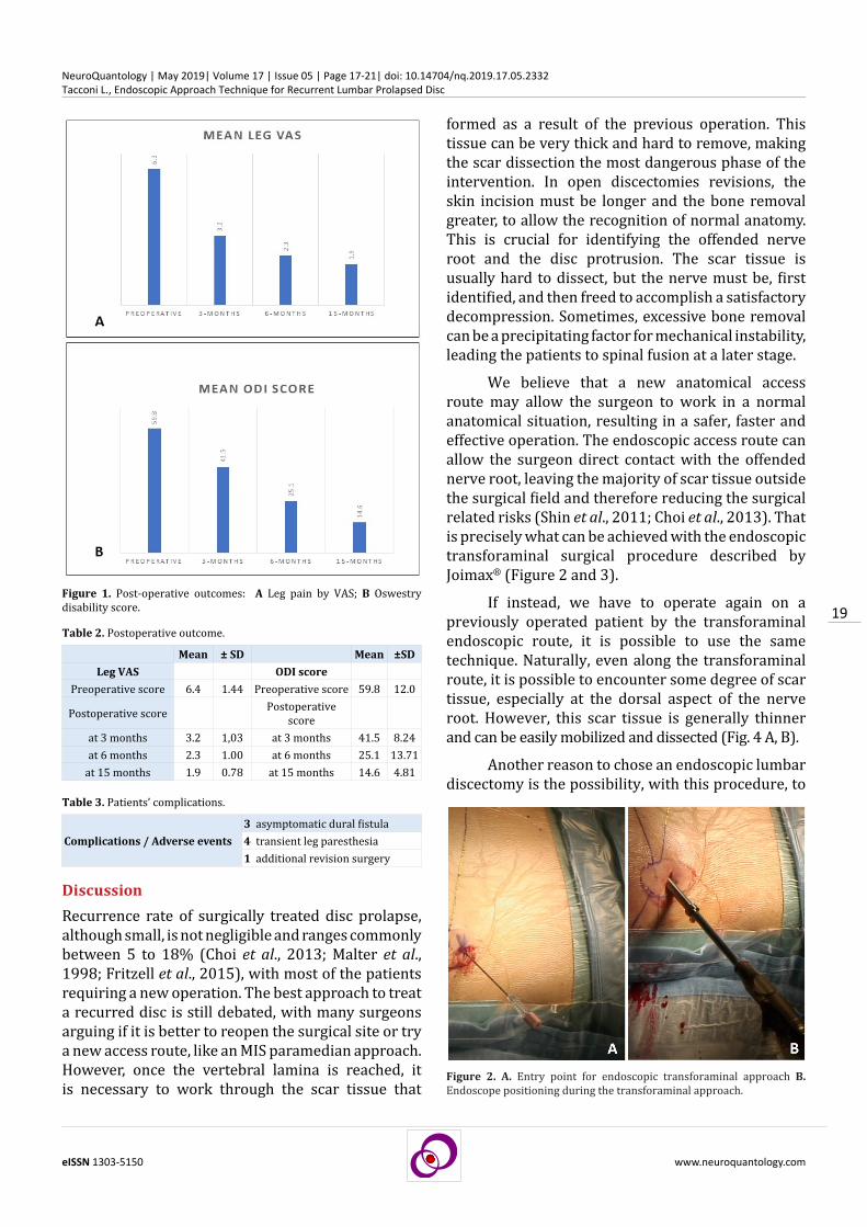
Median operative time was 52 minutes (range 44 to 79 minutes). The blood loss was negligible. The pain gradually improved in all cases, and after a median follow up of 18 months (range 15-24 months), 28 (93%) patients were pain-free. One patient experienced a recurrent disc 6 months later, another one was still feeling leg pain during physical activity. Leg VAS went from a preoperative mean value of 6.3 to a mean value of 3.2 at 3 months, 2.3 at 6 months, and 1.9 at 15 months (Fig. 1, A). ODI scores went from a mean preoperative score of 59.8 to 41.5 at 3 months, 25.1 at 6 months, and 14.6 at 15 months (Fig. 1, B).
Postoperative outcomes are summarized in Table 2.
There were no long-term complications detected. Perioperatively, four patients experienced painful paresthesia along the dermatomeric distribution of the operated nerve root. Those symptoms improved within a few weeks with medical therapy. Three patients experienced a dural tear without any clinical signs or symptoms for postoperative CSF leakage, while one patient needed to undergo open microdiscectomy for a newly recurred disc prolapsed (Table 3).
19
eISSN 1303-5150 www.neuroquantology.com
NeuroQuantology | May 2019| Volume 17 | Issue 05 | Page 17-21| doi: 10.14704/nq.2019.17.05.2332Tacconi L., Endoscopic Approach Technique for Recurrent Lumbar Prolapsed Disc
formed as a result of the previous operation. This tissue can be very thick and hard to remove, making the scar dissection the most dangerous phase of the intervention. In open discectomies revisions, the skin incision must be longer and the bone removal greater, to allow the recognition of normal anatomy. This is crucial for identifying the offended nerve root and the disc protrusion. The scar tissue is usually hard to dissect, but the nerve must be, first identified, and then freed to accomplish a satisfactory decompression. Sometimes, excessive bone removal can be a precipitating factor for mechanical instability, leading the patients to spinal fusion at a later stage.
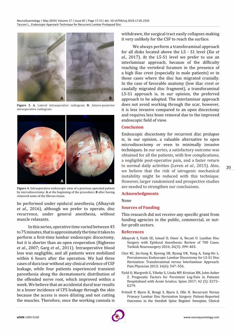
We believe that a new anatomical access route may allow the surgeon to work in a normal anatomical situation, resulting in a safer, faster and effective operation. The endoscopic access route can allow the surgeon direct contact with the offended nerve root, leaving the majority of scar tissue outside the surgical field and therefore reducing the surgical related risks (Shin et al., 2011; Choi et al., 2013). That is precisely what can be achieved with the endoscopic transforaminal surgical procedure described by Joimax® (Figure 2 and 3).
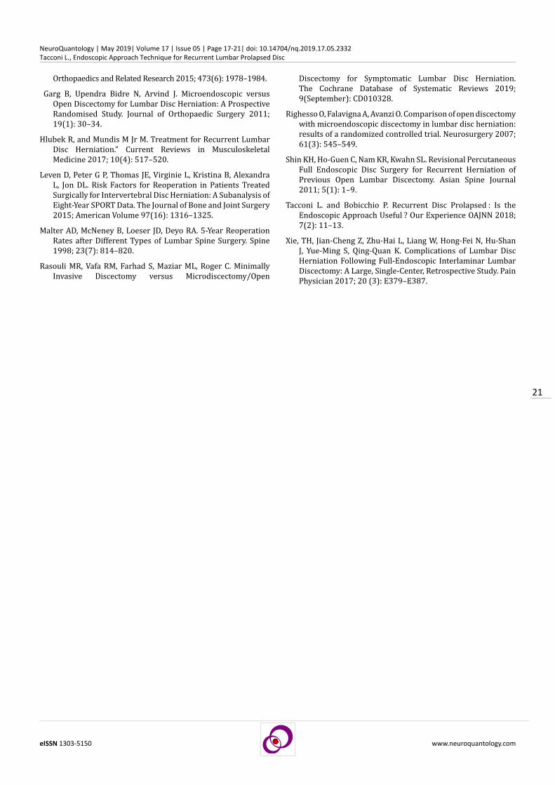
If instead, we have to operate again on a previously operated patient by the transforaminal endoscopic route, it is possible to use the same technique. Naturally, even along the transforaminal route, it is possible to encounter some degree of scar tissue, especially at the dorsal aspect of the nerve root. However, this scar tissue is generally thinner and can be easily mobilized and dissected (Fig. 4 A, B).
Another reason to chose an endoscopic lumbar discectomy is the possibility, with this procedure, to
Figure 1. Post-operative outcomes: A Leg pain by VAS; B Oswestry disability score.
Figure 2. A. Entry point for endoscopic transforaminal approach B. Endoscope positioning during the transforaminal approach.
Mean ± SD Mean ±SDLeg VAS ODI score
Preoperative score 6.4 1.44 Preoperative score 59.8 12.0
Postoperative score Postoperative score
at 3 months 3.2 1,03 at 3 months 41.5 8.24at 6 months 2.3 1.00 at 6 months 25.1 13.71
at 15 months 1.9 0.78 at 15 months 14.6 4.81
Table 2. Postoperative outcome.
Discussion Recurrence rate of surgically treated disc prolapse, although small, is not negligible and ranges commonly between 5 to 18% (Choi et al., 2013; Malter et al., 1998; Fritzell et al., 2015), with most of the patients requiring a new operation. The best approach to treat a recurred disc is still debated, with many surgeons arguing if it is better to reopen the surgical site or try a new access route, like an MIS paramedian approach. However, once the vertebral lamina is reached, it is necessary to work through the scar tissue that
Complications / Adverse events3 asymptomatic dural fistula4 transient leg paresthesia1 additional revision surgery
Table 3. Patients’ complications.
20
eISSN 1303-5150 www.neuroquantology.com
NeuroQuantology | May 2019| Volume 17 | Issue 05 | Page 17-21| doi: 10.14704/nq.2019.17.05.2332Tacconi L., Endoscopic Approach Technique for Recurrent Lumbar Prolapsed Disc
be performed under epidural anesthesia, (Albayrak et al., 2016), although we prefer to operate, disc recurrence, under general anesthesia, without muscle relaxants.
In this series, operative time varied between 45 to 75 minutes, that is approximately the time it takes to perform a first-time lumbar endoscopic discectomy, but it is shorter than an open reoperation (Righesso et al., 2007; Garg et al., 2011). Intraoperative blood loss was negligible, and all patients were mobilized within 6 hours after the operation. We had three cases of dura tear without any clinical evidence of CSF leakage, while four patients experienced transient paresthesia along the dermatomeric distribution of the offended nerve root, which improved within a week. We believe that an accidental dural tear results in a lesser incidence of CFS leakage through the skin because the access is more dilating and not cutting the muscles. Therefore, once the working cannula is
withdrawn, the surgical tract easily collapses making it very unlikely for the CSF to reach the surface.
We always perform a transforaminal approach for all disks located above the L5 - S1 level (Xie et al., 2017). At the L5-S1 level we prefer to use an interlaminar approach, because of the difficulty reaching the vertebral foramen in the presence of a high iliac crest (especially in male patients) or in those cases where the disc has migrated cranially. In the case of favorable anatomy (low iliac crest or caudally migrated disc fragment), a transforaminal L5-S1 approach is, in our opinion, the preferred approach to be adopted. The interlaminar approach does not avoid working through the scar, however, it is less invasive compared to an open discectomy and requires less bone removal due to the improved endoscopic field of view. Conclusion Endoscopic discectomy for recurrent disc prolapse is, in our opinion, a valuable alternative to open microdiscectomy or even to minimally invasive techniques. In our series, a satisfactory outcome was obtained for all the patients, with few complications, a negligible post-operative pain, and a faster return to normal daily activities (Leven et al., 2015). Also, we believe that the risk of iatrogenic mechanical instability might be reduced with this technique. However, larger randomized and prospective studies are needed to strengthen our conclusions.AcknowledgmentsNoneSources of FundingThis research did not receive any specific grant from funding agencies in the public, commercial, or not-for-profit sectors.References Albayrak S, Fatih SE, Ismail D, Omer A, Necati U. Lumbar Disc
Surgery with Epidural Anesthesia: Review of 700 Cases. Turkish Neurosurgery 2016; 26(3): 399–403.
Choi KC, Jin-Sung K, Kyeong SR, Byung UK, Yong A, Sang-Ho L. Percutaneous Endoscopic Lumbar Discectomy for L5-S1 Disc Herniation: Transforaminal versus Interlaminar Approach. Pain Physician 2013; 16(6): 547–556.
Fjeld O, Margreth G, Vibeke S, Linda MP, Kristian BN, John-Anker Z. Prognostic Factors for Persistent Leg-Pain in Patients Hospitalized with Acute Sciatica. Spine 2017; 42 (5): E272–E279.
Fritzell P, Bjorn K, Bengt S, Bjorn S, Olle H. Recurrent Versus Primary Lumbar Disc Herniation Surgery: Patient-Reported Outcomes in the Swedish Spine Register Swespine. Clinical
Figure 3. A. Lateral intraoperative radiogram B: Antero-posterior intraoperative radiogram.
Figure 4. Intraoperative endoscopic view of a previous operated patient by microdiscectomy: A at the beginning of the procedure; B after having removed some of the fibrous tissue.
21
eISSN 1303-5150 www.neuroquantology.com
NeuroQuantology | May 2019| Volume 17 | Issue 05 | Page 17-21| doi: 10.14704/nq.2019.17.05.2332Tacconi L., Endoscopic Approach Technique for Recurrent Lumbar Prolapsed Disc
Orthopaedics and Related Research 2015; 473(6): 1978–1984.
Garg B, Upendra Bidre N, Arvind J. Microendoscopic versus Open Discectomy for Lumbar Disc Herniation: A Prospective Randomised Study. Journal of Orthopaedic Surgery 2011; 19(1): 30–34.
Hlubek R, and Mundis M Jr M. Treatment for Recurrent Lumbar Disc Herniation.” Current Reviews in Musculoskeletal Medicine 2017; 10(4): 517–520.
Leven D, Peter G P, Thomas JE, Virginie L, Kristina B, Alexandra L, Jon DL. Risk Factors for Reoperation in Patients Treated Surgically for Intervertebral Disc Herniation: A Subanalysis of Eight-Year SPORT Data. The Journal of Bone and Joint Surgery 2015; American Volume 97(16): 1316–1325.
Malter AD, McNeney B, Loeser JD, Deyo RA. 5-Year Reoperation Rates after Different Types of Lumbar Spine Surgery. Spine 1998; 23(7): 814–820.
Rasouli MR, Vafa RM, Farhad S, Maziar ML, Roger C. Minimally Invasive Discectomy versus Microdiscectomy/Open
Discectomy for Symptomatic Lumbar Disc Herniation. The Cochrane Database of Systematic Reviews 2019; 9(September): CD010328.
Righesso O, Falavigna A, Avanzi O. Comparison of open discectomy with microendoscopic discectomy in lumbar disc herniation: results of a randomized controlled trial. Neurosurgery 2007; 61(3): 545–549.
Shin KH, Ho-Guen C, Nam KR, Kwahn SL. Revisional Percutaneous Full Endoscopic Disc Surgery for Recurrent Herniation of Previous Open Lumbar Discectomy. Asian Spine Journal 2011; 5(1): 1–9.
Tacconi L. and Bobicchio P. Recurrent Disc Prolapsed : Is the Endoscopic Approach Useful ? Our Experience OAJNN 2018; 7(2): 11–13.
Xie, TH, Jian-Cheng Z, Zhu-Hai L, Liang W, Hong-Fei N, Hu-Shan J, Yue-Ming S, Qing-Quan K. Complications of Lumbar Disc Herniation Following Full-Endoscopic Interlaminar Lumbar Discectomy: A Large, Single-Center, Retrospective Study. Pain Physician 2017; 20 (3): E379–E387.