Embed Size (px)
Citation preview

212
With accumulated experience with endoscopically as-sisted mechanical and laser lumbar diskectomy,1–18 theneed for a more efficient method to decompress thelateral recess19–26 and intervertebral neural foramenfrom very large or extruded disk protrusions, recurrentdisks, scar tissue, and spondylitic spurs became evident.The most frequently seen lumbar spinal disk disease inthe elderly is spinal and lateral foraminal stenosis.24,26
Lateral stenosis may be congenital or degenerative whensecondary to acute disk disease and spinal trauma. Theclassic wide posterior decompressive laminectomy withforaminotomy involves extensive muscle and soft tissuedissection for exposure, decompression, and resection ofthe posterior spinal elements. Despite varying degrees ofsuccess,24 it is associated with significant iatrogenic traumaand failed back syndrome.24,26 As a result, the search for aminimally invasive spinal surgery (MISS) technique began.This chapter will describe endoscopic lumbar foramino-plasty and diskectomy, including endoscopic laserforaminoplasty (ELF),10 as reported by Dr. Martin T. N.Knight, with laser application, and minimally invasivetransforaminal microdecompressive endoscopic assisteddiskectomy and foraminoplasty (TF-MEAD), a new systemof more aggressive mechanical instruments and laser ap-plication, developed at the California Center for Mini-mally Invasive Spine Surgery (C-MISS).
The pioneering work of Hijikata1 with percutaneousmanual diskectomy, Ascher2 and Choy3 with percuta-neous laser diskectomy, and the application ofendoscopy by Kambin4 and others27–32 resulted in moni-tored keyhole operations for removal of herniatedlumbar disks. The use of endoscopy and laser, especiallythe holmium side-firing laser, allows removal of large
protrusions and extruded disk fragments from theepidural space and stenotic foraminal decompressionwith ELF, which addresses one level and one side at atime.10,29 This technique takes 90 to 120 minutes to com-plete using primarily the laser for foraminal decompres-sion and diskectomy.9
Attention is now being directed to treatment ofepidural scarring, lateral recess and foraminal stenosis,and advanced degenerative changes that are often bilat-eral and occur at multiple levels.28 I have developed amore aggressive TF-MEAD system to address endoscopictransforaminal mechanical and laser microdecompressivediskectomy and foraminoplasty in a fast and effective man-ner for both unilateral single and bilateral multiple levels.These MISS procedures should now join the armamentar-ium of the spinal surgeon in treating advanced degenera-tive spinal stenosis and lateral foraminal stenosis.
Such procedures require the surgeon to be knowledge-able and competent in MISS, with a thorough knowledgeof the procedure of endoscopic lumbar diskectomy andforaminoplasty, pathoanatomy of the neuroforamen andthe spine, and the relationships of the lumbar exiting andtraversing nerve roots, dorsal root ganglion, facet joint,disks, and vertebrae.
Indications
Endoscopic lumbar foraminoplasty is indicated in thefollowing clinical situations10,16:
• Intractable low back pain with radiation down theleg (radicular pain)
• Symptoms of spinal neurogenic claudication
19
Endoscopic Lumbar ForaminoplastyJOHN C. CHIU
13693_C19.qxd 11/9/04 11:22 AM Page 212

CHAPTER 19 � Endoscopic Lumbar Foraminoplasty 213
• Compressive and irritative radiculopathy with sen-sorimotor impairment
• Disk extrusion or sequestration with predominantback or leg pain
• Degeneration and settlement of the spine withpredominant back pain, buttock pain, or leg pain
• Nonradicular low back pain persisting despite facetjoint injection
• Lateral recess stenosis with dynamic compressive ornoncompressive radiculopathy
• Prior failed conventional surgery with perineuralscarring and failed back syndrome
• No improvement of symptoms after a minimum of12 weeks of conservative therapy
• Spondylolytic spondylolisthesis• Diagnostic imaging, MRI, CT, 3D CT, and CT myel-
ogram that demonstrate disk herniation and/orextrusion or lateral recess stenosis
• Positive pre- or intraoperative diskogram and painprovocation test
• Positive electromyography• Multiple lumbar disks/levels that can be treated at
one sitting with TF-MEAD
Contraindications
The endoscopic lumbar foraminoplasty procedure iscontraindicated9,15 in the following clinical situations:
• Cauda equina syndrome• Painless motor deficit• Tumors• Clinical findings that suggest pathology other than
degenerative discogenic disease• ELF does not treat multilevel or bilateral disks
Instruments and Preparations
These surgical instruments are necessary to performendoscopic laser and/or lumbar foraminoplasty:
• Digital fluoroscopic equipment (C-arm) and monitor• Radiolucent C-arm/fluoroscopic carbon-fiber sur-
gical table• Endoscopic tower equipped with digital video mon-
itor, DVT/VHS recorder, light source, trichipdigital camera, and photo printer system
• Panoramic Plus Discoscope, 0 degree, 2.2 mmworking channel (Richard Wolf GmBH & Co.,Knittlingen, Germany)
• Percutaneous fiberoptic foraminoscope, 0 degree,6 mm OD, 3.9 mm working channel (Karl Storz,Tuttlingen, Germany)( Fig. 19–1A)
• Wide-angle posterolateral foraminoscope, 6 degree,6 mm OD, 3 mm working channel (Karl Storz)(Figs. 19–1A, 19–2E)
• C-MISS TF-MEAD system instruments (Figs. 19–1B,19–2B,D)
• Set of serial/progressive dilators, and cannulas ingraduated sizes (3.5–5.8 mm); a set of progressivecannulas with duckbill extensions (with variouslengths 5–10 mm on one side) (Fig. 19–1B)
• Aggressive toothed trephine set in graduated sizes• 9.9 mm tubular retractor system with a gradual dila-
tor set• Wide-angle endoscopes, 0 and 30 degree, 4 mm OD
(Karl Storz)• Trephine, curette, grasper and spoon forceps,
2 mm rotating bone punch (rongeurs), rasp, andburr
• Lumbar diskectomy sets (2.5, 3.5, 4.7 mm) (Black-stone Medical, Inc., Springfield, MA) with variousdiskectomies (Fig. 19–1C)
• Endoscopic grasping and cutting forceps, probe,knife, scissors, diskectomy rongeurs, and curette(Fig. 19–1A,B)
• Holmium: YAG laser generator (Trimedyne, Irvine,CA)
• Holmium 550-µm laser bare fiber with flat-tip andright-angle (side-firing) probes with and withoutirrigating system of various sizes, and 2 mm side-firing irrigating laser probe (Fig. 19–1A)
• Steerable Spinescope (Karl Storz) (Fig. 19–2A,C)with 2.5 mm working fiberscope (for laser applica-tion with a flexible tip that can bend up to 90 degrees and rotate to reach 360 degrees)
Anesthesia
Knight’s ELF procedure is done under neuroleptic(aware-state) analgesia in the prone position combinedwith local anesthesia. Patient feedback is essential inthese cases when one works around the nerve.
TF-MEAD patients are treated in an operating roomunder local anesthesia and monitored conscious seda-tion. The anesthesiologist maintains mild sedation, butthe patient is able to respond. Two grams Ancef and 8 mgdexamethasone are given intravenously at the start ofanesthesia. Surface EEG (SNAP, Nicolet Biomedical,Madison, WI) monitoring provides added precision ofanesthesia.
Patient Positioning
For TF-MEAD, if surgery is unilateral, the patient isplaced in a lateral decubitus position (Fig. 19–3B,C),
13693_C19.qxd 11/9/04 11:22 AM Page 213

214 SECTION 4 � Lumbar Spine
with the painful leg up and both hips and knees in mod-erate flexion. If the patient has an increased medicalrisk (i.e., pulmonary, cardiac, morbid obesity, and otherhigh-risk medical conditions), but requires a bilateralprocedure, the decubitus position may be used first onone side, then, turning the patient over, on the otherside to perform the bilateral procedure in one sitting.Bilateral operations are otherwise performed in theprone position on a radiolucent support similar to theWilson frame (as for ELF). The arms are supported onarm boards over the head. When local anesthesia andmild sedation are used, the extremities, buttocks, andshoulders are secured and restrained from sudden mo-tion with adhesive tape.
Localization
C-arm fluoroscopy is used to identify the lumbar levelsrelative to the sacrum. The midline, operative levels,and point of entry (operating portal) for surgery aremarked on the skin with a marking pen (Figs. 19–3,19–4A,B). The distance of the point of entry from themidline varies with the height and weight of the patient,
but it is ~12 cm at the affected disk level for an average-size patient. Positioning of the instruments is checkedthroughout the procedure by fluoroscopy in two planesas often as needed (Figs. 19–5, 19–6B, 19–7B–D). Atthe involved nerve roots distribution, sterile needle elec-trodes are placed for continuous intraoperative neuro-physiologic EMG monitoring.29
Surgical Technique for EndoscopicLaser Foraminoplasty
ELF9,16,28,31 begins with spinal probing and diskographyperformed at the suspected level, which is clinically ap-propriate to the site of back or peripheral radiatingpain or evidence of clinically related pathology. Knighthas described diagnostic spinal probing plus provoca-tive diskogram to identify any concordant pain-sensitive areas in the lateral recess and foraminal areasin a staged progression of the structures and scar tissueon the disk surface and around the nerve root, thefacets, and the walls of the foramen. Symptoms repro-duced by spinal probing and diskography determinethe extent of the surgical exploration required for the
FIGURE 19–1 Surgical instruments for endoscopic lumbarforaminoplasty and diskectomy. (A) Percutaneous fiberopticforaminoscope, 0 degrees, 6 mm OD, 3.9 mm workingchannel; posterolateral foraminoscope, 6 degrees, 6 mm ODworking channel; bare holmium laser fiber, side-firing laserprobe (Knight) (Trimedyne), and diskectomy forceps. (B) C-MISS TF-MEAD transforaminal decompressive system:endoscopes (0 and 30 degrees, 4 mm OD,) assisted tubularretractor, trephines, graduated duckbill cannulas, diskectomyrongeur, curette, and 2 mm bone punch. (C) Lumbar diskec-tomy set with dilators, working cannulas, trephines, andvarious discectomes.
A B
C
13693_C19.qxd 11/9/04 11:22 AM Page 214

CHAPTER 19 � Endoscopic Lumbar Foraminoplasty 215
FIGURE 19–2 Steerable spinoscope system, C-MISS TF-MEAD system, and foraminoscope for treatment of diskherniation and foramen stenosis. (A) Steerable spinoscope andcannular set for laser application and with flexible tip. (B) C-
MISS TF-MEAD system with a working channel 9.9 mmassisted by endoscopes (0 and 30 degrees, 4 mm OD). (C) Spin-oscope surgical application for TF-MEAD. (D) Lumbar foramino-plasty with C-MISS TF-MEAD system in surgical application.
A
C D
B
13693_C19.qxd 11/9/04 11:22 AM Page 215

216 SECTION 4 � Lumbar Spine
ELF procedure (second stage of ELF procedure), in-cluding endoscopic diskectomy, neurolysis, undercut-ting of facets, osteophytectomy, and/or laser ablationof painful tissue and foraminoplasty (Figs. 19–4A,B,19–6, 19–9A,D,E).
The diskography needle is replaced with aguidewire, and a 5 mm dilated tube is inserted into theexit root foramen. The procedure is performed underfluoroscopy. The trocar is then replaced with a work-ing endoscopic channel (Richard Wolf). A side-firing 2mm internal laser irrigation probe (Fig. 19–1A) is in-serted through the endoscope (Trimedyne) to per-form ELF (Figs. 19–6, 19–9A,D,E). The extraforami-nal zone and margin of the foramen are cleared. Thefacet joint surfaces are cleaned and undercut to allowthe endoscope to enter the epidural space. Vertebralbody and facet joint osteophytes, ligamentum flavumand superior foraminal ligaments, and perineuroand epidural scarring are ablated and the facet jointundercut until the anulus and epidural space arevisualized.
The exiting and traversing nerve roots are mobilizedand decompressed medially and laterally until the func-tional axilla of the root at the apex of the safe workingzone is displayed. The nerve is cleared of peripheral
fibrosis. Clearance and undercutting are extended alongthe bone margin to the superior notch with resection ofthe superior foraminal ligament. Exploration is continuedto the inferior pedicle, displacing the anulus and epiduralspace. Scarring is almost always associated with vascularbands. Inflamed soft tissue on the dorsum of the disk is ex-quisitely tender in some cases. Osteophytes along the as-cending joint of the superior notch, the dorsum of the tib-ial margin, and the vertebral shoulder are ablated underendoscopic vision by laser. Thermal modulation in con-junction with undercutting of the facet may increase thecross-section of the area of foramen and provide decom-pression. Endoscopic laser diskectomy and epidural ex-ploration and decompression complete the procedure.
Postoperative Care
The patient is discharged on the day following surgery.A muscle-balance physiotherapy regimen is recom-mended on the first day after discharge; later, the programis amplified with neural mobilization drills. Self-help drillsare encouraged twice a day thereafter. A pain diary ismaintained by the patient to identify the intensity andlocation of any residual pain.
FIGURE 19–2 (Continued) (E) Posterolateral foraminoscopywith wide-angle 6-degree, 3 mm working channel operatingforaminoscope.E
13693_C19.qxd 11/9/04 11:22 AM Page 216

CHAPTER 19 � Endoscopic Lumbar Foraminoplasty 217
Outcomes
Reported are a total of 958 ELF procedures performedon 716 patients who were evaluated at 6 and 12 weeks, 6months, and yearly following the surgery, unless clinicalsymptoms required closer supervision. The outcomefollowing ELF remains promising, with 61 to 70% ofcases achieving 50% gain in their preoperative OswestryDisability Index and visual analog pain score. Therewere 24 complications (1.6%), with diskitis in ninepatients, dural tear in one, wound infections in one, footdrop in two, myocardial infarction in one, erectile dys-function in one, and panic attack in one. Seven percent
of patients worsened after the operation. About 3% ofpatients required subsequent open surgery.
Surgical Technique for TransforaminalMicrodecompressive EndoscopicAssisted Diskectomy and Foraminoplasty
If a pain provocation test and diskogram were not donepreoperatively, they are done at the outset. If the disko-gram and pain provocation tests are confirmatory,surgery is performed (Figs. 19–2C–E, 19–4, 19–5, 19–7,
C
A B
FIGURE 19–3 Patient positioning and localization. (A) Patientin prone position. (B) Patient in lateral decubitus position.(C) Localization: skin marking and placement of needle (portal).
13693_C19.qxd 11/9/04 11:22 AM Page 217

218 SECTION 4 � Lumbar Spine
19–8, 19–9). An 18-gauge stylet is inserted and incre-mentally advanced under C-arm fluoroscopic guidancein two planes, at a 60-degree angle from the sagittalplane, targeting toward the center of the disk, throughthe safety zone, and into the desired interspace. Allinstrumentation is performed under C-arm fluoroscopiccontrol and endoscopy. The usual procedure for MISS isfollowed.15 The appropriate-size cannula and dilator arepassed over the stylet to the anulus. Under fluoroscopythe extended side of this cannula is turned to face thenerve root in order to retract and protect it.
The cannula retractors have variously shaped exten-sions like a duckbill (with various lengths, 5 to 10 mm onone side; Figs. 19–1B, 19–5E,F) to retract and protectthe nerve root once the cannula is inserted through theforamen into the epidural space and the extension isoriented properly toward the root. The larger, moreaggressively toothed trephines (Fig. 19–5B) are then
inserted and rotated to cut through anulus, disk protru-sion, spur, or spondylitic bar. The cannula is largeenough to admit a slim punch (rongeur), spinal diskforceps or pituitary forceps, and full-size curettes to aiddecompression of the foramen and the lateral recess(Fig. 19–5C–F). An endoscope can be passed through itinstead of the endoscope’s sheath to facilitate mechani-cal and laser decompression, foraminoplasty, anddiskectomy. The endoscope is useful in TF-MEAD sur-gery for decompression in the lateral recess and peri-foraminal area. Biting forceps, discectome, andholmium laser with continuous irrigation are used con-secutively to perform intradiscal diskectomy; lowerenergy nonablative laser is applied for shrinking andtightening of the disk (laser thermodiskoplasty).16,17
The decompression area can be enlarged with a largercannula retractor/trephine set. A small amount ofbleeding usually can be controlled with cold saline
A
C D
B
FIGURE 19–4 Safe surgical approach to neuroforamen forendoscopic lumbar foraminoplasty and diskectomy. (A) Pos-terolateral surgical approach to lumbar disk in cadaveric axialcryomicrotome. (B) Axial illustration of posterolateral lumbar
surgical approach for needle placement into intervertebralforamen. (C) Posterolateral surgical approach on prone axial CTimage at lumbar disk level. (D) Posterolateral surgical approachinto the foramen on axial view of lumbar spine 3D CT image.
13693_C19.qxd 11/9/04 11:22 AM Page 218

CHAPTER 19 � Endoscopic Lumbar Foraminoplasty 219
A
C
E F
D
B
FIGURE 19–5 Fluoroscopic view of lumbar endoscopicforaminoplasty and diskectomy with TF-MEAD system. (A)Stylet and dilator in disk space. (B) Trephine for disk and os-teophyte decompression. (C) Diskectomy forceps/rongeur for
diskectomy. (D) Bone punch for foraminoplasty. (E) Largediskectomy rongeur through duckbill cannula retractor fordiskectomy. (F) Duckbill cannula and bone punch in actionthrough TF-MEAD tubular retractor for foraminoplasty.
irrigation and rarely requires hemostasis by laser orbipolar coagulation. Holmium:YAG laser with a side-fir-ing probe or 550-µm holmium laser bare fiber (Fig.19–1A) is used to ablate the disk and to shrink and con-tract the disk, reducing the profile of protrusion andhardening the disk tissue (i.e., laser thermodiskoplasty).Disk removal is aided by a rocking excursion of the can-nula in a 25-degree arc, a “fan sweep maneuver”16,18
from side to side, that creates an inverted oval cone-shaped area of removed disk totaling up to 50 degrees.
Laser thermodiskoplasty can also cause sinovertebralneurolysis or denervation. The diskectomies is againused to remove charred debris.
The disk space and neural foramen can be directlyvisualized and examined by endoscopy to confirm ade-quate disk decompression and to perform furtherdecompression if necessary. If the foramen is compro-mised, the depth of insertion of the endoscope is ad-justed, the nerve root is again protected by the duckbillextension, and spurs are removed with curettes, bone
13693_C19.qxd 11/9/04 11:22 AM Page 219

220 SECTION 4 � Lumbar Spine
A
C D
B
FIGURE 19–6 Fluoroscopic view and illustration of laserside-firing probe for lumbar laser foraminoplasty (Knight).(A) Illustration of lateral spinal stenosis secondary to diskprotrusion, shoulder osteophytes, and facet hypertrophy.
(B) Fluoroscopic view of side-firing laser probe at foramen forlumbar facet decompression. (C) Illustration of lumbar laserdiskectomy. (D) Illustration of lumbar laser foraminoplasty forfacet hypertrophy.
13693_C19.qxd 11/9/04 11:22 AM Page 220

CHAPTER 19 � Endoscopic Lumbar Foraminoplasty 221
punches (rongeurs), and Kerrison rongeurs that canbe passed through the large cannula for decompressiveforaminoplasty, or laser application. An endoscopicallyassisted larger 9.9 mm tubular retractor system (Figs.19–1B, 19–2B,D, 19–5B,C,E) has been added to facili-tate foraminoplasty (Figs. 19–1B, 19–2B,D, 19–5B,C,E).
The steerable Spinescope (Karl Storz) can also beused to perform intradiscal lumbar laser diskectomy andlaser foraminoplasty (Figs. 19–2A,C, 19–7, 19–9B). TheSpinescope is fixed in a holding device, which allows thesurgeon to guide and steer very precisely a flexible fiber-scope and a working channel for a laser fiber of 0.6 mmdiameter to the pathologic part of the disk and the intra-and periforaminal tissue. The laser fiber can be ad-vanced or retracted millimeter by milllimeter inside thedisk under direct vision with the fiberoptic endoscopicsystem, within a given distance. Also, the tip of the appli-cator/laser fiber can be navigated and angulated from0 to 90 degrees with fine adjustment and rotatedthrough 360 degrees in all directions.
After removing all instruments, 0.25% Marcaine isinjected intradermally and into the incision and theparaspinal muscles along the path of the cannulationto prolong analgesia. A bandage is applied at the inci-sion sites.
Postoperative Care
Ambulation begins immediately after recovery, and thepatient is usually discharged 1 hour after surgery. Thepatient may shower the following day. Applying an icepack is helpful. NSAIDs are prescribed, and mild anal-gesics and muscle relaxants are recommended as needed.Patients typically return to usual activities in 10 days to3 weeks, provided heavy labor and prolonged sitting arenot involved.
Outcome
At C-MISS, the first 60 consecutive cases of the 180-plus cases treated with TF-MEAD to date (since year2000) included 31 males and 29 females. Thirty-six(60%) had surgery at one level, 24 (40%) at two ormore levels, eight bilaterally. All patients had com-plained of chronic low back pain with radicular pain,usually unilateral, and in eight instances, bilateral.Physical examination, as well as positive MRI and/orCT, EMG, and intraoperative or preoperative disko-grams appropriate to the symptomatic levels, con-firmed the diagnosis and levels. All had failed to
F
FIGURE 19–6 (Continued) (E) Illustration of lumbar laserforaminoplasty for upper shoulder osteophyte. (F) Illustration
of lumbar laser foraminoplasty for lower shoulder osteophyte.
E
13693_C19.qxd 11/9/04 11:22 AM Page 221

222 SECTION 4 � Lumbar Spine
A
B
FIGURE 19–7 Fluoroscopic view and illustration of laserside-firing probe in action for lumbar laser foraminoplasty withsteerable spinescope. (A) Illustrations of lumbar laserforaminoplasty with spinescope for facet hypertrophy (lateral
and axial views). (B) Illustration and fluoroscopic lateral viewsof lumbar laser foraminoplasty with spinescope for uppershoulder osteophyte.
13693_C19.qxd 11/9/04 11:22 AM Page 222

C
D
FIGURE 19–7 (Continued) (C) Illustration and fluoroscopiclateral views of lumbar laser foraminoplasty with spinescope
for lower shoulder osteophyte. (D) Illustration and fluoroscopiclateral views of lumbar laser diskectomy with spinescope.
13693_C19.qxd 11/9/04 11:22 AM Page 223

A
C
E F
D
B
FIGURE 19–8 Endoscopic view of lumbar mechanical de-compressive foraminoplasty and diskectomy. (A) Disk removalwith cutter forceps. (B) Disk decompression below the nerve.(C) Curette for osteophytic decompression. (D) Rasp for osteo-
phytic decompression. (E) Bone punch/rongeur for foraminaldecompression. (F) Disk and foramen appearance postforaminoplasty.
13693_C19.qxd 11/9/04 11:22 AM Page 224

CHAPTER 19 � Endoscopic Lumbar Foraminoplasty 225
A
C
E
D
B
FIGURE 19–9 Endoscopic views of lumbar laser foramino-plasty, disk decompression, and laser thermodiskoplasty. (A)Side-firing laser probe in action for disk decompression andforaminoplasty. (B) Disk defect after decompression and laser
thermodiskoplasty with bare laser fiber. (C) Lumbar nerveroot above the disk after disk decompression and foramino-plasty. (D) Disk before laser thermodiskoplasty. (E) Diskshrinkage after laser thermodiskoplasty.
13693_C19.qxd 11/9/04 11:22 AM Page 225
improve after at least 12 weeks of conservative therapy.There were no significant intraoperative or postopera-tive complications. Fifty-three patients (88.33%) had agood or excellent result (McNabb criteria). Six hadsome continuing complaints but were improved over-all. One did not significantly benefit from the proce-dure. Most patients found this procedure extremelygratifying.
Discussion
The TF-MEAD, with mechanical and laser applicationsusing newly devised, more aggressive instruments, allowswider and more complete removal of larger disks anddecompressive foraminoplasty at bilateral and multiplelevels in one sitting. It has proven to be safe and effec-tive. To become competent and avoid complications, theTF-MEAD or ELF surgeon must have a thorough knowl-edge of the surgical anatomy and the procedures, andspecific surgical training with hands-on experience in alaboratory and working closely with an endoscopicsurgeon expert at these procedures through the steepsurgical learning curve.
Advantages
With endoscopic lumbar foraminoplasty, decompres-sion of the lateral recess and foramen is accomplished ata single sitting and can be at multiple levels or bilateral.Other advantages are:
• Same-day outpatient procedure• Less traumatic, both physically and psychologically• Small incision and less scarring• Zero mortality• Minimal blood loss and little or no epidural bleeding• No dissection of muscle, bone, ligaments, or
manipulation of the dural sac or nerve roots• Does not promote further instability of spinal
segments or adjacent segment recurrent disks• Commonly done under local anesthesia; no gen-
eral anesthesia necessary• Multiple level diskectomy feasible and well tolerated28
• Least challenging to medically high-risk patientsand the obese
• Exercise programs can begin the same day as surgery• No significant incidence of infection• Direct endoscopic visualization and confirmation
of the adequacy of decompression• Minimal use of analgesics postoperatively• Earlier return to usual activities, including work• Costs less than conventional lumbar surgery
Disadvantages
Disadvantages of endoscopic lumbar foraminoplasty in-clude:
• More than grade I spondylolisthesis• Acute traumatic or neoplastic conditions• ELF unisegmental and unilateral• ELF requires overnight stay
Complications and Avoidance
A thorough knowledge of the endoscopic lumbarforaminoplasty and diskectomy procedures and surgicalanatomy of the lumbar spine and intervertebral fora-men, careful selection of patients and preoperative sur-gical planning with appropriate diagnostic evaluations,and meticulous intraoperative technique facilitate theELF and TF-MEAD procedures and prevent potentialcomplications. All potential complications of open lum-bar disk surgery are possible, but they are much lessfrequent in endoscopic lumbar foraminoplasty.26,28,31
• Inadequate decompression of disk material: Mini-mized by using such multiple modalities and instru-ments as forceps, discectome, and laser applicationboth to vaporize tissue and to perform ther-modiskoplasty (shrinking and hardening the diskwith laser energy at a lower level).
• Neural injury: Rare with MISS. Nerve root injury, al-though possible, can be avoided with the warning pro-vided by continuous intraoperative neurophysiologicmonitoring (EMG/NCV)31 and direct endoscopicvisualization. Operating strictly within the safe zoneor triangle minimizes the root’s exposure to injury.Sympathetic nerve injury is extremely remote becausethe procedure is largely intradiscal or in the foramen.Use of local anesthesia with a verbally responsivepatient also provides a further warning system.
• Ganglion (dorsal root) injury: One of the com-moner complications reported in posterolaterallumbar percutaneous approaches to the foramen isdysesthesia (the incidence of transient dysesthesiahas been reported as high as 25% transient at onecenter but usually less than 2 to 3%, and permanentless than 1%)31 in the leg on the operated side.
226 SECTION 4 � Lumbar Spine
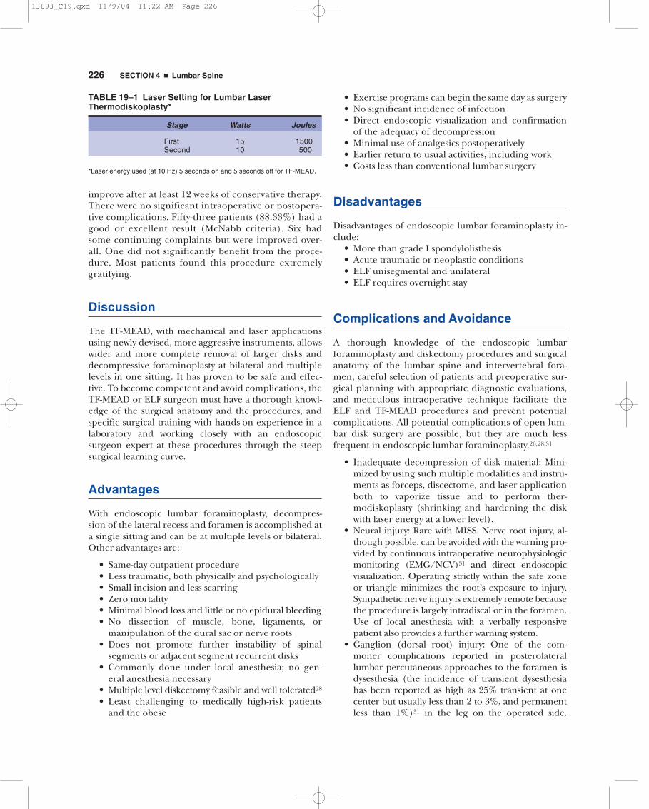
TABLE 19–1 Laser Setting for Lumbar LaserThermodiskoplasty*
Stage Watts Joules
First 15 1500Second 10 500
*Laser energy used (at 10 Hz) 5 seconds on and 5 seconds off for TF-MEAD.
13693_C19.qxd 11/9/04 11:22 AM Page 226

CHAPTER 19 � Endoscopic Lumbar Foraminoplasty 227
Careful technique guided by close endoscopic andC-arm fluoroscopic monitoring and knowledge ofthe surgical anatomy of the lumbar nerve root, dor-sal root ganglion, and foramen minimize this com-plication.
• Operating on wrong level: A major complication ofdisk surgery at any level of the spine is operating at
the wrong level. Proper use of digital C-arm fluo-roscopy for correct anatomical localization avoidsoperating at the wrong disk level. Routine painprovocation test and diskogram give additionalverification of the proper level.
• Infection: Avoided by careful sterile technique, themuch smaller incisional area, the absence of
A
C D
B
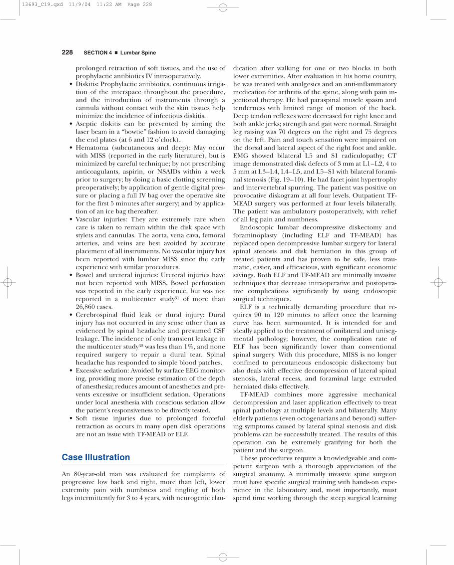
FIGURE 19–10 Axial CT images demonstrating severe bilateral lumbar foraminal stenosis secondary to posterior circumferentialdisk bulge/protrusion and facet osteophytic hypertrophy at (A) L1–L2, (B) L3–L4, (C) L4–L5, and (D) L5–S1.
13693_C19.qxd 11/9/04 11:22 AM Page 227
228 SECTION 4 � Lumbar Spine
prolonged retraction of soft tissues, and the use ofprophylactic antibiotics IV intraoperatively.
• Diskitis: Prophylactic antibiotics, continuous irriga-tion of the interspace throughout the procedure,and the introduction of instruments through acannula without contact with the skin tissues helpminimize the incidence of infectious diskitis.
• Aseptic diskitis can be prevented by aiming thelaser beam in a “bowtie” fashion to avoid damagingthe end plates (at 6 and 12 o’clock).
• Hematoma (subcutaneous and deep): May occurwith MISS (reported in the early literature), but isminimized by careful technique; by not prescribinganticoagulants, aspirin, or NSAIDs within a weekprior to surgery; by doing a basic clotting screeningpreoperatively; by application of gentle digital pres-sure or placing a full IV bag over the operative sitefor the first 5 minutes after surgery; and by applica-tion of an ice bag thereafter.
• Vascular injuries: They are extremely rare whencare is taken to remain within the disk space withstylets and cannulas. The aorta, vena cava, femoralarteries, and veins are best avoided by accurateplacement of all instruments. No vascular injury hasbeen reported with lumbar MISS since the earlyexperience with similar procedures.
• Bowel and ureteral injuries: Ureteral injuries havenot been reported with MISS. Bowel perforationwas reported in the early experience, but was notreported in a multicenter study31 of more than26,860 cases.
• Cerebrospinal fluid leak or dural injury: Duralinjury has not occurred in any sense other than asevidenced by spinal headache and presumed CSFleakage. The incidence of only transient leakage inthe multicenter study32 was less than 1%, and nonerequired surgery to repair a dural tear. Spinalheadache has responded to simple blood patches.
• Excessive sedation: Avoided by surface EEG monitor-ing, providing more precise estimation of the depthof anesthesia; reduces amount of anesthetics and pre-vents excessive or insufficient sedation. Operationsunder local anesthesia with conscious sedation allowthe patient’s responsiveness to be directly tested.
• Soft tissue injuries due to prolonged forcefulretraction as occurs in many open disk operationsare not an issue with TF-MEAD or ELF.
Case Illustration
An 80-year-old man was evaluated for complaints ofprogressive low back and right, more than left, lowerextremity pain with numbness and tingling of bothlegs intermittently for 3 to 4 years, with neurogenic clau-
dication after walking for one or two blocks in bothlower extremities. After evaluation in his home country,he was treated with analgesics and an anti-inflammatorymedication for arthritis of the spine, along with pain in-jectional therapy. He had paraspinal muscle spasm andtenderness with limited range of motion of the back.Deep tendon reflexes were decreased for right knee andboth ankle jerks; strength and gait were normal. Straightleg raising was 70 degrees on the right and 75 degreeson the left. Pain and touch sensation were impaired onthe dorsal and lateral aspect of the right foot and ankle.EMG showed bilateral L5 and S1 radiculopathy; CTimage demonstrated disk defects of 3 mm at L1–L2, 4 to5 mm at L3–L4, L4–L5, and L5–S1 with bilateral forami-nal stenosis (Fig. 19–10). He had facet joint hypertrophyand intervertebral spurring. The patient was positive onprovocative diskogram at all four levels. Outpatient TF-MEAD surgery was performed at four levels bilaterally.The patient was ambulatory postoperatively, with reliefof all leg pain and numbness.
Endoscopic lumbar decompressive diskectomy andforaminoplasty (including ELF and TF-MEAD) hasreplaced open decompressive lumbar surgery for lateralspinal stenosis and disk herniation in this group oftreated patients and has proven to be safe, less trau-matic, easier, and efficacious, with significant economicsavings. Both ELF and TF-MEAD are minimally invasivetechniques that decrease intraoperative and postopera-tive complications significantly by using endoscopicsurgical techniques.
ELF is a technically demanding procedure that re-quires 90 to 120 minutes to affect once the learningcurve has been surmounted. It is intended for andideally applied to the treatment of unilateral and uniseg-mental pathology; however, the complication rate ofELF has been significantly lower than conventionalspinal surgery. With this procedure, MISS is no longerconfined to percutaneous endoscopic diskectomy butalso deals with effective decompression of lateral spinalstenosis, lateral recess, and foraminal large extrudedherniated disks effectively.
TF-MEAD combines more aggressive mechanicaldecompression and laser application effectively to treatspinal pathology at multiple levels and bilaterally. Manyelderly patients (even octogenarians and beyond) suffer-ing symptoms caused by lateral spinal stenosis and diskproblems can be successfully treated. The results of thisoperation can be extremely gratifying for both thepatient and the surgeon.
These procedures require a knowledgeable and com-petent surgeon with a thorough appreciation of thesurgical anatomy. A minimally invasive spine surgeonmust have specific surgical training with hands-on expe-rience in the laboratory and, most importantly, mustspend time working through the steep surgical learning
13693_C19.qxd 11/9/04 11:22 AM Page 228

CHAPTER 19 � Endoscopic Lumbar Foraminoplasty 229
curve with an endoscopic spinal surgeon expert at thisprocedure.
REFERENCES
1. Hijikata S. Percutaneous nucleotomy: a new concept techniqueand 12 years’ experience. Clin Orthop. 1989;238:9–23.
2. Ascher PW. Application of the laser in neurosurgery. Laser SurgMed. 1986;2:91–97.
3. Choy DS. Percutaneous laser disc decompression (PLDD): twelveyears’ experience with 752 procedures in 518 patients. J Clin LaserMed Surg. 1998;16:325–331.
4. Kambin P, Saliffer PL. Percutaneous lumbar discectomy: reviewing100 patients and current practice. Clin Orthop. 1989;238:24–34.
5. Onik G, Maroon J, Davis G. Automated percutaneous discectomy: aprospective multi-institutional study. Neurosurgery. 1990;26:228–233.
6. Schreiber A, Suezawa Y, Leu HJ. Does percutaneous nucleotomywith discoscopy replace conventional discectomy? Eight years ofexperience and results in treatment of herniated lumbar disc. ClinOrthop. 1989;238:35–42.
7. Mayer HM, Brock M. Percutaneous endoscopic discectomy: surgi-cal technique and preliminary results compared to microsurgicaldiscectomy. J Neurosurg. 1993;78:216–225.
8. Savitz MH, Chiu JC, Yeung AT. History of minimalism in spinalmedicine and surgery. In: Savitz MH, Chiu JC, Yeung AD, eds. ThePractice of Minimally Invasive Spinal Technique. Richmond, VA:AAMISMS Education; 2000:1–12.
9. Jaikumar S, Kim DH, Kam A. History of minimally invasive spinesurgery. Neurosurgery. 2002;51(suppl 2):1–14.
10. Knight M, Goswami A, Patko J, Buxton N. Endoscopic foramino-plasty: an independent prospective evaluation. In: Gerber BE,Knight M, Seibert WE, eds. Laser in the Musculoskeletal System. NewYork: Springer-Verlag; 2001:320–329.
11. Savitz MH. Same day microsurgical arthroscopic lateral approachlaser assisted (SMALL) fluoroscopic discectomy. J Neurosurg. 1994;80:1039–1045.
12. Jaikumar S, Kim DH, Kam A. Minimally invasive spine instrumen-tation. Neurosurgery. 2002;51(suppl 2):15–22.
13. Perez-Cruet M, Fessler R, Perin N. Review: complications of minimallyinvasive spinal surgery. Neurosurgery. 2002;51(suppl 2):26–36.
14. Destandau J. Endoscopically assisted microdiscectomy. In: SavitzMH, Chiu JC, Yeung AD, eds. The Practice of Minimally InvasiveSpinal Technique. Richmond, VA: AAMISMS Education; 2000:187–
192.15. Chiu J, Clifford T, Princenthal R. The new frontier of minimally
invasive spine surgery through computer assisted technology. In:Lemke HU, Vannier MN, Invamura RD, eds. Computer AssistedRadiology and Surgery, CARS 2002. New York: Springer-Verlag; 2002:233–237.
16. Chiu J, Clifford T. Microdecompressive percutaneous discectomy:spinal discectomy with new laser thermodiskoplasty for nonextruded herniated nucleus pulposus. Surg Technol Int. 2000;8:343–351.
17. Chiu JC, Hansraj K, Akiyama C, Greenspan M. Percutaneous(endoscopic) decompressive discectomy for non-extruded cervi-cal herniated nucleus pulposus. Surg Technol Int. 1997;6:405–
411.18. Chiu JC, Clifford T, Greenspan M. Percutaneous microdecompres-
sive endoscopic cervical discectomy with laser thermodiskoplasty.Mt Sinai J Med. 2000;67:278–282.
19. Malis LI. Instrumentation and techniques in microsurgery. ClinNeurosurg. 1979;26:626–636.
20. Lin PM. Internal decompression for multiple levels of lumbarspinal stenosis: a technical note. Neurosurgery. 1982;11:546–549.
21. Caspar W, Campbell B, Barbier C, Kretschmmer R, Gottfried Y.The Caspar microsurgical discectomy and comparison with aconventional standard lumbar disc procedure. Neurosurgery. 1991;28:78–87.
22. Kambin P, Casey K, O’Brien E, Zhou I. Transforaminal arthroscopicdecompression of lateral recess stenosis J Neurosurg. 1996;84:462–467.
23. Atlas SJ, Keller RB, Robson D, Deyo RA, Singer DE. Surgical and non-surgical management of lumbar spinal stenosis: four-year outcomesfrom the Maine lumbar spine study. Spine. 2000;25:556–562.
24. Katz JN, Stucki G, Lipson SJ, Fossel AH, Grobler LJ, WeinsteinJ. Predictors of surgical outcome in degenerative lumbar spinalstenosis. Spine. 1999;24:2229–2233.
25. Haag M. Transforaminal endoscopic microdiscectomy: indicationsand short-term to intermediate-term results. Orthopade. 1999;28:615–621.
26. Khoo L, Fessler R. Microendoscopic decompressive laminotomyfor the treatment of lumbar stenosis. Neurosurgery. 2002;51(suppl 2):146–154.
27. Yeung AT, Tsou PM. Posterior lateral endoscopic excision for lumbardisc herniation: surgical technique, outcome, and complications.Spine. 2002;27:722–731.
28. Chiu JC, Clifford T. Multiple herniated discs at single and multiplespinal segments treated with endoscopic microdecompressivesurgery. J Minim Invasive Spinal Tech. 2001;1:15–19.
29. Knight M, Goswami A. Endoscopic laser foraminoplasty. In: SavitzMH, Chiu JC, Yeung AD. eds. The Practice of Minimally Invasive SpinalTechnique. Richmond, VA: AAMISMS Education; 2000:337–340.
30. Clifford T, Chiu JC, Rogers G. Neurophysiological monitoring ofperipheral nerve function during endoscopic laser discectomyJ Minim Invasive Spinal Tech. 2001;1:54–57.
31. Chiu JC, Clifford T, Savitz M, et al. Multicenter study of percuta-neous endoscopic discectomy (lumbar, cervical and thoracic).J Minim Invasive Spinal Tech. 2001;1:33–37.
32. Clifford TJ, Chiu JC, Batterjee KA. Transpinal approach forendoscopic discectomy at L5–S1. J Minim Invasive Spinal Tech. 2001;1:68–69.
13693_C19.qxd 11/9/04 11:22 AM Page 229