Embed Size (px)
Citation preview

25
MajorReview
Endogenous Endophthalmitis – A Review
Aji Sam Mathews MS, Gopal S Pillai MD, Natasha R MS, Mamta Shetty MS
Address for Correspondance: Amrita Institute of Medical Sciences. Kochi
Introduction
Metastatic Endophthalmitis is a potentially devastating albeit rare intraocular infection defined as inflammation of the anterior or posterior segment or both, with involvement of the adjacent ocular wall or as, an inflammatory reaction occurring as a result of intraocular colonization by bacteria, fungi, or rarely parasites [ESCRS]1.
Endophthalmitis can result from exogenous spread after intraocular surgery or trauma, or from adjacent structures. A less common form called endogenous endophthalmitis occurs due to hematogenous spread from a remote infectious site to the eye, resulting in intraocular colonization with inflammation, and subsequent severe loss of vision.
The incidence of endogenous endophthalmitis world wide is estimated to be 2 – 5 % of all cases of endophthalmitis2.Though the pathogen may vary between different geo- graphic areas, the most common bacterial organism is staphylococcus aureus, and the most common fungal organism is candida3.
Predisposing factors for endogenous endophthalmitis, ranges from chronic immune compromising illnesses[renal failure, diabetes mellitus] immunosuppressive diseases and treatment [post organ transplant, human immunodeficiency virus infection, long term intravenous catheters],recent invasive surgeries, gastrointestinal procedures, hepatobiliary tract infections, intravenous drug abuse, genitourinary infections, dental procedures4. Diagnosis is largely clinical with supplementary investigations like vitreous tap for smear and culture ,blood cultures.
The incidence of endogenous endophthalmitis has been rising due to longer life span and long term intravenous access. Though the final outcome in these cases is not very encouraging, prompt recognition, and aggressive treatment is the key to prevent severe visual loss in these patients.
This article attempts to review and concise the epidemiology, clinical findings, and management of endogenous endophthalmitis.
Classification of Endophthalmitis
Endophthalmitis has been defined in standard text books of ophthalmology as, the Inflammation of the anterior or posterior segment or both, with involvement of the adjacent ocular wall.
The European society of cataract and refractive surgeons
defines it as an inflammatory reaction occurring as a result of intraocular colonization by bacteria, fungi, or rarely parasites1.
Endophthalmitis is classified as Table 1.
Endogenous Endophthalmitis can be classified as in 5 Table 2 :
Table 1
Table 2
Epidemiology
Endogenous endophthalmitis, also termed metastatic endo- phthalmitis, comprises only a minority of endophthal- mitis cases, with an incidence rate of 2- 15%6, Puliafito et al in their series had reported an incidence of 2-5 %.Though metastatic endophthalmitis can occur at any age, Wong et al7 and connell et al8 have reported a mean age of 55 years and 57.5 years respectively, and has no sexual predilection but some series have reported a slightly more preponderance for males at 53.8 %8. The right eye is twice as often prone for a focus of infection than the left , because of the more proximal and direct blood flow to the right carotid artery5. Bilateral involvement occurs in approximately 25 % of cases9.

Vol. XXIII, No.1, Mar. 2011
26
Kerala Journal of Ophthalmology
Risk Factors
The risk factors for endogenous endophthalmitis can be varied, and with certain geographic predispositions. Common risk factors include Diabetes Mellitus, Malignancy, lymphoproliferative disorders, G.I tract infections, Immunosuppression, parenteral alimentation, recent extended surgical procedures, alcoholism, I.V drug abuse, catheters/UT.I, Endocarditis, Skin infections, Joint Infections, Hepatobiliary Tract Infections, COPD, End stage renal disorders.
Binder et al4, in their case series have found indwelling catheters/UTI as the primary risk factor, while Leibovitch et al10 had Diabetes Mellitus, COPD, End stage renal disease as the commonest risk factors. Okada et al11 had endocarditis and G.I tract infections, while Essman et al 12 found parenteral alimentation as the most common risk factor. Schielder et al13 in the case series of 21 patients found Indwelling catheters, Diabetes as the commonest risk factors, and PP Connell et al8 had a high incidence of IV drug abuse [38%] In their case series. In contrast to the above studies, Wong et al7 have found hepatobiliary tract infections and Diabetes as their commonest risk factors which they attributed to the higher incidence of hepatobiliary tract infections in their study population.
In our own experience with 12 cases[ 14 eyes] of metastatic endophthalmitis, with a mean follow up period of 5 months, Diabetes Mellitus[41.6 %] was the commonest risk factor, followed by liver pathology .Other risk factors were Malignancy, UTI, Liver transplant, and leukemia.
Etiology and Pathogenesis
Endogenous endophthalmitis can be caused by a spectrum of pathogens, ranging from gram positive bacteria ,to rarer organisms like citrobacter and non tuberculous mycobacteria.
Since it is metastatic in nature, the source of infection is from a site distant to the eye .The pathogen spreads hematogenously to the eye. Immuno compromised states such as diabetes mellitus , malignancy, patients on immmunosupression, are associated with reduced host defense and a high risk for endogenous endophthalmitis.
Retinal damage is multifactorial, which includes damage caused by microbial toxins and ischemia caused by septic emboli.
Gram positive organisms are responsible for 40% of cases9 and is more commonly seen in the western population11
Bacterial endophthalmitis Gram positive bacteria
Leibovitch et al 10 and Okada et al have found Streptococcus
spp as the commonest infective pathogen, while Staphylococcus sps. was the predominant organism in Jackson TC et al3 case series. Greenwald et al found a high incidence of Bacillus Cereus.
Hence the most common gram positive bacteria causing endogenous endophthalmitis is the Streptococcus spp.
Gram negative bacteria
Among Gram negative organisms, Jackson et al, Wong et al, and Vivian Schielder et al have reported Klebsiella as the single largest cause of metastatic endophthalmitis while Pseudomonas was the most frequent causative pathogen in the Leibovitch et al, and the Irvine WD et al 14 studies. Hence the commonest gram negative organism is Klebsiella pneumoniae, followed by Pseudomonas aeruginosa.Other offenders include E.coli,Serratia Marcescens,Listeria Monocytogens,and Neisseria Meningitides.
Fungal metastatic endophthalmitis
Studies on fungal endophthalmitis have shown the preponderance of aspergillus11,15 and candida10,13 as the most common pathogens .
Other causative fungi are, Pseudallecheria boydii, Histoplasma, Coccidoides, Blastomyces, Cryptococcus, Sporothrix, and Bipolar Hawaneseii among others.
Streptococcus pneomoniae and s.viridans are common causes of bacterial endogenous endophthalmitis secondary to meningitis and endocarditis respectively9. Group G streptococcal endophthalmitis has been reported in elderly patients with skin infections or malignant neoplasms , and group B streptococcal endophhtalmitis has been noted in neonates with meningitis and in immunocompromised adults16-18.Gram negative bacteria were found to be the most virulent7 .
In immonocompromised patients, intraocular infections represents dissemination of invasive diseases caused by candida species ,Aspergillus sps, Fusarium sps, Cryptococcus neoformans ,Pseudallescheria boydii and others19,20.
In our case series, Fungal metastatic endophthalmitis[50%] was slightly more commoner than bacterial[33.3%].
Clinical Findings
Endogenous endophthalmitis can occur a week, to a month after onset of systemic complaints /sepsis. Onset of symptoms was rapid in the case series of Okada et al11 and Schielder et al13 while it was more indolent in the Essman report.
Complaints of ocular pain, blurring of vision, ocular discharge, and photophobia , in a patient with the above risk factors, should be suspected to have Endogenous endophthalmitis. Signs suggestive of endophthalmitis include chemosis, flare

27
and cells in the anterior chamber, hypopyon, retinal changes such as Roth spots, and retinal periphlebitis (Figure 1). Ocular ultrasonography should be done if there is no view to the fundus, which can show vitreous opacities, exudates, membranes, retinochoroidal thickening and retinal detachment.
Systemic symptoms of sepsis include malaise, nausea, loss of appetite, and fever.
In Anterior focal disease, the anterior segment inflammation is mild, the infection is confined to discrete foci and may be seen as iris nodules or micro abscesses. On the other hand, in the diffuse type, the inflammation is more severe, with chemosis, lid swelling, corneal edema, fibrin in the anterior chamber, and hypopyon, with or without raised intraocular pressure.
Posterior focal disease presents as whitish nodules or plaques in the choroid which gradually involves the retina (Figure 2). Gram positive organisms may be multifocal, associated with Roth spots and retinal vasculitis, and tend to be severe. Gram negative infections usually cause a single large choroidal abscess involving the posterior pole9, with minimal injection of the conjunctiva, a relatively clear cornea, and mild to moderate anterior chamber reaction. Diffuse disease is more severe, with intense vitreous inflammation. Peri vascular hemorrhages inflammatory infiltrates and arterial emboli have been noted. This can further lead on to
retinal necrosis,detachment and perforation. Gram negative organisms like pseudomonas and Klebsiella can cause panophthalmitis.
Candidal endophthalmitis usually starts as a focal choroidits, gradually spreading to the retina, and breaking through to the vitreous, forming a string of pearls, characteristic of candida albicans (Figure 3).
Diagnosis/Investigations1. Hb,TC,DC,ESR.2. Blood/urine/csf/other cultures – Depending on suspected primary locus of infection.3. Chest X ray – To rule out pulmonary pathology.4. Echocardiography – If there is a possibility of endocarditis.5. CT/MRI/USG – Of the orbits for the diagnosis and monitoring of the response to the treatment of endogenous endophthalmitis in eyes with opaque media.6. Gallium -67 scan21 – May help in revealing areas of inflammation.7. Vitreous and aqueous tap – For fungal and bacterial smear and culture 8. Polymerase chain reaction.9. Semi-nested polymerase chain reaction - For rapid detection of panfungal genome directly from ocular specimens.
Aqueous tap can be obtained with a 26 G or 30 G stab incision at the limbus, aspirating 0.1 -0.2 ml of aqueous. Vitreous can be tapped using a 23 gauge needle at the end of a plastic 2cc syringe [0.2ml], through the pars plana, and intravitreal antibiotics delivered into the vitreous cavity through the same .Vitreous tap can also be done by using a 25 G 3 port pars plana vitrectomy ,with a tuberculin syringe attached to the end of the probe.
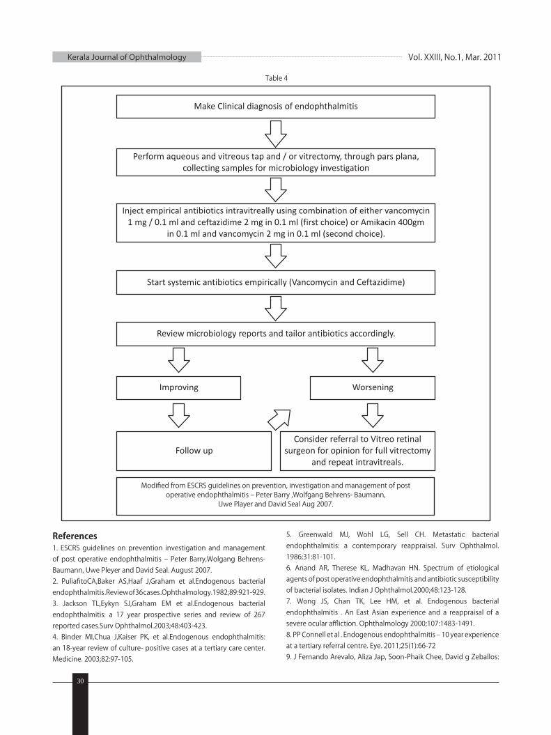
Treatment
The treatment of Metastatic endophthalmitis is the same as for the exogenous variety 1.The below given flow chart gives a overview of the line of management to be undertaken.
Aji Sam Mathews et al - Endogenous Endophthalmitis
Figure 1
Figure 2
Figure 3

Vol. XXIII, No.1, Mar. 2011
28
Kerala Journal of Ophthalmology

29
Role of intravitreal antibiotics
Intravitreal antibiotics are still the first line of treatment in all cases of endophthalmitis. Antibiotics can be given empirically to cover a broad spectrum of organisms or tailored against a particular species of pathogens.
The common antibiotics used intravitreally is given in Table 3
Table 3
The drugs of choice for gram positive and gram negative bacteria are ,vancomycin[ 1 mg/0.1 ml] and Ceftazidime [2 mg /0.1 ml ]
respectively.
Role of systemic antibiotics
Since the primary focus of infection is the source of the ocular infection, empiric broad spectrum antibiotic therapy with vancomycin and an aminoglycoside or a third generation cephalosporin is warranted22, in order to prevent continued bacteremia and reduce the chance of invasion of the other eye. The antibiotic therapy can be tailored depending on the culture reports. The drugs of choice against gram positive bacteria is Vancomycin, and is also preferred in patients known to abuse drugs covering the possibility of bacillus9. If patients history or culture suggest a fungal infection, amphotericin B, fluconazole,or itraconazole shold be included in the treatment.
Role of intravitreal steroids
Steroids have been used in bacterial endophthalmitis, because of their potential to suppress the associated inflammation, which plays a major role to play in the ocular damage associated with endophthalmitis. Intravitreal dexamethosone in the concentration of 400 microgram in 0.1 ml has been used but is contraindicated in fungal endophthalmitis.
Systemic steroids also have a role to play in the treatment of endophthalmitis, for the reasons mentioned above.
Role of vitrectomy
Vitrectomy in cases of endophthalmitis is useful, as it decreases the pathogen load, decreases the inflammatory mediators in the vitreous, ,adequate sampling is obtained for micro analysis, and it also helps in speedy visual recovery of the patient.
Timing of vitrectomy is controversial, with some investigators, for example Okada et al 11, and Yoon et al 23 favouring early vitrectomy. PP Connell eT al8 advocate aggressive early treatment with early vitrectomy in suspected bacterial metastatic endophthalmitis, and more conservative approach in suspected fungal cases. If a vitrectomy is performed, a 3 port pars plana complete vitrectomy is preferred over core vitrectomy. Complications like retinal detachment , hypotony and pthisis bulbi can occur in cases of vitrectomy performed in an inflamed eye, and have be borne in mind. Thus , vitrectomy definitely has a role to play in infections with virulent organisms, and in infections uncontrolled by systemic and /or intravitreal antibiotics, with definite indications for vitrectomy being, worsening of signs and symptoms, rapid progression, retinal necrosis, extensive subretinal abscess23, retinal detachment.
Visual acuity results
Among bacterial metastatic endophthalmitis, Vivian Schielder et al13 found 50% of patients has a final visual acuity of >20/400 ,and 50% PL/No PL.In the Okada et al study, 22 % of patients had a visual acuity of >= 20/400, and 38% PL /No PL.
In fungal endophthalmitis, the visual prognosis seems to be better, with Essmann et al reporting >65% of their patients with a final visual acuity of 20/400 and 30% PL/No PL.PP Connell et al also have reported fungal endophthalmitis patients faring better than their bacterial counterparts.
In our experience, fungal endophthalmitis patients had a worse initial visual acuity, and also final visual acuity, compared to the bacterial cases.
Conclusion
Metastatic endophthalmitis is a rare but potentially devastating intraocular infection occurring due to hematogenous spread from a remote infectious site to the eye, resulting in intraocular colonization with inflammation, and subsequent severe loss of vision. Patients with risk factors are prone to infection, with gram negative bacterial endophthalmitis patients faring worse than other infections. Early recognition of symptoms, and signs and prompt management with intravitreal antibiotics, systemic antibiotics and steroids along with vitrectomy is the key to minimize damage, and prevent involvement of the other eye.
Aji Sam Mathews et al - Endogenous Endophthalmitis
Vol. XXIII, No.1, Mar. 2011
30
Kerala Journal of Ophthalmology
Table 4
Modified from ESCRS guidelines on prevention, investigation and management of post operative endophthalmitis – Peter Barry ,Wolfgang Behrens- Baumann,
Uwe Player and David Seal Aug 2007.
References 1. ESCRS guidelines on prevention investigation and management of post operative endophthalmitis – Peter Barry,Wolgang Behrens- Baumann, Uwe Pleyer and David Seal. August 2007.2. PuliafitoCA,Baker AS,Haaf J,Graham et al.Endogenous bacterial endophthalmitis .Review of 36 cases .Ophthalmology.1982;89:921-929.3. Jackson TL,Eykyn SJ,Graham EM et al.Endogenous bacterial endophthalmitis: a 17 year prospective series and review of 267 reported cases.Surv Ophthalmol.2003;48:403-423.4. Binder MI,Chua J,Kaiser PK, et al.Endogenous endophthalmitis: an 18-year review of culture- positive cases at a tertiary care center.Medicine. 2003;82:97-105.
5. Greenwald MJ, Wohl LG, Sell CH. Metastatic bacterial endophthalmitis: a contemporary reappraisal. Surv Ophthalmol. 1986;31:81-101.6. Anand AR, Therese KL, Madhavan HN. Spectrum of etiological agents of post operative endophthalmitis and antibiotic susceptibility of bacterial isolates. Indian J Ophthalmol.2000;48:123-128.7. Wong JS, Chan TK, Lee HM, et al. Endogenous bacterial endophthalmitis . An East Asian experience and a reappraisal of a severe ocular affliction. Ophthalmology 2000;107:1483-1491.8. PP Connell et al . Endogenous endophthalmitis – 10 year experience at a tertiary referral centre. Eye. 2011;25(1):66-729. J Fernando Arevalo, Aliza Jap, Soon-Phaik Chee, David g Zeballos:

31
Endogenous Endophthalmitis in the Developing World. International Ophthalmology Clinics. 50(2) 173-187.10. Leibovitch I, Lai T, Raymond G,et al.Endogenous endophthalmitis: a 13 year review at a tertiary hospital in south Australia. Scand J Infect Dis. 2005;37:184-189.11. Okada AA, Johnson RP, Liles WC, et al. Endogenous bacterial endophthalmitis. Report of a ten year retrospective study. Ophthalmology. 1994;101:832-838.12. Essman TF, Flynn HW, Smiddy WE, et al. Treatment outcome in a 10-year study of endogenous fungal endophthalmitis. Ophthalmic Surg Lasers. 1997;28:185-194.13. Vivian Schielder:Culture proven endogenous endophhtalmitis: Clinical features and visual acuity outcomes. Am J Ophthalmol 2004;137:725-731.14.Irvine WD et al – Endogenous Endophthalmitis caused by gram negative organisms. Arch Ophthalmol. 1992;110[10]:1450-4.15. Chakrabarti A, Shivaprakash MR, Singh R, et al. Fungal endophthalmitis: fourteen year’s experience from a center in India. Retina. 2008[Epub ahead of print]16. Tan JH, Newman DK, Burton RL. Endogenous endophthalmitis
due to Group G streptococcus . Eye. 1999;13:116-117. 17. Nagelberg HP, Petashnick DE, To KW, et al. Group B streptococcal metastatic endophthalmitis. Am J Ophthalmol. 1994;117:498-500.18. Lee SY, Chee SP. Group B Streptococcus endogenous endophthalmitis: case reports and review of the literature. Ophthalmology. 2002;109:1879-1886.19. Graham DA, Kinyoun JL, George DP. Endogenous Aspergillus endophthalmitis after lung transplantation. Am J Ophthalmol. 1995;119:107-109.20. Sheu SJ, Chen YC, Kuo NW, et al. Endogenous cryptococcal endophhtalmitis.Ophthalmology. 1998;105:377-381.21. Kao PF, Tzen KY, Tsai MF, et al. Gallium-67 scanning in endogenous endophthalmitiswith unknown primary focus. Scand J Infect Dis. 2000;32:326-328.22. Romero CF, Rai MK, Lowder CY, et al.Endogenous endophthalmitis: case report and brief review. Am Fam Physian. 1999;60:510-514.23. Yoon YH, Lee SU, Sohn JH, et al. Result of early vitrectomy for endogenous Klebsiella pneumoniae endophthalmitis. Retina. 2003;23:366-370.
Aji Sam Mathews et al - Endogenous Endophthalmitis
Vol. XXIII, No.1, Mar. 2011
32
Kerala Journal of Ophthalmology