Embed Size (px)
Citation preview
Vol.4,n.1,pp.05-07 (Jan - Mar 2015) Journal of Surgical and Clinical Dentistry - JSCD
JSCD Openly accessible at http://www.mastereditora.com.br/jscd
ENDODONTIC TREATMENT IN 2ND PREMOLAR WITHANATOMICAL COMPLEXITY: CASE REPORT
RAISSA PEDROSO BATISTA1, PATRÍCIA DE ANGELO MATHEOS1, VANESSA RODRIGUES2, LUIZFERNANDO TOMAZINHO3
1. Undergraduate student of Dentistry, Paranaense University – UNIPAR; 2. Associate Professor of Graduation Course of Dentistry of the ParanaenseUniversity – UNIPAR; 3. Full Professor of Endodontics of Graduation Course of Dentistry of the Paranaense University – UNIPAR.
* Rua Inaja, 3560, Ap. 42, Centro, Umuarama, Paraná, Brazil. CEP: 87501-160. [email protected]
Received: 10/09/2014. Accepted: 24/10/2014
ABSTRACTThe lower second premolar presents commonly, simply, with asingle root, tapered and straight. However, different morphologicalaspects of this tooth root and canal are not uncommon occurrencesmay appear with bow root, double and canals with bifurcations inremote places to perform the complete cleaning and shaping, whilealso having the tooth inclination in the arcade one the factors ofdifficulty in performing coronary opening and the location of thecanal. In this case, it is of a lower second premolar having threecanals and two foramina opening where it was held, instrumenta-tion and root canal filling. Therefore, the purpose of the authors ofthis study is to describe step by step the case report of the patientwho possess the anatomical differentiation mentioned above, sothat it may contribute to the elucidation of common questionsamong endodontists and general clinical to the subject.
KEYWORDS: Endodontic treatment, second premolar, ana-tomical variation.
1. INTRODUCTIONThe lower premolars may represent one of the great
difficulties to perform a root canal treatment successfulwhen compared to other teeth because the canal haslarge anatomical variation, which associated with lack ofknowledge of root morphology leads to a high failurerate1. Their internal anatomy can present very complex,such that some authors claim that a root with taperedcanal and a single foramen is an exception and not arule2. Errors such as canal drift during instrumentation,several drilling or iatrogenic are commonly committeddue to lack of anatomical knowledge, leading some pa-tients to feel pain after the operation and/ or tooth loss.
For a correct procedure, it is necessary to clean,shape and filling the space of the canal in all its dimen-sions. There will be an adequate and satisfactory sealing,always assuming that the tooth may have roots and/ orextra canals: one canal and one foramen; one channelthat forks in the middle third forming two separate fo-ramina; one foramen that bifurcates at the apical third,
forming two separate foramina; two canals from the cer-vical third and apical third, forming two separate foram-ina; two canal that forking in any third of, or can befound with anatomical variations canals, that are blendedto form two or three canals3.
The objective of this study is to report the case of apatient who has as anatomical variation a lower premolarwith three channels from its initial radiograph to the fill-ing of the canal, with clinical and radiographic accom-paniment for a period of 6 months.
2. CASE REPORTPatient aged 40, serviced in the dental office, report-
ed sensitivity to touch in the posterior region of the leftmandible. In clinical and physical examination, the ele-ment 35 had to be sensitive to vertical and horizontalpercussion test, and clinically show a fistula in the apicalregion.
Radiographically, the element in a matter showed thepresence of an extensive radiolucent image in the apicalregion of the mesial root. After a careful history andphysical examination, the patient reported that the dentalelement was subjected to a replacement of an amalgamrestoration with a resin to four years ago. The digitalperiapical element proved the presence of a differentconventional anatomy. The main conduit was clear onlyuntil the end of the middle third of the root, disappearingto the apex. This image, suggested the presence of morethan one canal in the apical third of the root.
In the initial consultation, after the performance ofprophylaxis in the tooth 35, anesthesia of the region wascarried out using two tubes of mepivacaine (DFL. RJ-RJ.Brazil), with the total isolation of the element. We thencarried out the coronal opening and obtained access tothe mouth of the canal. For the initial operation of thecanal was used files type K-08 (Dentsply-Malleifer.
Batista et al. / J. Surg. Clin. Dent. V.4,n.1,pp.05-07 (Jan - Mar 2015)
JSCD Openly accessible at http://www.mastereditora.com.br/jscd
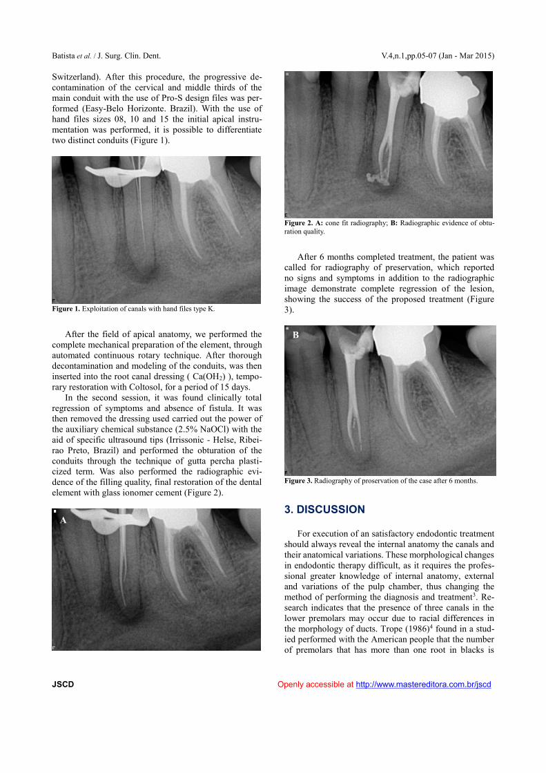
Switzerland). After this procedure, the progressive de-contamination of the cervical and middle thirds of themain conduit with the use of Pro-S design files was per-formed (Easy-Belo Horizonte. Brazil). With the use ofhand files sizes 08, 10 and 15 the initial apical instru-mentation was performed, it is possible to differentiatetwo distinct conduits (Figure 1).
Figure 1. Exploitation of canals with hand files type K.
After the field of apical anatomy, we performed thecomplete mechanical preparation of the element, throughautomated continuous rotary technique. After thoroughdecontamination and modeling of the conduits, was theninserted into the root canal dressing ( Ca(OH2) ), tempo-rary restoration with Coltosol, for a period of 15 days.
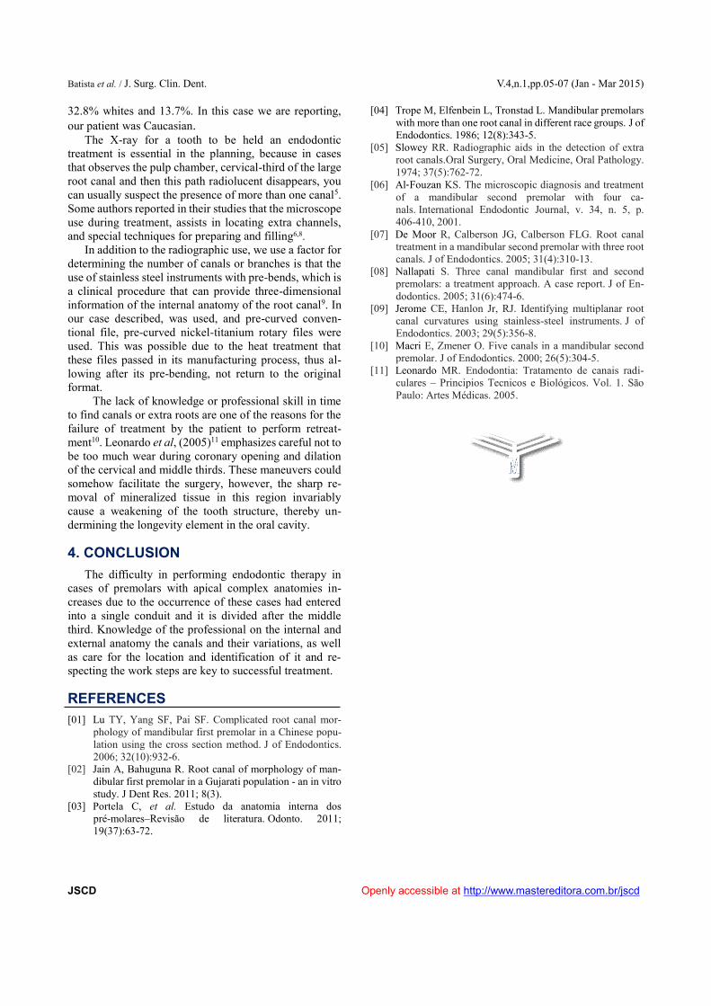
In the second session, it was found clinically totalregression of symptoms and absence of fistula. It wasthen removed the dressing used carried out the power ofthe auxiliary chemical substance (2.5% NaOCl) with theaid of specific ultrasound tips (Irrissonic - Helse, Ribei-rao Preto, Brazil) and performed the obturation of theconduits through the technique of gutta percha plasti-cized term. Was also performed the radiographic evi-dence of the filling quality, final restoration of the dentalelement with glass ionomer cement (Figure 2).
Figure 2. A: cone fit radiography; B: Radiographic evidence of obtu-ration quality.
After 6 months completed treatment, the patient wascalled for radiography of preservation, which reportedno signs and symptoms in addition to the radiographicimage demonstrate complete regression of the lesion,showing the success of the proposed treatment (Figure3).
Figure 3. Radiography of proservation of the case after 6 months.
3. DISCUSSION
For execution of an satisfactory endodontic treatmentshould always reveal the internal anatomy the canals andtheir anatomical variations. These morphological changesin endodontic therapy difficult, as it requires the profes-sional greater knowledge of internal anatomy, externaland variations of the pulp chamber, thus changing themethod of performing the diagnosis and treatment3. Re-search indicates that the presence of three canals in thelower premolars may occur due to racial differences inthe morphology of ducts. Trope (1986)4 found in a stud-ied performed with the American people that the numberof premolars that has more than one root in blacks is
A
B
Batista et al. / J. Surg. Clin. Dent. V.4,n.1,pp.05-07 (Jan - Mar 2015)
JSCD Openly accessible at http://www.mastereditora.com.br/jscd
32.8% whites and 13.7%. In this case we are reporting,our patient was Caucasian.
The X-ray for a tooth to be held an endodontictreatment is essential in the planning, because in casesthat observes the pulp chamber, cervical-third of the largeroot canal and then this path radiolucent disappears, youcan usually suspect the presence of more than one canal5.Some authors reported in their studies that the microscopeuse during treatment, assists in locating extra channels,and special techniques for preparing and filling6,8.
In addition to the radiographic use, we use a factor fordetermining the number of canals or branches is that theuse of stainless steel instruments with pre-bends, which isa clinical procedure that can provide three-dimensionalinformation of the internal anatomy of the root canal9. Inour case described, was used, and pre-curved conven-tional file, pre-curved nickel-titanium rotary files wereused. This was possible due to the heat treatment thatthese files passed in its manufacturing process, thus al-lowing after its pre-bending, not return to the originalformat.
The lack of knowledge or professional skill in timeto find canals or extra roots are one of the reasons for thefailure of treatment by the patient to perform retreat-ment10. Leonardo et al, (2005)11 emphasizes careful not tobe too much wear during coronary opening and dilationof the cervical and middle thirds. These maneuvers couldsomehow facilitate the surgery, however, the sharp re-moval of mineralized tissue in this region invariablycause a weakening of the tooth structure, thereby un-dermining the longevity element in the oral cavity.
4. CONCLUSIONThe difficulty in performing endodontic therapy in
cases of premolars with apical complex anatomies in-creases due to the occurrence of these cases had enteredinto a single conduit and it is divided after the middlethird. Knowledge of the professional on the internal andexternal anatomy the canals and their variations, as wellas care for the location and identification of it and re-specting the work steps are key to successful treatment.
REFERENCES[01] Lu TY, Yang SF, Pai SF. Complicated root canal mor-
phology of mandibular first premolar in a Chinese popu-lation using the cross section method. J of Endodontics.2006; 32(10):932-6.
[02] Jain A, Bahuguna R. Root canal of morphology of man-dibular first premolar in a Gujarati population - an in vitrostudy. J Dent Res. 2011; 8(3).
[03] Portela C, et al. Estudo da anatomia interna dospré-molares–Revisão de literatura. Odonto. 2011;19(37):63-72.
[04] Trope M, Elfenbein L, Tronstad L. Mandibular premolarswith more than one root canal in different race groups. J ofEndodontics. 1986; 12(8):343-5.
[05] Slowey RR. Radiographic aids in the detection of extraroot canals.Oral Surgery, Oral Medicine, Oral Pathology.1974; 37(5):762-72.
[06] Al‐Fouzan KS. The microscopic diagnosis and treatmentof a mandibular second premolar with four ca-nals. International Endodontic Journal, v. 34, n. 5, p.406-410, 2001.
[07] De Moor R, Calberson JG, Calberson FLG. Root canaltreatment in a mandibular second premolar with three rootcanals. J of Endodontics. 2005; 31(4):310-13.
[08] Nallapati S. Three canal mandibular first and secondpremolars: a treatment approach. A case report. J of En-dodontics. 2005; 31(6):474-6.
[09] Jerome CE, Hanlon Jr, RJ. Identifying multiplanar rootcanal curvatures using stainless-steel instruments. J ofEndodontics. 2003; 29(5):356-8.
[10] Macri E, Zmener O. Five canals in a mandibular secondpremolar. J of Endodontics. 2000; 26(5):304-5.
[11] Leonardo MR. Endodontia: Tratamento de canais radi-culares – Principios Tecnicos e Biológicos. Vol. 1. SãoPaulo: Artes Médicas. 2005.