Embed Size (px)
Citation preview
Endodontic microsurgery. Part two: armamentarium and techniqueSarah Jadun,1 Liam Monaghan2 and James Darcey*3
Introduction
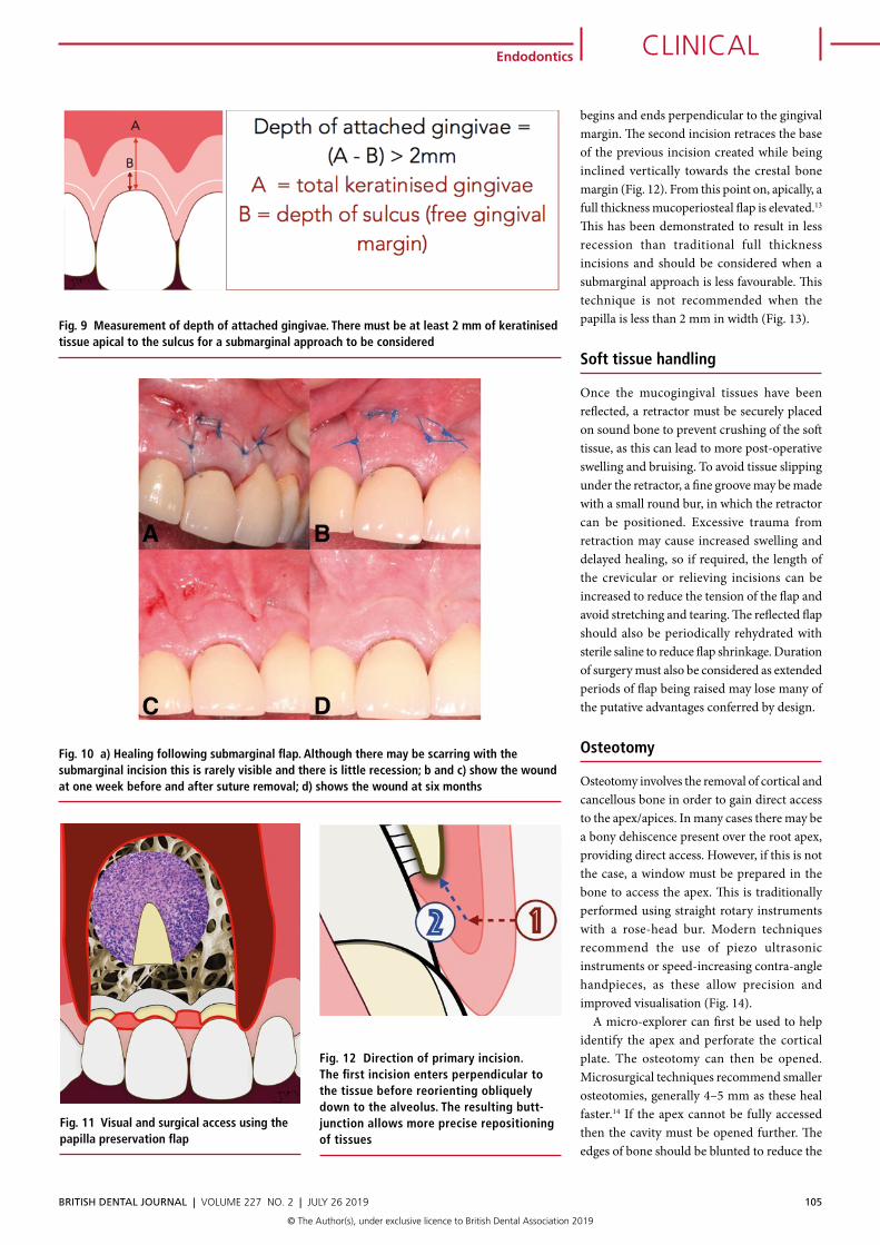
Modern endodontic microsurgery has a reported success rate of up to 93.5% through reduction of periapical lesion size rather than complete healing, and a success rate of up to 70–80% using strict criteria, making it a viable treatment option in the management of periapical disease when orthograde root treatment is not possible or inappropriate.1,2 Such high success rates are intimately related to advanced training in techniques that have allowed dentists to overcome the historical barriers to success seen in traditional surgical endodontics.3 Table 1 highlights some of the key differences between traditional techniques and endodontic microsurgery.3
Armamentarium
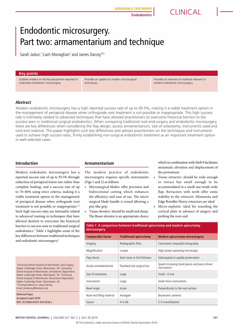
The modern practice of endodontic microsurgery requires specific instruments (Figs 1 and 2) as follows:• Microsurgical blades: offer precision and
bidirectional cutting which enhances the efficiency and ease of use. The micro surgical blade handle is round allowing a pen-like grip
• Tissue elevator: should be small and sharp. The Buser elevator is an appropriate choice
which in combination with Molt 9 facilitates atraumatic elevation and displacement of the periosteum
• Tissue retractor: should be wide enough to retract but small enough to be accommodated in a small one-tooth-wide flap. Retractors with teeth offer some stability to the retractor. Minnesota and Edge Bowdler Henry retractors are ideal
• Micro-explorer: ideal for sounding the cortical plate in advance of surgery and probing the root-end
Updates readers on the key equipment required to undertake endodontic microsurgery.
Provides an update on modern microsurgical techniques.
Provides an overview of materials relevant to modern endodontic microsurgery.
Key points
AbstractModern endodontic microsurgery has a high reported success rate of up to 93.5%, making it a viable treatment option in the management of periapical disease when orthograde root treatment is not possible or inappropriate. This high success rate is intimately related to advanced techniques that have allowed practitioners to overcome historical barriers to the success seen in traditional surgical endodontics. When comparing traditional root-end surgery and endodontic microsurgery there are key differences when considering the flap design, access armamentarium, size of osteotomy, instruments used and root-end material. This paper highlights such key differences and advises practitioners on the techniques and instruments used to achieve high success rates, firmly establishing non-surgical endodontic treatment as an important treatment option in well-selected cases.
1University Dental Hospital of Manchester, Oral Surgery, Higher Cambridge Street, Manchester, UK; 2University Dental Hospital of Manchester, Orthodontic Department, Higher Cambridge Street, Manchester, UK. 3University Dental Hospital of Manchester, Restorative Department, Higher Cambridge Street, Manchester, UK. *Correspondence to: James Darcey Email: [email protected]
Refereed Paper.Accepted 4 April 2019DOI: 10.1038/s41415-019-0516-z
Comparable factor Traditional apicectomy Modern apicectomy microsurgery
Imaging Radiographic films Cone beam computed tomography
Magnification Loupes High-power operating microscope
Flap choice Semi-lunar or full thickness Submarginal or papilla preservation
Access armamentarium Standard size surgical bur Speed increasing hand pieces and piezo-driven instruments
Size of osteotomy Large Small: >5 mm
Instruments Large Small micro-instruments
Bevel angle Acute Perpendicular to the root surface
Root-end filling material Amalgam Bioceramic cements
Suture 4-0 silk 5-0 monofilament
Table 1 A comparison between traditional apicectomy and modern apicectomy microsurgery
BRITISH DENTAL JOURNAL | VOLUME 227 NO. 2 | JULy 26 2019 101
CLINICALEndodontics
VERIFIABLE CPD PAPER
© The Author(s), under exclusive licence to British Dental Association 2019
• Micro-curettes: for lifting soft tissue from the cavity and winkling out root tips
• Micro-mirror: for inspection of the osteotomy, retro-preparation and obturation. These may be small front surface reflecting mirrors or stainless steel
• Micro-plugger: for compacting GP within the apex and compacting the root-end-filling.
Clinicians may also choose specialised instruments for suturing such as Castroviejo or Barraquer needle holders and Lachal scissors. Though not strictly necessary, these fine instruments are invaluable when manipulating 5-0 or 6-0 sutures.
Local anaesthesia
Anxious patients should be reassured that apical surgery will not be an uncomfortable procedure if undertaken carefully. This is important as the anticipation of pain can serve to heighten the level of pain experienced during the procedure.4 This last point is often overlooked, but not only does lasting anaesthesia provide immediate post-operative pain relief but reduces longer-term pain by reducing central sensitisation.5
Properties of the most commonly used local anaesthetic solutions are displayed in Table 2.
Often a combination of an infra-orbital or inferior alveolar nerve block, alongside an infiltration of solution at the surgical site, will provide good anaesthesia and haemostasis. As many lesions expand to, or through the palatal/lingual plates, supplemental anaesthesia is essential in these areas. While articaine provides no benefit over lidocaine when used
for inferior alveolar nerve blocks,6 articaine has been shown to be superior to lidocaine in providing successful anaesthesia using buccal infiltrations, and a buccal infiltration of articaine acts to enhance the effectiveness of an inferior alveolar nerve block made with lidocaine.7 The vasoconstrictor felypressin is used as an alternative to adrenaline in some solutions; however, its haemostatic properties are no greater to that of adrenaline. For this reason Scandonest and Citanest should not be considered anaesthetics of choice. Bupivacaine is longer lasting and would make for a very useful anaesthetic were it readily available.
Magnification and illumination
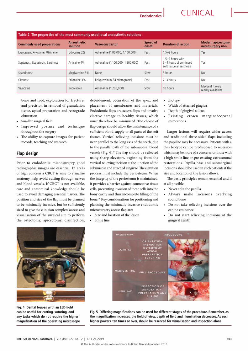
Operating microscopes have become essential to visualise the effectiveness of the disinfection, debridement, membrane and scaffold placement, root canal obturation
and endodontic microsurgery steps, which are essential to the successful outcome of endodontic microsurgery.8 Historically, loupes have been the most common form of magnification, and they can still be useful to help visualise dental tissues, but they cannot provide the higher magnifications that the clinician needs for detailed endodontic microsurgery. Moreover, a microscope allows the clinician to maintain an upright posture. When using a microscope, different magnification ranges are recommended depending on the stage of surgical endodontic treatment. A further advantage of loupe magnification or microscopy is the incorporation of LED illumination. Head-mounted and in-built lights support excellent visualisation of the operating field (Figs 3, 4, 5). An operating microscope fitted with a digital camera permits:• More meticulous inspection of the surgical
field, facilitating the distinction between
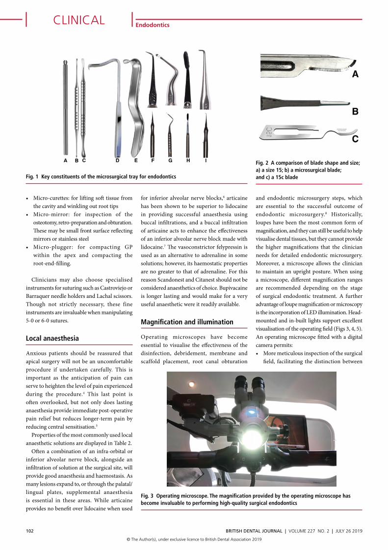
Fig. 2 A comparison of blade shape and size; a) a size 15; b) a microsurgical blade; and c) a 15c bladeFig. 1 Key constituents of the microsurgical tray for endodontics
Fig. 3 Operating microscope. The magnification provided by the operating microscope has become invaluable to performing high-quality surgical endodontics
102 BRITISH DENTAL JOURNAL | VOLUME 227 NO. 2 | JULy 26 2019
CLINICAL Endodontics
© The Author(s), under exclusive licence to British Dental Association 2019
bone and root, exploration for fractures and precision in removal of granulation tissue, apical preparation and retrograde obturation
• Smaller surgical field• Improved posture and technique
throughout the surgery• The ability to capture images for patient
records, teaching and research.
Flap design
Prior to endodontic micoosurgery good radiographic images are essential. In areas of high concern a CBCT is wise to visualise anatomy, help avoid cutting through nerves and blood vessels. If CBCT is not available, care and anatomical knowledge should be used to avoid damaging essential tissues. The position and size of the flap must be planned to be minimally-invasive, but be sufficiently sized to give the clinician complete access and visualisation of the surgical site to perform the osteotomy, apicectomy, disinfection,
debridement, obturation of the apex, and placement of membranes and materials. Endodontic flaps are access flaps and involve elective damage to healthy tissues, which must therefore be minimised. The choice of flap design should allow the maintenance of a sufficient blood supply to all parts of the soft tissues. Vertical relieving incisions must be near parallel to the long axis of the teeth, due to the parallel path of the submucosal blood vessels (Fig. 6).9 The flap should be reflected using sharp elevators, beginning from the vertical relieving incision at the junction of the submucosa and attached gingivae. The elevation process must include the periosteum. When the integrity of the periosteum is maintained, it provides a barrier against connective tissue cells, preventing invasion of these cells into the bony cavity and thus incomplete filling of the bone.10 Key considerations for positioning and planning the minimally-invasive endodontic microsurgery access flap are:• Size and location of the lesion• Smile line
• Biotype• Width of attached gingiva• Depth of gingival sulcus• Exist ing crown margins/coronal
restorations.
Larger lesions will require wider access and traditional three-sided flaps including the papillae may be necessary. Patients with a thin biotype can be predisposed to recession which may be more of a concern for those with a high smile line or pre-existing extracoronal restorations. Papilla base and submarginal incisions should be used in such patients if the size and location of the lesion allows.
The basic principles remain essential and if at all possible:• Never split the papilla• Always make incisions overlying
sound bone• Do not take relieving incisions over the
canine eminence• Do not start relieving incisions at the
gingival zenith
Commonly used preparations Anaesthetic solution Vasoconstrictor Speed of
onset Duration of action Modern apicectomy microsurgery use?
Lignospan, Xylocaine, Utilicaine Lidocaine 2% Adrenaline (1:80,000; 1:100,000) Fast 1.5–2 hours Yes
Septanest, Espestesin, Bartinest Articaine 4% Adrenaline (1:100,000; 1:200,000) Fast1.5–2 hours with 3–4 hours of continued soft tissue anaesthesia
Yes
Scandonest Mepivacaine 3% None Slow 3 hours No
Citanest Prilocaine 3% Felypressin (0.54 micrograms) Fast 2–3 hours No
Vivacaine Bupivacain Adrenaline (1:200,000) Slow 10 hours Maybe if it were readily available!
Table 2 The properties of the most commonly used local anaesthetic solutions
Fig. 5 Differing magnifications can be used for different stages of the procedure. Remember, as the magnification increases, the field of view, depth of field and illumination decreases. As such higher powers, ten times or over, should be reserved for visualisation and inspection alone
Fig. 4 Dental loupes with an LED light can be useful for cutting, suturing, and any tasks which do not require the higher magnification of the operating microscope
BRITISH DENTAL JOURNAL | VOLUME 227 NO. 2 | JULy 26 2019 103
CLINICALEndodontics
© The Author(s), under exclusive licence to British Dental Association 2019

• Avoid relieving incisions that cross the apices of the lower premolar teeth.
Conventional crevicular incision
Conventional flaps can be classified by the number of sides created by relieving incisions. Flaps requiring crevicular incisions (Fig. 7) may carry an increased risk of gingival recession post-operatively; therefore, meticulous surgical technique is required.11
An envelope flap is a single sided flap raised when a crevicular incision is made to include multiple teeth. Envelope flaps rarely allow enough tissue mobility for apical surgery and it is necessary to incorporate one or two relieving incisions which should extend from the point of incision away from the apex to make the flap slightly trapezoidal in shape.
Triangular (two-sided) flaps utilise a single reliving incision, mainly reserved for posterior teeth. The edges of two-sided flaps reposition easily and the shape allows a good blood supply to be maintained. However, access can still prove challenging, especially if the extent of the lesion is unknown. The rectangular (three-sided) flap involves two relieving incisions, allowing better access and visualisation. It also minimises flap tension and the risk of tearing; this may be preferable for anterior teeth.
Submarginal or Leubke-Oschenbein incision
Submarginal flaps consist of two vertical incisions connected by a scalloped horizontal incision, roughly parallel to the marginal contour of the gingiva (Fig. 8). Before using
this type of incision, it is important to determine whether there is sufficient attached gingiva and a minimum of 2 mm considered as sufficient (Fig. 9).12 Any incision must also remain on sound bone to facilitate healing. This may not be possible in larger lesions with extensive buccal plate erosion and such tissue breakdown will lead to major recession and an unaesthetic result. This technique is associated with scar tissue formation; however, this is less of a concern if not visible when the patient smiles. When performed with appropriate planning and surgical technique, it is easy to achieve precise primary closure. This method guarantees good surgical access and keeps the incision free of marginal tissue, minimising the risk of gingival recession (Fig. 10).
Papilla base preservation technique
This technique involves preservation of the entire papilla, thus eliminating any loss of height as a result of the surgical or healing process (Fig. 11). This may require considerable surgical finesse. This flap consists of two releasing vertical incisions, connected by the papilla-base incision and intrasulcular incision in the cervical area of the tooth. The papilla-base incision requires two different incisions at the base of the papilla. The first shallow incision severs the epithelium and connective tissue to the depth of 1.5 mm from the surface of the gingiva. The path is a curved line, connecting one side of the papilla to the other. The incision
Fig. 6 Direction of blood supply to gingival tissues and placement of relieving incisions. The capillary supply to the gingival tissues run vertically thus wide, trapezoidal incisions will cut more vessels and result in greater bleeding. As such, vertical or near relieving incisions should be favoured unless the lesion size demands wider access
Fig. 7 a) Visual and surgical access using two-sided flap; b) visual and surgical access using three-sided flap. The two-sided or triangular flap may limit surgical damage but may not allow full visualisation of the lesion. The three-sided flap offers greater visibility but comes at the cost of greater collateral damage
Fig. 8 Visual and surgical access using submarginal flap. The submarginal incision is remote to the aesthetic soft-tissue zone and allows accurate repositioning but is dependent upon sufficient keratinised tissue. It can be inadvertently be made over a large lesion but may result in scar formation
104 BRITISH DENTAL JOURNAL | VOLUME 227 NO. 2 | JULy 26 2019
CLINICAL Endodontics
© The Author(s), under exclusive licence to British Dental Association 2019
begins and ends perpendicular to the gingival margin. The second incision retraces the base of the previous incision created while being inclined vertically towards the crestal bone margin (Fig. 12). From this point on, apically, a full thickness mucoperiosteal flap is elevated.13 This has been demonstrated to result in less recession than traditional full thickness incisions and should be considered when a submarginal approach is less favourable. This technique is not recommended when the papilla is less than 2 mm in width (Fig. 13).
Soft tissue handling
Once the mucogingival tissues have been reflected, a retractor must be securely placed on sound bone to prevent crushing of the soft tissue, as this can lead to more post-operative swelling and bruising. To avoid tissue slipping under the retractor, a fine groove may be made with a small round bur, in which the retractor can be positioned. Excessive trauma from retraction may cause increased swelling and delayed healing, so if required, the length of the crevicular or relieving incisions can be increased to reduce the tension of the flap and avoid stretching and tearing. The reflected flap should also be periodically rehydrated with sterile saline to reduce flap shrinkage. Duration of surgery must also be considered as extended periods of flap being raised may lose many of the putative advantages conferred by design.
Osteotomy
Osteotomy involves the removal of cortical and cancellous bone in order to gain direct access to the apex/apices. In many cases there may be a bony dehiscence present over the root apex, providing direct access. However, if this is not the case, a window must be prepared in the bone to access the apex. This is traditionally performed using straight rotary instruments with a rose-head bur. Modern techniques recommend the use of piezo ultrasonic instruments or speed-increasing contra-angle handpieces, as these allow precision and improved visualisation (Fig. 14).
A micro-explorer can first be used to help identify the apex and perforate the cortical plate. The osteotomy can then be opened. Microsurgical techniques recommend smaller osteotomies, generally 4–5 mm as these heal faster.14 If the apex cannot be fully accessed then the cavity must be opened further. The edges of bone should be blunted to reduce the
Fig. 12 Direction of primary incision. The first incision enters perpendicular to the tissue before reorienting obliquely down to the alveolus. The resulting butt-junction allows more precise repositioning of tissues
Fig. 10 a) Healing following submarginal flap. Although there may be scarring with the submarginal incision this is rarely visible and there is little recession; b and c) show the wound at one week before and after suture removal; d) shows the wound at six months
Fig. 11 Visual and surgical access using the papilla preservation flap
Fig. 9 Measurement of depth of attached gingivae. There must be at least 2 mm of keratinised tissue apical to the sulcus for a submarginal approach to be considered
BRITISH DENTAL JOURNAL | VOLUME 227 NO. 2 | JULy 26 2019 105
CLINICALEndodontics
© The Author(s), under exclusive licence to British Dental Association 2019

risk of sequestration. There must be at least 2–3 mm of crestal bone remaining in the cavity following preparation to reduce the risk of recession and provide appropriate periodontal support for the tooth.
When cutting through bone, careful consideration must be given to potential trauma inflicted on the tissue through mechanical forces, and also through the generation of heat. The tissues may be even more vulnerable to injury following reduction in blood supply due to the vasoconstrictive nature of the adrenaline in local anaesthetic. The threshold for irreversible damage to bone is at 47 degrees Celsius. The use of water or saline coolant applied directly to the cutting surface will reduce the rise in temperature, and limit or prevent permanent damage.15 Table 3 highlights some of the principal challenges to the ideal microsurgical cases.
Once the root apex has been accessed, all soft granulation tissue present within the bony crypt must be removed with sharp curettes. It may be sensible to send this tissue for histopathological analysis. Occasionally this can be challenging and the apex may require amputation before full curettage is possible.
Resection of the root
Once the apical root is visible, it should be inspected for any sign of fractures, which may no longer make the tooth a candidate for apicectomy and make extraction unavoidable. If there appears to be no radiographic or visible microscopic complications, apicectomy may proceed by removing 3 mm of the root. This 3mm will include the apical delta and the majority of lateral canals which are impossible to disinfect, and can harbour bacterial and necrotic tissues.3,16 Greater than 3 mm of the root can be removed if there are anatomical variations, separated instruments or perforations, provided that the tooth has an
adequate remaining root length to be stable.16
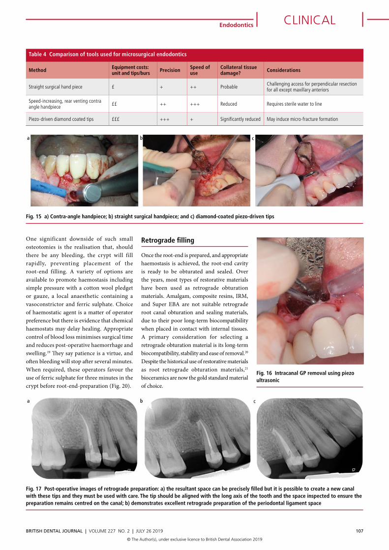
The root tip should be resected perpendicular to the long axis of the tooth, demonstrating a zero-degree bevel. This allows a 90 degree cavo-surface margin for the root-end filling. Bevelling the tip is not recommended as it exposes more dentine tubules, allowing the communication of any remaining intra-radicular microorganisms and extra-radicular nutrients. There are three principle tools for root-end resection, traditional straight surgical handpieces, speed increasing, rear-venting contra-angle handpieces and piezo tips (Table 4 compares the techniques). The use of the piezo for resection as well as osteotomy and apical preparation offers many advantages (Fig. 15), not least the simplification of the surgical tray and preparation; one machine to rule them all!
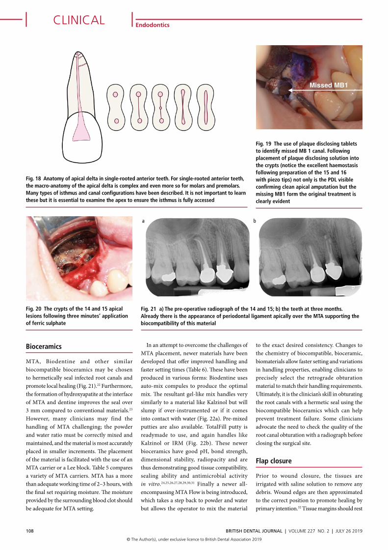
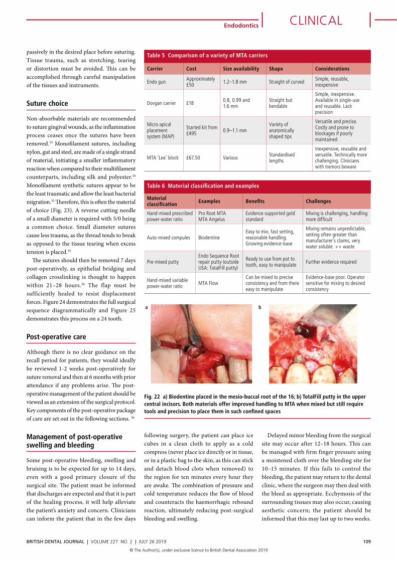
Once resected, the apical cavity requires preparation. The depth of the root-end preparation should be at least 3 mm in order to optimise the seal provided by the root-end filling.17 Piezo/ultrasonic tips demonstrate a
higher success rate when used to prepare the root-end when compared to burs (Figs 16 and 17).18 The osteotomy should therefore be large enough to allow the insertion and engagement of the exposed canal with the instrument. The surgeon should have unimpeded visual access throughout. The roots of molar and premolar teeth must be inspected for the presence of isthmuses and fins connecting the canals (Fig. 18). If present, these should be debrided with the piezo tips. Plaque-disclosing solution can be placed in the crypt and this will stain the periodontal ligament and isthmus, providing confirmation the full root tip has been resected and helping to identify the anatomy (Fig. 19). Once the retropreparation is complete the canal should be inspected with the micro-mirror. Micro pluggers may be used to compact any GP that has become softened with the ultrasonics.
Crypt management
Following root resection, the bony crypt must be kept dry to allow for obturation.
Factors Complication Treatment considerations
Size of periapical lesion Extension of lesion to adjacent roots of teeth or both cortical plates are eroded
Soft tissue should be gently curetted as far as is accessible with care over the apices and not to perforate palatally
Maxillary sinusRisk of perforation of the Schneiderian membrane, and displacement of materials into the sinus
If, from radiographic assessment, the apex lies in the sinus the osteotomy should begin more inferiorly and proceed with caution. Root amputation should be performed with piezo ultrasonic tips to minimise damage to the sinus lining and the suction on hand to capture the apical fragment if necessary. Care should be taken to avoid displacement of either the root tip or retrograde filling material into the sinus
ID nerveMechanical compression, chemical neurotoxicity and local infection may cause nerve damage
All surgery planned in the lower premolar region should be supported with CBCT to determine proximity to the nerve and formally consent the patient to the risks. The mental nerve should be identified and protected
Table 3 Challenges to the ideal microsurgical cases
Fig. 13 A two sided papilla preservation flap at surgery and at three-month review. There is minimal recession and scarring
Fig. 14 The use of piezo driven diamond-coated tips allows safe and minimally traumatic osteotomy and avoids the need for a separate straight surgical handpiece. It is, however, more time consuming
106 BRITISH DENTAL JOURNAL | VOLUME 227 NO. 2 | JULy 26 2019
CLINICAL Endodontics
© The Author(s), under exclusive licence to British Dental Association 2019
One significant downside of such small osteotomies is the realisation that, should there be any bleeding, the crypt will fill rapidly, preventing placement of the root-end filling. A variety of options are available to promote haemostasis including simple pressure with a cotton wool pledget or gauze, a local anaesthetic containing a vasoconstrictor and ferric sulphate. Choice of haemostatic agent is a matter of operator preference but there is evidence that chemical haemostats may delay healing. Appropriate control of blood loss minimises surgical time and reduces post-operative haemorrhage and swelling.19 They say patience is a virtue, and often bleeding will stop after several minutes. When required, these operators favour the use of ferric sulphate for three minutes in the crypt before root-end-preparation (Fig. 20).
Retrograde filling
Once the root-end is prepared, and appropriate haemostasis is achieved, the root-end cavity is ready to be obturated and sealed. Over the years, most types of restorative materials have been used as retrograde obturation materials. Amalgam, composite resins, IRM, and Super EBA are not suitable retrograde root canal obturation and sealing materials, due to their poor long-term biocompatibility when placed in contact with internal tissues. A primary consideration for selecting a retrograde obturation material is its long-term biocompatibility, stability and ease of removal.20 Despite the historical use of restorative materials as root retrograde obturation materials,21 bioceramics are now the gold standard material of choice.
Method Equipment costs: unit and tips/burs Precision Speed of
useCollateral tissue damage? Considerations
Straight surgical hand piece £ + ++ Probable Challenging access for perpendicular resection for all except maxillary anteriors
Speed-increasing, rear venting contra angle handpiece ££ ++ +++ Reduced Requires sterile water to line
Piezo-driven diamond coated tips £££ +++ + Significantly reduced May induce micro-fracture formation
Table 4 Comparison of tools used for microsurgical endodontics
Fig. 15 a) Contra-angle handpiece; b) straight surgical handpiece; and c) diamond-coated piezo-driven tips
Fig. 17 Post-operative images of retrograde preparation: a) the resultant space can be precisely filled but it is possible to create a new canal with these tips and they must be used with care. The tip should be aligned with the long axis of the tooth and the space inspected to ensure the preparation remains centred on the canal; b) demonstrates excellent retrograde preparation of the periodontal ligament space
Fig. 16 Intracanal GP removal using piezo ultrasonic
BRITISH DENTAL JOURNAL | VOLUME 227 NO. 2 | JULy 26 2019 107
CLINICALEndodontics
© The Author(s), under exclusive licence to British Dental Association 2019
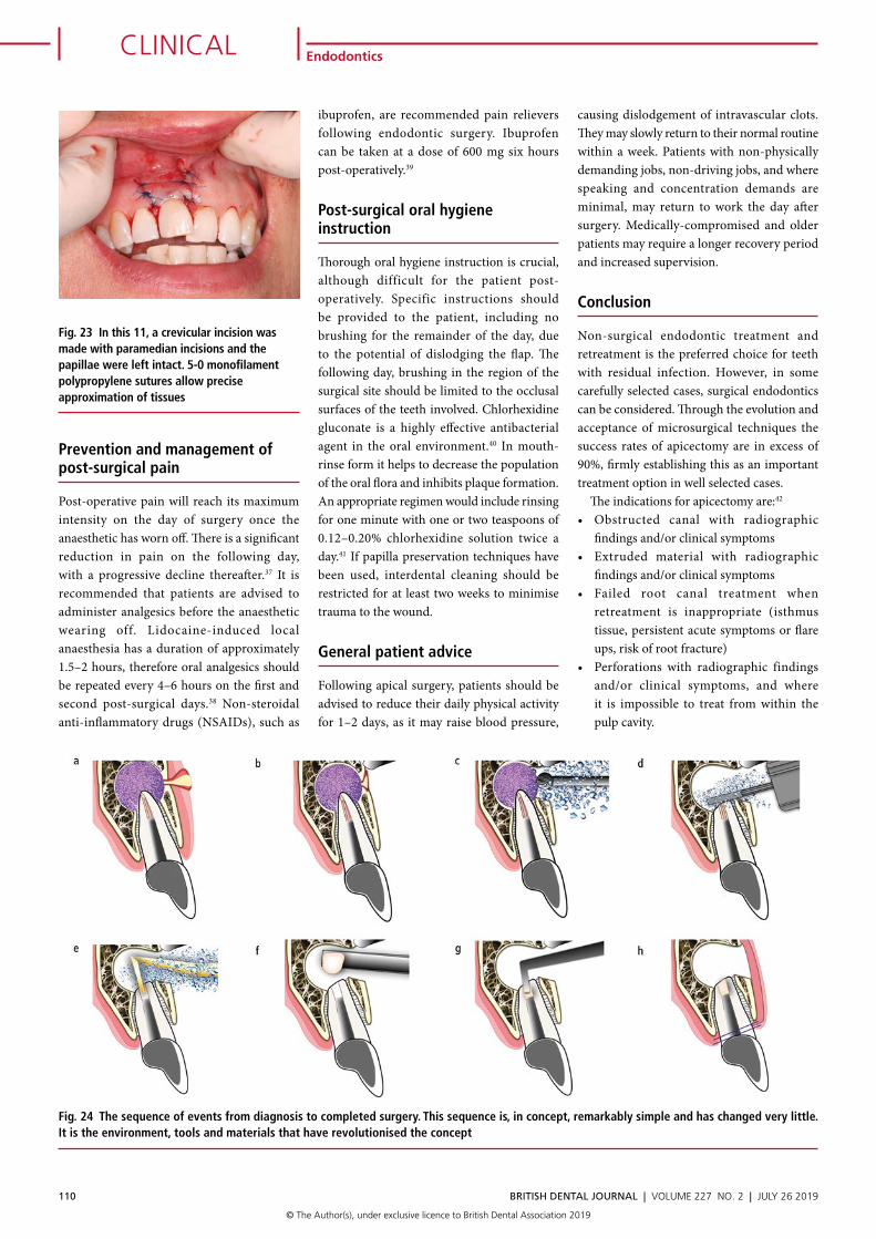
Bioceramics
MTA, Biodentine and other similar biocompatible bioceramics may be chosen to hermetically seal infected root canals and promote local healing (Fig. 21).22 Furthermore, the formation of hydroxyapatite at the interface of MTA and dentine improves the seal over 3 mm compared to conventional materials.23 However, many clinicians may find the handling of MTA challenging; the powder and water ratio must be correctly mixed and maintained, and the material is most accurately placed in smaller increments. The placement of the material is facilitated with the use of an MTA carrier or a Lee block. Table 5 compares a variety of MTA carriers. MTA has a more than adequate working time of 2–3 hours, with the final set requiring moisture. The moisture provided by the surrounding blood clot should be adequate for MTA setting.
In an attempt to overcome the challenges of MTA placement, newer materials have been developed that offer improved handling and faster setting times (Table 6). These have been produced in various forms: Biodentine uses auto-mix compules to produce the optimal mix. The resultant gel-like mix handles very similarly to a material like Kalzinol but will slump if over-instrumented or if it comes into contact with water (Fig. 22a). Pre-mixed putties are also available. TotalFill putty is readymade to use, and again handles like Kalzinol or IRM (Fig. 22b). These newer bioceramics have good pH, bond strength, dimensional stability, radiopacity and are thus demonstrating good tissue compatibility, sealing ability and antimicrobial activity in vitro.24,25,26,27,28,29,30,31 Finally a newer all-encompassing MTA Flow is being introduced, which takes a step back to powder and water but allows the operator to mix the material
to the exact desired consistency. Changes to the chemistry of biocompatible, bioceramic, biomaterials allow faster setting and variations in handling properties, enabling clinicians to precisely select the retrograde obturation material to match their handling requirements. Ultimately, it is the clinician’s skill in obturating the root canals with a hermetic seal using the biocompatible bioceramics which can help prevent treatment failure. Some clinicians advocate the need to check the quality of the root canal obturation with a radiograph before closing the surgical site.
Flap closure
Prior to wound closure, the tissues are irrigated with saline solution to remove any debris. Wound edges are then approximated to the correct position to promote healing by primary intention.32 Tissue margins should rest
Fig. 18 Anatomy of apical delta in single-rooted anterior teeth. For single-rooted anterior teeth, the macro-anatomy of the apical delta is complex and even more so for molars and premolars. Many types of isthmus and canal configurations have been described. It is not important to learn these but it is essential to examine the apex to ensure the isthmus is fully accessed
Fig. 19 The use of plaque disclosing tablets to identify missed MB 1 canal. Following placement of plaque disclosing solution into the crypts (notice the excellent haemostasis following preparation of the 15 and 16 with piezo tips) not only is the PDL visible confirming clean apical amputation but the missing MB1 form the original treatment is clearly evident
Fig. 20 The crypts of the 14 and 15 apical lesions following three minutes’ application of ferric sulphate
Fig. 21 a) The pre-operative radiograph of the 14 and 15; b) the teeth at three months. Already there is the appearance of periodontal ligament apically over the MTA supporting the biocompatibility of this material
108 BRITISH DENTAL JOURNAL | VOLUME 227 NO. 2 | JULy 26 2019
CLINICAL Endodontics
© The Author(s), under exclusive licence to British Dental Association 2019
passively in the desired place before suturing. Tissue trauma, such as stretching, tearing or distortion must be avoided. This can be accomplished through careful manipulation of the tissues and instruments.
Suture choice
Non-absorbable materials are recommended to suture gingival wounds, as the inflammation process ceases once the sutures have been removed.33 Monofilament sutures, including nylon, gut and steel, are made of a single strand of material, initiating a smaller inflammatory reaction when compared to their multifilament counterparts, including silk and polyester.34 Monofilament synthetic sutures appear to be the least traumatic and allow the least bacterial migration.33 Therefore, this is often the material of choice (Fig. 23). A reverse cutting needle of a small diameter is required with 5/0 being a common choice. Small diameter sutures cause less trauma, as the thread tends to break as opposed to the tissue tearing when excess tension is placed.35
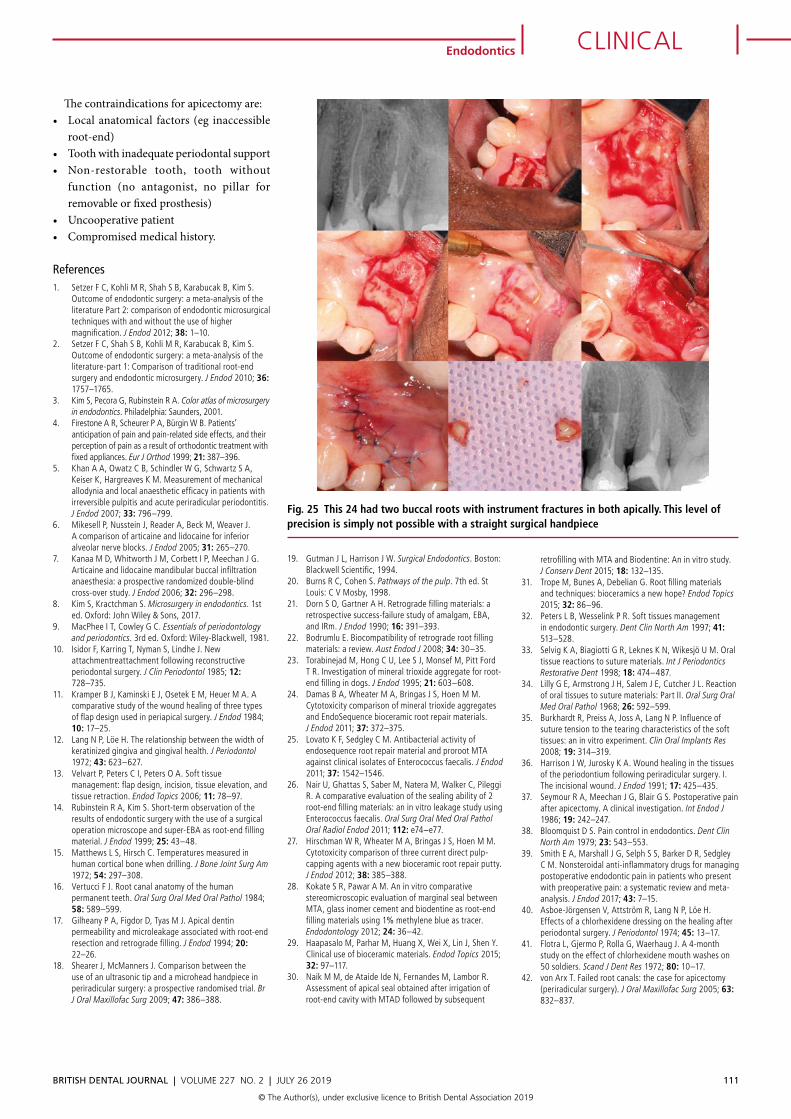
The sutures should then be removed 7 days post-operatively, as epithelial bridging and collagen crosslinking is thought to happen within 21–28 hours.36 The flap must be sufficiently healed to resist displacement forces. Figure 24 demonstrates the full surgical sequence diagrammatically and Figure 25 demonstrates this process on a 24 tooth.
Post-operative care
Although there is no clear guidance on the recall period for patients, they would ideally be reviewed 1-2 weeks post-operatively for suture removal and then at 6 months with prior attendance if any problems arise. The post-operative management of the patient should be viewed as an extension of the surgical protocol. Key components of the post-operative package of care are set out in the following sections. 36
Management of post-operative swelling and bleeding
Some post-operative bleeding, swelling and bruising is to be expected for up to 14 days, even with a good primary closure of the surgical site. The patient must be informed that discharges are expected and that it is part of the healing process, it will help alleviate the patient’s anxiety and concern. Clinicians can inform the patient that in the few days
following surgery, the patient can place ice cubes in a clean cloth to apply as a cold compress (never place ice directly or in tissue, or in a plastic bag to the skin, as this can stick and detach blood clots when removed) to the region for ten minutes every hour they are awake. The combination of pressure and cold temperature reduces the flow of blood and counteracts the haemorrhagic rebound reaction, ultimately reducing post-surgical bleeding and swelling.
Delayed minor bleeding from the surgical site may occur after 12–18 hours. This can be managed with firm finger pressure using a moistened cloth over the bleeding site for 10–15 minutes. If this fails to control the bleeding, the patient may return to the dental clinic, where the surgeon may then deal with the bleed as appropriate. Ecchymosis of the surrounding tissues may also occur, causing aesthetic concern; the patient should be informed that this may last up to two weeks.
Material classification Examples Benefits Challenges
Hand-mixed prescribed power-water ratio
Pro Root MTAMTA Angelus
Evidence-supported gold standard
Mixing is challenging, handling more difficult
Auto-mixed compules BiodentineEasy to mix, fast setting, reasonable handling. Growing evidence-base
Mixing remains unpredictable, setting often greater than manufacturer’s claims, very water soluble. ++ waste
Pre-mixed puttyEndo Sequence Root repair putty (outside USA: TotalFill putty)
Ready to use from pot to tooth, easy to manipulate Further evidence required
Hand-mixed variable power-water ratio MTA Flow
Can be mixed to precise consistency and from there easy to manipulate
Evidence-base poor. Operator sensitive for mixing to desired consistency
Table 6 Material classification and examples
Carrier Cost Size availability Shape Considerations
Endo gun Approximately £50 1.2–1.8 mm Straight of curved Simple, reusable,
inexpensive
Dovgan carrier £18 0.8, 0.99 and 1.6 mm
Straight but bendable
Simple, inexpensive. Available in single-use and reusable. Lack precision
Micro apical placement system (MAP)
Started kit from £495 0.9–1.1 mm
Variety of anatomically shaped tips
Versatile and precise. Costly and prone to blockages if poorly maintained
MTA ‘Lee’ block £67.50 Various Standardised lengths
Inexpensive, reusable and versatile. Technically more challenging. Clinicians with tremors beware
Table 5 Comparison of a variety of MTA carriers
Fig. 22 a) Biodentine placed in the mesio-buccal root of the 16; b) TotalFill putty in the upper central incisors. Both materials offer improved handling to MTA when mixed but still require tools and precision to place them in such confined spaces
BRITISH DENTAL JOURNAL | VOLUME 227 NO. 2 | JULy 26 2019 109
CLINICALEndodontics
© The Author(s), under exclusive licence to British Dental Association 2019
Prevention and management of post-surgical pain
Post-operative pain will reach its maximum intensity on the day of surgery once the anaesthetic has worn off. There is a significant reduction in pain on the following day, with a progressive decline thereafter.37 It is recommended that patients are advised to administer analgesics before the anaesthetic wearing off. Lidocaine-induced local anaesthesia has a duration of approximately 1.5–2 hours, therefore oral analgesics should be repeated every 4–6 hours on the first and second post-surgical days.38 Non-steroidal anti-inflammatory drugs (NSAIDs), such as
ibuprofen, are recommended pain relievers following endodontic surgery. Ibuprofen can be taken at a dose of 600 mg six hours post-operatively.39
Post-surgical oral hygiene instruction
Thorough oral hygiene instruction is crucial, although difficult for the patient post-operatively. Specific instructions should be provided to the patient, including no brushing for the remainder of the day, due to the potential of dislodging the flap. The following day, brushing in the region of the surgical site should be limited to the occlusal surfaces of the teeth involved. Chlorhexidine gluconate is a highly effective antibacterial agent in the oral environment.40 In mouth-rinse form it helps to decrease the population of the oral flora and inhibits plaque formation. An appropriate regimen would include rinsing for one minute with one or two teaspoons of 0.12–0.20% chlorhexidine solution twice a day.41 If papilla preservation techniques have been used, interdental cleaning should be restricted for at least two weeks to minimise trauma to the wound.
General patient advice
Following apical surgery, patients should be advised to reduce their daily physical activity for 1–2 days, as it may raise blood pressure,
causing dislodgement of intravascular clots. They may slowly return to their normal routine within a week. Patients with non-physically demanding jobs, non-driving jobs, and where speaking and concentration demands are minimal, may return to work the day after surgery. Medically-compromised and older patients may require a longer recovery period and increased supervision.
Conclusion
Non-surgical endodontic treatment and retreatment is the preferred choice for teeth with residual infection. However, in some carefully selected cases, surgical endodontics can be considered. Through the evolution and acceptance of microsurgical techniques the success rates of apicectomy are in excess of 90%, firmly establishing this as an important treatment option in well selected cases.
The indications for apicectomy are:42
• Obstructed canal with radiographic findings and/or clinical symptoms
• Extruded material with radiographic findings and/or clinical symptoms
• Failed root canal treatment when retreatment is inappropriate (isthmus tissue, persistent acute symptoms or flare ups, risk of root fracture)
• Perforations with radiographic findings and/or clinical symptoms, and where it is impossible to treat from within the pulp cavity.
Fig. 24 The sequence of events from diagnosis to completed surgery. This sequence is, in concept, remarkably simple and has changed very little. It is the environment, tools and materials that have revolutionised the concept
Fig. 23 In this 11, a crevicular incision was made with paramedian incisions and the papillae were left intact. 5-0 monofilament polypropylene sutures allow precise approximation of tissues
110 BRITISH DENTAL JOURNAL | VOLUME 227 NO. 2 | JULy 26 2019
CLINICAL Endodontics
© The Author(s), under exclusive licence to British Dental Association 2019
The contraindications for apicectomy are:• Local anatomical factors (eg inaccessible
root-end)• Tooth with inadequate periodontal support• Non-restorable tooth, tooth without
function (no antagonist, no pillar for removable or fixed prosthesis)
• Uncooperative patient• Compromised medical history.
References1. Setzer F C, Kohli M R, Shah S B, Karabucak B, Kim S.
Outcome of endodontic surgery: a meta-analysis of the literature Part 2: comparison of endodontic microsurgical techniques with and without the use of higher magnification. J Endod 2012; 38: 1–10.
2. Setzer F C, Shah S B, Kohli M R, Karabucak B, Kim S. Outcome of endodontic surgery: a meta-analysis of the literature-part 1: Comparison of traditional root-end surgery and endodontic microsurgery. J Endod 2010; 36: 1757–1765.
3. Kim S, Pecora G, Rubinstein R A. Color atlas of microsurgery in endodontics. Philadelphia: Saunders, 2001.
4. Firestone A R, Scheurer P A, Bürgin W B. Patients’ anticipation of pain and pain-related side effects, and their perception of pain as a result of orthodontic treatment with fixed appliances. Eur J Orthod 1999; 21: 387–396.
5. Khan A A, Owatz C B, Schindler W G, Schwartz S A, Keiser K, Hargreaves K M. Measurement of mechanical allodynia and local anaesthetic efficacy in patients with irreversible pulpitis and acute periradicular periodontitis. J Endod 2007; 33: 796–799.
6. Mikesell P, Nusstein J, Reader A, Beck M, Weaver J. A comparison of articaine and lidocaine for inferior alveolar nerve blocks. J Endod 2005; 31: 265–270.
7. Kanaa M D, Whitworth J M, Corbett I P, Meechan J G. Articaine and lidocaine mandibular buccal infiltration anaesthesia: a prospective randomized double-blind cross-over study. J Endod 2006; 32: 296–298.
8. Kim S, Kractchman S. Microsurgery in endodontics. 1st ed. Oxford: John Wiley & Sons, 2017.
9. MacPhee I T, Cowley G C. Essentials of periodontology and periodontics. 3rd ed. Oxford: Wiley-Blackwell, 1981.
10. Isidor F, Karring T, Nyman S, Lindhe J. New attachmentreattachment following reconstructive periodontal surgery. J Clin Periodontol 1985; 12: 728–735.
11. Kramper B J, Kaminski E J, Osetek E M, Heuer M A. A comparative study of the wound healing of three types of flap design used in periapical surgery. J Endod 1984; 10: 17–25.
12. Lang N P, Löe H. The relationship between the width of keratinized gingiva and gingival health. J Periodontol 1972; 43: 623–627.
13. Velvart P, Peters C I, Peters O A. Soft tissue management: flap design, incision, tissue elevation, and tissue retraction. Endod Topics 2006; 11: 78–97.
14. Rubinstein R A, Kim S. Short-term observation of the results of endodontic surgery with the use of a surgical operation microscope and super-EBA as root-end filling material. J Endod 1999; 25: 43–48.
15. Matthews L S, Hirsch C. Temperatures measured in human cortical bone when drilling. J Bone Joint Surg Am 1972; 54: 297–308.
16. Vertucci F J. Root canal anatomy of the human permanent teeth. Oral Surg Oral Med Oral Pathol 1984; 58: 589–599.
17. Gilheany P A, Figdor D, Tyas M J. Apical dentin permeability and microleakage associated with root-end resection and retrograde filling. J Endod 1994; 20: 22–26.
18. Shearer J, McManners J. Comparison between the use of an ultrasonic tip and a microhead handpiece in periradicular surgery: a prospective randomised trial. Br J Oral Maxillofac Surg 2009; 47: 386–388.
19. Gutman J L, Harrison J W. Surgical Endodontics. Boston: Blackwell Scientific, 1994.
20. Burns R C, Cohen S. Pathways of the pulp. 7th ed. St Louis: C V Mosby, 1998.
21. Dorn S O, Gartner A H. Retrograde filling materials: a retrospective success-failure study of amalgam, EBA, and IRm. J Endod 1990; 16: 391–393.
22. Bodrumlu E. Biocompatibility of retrograde root filling materials: a review. Aust Endod J 2008; 34: 30–35.
23. Torabinejad M, Hong C U, Lee S J, Monsef M, Pitt Ford T R. Investigation of mineral trioxide aggregate for root-end filling in dogs. J Endod 1995; 21: 603–608.
24. Damas B A, Wheater M A, Bringas J S, Hoen M M. Cytotoxicity comparison of mineral trioxide aggregates and EndoSequence bioceramic root repair materials. J Endod 2011; 37: 372–375.
25. Lovato K F, Sedgley C M. Antibacterial activity of endosequence root repair material and proroot MTA against clinical isolates of Enterococcus faecalis. J Endod 2011; 37: 1542–1546.
26. Nair U, Ghattas S, Saber M, Natera M, Walker C, Pileggi R. A comparative evaluation of the sealing ability of 2 root-end filling materials: an in vitro leakage study using Enterococcus faecalis. Oral Surg Oral Med Oral Pathol Oral Radiol Endod 2011; 112: e74–e77.
27. Hirschman W R, Wheater M A, Bringas J S, Hoen M M. Cytotoxicity comparison of three current direct pulp-capping agents with a new bioceramic root repair putty. J Endod 2012; 38: 385–388.
28. Kokate S R, Pawar A M. An in vitro comparative stereomicroscopic evaluation of marginal seal between MTA, glass inomer cement and biodentine as root-end filling materials using 1% methylene blue as tracer. Endodontology 2012; 24: 36–42.
29. Haapasalo M, Parhar M, Huang X, Wei X, Lin J, Shen Y. Clinical use of bioceramic materials. Endod Topics 2015; 32: 97–117.
30. Naik M M, de Ataide Ide N, Fernandes M, Lambor R. Assessment of apical seal obtained after irrigation of root-end cavity with MTAD followed by subsequent
retrofilling with MTA and Biodentine: An in vitro study. J Conserv Dent 2015; 18: 132–135.
31. Trope M, Bunes A, Debelian G. Root filling materials and techniques: bioceramics a new hope? Endod Topics 2015; 32: 86–96.
32. Peters L B, Wesselink P R. Soft tissues management in endodontic surgery. Dent Clin North Am 1997; 41: 513–528.
33. Selvig K A, Biagiotti G R, Leknes K N, Wikesjö U M. Oral tissue reactions to suture materials. Int J Periodontics Restorative Dent 1998; 18: 474–487.
34. Lilly G E, Armstrong J H, Salem J E, Cutcher J L. Reaction of oral tissues to suture materials: Part II. Oral Surg Oral Med Oral Pathol 1968; 26: 592–599.
35. Burkhardt R, Preiss A, Joss A, Lang N P. Influence of suture tension to the tearing characteristics of the soft tissues: an in vitro experiment. Clin Oral Implants Res 2008; 19: 314–319.
36. Harrison J W, Jurosky K A. Wound healing in the tissues of the periodontium following periradicular surgery. I. The incisional wound. J Endod 1991; 17: 425–435.
37. Seymour R A, Meechan J G, Blair G S. Postoperative pain after apicectomy. A clinical investigation. Int Endod J 1986; 19: 242–247.
38. Bloomquist D S. Pain control in endodontics. Dent Clin North Am 1979; 23: 543–553.
39. Smith E A, Marshall J G, Selph S S, Barker D R, Sedgley C M. Nonsteroidal anti-inflammatory drugs for managing postoperative endodontic pain in patients who present with preoperative pain: a systematic review and meta-analysis. J Endod 2017; 43: 7–15.
40. Asboe-Jörgensen V, Attström R, Lang N P, Löe H. Effects of a chlorhexidene dressing on the healing after periodontal surgery. J Periodontol 1974; 45: 13–17.
41. Flotra L, Gjermo P, Rolla G, Waerhaug J. A 4-month study on the effect of chlorhexidene mouth washes on 50 soldiers. Scand J Dent Res 1972; 80: 10–17.
42. von Arx T. Failed root canals: the case for apicectomy (periradicular surgery). J Oral Maxillofac Surg 2005; 63: 832–837.
Fig. 25 This 24 had two buccal roots with instrument fractures in both apically. This level of precision is simply not possible with a straight surgical handpiece
BRITISH DENTAL JOURNAL | VOLUME 227 NO. 2 | JULy 26 2019 111
CLINICALEndodontics
© The Author(s), under exclusive licence to British Dental Association 2019