Embed Size (px)
Citation preview

C H A P T E R F I V E
Endocytosis and Intracellular Trafficking of Notch and Its Ligands
Shinya Yamamoto,*,1 Wu-Lin Charng,*,1 and Hugo J. Bellen *,†,‡,§
Contents 1. Notch Signaling and its Regulation
by Endocytosis and Vesicle Trafficking 166 1.1. Introduction 166 1.2. Intracellular trafficking of Notch and DSL ligands 167 1.3. Endocytosis is essential for Notch signaling 167 1.4. Proteins and molecules involved in endocytosis 169 1.5. Proteins involved in endocytic trafficking, sorting, recycling, and
degradation 169 2. Ligand Endocytosis and Trafficking 171
2.1. The role of endocytosis of DSL ligands in the signal-sending cells 171
2.2. The role of ubiquitin, E3 ligases, and ubiquitin interacting proteins in DSL ligand trafficking 171
2.3. Two theories on the function of DSL ligand endocytosis 174 3. Notch Receptor Endocytosis and Endosomal Trafficking 177
3.1. The role of endocytosis of the Notch receptor in signal-receiving cells 177
3.2. The controversy on the requirement of endocytosis for S3 cleavage 179
3.3. Degradation of Notch receptors through the lysosomal pathway 182
4. Regulation of Notch Signaling by Endocytosis and Vesicle Trafficking During Mechanosensory Organ Development in Drosophila 185 4.1. Introduction to mechanosensory organ development 185 4.2. Setting up the asymmetry in the SOP cell 186 4.3. Role of asymmetrically segregated Neuralized
and Delta recycling in the pIIb cell 187
* Program in Developmental Biology, Baylor College of Medicine, Houston, TX, USA † Department of Molecular and Human Genetics, Baylor College of Medicine, Houston, TX, USA ‡ Department of Neuroscience, Baylor College of Medicine, Houston, TX, USA § Howard Hughes Medical Institute, Baylor College of Medicine, Houston, TX, USA 1 Shinya Yamamoto and Wu-Lin Charng have contributed equally to the manuscript.
Current Topics in Developmental Biology, Volume 92 � 2010 Elsevier Inc. ISSN 0070-2153, DOI 10.1016/S0070-2153(10)92005-X All rights reserved.
165

166 Shinya Yamamoto et al.
4.4. Role of asymmetrically segregated Numb in the pIIb cell 187 4.5. Role of asymmetrically segregated Sara-endosomes
in the pIIa cell 189 5. Conclusion and Future Directions 190 Acknowledgments 191 References 191
Abstract Notch signaling occurs through direct interaction between Notch, the receptor, and its ligands, presented on the surface of neighboring cells. Endocytosis has been shown to be essential for Notch signal activation in both signal-sending and signal-receiving cells, and numerous genes involved in vesicle trafficking have recently been shown to act as key regulators of the pathway. Defects in vesicle trafficking can lead to gain- or loss-of-function defects in a context-dependent manner. Here, we discuss how endocytosis and vesicle trafficking regulate Notch signaling in both signal-sending and signal-receiving cells. We will introduce the key players in different trafficking steps, and further illustrate how they impact the signal outcome. Some of these players act as general factors and modulate Notch signaling in all contexts, whereas others modulate signaling in a context-specific fashion. We also discuss Notch signaling during mechanosensory organ development in the fly to exemplify how endocytosis and vesicle trafficking are effectively used to determine correct cell fates. In summary, endocytosis plays an essential role in Notch signaling, whereas intracellular vesicle trafficking often plays a context-dependent or regulatory role, leading to divergent outcomes in different developmental contexts.
1. Notch Signaling and its Regulation by Endocytosis and Vesicle Trafficking
1.1. Introduction
Notch signaling is an evolutionally conserved signaling pathway which takes place between neighboring cells. When Notch receptors are activated by DSL (Delta/Serrate/LAG-2) ligands, Notch undergoes a set of serial proteolytic cleavages resulting in the release of the Notch intracellular domain (NICD). NICD translocates into the nucleus to form a positive transcriptional complex with a key transcription factor CSL for CBF-1/Su(H)/LAG-1 (C-promoter binding factor-1/Suppressor of Hairless/Lin-12-and-GLP-1) and a coactivator, Mastermind (Kopan and Ilagan, 2009). This CSL-dependent process is referred to as canonical Notch
167 Notch Signaling and Endocytosis
signaling. It has also been shown that in certain contexts, Notch signaling activity can be mediated through a CSL-independent pathway, which is usually referred to as noncanonical Notch signaling (Ligoxygakis et al., 1998; Ordentlich et al., 1998; Ramain et al., 2001; Zecchini et al., 1999). Since both ligands and receptors are transmembrane proteins, endocytosis and vesicle trafficking play a critical role in the regulation of this signaling pathway.
1.2. Intracellular trafficking of Notch and DSL ligands
Notch receptors and DSL ligands are produced in the endoplasmic reticulum (ER) and traffic through the Golgi apparatus to reach the plasma membrane (Fig. 5.1). From the cell surface, they re-enter the cell via endocytosis, a process by which vesicles invaginate from the plasma membrane into the cytoplasm. These endocytic vesicles typically fuse with an early endosome, a sorting center of the endocytic pathway, often referred to as the “sorting endosome.” From this early/sorting endosomes, proteins can be recycled back to the plasma membrane, transported to the Golgi apparatus, or transported to the late endosome, which eventually fuses with the lysosome for protein degradation (Doherty and McMahon, 2009). In the past, endocytosis was considered to only play a negative role in signaling pathways by removing receptors from the membrane. However, more and more evidence suggests that endocytosis also plays a positive role. Signaling may occur not only at the cell membrane but also in endocytosed vesicles or endosomes. Indeed, numerous signaling pathways, including Notch signaling, have been shown to depend on endocytosis for their full activation (Sorkin and von Zastrow, 2009).
1.3. Endocytosis is essential for Notch signaling
Endocytosis and endosomal trafficking have been shown to play an important role in the activation and regulation of Notch signaling. The first hint came from the phenotype associated with the Drosophila shibire (shi) mutant. shi was initially identified as a temperature-sensitive mutation that leads to embryonic lethality at restrictive temperatures (Poodry et al., 1973). The gene was later shown to encode dynamin, a GTPase essential for most, if not all, forms of endocytosis (Chen et al., 1991; van der Bliek and Meyerowitz, 1991). Interestingly, shits1 embryos, raised at the restrictive temperature during neuroblasts segregation, contain excessive neuroblasts and neurons (Poodry, 1990), a neurogenic phenotype that resembles the loss of Notch phenotype (Poulson, 1937). Further studies based on clonal analysis and genetic interaction assays provided the first evidence that endocytosis is required for ligand-dependent Notch activation in both signal-sending and signal-receiving cells (Seugnet et al., 1997).

Clathrin-
Clathrindependent
independent endocytosis Dynein
endocytosis
Golgi apparatus Auxilin Hsc70
Rab5
Recycling endosome
Rab11 Sec15
Avl Rab4
Early/sorting Hrs endosomeESCRT
Endoplasmic reticulum
Rab7 HOPS AP-3 Exosome
MVB/ Late endosome
Transmembrane proteins Adaptor proteins Clathrin
Nucleus DynaminLysosome
168 Shinya Yamamoto et al.
Figure 5.1 Overview of endocytosis and vesicle trafficking. Transmembrane proteins are made in the ER and traffic through the Golgi apparatus to reach the plasma membrane. From the cell surface, these proteins can re-enter the cell via various endocytosis pathways. Clathrin-dependent endocytosis is usually referred to as “canonical endocytosis.” Clathrin adaptor proteins, such as the AP-2 complex, recruit clathrin and cargo transmembrane proteins to the site of endocytosis. The clathrin-coated endocytic vesicle is pinched off by the action of dynamin GTPase, and the clathrin coat is then removed by molecular chaperone Hsc70 via the assistance of auxilin. On the other hand, endocytosis can also occur without clathrin and is referred to as “noncanonical endocytosis” or “clathrinindependent endocytosis.” After endocytosis, small GTPase Rab5 and SNARE protein Avalanche (Avl) mediate the fusion of endocytic vesicles with the early/sorting endosome. From the early endosome, endocytosed proteins can recycle back to the plasma membrane directly in a Rab4-dependent manner or indirectly through the recycling endosome in a Rab11-dependent manner. Alternatively, they can return back to the Golgi or travel to the late endosome and lysosome for degradation. Proteins destined for degradation are sorted into Rab7-positive late endosome or multivesicular bodies (MVB). Packaging of transmembrane proteins into intraluminal vesicles is mediated by the ESCRT complexes. In certain cell contexts, MVB can secrete their contents to extracellular regions. These secreted MVBs are referred to as exosomes. Finally, through HOPS and AP3 complexes, MVB/late endosomes fuse with the lysosome and transmembrane proteins are degraded by proteases and acid hydrolases. (See Color Insert.)
Based on these pioneering studies, various labs have focused on understanding how endocytosis regulates Notch signaling through forward and reverse genetic approaches. First, we will briefly review key steps in endocytosis and the molecular players that have been shown to affect

169 Notch Signaling and Endocytosis
Notch signaling. Specific players that seemingly only affect endocytosis of Notch signaling components in a cell context-dependent manner will be discussed later.
1.4. Proteins and molecules involved in endocytosis
Canonical endocytosis requires the assembly of a clathrin lattice to form a clathrin-coated pit, which is then pinched off by the action of a GTPase, dynamin (Seugnet et al., 1997; Traub, 2009). Clathrin is composed of heavy and light chains which form a triskelion upon multimerization. Clathrin is recruited to the site of endocytosis in the membrane through adaptor proteins, including the assembly protein-2 (AP-2) complex (Berdnik et al., 2002). These and other adaptor proteins bind to transmembrane proteins that are targeted for endocytosis and recruited into clathrin-coated pits. The lipid composition of the plasma membrane also plays an important role in endocytosis. For example, phosphatidylinositol (4,5) diphosphate (PI(4,5) P2) is enriched in the plasma membrane at sites where endocytosis occurs, and the recruitment of many adaptor proteins depends on their binding to this lipid (Di Paolo and De Camilli, 2006; Poccia and Larijani, 2009).
A key signal to promote endocytosis of transmembrane proteins relies on the monoubiquitination of intracellular lysine residues by E3 ubiquitin ligases. The ubiquitin tag can promote the interaction with adaptor proteins and lead to recruitment and enrichment into clathrin-coated pits (d’Azzo et al., 2005). Ubiquitinated proteins can be recognized by proteins that contain ubiquitin interaction motifs. Upon invagination and pinching off, vesicles are stripped of their clathrin coat by molecular chaperones such as Hsc70 with the assistance of auxilin (Eisenberg and Greene, 2007; Eun et al., 2008; Hagedorn et al., 2006).
Alternatively, endocytosis can also occur without the assembly of clathrin-coated pits, a process often referred to as noncanonical endocytosis or clathrin-independent endocytosis (Doherty and McMahon, 2009; Hansen and Nichols, 2009). However, compared to the well-established role of clathrin-dependent endocytosis in signaling pathways, its involvement in signal regulation is poorly understood.
1.5. Proteins involved in endocytic trafficking, sorting, recycling, and degradation
Upon the uncoating of internalized vesicles, the small GTPase Rab5 and the SNARE (Soluble N-Ethylmaleimide-Sensitive Factor Adaptor Protein Receptor) protein syntaxin 7 mediate fusion of the endocytosed vesicles with the early endosome (Lu and Bilder, 2005; Vaccari et al., 2008). From the early endosome, endocytosed proteins can either be recycled to the

170 Shinya Yamamoto et al.
plasma membrane, return to the Golgi and ER, or travel to the late endosome and lysosome for degradation. Some proteins can recycle to the plasma membrane directly from the early endosome in a GTPase Rab4dependent manner, whereas most proteins enter the recycling endosomes prior to returning to the cell surface (Grant and Donaldson, 2009). The latter slower recycling process depends on the function of Rab11 and the exocyst complex, a multiprotein complex including Sec15 (Emery et al., 2005; Jafar-Nejad et al., 2005). In addition, some internalized proteins can travel to the Golgi apparatus and further to the ER with the assistance of the retromer complex, but the role of this trafficking route in Notch signaling has not been investigated. Proteins destined for degradation are sorted into Rab7-positive late endosomal compartments. During the transition between the early and the late endosome, transmembrane proteins are packaged into intraluminal vesicles also called multivesicular bodies (MVB) (Doherty and McMahon, 2009).
Sorting of cargos into intraluminal vesicles is mediated by the endosomal sorting complex required for transport (ESCRT) complexes. ESCRT-0 recognizes ubiquitinated receptors and recruits ESCRT-I, resulting in the activation of ESCRT-II, which assists the assembly of ESCRT-III (Herz and Bergmann, 2009; Raiborg and Stenmark, 2009). In certain cell contexts, MVB can be recycled to the plasma membrane and secrete their contents, the intraluminal vesicles (Simons and Raposo, 2009). These secreted MVBs, called exosomes, have been proposed to play a role in Notch signaling through secretion of active Delta (Chitnis, 2006; Le Borgne and Schweisguth, 2003a) but their in vivo role in Notch signaling awaits testing.
Finally, MVB/late endosomes fuse with the lysosome where the internalized cargos are degraded by proteases and acid hydrolases. The AP-3 complex is involved in endosomal trafficking to the lysosome, and the homotypic fusion and vacuole protein sorting (HOPS) complex is involved in the late endosome maturation/lysosomal fusion step (Dell’Angelica, 2009; Wilkin et al., 2008).
Along the endocytic trafficking process, the luminal pH of endosomal compartments becomes gradually more acidic (Marshansky and Futai, 2008). The low pH assists in the dissociation of certain protein–protein interactions, as well as provides the optimal environment for enzymatic activity of certain proteases. Therefore, proteins involved in the acidification of endosomes can influence the strength of protein interactions and efficiency of protein cleavage/degradation. For example, vacuolar (Hþ)ATPase (V-ATPase), a proton transporter involved in the acidification of endosomal compartments, has been reported to influence the processing and activation of Notch receptors (Yan et al., 2009).
Here, we will discuss how DSL ligands and Notch receptors functions are regulated/affected by endocytosis and intracellular vesicle trafficking.

171 Notch Signaling and Endocytosis
It is important to note that endocytosis and vesicle trafficking play distinct functions in signal-sending and in signal-receiving cells, respectively. Since most of these studies used Drosophila as a model organism, we will mainly focus on the results from Drosophila and cover findings in other organisms where appropriate.
2. Ligand Endocytosis and Trafficking
2.1. The role of endocytosis of DSL ligands in the signal-sending cells
The DSL ligands are type-1 transmembrane proteins that contain a characteristic DSL domain at their N terminus followed by multiple epithelial growth factor-like repeats (EGF-r), a single transmembrane domain (TMD), and an intracellular domain (Kopan and Ilagan, 2009). These ligands can be subdivided into the Serrate/Jagged group ligands, which contain a cysteine-rich domain between the EGF-r and TMD, and Delta group ligands that lack this motif. The N terminus of DSL ligands including the DSL domain is required for ligand–receptor interaction and signaling activity (Glittenberg et al., 2006; Henderson et al., 1997; Parks et al., 2006; Shimizu et al., 1999). Early studies on the subcellular localization of Delta in Drosophila embryos and imaginal discs documented the presence of Delta in intracellular vesicles (Kooh et al., 1993). Analysis of endocytic mutants such as shi (Parks et al., 1995; Seugnet et al., 1997) and hook (Kramer and Phistry, 1996, 1999) revealed that these vesicles are endocytic in nature, and a block in endocytosis of DSL ligands attenuated Notch signaling (Parks et al., 2000; Seugnet et al., 1997). Although many agree that endocytosis is essential for the activity of DSL ligands for canonical Notch signaling, the precise function of endocytosis is still debated. Here, we will first introduce the players in DSL ligand endocytosis and trafficking, and then discuss two nonmutually exclusive theories that have been proposed (Fig. 5.2).
2.2. The role of ubiquitin, E3 ligases, and ubiquitin interacting proteins in DSL ligand trafficking
In vivo structure function analysis of Delta using specific point mutant alleles showed that certain EGF-r as well as certain intracellular lysine residues are necessary for endocytosis and proper signaling (Parks et al., 2006). Similar results have been obtained from structure function analysis of Serrate using an ectopic overexpression assay system in vivo (Glittenberg et al., 2006). Lysine residues can be posttranslationally modified by ubiquitin, which serves as a signal for endocytosis, sorting, and/or degradation (Acconcia

172 Shinya Yamamoto et al.
DSL ligands
S1 cleaved Notch
S2 cleaved Notch
S2 cleavage Signal receiving
Signal sending
“Pulling force”
“Ligand activation”
ADAM
Figure 5.2 Endocytosis and trafficking of DSL ligands in the signal-sending cell. DLS ligands are synthesized in the ER and traffic to the cell surface through the secretory pathway. Endocytosis is required in the signal-sending cell for activation of the canonical Notch signaling pathway, but there are two nonmutually exclusive hypotheses to explain how endocytosis promotes DSL ligand activity. In the “Ligand Activation” theory, DSL proteins that have just been synthesized and reached the cell surface are still inactive and do not have the capacity to activate the Notch receptor on the signal-receiving cell. DSL are endocytosed and sorted into a unique endocytic compartment where they become “activated.” The activated ligands return to the cell surface via the recycling pathway where they interact with and activate the Notch receptor. In contrast, the “pulling force” model insists that when DSL ligands and Notch receptor interact, endocytosis in the signal-sending cell generates a mechanical force that leads to a conformational change in the Notch receptor. This force mediates the separation of the Notch heterodimer and allows the S2 cleavage mediated by ADAM proteases. The extracellular portion of Notch is trans-endocytosed into the signal-sending cell and assumed to be degraded through the lysosomal pathway along with the DSL ligands. (See Color Insert.)
et al., 2009; Hicke and Dunn, 2003). Ubiquitination is mediated by E3 ligases which recognize their specific target proteins and recruit E2 ligases for transfer of ubiquitin on to the lysine residues. The neuralized (neur) gene, whose loss causes a neurogenic phenotype similar to Notch and Delta mutants (Lehmann et al., 1981), encodes an E3 ligase with a C-terminal RING domain that is necessary for its E3 activity and two neuralized homology repeats (NHR1 and NHR2) (Deblandre et al., 2001; Lai et al., 2001; Pavlopoulos et al., 2001; Yeh et al., 2001). NRH1 has been shown

173 Notch Signaling and Endocytosis
to be necessary and sufficient for its interaction with Delta (Commisso and Boulianne, 2007). Mind bomb (Mib), first isolated in zebrafish to cause a neurogenic phenotype, encodes an E3 ligase with a C-terminal RING domain that ubiquitinates DSL ligands (Itoh et al., 2003). Both Neur and Mib are conserved in Drosophila and vertebrates, and loss of function of either gene in Drosophila shows a loss of Notch signaling phenotype in a tissue-specific manner (Boulianne et al., 1991; Lai et al., 2005; Le Borgne et al., 2005; Pitsouli and Delidakis, 2005; Wang and Struhl, 2005). These two E3 ligases are necessary for Notch signaling in the signal-sending cells, and loss of function leads to defects in endocytosis/sorting of DSL ligands. It has been suggested that Neur is likely to be involved in Delta-dependent signaling events, whereas Mib functions in Serrate/Jagged-dependent signaling events. However, the two proteins overlap in their functions, since they can rescue the loss-of-function phenotype of each other upon ectopic expression in a mutant background (Lai et al., 2005; Le Borgne et al., 2005; Pitsouli and Delidakis, 2005; Wang and Struhl, 2005). Both Neur and Mib are localized to the plasma membrane, where they can interact with DSL proteins. Neuralized can be recruited to the membrane via interaction with DSL ligands through its NHR1 domain (Commisso and Boulianne, 2007), via an interaction with phosphoinositides through its N-terminal polybasic domain (Skwarek et al., 2007), and/or via N-myristoylation of the N-terminal glycine residue (Koutelou et al., 2008). Notch signaling can be fine-tuned by regulating the activity of Neur by Bearded family proteins. Bearded family proteins, such as Bearded (Brd) and Twin of m4 (Tom), are negative regulators of Neur function, encoded by multiple genes clustered in the Bearded complex locus and Enhancer of Split complex locus (Bardin and Schweisguth, 2006; De Renzis et al., 2006; Fontana and Posakony, 2009; Leviten et al., 1997). Initially, gain-of-function mutations of Bearded were identified to cause loss of Notch signaling during lateral inhibition of mechanosensory organ precursors in Drosophila (Leviten and Posakony, 1996). Bearded family proteins posses Neur interaction motifs that allow them to bind to Neur and inhibit its function. However, in contrast to their gain-of-function phenotype, loss of function of all eight Bearded family genes show only partial Notch signaling defects during mechanosensory organ precursor specification and mesectoderm specification during embryogenesis, suggesting a context-specific role in vivo (Chanet et al., 2009).
Ubiquitinated DSL ligands are potentially recognized by Epsin (encoded by the liquid facet gene in Drosophila). Epsin is a ubiquitin binding protein that interacts with PI(4,5)P2 as well as several endocytic proteins such as clathrin and AP-2 (Chen et al., 1998; De Camilli et al., 2002; Polo et al., 2002). Loss of function of epsin shows a loss of Notch signaling phenotype in flies (Overstreet et al., 2003, 2004; Tian et al., 2004; Wang and Struhl, 2004) and in mice (Chen et al., 2009). Epsin is required in the signal-sending cell, supporting the idea that epsin mediates the trafficking of ubiquitinated

174 Shinya Yamamoto et al.
DSL ligands. The activity of epsin is positively regulated by Fat facets (Faf), a de-ubiquitinating enzyme that stabilizes epsin. Loss of function of Faf causes defects similar to those observed on epsin mutants. However, some epsin-dependent Notch signaling events are Faf-independent, suggesting that epsin activity can be regulated by other factors than Faf (Overstreet et al., 2004). How does epsin regulate Notch signaling? Several groups propose that epsin affects Notch signaling by promoting endocytosis of Delta. However, one group has observed that the bulk endocytosis of Delta is not affected in epsin mutants (Wang and Struhl, 2005), leading to an alternative hypothesis that epsin is required for sorting of internalized DSL ligands into a recycling pathway that leads to activation of Delta. Ubiquitinated DSL proteins have been proposed to undergo clathrindependent endocytosis, since mutations in auxilin, an adaptor molecule that recruits Hsc70 to clathrin-coated vesicles for uncoating, exhibit defects in DSL endocytosis (Eun et al., 2008; Kandachar et al., 2008). However, there is evidence indicating that DSL ligands are endocytosed through multiple distinct endocytic routes (Wang and Struhl, 2005), and epsin has been implicated in nonclathrin-mediated endocytosis (Sigismund et al., 2005). A recent study supports this model based on the observation that loss of clathrin heavy chain in the signal-sending cell is capable of signaling during oogenesis. However, epsin is essential, suggesting that endocytosis of Delta by epsin is clathrin independent (Windler and Bilder, 2010). Since the DSL ligands are present in lipid raft compartments and cofractionate with caveolin (Heuss et al., 2008), DSL ligand endocytosis may depend on a clathrin-independent lipid raft-mediated endocytic pathway for ligand activation. Further studies on endocytosis and trafficking of DSL ligands are needed to resolve these controversies.
2.3. Two theories on the function of DSL ligand endocytosis
DSL ligand endocytosis is necessary for canonical Notch signaling activation, and two models have been proposed to explain how DSL endocytosis leads to successful signal activation. One is the “ligand activation” model; the other is the “pulling force” hypothesis. It is important to note that these two models are not mutually exclusive.
2.3.1. The “ligand activation” theory Bulk endocytosis of Delta is not affected in epsin mutants, yet Delta is unable to signal (Wang and Struhl, 2004). Based on this observation, it was proposed that newly synthesized Delta does not have the capacity to signal and that it needs to be endocytosed and sorted into a specialized endocytic compartment that depends on epsin and ubiquitination of Delta. Delta then traffics back to the cell surface through a recycling pathway. During

175 Notch Signaling and Endocytosis
this process, Delta is thought to become “activated” and acquires its signaling capability. Following this study, several mutants that exhibit Delta recycling defects and lead to Notch loss-of-function phenotypes were identified from forward genetic screens. Mutations in sec15, a component of the exocyst complex, cause a Notch signaling defect in the mechanosensory organ lineage in Drosophila (Jafar-Nejad et al., 2005). In these mutants, Delta can be detected at the cell surface and can be internalized from the cell surface, suggesting that there is no defect in exo- or endocytosis. In wild-type cells, Delta enters recycling endosomes and returns to the plasma membrane, but this recycling fails to take place in sec15 mutant cells. In addition, mutations in Arp3, a subunit of the Arp2/3 complex which regulates actin polymerization (Goley and Welch, 2006), cause very similar Delta recycling defects (Rajan et al., 2009). Similarly, other proteins in Arp2/3 complex and WASp, an activator of the Arp2/3 complex, are required for Notch signaling (Ben-Yaacov et al., 2001; Tal et al., 2002). The activity of actin polymerization via the Arp2/3–WASp complex is therefore proposed to be critical for Delta recycling upon its internalization for successful canonical Notch signaling (see Section 4). Studies in mammalian cultured cells have shown that overexpression of dominant-negative forms of Rab11 in signal-sending cells leads to recycling defects and a less active signaling ability of Dll1 (Emery et al., 2005), suggesting that the role of recycling pathway in DSL ligand activation may be evolutionally conserved. Further studies in a cell culture model support this ligand activation theory, and these authors also proposed the involvement of lipid rafts in the activation of Dll1 (Heuss et al., 2008). However, the molecular mechanism of this mysterious “activation” remains to be identified. The activation has been proposed to consist of clustering of ligands, trafficking into lipid microdomains, proteolytic cleavage, or other posttranscriptional modification (Chitnis, 2006; Le Borgne and Schweisguth, 2003a; Wang and Struhl, 2004). In addition, although the role of the recycling pathway and actin polymerization in Delta activation have been well established in the cell fate determination during Drosophila mechanosensory lineage, in development of the wing margin, and in oogenesis (Ben-Yaacov et al., 2001; Jafar-Nejad et al., 2005; Rajan et al., 2009; Tal et al., 2002), this requirement may be context specific. Sec15 mutant cells undergo normal photoreceptor development (Mehta et al., 2005), a process in which Notch signaling is utilized reiteratively. Moreover, WASp mutant cells have been reported to not exhibit defects in lateral inhibition during mechanosensory organ development, another context where Notch signaling is required (Ben-Yaacov et al., 2001). Furthermore, a recent study reports that during oogenesis, ligand internalization through dynamin in the signal-sending germ line cell is necessary but clathrin heavy chain, Rab5, Rab11, and Sec15 are dispensable in the germ line cell during Notch signaling in this context (Windler and Bilder, 2010). Therefore, Delta activation through endocytosis and trafficking through the recycling pathway

176 Shinya Yamamoto et al.
may be required to further potentiate the activity of Delta in contexts where robust Notch signaling is required.
2.3.2. The “Pulling force” theory Studies using Drosophila cell lines have indicated that Notch can be “transendocytosed” into the signal-sending cell. This trans-endocytosis can be inhibited upon blockage of dynamin in the signal-sending cell (Klueg and Muskavitch, 1999; Klueg et al., 1998). Trans-endocytosis of Notch into Delta expressing cell has been proposed to take place in vivo as well, based on immunohistochemistry studies of Drosophila eye and wing vein tissue (Parks et al., 2000). Initially, Delta had also been suggested to be transendocytosed into Notch expressing cells based on cell culture studies (Fehon et al., 1990; Klueg et al., 1998), but this has not been confirmed in vivo. In addition, early studies have suggested that full-length Notch is trans-endocytosed into Delta expressing cells (Klueg and Muskavitch, 1999; Klueg et al., 1998), but later studies in Drosophila and in mammalian cultured cells have identified that it is only the extracellular portion of Notch that is trans-endocytosed and that the NICD remains in the signal-receiving cell (Nichols et al., 2007; Parks et al., 2000). Based on these observations, and together with the fact that endocytosis is necessary for activation of the Notch receptor, Parks et al. (2000) initially proposed that the interaction of DSL ligands with Notch and subsequent endocytosis of DSL ligands mediate some kind of a conformational change in Notch that leads to successful S2 cleavage via the ADAM (A Disintegrin And Metalloprotease) family proteases. This in turn would allow the extracellular portion of Notch to be trans-endocytosed. In mammalian cells, it has been shown that blocking S2 cleavage does not affect trans-endocytosis of Notch, suggesting that endocytosis of DSL ligands may generate a physical force to separate the Notch heterodimer that is linked together by non-covalent interactions within the extracellular heterodimerization domain (Nichols et al., 2007). Indeed, this heterodimerization of Notch is mediated by furin-dependent S1 cleavage in the Golgi, after which Notch traffics to the cell surface for ligand-mediated activation (Logeat et al., 1998). Together, endocytosis is proposed to generate a pulling force that separates the S1 cleaved heterodimer and as a consequence, the extracellular domain of Notch becomes trans-endocytosed into the signal-sending cell. The stretched Notch receptor becomes a substrate of ADAM-mediated S2 cleavage which leads to generation of Notch extracellular truncation (NEXT). Membrane attached NEXT then is cleaved by γ-secretase, termed S3 cleavage, to generate NICD. Furin-mediated S1 cleavage of Notch is still somewhat controversial in Drosophila as Kidd and Lieber (2002) have argued that furin cleavage is not required for Notch function

177 Notch Signaling and Endocytosis
and the majority of Drosophila Notch proteins do not undergo S1 cleavage. However, recent studies suggest that S1 cleavage plays a positive regulatory role, and they also indicate that the S1 cleavage that is mediated by a furinlike protease does take place in Drosophila (Lake et al., 2009). Hence, the model proposed by Nichols et al. (2007) may be evolutionally conserved. In support of this model, structural studies using X-ray crystallography have determined that the S2 cleavage site of Notch is buried deep within the heterodimerization domain and protected by three LNR domains, suggesting that a physical pulling force is required to expose this site for interaction with the ADAM protease (Gordon et al., 2007, 2008, 2009). In addition, using atomic force microscopy, the binding force between Notch and DSL ligands has been shown to be relatively strong (Ahimou et al., 2004). Furthermore, work from various groups has shown that most secreted form of DSL ligands can interact with Notch but cannot activate Notch signaling. Rather they act in a dominant-negative fashion (Hukriede et al., 1997; Sun and Artavanis-Tsakonas, 1997). However, when secreted DSL ligands are cross-linked, clustered or immobilized, they can activate Notch signaling in cultured cells (Morrison et al., 2000; Varnum-Finney et al., 2000), in support of the idea that tension and force generated between the Notch and DSL ligand complex is necessary and sufficient for Notch activation.
In summary, there are data that support both the “ligand activation” and the “pulling force” models. It is important to keep in mind that these two models are not mutually exclusive. In addition, there may be some context specificity for the requirement for the recycling pathway to modify Delta to make it a more potent ligand, together with the pulling force generated by ligand endocytosis to promote the conformational change in Notch in order to expose the S2 cleavage site.
3. Notch Receptor Endocytosis and Endosomal Trafficking
3.1. The role of endocytosis of the Notch receptor in signal-receiving cells
Endocytosis of the Notch receptor in signal-receiving cells plays both positive and negative roles in Notch signaling. As mentioned earlier, endocytosis is required for ligand-dependent Notch activation (canonical Notch signaling) in both signal-sending and receiving cells. However, the exact step in which endocytosis is required in the signal-receiving cells remains unclear and has been a topic of debate (Fig. 5.3). Moreover, Notch receptors that do not bind to DSL ligands, and remain inactive,

Notch signaling activation
S2 cleavage ADAM
S2 cleavage Endocytosis γ-Secretase Acidification
(B)Dynamin
V-ATPase Rab5 Rbcn-3A/B
NEXT avalanche
(A)
S3 cleavage γ-Secretase NICD
Signal Signal sending receiving
178 Shinya Yamamoto et al.
Figure 5.3 Endocytosis and trafficking of Notch during canonical signal activation. Canonical Notch signaling occurs in a cell–cell contact dependent manner, in which membrane-bounded DSL (Delta/Serrate/LAG-2) ligands activate Notch receptor on the neighboring cell. This interaction with ligands leads to a conformational change in Notch receptors and exposes the S2 cleavage site, which is cleaved by ADAM metalloprotease to produce Notch extracellular truncation (NEXT). NEXT then undergoes S3/S4 cleavage via γ-secretase to generate Notch intracellular domain (NICD). During this process, whether endocytosis is required for NEXT cleavage by γ-secretase is controversial. Two possible models are shown as dashed lines: (A) S3 cleavage takes place on the cell surface and does not require endocytosis. Instead, endocytosis might play a negative tuning role since γ-secretase prefers the generation of unstable form of NICD in endosomal compartments. (B) Endocytosis is required for S3 cleavage. Dynamin and two early endosomal proteins, Rab5 and Avl, are important in internalization and endocytic trafficking of NEXT fragment. Rbcn-3A, Rbcn-3B, and V-ATPase function in acidification of endosomal compartments where γ-secretase is more active and S3 cleavage is thus more efficient. (See Color Insert.)
are constitutively endocytosed and recycled to the cell surface (McGill et al., 2009) or degraded in the lysosome (Jehn et al., 2002) of cultured cells (Fig. 5.4). Recent data indicate that when endocytic trafficking of the Notch receptor destined for lysosomal degradation is disrupted, the Notch receptor can undergo proteolytic cleavage in a ligand-independent manner (Fortini, 2009; Fortini and Bilder, 2009; Furthauer and Gonzalez-Gaitan, 2009). This in turn can lead to ectopic activation of Notch signaling. As several components in this pathway have been associated with tumor progression, ligand-independent constitutive activation

Notch receptor degradation
Acidification
V-ATPase AP-3 Recycling Rbcn-3A/B HOPS
Hrs Lysosome Lgd
ESCRT
MVB Avl
Rab5
Early endosome γ-Secretase
S3 cleavage
Ubiquitination NICD
Dx Su(Dx) Nedd4
Ligand-independent Notch signaling
179 Notch Signaling and Endocytosis
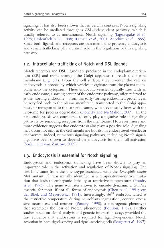
Figure 5.4 Endocytosis and trafficking of Notch receptor during lysosomal degradation and noncanonical activation. Inactive Notch receptors undergo constitutive endocytosis, endocytic trafficking, and are finally degraded in the lysosome. Full-length Notch receptors are firstly monoubiquitinated by E3 ligase Deltex, Su(dx), or DNedd4 for internalization. With the help of Rab5 and Avl, Notch receptors enter into the early endosome, from which they can recycle back to the cell surface or further progress into MVB/late endosomes. Sorting of Notch receptors into intraluminal vesicles is mediated by Hrs and ESCRT complexes (I, II, and III). Lgd, a C2-containing phospholipid binding protein, is placed between Hrs and ESCRT complexes based on epistatic analysis results. HOPS and AP-3 complexes are involved in endocytic trafficking/fusion between late endosome and lysosome. They are required for Dx-dependent Notch signaling activation. When endocytic trafficking of full-length Notch receptor for lysosomal degradation is disrupted, Notch receptor can undergo proteolytic cleavages in a ligand-independent manner (dashed line), which leads to ectopic activation of Notch signaling. (See Color Insert.)
of Notch signaling is proposed to be critical in certain cancers (Tanaka et al., 2008).
3.2. The controversy on the requirement of endocytosis for S3 cleavage
Proteins involved in early steps of endocytosis, including dynamin, Rab5, and Avl, are required in the signal-receiving cell for Notch activation

180 Shinya Yamamoto et al.
(Lu and Bilder, 2005; Vaccari et al., 2008). However, it is still not clear which step in canonical Notch signaling activation requires endocytosis. Since the ligand–receptor interaction and S2 cleavage of Notch receptors takes place on the cell surface, the current debate is focused on whether endocytosis is essential for effective S3 cleavage and precisely where the cleavage occurs.
3.2.1. Endocytosis is required for S3 cleavage The S3 cleavage of Notch is mediated by the γ-secretase, an intramembrane protease complex (De Strooper et al., 1999). Since mutations in γ-secretase components, like presenilin, are associated with Alzheimer’s disease via aberrant cleavage of amyloid precursor protein and production of pathogenic Aβ (Levy-Lahad et al., 1995; Rogaev et al., 1995; Sherrington et al., 1995), there has been much interest in understanding how, when, and where this cleavage takes place. The γ-secretase complex is present on plasma membranes, endocytic compartments, lysosomes, ER, and Golgi apparatus (Small and Gandy, 2006). Since the endocytic pathway is involved in Aβ production (Koo and Squazzo, 1994), endocytosis may play a positive role in γ-secretase-mediated S3 cleavage of Notch.
Several observations support a requirement of endocytosis for S3 cleavage. The activity of γ-secretase has been suggested to be higher in acidic environments, implicating that the S3 cleavage is more efficient in the endocytic compartments where the pH is lower (Pasternak et al., 2003). In support of this idea, defects in proteins involved in acidification of endosome affect Notch signaling. Mutations in Rabconnectin (Rbcn)3A, and Rbcn-3B, which assist the assembly of V-ATPase, as well as mutations in a subunit of V-ATPase, impair the acidification of endosomal compartments and lead to accumulation of the Notch receptor in enlarged late endosomes in Drosophila follicle cells and imaginal disc cells. This disruption of Notch signaling occurs after S2 cleavage in the receiving cells, supporting the idea that γ-secretase cleavage may be defective in these mutants (Yan et al., 2009). A mutation in big brain (bib), a gene encoding a monovalent cation (including Hþ) transporter (Yanochko and Yool, 2002), was initially reported to cause a Notch endocytic trafficking and signaling defect (Kanwar and Fortini, 2008). Note that the endocytic trafficking defects in bib mutants were later found to be the result of a second-site mutation while the cleaned bib mutant chromosome still exhibits Notch signaling defects (Fortini and Bilder, 2009). The Notch signaling defects in bib mutants may be due to improper pH environment in endosomes and lysosomes as acidification of these organelles in bib mutant cells is strongly attenuated (Fortini and Bilder, 2009; Kanwar and Fortini, 2008).

181 Notch Signaling and Endocytosis
In Drosophila Rab5 and Avl mutants, Notch accumulates at or near the cell surface and NICD production by γ-secretase is largely reduced, suggesting that S3 cleavage occurs less efficiently at the plasma membrane (Vaccari et al., 2008). In addition, with a point mutation at the S3 cleavage site in the NEXT-like fragment, this modified fragment can be detected in the endosomes, suggesting that S3 cleavage might occur in the endosomal compartments (Gupta-Rossi et al., 2004). These data suggest that upon S2 cleavage, Notch is endocytosed into an acidified endosomal compartment where S3 cleavage takes place by the g-secretase complex.
3.2.2. S3 cleavage can occur WITHOUT endocytosis of Notch Some data argue that there is no requirement of dynamin for γ-secretasemediated S3 cleavage of Notch following removal of the Notch ectodomain and generation of NEXT (Struhl and Adachi, 2000). For example, NEXT remains associated with the apical membrane in γ-secretase mutant cells. Importantly, overexpressed NEXT-like fragments can be cleaved in shi mutant pupal nota and embryos (Lopez-Schier and St Johnston, 2002; Seugnet et al., 1997). Furthermore, in a mammalian cell culture system, active γ-secretase complex can be purified from the plasma membrane that still contains Notch fragment cleavage activity (Chyung et al., 2005). Several reports document that the optimal pH environment for γ-secretase is 6.8–7.4, suggesting that acidic endosomes are not required for S3 cleavage (Lee et al., 2002; McLendon et al., 2000; Zhang et al., 2001). Moreover, a NEXT-like fragment with a point mutation at the ubiquitination site cannot be monoubiquitinated and endocytosed. Though the mutated NEXT fragment was thought to be unable to be cleaved into NICD (Gupta-Rossi et al., 2004), it was found later that this fragment can still be cleaved at the plasma membrane producing a less stable form of NICD with a shift in the cleavage position (Tagami et al., 2008). Tagami et al. (2008) further argued that during S3 cleavage, γ-secretase can process NEXT into various forms of NICD depending on the exact position of the cleavage, including NICD-S(þ3), NICD-L(þ1), NICD-L(þ2), and NICD-V (the most stable one; refer to the original NICD), with similar transactivation activity. Cleavage at the plasma membrane preferred the production of the more stable NICD-V, while cleavage in the endosomes leads to the production of the less stable NICD-S(þ3), arguing against the idea that endocytosis promotes γ-secretase cleavage. All together, these data suggest that endocytosis is not essential for S3 cleavage of Notch and that γ-secretase is able to mediate the cleavage at the plasma membrane.
In summary, whether endocytosis promotes γ-secretase cleavage or not is still a matter of debate. The controversy may be due to the fact that
182 Shinya Yamamoto et al.
conclusions are based on different model systems (Drosophila vs. mammalian cell culture) and that many of the data are based on overexpression strategies to generate Notch fragment intermediates. Considering the stringent dosage dependence of Notch signaling in different contexts, endocytosis may play either a positive or a negative role in γ-secretase cleavage in different contexts. Additional studies are required to reveal a more detailed picture of the relationship between endocytosis and canonical Notch signal activation.
3.3. Degradation of Notch receptors through the lysosomal pathway
Inactive Notch receptors undergo constitutive endocytosis, endocytic trafficking, and are eventually degraded in the lysosome (Jehn et al., 2002). However, the acidic environment during trafficking might promote the dissociation of the Notch heterodimer and produce membrane-tethered NEXT, which can be further processed by γ-secretase in the endosomes. Production of NICD may bypass the requirement for ligand binding as well as S2 cleavage and even occur on lysosomal membranes (Wilkin et al., 2008).
Mutations in genes involved in the degradation pathway can lead to an accumulation of Notch in endosomes and ectopic activation of Notch signaling in a ligand-independent manner. In other words, some proteins must prevent the ectopic activation of Notch and act as negative regulators of Notch signaling. These include proteins that regulate the ubiquitination of Notch, proteins involved in the maturation of early endosomes into MVB, and proteins that mediate the endocytic trafficking and fusion between late endosomes and lysosomes. The regulation of Notch signaling by these proteins is discussed below.
3.5.1. E3 ligases for ubiquitination of Notch receptors Multiple E3 ligases ubiquitinate Notch and promote its internalization to regulate its signaling activity. These include Deltex (dx), Su(dx) (Suppressor of deltex), and DNedd4 (Brennan and Gardner, 2002; Kanwar and Fortini, 2004).
Dx, a RING finger E3 ubiquitin ligase, was originally identified as a positive regulator of Notch signaling based on genetic studies (Diederich et al., 1994; Matsuno et al., 1995; Xu and Artavanis-Tsakonas, 1990). Loss of dx leads to a Notch signaling impairment in certain cell contexts (Drosophila eye and wing imaginal discs). In addition, overexpression of dx results in Notch signaling activation in the dorsal–ventral boundary of the wing, independent of DSL ligands and CSL. This ectopic signaling activation requires the internalization of Notch into Rab7-positive late endosomes
183 Notch Signaling and Endocytosis
(Fuwa et al., 2006; Hori et al., 2004; Wilkin et al., 2004, 2008). However, in contrast to its positive role in Notch signaling activation, Dx was also found to promote Notch receptor degradation when it forms a complex with Kurtz (Krz), the Drosophila homolog of a nonvisual β-arrestin (Mukherjee et al., 2005). Therefore, Dx can act both in a positive and in a negative manner in Notch signaling depending on the context and interacting partners.
The Drosophila HECT (Homologous to the E6-AP Carboxyl Terminus) as full form of HECT domain-containing family of E3 ligases includes three members: Su(dx), DNedd4, and D-smurf. Much of data related to these studies were obtained from studies in the Drosophila wing margin, ovary development, and cultured S2 cells. Su(dx) and dNedd4 can ubiquitinate full-length Notch in a ligand-independent manner and promote its entry in the lysosomal degradation pathway (Sakata et al., 2004; Wilkin et al., 2004). Loss of Su(dx) and dNedd4 causes Notch gain-of-function phenotypes, while overexpression causes Notch loss-of-function phenotypes. Su(dx) and dNedd4 mutations can also suppress Notch partial loss-of-function phenotypes, supporting the idea that these proteins play negative roles in Notch signaling (Cornell et al., 1999; Fostier et al., 1998; Mazaleyrat et al., 2003; Qiu et al., 2000; Sakata et al., 2004; Wilkin et al., 2004). The third member of Nedd4 family, D-Smurf, has been suspected to have some functional redundancy with the other two members, but a direct role in Notch signaling has yet to be demonstrated (Wilkin et al., 2004). Finally, the protein levels of Dx are negatively correlated to the expression level of Nedd4 family proteins, implicating their role in regulation of the Dx protein level (Wilkin et al., 2004). Thus, Nedd4 family proteins might regulate Notch signaling by directly promoting lysosomal Notch degradation and by regulating the protein level of Dx.
In mammals, the homolog of Su(dx) (named AIP4/Itch) has also been reported to ubiquitinate full-length Notch-1 in a ligand-independent manner and to promote its lysosomal degradation (Chastagner et al., 2008; Qiu et al., 2000). The adaptor protein Numb can interact with AIP4/Itch to promote this degradation process (McGill et al., 2009; McGill and McGlade, 2003). In addition, AIP4/Itch was also shown to mediate lysosomal degradation of Dx through ubiquitination (Chastagner et al., 2006). Another mammalian RING type E3 ubiquitin ligase c-Cbl can also promote the degradation of the Notch-1 NEXT fragment (Jehn et al., 2002). However, whether c-Cbl can target full-length Notch-1 for degradation is still unclear. Although c-Cbl is conserved in Drosophila, it awaits to be tested whether it can function in regulating Notch signaling.
3.3.2. Lgd and ESCRT complex Proteins involved in the maturation of early endosomes into MVB, such as ESCRT complexes and Lethal (2) giant discs (Lgd), have been shown

184 Shinya Yamamoto et al.
to function in Notch degradation. ESCRT 0 or STAM/Hrs is localized to early endosomes and can bind and transfer ubiquitinated cargo to the ESCRT I/II/III complexes (Raiborg and Stenmark, 2009). Lgd is a C2 domain-containing protein that binds to phospholipids. lgd mutants have been shown to exhibit general protein sorting defects. Loss of lgd leads to accumulation of Notch in Hrs-positive endosomes as well as ectopic Notch signaling activation. This ectopic Notch activation is ligand-independent since the activation is also observed in lgd Dl Ser triple mutant clones (Childress et al., 2006; Jaekel and Klein, 2006). In hrs lgd double mutant clones, this ligand-independent Notch activation is blocked while ligand-dependent activation remains unaffected. Thus, Lgd functions to prevent ligand-independent Notch activation in an Hrs-dependent manner (Childress et al., 2006; Gallagher and Knoblich, 2006; Jaekel and Klein, 2006; Klein, 2003). Interestingly, in hrs single mutant, Notch receptors accumulate in the Avl-positive early endosomes but remain inactive (Jekely and Rorth, 2003; Lloyd et al., 2002; Lu and Bilder, 2005; Thompson et al., 2005). Therefore, Hrs is only required for ectopic activation of Notch signaling in the lgd mutant background but not in a wild-type fly.
Mutations in Drosophila ESCRT I (tsg101/erupted and vps28), ESCRT II (vps22, vps25, and vps36), and ESCRT III (vps2, vps20, and vps32) complexes all result in accumulation of Notch receptors in early endosomes and most of them cause ectopic Notch signaling activation in developing imaginal discs (Herz et al., 2006, 2009; Moberg et al., 2005; Thompson et al., 2005; Vaccari and Bilder, 2005; Vaccari et al., 2009). The ectopic activation of Notch signaling up-regulates the expression of the ligand of JAK/STAT pathway, Unpaired, which in turn promotes the overgrowth of surrounding wild-type cells in a nonautonomous manner. Although ESCRT and lgd mutants both exhibit accumulation of Notch receptors and ectopic Notch signaling activity, they have also distinct phenotypes. ESCRT mutant cells lose epithelial organization and eventually die while inducing non-cell-autonomous tissue growth (Herz et al., 2006, 2009; Moberg et al., 2005; Thompson et al., 2005; Vaccari and Bilder, 2005; Vaccari et al., 2009). Conversely, lgd mutant cells display cell-autonomous overgrowth and apoptosis while still maintaining their epithelial organization (Childress et al., 2006; Gallagher and Knoblich, 2006; Jaekel and Klein, 2006; Klein, 2003).
3.3.3. Other genes involved in Notch receptor trafficking Several other genes/proteins have been shown to affect Notch signaling by affecting endocytosis and trafficking of Notch receptors. Mutations in genes involved in trafficking/fusion between late endosomes and lysosomes, such as the HOPS and AP-3 complexes, play a regulatory role in Notch

185 Notch Signaling and Endocytosis
degradation (Wilkin et al., 2008). Loss-of-function mutations in these genes act as genetic modifiers of ectopic Notch signaling caused by overexpression of dx. However, these mutants only show minor, if any, Notch signaling defects on their own. Tumor suppressors Merlin (Mer) and expanded (ex) encodes proteins that belong to the FERM (four-point one, ezrin, radixin, moesin) domain superfamily and promote endocytosis and clearance of Notch receptors from the cell surface (Maitra et al., 2006). In Mer ex double mutant flies, Notch accumulates at the plasma membrane, but these mutants do not exhibit any obvious defects in Notch signaling.
Proteins involved in membrane lipid biosynthesis can also modulate Notch signaling activity, possibly through their effect on the endocytic processes by altering the phospholipid composition in the plasma membrane and endosomal membranes. For example, the Caenorhabditis elegans BRE-5 (BT-toxin resistance) catalyzes the biosynthesis of glycosphingolipids (GSL), which are enriched in the lipid raft. Knockdown of BRE-5 can suppress hypermorphic LIN-12 (C. elegans homolog of Notch) egg-laying phenotypes (Katic et al., 2005). In Drosophila, mutations in cytidylyltransferase-1, a rate-limiting enzyme in phosphatidylcholine (PC) biosynthesis, show reduction in Notch signaling and an increased late endosomal localization of Notch receptor (Weber et al., 2003). These data suggest a positive role for GSL and PC in Notch signaling.
In brief, degradation of inactive Notch receptors through the endocytic pathway provides a mechanism to prevent ectopic Notch activation. Mutations in endocytosis-related genes, including E3 ubiquitin ligases, endocytic trafficking proteins, and enzymes involved in phospholipid biosynthesis, can cause abnormal Notch signaling activity. These similar but distinct mutant phenotypes, combined with their context dependence, reveal their different roles in Notch degradation and their partial redundancy in protein sorting and vesicle trafficking.
4. Regulation of Notch Signaling by Endocytosis and Vesicle Trafficking During Mechanosensory Organ Development in Drosophila
4.1. Introduction to mechanosensory organ development
The development of the mechanosensory organs of the Drosophila peripheral nervous system has served as a model system to understand many aspects of Notch signaling including endocytic trafficking. The body of an adult fly is covered by hundreds of mechanosensory bristles that act as sensors. Each

186 Shinya Yamamoto et al.
bristle is composed of four different cell types: socket, hair, sheath, and neuron (Hartenstein and Posakony, 1989). These cells arise from series of asymmetric cell division of a sensory organ precursor (SOP) cell and subsequent unidirectional Notch signaling between the daughter cells during pupariation (Jan and Jan, 2001). In the dorsal thorax or notum, the division of the SOP occurs in parallel to the anterior–posterior body axis. The anterior cell becomes the signal-sending cell (pIIb), whereas the posterior cell becomes the signal-receiving cell (pIIa), leading to asymmetric activation of Notch signaling. The pIIb gives rise to the internal cells (sheath and neuron), whereas the pIIa becomes the progenitor of the external cells (socket and hair). When Notch signaling is lost, the pIIa transforms into a pIIb, leading to loss of external cells and gain of internal cells (de Celis et al., 1991; Hartenstein and Posakony, 1990; Zeng et al., 1998). Conversely, when Notch signaling is ectopically activated in both cells, there is a pIIb-to-pIIa cell fate change, leading to the gain of external cells and loss of internal cells (Frise et al., 1996; Guo et al., 1996). Notch signaling needs to be tightly controlled and the cells of the bristle lineage achieve this via an asymmetric segregation of endocytic factors, which are often referred to as “cell fate determinants.” Here, we will especially focus on the specification of the pIIa and pIIb cells, and discuss how the endocytic and trafficking machinery is employed to bias Notch signaling.
4.2. Setting up the asymmetry in the SOP cell
During division of the SOP, the cell fate determinants Neur and Numb become enriched at the anterior pole, forming a crescent (Le Borgne and Schweisguth, 2003b; Rhyu et al., 1994) (Fig. 5.5A). The division then allows the anterior pIIb to inherit these two factors, whereas the posterior pIIa does not. The formation of the anterior crescent is determined by cell polarity factors (Bardin et al., 2004; Betschinger and Knoblich, 2004) and planar cell polarity cues to assure that the division of the SOP occurs along the anterior–posterior axis so that the cell fate determinants are properly segregated into the pIIb cell. In parallel to the asymmetric segregation of Neur and Numb, the inheritance of endocytic compartments are also biased between pIIa and pIIb cells. Rab11-positive recycling endosomes become enriched in pIIb cells, due to asymmetric enrichment of nuclear fallout (Nuf) around the pIIb cell centrosome after mitosis. Nuf is the Drosophila homolog of arfophilins, an effector of Rab11 that is required for recycling endosome formation and function (Emery et al., 2005). On the other hand, endosomes that are positive for Sara becomes asymmetrically segregated into the pIIa cell (Coumailleau et al., 2009). The asymmetric inheritance of cell fate determinants together with asymmetric redistribution of endocytic compartments works in concert to assure that Notch signaling occurs unidirectionally. Here, we will next discuss how each

187 Notch Signaling and Endocytosis
of these factors contributes to establishment of a signal-sending cell and a signal-receiving cell during pIIa vs. pIIb cell fate determination.
4.3. Role of asymmetrically segregated Neuralized and Delta recycling in the pIIb cell
In the pIIb cell, Neur ubiquitinates Delta and promotes its endocytosis (Le Borgne and Schweisguth, 2003b) (Fig. 5.5B). Endocytosed Delta is then sorted into a basal Rab11-positive compartment (Emery et al., 2005; Jafar-Nejad et al., 2005). In cells mutant for Sec15, a component of the exocyst complex and an effector of Rab11 (Wu et al., 2005), Delta is stuck in this basal compartment and it cannot recycle back to the cell surface (Jafar-Nejad et al., 2005). Similar phenotypes are observed in cells mutant for the Arp2/3 complex and WASp, mediators of branched actin polymerization (Rajan et al., 2009). Based on detailed phenotypic analysis of these mutants, a model has been proposed where Delta recycles back to the apical plasma membrane from the basal recycling endosomes with the help of the exocyst complex and via actin polymerization through the Arp2/3–WASp complex in the pIIb cell. One interesting possibility is that basal Delta-positive vesicles may recruit Arp2/3 complex to propel them to the apical region via a force generated by actin polymerization, analogous to Listeria monocytogenes recruiting and activating Arp2/3 for intracellular motility (Lambrechts et al., 2008). This Neur-mediated recycling of Delta is essential for Notch signaling in the mechanosensory lineage since mutations in Sec15 and Arp2/3–WASp complex lead to loss of Notch signaling. Since the distribution of Neur is restricted to the pIIb cell, Delta in the pIIa cell cannot be endocytosed/recycled, and hence cannot activate the Notch receptor on the pIIb cell.
Recently, it was reported that the apical membranes of the pIIa and pIIb cells are enriched in polymerized actin (Rajan et al., 2009). This structure, which was referred to as an apical actin-rich structure (ARS), is rich in microvilli and recycled Delta. The microvilli of the ARS may be the site of ligand–receptor interaction, and the microvilli may promote Notch signaling by increasing the surface area between the signaling cells. In Arp2/3– WASp complex mutants, the ARS becomes smaller, which may contribute to the Notch loss-of-function defects in these cells.
4.4. Role of asymmetrically segregated Numb in the pIIb cell
The pIIb cell not only inherits Neuralized but also inherits Numb, which acts as a negative regulator of signal reception (Rhyu et al., 1994) (Fig. 5.5B). Numb is an endocytic protein that interacts with the AP-2 complex (Santolini et al., 2000). In the pIIb cell, Numb binds α-adaptin of the AP-2 complex (Berdnik et al., 2002) and promotes the endocytosis of

(A) Anterior Posterior [SOP]
NumbCell polarity Neurfactors
[pllb] [plla]
Rab11 Sara endosomes endosomes
(B)
[pllb] [plla]
Signal Signal sending receiving
Spdo Sara Dynamin Neur
Sec15 Arp2/3 WASp
Rab11
Numb AP-2
188 Shinya Yamamoto et al.
Sanpodo (Spdo) (Hutterer and Knoblich, 2005; Langevin et al., 2005; Roegiers et al., 2005). Spdo is a four transmembrane protein that acts as a positive regulator of Notch signaling when present at the plasma membrane through an unknown mechanism (Babaoglan et al., 2009; O’Connor-Giles
Figure 5.5 (Continued)

189 Notch Signaling and Endocytosis
and Skeath, 2003). In the pIIb cell, Spdo is sequestered away from the plasma membrane in an endocytic compartment, where it is thought to be nonfunctional. In contrast, Spdo is localized at the plasma membrane in the pIIa cell, where Numb is absent, and Notch signaling reception is promoted. Thus, by segregating Neur and Numb into the pIIb cell, this cell becomes the signal-sending cell, whereas the pIIa becomes the signal-receiving cell.
4.5. Role of asymmetrically segregated Sara-endosomes in the pIIa cell
Recently, endosomes that are marked by Sara (Sara-endosomes) have been identified as a third cell fate determinant during pIIa–pIIb cell fate specification (Coumailleau et al., 2009). Sara is a FYVE domain-containing adaptor protein that localizes to a subpopulation of PI3P-containing endosomes (Bokel et al., 2006; Tsukazaki et al., 1998). In the SOP, a population of Notch and Delta are endocytosed into Sara-endosomes that become segregated into the pIIa cell. Although loss of function of Sara does not exhibit any defect in the bristle lineage, overexpression of Sara or inheritance of a
Figure 5.5 Regulation of Notch signaling via endocytosis and vesicle trafficking during mechanosensory organ development in Drosophila. (A) During mitosis of the sensory organ precursor (SOP) cell, cell fate determinants Neuralized (Neur) and Numb are asymmetrically segregated into the anterior crescent which is determined through interactions between cell polarity factors. Upon cytokinesis, Neur and Numb are both inherited by the anterior pIIb cell, whereas the posterior cell fails lacks these factors. In parallel to the asymmetric segregation of the two cell fate determinants, Rab11-positive recycling endosomes become enriched in the pIIb cell, whereas Sara-positive endosomes are sorted into the pIIa cell. This asymmetric segregation of cell fate determinants and specific endocytic compartment biases the following Notch signaling between the pIIa and the pIIb cell. (B) In the pIIb cell, Neur promotes the endocytosis and sorting of Delta for activation. Activated Delta traffics through Rab11-positive endosomes and recycle back to the apical cell surface where there is an enrichment of actin filaments and microvilli, referred to as the ARS (apical actin-rich structure). Sec15, a Rab11 effector and component of the exocyst complex, is required for this apical recycling of Delta. In addition, Arp2/3 and WASp, positive regulators of actin polymerization, are also required for recycling of Delta, possibly through mobilization of Delta-positive vesicles and/or facilitation of ARS formation. Activated Delta that returned to the cell surface interacts with and activates Notch in the pIIa cell. Sanpodo (Spdo), a four transmembrane domain protein, is present at the cell surface of the pIIa cell to promote the reception of this signal. pIIa cell cannot signal to the pIIb since they are not able to activate Delta due to lack of Neur. In addition, pIIb cannot receive Notch signaling since Spdo is endocytosed by Numb and kept in an inactive form. As a third mechanism, Sara-positive endosome has been recently been proposed to bias Notch signaling by actively recruiting Notch and Delta into the pIIa cell. γ-secretase cleavage of Notch has been proposed to be happening at or before Notch entering the Sara-positive endosome, but the exact mechanism and role of Sara is not fully understood. (See Color Insert.)
190 Shinya Yamamoto et al.
single giant Sara-endosome generated by constitutively active Rab5 expression into the pIIb cell, can mediate pIIb-to-pIIa cell fate transformation, caused by a Notch gain of function. Together with the observation that γsecretase-dependent cleavage of Notch may be taking place at or before entry into Sara-endosomes in the pIIa, segregation of Delta and Notch through Sara-endosomes into pIIa may create a small asymmetry in the signaling activity between the pIIa and pIIb, which is further amplified by actions of asymmetric segregated Neur and Numb. However, it is important to note that loss of function of Sara as well as artificial segregation of Sara-endosome away from pIIa cell does not lead to loss of Notch signaling defects, suggesting that the Sara-endosome is not an essential component of Notch activation and plays a regulatory role during bristle development.
In summary, cells of the mechanosensory lineage utilize the endocytic pathway in order to restrict and regulate the activity of Delta and Notch to achieve the proper fate via unidirectional Notch signaling. Since loss of function in genes such as Numb and Sec15 show defects in bristle development and other binary cell fate determination events but not in all Notch signaling-dependent events (Jafar-Nejad et al., 2005; Mehta et al., 2005; O’Connor-Giles and Skeath, 2003), there seems to be context specificity in the utilization of the endocytic pathway to achieve successful Notch signaling mediated decisions in vivo. Since defects in mechanosensory organ development can be subjected to high-throughput forward genetic screens using clonal analysis (Berdnik et al., 2002; Jafar-Nejad et al., 2005) identification of novel genes and endocytic pathways that regulate Notch signaling are likely to continue to be discovered using this model system.
5. Conclusion and Future Directions
In both signal-sending and receiving cells, the vesicle trafficking routes not only activate Notch signaling but also fine tune the signal output. Endocytosis and vesicle trafficking mediate the activation of DSL ligands through the recycling pathway, generate a pulling force to promote the S2 cleavage of Notch upon ligand–receptor interaction, may regulate the S3 cleavage to release the active NICD fragment, promote degradation of inactive Notch, and control ligand-independent activation of the pathway. It is important to keep in mind that different cell types have distinct trafficking properties and that cell context is important.
There are numerous questions that remain unanswered in this area. What is the activated state of DSL ligands upon entry into the specialized recycling pathway? What is the exact function of endocytosis in the signal-receiving cells? Which endocytic factors are true universal regulators and which factors act in context/species-specific manner? Are ARS and apically

191 Notch Signaling and Endocytosis
enriched microvilli the sites where Notch signaling takes place and are these structures seen in other cell types that signal though Notch? What other genes and trafficking pathways regulate Notch? What fraction of developmental disorders and human diseases are caused by defects in endocytosis and trafficking of Notch signal components? We believe that the answers to these and many other questions will come from integration of various studies from different fields. We have emphasized genetic approaches in Drosophila in this chapter because much of the key observations related to endocytosis and vesicle trafficking in Notch signaling were first made in Drosophila. We hope that further insights into the importance and various roles of endocytosis and vesicle trafficking in Notch signaling will be forthcoming, not only from the fly field but also from experiments in vertebrates.
ACKNOWLEDGMENTS
We would like to thank Mark Fortini, Nikolaos Giagtzoglou, and An-Chi Tien for useful suggestions. We apologize to all our colleagues for not being able to cite their work given the length restrictions. SY is supported by the Nakajima Foundation and HJB is Investigator with the Howard Hughes Medical Institute.
REFERENCES
Acconcia, F., Sigismund, S., and Polo, S. (2009). Ubiquitin in trafficking: the network at work. Exp. Cell Res. 315, 1610–1618.
Ahimou, F., Mok, L. P., Bardot, B., and Wesley, C. (2004). The adhesion force of Notch with Delta and the rate of Notch signaling. J. Cell Biol. 167, 1217–1229.
Babaoglan, A. B., O’Connor-Giles, K. M., Mistry, H., Schickedanz, A., Wilson, B. A., and Skeath, J. B. (2009). Sanpodo: a context-dependent activator and inhibitor of Notch signaling during asymmetric divisions. Development 136, 4089–4098.
Bardin, A. J., Le Borgne, R., and Schweisguth, F. (2004). Asymmetric localization and function of cell-fate determinants: a fly’s view. Curr. Opin. Neurobiol. 14, 6–14.
Bardin, A. J., and Schweisguth, F. (2006). Bearded family members inhibit Neuralizedmediated endocytosis and signaling activity of Delta in Drosophila. Dev. Cell 10, 245–255.
Ben-Yaacov, S., Le Borgne, R., Abramson, I., Schweisguth, F., and Schejter, E. D. (2001). Wasp, the Drosophila Wiskott-Aldrich syndrome gene homologue, is required for cell fate decisions mediated by Notch signaling. J. Cell Biol. 152, 1–13.
Berdnik, D., Torok, T., Gonzalez-Gaitan, M., and Knoblich, J. A. (2002). The endocytic protein alpha-Adaptin is required for numb-mediated asymmetric cell division in Drosophila. Dev. Cell 3, 221–231.
Betschinger, J., and Knoblich, J. A. (2004). Dare to be different: asymmetric cell division in Drosophila, C. elegans and vertebrates. Curr. Biol. 14, R674–R685.
Bokel, C., Schwabedissen, A., Entchev, E., Renaud, O., and Gonzalez-Gaitan, M. (2006). Sara endosomes and the maintenance of Dpp signaling levels across mitosis. Science 314, 1135–1139.
Boulianne, G. L., de la Concha, A., Campos-Ortega, J. A., Jan, L. Y., and Jan, Y. N. (1991). The Drosophila neurogenic gene neuralized encodes a novel protein and is expressed in precursors of larval and adult neurons. EMBO J. 10, 2975–2983.
Brennan, K., and Gardner, P. (2002). Notching up another pathway. Bioessays 24, 405–410.

192 Shinya Yamamoto et al.
Chanet, S., Vodovar, N., Mayau, V., and Schweisguth, F. (2009). Genome engineering-based analysis of Bearded family genes reveals both functional redundancy and a nonessential function in lateral inhibition in Drosophila. Genetics 182, 1101–1108.
Chastagner, P., Israel, A., and Brou, C. (2006). Itch/AIP4 mediates Deltex degradation through the formation of K29-linked polyubiquitin chains. EMBO Rep. 7, 1147–1153.
Chastagner, P., Israel, A., and Brou, C. (2008). AIP4/Itch regulates Notch receptor degradation in the absence of ligand. PLoS ONE 3, e2735.
Chen, H., Fre, S., Slepnev, V. I., Capua, M. R., Takei, K., Butler, M. H., Di Fiore, P. P., and De Camilli, P. (1998). Epsin is an EH-domain-binding protein implicated in clathrin-mediated endocytosis. Nature 394, 793–797.
Chen, H., Ko, G., Zatti, A., Di Giacomo, G., Liu, L., Raiteri, E., Perucco, E., Collesi, C., Min, W., Zeiss, C., De Camilli, P., and Cremona, O. (2009). Embryonic arrest at midgestation and disruption of Notch signaling produced by the absence of both epsin 1 and epsin 2 in mice. Proc. Natl. Acad. Sci. U.S.A.106, 13838–13843.
Chen, M. S., Obar, R. A., Schroeder, C. C., Austin, T. W., Poodry, C. A., Wadsworth, S. C., and Vallee, R. B. (1991). Multiple forms of dynamin are encoded by shibire, a Drosophila gene involved in endocytosis. Nature 351, 583–586.
Childress, J. L., Acar, M., Tao, C., and Halder, G. (2006). Lethal giant discs, a novel C2domain protein, restricts notch activation during endocytosis. Curr. Biol. 16, 2228–2233.
Chitnis, A. (2006). Why is delta endocytosis required for effective activation of notch? Dev. Dyn. 235, 886–894.
Chyung, J. H., Raper, D. M., and Selkoe, D. J. (2005). Gamma-secretase exists on the plasma membrane as an intact complex that accepts substrates and effects intramembrane cleavage. J. Biol. Chem. 280, 4383–4392.
Commisso, C., and Boulianne, G. L. (2007). The NHR1 domain of Neuralized binds Delta and mediates Delta trafficking and Notch signaling. Mol. Biol. Cell 18, 1–13.
Cornell, M., Evans, D. A., Mann, R., Fostier, M., Flasza, M., Monthatong, M., Artavanis-Tsakonas, S., and Baron, M. (1999). The Drosophila melanogaster Suppressor of deltex gene, a regulator of the Notch receptor signaling pathway, is an E3 class ubiquitin ligase. Genetics 152, 567–576.
Coumailleau, F., Furthauer, M., Knoblich, J. A., and Gonzalez-Gaitan, M. (2009). Directional Delta and Notch trafficking in Sara endosomes during asymmetric cell division. Nature 458, 1051–1055.
d’Azzo, A., Bongiovanni, A., and Nastasi, T. (2005). E3 ubiquitin ligases as regulators of membrane protein trafficking and degradation. Traffic 6, 429–441.
De Camilli, P., Chen, H., Hyman, J., Panepucci, E., Bateman, A., and Brunger, A. T. (2002). The ENTH domain. FEBS Lett. 513, 11–18.
de Celis, J. F., Mari-Beffa, M., and Garcia-Bellido, A. (1991). Cell-autonomous role of Notch, an epidermal growth factor homologue, in sensory organ differentiation in Drosophila. Proc. Natl. Acad. Sci. U.S.A. 88, 632–636.
De Renzis, S., Yu, J., Zinzen, R., and Wieschaus, E. (2006). Dorsal-ventral pattern of Delta trafficking is established by a Snail-Tom-Neuralized pathway. Dev. Cell 10, 257–264.
De Strooper, B., Annaert, W., Cupers, P., Saftig, P., Craessaerts, K., Mumm, J. S., Schroeter, E. H., Schrijvers, V., Wolfe, M. S., Ray, W. J., Goate, A., and Kopan, R., (1999). A presenilin-1dependent gamma-secretase-like protease mediates release of Notch intracellular domain. Nature 398, 518–522.
Deblandre, G. A., Lai, E. C., and Kintner, C. (2001). Xenopus neuralized is a ubiquitin ligase that interacts with XDelta1 and regulates Notch signaling. Dev. Cell 1, 795–806.
Dell’Angelica, E. C. (2009). AP-3-dependent trafficking and disease: the first decade. Curr. Opin. Cell Biol. 21, 552–559.
Di Paolo, G., and De Camilli, P. (2006). Phosphoinositides in cell regulation and membrane dynamics. Nature 443, 651–657.

193 Notch Signaling and Endocytosis
Diederich, R. J., Matsuno, K., Hing, H., and Artavanis-Tsakonas, S. (1994). Cytosolic interaction between deltex and Notch ankyrin repeats implicates deltex in the Notch signaling pathway. Development 120, 473–481.
Doherty, G. J., and McMahon, H. T. (2009). Mechanisms of endocytosis. Annu. Rev. Biochem. 78, 857–902.
Eisenberg, E., and Greene, L. E. (2007). Multiple roles of auxilin and Hsc70 in clathrinmediated endocytosis. Traffic 8, 640–646.
Emery, G., Hutterer, A., Berdnik, D., Mayer, B., Wirtz-Peitz, F., Gaitan, M. G., and Knoblich, J. A. (2005). Asymmetric Rab 11 endosomes regulate delta recycling and specify cell fate in the Drosophila nervous system. Cell 122, 763–773.
Eun, S. H., Banks, S. M., and Fischer, J. A. (2008). Auxilin is essential for Delta signaling. Development 135, 1089–1095.
Fehon, R. G., Kooh, P. J., Rebay, I., Regan, C. L., Xu, T., Muskavitch, M. A., and Artavanis-Tsakonas, S. (1990). Molecular interactions between the protein products of the neurogenic loci Notch and Delta, two EGF-homologous genes in Drosophila. Cell 61, 523–534.
Fontana, J. R., and Posakony, J. W. (2009). Both inhibition and activation of Notch signaling rely on a conserved Neuralized-binding motif in Bearded proteins and the Notch ligand Delta. Dev. Biol. 333, 373–385.
Fortini, M. E. (2009). Notch signaling: the core pathway and its posttranslational regulation. Dev. Cell 16, 633–647.
Fortini, M. E., and Bilder, D. (2009). Endocytic regulation of Notch signaling. Curr. Opin. Genet. Dev. 19, 323–328.
Fostier, M., Evans, D. A., Artavanis-Tsakonas, S., and Baron, M. (1998). Genetic characterization of the Drosophila melanogaster Suppressor of deltex gene: a regulator of notch signaling. Genetics 150, 1477–1485.
Frise, E., Knoblich, J. A., Younger-Shepherd, S., Jan, L. Y., and Jan, Y. N. (1996). The Drosophila Numb protein inhibits signaling of the Notch receptor during cell-cell interaction in sensory organ lineage. Proc. Natl. Acad. Sci. U.S.A. 93, 11925–11932.
Furthauer, M., and Gonzalez-Gaitan, M. (2009). Endocytic regulation of notch signalling during development. Traffic 10, 792–802.
Fuwa, T. J., Hori, K., Sasamura, T., Higgs, J., Baron, M., and Matsuno, K. (2006). The first deltex null mutant indicates tissue-specific deltex-dependent Notch signaling in Drosophila. Mol. Genet. Genomics 275, 251–263.
Gallagher, C. M., and Knoblich, J. A. (2006). The conserved c2 domain protein lethal (2) giant discs regulates protein trafficking in Drosophila. Dev. Cell 11, 641–653.
Glittenberg, M., Pitsouli, C., Garvey, C., Delidakis, C., and Bray, S. (2006). Role of conserved intracellular motifs in Serrate signalling, cis-inhibition and endocytosis. EMBO J. 25, 4697–4706.
Goley, E. D., and Welch, M. D. (2006). The ARP2/3 complex: an actin nucleator comes of age. Nat. Rev. 7, 713–726.
Gordon, W. R., Arnett, K. L., and Blacklow, S. C. (2008). The molecular logic of Notch signaling–a structural and biochemical perspective. J. Cell Sci. 121, 3109–3119.
Gordon, W. R., Roy, M., Vardar-Ulu, D., Garfinkel, M., Mansour, M. R., Aster, J. C., and Blacklow, S. C. (2009). Structure of the Notch1-negative regulatory region: implications for normal activation and pathogenic signaling in T-ALL. Blood 113, 4381–4390.
Gordon, W. R., Vardar-Ulu, D., Histen, G., Sanchez-Irizarry, C., Aster, J. C., and Black-low, S. C. (2007). Structural basis for autoinhibition of Notch. Nat. Struct. Mol. Biol. 14, 295–300.
Grant, B. D., and Donaldson, J. G. (2009). Pathways and mechanisms of endocytic recycling. Nat. Rev. 10, 597–608.
Guo, M., Jan, L. Y., and Jan, Y. N. (1996). Control of daughter cell fates during asymmetric division: interaction of Numb and Notch. Neuron 17, 27–41.

194 Shinya Yamamoto et al.
Gupta-Rossi, N., Six, E., LeBail, O., Logeat, F., Chastagner, P., Olry, A., Israel, A., and Brou, C. (2004). Monoubiquitination and endocytosis direct gamma-secretase cleavage of activated Notch receptor. J. Cell Biol. 166, 73–83.
Hagedorn, E. J., Bayraktar, J. L., Kandachar, V. R., Bai, T., Englert, D. M., and Chang, H. C. (2006). Drosophila melanogaster auxilin regulates the internalization of Delta to control activity of the Notch signaling pathway. J. Cell Biol. 173, 443–452.
Hansen, C. G., and Nichols, B. J. (2009). Molecular mechanisms of clathrin-independent endocytosis. J. Cell Sci. 122, 1713–1721.
Hartenstein, V., and Posakony, J. W. (1989). Development of adult sensilla on the wing and notum of Drosophila melanogaster. Development 107, 389–405.
Hartenstein, V., and Posakony, J. W. (1990). Sensillum development in the absence of cell division: the sensillum phenotype of the Drosophila mutant string. Dev. Biol. 138, 147–158.
Henderson, S. T., Gao, D., Christensen, S., and Kimble, J. (1997). Functional domains of LAG-2, a putative signaling ligand for LIN-12 and GLP-1 receptors in Caenorhabditis elegans. Mol. Biol. Cell 8, 1751–1762.
Herz, H. M., and Bergmann, A. (2009). Genetic analysis of ESCRT function in Drosophila: a tumour model for human Tsg101. Biochem. Soc. Trans. 37, 204–207.
Herz, H. M., Chen, Z., Scherr, H., Lackey, M., Bolduc, C., and Bergmann, A. (2006). vps25 mosaics display non-autonomous cell survival and overgrowth, and autonomous apoptosis. Development 133, 1871–1880.
Herz, H. M., Woodfield, S. E., Chen, Z., Bolduc, C., and Bergmann, A. (2009). Common and distinct genetic properties of ESCRT-II components in Drosophila. PLoS ONE 4, e4165.
Heuss, S. F., Ndiaye-Lobry, D., Six, E. M., Israel, A., and Logeat, F. (2008). The intracellular region of Notch ligands Dll1 and Dll3 regulates their trafficking and signaling activity. Proc. Natl. Acad. Sci. U.S.A. 105, 11212–11217.
Hicke, L., and Dunn, R. (2003). Regulation of membrane protein transport by ubiquitin and ubiquitin-binding proteins. Annu. Rev. Cell Dev. Biol. 19, 141–172.
Hori, K., Fostier, M., Ito, M., Fuwa, T. J., Go, M. J., Okano, H., Baron, M., and Matsuno, K. (2004). Drosophila deltex mediates suppressor of Hairless-independent and late-endosomal activation of Notch signaling. Development 131, 5527–5537.
Hukriede, N. A., Gu, Y., and Fleming, R. J. (1997). A dominant-negative form of Serrate acts as a general antagonist of Notch activation. Development 124, 3427–3437.
Hutterer, A., and Knoblich, J. A. (2005). Numb and alpha-Adaptin regulate Sanpodo endocytosis to specify cell fate in Drosophila external sensory organs. EMBO Rep. 6, 836–842.
Itoh, M., Kim, C. H., Palardy, G., Oda, T., Jiang, Y. J., Maust, D., Yeo, S. Y., Lorick, K., Wright, G. J., Ariza-McNaughton, L., et al. (2003). Mind bomb is a ubiquitin ligase that is essential for efficient activation of Notch signaling by Delta. Dev. Cell 4, 67–82.
Jaekel, R., and Klein, T. (2006). The Drosophila Notch inhibitor and tumor suppressor gene lethal (2) giant discs encodes a conserved regulator of endosomal trafficking. Dev. Cell 11, 655–669.
Jafar-Nejad, H., Andrews, H. K., Acar, M., Bayat, V., Wirtz-Peitz, F., Mehta, S. Q., Knoblich, J. A., and Bellen, H. J. (2005). Sec15, a component of the exocyst, promotes notch signaling during the asymmetric division of Drosophila sensory organ precursors. Dev. Cell 9, 351–363.
Jan, Y. N., and Jan, L. Y. (2001). Asymmetric cell division in the Drosophila nervous system. Nat. Rev. 2, 772–779.
Jehn, B. M., Dittert, I., Beyer, S., von der Mark, K., and Bielke, W. (2002). c-Cbl binding and ubiquitin-dependent lysosomal degradation of membrane-associated Notch1. J. Biol. Chem. 277, 8033–8040.

195 Notch Signaling and Endocytosis
Jekely, G., and Rorth, P. (2003). Hrs mediates downregulation of multiple signalling receptors in Drosophila. EMBO Rep. 4, 1163–1168.
Kandachar, V., Bai, T., and Chang, H. C. (2008). The clathrin-binding motif and the J-domain of Drosophila Auxilin are essential for facilitating Notch ligand endocytosis. BMC Dev. Biol. 8, 50.
Kanwar, R., and Fortini, M. E. (2004). Notch signaling: a different sort makes the cut. Curr. Biol. 14, R1043–R1045.
Kanwar, R., and Fortini, M. E. (2008). The big brain aquaporin is required for endosome maturation and notch receptor trafficking. Cell 133, 852–863.
Katic, I., Vallier, L. G., and Greenwald, I. (2005). New positive regulators of lin-12 activity in Caenorhabditis elegans include the BRE-5/Brainiac glycosphingolipid biosynthesis enzyme. Genetics 171, 1605–1615.
Kidd, S., and Lieber, T. (2002). Furin cleavage is not a requirement for Drosophila Notch function. Mech. Dev. 115, 41–51.
Klein, T. (2003). The tumour suppressor gene l(2)giant discs is required to restrict the activity of Notch to the dorsoventral boundary during Drosophila wing development. Dev. Biol. 255, 313–333.
Klueg, K. M., and Muskavitch, M. A. (1999). Ligand-receptor interactions and trans-endocytosis of Delta, Serrate and Notch: members of the Notch signalling pathway in Drosophila. J. Cell Sci. 112(Pt 19), 3289–3297.
Klueg, K. M., Parody, T. R., and Muskavitch, M. A. (1998). Complex proteolytic processing acts on Delta, a transmembrane ligand for Notch, during Drosophila development. Mol. Biol. Cell 9, 1709–1723.
Koo, E. H., and Squazzo, S. L. (1994). Evidence that production and release of amyloid beta-protein involves the endocytic pathway. J. Biol. Chem. 269, 17386–17389.
Kooh, P. J., Fehon, R. G., and Muskavitch, M. A. (1993). Implications of dynamic patterns of Delta and Notch expression for cellular interactions during Drosophila development. Development 117, 493–507.
Kopan, R., and Ilagan, M. X. (2009). The canonical Notch signaling pathway: unfolding the activation mechanism. Cell 137, 216–233.
Koutelou, E., Sato, S., Tomomori-Sato, C., Florens, L., Swanson, S. K., Washburn, M. P., Kokkinaki, M., Conaway, R. C., Conaway, J. W., and Moschonas, N. K. (2008). Neuralized-like 1 (Neurl1) targeted to the plasma membrane by N-myristoylation regulates the Notch ligand Jagged1. J. Biol. Chem. 283, 3846–3853.
Kramer, H., and Phistry, M. (1996). Mutations in the Drosophila hook gene inhibit endocytosis of the boss transmembrane ligand into multivesicular bodies. J. Cell Biol. 133, 1205–1215.
Kramer, H., and Phistry, M. (1999). Genetic analysis of hook, a gene required for endocytic trafficking in drosophila. Genetics 151, 675–684.
Lai, E. C., Deblandre, G. A., Kintner, C., and Rubin, G. M. (2001). Drosophila neuralized is a ubiquitin ligase that promotes the internalization and degradation of delta. Dev. Cell 1, 783–794.
Lai, E. C., Roegiers, F., Qin, X., Jan, Y. N., and Rubin, G. M. (2005). The ubiquitin ligase Drosophila Mind bomb promotes Notch signaling by regulating the localization and activity of Serrate and Delta. Development 132, 2319–2332.
Lake, R. J., Grimm, L. M., Veraksa, A., Banos, A., and Artavanis-Tsakonas, S. (2009). In vivo analysis of the Notch receptor S1 cleavage. PLoS ONE 4, e6728.
Lambrechts, A., Gevaert, K., Cossart, P., Vandekerckhove, J., and Van Troys, M. (2008). Listeria comet tails: the actin-based motility machinery at work. Trends Cell Biol. 18, 220–227.
Langevin, J., Le Borgne, R., Rosenfeld, F., Gho, M., Schweisguth, F., and Bellaiche, Y. (2005). Lethal giant larvae controls the localization of notch-signaling regulators numb, neuralized, and Sanpodo in Drosophila sensory-organ precursor cells. Curr. Biol. 15, 955–962.

196 Shinya Yamamoto et al.
Le Borgne, R., Remaud, S., Hamel, S., and Schweisguth, F. (2005). Two distinct E3 ubiquitin ligases have complementary functions in the regulation of delta and serrate signaling in Drosophila. PLoS Biol. 3, e96.
Le Borgne, R., and Schweisguth, F. (2003). Notch signaling: endocytosis makes delta signal better. Curr. Biol. 13, R273–R275.
Le Borgne, R., and Schweisguth, F. (2003). Unequal segregation of Neuralized biases Notch activation during asymmetric cell division. Dev. Cell 5, 139–148.
Lee, J. M., Petrucelli, L., Fisher, G., Ramdath, S., Castillo, J., Di Fiore, M. M., and D’Aniello, A. (2002). Evidence for D-aspartyl-beta-amyloid secretase activity in human brain. J. Neuropathol. Exp Neurol. 61, 125–131.
Lehmann, R., Dietrich, U., Jiménez, F., and Campos-Ortega, J. A. (1981). Mutations of early neurogenesis in Drosophila. Dev. Genes Evol. 190, 226–229.
Leviten, M. W., Lai, E. C., and Posakony, J. W. (1997). The Drosophila gene Bearded encodes a novel small protein and shares 3′ UTR sequence motifs with multiple Enhancer of split complex genes. Development 124, 4039–4051.
Leviten, M. W., and Posakony, J. W. (1996). Gain-of-function alleles of Bearded interfere with alternative cell fate decisions in Drosophila adult sensory organ development. Dev. Biol. 176, 264–283.
Levy-Lahad, E., Wasco, W., Poorkaj, P., Romano, D. M., Oshima, J., Pettingell, W. H., Yu, C. E., Jondro, P. D., Schmidt, S. D., Wang, K., et al. (1995). Candidate gene for the chromosome 1 familial Alzheimer’s disease locus. Science 269, 973–977.
Ligoxygakis, P., Yu, S. Y., Delidakis, C., and Baker, N. E. (1998). A subset of notch functions during Drosophila eye development require Su(H) and the E(spl) gene complex. Development 125, 2893–2900.
Lloyd, T. E., Atkinson, R., Wu, M. N., Zhou, Y., Pennetta, G., and Bellen, H. J. (2002). Hrs regulates endosome membrane invagination and tyrosine kinase receptor signaling in Drosophila. Cell 108, 261–269.
Logeat, F., Bessia, C., Brou, C., LeBail, O., Jarriault, S., Seidah, N. G., and Israel, A. (1998). The Notch1 receptor is cleaved constitutively by a furin-like convertase. Proc. Natl. Acad. Sci. U.S.A. 95, 8108–8112.
Lopez-Schier, H., and St Johnston, D. (2002). Drosophila nicastrin is essential for the intramembranous cleavage of notch. Dev. Cell 2, 79–89.
Lu, H., and Bilder, D. (2005). Endocytic control of epithelial polarity and proliferation in Drosophila. Nat. Cell Biol. 7, 1232–1239.
Maitra, S., Kulikauskas, R. M., Gavilan, H., and Fehon, R. G. (2006). The tumor suppressors Merlin and Expanded function cooperatively to modulate receptor endocytosis and signaling. Curr. Biol. 16, 702–709.
Marshansky, V., and Futai, M. (2008). The V-type Hþ-ATPase in vesicular trafficking: targeting, regulation and function. Curr. Opin. Cell Biol. 20, 415–426.
Matsuno, K., Diederich, R. J., Go, M. J., Blaumueller, C. M., and Artavanis-Tsakonas, S. (1995). Deltex acts as a positive regulator of Notch signaling through interactions with the Notch ankyrin repeats. Development 121, 2633–2644.
Mazaleyrat, S. L., Fostier, M., Wilkin, M. B., Aslam, H., Evans, D. A., Cornell, M., and Baron, M. (2003). Down-regulation of Notch target gene expression by Suppressor of deltex. Dev. Biol. 255, 363–372.
McGill, M. A., Dho, S. E., Weinmaster, G., and McGlade, C. J. (2009). Numb regulates post-endocytic trafficking and degradation of Notch1. J. Biol. Chem. 284, 26427–26438.
McGill, M. A., and McGlade, C. J. (2003). Mammalian numb proteins promote Notch1 receptor ubiquitination and degradation of the Notch1 intracellular domain. J. Biol. Chem. 278, 23196–23203.

197 Notch Signaling and Endocytosis
McLendon, C., Xin, T., Ziani-Cherif, C., Murphy, M. P., Findlay, K. A., Lewis, P. A., Pinnix, I., Sambamurti, K., Wang, R., Fauq, A., et al. (2000). Cell-free assays for gammasecretase activity. FASEB J. 14, 2383–2386.
Mehta, S. Q., Hiesinger, P. R., Beronja, S., Zhai, R. G., Schulze, K. L., Verstreken, P., Cao, Y., Zhou, Y., Tepass, U., Crair, M. C., et al. (2005). Mutations in Drosophila sec15 reveal a function in neuronal targeting for a subset of exocyst components. Neuron 46, 219–232.
Moberg, K. H., Schelble, S., Burdick, S. K., and Hariharan, I. K. (2005). Mutations in erupted, the Drosophila ortholog of mammalian tumor susceptibility gene 101, elicit non-cell-autonomous overgrowth. Dev. Cell 9, 699–710.
Morrison, S. J., Perez, S. E., Qiao, Z., Verdi, J. M., Hicks, C., Weinmaster, G., and Anderson, D. J. (2000). Transient Notch activation initiates an irreversible switch from neurogenesis to gliogenesis by neural crest stem cells. Cell 101, 499–510.
Mukherjee, A., Veraksa, A., Bauer, A., Rosse, C., Camonis, J., and Artavanis-Tsakonas, S. (2005). Regulation of Notch signalling by non-visual beta-arrestin. Nat. Cell Biol. 7, 1191–1201.
Nichols, J. T., Miyamoto, A., Olsen, S. L., D’Souza, B., Yao, C., and Weinmaster, G. (2007). DSL ligand endocytosis physically dissociates Notch1 heterodimers before activating proteolysis can occur. J. Cell Biol. 176, 445–458.
Ordentlich, P., Lin, A., Shen, C. P., Blaumueller, C., Matsuno, K., Artavanis-Tsakonas, S., and Kadesch, T. (1998). Notch inhibition of E47 supports the existence of a novel signaling pathway. Mol. Cell Biol. 18, 2230–2239.
Overstreet, E., Chen, X., Wendland, B., and Fischer, J. A. (2003). Either part of a Drosophila epsin protein, divided after the ENTH domain, functions in endocytosis of delta in the developing eye. Curr. Biol. 13, 854–860.
Overstreet, E., Fitch, E., and Fischer, J. A. (2004). Fat facets and Liquid facets promote Delta endocytosis and Delta signaling in the signaling cells. Development 131, 5355–5366.
O’Connor-Giles, K. M., and Skeath, J. B. (2003). Numb inhibits membrane localization of Sanpodo, a four-pass transmembrane protein, to promote asymmetric divisions in Drosophila. Dev. Cell 5, 231–243.
Parks, A. L., Klueg, K. M., Stout, J. R., and Muskavitch, M. A. (2000). Ligand endocytosis drives receptor dissociation and activation in the Notch pathway. Development 127, 1373–1385.
Parks, A. L., Stout, J. R., Shepard, S. B., Klueg, K. M., Dos Santos, A. A., Parody, T. R., Vaskova, M., and Muskavitch, M. A. (2006). Structure-function analysis of delta trafficking, receptor binding and signaling in Drosophila. Genetics 174, 1947–1961.
Parks, A. L., Turner, F. R., and Muskavitch, M. A. (1995). Relationships between complex Delta expression and the specification of retinal cell fates during Drosophila eye development. Mech. Dev. 50, 201–216.
Pasternak, S. H., Bagshaw, R. D., Guiral, M., Zhang, S., Ackerley, C. A., Pak, B. J., Callahan, J. W., and Mahuran, D. J. (2003). Presenilin-1, nicastrin, amyloid precursor protein, and gamma-secretase activity are co-localized in the lysosomal membrane. J. Biol. Chem. 278, 26687–26694.
Pavlopoulos, E., Pitsouli, C., Klueg, K. M., Muskavitch, M. A., Moschonas, N. K., and Delidakis, C. (2001). Neuralized Encodes a peripheral membrane protein involved in delta signaling and endocytosis. Dev. Cell 1, 807–816.
Pitsouli, C., and Delidakis, C. (2005). The interplay between DSL proteins and ubiquitin ligases in Notch signaling. Development 132, 4041–4050.
Poccia, D., and Larijani, B. (2009). Phosphatidylinositol metabolism and membrane fusion. Biochem. J. 418, 233–246.
Polo, S., Sigismund, S., Faretta, M., Guidi, M., Capua, M. R., Bossi, G., Chen, H., De Camilli, P., and Di Fiore, P. P. (2002). A single motif responsible for ubiquitin recognition and monoubiquitination in endocytic proteins. Nature 416, 451–455.

198 Shinya Yamamoto et al.
Poodry, C. A. (1990). Shibire, a neurogenic mutant of Drosophila. Dev. Biol. 138, 464–472. Poodry, C. A., Hall, L., and Suzuki, D. T. (1973). Developmental properties of Shibire: a
pleiotropic mutation affecting larval and adult locomotion and development. Dev. Biol. 32, 373–386.
Poulson, D. F. (1937). Chromosomal Deficiencies and the Embryonic Development of Drosophila Melanogaster. Proc. Natl. Acad. Sci. U.S.A. 23, 133–137.
Qiu, L., Joazeiro, C., Fang, N., Wang, H. Y., Elly, C., Altman, Y., Fang, D., Hunter, T., and Liu, Y. C. (2000). Recognition and ubiquitination of Notch by Itch, a hect-type E3 ubiquitin ligase. J. Biol. Chem. 275, 35734–35737.
Raiborg, C., and Stenmark, H. (2009). The ESCRT machinery in endosomal sorting of ubiquitylated membrane proteins. Nature 458, 445–452.
Rajan, A., Tien, A. C., Haueter, C. M., Schulze, K. L., and Bellen, H. J. (2009). The Arp2/3 complex and WASp are required for apical trafficking of Delta into microvilli during cell fate specification of sensory organ precursors. Nat. Cell Biol. 11, 815–824.
Ramain, P., Khechumian, K., Seugnet, L., Arbogast, N., Ackermann, C., and Heitzler, P. (2001). Novel Notch alleles reveal a Deltex-dependent pathway repressing neural fate. Curr. Biol. 11, 1729–1738.
Rhyu, M. S., Jan, L. Y., and Jan, Y. N. (1994). Asymmetric distribution of numb protein during division of the sensory organ precursor cell confers distinct fates to daughter cells. Cell 76, 477–491.
Roegiers, F., Jan, L. Y., and Jan, Y. N. (2005). Regulation of membrane localization of Sanpodo by lethal giant larvae and neuralized in asymmetrically dividing cells of Drosophila sensory organs. Mol. Biol. Cell 16, 3480–3487.
Rogaev, E. I., Sherrington, R., Rogaeva, E. A., Levesque, G., Ikeda, M., Liang, Y., Chi, H., Lin, C., Holman, K., Tsuda, T., et al. (1995). Familial Alzheimer’s disease in kindreds with missense mutations in a gene on chromosome 1 related to the Alzheimer’s disease type 3 gene. Nature 376, 775–778.
Sakata, T., Sakaguchi, H., Tsuda, L., Higashitani, A., Aigaki, T., Matsuno, K., and Hayashi, S. (2004). Drosophila Nedd4 regulates endocytosis of notch and suppresses its ligand-independent activation. Curr. Biol. 14, 2228–2236.
Santolini, E., Puri, C., Salcini, A. E., Gagliani, M. C., Pelicci, P. G., Tacchetti, C., and Di Fiore, P. P. (2000). Numb is an endocytic protein. J. Cell Biol. 151, 1345–1352.
Seugnet, L., Simpson, P., and Haenlin, M. (1997). Requirement for dynamin during Notch signaling in Drosophila neurogenesis. Dev. Biol. 192, 585–598.
Sherrington, R., Rogaev, E. I., Liang, Y., Rogaeva, E. A., Levesque, G., Ikeda, M., Chi, H., Lin, C., Li, G., Holman, K., et al. (1995). Cloning of a gene bearing missense mutations in early-onset familial Alzheimer’s disease. Nature 375, 754–760.
Shimizu, K., Chiba, S., Kumano, K., Hosoya, N., Takahashi, T., Kanda, Y., Hamada, Y., Yazaki, Y., and Hirai, H. (1999). Mouse jagged1 physically interacts with notch2 and other notch receptors. Assessment by quantitative methods. J. Biol. Chem. 274, 32961–32969.
Sigismund, S., Woelk, T., Puri, C., Maspero, E., Tacchetti, C., Transidico, P., Di Fiore, P. P., and Polo, S. (2005). Clathrin-independent endocytosis of ubiquitinated cargos. Proc. Natl. Acad. Sci. U.S.A. 102, 2760–2765.
Simons, M., and Raposo, G. (2009). Exosomes–vesicular carriers for intercellular communication. Curr. Opin. Cell Biol. 21, 575–581.
Skwarek, L. C., Garroni, M. K., Commisso, C., and Boulianne, G. L. (2007). Neuralized contains a phosphoinositide-binding motif required downstream of ubiquitination for delta endocytosis and notch signaling. Dev. Cell 13, 783–795.
Small, S. A., and Gandy, S. (2006). Sorting through the cell biology of Alzheimer’s disease: intracellular pathways to pathogenesis. Neuron 52, 15–31.

199 Notch Signaling and Endocytosis
Sorkin, A., and von Zastrow, M. (2009). Endocytosis and signalling: intertwining molecular networks. Nat. Rev. Mol. Cell Biol. 10, 609–622.
Struhl, G., and Adachi, A. (2000). Requirements for presenilin-dependent cleavage of notch and other transmembrane proteins. Mol. Cell 6, 625–636.
Sun, X., and Artavanis-Tsakonas, S. (1997). Secreted forms of DELTA and SERRATE define antagonists of Notch signaling in Drosophila. Development 124, 3439–3448.
Tagami, S., Okochi, M., Yanagida, K., Ikuta, A., Fukumori, A., Matsumoto, N., Ishizuka-Katsura, Y., Nakayama, T., Itoh, N., Jiang, J., et al. (2008). Regulation of Notch signaling by dynamic changes in the precision of S3 cleavage of Notch-1. Mol. Cell Biol. 28, 165–176.
Tal, T., Vaizel-Ohayon, D., and Schejter, E. D. (2002). Conserved interactions with cytoskeletal but not signaling elements are an essential aspect of Drosophila WASp function. Dev. Biol. 243, 260–271.
Tanaka, N., Kyuuma, M., and Sugamura, K. (2008). Endosomal sorting complex required for transport proteins in cancer pathogenesis, vesicular transport, and non-endosomal functions. Cancer Sci. 99, 1293–1303.
Thompson, B. J., Mathieu, J., Sung, H. H., Loeser, E., Rorth, P., and Cohen, S. M. (2005). Tumor suppressor properties of the ESCRT-II complex component Vps25 in Drosophila. Dev. Cell 9, 711–720.
Tian, X., Hansen, D., Schedl, T., and Skeath, J. B. (2004). Epsin potentiates Notch pathway activity in Drosophila and C. elegans. Development 131, 5807–5815.
Traub, L. M. (2009). Clathrin couture: fashioning distinctive membrane coats at the cell surface. PLoS Biol. 7, e1000192.
Tsukazaki, T., Chiang, T. A., Davison, A. F., Attisano, L., and Wrana, J. L. (1998). SARA, a FYVE domain protein that recruits Smad2 to the TGFbeta receptor. Cell 95, 779–791.
Vaccari, T., and Bilder, D. (2005). The Drosophila tumor suppressor vps25 prevents non-autonomous overproliferation by regulating notch trafficking. Dev. Cell 9, 687–698.
Vaccari, T., Lu, H., Kanwar, R., Fortini, M. E., and Bilder, D. (2008). Endosomal entry regulates Notch receptor activation in Drosophila melanogaster. J. Cell Biol. 180, 755–762.
Vaccari, T., Rusten, T. E., Menut, L., Nezis, I. P., Brech, A., Stenmark, H., and Bilder, D. (2009). Comparative analysis of ESCRT-I, ESCRT-II and ESCRT-III function in Drosophila by efficient isolation of ESCRT mutants. J. Cell Sci. 122, 2413–2423.
van der Bliek, A. M., and Meyerowitz, E. M. (1991). Dynamin-like protein encoded by the Drosophila shibire gene associated with vesicular traffic. Nature 351, 411–414.
Varnum-Finney, B., Wu, L., Yu, M., Brashem-Stein, C., Staats, S., Flowers, D., Griffin, J. D., and Bernstein, I. D. (2000). Immobilization of Notch ligand, Delta-1, is required for induction of notch signaling. J. Cell Sci. 113(Pt 23), 4313–4318.
Wang, W., and Struhl, G. (2004). Drosophila Epsin mediates a select endocytic pathway that DSL ligands must enter to activate Notch. Development 131, 5367–5380.
Wang, W., and Struhl, G. (2005). Distinct roles for Mind bomb, Neuralized and Epsin in mediating DSL endocytosis and signaling in Drosophila. Development 132, 2883–2894.
Weber, U., Eroglu, C., and Mlodzik, M. (2003). Phospholipid membrane composition affects EGF receptor and Notch signaling through effects on endocytosis during Drosophila development. Dev. Cell 5, 559–570.
Wilkin, M. B., Carbery, A. M., Fostier, M., Aslam, H., Mazaleyrat, S. L., Higgs, J., Myat, A., Evans, D. A., Cornell, M., and Baron, M. (2004). Regulation of notch endosomal sorting and signaling by Drosophila Nedd4 family proteins. Curr. Biol. 14, 2237–2244.
Wilkin, M., Tongngok, P., Gensch, N., Clemence, S., Motoki, M., Yamada, K., Hori, K., Taniguchi-Kanai, M., Franklin, E., Matsuno, K., et al. (2008). Drosophila HOPS and AP-3 complex genes are required for a Deltex-regulated activation of notch in the endosomal trafficking pathway. Dev. Cell 15, 762–772.
200 Shinya Yamamoto et al.
Windler, S. L., and Bilder, D. (2010). Endocytic internalization routes required for Delta/ Notch signaling. Curr. Biol. 20, 538–543.
Wu, S., Mehta, S. Q., Pichaud, F., Bellen, H. J., and Quiocho, F. A. (2005). Sec15 interacts with Rab11 via a novel domain and affects Rab11 localization in vivo. Nat. Struct. Mol. Biol. 12, 879–885.
Xu, T., and Artavanis-Tsakonas, S. (1990). Deltex, a locus interacting with the neurogenic genes, Notch, Delta and mastermind in Drosophila melanogaster. Genetics 126, 665–677.
Yan, Y., Denef, N., and Schupbach, T. (2009). The vacuolar proton pump, V-ATPase, is required for notch signaling and endosomal trafficking in Drosophila. Dev. Cell 17, 387–402.
Yanochko, G. M., and Yool, A. J. (2002). Regulated cationic channel function in Xenopus oocytes expressing Drosophila big brain. J. Neurosci. 22, 2530–2540.
Yeh, E., Dermer, M., Commisso, C., Zhou, L., McGlade, C. J., and Boulianne, G. L. (2001). Neuralized functions as an E3 ubiquitin ligase during Drosophila development. Curr. Biol. 11, 1675–1679.
Zecchini, V., Brennan, K., and Martinez-Arias, A. (1999). An activity of Notch regulates JNK signalling and affects dorsal closure in Drosophila. Curr. Biol. 9, 460–469.
Zeng, C., Younger-Shepherd, S., Jan, L. Y., and Jan, Y. N. (1998). Delta and Serrate are redundant Notch ligands required for asymmetric cell divisions within the Drosophila sensory organ lineage. Genes Dev. 12, 1086–1091.
Zhang, L., Song, L., Terracina, G., Liu, Y., Pramanik, B., and Parker, E. (2001). Biochemical characterization of the gamma-secretase activity that produces beta-amyloid peptides. Biochemistry 40, 5049–5055.


![Intracellular Trafficking Network of Protein Nanocapsules: Endocytosis… · 2016-09-13 · endocytosis, recycling endocytosis and exocytosis pathways [22]. Rab5 and Rab7 have been](https://img.pdfslide.us/doc/110x75/5f34351cd6125f288673d8b5/intracellular-trafficking-network-of-protein-nanocapsules-endocytosis-2016-09-13.jpg)