Embed Size (px)
Citation preview
A Role for the Clathrin Assembly Domain of AP180 in SynapticVesicle Endocytosis
Jennifer R. Morgan,1,3 Xiaojun Zhao,2 Mary Womack,1,3 Kondury Prasad,2,3 George J. Augustine,1,3 andEileen M. Lafer2,3
1Department of Neurobiology, Duke University Medical Center, Durham, North Carolina 27710, 2Department of MolecularMedicine, Institute of Biotechnology, University of Texas Health Science Center at San Antonio, San Antonio, Texas78245, and 3Marine Biological Laboratory, Woods Hole, Massachusetts 02543
We have used the squid giant synapse to determine whetherclathrin assembly by AP180 is important for synaptic vesicleendocytosis. The squid homolog of AP180 encodes a 751amino acid protein with 40% sequence identity to mouseAP180. Alignment of squid AP180 with other AP180 homologsshows that amino acid identity was highest in the N-terminalinositide-binding domain of the protein and weakest in theC-terminal clathrin assembly domain. Recombinant squidAP180 was able to assemble clathrin in vitro, suggesting aconserved three-dimensional structure that mediates clathrinassembly despite the divergent primary sequence of theC-terminal domain. Microinjection of the C-terminal domains ofeither mouse or squid AP180 into the giant presynaptic terminalof squid enhanced synaptic transmission. Conversely, a pep-tide from the C-terminal domain of squid AP180 that inhibitedclathrin assembly in vitro completely blocked synaptic trans-mission when it was injected into the giant presynaptic terminal.
This inhibitory effect occurred over a time scale of minuteswhen the synapse was stimulated at low (0.03 Hz), physiolog-ical rates. Electron microscopic analysis revealed several struc-tural changes consistent with the inhibition of synaptic vesicleendocytosis; peptide-injected terminals had far fewer synapticvesicles, were depleted of coated vesicles, and had a largerplasma membrane perimeter than terminals injected with con-trol solutions. In addition, the remaining synaptic vesicles weresignificantly larger in diameter. We conclude that the clathrinassembly domain of AP180 is important for synaptic vesiclerecycling at physiological rates of activity and that assembly ofclathrin by AP180 is necessary for maintaining a pool of releas-able synaptic vesicles.
Key words: membrane retrieval; synaptic vesicle; coated ves-icle; clathrin-mediated endocytosis; squid giant synapse;AP180 homologs
Synaptic transmission relies on exocytosis and endocytosis reac-tions that involve synaptic vesicles and occur locally within pre-synaptic terminals. Although much progress has been made inidentifying the proteins underlying synaptic vesicle exocytosis,molecular analysis of endocytosis is less developed (Rothman,1994; Scheller, 1995; Sudhof, 1995; Augustine et al., 1996; DeCamilli and Takei, 1996). Part of the problem is that there is stilldebate about the vesicle trafficking pathway used during endocy-tosis. Heuser and Reese (1973) proposed that synaptic vesicleendocytosis is mediated by coated vesicles. In contrast, it has beenargued that endocytosis proceeds via a mechanism that does notuse coated vesicles (Ceccarelli et al., 1973, 1979; Palfrey andArtalejo, 1998).
Central to this debate is the role of a key component of coated
vesicles, the coat protein clathrin (Pearse, 1976; Keen, 1987;Maycox et al., 1992; Takei et al., 1996; Cremona and De Camilli,1997). We have evaluated the mechanism of synaptic vesicleendocytosis by perturbing clathrin function at the squid giantsynapse. Because the presynaptic terminal is so large (Young,1939), it is possible to microinject specific macromolecules whilemonitoring the effect of the macromolecules on presynaptic func-tion and structure (Burns and Augustine, 1999). To examine therole of clathrin-mediated processes in synaptic vesicle endocyto-sis, we have focused on the synapse-specific protein AP180 (pre-viously called pp155, NP185, F1–20, and AP-3; Ahle and Un-gewickell, 1986; Zhou et al., 1992, 1993; McMahon, 1999). Invitro, this protein binds to clathrin, assembles clathrin triskeliainto three-dimensional cages that resemble the coats of coatedvesicles (Lindner and Ungewickell, 1992), and can form suchcoats over uncoated vesicles (Prasad and Lippoldt, 1988). Inaddition to its interactions with clathrin, AP180 is a major cellularligand for inositides (Norris et al., 1995; Ye et al., 1995; Hao et al.,1997). Recent experiments show that deletion of the DrosophilaAP180 gene impairs synaptic transmission and depletes synapticvesicles, consistent with a block of endocytosis (Zhang et al.,1998). However, the sequence of Drosophila AP180 is quite di-vergent from mammalian AP180, and it has not been character-ized biochemically. Thus, it remains to be determined whetherthe mutant phenotype is caused by the loss of possible inositidebinding, clathrin assembly, or some other biochemical activity of
Received July 6, 1999; revised Aug. 8, 1999; accepted Sept. 14, 1999.This work was supported by National Institutes of Health Grant NS29051, a grant
from the Muscular Dystrophy Association, and a Howard Hughes Medical Institutefaculty development award (to E.M.L.); and by National Institutes of Health GrantNS21624 and a Human Frontier Science Program grant (to G.J.A.). We thank DeDieu for superb technical assistance and Larry Hawkey for performing the electionmicroscopy. We thank Dr. James Battey (National Institutes of Health, Bethesda,MD) for providing the squid cDNA libraries. The reported sequences have beendeposited in GenBank and assigned accession numbers AF182339 for squid AP180and AF182340 for Xenopus AP180–1.
Correspondence should be addressed to Dr. Eileen M. Lafer, Department ofMolecular Medicine, Institute of Biotechnology, University of Texas Health ScienceCenter at San Antonio, 15355 Lambda Drive, San Antonio, TX 78245. E-mail:[email protected] © 1999 Society for Neuroscience 0270-6474/99/1910201-12$05.00/0
The Journal of Neuroscience, December 1, 1999, 19(23):10201–10212
the protein. In addition, chronic genetic manipulations may allowdevelopmental or compensatory effects that could indirectly affectendocytosis.
We find that treatments that acutely perturb clathrin assemblyin vitro affect synaptic transmission in vivo. In particular, blockingclathrin assembly by AP180 in vivo rapidly blocks synaptic vesicleendocytosis, similar to what was observed by the genetic mutationof AP180. Our results indicate that clathrin and its assembly byAP180 play predominant roles in synaptic vesicle endocytosis.
MATERIALS AND METHODS
Cloning of squid AP180. A lgt10 cDNA library prepared from squidstellate ganglion (kindly provided by Dr. James Battey, National Insti-tutes of Health, Bethesda, MD) was screened by low-stringency hybrid-ization with a mouse AP180 cDNA probe corresponding to nucleotides504–1408 (amino acids 168–469) (Zhou et al., 1992). Nitrocellulosemembranes were hybridized with a- 32P-dCTP-labeled mouse AP180cDNA probe at 50°C overnight in hybridization solution (63 SSC, 53Denhardt’s, 0.1% SDS, and 100 mg/ml of denatured salmon sperm
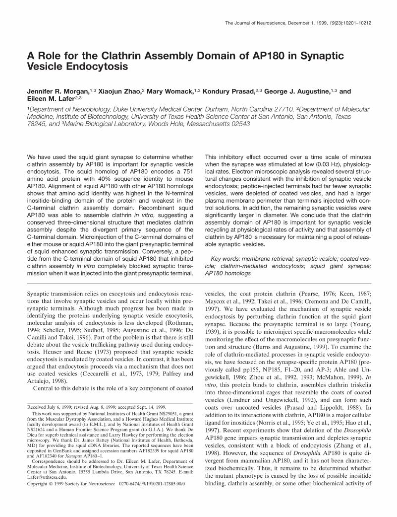
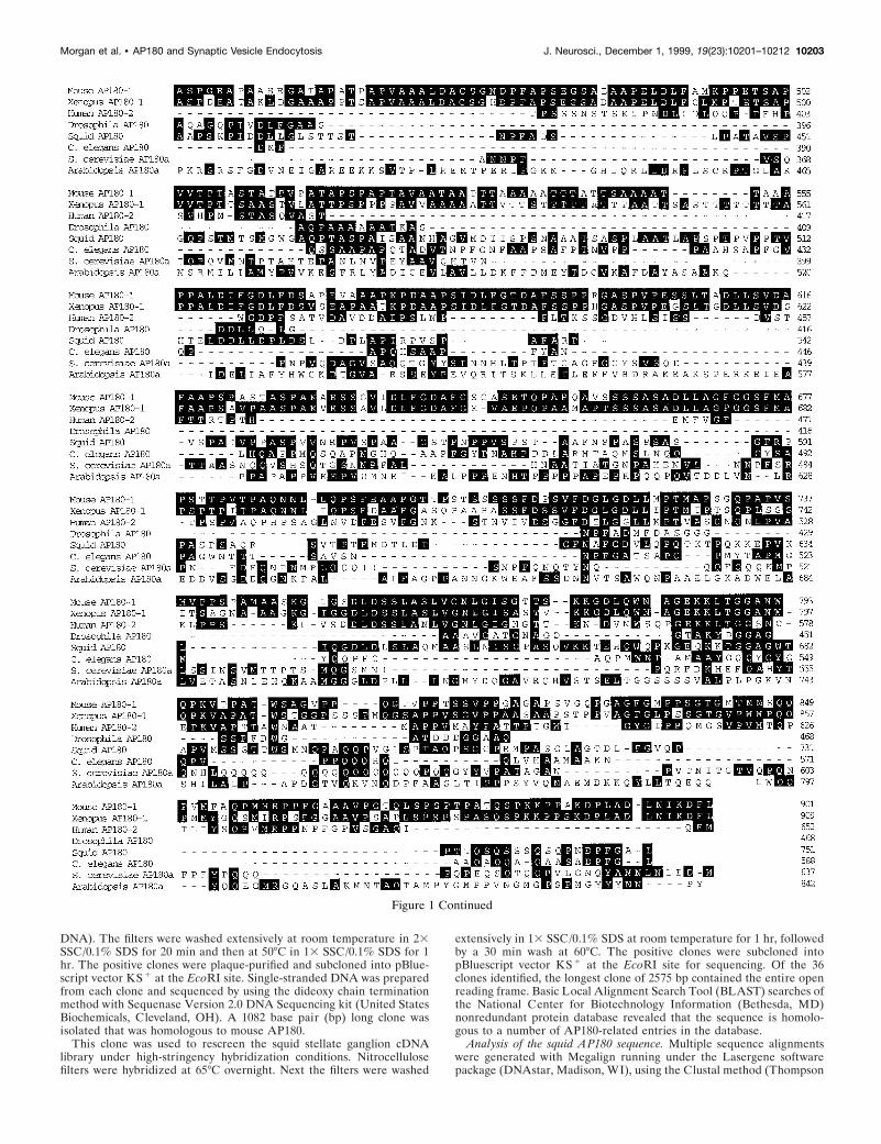
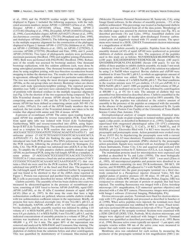
Figure 1. Amino acid sequence alignment of the AP180 family members. Residues present in two or more family members are highlighted in black.
10202 J. Neurosci., December 1, 1999, 19(23):10201–10212 Morgan et al. • AP180 and Synaptic Vesicle Endocytosis
DNA). The filters were washed extensively at room temperature in 23SSC/0.1% SDS for 20 min and then at 50°C in 13 SSC/0.1% SDS for 1hr. The positive clones were plaque-purified and subcloned into pBlue-script vector KS 1 at the EcoRI site. Single-stranded DNA was preparedfrom each clone and sequenced by using the dideoxy chain terminationmethod with Sequenase Version 2.0 DNA Sequencing kit (United StatesBiochemicals, Cleveland, OH). A 1082 base pair (bp) long clone wasisolated that was homologous to mouse AP180.
This clone was used to rescreen the squid stellate ganglion cDNAlibrary under high-stringency hybridization conditions. Nitrocellulosefilters were hybridized at 65°C overnight. Next the filters were washed
extensively in 13 SSC/0.1% SDS at room temperature for 1 hr, followedby a 30 min wash at 60°C. The positive clones were subcloned intopBluescript vector KS 1 at the EcoRI site for sequencing. Of the 36clones identified, the longest clone of 2575 bp contained the entire openreading frame. Basic Local Alignment Search Tool (BLAST) searches ofthe National Center for Biotechnology Information (Bethesda, MD)nonredundant protein database revealed that the sequence is homolo-gous to a number of AP180-related entries in the database.
Analysis of the squid AP180 sequence. Multiple sequence alignmentswere generated with Megalign running under the Lasergene softwarepackage (DNAstar, Madison, WI), using the Clustal method (Thompson
Figure 1 Continued
Morgan et al. • AP180 and Synaptic Vesicle Endocytosis J. Neurosci., December 1, 1999, 19(23):10201–10212 10203
et al., 1994) and the PAM250 residue weight table. The alignmentdisplayed in Figure 1 included the following sequences, with the indi-cated accession numbers: mouse AP180–1 (2492687) (Zhou et al., 1992),Xenopus AP180–1 (AF182340) (Zhou, 1994), human AP180–2(1373146) (Dreyling et al., 1996), Drosophila AP180 (4160434) (Zhang etal., 1998), Caenorhabditis elegans AP180 (AF144257) (Nonet et al., 1999),Saccharomyces cerevisiae AP180a (731735) (Wendland and Emr, 1998),and Arabidopsis AP180a (2864615). The following sequences were addedto the multiple sequence alignment for the construction of the phylogenydisplayed in Figure 2: human AP180–1 (3327126) (Ishikawa et al., 1998),rat AP180–1 (2492686) (Morris et al., 1993), rat AP180–2 (2792502), S.cerevisiae AP180b (1723758) (Wendland and Emr, 1998), and S. pombeAP180 (3150136). Two types of phylogenetic analysis were performed,maximum parsimony (Swofford, 1998) and neighbor joining (Felsenstein,1985). Both were performed with PAUP4.0b2 (Swofford, 1998). Robust-ness of the results was assessed by bootstrap analysis. One thousandbootstrap replications, with five random additions each, were done byusing unweighted parsimony. One thousand bootstrap replications weredone by using neighbor joining, with tree-bisection reconnection branchswapping to define the shortest tree. The results of the two analyses werein agreement, although the level of support for particular nodes differed.The tree was rooted by using the most divergent sequence, ArabidopsisAP180a, as the out-group (data not shown). Bootstrap values are shownabove each node (parsimony/neighbor joining). The reported pairwiseidentities (see Table 1 and text) were calculated by dividing the numberof positions with identical residues in the multiple sequence alignment(see Fig. 1) by the shortest of the two sequences being compared. The 33kDa N-terminal domain of mouse AP180 already has been defined ascomprising amino acids 1–304, and the 58 kDa C-terminal domain ofmouse AP180 has been defined as comprising amino acids 305–901 (Yeand Lafer, 1995a,b). For each of the AP180 family members that wasanalyzed, the last residue of the N-terminal domain was taken to be theresidue that aligned with amino acid 304 of mouse AP180.
Expression of recombinant AP180. The entire open reading frame ofsquid AP180 was amplified by reverse transcription PCR. Total RNAfrom squid optic lobe was isolated with Trizol (Life Technologies,Gaithersburg, MD) and reverse-transcribed into cDNA, using Super-Script II RT (Life Technologies). The synthesized first-strand cDNA wasused as a template for a PCR reaction that used sense primer (59-AGCGAGCTCCCCGGGATGTCTGGACAGAGTATAATG-39) andantisense primer (59-GCTCTCGAGCCCGGGTCACAAAGCACCAAATGGATC-39). An XbaI site flanking the open reading frame wasintroduced into both PCR primers. Pfu DNA polymerase was used forthe PCR reaction, following the protocol provided by Stratagene (LaJolla, CA). The PCR product was subcloned into pGEX-X at the XbaIsite. To amplify the 45 kDa putative clathrin assembly domain of squidAP180, we performed PCR, using the full-length AP180 as a template. Asense primer (59-AGCGAGCTCCCGGGTTCAACATCTAATGG-TGTGTCA-39) that contains a SmaI site and an antisense primer (59GCTCCCGGGCTCGAGTCACAAAGCACCAAATGGATC-39) that con-tains a XhoI site were used for the PCR reaction. The PCR product wassubcloned into pGEX-4T-1 at the SmaI and XhoI sites. The sequence ofthe full-length squid AP180 insert was confirmed further by sequencingand was found to be identical to that of the cDNA clone reported inFigure 1. Protein was expressed and purified from suitably transformedBL21 cells as previously described for mouse AP180 (Zhou et al., 1993).
Clathrin assembly. The assembly of bovine brain clathrin into coats wasmeasured by ultracentrifugation in the presence of recombinant pro-teins, consisting of GST fused to bovine AP180 (bAP180), squid GST-AP180 (sAP180), or the 45 kDa C-terminal domain of squid AP180(sC45) (Hao et al., 1997). In this assay the coats that have a highsedimentation coefficient are pelleted, whereas soluble clathrin triskeliawith low sedimentation coefficient remain in the supernatant. Briefly, allproteins first were dialyzed overnight into 10 mM Tris-HCl, pH 8.5, at4°C. Individually, bAP180, sAP180, sC45, or GST was combined withbovine clathrin, and the assembly reaction was initiated by adding 1:10volume of 1 M MES-NaOH, pH 6.5. The final conditions in the reactionwere 0.8 mM clathrin, 0.1 M MES-NaOH, 9 mM Tris-HCl, pH 6.5, and theindicated concentrations of assembly proteins in a volume of 200 ml. Themixture was incubated on ice for 60 min and then was centrifuged at400,000 3 g, 4°C for 6 min. The upper 80% of the supernatant wasremoved and analyzed by SDS-PAGE and Coomassie blue staining. Thepercentage of clathrin that was assembled was determined by the relativedepletion of clathrin from the solutions before and after centrifugation.This was quantified by densitometry of Coomassie blue-stained gels
(Molecular Dynamics Personal Densitometer SI, Sunnyvale, CA), usingImage Quant software. In the absence of assembly proteins, ;5% of theclathrin sedimented. This percentage was treated as background clathrinassembly and was subtracted from all of the data points. Morphology ofthe clathrin cages was assessed by electron microscopy (see Fig. 3E), asdescribed previously (Ye and Lafer, 1995a). Assembled clathrin coatstructures were applied to freshly glow-discharged Formvar carbon-coated grids and negatively stained with 1% uranyl acetate. The speci-mens were viewed in a JEOL 1200 EX Transmission electron microscopeat a magnification of 40,0003.
Inhibition of clathrin assembly by peptides. Peptides from the clathrinassembly domain (sC45) of squid AP180 were synthesized as potentialinhibitors of clathrin assembly in vitro. The sequences of the peptidesused in this study are as follows: NGVSDEEKKKMLDDENQRLNQ(s180 pep); KSLGEDDNRNMVEEDKNQLQK (Scram s180 pep1);QNEDSMKDVQKNLENLKGDRE (Scram s180 pep2). To test theability of these peptides to inhibit clathrin assembly, we made 5 mM stocksolutions in 10 mM Tris-HCl, pH 8.5. The pH of the solution was adjustedby using 2 M NaOH when required. Bovine clathrin and sAP180 werecombined in 10 mM Tris-HCl, pH 8.5, to which an appropriate amount ofthe peptide solution was added. The assembly was initiated by theaddition of 1:10 volume of 1 M MES-NaOH, pH 6.7. The final conditionsin the reaction mixture were 0.5 mM clathrin, 1.5–2.0 mM sAP180, 0.1 MMES-NaOH, pH 6.7, and 0–1000 mM peptide in a final volume of 200 ml.The mixture was incubated on ice for 45 min, followed by centrifugationat 400,000 3 g, at 4°C for 6 min. The amount of clathrin that wasassembled was determined as described above. Clathrin assembled in thepresence of sAP180, but without peptides, served as 100% assembly. Theinhibition by the peptides was calculated as the relative amount ofassembly in the presence of the peptides as compared with the assemblyin the absence of the peptides. Peptides were synthesized by Dr. LyndaBonewald at the Protein Core Facility of the University of Texas HealthScience Center (San Antonio, TX).
Electrophysiolog ical analysis of synaptic transmission. Electrical mea-surements were made on giant synapses in isolated stellate ganglia of thesquid, Loligo pealei, as described in Bommert et al., (1993). Ganglia weresuperfused with oxygenated physiological saline (10–15°C) containing(in mM) 466 NaCl, 54 MgCl2, 11 CaCl2, 10 KCl, 3 NaHCO3, and 10HEPES, pH 7.2. Electrodes filled with 3 M KCl were inserted into thepresynaptic and postsynaptic axons. Action potentials were evoked every30 sec (0.033 Hz) by depolarizing the presynaptic axon with a currentpulse (0.7–1.9 mA, 0.5 msec duration). Transmitter release was measuredby recording postsynaptic potentials (PSPs) elicited by the presynapticaction potentials. Signals were recorded with an Axoclamp-2A amplifier(Axon Instruments, Foster City, CA) and acquired and analyzed withAxobasic programs written by F. Schweizer (UCLA, Los Angeles, CA).
The mammalian proteins used for microinjection were expressed andpurified as described by Ye and Lafer (1995b). The most biologicallyabundant isoform of mouse AP180 (AS108 1AS15 2) was used (Zhou etal., 1993). All microinjected peptides and proteins were dissolved in aninjection solution containing (in mM) 250 K-isothionate, 200 KCl, 100taurine, and 50 HEPES, pH-adjusted to 7.4 with NaOH. These solutionswere microinjected into the giant presynaptic terminal via a third elec-trode connected to a Picospritzer injector (General Valve, NJ) thatapplied pulses of positive pressure (10–80 msec; 10–100 psi; N2 gas).FITC-dextran (3 kDa MW; 100 mM; Molecular Probes, Eugene, OR) wascoinjected to estimate the amount of protein or peptide that had beeninjected. The resulting fluorescence was imaged with a Zeiss Axioskopmicroscope (103 magnification, 0.25 numerical aperture objective) anddetected with a Cohu SIT camera. Fluorescence images were processedwith Image-1 software (Universal Imaging, Philadelphia, PA).
Electron microscopic analysis. Terminals were fixed for electron micros-copy with 2.5% glutaraldehyde and processed as described in Sanchez etal. (1990). When active peptides were injected, the terminals were fixedafter synaptic transmission was inhibited by 80% or more. Fixed termi-nals were sectioned (80 nm thickness) at intervals of 50 or 75 mm throughthe entire length of the synaptic terminal. Sections were examined witha JEM-1200 ExII (JOEL USA, Peabody, MA) electron microscope, andimages were digitized and analyzed with Image-1 software. Terminalstructure was quantified as described previously (Hess et al., 1993; Burnset al., 1998). Because active zones typically are spaced ;1 mm apart, onlysynaptic vesicles within 500 nm of the active zone were measured toensure that each vesicle was counted only once.
Membrane area was calculated for each section by measuring theperimeter of the presynaptic terminal in low magnification (5003) im-
10204 J. Neurosci., December 1, 1999, 19(23):10201–10212 Morgan et al. • AP180 and Synaptic Vesicle Endocytosis
ages. Such measurements probably represent underestimates because ofsmall plasma membrane invaginations that could not be detected at lowmagnification. To determine the magnitude of this effect, we measuredactual plasma membrane distance from selected regions in high-magnification images (12,0003). These measurements indicated that theplasma membrane perimeter of terminals injected with s180 peptide was36% larger than estimated from the low-magnification images, whereasthe control terminals were underestimated by 30%. Then the perimetermeasurements made at low magnification were increased by theseamounts. Plasma membrane perimeter was multiplied by the thickness ofthe section (80 nm) to calculate the surface area of plasma membrane ineach section. The surface area of synaptic and coated vesicles wascalculated as 4pr 2, where r is the measured radius of these vesicles. Thesurface area per vesicle was multiplied by the mean number of vesiclesper active zone, yielding the amount of vesicle area per active zone, andthen was multiplied by the number of active zones per section to yield themean surface area in synaptic and coated vesicles in each section.
RESULTSCloning of the squid homolog of AP180Low-stringency screening of a squid lgt10 cDNA library, byhybridization with a mouse AP180 cDNA probe, identified a 1082
bp clone that had high homology to mouse AP180. This clone wasused to rescreen the cDNA library by high-stringency hybridiza-tion and identified 36 additional clones. The sequence of thelongest clone was 2575 nucleotides, and it covered 90 nucleotidesof the 59 untranslated region, the entire coding region, and theentire 39 untranslated region to the poly(A1) tail. It contained asingle open reading frame with two upstream in-frame stopcodons. The open reading frame of squid AP180 encoded a 751amino acid protein with a predicted molecular weight of 80,200 Da.
The sequence of squid AP180 is homologous to a number ofAP180-related entries in the protein database of the NationalCenter for Biotechnology Information. Because several of theseentries came from genome-sequencing projects and the predictedproteins have not yet been named, we devised a uniform nomen-clature for the AP180 family members. Mouse AP180, the firstvertebrate AP180 to be cloned and sequenced, was assigned thename AP180–1. All vertebrate sequences highly homologous tomouse AP180–1 also were assigned the name AP180–1. MouseAP180–1 mRNA is restricted to neuronal cells, and its protein issynapse-specific (Perry et al., 1991, 1992; Sousa et al., 1992); this maybe the case for the other members of the AP180–1 group. A secondvertebrate AP180-related gene with 48% identity to AP180–1 wasisolated from a human myeloid leukemia cell line and was calledclathrin assembly lymphoid myeloid leukemia gene, or CALM(Dreyling et al., 1996). We assigned the name AP180–2 to humanCALM as well as to all vertebrate sequences highly homologous tohuman AP180–2. AP180–2 mRNA is expressed ubiquitously in awide range of human cell types, and this may be true for othermembers of the AP180–2 group (Dreyling et al., 1996). The AP180-related genes of nonvertebrates are called AP180, with multiplegenes distinguished by letters according to the precedent set by theS. cerevisiae AP180 genes (Wendland and Emr, 1998).
The relationship between squid AP180 and other representa-tives of the AP180 family was revealed by a multiple sequencealignment (Fig. 1). When more than one alternatively splicedisoform has been reported for a family member, we used thelongest available isoform in constructing the alignment. Gaps inthe alignment reflect the diverse range of sizes of the AP180family members, especially within the C-terminal domain. Thesmallest reported family member is Drosophila AP180, which has469 amino acids (Zhang et al., 1998). The largest reported familymember is rat AP180, which has 915 amino acids (Morris et al.,1993). Examination of the alignment reveals a protein family thatis much better conserved in the ;33 kDa N-terminal domain thanin the variably sized C-terminal domain (Fig. 1). This observationis supported by an analysis of the pairwise identity scores of thealigned sequences (Table 1). For example, whereas the overallidentity between squid and mouse AP180 is 40%, the identitybetween their N-terminal domains is 66%, and the identity be-tween their C-terminal domains is 24%.
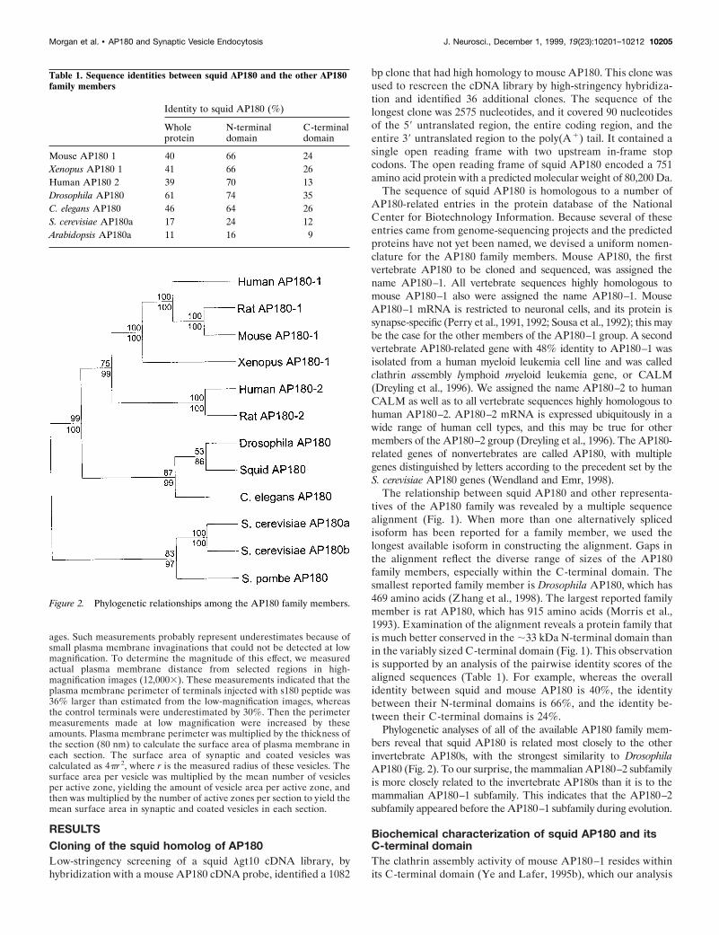
Phylogenetic analyses of all of the available AP180 family mem-bers reveal that squid AP180 is related most closely to the otherinvertebrate AP180s, with the strongest similarity to DrosophilaAP180 (Fig. 2). To our surprise, the mammalian AP180–2 subfamilyis more closely related to the invertebrate AP180s than it is to themammalian AP180–1 subfamily. This indicates that the AP180–2subfamily appeared before the AP180–1 subfamily during evolution.
Biochemical characterization of squid AP180 and itsC-terminal domainThe clathrin assembly activity of mouse AP180–1 resides withinits C-terminal domain (Ye and Lafer, 1995b), which our analysis
Figure 2. Phylogenetic relationships among the AP180 family members.
Table 1. Sequence identities between squid AP180 and the other AP180family members
Identity to squid AP180 (%)
Wholeprotein
N-terminaldomain
C-terminaldomain
Mouse AP180 1 40 66 24Xenopus AP180 1 41 66 26Human AP180 2 39 70 13Drosophila AP180 61 74 35C. elegans AP180 46 64 26S. cerevisiae AP180a 17 24 12Arabidopsis AP180a 11 16 9
Morgan et al. • AP180 and Synaptic Vesicle Endocytosis J. Neurosci., December 1, 1999, 19(23):10201–10212 10205
indicates is not well conserved among other members of theAP180 family (see Fig. 1). Thus, it is possible that other AP180forms do not assemble clathrin. For this reason we asked whethersquid AP180 can assemble clathrin. To study the functionalproperties of squid AP180 in vitro, we expressed in bacteriaglutathione S-transferase (GST) fusions of full-length squidAP180 (aa 1–751) (sAP180) and its C-terminal domain (aa 313–751) (sC45). Because squid AP180 is shorter than mouse AP180,the amino acids that correspond to the 58 kDa clathrin assemblydomain of mouse AP180 (mC58; Ye and Lafer, 1995b) encode a45 kDa protein in squid. The abilities of sAP180, sC45, GST, andbovine AP180 (bAP180) to assemble bovine clathrin were com-pared in an in vitro assay that measured the incorporation ofclathrin triskelia into cages (Fig. 3). Both sAP180 and sC45 were
able to assemble bovine clathrin in a concentration-dependentmanner (Fig. 3A,B). In terms of the concentrations required forclathrin assembly, as well as the maximal amount of assembly,both squid constructs were very similar to bovine AP180 (Fig.3C). GST alone did not assemble clathrin (Fig. 3D), showing thatthe clathrin assembly activity of the fusions was attributable tothe AP180 rather than to the GST. The cages assembled bysAP180 were homogeneous in size (Fig. 3E), as has been reportedfor mammalian AP180s (Ye and Lafer, 1995a). These resultsindicate that squid AP180, like its mammalian homologs, is aclathrin assembly protein. Further, the ability to assemble clathrinresides in the 45 kDa C-terminal domain of squid AP180, whichindicates that enough tertiary structure is conserved to mediateclathrin assembly despite the divergent primary sequences in theC-terminal domains of mammalian and squid AP180.
The clathrin assembly domain of AP180 enhancessynaptic transmissionTo determine whether clathrin assembly by AP180 is importantfor synaptic vesicle endocytosis, we microinjected various formsof AP180 into the squid giant presynaptic terminal to determinetheir actions on neurotransmitter release. If AP180 stimulatesclathrin assembly in vivo and clathrin assembly is essential forsynaptic vesicle endocytosis, then injecting AP180 might enhancesynaptic transmission. Synaptic transmission was evoked by singleaction potentials elicited every 30 sec (0.03 Hz) by currentsinjected directly into the presynaptic axon, and postsynaptic re-sponses were recorded during the injection of AP180 reagents.Although full-length mouse GST-AP180 (mAP180) and sAP180had no effect on synaptic transmission (Table 2), the injection of
Table 2. Effects of microinjected AP180 proteins and peptides onsynaptic transmission
AP180 reagent Number of injections Mean effect (SEM) (%)
mAP180 9 8.1 (4.0)mC58 13 17.9 (6.5)mM42 4 5.8 (7.1)mN33 2 5.1 (5.1)sAP180 8 3.8 (7.6)sC45 10 16.6 (9.2)GST 10 5.1 (4.1)Carrier solution 13 20.1 (2.3)s180 pep 17 235.4 (7.2)Scram s180 pep1 2 23.8 (2.4)
Figure 3. Squid GST-AP180 and squid GST-c45, but not GST, assemble clathrin as efficiently as bovine AP180. A–D, Clathrin assembly by squidGST-AP180 (A), squid GST-c45 (B), bovine AP180 (C), and GST (D). Points represent the mean of three to four independent experiments, and errorbars indicate the SEM values. Half-maximal concentrations for clathrin assembly, determined by fits to a rectangular hyperbola function (solid lines),were 0.3 mM for squid GST-AP180, 0.4 mM for GST-c45, and 0.6 mM for bovine AP180. The maximum amount of assembly ranged from 85 to 94% forthese three proteins. E, Electron micrograph of clathrin coats assembled by squid GST-AP180.
10206 J. Neurosci., December 1, 1999, 19(23):10201–10212 Morgan et al. • AP180 and Synaptic Vesicle Endocytosis
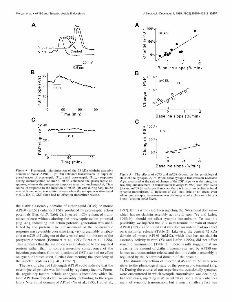
the clathrin assembly domains of either squid (sC45) or mouseAP180 (mC58) enhanced PSPs produced by presynaptic actionpotentials (Fig. 4A,B, Table 2). Injected mC58 enhanced trans-mitter release without altering the presynaptic action potential(Fig. 4A), indicating that action potential generation was unaf-fected by the protein. The enhancement of the postsynapticresponse was reversible over time (Fig. 4B), presumably attribut-able to mC58 diffusing out of the terminal and into the rest of thepresynaptic neuron (Bommert et al., 1993; Burns et al., 1998).This indicates that the inhibition was attributable to the injectedprotein rather than to some irreversible consequence of theinjection procedure. Control injections of GST also had no effecton synaptic transmission, further documenting the specificity ofthe injected proteins (Fig. 4C, Table 2).
The lack of effect of full-length AP180 could indicate that themicroinjected protein was inhibited by regulatory factors. Poten-tial regulatory factors include endogenous inositides, which in-hibit AP180-mediated clathrin assembly by binding to the regu-latory N-terminal domain of AP180 (Ye et al., 1995; Hao et al.,
1997). If this is the case, then injecting the N-terminal domain—which has no clathrin assembly activity in vitro (Ye and Lafer,1995a,b)—should not affect synaptic transmission. To test thispossibility, we injected the 33 kDa N-terminal domain of mouseAP180 (mN33) and found that this domain indeed had no effecton transmitter release (Table 2). Likewise, the central 42 kDadomain of mouse AP180 (mM42), which also has no clathrinassembly activity in vitro (Ye and Lafer, 1995b), did not affectsynaptic transmission (Table 2). These results suggest that in-creasing the amount of clathrin assembly in vivo by AP180 en-hances neurotransmitter release and that this clathrin assembly isregulated by the N-terminal domain of the protein.
The stimulatory actions of injected sC45 and mC58 were sen-sitive to the physiological state of the presynaptic terminal (Fig.5). During the course of our experiments, occasionally synapseswere encountered in which synaptic transmission was declining.In these cases, injecting sC45 or mC58 caused a large enhance-ment of synaptic transmission, but a much smaller effect was
Figure 4. Presynaptic microinjection of the 58 kDa clathrin assemblydomain of mouse AP180–1 (mC58) enhances transmission. A, Superim-posed traces of presynaptic (Vpre ) and postsynaptic (Vpost ) responsesduring microinjection of mC58. mC58 enhanced the postsynaptic re-sponses, whereas the presynaptic response remained unchanged. B, Timecourse of response to the injection of mC58 (10 mM; during bar). mC58reversibly enhanced transmitter release when the synapse was stimulatedat 0.03 Hz. C, GST alone had no effect on transmitter release.
Figure 5. The effects of sC45 and mC58 depend on the physiologicalstate of the synapse. A, B, When basal synaptic transmission (Baselineslope, measured as the rate of change of the PSP slope) was declining, theresulting enhancement in transmission (Change in PSP) seen with sC45(A) and mC58 (B) is larger than when there is little or no decline in basalsynaptic transmission. C, Injection of GST had little or no effect, evenwhen basal synaptic transmission was declining rapidly. Data were fit by alinear function (solid lines).
Morgan et al. • AP180 and Synaptic Vesicle Endocytosis J. Neurosci., December 1, 1999, 19(23):10201–10212 10207
observed in synapses in which transmission was stable beforeinjecting these proteins. The influence of the stability of basalsynaptic transmission on the effects of injected proteins is shownin Figure 5. There was a strong correlation between the stabilityof basal synaptic transmission, measured as the rate of change inthe baseline slope of the PSPs, and the effect of injected sC45(Fig. 5A) or mC58 (Fig. 5B) on PSPs. In contrast, injections ofGST (Fig. 5C) did not enhance synaptic transmission signifi-cantly, even when baseline transmission was steeply declining.The difference between the effects of sC45 and GST was statis-tically significant ( p , 0.01; Student’s t test) as was the differencebetween mC58 and GST ( p , 0.1). Although we do not know thereason for the decline in basal synaptic transmission, we suspectthat it is attributable to a high rate of spontaneous transmitterrelease and subsequent depletion of the readily releasable pool ofsynaptic vesicles (Charlton et al., 1982). Thus, the clathrin assem-bly domains of squid and mouse AP180 may enhance transmitterrelease when the terminal is highly active.
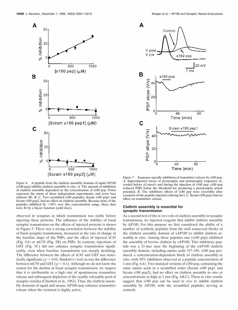
Clathrin assembly is essential forsynaptic transmissionAs a second test of the in vivo role of clathrin assembly in synaptictransmission, we injected reagents that inhibit clathrin assemblyby AP180. For this purpose we first considered the ability of anumber of synthetic peptides from the well conserved blocks ofthe clathrin assembly domain of sAP180 to inhibit clathrin as-sembly in vitro. Among these peptides one (s180 pep) inhibitedthe assembly of bovine clathrin by sAP180. This inhibitory pep-tide was a 21-mer near the beginning of the sAP180 clathrinassembly domain, including amino acids 337–356. s180 pep pro-duced a concentration-dependent block of clathrin assembly invitro, with 38% inhibition observed at a peptide concentration of1 mM (Fig. 6A). Two mutated versions of s180 pep, containing thesame amino acids in a scrambled order (Scram s180 pep1 andScram s180 pep2), had no effect on clathrin assembly in vitro atconcentrations as high as 1 mM (Fig. 6B,C). These in vitro resultssuggest that s180 pep can be used in vivo to inhibit clathrinassembly by AP180, with the scrambled peptides serving ascontrols.
Figure 6. A peptide from the clathrin assembly domain of squid AP180(s180 pep) inhibits clathrin assembly in vitro. A, The amount of inhibitionof clathrin assembly depended on the concentration of s180 pep. Pointsrepresent the mean of three independent experiments, and error barsindicate SE. B, C, Two scrambled s180 peptides, Scram s180 pep1 andScram s180 pep2, had no effect on clathrin assembly. Because none of thepeptides inhibited by .50% over this concentration range, these datawere fit by a linear function (solid lines).
Figure 7. Sequence-specific inhibition of transmitter release by s180 pep.A, Superimposed traces of presynaptic and postsynaptic responses re-corded before (Control ) and during the injection of s180 pep. s180 pepreduced PSPs below the threshold for producing a postsynaptic actionpotential. B, The inhibitory effects of s180 pep were reversible aftercessation of the peptide injection (during bar). C, Scram s180 pep1 had noeffect on transmitter release.
10208 J. Neurosci., December 1, 1999, 19(23):10201–10212 Morgan et al. • AP180 and Synaptic Vesicle Endocytosis
We next injected s180 pep into the squid giant presynapticterminal to ask whether clathrin assembly by AP180 is necessaryfor synaptic vesicle endocytosis in vivo. If clathrin is important forendocytosis in the nerve terminal, then the block of clathrinassembly should have several effects: (1) prevent fused vesicularmembrane from being retrieved from the plasma membrane andthereby increase presynaptic surface area, (2) inhibit the forma-tion of clathrin-coated vesicles, (3) reduce the number of synapticvesicles, and (4) inhibit transmitter release by reducing the num-ber of synaptic vesicles. We found that s180 pep produced all ofthese lesions.
First, presynaptic injection of sAP180 pep inhibited evokedtransmitter release without affecting the presynaptic action po-tential (Fig. 7A). On average, sAP180 pep injection inhibitedPSPs by 36% (n 5 17; Table 2), although injection of a sufficientamount of this peptide (estimated to be several millimolar) com-pletely inhibited synaptic transmission. Inhibition of the PSP wasreversible over time (Fig. 7B), indicating that the inhibition wasnot attributable to damage of the presynaptic terminal. Scrams180 pep1, which had no effect on clathrin assembly in vitro, alsohad no effect on transmitter release (Fig. 7C, Table 2). Thisindicates that the inhibition of synaptic transmission seen withs180 pep is sequence-specific and that clathrin assembly in vivo byAP180 is essential for neurotransmitter release.
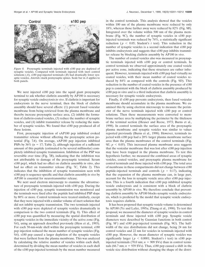
We next used electron microscopy to examine the ultrastruc-ture of presynaptic terminals injected with s180 pep. During theinjection of s180 pep, synaptic transmission was monitored andthe terminals were fixed after the peptide had inhibited PSP slopeby 80% or more. Control terminals were treated similarly, exceptthat they were injected with a similar volume of inert solution thatdid not inhibit synaptic transmission. The two terminals injectedwith s180 pep were depleted of synaptic vesicles dramatically incomparison to the two control terminals (Fig. 8). This effect ofs180 pep was quantified by measuring the spatial distribution ofsynaptic vesicles in the immediate vicinity of the active zone (Fig.9A), using an approach described previously (Hess et al., 1993).For each 50-nm-wide shell within the presynaptic terminal, s180pep injection reduced the mean number of synaptic vesicles (Fig.9A). s180 pep caused a larger depletion of the synaptic vesiclesthat were farthest from the plasma membrane. This was analyzedby calculating the relative number of vesicles within each shell,determined by dividing the mean number of vesicles in each shellof the s180 pep-injected terminals by the mean number measured
in the control terminals. This analysis showed that the vesicleswithin 100 nm of the plasma membrane were reduced by only18%, whereas those farther away were reduced by 82% (Fig. 9B).Integrated over the volume within 500 nm of the plasma mem-brane (Fig. 9C), the number of synaptic vesicles in s180 pep-injected terminals was reduced by 74%, a statistically significantreduction ( p , 0.05; Student’s t test). This reduction in thenumber of synaptic vesicles is a second indication that s180 pepinhibits endocytosis and suggests that s180 pep inhibits transmit-ter release by blocking clathrin assembly by AP180 in vivo.
The number of coated vesicles also was measured in presynap-tic terminals injected with s180 pep or control terminals. Incontrol terminals we observed approximately one coated vesicleper active zone, indicating that these structures are rather infre-quent. However, terminals injected with s180 pep had virtually nocoated vesicles, with their mean number of coated vesicles re-duced by 84% as compared with the controls (Fig. 9D). Thisreduction in the number of coated vesicles in the presence of s180pep is consistent with the block of clathrin assembly produced bys180 pep in vitro and is a third indication that clathrin assembly isnecessary for synaptic vesicle endocytosis.
Finally, if s180 pep prevents endocytosis, then fused vesicularmembrane should accumulate in the plasma membrane. We ex-amined this by using electron microscopy to measure the perim-eter of the nerve terminals injected with s180 pep or controlsolutions. Then these measurements were converted to mem-brane surface area by multiplying the perimeter by the thicknessof the terminal section (Heuser and Reese, 1973; Burns et al.,1998). In control terminals the surface area of the presynapticplasma membrane and synaptic vesicles was similar to valuesreported previously (Burns et al., 1998). However, terminals in-jected with s180 pep had a 20% larger plasma membrane surfacearea than the plasma membranes of the control terminals (Fig.9E; p , 0.05). This increased plasma membrane area suggeststhat the vesicular membrane that was lost after s180 pep injectionmay have been trapped in the plasma membrane. To test thishypothesis further, we measured the combined areas of synapticvesicles, coated vesicles, and presynaptic plasma membrane forcontrol terminals and those injected with s180 pep. The total areaof membrane in these compartments did not change between s180peptide-injected terminals and controls ( p 5 0.17), indicatingthat the expansion of the plasma membrane can, in large part,account for the loss in synaptic vesicle area after s180 pep injec-tion. This is a fourth indication that s180 pep inhibited synapticvesicle endocytosis and is consistent with a block of clathrinassembly by AP180 in vivo. We therefore conclude that prevent-ing clathrin assembly by AP180 blocks synaptic vesicle endocyto-sis, which is predicted by the model that synaptic vesicle endocy-tosis requires clathrin.
It has been proposed that synaptic vesicle volume is determinedby AP180 (Ye and Lafer, 1995a; Zhang et al., 1998). To test thisproposal, we measured the diameter of synaptic vesicles in controlterminals and those injected with s180 pep. Synaptic vesiclediameters were described by Gaussian functions in both control(Fig. 9F) and s180 pep-injected terminals (Fig. 9G). The half-width of the size distributions did not change, being 24 nm forcontrol vesicles and 22 nm for vesicles in terminals injected withs180 pep. However, the mean diameter of synaptic vesicles wassignificantly larger ( p , 0.0001; Student’s t test) in s180 pep-injected terminals (70.0 nm; n 5 909 SVs) than in control termi-nals (66.7 nm; n 5 939 SVs). Thus, s180 pep caused a shift in thevesicle size distribution without changing the shape of the distri-
Figure 8. Presynaptic terminals injected with s180 pep are depleted ofsynaptic vesicles. Compared with terminals injected with inert controlsolutions (A), s180 pep-injected terminals (B) had drastically fewer syn-aptic vesicles. Asterisks mark postsynaptic spines. Scale bar in A applies toboth panels.
Morgan et al. • AP180 and Synaptic Vesicle Endocytosis J. Neurosci., December 1, 1999, 19(23):10201–10212 10209
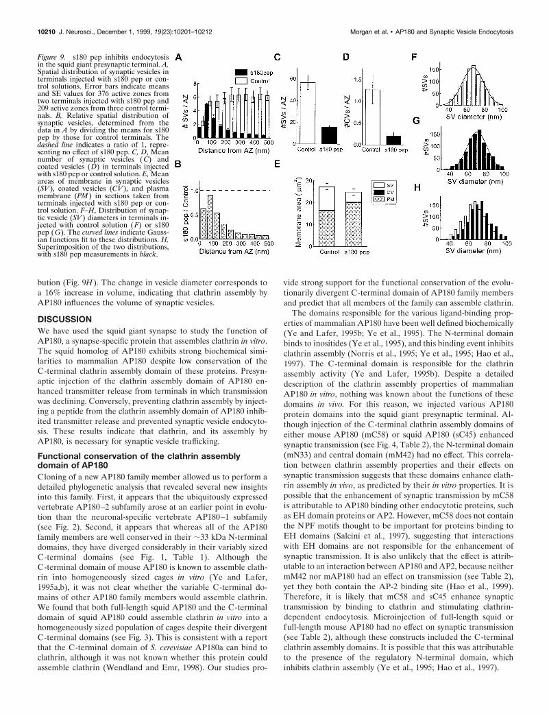
bution (Fig. 9H). The change in vesicle diameter corresponds toa 16% increase in volume, indicating that clathrin assembly byAP180 influences the volume of synaptic vesicles.
DISCUSSIONWe have used the squid giant synapse to study the function ofAP180, a synapse-specific protein that assembles clathrin in vitro.The squid homolog of AP180 exhibits strong biochemical simi-larities to mammalian AP180 despite low conservation of theC-terminal clathrin assembly domain of these proteins. Presyn-aptic injection of the clathrin assembly domain of AP180 en-hanced transmitter release from terminals in which transmissionwas declining. Conversely, preventing clathrin assembly by inject-ing a peptide from the clathrin assembly domain of AP180 inhib-ited transmitter release and prevented synaptic vesicle endocyto-sis. These results indicate that clathrin, and its assembly byAP180, is necessary for synaptic vesicle trafficking.
Functional conservation of the clathrin assemblydomain of AP180Cloning of a new AP180 family member allowed us to perform adetailed phylogenetic analysis that revealed several new insightsinto this family. First, it appears that the ubiquitously expressedvertebrate AP180–2 subfamily arose at an earlier point in evolu-tion than the neuronal-specific vertebrate AP180–1 subfamily(see Fig. 2). Second, it appears that whereas all of the AP180family members are well conserved in their ;33 kDa N-terminaldomains, they have diverged considerably in their variably sizedC-terminal domains (see Fig. 1, Table 1). Although theC-terminal domain of mouse AP180 is known to assemble clath-rin into homogeneously sized cages in vitro (Ye and Lafer,1995a,b), it was not clear whether the variable C-terminal do-mains of other AP180 family members would assemble clathrin.We found that both full-length squid AP180 and the C-terminaldomain of squid AP180 could assemble clathrin in vitro into ahomogeneously sized population of cages despite their divergentC-terminal domains (see Fig. 3). This is consistent with a reportthat the C-terminal domain of S. cerevisiae AP180a can bind toclathrin, although it was not known whether this protein couldassemble clathrin (Wendland and Emr, 1998). Our studies pro-
vide strong support for the functional conservation of the evolu-tionarily divergent C-terminal domain of AP180 family membersand predict that all members of the family can assemble clathrin.
The domains responsible for the various ligand-binding prop-erties of mammalian AP180 have been well defined biochemically(Ye and Lafer, 1995b; Ye et al., 1995). The N-terminal domainbinds to inositides (Ye et al., 1995), and this binding event inhibitsclathrin assembly (Norris et al., 1995; Ye et al., 1995; Hao et al.,1997). The C-terminal domain is responsible for the clathrinassembly activity (Ye and Lafer, 1995b). Despite a detaileddescription of the clathrin assembly properties of mammalianAP180 in vitro, nothing was known about the functions of thesedomains in vivo. For this reason, we injected various AP180protein domains into the squid giant presynaptic terminal. Al-though injection of the C-terminal clathrin assembly domains ofeither mouse AP180 (mC58) or squid AP180 (sC45) enhancedsynaptic transmission (see Fig. 4, Table 2), the N-terminal domain(mN33) and central domain (mM42) had no effect. This correla-tion between clathrin assembly properties and their effects onsynaptic transmission suggests that these domains enhance clath-rin assembly in vivo, as predicted by their in vitro properties. It ispossible that the enhancement of synaptic transmission by mC58is attributable to AP180 binding other endocytotic proteins, suchas EH domain proteins or AP2. However, mC58 does not containthe NPF motifs thought to be important for proteins binding toEH domains (Salcini et al., 1997), suggesting that interactionswith EH domains are not responsible for the enhancement ofsynaptic transmission. It is also unlikely that the effect is attrib-utable to an interaction between AP180 and AP2, because neithermM42 nor mAP180 had an effect on transmission (see Table 2),yet they both contain the AP-2 binding site (Hao et al., 1999).Therefore, it is likely that mC58 and sC45 enhance synaptictransmission by binding to clathrin and stimulating clathrin-dependent endocytosis. Microinjection of full-length squid orfull-length mouse AP180 had no effect on synaptic transmission(see Table 2), although these constructs included the C-terminalclathrin assembly domains. It is possible that this was attributableto the presence of the regulatory N-terminal domain, whichinhibits clathrin assembly (Ye et al., 1995; Hao et al., 1997).
Figure 9. s180 pep inhibits endocytosisin the squid giant presynaptic terminal. A,Spatial distribution of synaptic vesicles interminals injected with s180 pep or con-trol solutions. Error bars indicate meansand SE values for 376 active zones fromtwo terminals injected with s180 pep and209 active zones from three control termi-nals. B, Relative spatial distribution ofsynaptic vesicles, determined from thedata in A by dividing the means for s180pep by those for control terminals. Thedashed line indicates a ratio of 1, repre-senting no effect of s180 pep. C, D, Meannumber of synaptic vesicles ( C) andcoated vesicles (D) in terminals injectedwith s180 pep or control solution. E, Meanareas of membrane in synaptic vesicles(SV ), coated vesicles (CV ), and plasmamembrane (PM ) in sections taken fromterminals injected with s180 pep or con-trol solution. F–H, Distribution of synap-tic vesicle (SV ) diameters in terminals in-jected with control solution ( F) or s180pep (G). The curved lines indicate Gauss-ian functions fit to these distributions. H,Superimposition of the two distributions,with s180 pep measurements in black.
10210 J. Neurosci., December 1, 1999, 19(23):10201–10212 Morgan et al. • AP180 and Synaptic Vesicle Endocytosis
Manipulating clathrin assembly by AP180 alterssynaptic vesicle traffickingOur results provide several lines of support for a role for clathrinin synaptic vesicle trafficking. Our observation that clathrin as-sembly domains enhanced synaptic transmission is the first dem-onstration that stimulation of clathrin-mediated endocytosis canaffect synaptic transmission. We presume that the enhancementarises from increasing the number of synaptic vesicles in thereadily releasable pool of vesicles, attributable to the stimulationof vesicle endocytosis and subsequent reformation of synapticvesicles. Consistent with this explanation, we found that theenhancement of synaptic transmission by these domains waslargest in terminals in which basal synaptic transmission wasdeclining. We interpreted this to suggest that the concentration ofAP180 is only limiting in very active terminals, although alterna-tive explanations cannot be excluded without understanding whybasal synaptic transmission declines.
Stronger support for the hypothesis that clathrin is importantfor synaptic vesicle endocytosis comes from s180 pep, whichprevents clathrin assembly in vitro (see Fig. 6). Injecting s180 pepinto the squid presynaptic terminal inhibited synaptic transmis-sion reversibly (see Fig. 7). The fact that synaptic transmissioncould be inhibited completely by s180 pep indicates that clathrinassembly by AP180 is essential for synaptic transmission. Further,s180 pep injection reduced the number of synaptic vesicles andcoated vesicles and increased the surface area of the presynapticterminal (see Fig. 9). All of these changes indicate that impair-ment of clathrin assembly prevents synaptic vesicle endocytosis.Our results complement recent work showing that mutation of theDrosophila AP180 gene impairs synaptic transmission and de-pletes synaptic vesicles (Zhang et al., 1998). Because DrosophilaAP180 is not known to assemble clathrin, it was not clear whetherthe resulting phenotype represents the loss of clathrin assemblyactivity or some other function of AP180. Our results suggest thatlack of clathrin assembly is the likely explanation for the conse-quences of genetic deletion of AP180.
Based on the common definition that docked vesicles are thoseclosest to the plasma membrane (Schweizer et al., 1995), s180 pepcaused the number of docked synaptic vesicles to decrease by;40% (see first column, Fig. 9B), yet evoked transmitter releasecompletely stopped. There are several possible explanations forthe inability of the remaining docked vesicles to fuse. It is possiblethat these vesicles are generated by a pathway incapable of cor-rectly sorting vesicular proteins, so these vesicles do not containproteins required for fusion. Alternatively, transmitter releasecould be disrupted by accumulation of vesicular membrane in theplasma membrane. Regardless of the specific mechanism, ourresults indicate that nerve terminals cannot sustain transmitterrelease in the absence of clathrin-mediated endocytosis.
Molecular pathways for synaptic vesicle recyclingA role for clathrin-dependent endocytosis in synaptic vesicletrafficking originally was suggested by Heuser and Reese (1973).Because their experiments used heavy and possibly nonphysi-ological rates of synaptic activity, it has been suggested thatclathrin-independent mechanisms may be involved in vesicle traf-ficking under more physiological conditions (for review, see Pal-frey and Artalejo, 1998). Our experiments argue strongly to thecontrary by showing that clathrin-mediated endocytosis is impor-tant even during physiological levels of synaptic activity. How-ever, terminals injected with s180 pep still had approximatelyone-fourth of their synaptic vesicles remaining. It is possible that
the peptide may not have inhibited clathrin-mediated endocytosiscompletely, allowing other clathrin assembly proteins, such asAP2, to sustain endocytosis at a low level. This also could accountfor the presence of a few coated vesicles in peptide-injectedterminals (see Fig. 9D) and the existence of synaptic vesicles inDrosophila AP180 mutants (Zhang et al., 1998). Alternatively, theremaining synaptic vesicles may have been endocytosed via asecond, clathrin-independent pathway (Koenig and Ikeda, 1996;Klingauf et al., 1998; Kuromi and Kidokoro, 1998; Shi et al.,1998).
Clathrin cages assembled in vitro in the presence of mammalianAP180 are smaller and more uniform than those assembled with-out AP180, suggesting that AP180 may control the volume ofsynaptic vesicles (Ye and Lafer, 1995a). Support for this hypoth-esis comes from our finding that the synaptic vesicles remaining interminals injected with s180 pep had a 16% larger mean volumethan control terminals (see Fig. 9F–H), as well as observationsthat mutation of the AP180 gene reduces vesicle volume (Zhanget al., 1998; Nonet et al., 1999). These results indicate thatsynaptic vesicles of normal size require AP180 and, hence,clathrin-coated vesicles. However, the source of these vesicles isstill uncertain. Coated vesicles clearly bud off from the plasmamembrane (Heuser and Reese, 1973; Takei et al., 1996), andinterfering with AP180 must prevent such budding because s180pep increases presynaptic plasma membrane area. However, it isalso possible that coated vesicles bud off from endosomes (Heuserand Reese, 1973; Schweizer et al., 1995; Sudhof, 1995). Thus, ourresults cannot resolve the long-standing question of whetherclathrin-based budding from endosomes is responsible for thegeneration of synaptic vesicles (Takei et al., 1996; Murthy andStevens, 1998; Shi et al., 1998).
In summary, our results show that clathrin assembly by AP180is important for endocytosis in nerve terminals experiencingphysiological amounts of activity. AP180 also is essential forneurotransmitter release and for the formation of synaptic vesi-cles of normal size. Our results support a clathrin-based mecha-nism for synaptic vesicle recycling, although it remains to bedetermined whether an endosomal intermediate is involved inendocytosis and why some synaptic vesicles are present whenAP180-mediated clathrin assembly is impaired. Althoughclathrin-based endocytosis is the predominant mechanism ofendocytosis, it is still possible that clathrin-independent mecha-nisms participate in synaptic vesicle endocytosis.
REFERENCESAhle S, Ungewickell E (1986) Purification and properties of a new clath-
rin assembly protein. EMBO J 5:3143–3149.Augustine GJ, Burns ME, DeBello WM, Pettit DL, Schweizer FE (1996)
Exocytosis: proteins and perturbations. Annu Rev Pharmacol Toxicol36:659–701.
Bommert K, Charlton MP, DeBello WM, Chin GJ, Betz H, AugustineGJ (1993) Inhibition of neurotransmitter release by C2-domain pep-tides implicates synaptotagmin in exocytosis. Nature 363:163–165.
Burns ME, Augustine GJ (1999) Functional studies of presynaptic pro-teins at the squid giant synapse. In: Neurotransmitter release: frontiersin molecular biology (Bellen H, ed), 237–264. New York: Oxford UP.
Burns ME, Sasaki T, Takai Y, Augustine GJ (1998) Rabphilin-3A: amultifunctional regulator of synaptic vesicle traffic. J Gen Physiol111:243–255.
Ceccarelli B, Hurlbut WP, Mauro A (1973) Turnover of transmitter andsynaptic vesicles at the frog neuromuscular junction. J Cell Biol57:499–524.
Ceccarelli B, Grohovaz F, Hurlbut WP (1979) Freeze-fracture studies offrog neuromuscular junctions during intense release of neurotransmit-ter. II. Effects of electrical stimulation and high potassium. J Cell Biol81:178–192.
Morgan et al. • AP180 and Synaptic Vesicle Endocytosis J. Neurosci., December 1, 1999, 19(23):10201–10212 10211
Charlton MP, Smith SJ, Zucker RS (1982) Role of presynaptic calciumions and channels in synaptic facilitation and depression at the squidgiant synapse. J Physiol (Lond) 323:173–193.
Cremona O, De Camilli P (1997) Synaptic vesicle endocytosis. CurrOpin Neurobiol 7:323–330.
De Camilli P, Takei K (1996) Molecular mechanisms in synaptic vesicleendocytosis and recycling. Neuron 16:481–486.
Dreyling MH, Martinez-Climent JA, Zheng M, Mao J, Rowley JD,Bohlander SK (1996) The t(10;11)(p13;q14) in the U937 cell lineresults in the fusion of the AF10 gene and CALM, encoding a newmember of the AP-3 clathrin assembly protein family. Proc Natl AcadSci USA 93:4804–4809.
Felsenstein J (1985) Confidence limits on phylogenies: an approach us-ing the bootstrap. Evolution 39:783–791.
Hao W, Tan Z, Prasad K, Reddy KK, Chen J, Prestwich GD, Falck JR,Shears SB, Lafer EM (1997) Regulation of AP-3 function by inositi-des. Identification of phosphatidylinositol 3:4,5-trisphosphate as a po-tent ligand. J Biol Chem 272:6393–6398.
Hao W, Luo Z, Zheng L, Prasad K, Lafer EM (1999) AP180 and AP2interact directly in a complex that cooperatively assembles clathrin.J Biol Chem 274:22785–22794.
Hess SD, Doroshenko PA, Augustine GJ (1993) A functional role forGTP-binding proteins in synaptic vesicle cycling. Science259:1169–1172.
Heuser JE, Reese TS (1973) Evidence for recycling of synaptic vesiclemembrane during transmitter release at the frog neuromuscular junc-tion. J Cell Biol 57:315–344.
Ishikawa K, Nagase T, Suyama M, Miyajima N, Tanaka A, Kotani H,Nomura N, Ohara O (1998) Prediction of the coding sequences ofunidentified human genes. X. The complete sequences of 100 newcDNA clones from brain which can code for large proteins in vitro.DNA Res 5:169–176.
Keen JH (1987) Clathrin assembly proteins: affinity purification and amodel for coat assembly. J Cell Biol 105:1989–1998.
Klingauf J, Kavalali ET, Tsien RW (1998) Kinetics and regulation of fastendocytosis at hippocampal synapses. Nature 394:581–585.
Koenig JH, Ikeda K (1996) Synaptic vesicles have two distinct recyclingpathways. J Cell Biol 135:797–808.
Kuromi H, Kidokoro Y (1998) Two distinct pools of synaptic vesicles insingle presynaptic boutons in a temperature-sensitive Drosophila mu-tant, shibire. Neuron 20:917–925.
Lindner R, Ungewickell E (1992) Clathrin-associated proteins of bovinebrain coated vesicles. An analysis of their number and assembly-promoting activity. J Biol Chem 267:16567–16573.
Maycox PR, Link E, Reetz A, Morris SA, Jahn R (1992) Clathrin-coated vesicles in nervous tissue are involved primarily in synapticvesicle recycling. J Cell Biol 118:1379–1388.
McMahon HT (1999) Endocytosis: an assembly protein for clathrincages. Curr Biol 9:R332–R335.
Morris SA, Schroder S, Plessmann U, Weber K, Ungewickell E (1993)Clathrin assembly protein AP180: primary structure, domain organi-zation, and identification of a clathrin binding site. EMBO J12:667–675.
Murthy VN, Stevens CF (1998) Synaptic vesicles retain their identitythrough the endocytic cycle. Nature 392:497–501.
Nonet ML, Holgado AM, Brewer F, Serpe CJ, Norbeck BA, Holleran J,Wei L, Hartwieg E, Jorgensen EM, Alfonso A (1999) UNC-11, aCaenorhabditis elegans AP180 homologue, regulates the size and pro-tein composition of synaptic vesicles. Mol Biol Cell 10:2343–2360.
Norris FA, Ungewickell E, Majerus PW (1995) Inositol hexakisphos-phate binds to clathrin assembly protein 3 (AP-3/AP180) and inhibitsclathrin cage assembly in vitro. J Biol Chem 270:214–217.
Palfrey HC, Artalejo CR (1998) Vesicle recycling revisited: rapid endo-cytosis may be the first step. Neuroscience 83:969–989.
Pearse BM (1976) Clathrin: a unique protein associated with intracellu-lar transfer of membrane by coated vesicles. Proc Natl Acad Sci USA73:1255–1259.
Perry DG, Hanson V, Benuck ML, Puszkin S (1991) Neuronal proteinNP185 in avian and murine cerebellum: expression during develop-ment and evidence for its presence in nerve endings. J HistochemCytochem 39:1461–1470.
Perry DG, Li S, Hanson V, Puszkin S (1992) Neuromuscular junctionscontain NP185: the multifunctional protein is located at the presynap-tic site. J Neurosci Res 33:408–417.
Prasad K, Lippoldt RE (1988) Molecular characterization of the AP180coated vesicle assembly protein. Biochemistry 27:6098–6104.
Rothman JE (1994) Mechanisms of intracellular protein transport. Na-ture 372:55–63.
Salcini AE, Confalonieri S, Doria M, Santolini E, Tassi E, Minenkova O,Cesareni, G, Pelicci PG, Di Fiore PP (1997) Binding specificity and invivo targets of the EH domain, a novel protein–protein interactionmodule. Genes Dev 11:2239–2249.
Sanchez ME, Nuno CM, Buchanan J, Augustine GJ (1990) Contractionsof the squid stellate ganglion. J Exp Biol 152:369–387.
Scheller RH (1995) Membrane trafficking in the presynaptic nerve ter-minal. Neuron 14:893–897.
Schweizer FE, Betz H, Augustine GJ (1995) From vesicle docking toendocytosis: intermediate reactions of exocytosis. Neuron 14:689–696.
Shi G, Faundez V, Roos J, Dell’Angelica EC, Kelly RB (1998) Neuroen-docrine synaptic vesicles are formed in vitro by both clathrin-dependentand clathrin-independent pathways. J Cell Biol 143:947–955.
Sousa R, Tannery NH, Zhou S, Lafer EM (1992) Characterization of anovel synapse-specific protein. I. Developmental expression and cellu-lar localization of the F1–20 protein and mRNA. J Neurosci12:2130–2143.
Sudhof TC (1995) The synaptic vesicle cycle: a cascade of protein–protein interactions. Nature 375:645–653.
Swofford DL (1998) PAUP*. Phylogenetic analysis using parsimony(*and other methods), Version 4. Sunderland, MA: Sinauer.
Takei K, Mundigl O, Daniell L, De Camilli P (1996) The synaptic vesiclecycle: a single vesicle budding step involving clathrin and dynamin.J Cell Biol 133:1237–1250.
Thompson JD, Higgins DG, Gibson TJ (1994) CLUSTAL W: improvingthe sensitivity of progressive multiple sequence alignment throughsequence weighting, position-specific gap penalties, and weight matrixchoice. Nucleic Acids Res 22:4673–4680.
Wendland B, Emr SD (1998) Pan1p, yeast eps15, functions as a multi-valent adaptor that coordinates protein–protein interactions essentialfor endocytosis. J Cell Biol 141:71–84.
Ye W, Lafer EM (1995a) Bacterially expressed F1–20/AP-3 assemblesclathrin into cages with a narrow size distribution: implications for theregulation of quantal size during neurotransmission. J Neurosci Res41:15–26.
Ye W, Lafer EM (1995b) Clathrin binding and assembly activities ofexpressed domains of the synapse-specific clathrin assembly proteinAP-3. J Biol Chem 270:10933–10939.
Ye W, Ali N, Bembenek ME, Shears SB, Lafer EM (1995) Inhibition ofclathrin assembly by high affinity binding of specific inositol polyphos-phates to the synapse-specific clathrin assembly protein AP-3. J BiolChem 270:1564–1568.
Young JZ (1939) Fused neurons and synaptic contacts in the giant nervefibres in cephalopods. Philos Trans R Soc Lond [Biol] 229:465–503.
Zhang B, Koh YH, Beckstand RB, Budnik V, Ganetzky B, Bellen HJ(1998) Synaptic vesicle size and number are regulated by a clathrinadaptor protein required for endocytosis. Neuron 21:1465–1475.
Zhou S (1994) Molecular characterization of F1–20: a novel synapse-specific clathrin assembly protein. PhD dissertation, University ofPittsburgh.
Zhou S, Sousa R, Tannery NH, Lafer EM (1992) Characterization of anovel synapse-specific protein. II. cDNA cloning and sequence analysisof the F1–20 protein. J Neurosci 12:2144–2155.
Zhou S, Tannery NH, Yang J, Puszkin S, Lafer EM (1993) The synapse-specific phosphoprotein F1–20 is identical to the clathrin assemblyprotein AP-3. J Biol Chem 268:12655–12662.
10212 J. Neurosci., December 1, 1999, 19(23):10201–10212 Morgan et al. • AP180 and Synaptic Vesicle Endocytosis


![Differential Regulation of Clathrin and Its Adaptor Proteins during … · Differential Regulation of Clathrin and Its Adaptor Proteins during Membrane Recruitment for Endocytosis1[OPEN]](https://img.pdfslide.us/doc/110x75/5edaa53945e36b503a7c8bfb/differential-regulation-of-clathrin-and-its-adaptor-proteins-during-differential.jpg)