Embed Size (px)
Citation preview
Endocrine SystemANS 215
Physiology and Anatomy ofDomesticated Animals
I. IntroductionA. Considered to be part of animals communication system
1. Nervous system uses physical structures for communication2. Endocrine system uses body fluids to transport messages (hormones)
a. referred to as humeral versus neural control
II. HormonesA. Classically, hormones are defined as chemical substances produced by ductless
glands and secreted into the blood supply to affect a tissue distant from the gland,but now it is understood that hormones can be produced by single cells as well.
1. epicrinea. hormones pass through gap junctions of adjacent cells without
entering extracellular fluid2. neurocrine
a. hormones pass through synaptic clefts between neurons as do neurotransmitters; hormones can also be synthesized in a neuron (e.g. oxytocin) and secreted directly into the blood from the axon
3. paracrinea. hormones diffuse through interstitial fluid (e.g. prostaglandins)
4. endocrinea. hormones are delivered via the bloodstream (e.g. growth hormone)
5. exocrinea. chemical substance is secreted to exterior of the body (e.g.
phermones) or into the digestive tract (e.g. somatostatin)B. Biochemestry
1. Hormones are biochemically categorized as amines, peptides, steroids, or prostaglandins.
a. amines – thyroid hormones, adrenal catecholaminesb. peptides – growth hormone, prolactin, luteinizing hormonec. steroids – estradiol, testosterone, cortisol, progesteroned. prostaglandins – PGF2
III. Pituitary Gland (hypophysis cerebri)A. Has two distinct parts
1. anterior lobe (adenohypophysis)2. posterior lobe (neurohypophysis)
B. Located in a bony recess (sella turcica) at the base of the brain
C. Connected to the brain by the hypothalamus and a portal blood supply
1
1. vein draining the hypothalamus breaks up into a capillary bed within the anterior pituitary
2. route by which releasing factors from the hypothalamus travel to cause release of hormones from the anterior pituitary
Schematic representation of the pituitary. 1. Hypothalamic-hypophyseal portal circulation and thepathway followed by the hypothalamic-hypophysiotropic hormones (releasing and inhibiting) in reaching
the anterior pituitary (left). 2. Pathway by which the posterior pituitary hormone oxytocin and ADH(antidiuretic hormone) reach the capillaries of the posterior lobe (right). Hormone-containing blood leaveseach lobe of the pituitary through a number of hypophyseal veins. The orange arrows indicate the direction
of blood flow; the black arrowheads indicate the path taken by the hormones involved.
3. Posterior pituitarya. is an outgrowth of the hypothalamus and contains the terminal
axons from two pairs of nucleii. supraoptic – secrets antidiuretic hormoneii. paraventricular – secretes oxytocin
b. action potentials generated by the need for each of the stored hormones causes release of the substance into the blood supply
2
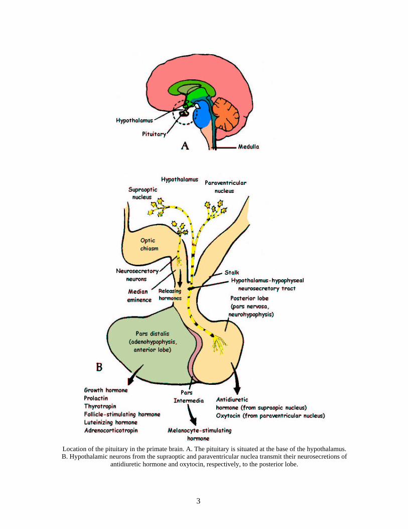
Location of the pituitary in the primate brain. A. The pituitary is situated at the base of the hypothalamus.B. Hypothalamic neurons from the supraoptic and paraventricular nuclea transmit their neurosecretions of
antidiuretic hormone and oxytocin, respectively, to the posterior lobe.
3
4. Anterior pituitarya. Anterior pituitary lies forward from the posterior pituitary and is
composed of three partsi. pars distalis
- principal part – 5 cell types. somatotropes –growth hormone. corticotropes – corticotropin releasing hormone. mammotropes – prolactin
ii. pars intermedia. melanocyte stimulating hormone
iii. pars tuberalis. layer of cells surrounding the neural stalk with no knowfunction
D. Functions of anterior pituitary hormones1. growth hormone – regulates growth, metabolism
a. acts on liver to cause production of somatomedins and somatomedin binding proteins
2. adrenocorticotrophic hormonea. stimulates adrenal cortex to release cortisol and aldosterone,
involved in metabolic regulation3. gonadotrophic hormones
a. follicle stimulating hormonei. stimulates follicular growth in the ovary
b. luteinizing hormonei. stimulates ovulation and growth of corpora lutea
4. thyroid stimulating hormonea. stimulates release of thyroxine from thyroid gland
5. prolactina. stimulates mammary development and lactation
6. beta-lipotrophina. secreted by corticotropesb. involved in pain relief
E. Functions of posterior pituitary hormones1. ADH – regulates reabsorption of water in the kidney3. Oxytocin – milk ejection reflex
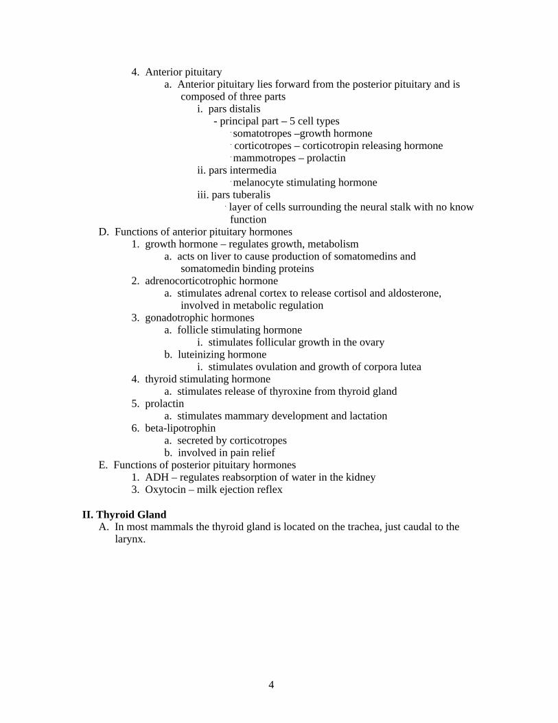
II. Thyroid GlandA. In most mammals the thyroid gland is located on the trachea, just caudal to the
larynx.
4
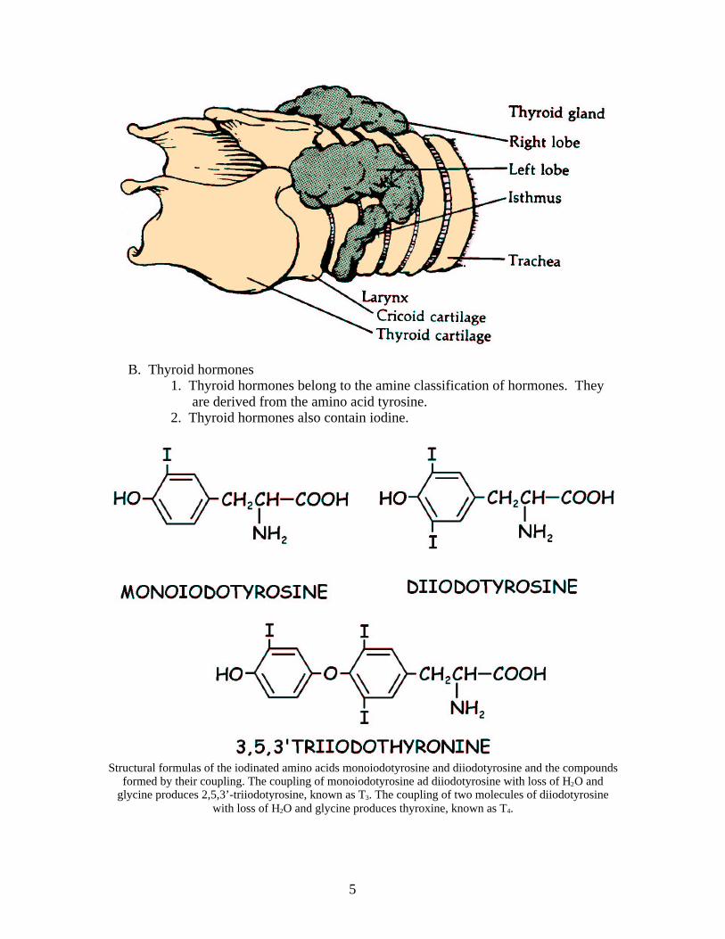
B. Thyroid hormones1. Thyroid hormones belong to the amine classification of hormones. They
are derived from the amino acid tyrosine.2. Thyroid hormones also contain iodine.
Structural formulas of the iodinated amino acids monoiodotyrosine and diiodotyrosine and the compoundsformed by their coupling. The coupling of monoiodotyrosine ad diiodotyrosine with loss of H2O and
glycine produces 2,5,3’-triiodotyrosine, known as T3. The coupling of two molecules of diiodotyrosinewith loss of H2O and glycine produces thyroxine, known as T4.
5
3. Thyroid hormones increase basal metabolism and heat production4. secretion of thyroid hormones (thyroxine and triiodothyronine) is regulated
by thyroid stimulating hormone (TSH) from the anterior pituitary5. Thyroglobulin is a large glyporprotein molecule secreted into the thyroid
follicles by the cells lining the follicles.6. Thyroid deficiency typically results from iodine deficiency. The thyroid
gland enlarges because of accumulation of thyroglobulin.7. Hypo and hyper thyroidism are rare in domestic animals, bud it does occur
in dogs and cats.8. calcitonin
a. hormone of the thyroid gland secreted by the parafollicular or C cells
b. inhibits calcium reabsorption at the kidney and stimulates calcium deposition in bone – lowers blood calcium
III. Parathyroid GlandA. One or two glands which are located near or embedded in the thyroid glands.B. Low calcium levels in plasma stimulate parathyroid hormone (PTH) release.C. PTH stimulates the release of calcium and phosphorus from the bones.D. PTH also stimulates osteoclasts to dissolve boneE. PTH enhances both calcium and phosphate absorption from the intestine by
increasing the rate of formation of 1,25-dihydroxycholecalciferol, the active form of vitamin D.
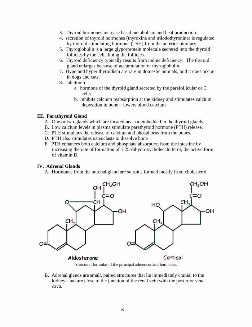
IV. Adrenal GlandsA. Hormones from the adrenal gland are steroids formed mostly from cholesterol.
Structural formulas of the principal adrenocortical hormones.
B. Adrenal glands are small, paired structures that lie immediately cranial to the kidneys and are close to the junction of the renal vein with the posterior vena cava.
6
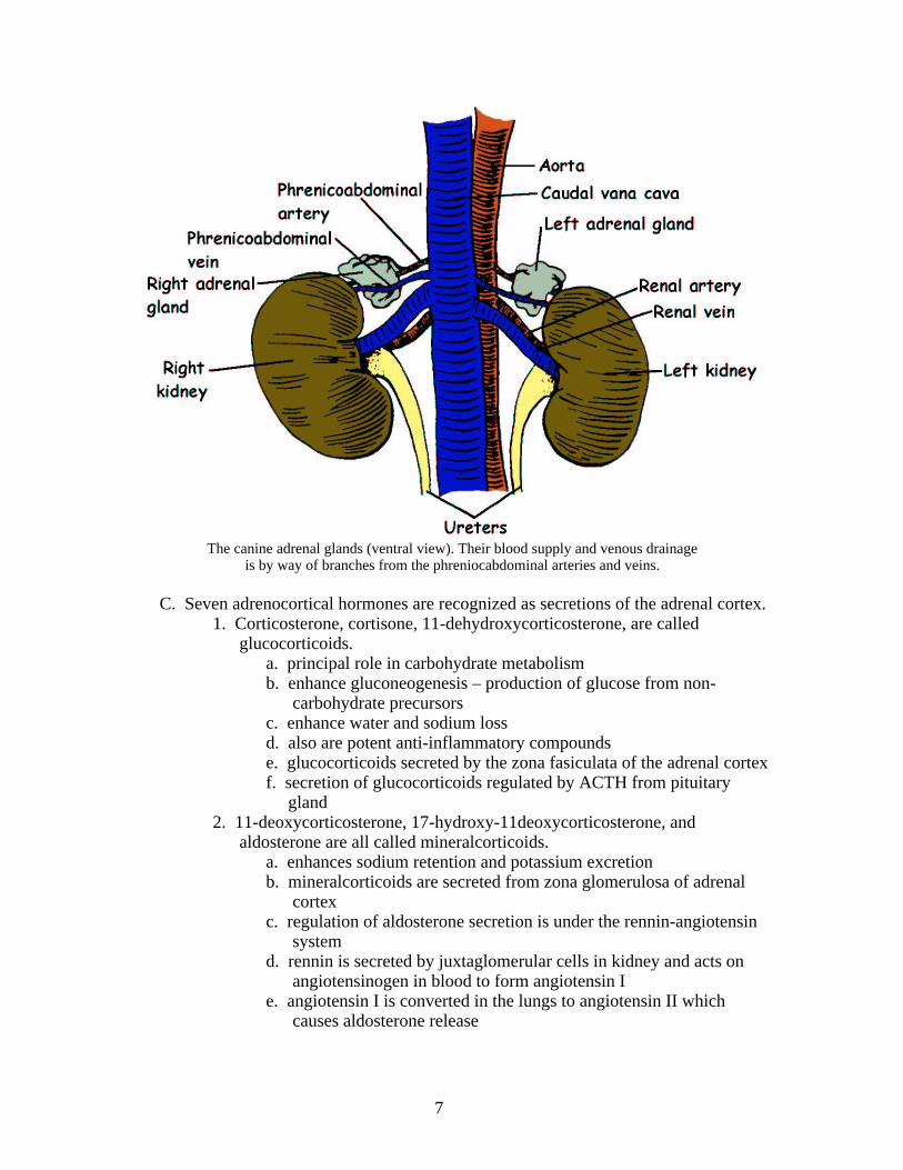
The canine adrenal glands (ventral view). Their blood supply and venous drainageis by way of branches from the phreniocabdominal arteries and veins.
C. Seven adrenocortical hormones are recognized as secretions of the adrenal cortex.1. Corticosterone, cortisone, 11-dehydroxycorticosterone, are called
glucocorticoids.a. principal role in carbohydrate metabolismb. enhance gluconeogenesis – production of glucose from non-
carbohydrate precursorsc. enhance water and sodium lossd. also are potent anti-inflammatory compoundse. glucocorticoids secreted by the zona fasiculata of the adrenal cortexf. secretion of glucocorticoids regulated by ACTH from pituitary gland
2. 11-deoxycorticosterone, 17-hydroxy-11deoxycorticosterone, and aldosterone are all called mineralcorticoids.
a. enhances sodium retention and potassium excretionb. mineralcorticoids are secreted from zona glomerulosa of adrenal
cortex c. regulation of aldosterone secretion is under the rennin-angiotensin
systemd. rennin is secreted by juxtaglomerular cells in kidney and acts on
angiotensinogen in blood to form angiotensin Ie. angiotensin I is converted in the lungs to angiotensin II which
causes aldosterone release
7
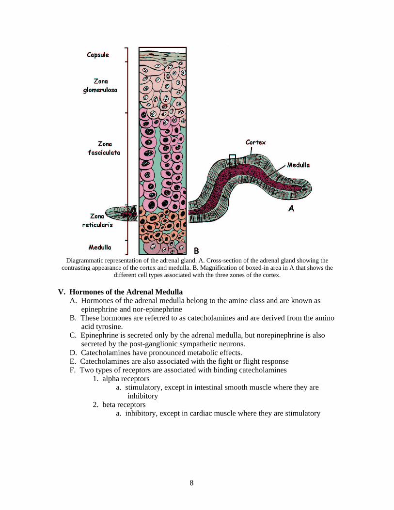
Diagrammatic representation of the adrenal gland. A. Cross-section of the adrenal gland showing thecontrasting appearance of the cortex and medulla. B. Magnification of boxed-in area in A that shows the
different cell types associated with the three zones of the cortex.
V. Hormones of the Adrenal MedullaA. Hormones of the adrenal medulla belong to the amine class and are known as
epinephrine and nor-epinephrineB. These hormones are referred to as catecholamines and are derived from the amino
acid tyrosine.C. Epinephrine is secreted only by the adrenal medulla, but norepinephrine is also
secreted by the post-ganglionic sympathetic neurons.D. Catecholamines have pronounced metabolic effects.E. Catecholamines are also associated with the fight or flight responseF. Two types of receptors are associated with binding catecholamines
1. alpha receptorsa. stimulatory, except in intestinal smooth muscle where they are
inhibitory2. beta receptors
a. inhibitory, except in cardiac muscle where they are stimulatory
8

Structural formulas of catecholamine hormones. They are formed from the amino acid tyrosine and are derivatives of catechol. The abbreviation “dop” is derived from
the German name of this compound, dioxyphenylalamine.
VI. PancreasA. Hormones of the pancreas are insulin, glucagon, somatostatin, and pancreatic
polypeptide.B. Each hormone is secreted by a specific cell type in the pancreatic islets.
1. alpha cells – glucagona. stimulates production of glucose from liver
2. beta cells – insulina. stimulate uptake of glucose
3. delta cells – somatostatina. inhibits secretion of insulin and glucagonb. inhibits secretion of gastrin, secretin, and gastric acid
4. F cells – pancreatic polypeptidea. secretion is stimulated by protein intake, exercise, and fasting –
function unknownVII. Prostaglandins and Their Functions
A. First isolated from prostate gland and hence named prostaglandinsB. Derived from arachidonic acid
9
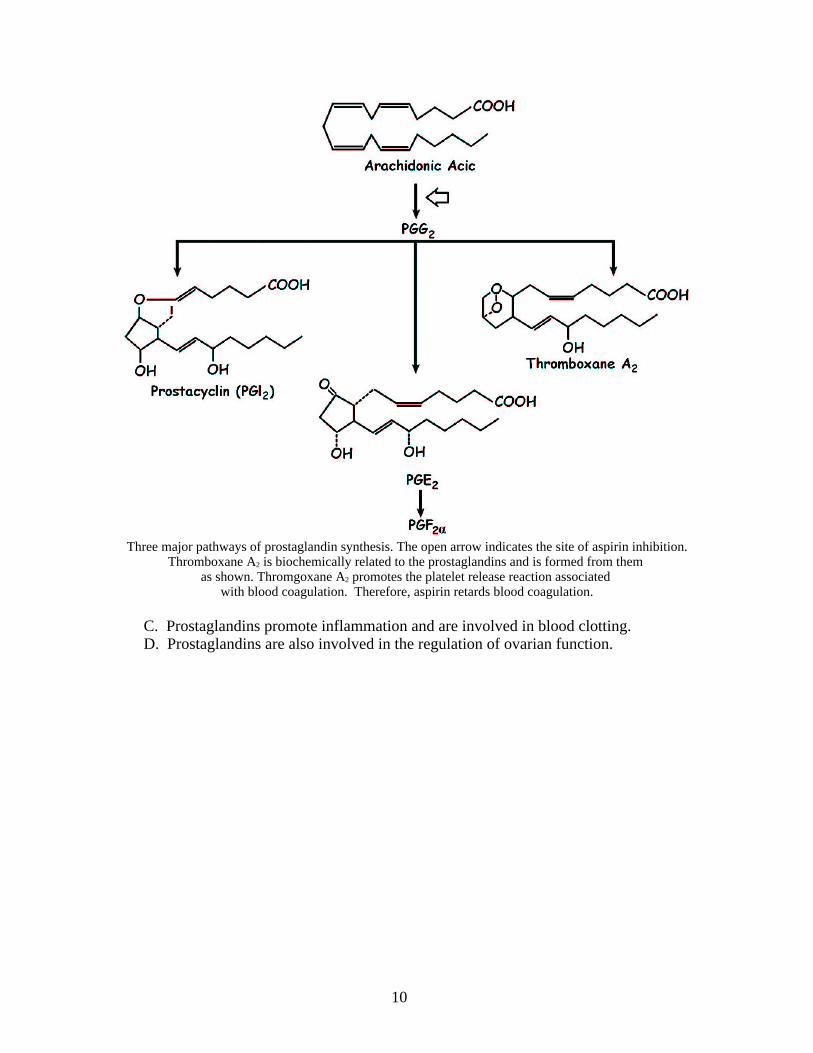
Three major pathways of prostaglandin synthesis. The open arrow indicates the site of aspirin inhibition.Thromboxane A2 is biochemically related to the prostaglandins and is formed from them
as shown. Thromgoxane A2 promotes the platelet release reaction associated with blood coagulation. Therefore, aspirin retards blood coagulation.
C. Prostaglandins promote inflammation and are involved in blood clotting.D. Prostaglandins are also involved in the regulation of ovarian function.
10


![EUROGROUP FOR ANIMALS · more unusual species of animals, often referred to as ‘exotic pets’ [9]. This is a notable shift from the more traditional, domesticated pet animals such](https://img.pdfslide.us/doc/110x75/5f1506009d4e604d0177c573/eurogroup-for-animals-more-unusual-species-of-animals-often-referred-to-as-aexotic.jpg)