Embed Size (px)
Citation preview
Sensory Organs
ANS 215
Anatomy & Physiology
Of Domesticated
Animals
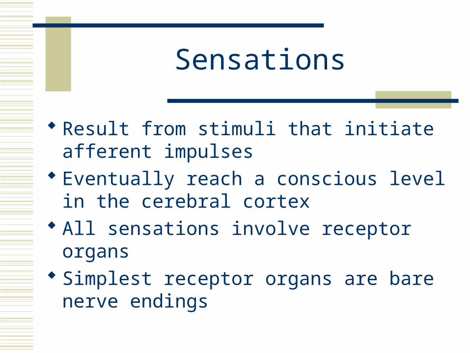
Sensations
Result from stimuli that initiate afferent impulses
Eventually reach a conscious level in the cerebral cortex
All sensations involve receptor organs Simplest receptor organs are bare nerve
endings
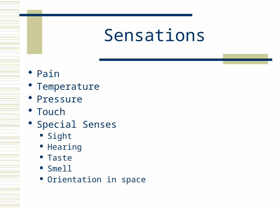
Sensations
Pain Temperature Pressure Touch Special Senses
Sight Hearing Taste Smell Orientation in space
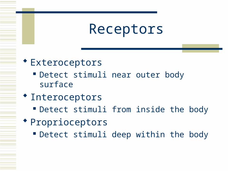
Receptors
Exteroceptors Detect stimuli near outer body surface
Interoceptors Detect stimuli from inside the body
Proprioceptors Detect stimuli deep within the body
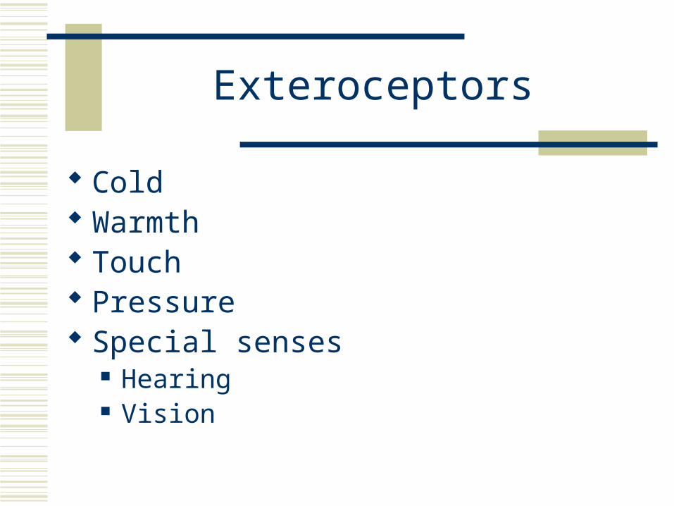
Exteroceptors
Cold Warmth Touch Pressure Special senses
Hearing Vision
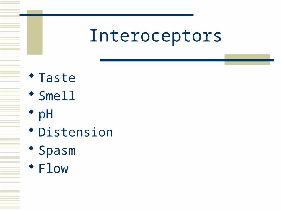
Interoceptors
Taste Smell pH Distension Spasm Flow
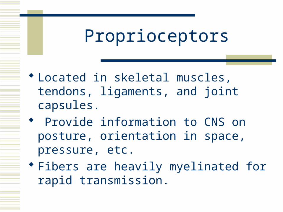
Proprioceptors
Located in skeletal muscles, tendons, ligaments, and joint capsules.
Provide information to CNS on posture, orientation in space, pressure, etc.
Fibers are heavily myelinated for rapid transmission.
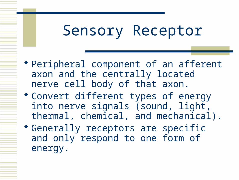
Sensory Receptor
Peripheral component of an afferent axon and the centrally located nerve cell body of that axon.
Convert different types of energy into nerve signals (sound, light, thermal, chemical, and mechanical).
Generally receptors are specific and only respond to one form of energy.
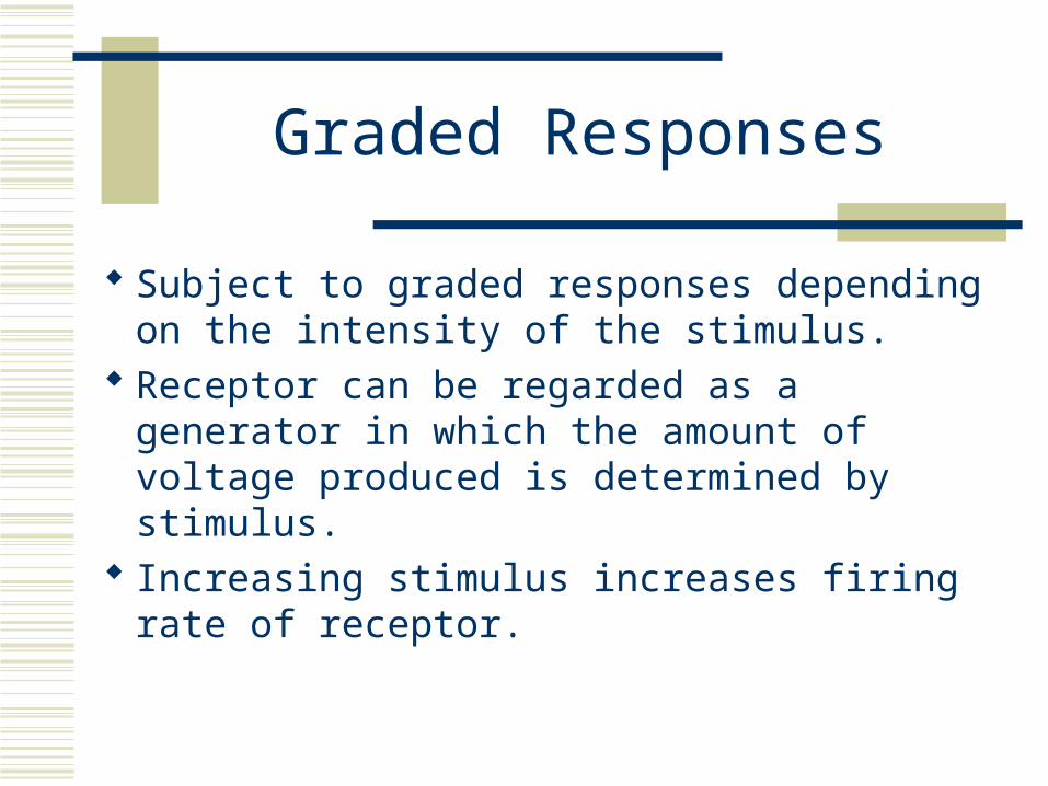
Graded Responses
Subject to graded responses depending on the intensity of the stimulus.
Receptor can be regarded as a generator in which the amount of voltage produced is determined by stimulus.
Increasing stimulus increases firing rate of receptor.
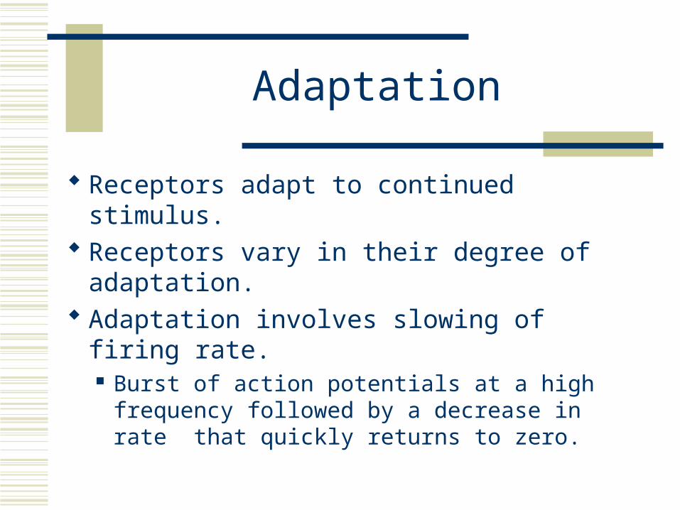
Adaptation
Receptors adapt to continued stimulus. Receptors vary in their degree of adaptation. Adaptation involves slowing of firing rate.
Burst of action potentials at a high frequency followed by a decrease in rate that quickly returns to zero.
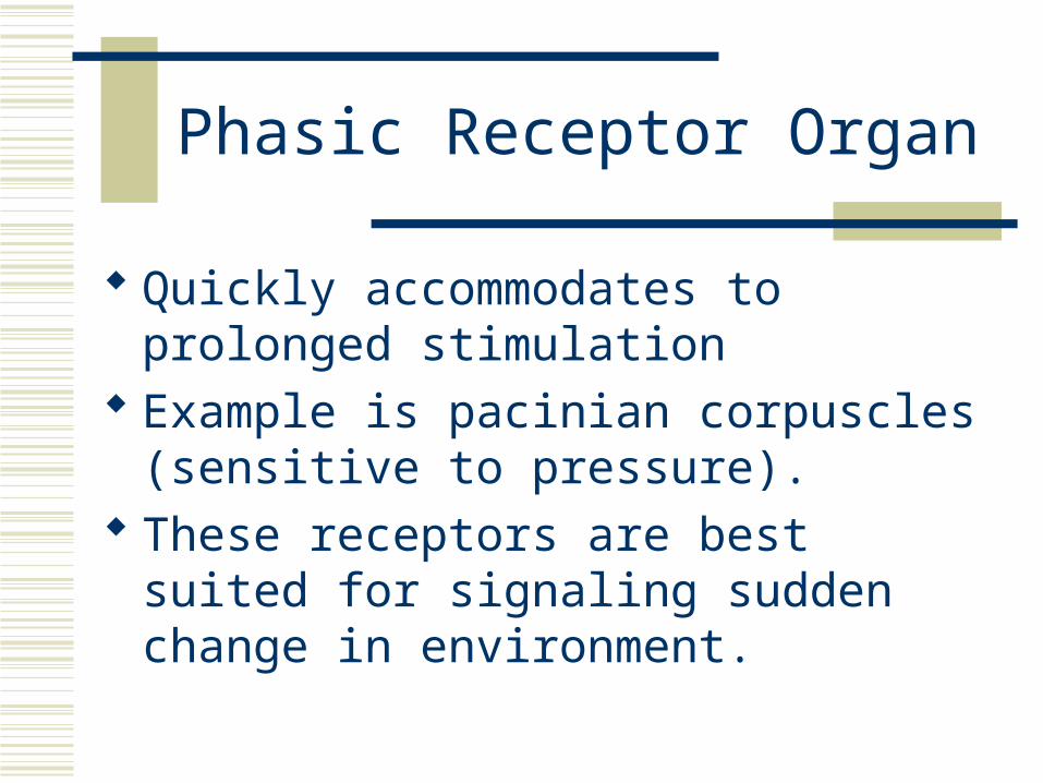
Phasic Receptor Organ
Quickly accommodates to prolonged stimulation
Example is pacinian corpuscles (sensitive to pressure).
These receptors are best suited for signaling sudden change in environment.
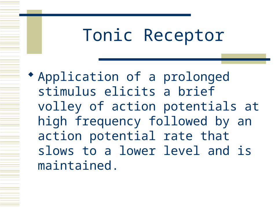
Tonic Receptor
Application of a prolonged stimulus elicits a brief volley of action potentials at high frequency followed by an action potential rate that slows to a lower level and is maintained.
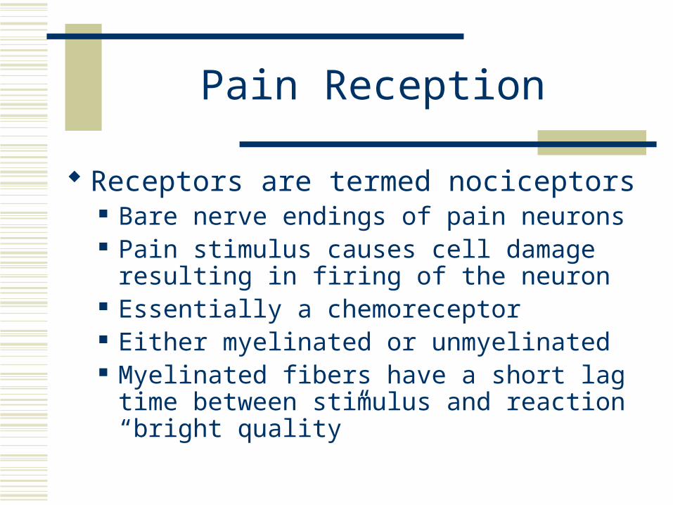
Pain Reception
Receptors are termed nociceptors Bare nerve endings of pain neurons Pain stimulus causes cell damage resulting in
firing of the neuron Essentially a chemoreceptor Either myelinated or unmyelinated Myelinated fibers have a short lag time between
stimulus and reaction “bright quality”
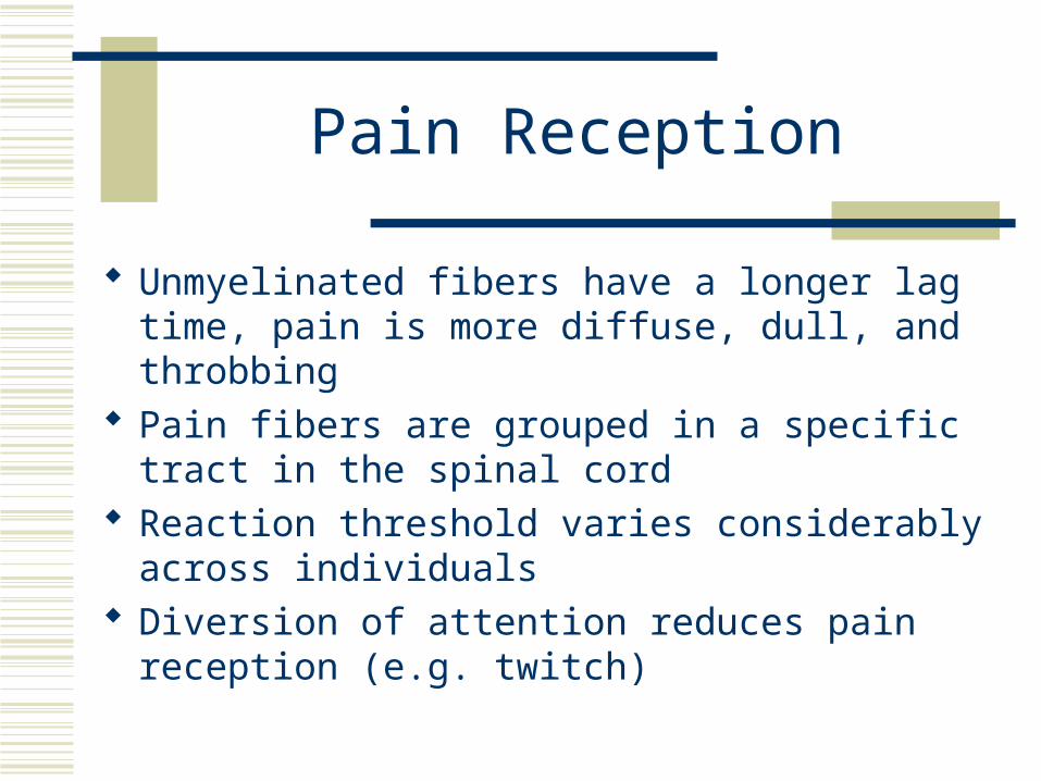
Pain Reception
Unmyelinated fibers have a longer lag time, pain is more diffuse, dull, and throbbing
Pain fibers are grouped in a specific tract in the spinal cord
Reaction threshold varies considerably across individuals
Diversion of attention reduces pain reception (e.g. twitch)
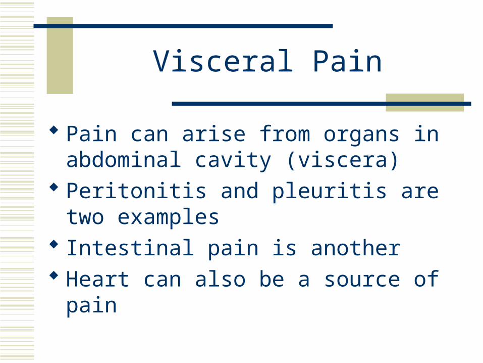
Visceral Pain
Pain can arise from organs in abdominal cavity (viscera)
Peritonitis and pleuritis are two examples Intestinal pain is another Heart can also be a source of pain
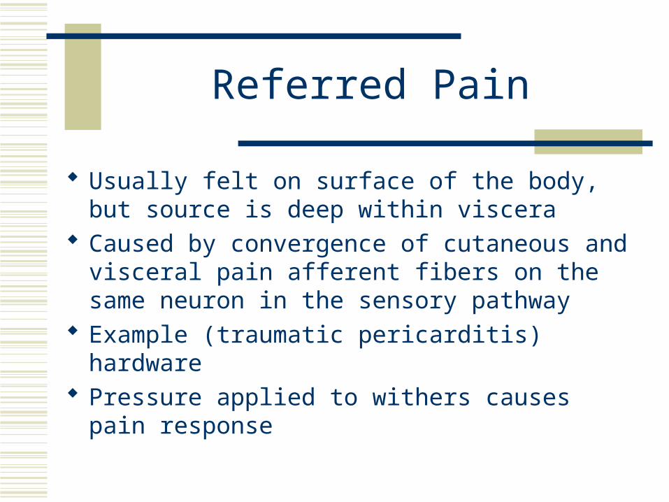
Referred Pain
Usually felt on surface of the body, but source is deep within viscera
Caused by convergence of cutaneous and visceral pain afferent fibers on the same neuron in the sensory pathway
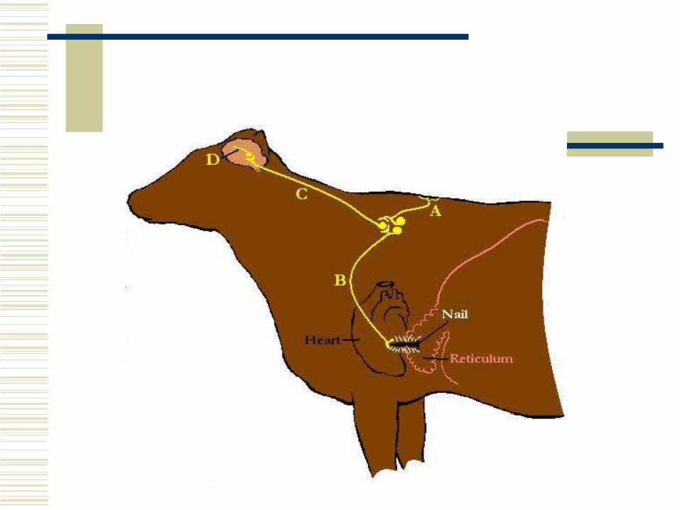
Example (traumatic pericarditis) hardware Pressure applied to withers causes pain response
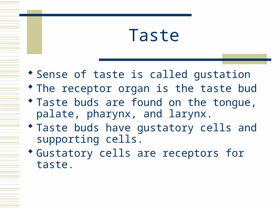
Taste
Sense of taste is called gustation The receptor organ is the taste bud Taste buds are found on the tongue, palate,
pharynx, and larynx. Taste buds have gustatory cells and
supporting cells. Gustatory cells are receptors for taste.
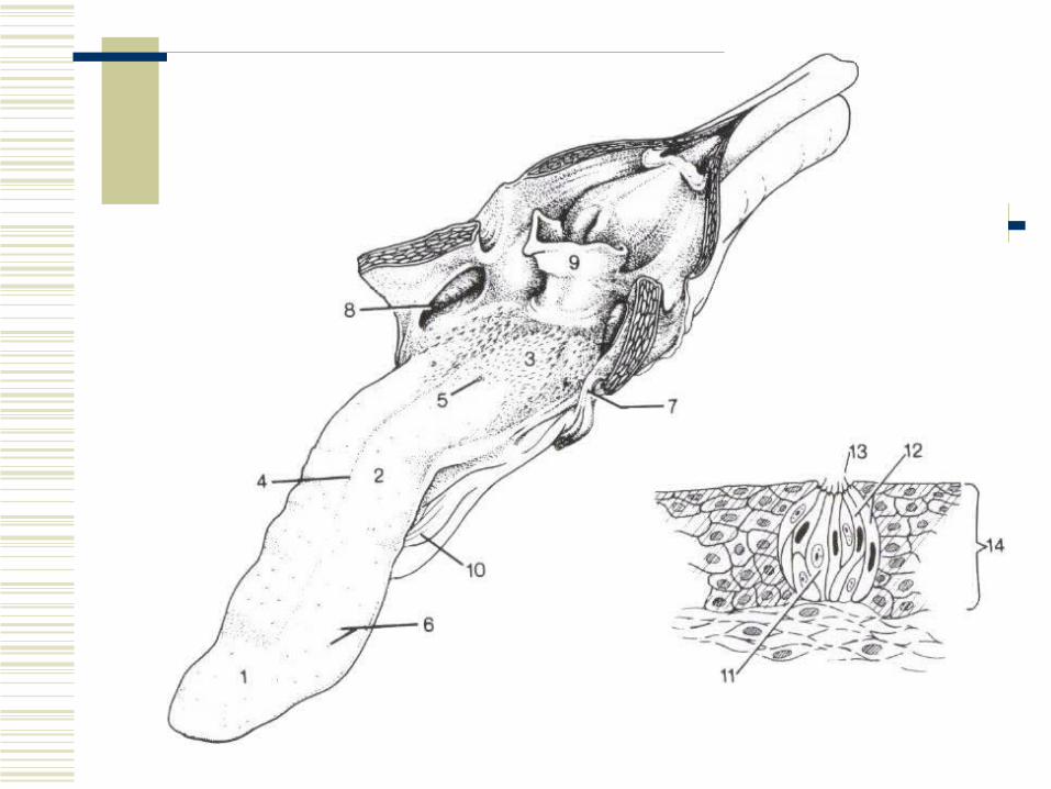
Taste Reception
Taste bud pit communicates with the oral cavity by way of the pore.
Any substance tasted must get into solution and enter the pore of the taste bud.
Hair of the gustatory cell is affected causing stimulation of the gustatory cell.
The impulse is transmitted by cranial nerves VII and IX to the brain.
Taste Sensations
Classified as salty, sweet, bitter, or sour. Each taste sensation is some combination of
the above. Taste perception by animals is based on
preference. Considerable variation within a species
Temperature and Taste
In humans, the temperature of a beverage or food markedly affects its taste.
In humans, the temperature of a beverage or food markedly affects its taste.

Smell
As evolution progressed, nerve cell bodies migrated centrally so that only the nerve fibers remained in a peripheral position.
This provided protection for nerve cells, which do not regenerate.
Central migration did not occur for the nerve cell bodies of Cranial Nerve I (olfactory).
Cell bodies of Cranial Nerve I are found in the mucous membrane of the nasal cavity.
Smell
This location is known as the olfactory region.
The size of this region is directly related to the development of the sense of smell.
Dogs can detect substances 1:1000 of that detectable by humans.
Sensation of smell is known as olfaction.
Smell
Animals with greatly developed sense of smell are macrosmatic.
Animals with less developed sense are microsmatic. (e.g. humans and monkeys)
Animals with no sense of smell are anosmatic. (many aquatic animals)
Smell
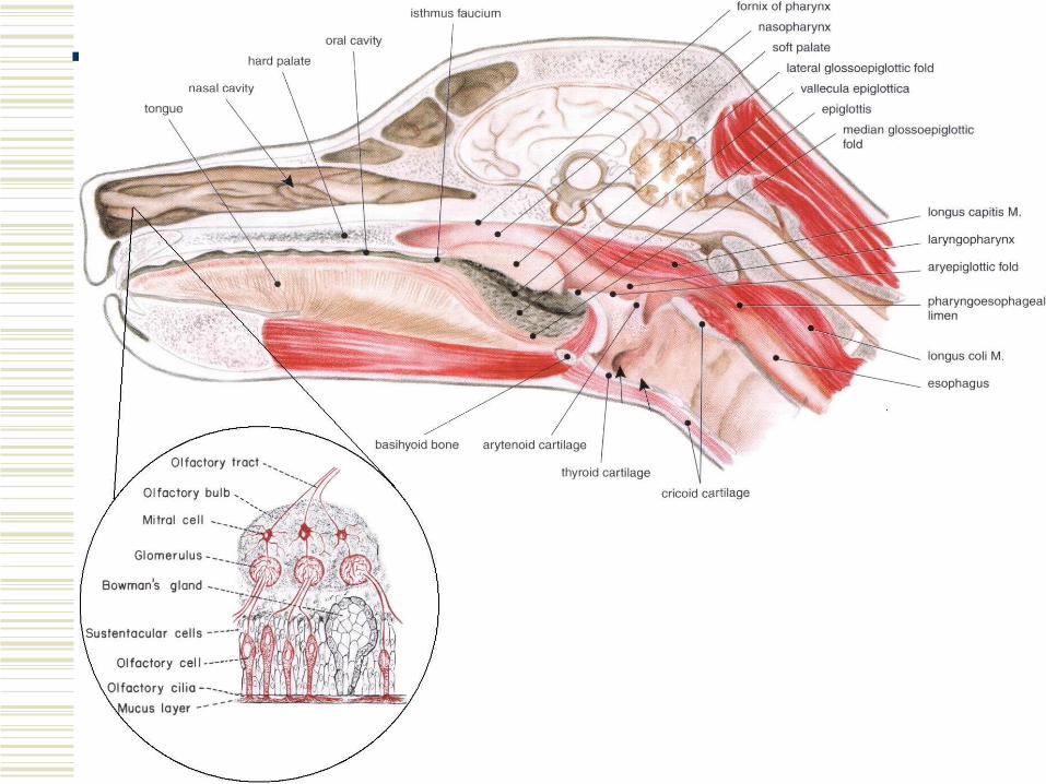
Each olfactory receptor has a cell body and a nerve fiber extending from each end. One is an axon and the other a dendrite.
The dendritic process of the olfactory cell extends to the outside of the olfactory region membrane in crevices between sustentacular cells.
Smell
Sustentacular cells provide major support to the dendritic processes and shield the nerve cell body from the olfactory cavity.
Dendritic processes form hair-like structures (olfactory cilia) that extend into the nasal cavity.
Cilia are covered with secretions from the glands of Bowman.
Smell
Ducts from the glands of Bowman lead through the epithelium of the nasal cavity to its surface.
Secretions constantly refresh the thin layer of fluid bathing the olfactory cilia.
Sniffing allows for back-and-forth movement of air, providing a greater chance for substances to go into solution.
Smell
Once the compound is in solution it binds to olfactory cilia and provides a stimulus for the impulse to be transmitted.
Axons of the olfactory cells join and proceed as fibers and branches of the olfactory nerves.
Basal cells divide and become sustentacular cells or olfactory cells replacing those lost.
Smell
It is unlikely that a specific olfactory cell exists for each smell.
It is probable that the basic smells combine to provide the sensation of a particular odor.
Only one odor can be perceived at any one time.
Olfactory cells adapt to odors.
Phermones
Animals use odors to communicate with each other.
A chemical secreted by one animal which influences the behavior of another is called a pheromone.
Pheromones are used to identify species, mark territories, emit alarms, mark food location, and identify animals in estrus.
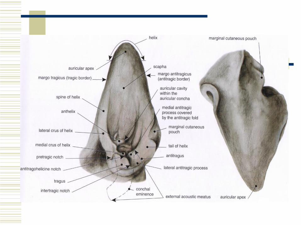
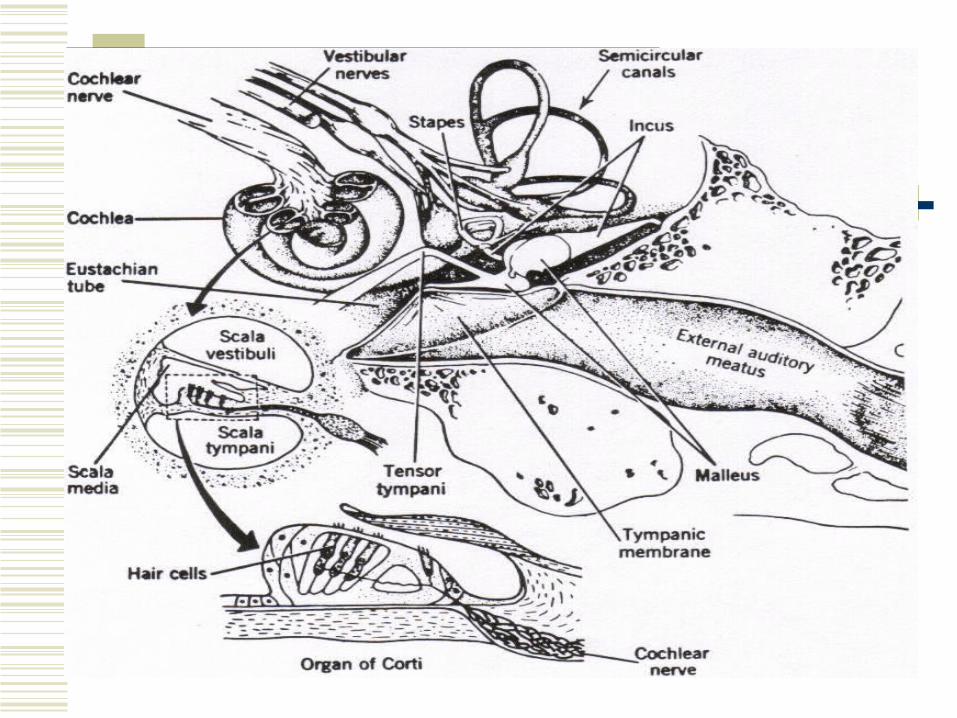
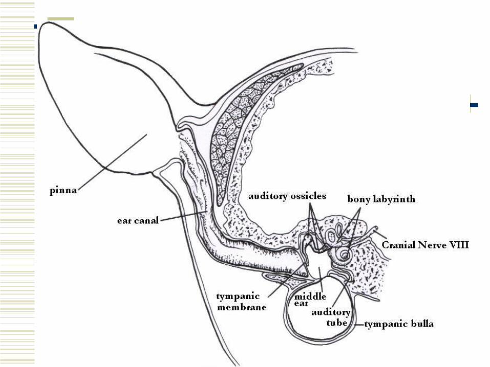
The Ear
Three regions:1. External ear
2. Middle ear
3. Inner ear
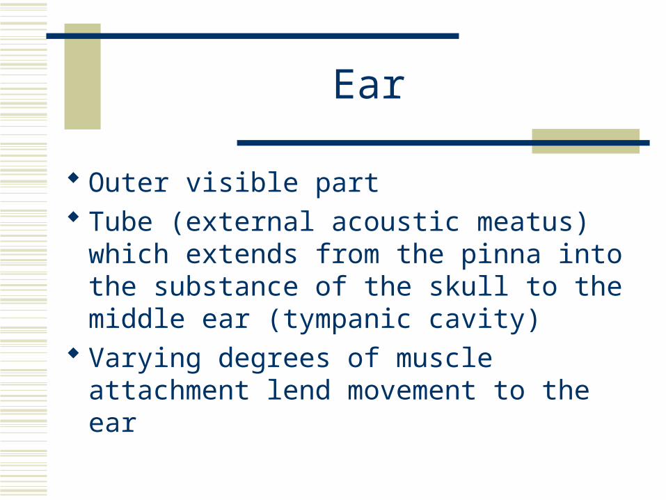
Ear
Outer visible part Tube (external acoustic meatus) which
extends from the pinna into the substance of the skull to the middle ear (tympanic cavity)
Varying degrees of muscle attachment lend movement to the ear
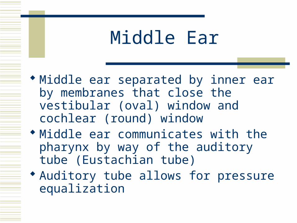
Middle Ear
Middle ear separated by inner ear by membranes that close the vestibular (oval) window and cochlear (round) window
Middle ear communicates with the pharynx by way of the auditory tube (Eustachian tube)
Auditory tube allows for pressure equalization
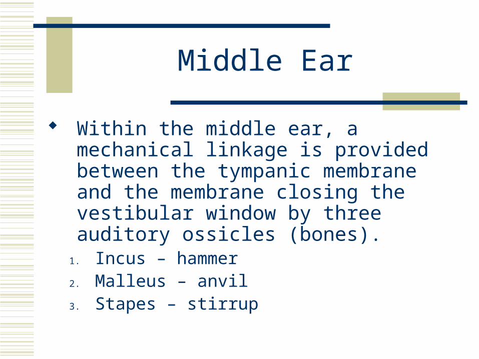
Middle Ear
Within the middle ear, a mechanical linkage is provided between the tympanic membrane and the membrane closing the vestibular window by three auditory ossicles (bones).
1. Incus – hammer 2. Malleus – anvil 3. Stapes – stirrup
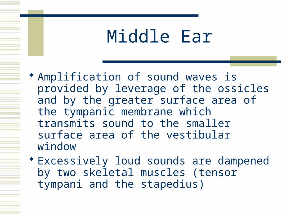
Middle Ear
Amplification of sound waves is provided by leverage of the ossicles and by the greater surface area of the tympanic membrane which transmits sound to the smaller surface area of the vestibular window
Excessively loud sounds are dampened by two skeletal muscles (tensor tympani and the stapedius)
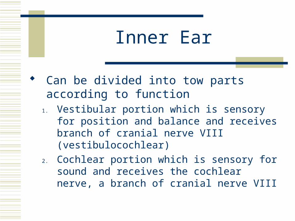
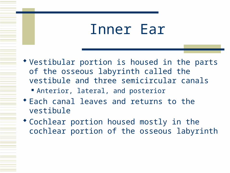
Inner Ear
Can be divided into tow parts according to function
1. Vestibular portion which is sensory for position and balance and receives branch of cranial nerve VIII (vestibulocochlear)
2. Cochlear portion which is sensory for sound and receives the cochlear nerve, a branch of cranial nerve VIII
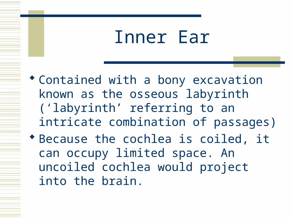
Inner Ear
Contained with a bony excavation known as the osseous labyrinth (‘labyrinth’ referring to an intricate combination of passages)
Because the cochlea is coiled, it can occupy limited space. An uncoiled cochlea would project into the brain.
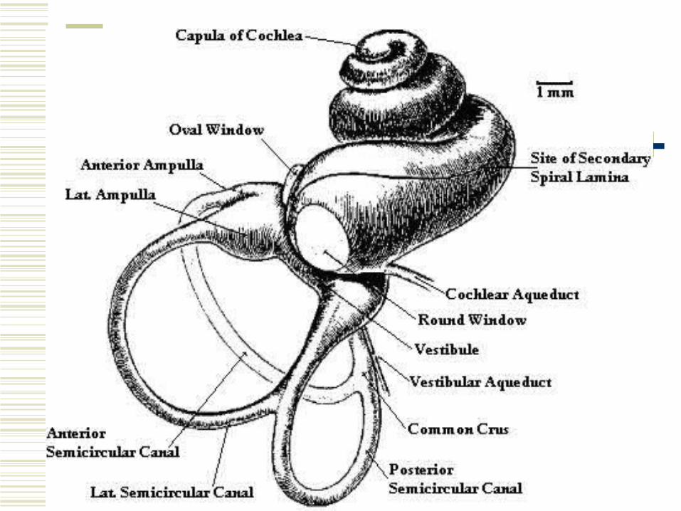
Inner Ear
Vestibular portion is housed in the parts of the osseous labyrinth called the vestibule and three semicircular canals Anterior, lateral, and posterior
Each canal leaves and returns to the vestibule Cochlear portion housed mostly in the
cochlear portion of the osseous labyrinth
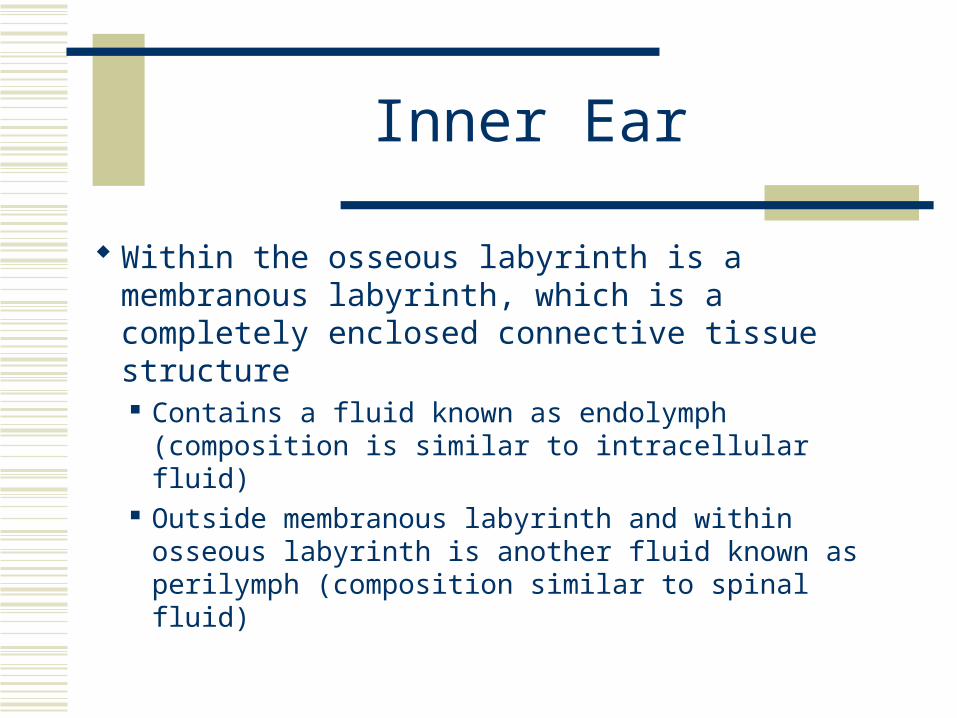
Inner Ear
Within the osseous labyrinth is a membranous labyrinth, which is a completely enclosed connective tissue structure Contains a fluid known as endolymph
(composition is similar to intracellular fluid) Outside membranous labyrinth and within
osseous labyrinth is another fluid known as perilymph (composition similar to spinal fluid)

Inner Ear
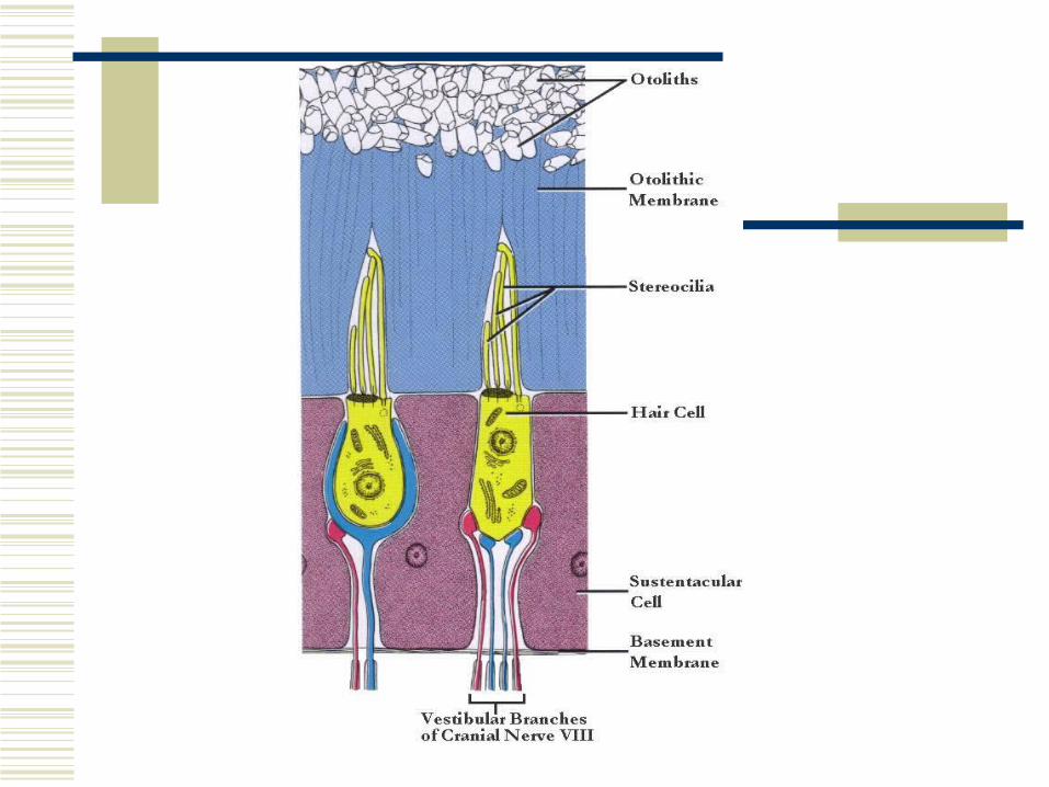
Within the vestibular portion, the membranous labyrinth also includes three semicircular canals and two sacs within the vestibule known as the utricle and saccule.
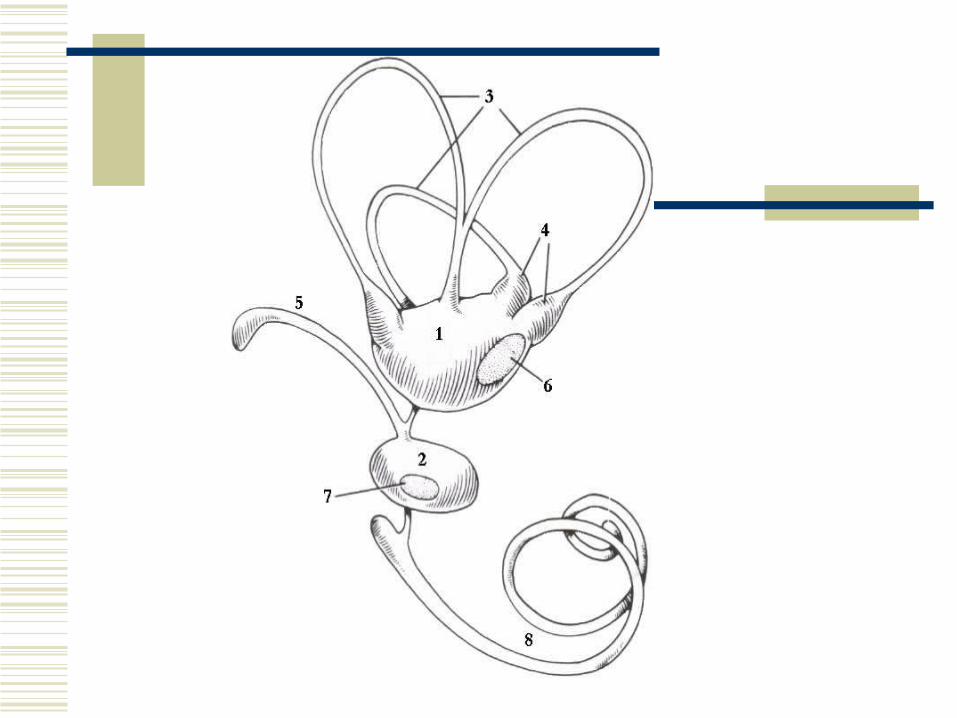
Inner Ear
As each membranous labyrinth occupying the semicircular canals leaves the utricle, a dilated portion is noted – the ampula
Each of the three ampullae contains sensory receptors for equilibrium known as the crista ampullaris. The utricle and saccule each contains a sensory receptor area known as the macula. Macula receptors are more or less stimulated depending
on the position of the head in space. The cristae are stimulated during head movement

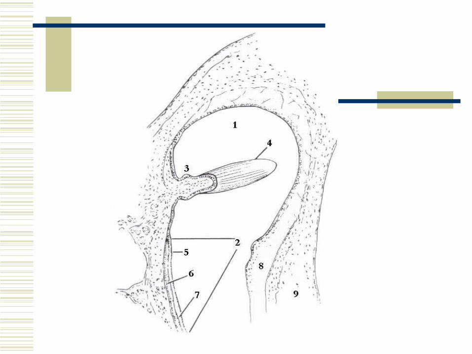
Inner Ear
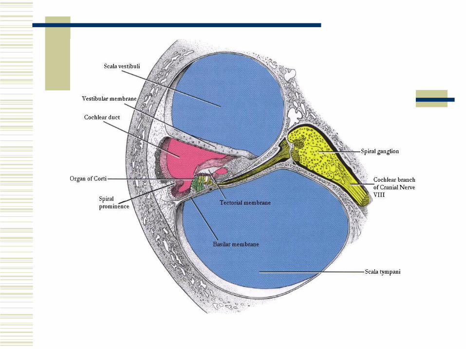
Extension of the membranous labyrinth into the cochlea is known as the cochlear duct or scala media. This divides the cochlea into a part above the scala media (scala vestibuli) and a part below (scala tympani).
Inner Ear
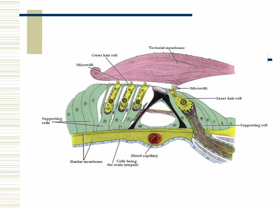
Along the length of the scala media are a large number of structures each individually called an organ of corti. Convert sound waves to nerve impulses Location of organ of corti within scala media determines
frequency of sound perceived Organ of cortis is composed of hair cells that have hairs
projecting toward the tectorial membrane. Displacement of the hair cell cilia against the tectorial membrane by oscillations of the basilar membrane causes the hair cells to depolarize and create a nerve impulse.
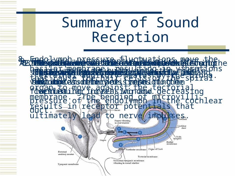
Summary of Sound Reception
1. Sound wave is directed into the external auditory meatus
by the pinna. 2. Sound wave strikes the tympanic membrane (eardrum)
and sets it in motion. 3. The motion of the eardrum is transmitted through the
middle ear by the auditory ossicles to the vestibular (oval) window.
4. The stapes moves back and forth pushing the membrane of the oval window in and out.
5. The movement of the oval window sets up fluid pressure waves in the incompressible perilymph of the cochlea.
6. Pressure waves are transmitted through the scala vestibuli. 7. The pressure waves deform the walls of the scala vestibuli, scala tympani, and vestibular membrane, resulting in fluctuating increasing and decreasing pressure of the endolymph in the cochlear duct.
8. Endolymph pressure fluctuations move the basilar membrane, resulting in vibrations that cause the hair cells of the spiral organ to move against the tectorial membrane. The bending of microvilli results in receptor potentials that ultimately lead to nerve impulses.
9. Pressure waves eventually cause the round window to bulge into the middle ear.
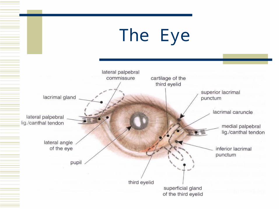
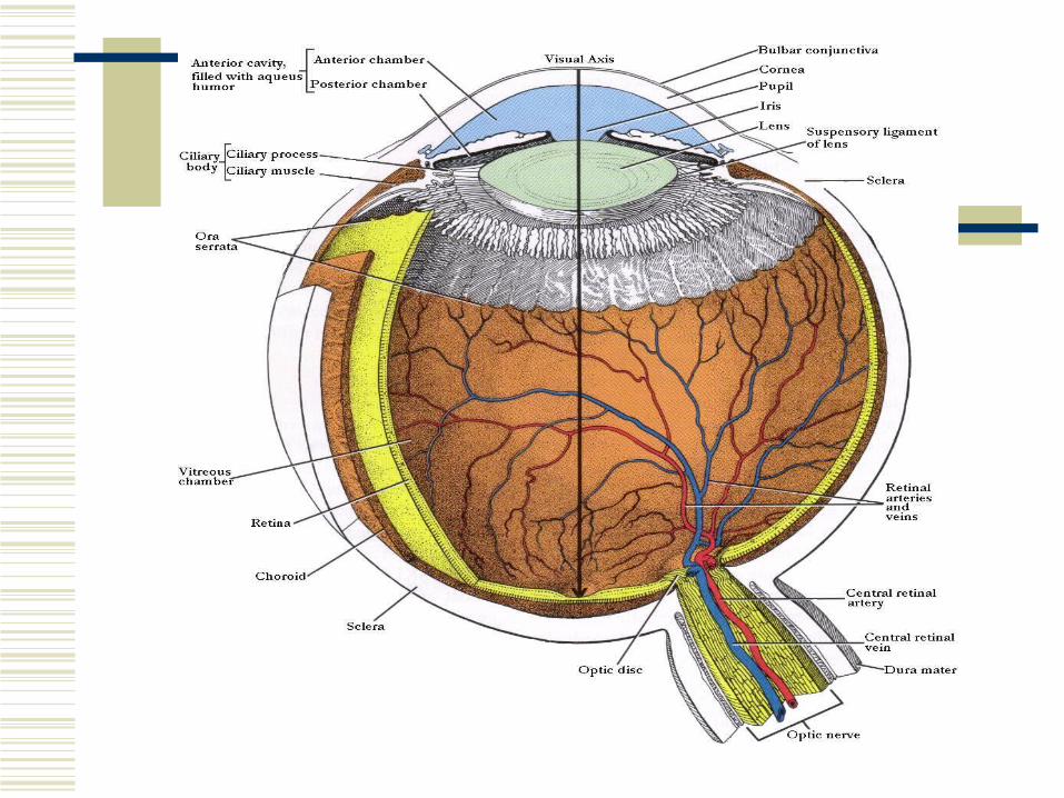
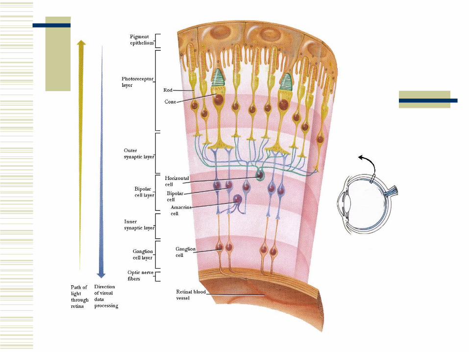
The Eye
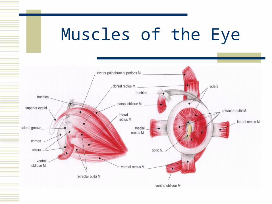
Muscles of the Eye