Embed Size (px)
Citation preview

REVIEW
Emerging Research and Clinical Development Trends of Liposomeand Lipid Nanoparticle Drug Delivery Systems
JOHN C. KRAFT, JENNIFER P. FREELING, ZIYAO WANG, RODNEY J. Y. HO
Department of Pharmaceutics, University of Washington, Seattle, Washington
Received 23 August 2013; revised 11 October 2013; accepted 14 October 2013
Published online 25 November 2013 in Wiley Online Library (wileyonlinelibrary.com). DOI 10.1002/jps.23773
ABSTRACT: Liposomes are spherical-enclosed membrane vesicles mainly constructed with lipids. Lipid nanoparticles are loaded withtherapeutics and may not contain an enclosed bilayer. The majority of those clinically approved have diameters of 50–300 nm. The growinginterest in nanomedicine has fueled lipid–drug and lipid–protein studies, which provide a foundation for developing lipid particles thatimprove drug potency and reduce off-target effects. Integrating advances in lipid membrane research has enabled therapeutic development.At present, about 600 clinical trials involve lipid particle drug delivery systems. Greater understanding of pharmacokinetics, biodistribution,and disposition of lipid–drug particles facilitated particle surface hydration technology (with polyethylene glycol) to reduce rapid clearanceand provide sufficient blood circulation time for drug to reach target tissues and cells. Surface hydration enabled the liposome-encapsulatedcancer drug doxorubicin (Doxil) to gain clinical approval in 1995. Fifteen lipidic therapeutics are now clinically approved. Although muchresearch involves attaching lipid particles to ligands selective for occult cells and tissues, preparation procedures are often complex andpose scale-up challenges. With emerging knowledge in drug target and lipid–drug distribution in the body, a systems approach thatintegrates knowledge to design and scale lipid–drug particles may further advance translation of these systems to improve therapeuticsafety and efficacy. C© 2013 Wiley Periodicals, Inc. and the American Pharmacists Association J Pharm Sci 103:29–52, 2014Keywords: drug delivery systems; lipids; liposomes; phospholipids; micelle; disposition; nanotechnology; nanoparticles; pegylation
INTRODUCTION
It begins in the late 1950s with the discovery by Saunders andThomas1 and Bangham and Horne2 that simple hydration ofdry lipid film coated on a glass surface produces spherical vesi-cles or liposomes. This basic observation not only enabled theexploration of lipid–drug and lipid–protein interactions, butit spurred the development of liposomes and lipid nanoparti-cles as drug carriers to enhance therapeutic benefits. Today,liposomes or lipid vesicles are a pivotal biocompatible andbiodegradable drug delivery and formulation platform. Theyare typically constructed with a synthetic lipid bilayer mem-brane, a biomimetic of cell membranes, to entrap drug insidean aqueous core. Under the protection of the lipid membrane, awell-subscribed early concept was that drug inside the aqueouscompartment could be transported to tissue, cell, or intracel-lular targets. Incorporating drug molecules in these particles
Abbreviations Used: APC, antigen-presenting cell; DMPC, dimyristoylphos-phatidylcholine; DOPC, dioleoylphosphatidylcholine; DPPC, dipalmitoylphos-phatidylcholine; DSPC, distearoylphosphatidylcholine; DTPA, diethylenetriamine pentaacetic acid; EDC, ethyl(dimethylaminopropyl) carbodiimide;NHS, N-hydroxysuccinimide; EPR, enhanced permeability and retention;FDA, US Food and Drug Administration; HER-2, human epidermal growthfactor receptor 2; HIFU, high-intensity focused ultrasound; MLV, multilamel-lar vesicle; MMP, matrix metalloproteinase; MPS, mononuclear phagocytesystem; MRI, magnetic resonance imaging; MUC1, mucin 1; MVL, multi-vesicular liposome; NGPE, N-glutaryl phosphatidylethanolamine; PC, phos-phatidylcholine; pDNA, plasmid DNA; PE, phosphatidylethanolamine; PEG,polyethylene glycol; PET, positron emission tomography; PG, phosphatidyl-glycerol; PRR, pattern recognition receptor; PSA, prostate-specific antigen;PSMA, prostate-specific membrane antigen; RGD, arginine–glycine–aspartate;PS, phosphatidylserine; SMPB, succinimidyl-4-(p-maleimidophenyl) butyrate;SPH, sphingomyelin; Tc, lipid-phase transition temperature; TLR, Toll-likereceptor.
Correspondence to: Rodney J. Y. Ho (Telephone: +206-543-9434; Fax: +206-543-3204; E-mail: [email protected])
Journal of Pharmaceutical Sciences, Vol. 103, 29–52 (2014)C© 2013 Wiley Periodicals, Inc. and the American Pharmacists Association
was proposed to shield healthy bystander tissues and cells fromdrug toxicity while the drug is en route to sites of pharmaco-logical action or disease (effect) sites. In theory, water-soluble(hydrophilic) agents can be encapsulated in the aqueous coreenveloped by the lipid membrane or attached on the membranesurface with lipid conjugated to soluble agents. The potential tocarry both hydrophobic and hydrophilic compounds has madeliposomes one of the favorite research topics in drug carrier re-search for scientists across disciplines. However, it was soon dis-covered that each liposome and lipid nanoparticle (constructedwith different lipid mixtures) exhibits distinct physical stabil-ity, distribution, and patterns of elimination time course in thebody. Many years passed before scientists began to appreciatethe challenges of premature liposome degradation and clear-ance, and found lipid compositions that produce stable lipo-somes that circulate for a sufficient amount of time in the body.Together, physical stability (in storage and in the body) andpharmacokinetics (time course study) of liposomes intended toreduce rapid elimination or clearance are some of the keys tosuccessful translation of liposome drug delivery systems intotherapeutic products.
Depending on design, liposomes may contain a single ormultiple (onion-like concentric) bilayered lipid membrane com-posed of natural or synthetic lipids, with diameters rangingfrom tens of nanometers to micrometers.3 However, not alllipid nanoparticles have a contiguous bilayer that would qual-ify them as lipid vesicles or liposomes. For example, somelipid nanoparticles may have up to 33% of drug bound tolipid molecules.4 Although these lipid nanoparticles may bephysically stable, the membrane with high densities of drugmolecules may not behave as a liposomal membrane capableof encapsulating aqueous contents. Thus, we qualify this vari-ability by discussing both liposomes and lipid nanoparticles in
Kraft et al., JOURNAL OF PHARMACEUTICAL SCIENCES 103:29–52, 2014 29

30 REVIEW
this manuscript. In some cases, lipid–drug aggregates may as-sume micelle-like structures. Micelles are thermodynamicallystable multimeric nanoaggregate structures of amphipathic li-pidic molecules in solution about 5–10 nm in diameter. Typicalmicelles contain a hydrophobic core; however, inverted micelleshave a small hydrophilic interior. Other lipid nanoparticles oflipid–drug complexes may be prepared as water-in-oil or oil-in-water emulsions and conform into colloidal dispersions. Lipidsand derivatives exhibiting a range of biochemical and biophys-ical properties (size, charge, and surface structure) can be syn-thesized and engineered to develop drug carriers for specifictherapeutic applications. This potential flexibility and associ-ated potential number of variations in lipid–drug combinations(because of the unique lipid–drug interactions) and therapeutictarget design result in wide-ranging lipid–drug compositions.Thus, with no two liposomes or lipid nanoparticles being iden-tical, it makes rigorous manufacturing control imperative.
Since their discovery, liposomes have enjoyed significant at-tention in laboratory and pharmaceutical research because of anumber of attributes. The bilayer membrane could protect drugfrom hydrolysis or oxidative degradation, thereby minimizingtoxicity (i.e., improving the therapeutic index). Prolonged drugcirculation or residence time in the body may increase drugbioavailability (reduce clearance) and provide sufficient timefor drug molecules to arrive at disease targets. Other potentialadvantages include the ability to carry multiple drugs at once;the addition of targeting moieties, such as antibodies; and thebio-degradable and tunable drug release in response to temper-ature, pH, or other environmental inputs.
It took about 35 years after the late 1950s discovery to re-alize the first clinical liposome application in drug delivery.
In 1995, Doxil (PEGylated liposome-encapsulated doxorubicin)became the first liposome drug delivery system approved forhuman use by the US Food and Drug Administration (FDA).5,6
Today, Doxil and other liposomal doxorubicin and daunorubicinare widely used to treat ovarian cancer and Kaposi’s sarcoma(over 300,000 patients are treated each year), and to protect pa-tients from anthracycline cardiotoxicity.7 Moreover, Doxil wasreported to improve doxorubicin levels in Kaposi’s sarcoma tis-sues by as much as 22-fold compared with healthy normal skintissues.8,9 Several drugs and molecules, such as anticancer andantibacterial agents, imaging and probing agents, peptide hor-mones, proteins, enzymes, vaccines, and genetic material, havebeen loaded into the aqueous compartment or lipid phases ofliposomes.
As shown in Table 1, about 15 liposome and lipid-based drugformulations are approved for human use. Six are treatmentsintended for cancers; others are for fungi, microbes, preventivevaccination, analgesia, macular degeneration, and hormonereplacement. Select lipid-based drug candidates in late-stage(Phase II/III) clinical trials are presented in Table 2. Currently,all human clinical trials intended for product licensing approvalby the FDA must be registered with ClinicalTrials.gov, a USDepartment of Health and Human Services sponsored clinicaltrial registry. According to ClinicalTrials.gov, there are 589 in-terventional drug studies with a liposome platform as of May2013. Interestingly, no FDA-licensed liposome or lipid nanopar-ticle is coated with ligands or targeting moieties for homingdrug to target tissues, cells, or subcellular organelles. Such tar-geted therapeutics (with or without precise and controlled drugrelease) are an emerging area of research. These ligand-coatedparticles, often referred to as actively targeted liposomes, are a
Table 1. Marketed Liposomal and Lipid-Based Products
Trade Name (Company) Lipid Platform Drug Size Indication
AnticancerDoxil/Caelyx (Janssen) PEG–liposome Doxorubicin 100 nm Kaposi’s sarcoma, ovarian cancer, breast
cancer, combination with bortezomib inmultiple myeloma
DaunoXome (Galen) Liposome Daunorubicin 45–80 nm Kaposi’s sarcomaDepoCyt (Pacira) Liposome Cytarabine 20 :m Malignant lymphomatous meningitisMarqibo (Talon) Liposome Vincristine 100 nm Acute lymphoblastic leukemiaMyocet (Cephalon) Liposome Doxorubicin 80–90 nm Combination therapy with
cyclophosphamide in breast cancer
AntifungalAbelcet (Sigma–Tau) Lipid drug particles Amphotericin B 2–5 :m AspergillosisAmBisome (Astellas) Liposome Amphotericin B <100 nm Antifungal, leishmaniasisAmphotec (Alkopharma) Micelle Amphotericin B 115 nm Aspergillosis
VaccineEpaxal (Crucell) Virosome Hepatitis A antigen 150 nm Hepatitis AInflexal V (Crucell) Virosome Influenza antigen 150 nm Influenza
AnalgesicsDiprivan (Fresenius Kabi) Lipid emulsion Propofol 180 nm AnesthesiaDepoDur (Pacira) MV liposome Morphine 17–23 :m Postsurgical painExparel (Pacira) MV liposome Bupivacaine 24–31 :m Analgesia
OtherVisudyne (QLT) Liposome Verteporfin – Age-related macular degenerationEstrasorb (Medicis) Micelle Estradiol 600 :m Menopausal therapy
MV, multivesicular.
Kraft et al., JOURNAL OF PHARMACEUTICAL SCIENCES 103:29–52, 2014 DOI 10.1002/jps.23773

REVIEW 31
Table 2. Select Lipid-Based Products in Clinical Development
Therapeutic Product Name Sponsor Indication Trial Phase
BLP-25a Stimuvax Merck Nonsmall cell lung cancer Phase IIICytarabine CPX-351 Celator Acute myeloid leukemia Phase IIIMHC Ib Allovectin-7 Vical Inc. Metastatic melanoma Phase IIICisplatin Lipoplatin Regulon Nonsmall cell lung cancer Phase III
SPI-77 NYU Ovarian cancer Phase IIAroplatin NYU Malignant mesothelioma Phase II
Doxorubicin ThermoDox Celsion Primary hepatocellular carcinoma Phase IIIRefractory chest wall breast cancer Phase IIColorectal liver metastases Phase II
2B3-101 To-BBB Brain metastases and glioma Phase IIMeningeal carcinomatosis Phase II
MPL/QS21c RTS,S/ASO1B GSK Malaria Phase IIOxaliplatin MBP-426 Mebiopharm Gastrointestinal adenocarcinoma Phase IIPaclitaxel LEP—ETU Insys Breast cancer Phase II
EndoTAG-1 MediGene Breast cancer Phase IIPNU-91934 MSKCC Esophageal cancer Phase II
SN38d CPX-1 Celator Colorectal cancer Phase IILE-SN38 C&L Grp B Metastatic colorectal cancer Phase IIMM-398 Merrimack Gastric and pancreatic cancer Phase II
aThe BLP-25 lipopeptide is a 25-amino-acid protein sequence (STAPPAHGVTSAPDTRPAPGSTAPP) containing a palmitoyl lysine residue at the carboxy terminal.BLP-25 provides specificity of the mucin 1 (MUC1) integral membrane protein to stimulate an anti-MUC1 immune response.
bAllovectin-7 is a cancer immunotherapeutic formulated as a plasmid/cationic lipid complex containing DNA sequences encoding HLA-B7 and beta-2-microglobulin—the heavy and light chains of the major histocompatibility complex (MHC) class I, respectively.
cRTS,S/AS01B is a recombinant hybrid peptide malaria candidate vaccine formulated as a liposome adjuvant system with immunostimulants monophosphoryllipid A (MPL) and QS21 (a natural saponin that is the purification fraction 21 from the bark of the South American tree Quillaja saponaria).
dSN38 (7-ethyl-10-hydroxy-camptothecin) is the active metabolite of prodrug irinotecan (CPT-11), converted through carboxylesterase enzymes.C&L Grp B, Cancer and Leukemia Group B; GSK, GlaxoSmithKline; MSKCC, Memorial Sloan-Kettering Cancer Center; and NYU, New York University School
of Medicine.
challenge to reproduce and manufacture at clinically meaning-ful scales, even if validated in small animals. Optimization ofphysiochemical properties involved in stability, toxicity, and im-mune surveillance, and the development of robust scale-up andmanufacturing processes could be challenging in some cases.Although the first-generation liposome and lipid nanoparticletherapeutic products proved this platform to be safe and effec-tive for delivery of drugs and vaccines, their use for nucleic acidand gene therapeutics continues to be explored.
Since our last review on liposome drug delivery systems,10
research continues to fuel development of liposomes and lipidnanoparticles that improve the pharmacokinetics and ther-apeutic index of drugs by extending their margin of safetyand efficacy. This manuscript discusses the emerging researchand clinical developments in liposome and lipid nanoparti-cle delivery of therapeutics. We highlight opportunities forvalue-added clinical translation of compounds based on thisplatform. To do so, we first discuss physiochemical proper-ties that are key to characterize and optimize prior to invivo scaling.11 As recent reviews focus on biophysical andchemical aspects of liposome preparation, characterization,targeting, and optimization, we briefly discuss basic proper-ties of liposomes and lipid nanoparticles.3,11–14 We next dis-cuss scale-up considerations then in vivo delivery and cur-rent advances in passive and active drug targeting. This isfollowed by applications of liposomes and lipid nanoparti-cles as multifunctional carriers, vaccines, gene therapeutics,and oral drug delivery systems. We conclude with a highlighton future directions and innovations in liposome and lipidnanoparticle therapeutics.
BASIC PROPERTIES OF LIPOSOMES AND LIPIDNANOPARTICLES
Lipid vesicles or liposomes are colloidal particles composed ofphospholipid molecules that form contiguous membrane bilay-ers able to entrap solute. Although liposomes and lipid nanopar-ticles may be prepared with nonphospholipid molecules suchas cardiolipin and other synthetic derivatives, to date most allcore lipids derive from a phospholipid backbone structure. Lipidnanoparticles, on the contrary, may have a significant fractionof drug and other lipid-bound molecules such that thermody-namically stable lipidic nanoparticles are formed. They may ormay not stably encapsulate a solute within the aqueous com-partment. Although the specific composition and constituentsfor each liposome or lipid nanoparticle varies, most pharmaceu-tical formulations use synthetic products of natural phospho-lipids and their derivatives. Some of the major phospholipidstypically used in pharmaceutical applications are presented inFigure 1. Liposome and lipid nanoparticle-based therapeuticdrugs approved for humans typically contain phosphatidyl-choline (PC; neutral charge) as a major membrane buildingblock, with fatty acyl chains of varying lengths and saturation(Table 3). In some cases, cholesterol (∼30 mol % of total lipid) isincluded to increase rigidity and reduce serum-induced mem-brane instability because of serum protein binding.15 Cellularand physiological mechanisms may also influence lipid parti-cle surface charge, membrane fluidity, surface hydration, size,and distribution and clearance of lipid-associated drug from thebody.
Depending on lipid composition, preparation methods, andphysical structure, lipidic particles may assume a configuration
DOI 10.1002/jps.23773 Kraft et al., JOURNAL OF PHARMACEUTICAL SCIENCES 103:29–52, 2014

32 REVIEW
Figure 1. A schematic presentation of commonly used phospholipids. Most of the commonly used lipids are presented with hydrophobic R1and R2 fatty acyl tail groups and a hydrophilic head group carrying a net charge at neutral pH 7. The head group determines the charge of aphospholipid, whereas the lipid tail group contributes no charge. The lipids with head groups (oval shape shaded area) for sphingomyelin (SPH),phosphatidylcholine, and phosphatidylethanolamine exhibit neutral net charge. Phosphatidylserine and phosphatidylglycerol carry a negativenet charge at neutral pH 7. The tail groups (R1 and R2) for each phospholipid can have various lengths (typically C14–C18) and degrees ofsaturation. SPH contains a sphingoid base backbone (unshaded) and the other four phospholipids contain a glycerol backbone (unshaded). Inaddition, R1 of SPH is a C15-saturated carbon chain and R2 is a fatty acid residue connected to the sphingoid base backbone through an amidefunctional group. The fatty acid residues for the other four phospholipids are attached to the glycerol backbone via an ester functional group.The detailed effects on the physical properties of phospholipids because of charge and variation in R1 and R2 are described in Table 3.
other than liposomes. As schematically shown in Figure 1,lipids and phospholipids contain a charged or hydrophilic do-main and two fatty acyl chains (tails) typically 14–18 carbons inlength. In solution, phospholipids and adjacent lipid moleculesinteract and align to form contiguous bilayer sheets. The bi-layer sheets in solution form enclosed vesicles analogous tocells with a spherical membrane. Depending on the fatty acylchain length of lipids and lipid structure, each lipid bilayer orlamellae assumes a thickness of 3–6 nm. Liposomes can alsohave more than one lipid bilayer—multilamellar vesicles (orMLVs) consist of several concentric (multiple onion-like) bilay-ers and have spherical diameters of 500–5000 nm. Multivesic-ular liposomes (MVLs)—the lipid platform for DepoDur and
Exparel (Table 1)—are structurally distinct from multilamel-lar liposomes. They are aggregates of hundreds of water-filledpolyhedral compartments separated by lipid bilayer septa andare 5000–50,000 nm in diameter.16,17 These large MVLs are alsoknown as DepoFoam.
Micelles, on the contrary, are lipid aggregates with alipophilic core and polar surface (Fig. 2a). In some cases, mi-celles may contain a small polar core and lipophilic surfaceexposed to aqueous environments as the thermodynamicallymost favorable aggregates (Fig. 2b). These inverted micellesare formed by phospholipids with a smaller head group, suchas phosphatidylethanolamine (PE; compared with PC with alarger head group diameter), and a moderately unsaturated
Table 3. Attributes of Head and Fatty Acyl (Tail) Groups for Commonly Used Phospholipids
Phospholipid PropertyEffect on Liposome Membrane andNanoparticle Characteristics Functional Attributes
Head groupSPH/choline: –(CH2)2–N(CH2)3
+ Some surface hydration Neutral chargeEthanolamine: –(CH2)2–NH3
+ Minimal surface hydration Neutral chargeSerine: –CH2–CH(COO−)–NH3
+ Some surface hydration Negative chargeGlycerol: –CH2–CH(OH)–CH2OH Some surface hydration Negative chargePEG (ethanolamine): –(CH2)2–NH–PEGa Enhanced surface hydration and steric
effectNegative charge
Tail group—fatty acyl chains: R1 and R2 (C14–18 in length)Increase in the degree of saturation More rigidity; less fluidity Elevated TcIncrease in the chain length of R1 and R2 Increased thickness of bilayer Elevated TcVarying degree of saturation and chain length on
R1 and R2
Decreased order of membrane packing Reduced Tc (compared with phospholipidwith two identical fatty acyl tails)
aPEG: –[O–(CH2)2]n–OH.Tc, lipid-phase transition temperature.
Kraft et al., JOURNAL OF PHARMACEUTICAL SCIENCES 103:29–52, 2014 DOI 10.1002/jps.23773

REVIEW 33
Figure 2. A schematic presentation of lipids and derivatives that form micelles and inverted micelles. (a) Lipidic micelles (hexagonal HI) areformed because of a large hydrophilic head group, such as a lyso-phosphatidylcholine with a choline head group and a saturated fatty acid. Theyform stable molecular aggregates that resemble sheets of tubes with an internal lipidic core. (b) Inverted micelles (hexagonal HII) are formedbecause of phospholipid with a neutral and small head group, such as phosphatidylethanolamine, with unsaturated fatty acyl tails that tend toform inverted cone structures in solution (e.g., 1,2-dioleoyl-sn-glycero-3-phosphoethanolamine or DOPE). They form stable molecular aggregatesthat resemble sheets of tubes with an internal aqueous space.
fatty acid tail. In solution, inverted micelles tend to formhigher-order tube-like aggregates constructed of sheets of ex-tended parallel stacks. These structures are known as thehexagonal (HII) lipid polymorphic phase.18 Although liposomescan serve as a drug carrier for tissue, cell, and intracellulartargeted delivery, micelles may act as a solubilizer for water-insoluble drugs. Micelles enable injectable preparations ofotherwise insoluble drugs into a colloidal emulsion or solutionsuitable for human administration. These small lipid nanopar-ticles, while physically stable, may not necessarily have a lipidmembrane, nor enclosed aqueous or lipophilic core. Instead,they may exist as a lipid matrix of one or several lipid monolay-ers or bilayers, within or encapsulating other materials such aspolymers, quantum dots, gold, iron oxide, or silica.
Regardless, it suffices to say that most liposome and lipidnanoparticle formulations use synthetic products of naturalphospholipid carrying fatty acyl chains of various lengths anddegrees of saturation. Although a mammalian cell membranecontains about 500–1000 different lipid species,19 liposometherapeutic products are constructed with one or two phospho-lipids in the final composition to simplify characterization andscale-up preparation of licensed products. A simple and min-imalist approach to selecting a lipid composition is necessaryfor clinical translation. The key consideration is to select a setof physical characteristics that provide optimal liposome andlipid nanoparticle stability in storage as well as specified clin-ical pharmacokinetic (disposition in vivo, particularly plasmaclearance) characteristics. Such a focused approach has proved
successful for developing therapeutic products based on thisdrug delivery system.
Surface Charge
Depending on the lipid composition and the head group of lipids,liposomes and lipid nanoparticles may carry a negative, neu-tral, or positive net charge (Fig. 1, Table 3). The overall netcharge of the particles is typically expressed as surface or zetapotential. Particles without charge have higher tendency to ag-gregate than those with net charge. In solution, surface chargeof particles depends on the lipid head group composition, salt,and pH. At physiologic pH 7.4, therapeutic liposomes and lipidnanoparticles composed of sphingomyelin (SPH), PC, or PEcarry a neutral net charge, whereas phosphatidylserine (PS)and phosphatidylglycerol (PG) exhibit one negative net charge(Fig. 1).
The nature and density of the surface charge may impactstability, pharmacokinetics, biodistribution, and cellular affin-ity and drug internalization. Upon entering the circulation,negatively charged liposomes are subjected to opsonin proteinbinding (liposome opsonization). Although opsonization of bac-teria and viruses (which often carry a negative net charge)reduces the electrostatic surface repulsion between invadingmicrobes and phagocytic cells (macrophages) of the mononu-clear phagocyte system (MPS), whether this mechanism iskey to the observation that negative charge enhances cellularuptake in vivo is not clear. Nevertheless, negatively charged
DOI 10.1002/jps.23773 Kraft et al., JOURNAL OF PHARMACEUTICAL SCIENCES 103:29–52, 2014

34 REVIEW
particles containing PS or PG have been shown to enhancecellular uptake through endocytosis at a faster rate and to agreater extent than neutral counterparts.20,21 Moreover, nega-tive surface charge is recognized by receptors found on a vari-ety of cells, including macrophages.20,22 Inclusion of glycolipids,such as the ganglioside GM1 or phosphatidylinositol signifi-cantly reduces uptake by macrophages and MPS cells, resultingin prolonged blood circulation times. A small amount of neg-atively charged lipids may stabilize neutral liposomes againstan aggregation-dependent phagocytic uptake mechanism.23 Onthe contrary, when positive charges are not fully neutralized bynegatively charged DNA, cationic liposomes and lipid nanopar-ticles with net positive charge have a tendency to interact withproteins in serum. These interactions may potentially lead tocompliment activation by certain serum proteins adsorbing tothe particle surface. In some instances, this process may alsoenhance uptake by the MPS and cause eventual clearance bythe lung, liver, or spleen.24
Recently, it was reported that macrophage uptake of polysac-charide nanoparticles with 150 nm diameter increases whennegative and positive charge density increases; however, up-take of particles with positive charge appeared to be nearlytwofold higher than negative particles.25 Thus, for equivalentand larger particles, carrying net positive charge tends to en-hance macrophage and other phagocytic uptake. At high lipiddoses, cationic liposomes activate the classical complementpathway, and negatively charged liposomes activate the alter-nate (lectin) pathway.26,27 Interestingly, complement activationis sensitive to the negative charge on the phosphate head groupand appears to be linked to the charge on phosphate. Negativeliposomes without a phosphate group failed to induce comple-ment activation.28,29 Thus, not all negatively charged liposomeshave complement-activating potential. Taken together, posi-tively charged liposomes increase plasma protein adsorptionand exhibit higher tendency for untoward effects because of ahigher rate of nonspecific cellular uptake. Negatively chargedlipid particles are common to most FDA-approved therapeuticlipid–drug formulations.
Fluidity of Lipid Membrane and Lipid Nanoparticles
Organized in a thermodynamically most stable bilayer struc-ture, lipid molecules in liposomes and lipid nanoparticles mayexhibit a well ordered or gel phase below the respective lipidphase transition temperature (Tc), and a disordered or fluidphase above Tc. The Tc is measured by a number of method-ologies including fluorescence probe polarization, calorimetry,and electron spin resonance of membrane spin probes. The Tc issometimes referred to as the lipid melting temperature or Tm.At the Tc, equal proportions of the two phases coexist. Becauseof the formation of segregated gel and fluid domains within thebilayer at Tc, a maximum in liposome leakiness is observed.30
Overall, the phase behavior of a liposome membrane deter-mines permeability, aggregation, protein binding, and to alesser degree, fusion of liposomes. As outlined in Table 3,the Tc of each lipid molecule depends on the length and na-ture (saturated or unsaturated) of its fatty acid chains. Thus,by selection and appropriate combinations of lipids, the fluidityof lipid bilayers can be predicted for physiological temperature(37◦C). For instance, liposomes with distearoylphosphatidyl-choline (DSPC; Tc = 55◦C) with its 18-carbon fatty acyl chainswould exhibit the gel phase, whereas dimyristoylphosphatidyl-
choline (DMPC; Tc = 24◦C) with two symmetrical 14-carbonfatty acyl chains would be in the fluid phase at physiologicaltemperature. The intermediate 16-carbon saturated fatty acylchain containing dipalmitoylphosphatidylcholine (DPPC; Tc =41◦C) would form mostly the gel phase at 37◦C. Introduction ofa double bond or unsaturated fatty acid to DPPC, that is, di-oleoylphosphatidylcholine (DOPC) with its two oleoyl C18:1c9,reduces the Tc to −17◦C. Incorporation of other lipidic moleculessuch as cholesterol (up to 30% of the total amount of mem-brane PC) into a PC bilayer may lead to an increase in mem-brane fluidity and broaden the temperature range in whichthe lipid membrane goes into transition.31 In other words, suchaddition has a buffering effect on the Tc. More recently, addi-tional derivatives of cholesterol including chimera cholesterol–PC derivatives have been reported to further improve mem-brane stability.32
Phase transition behavior of lipid bilayers has been exploitedto enhance liposome aggregation, lipid transfer, and drug re-lease. It is important to note that while desirable, fusion be-tween liposomes and cells requires high activation energy be-cause of membrane-bound water. Thus, fusion is a rare eventwithout the help of fusion proteins or significant energy inputsuch as pH, temperature, or other environmental sources. Incontrast, liposome aggregation (requiring a lower energy) couldmediate membrane destabilization that leads to the releaseof encapsulated drug. Following administration, the tempera-ture of gel phase liposomes or lipid nanoparticles accumulatedin local tissue can be raised to Tc with external heat sourcessuch as infrared, microwave, ultrasound, or lasers. However,such strategies must account (compensate as necessary) for theTc depression because of drugs bound to lipid membranes orprotein-bound lipid membranes. In some instances, drug bind-ing may abrogate the phase transition behavior altogether.33,34
Additionally, binding of serum proteins may influence the phasetransition behavior and also the premature release rate of drugtrapped within the aqueous compartment of liposomes.35 More-over, fluidity, in particular liposomes that exhibit phase transi-tion behavior at or near physiologic temperatures (37◦C), mayenhance the activity of cell surface phospholipases that degradelipids and generate lysophospholipids (by deacylation at the A1
or A2 positions of phospholipids). In another scenario, the for-mation of micelles within the lipid bilayer because of increasingconcentrations of lysophospholipids may accelerate the drugrelease rate because of the surfactant property of lysolipid mi-celles. Intrathecally administered lysophospholipids have beenshown to elicit neurobehavioral toxicity in rats.36 Collectively,appropriate lipid compositions that provide fluidity necessaryto maintain lipid structure, as well as physical properties atphysiological temperature, are key considerations in designingliposome and lipid nanoparticle drug delivery systems.
Surface Hydration or Steric Effect
It has been known for quite some time that the degree of hydra-tion on the membrane surface plays a role in liposome aggrega-tion. Increasing the hydration shell on the membrane tendsto reduce liposome aggregation and phagocytic cell uptake.Thus, in the 1980s, attempts were made to increase membranehydration to reduce aggregation and avoid recognition of theMPS by coating the membrane surface with hydrophilic poly-mers. Initial efforts used glycolipids and gangliosides, such asGM1 or lipids that are chemically conjugated to hygroscopic or
Kraft et al., JOURNAL OF PHARMACEUTICAL SCIENCES 103:29–52, 2014 DOI 10.1002/jps.23773

REVIEW 35
hydrophilic polymers, including various lengths and branchingof polyethylene glycol (PEG) and polymeric glycosytic chains. Itwas later found that lipid-conjugated PEGs of varying lengthswith a long-standing human safety profile are cost-effective,and provide a sufficient degree of surface hydration for phar-maceutical product development. The technology using PEG-modified liposomes and lipid nanoparticles is similar to proteinPEGylation. For liposome incorporation, PEG can be conju-gated to the terminal amine of PE, instead of conjugating PEGto therapeutic proteins such as adenosine deaminase (Alderase,for treatment of severe combined immunodeficiency syndrome)to reduce immune recognition and rapid clearance.37 PEG canalso be conjugated to molecules such as cholesterol that an-chor into the lipid bilayer, which has been explored for folatetargeting.38 These PEG–cholesterol derivatives and PEGylatedlipids are commercially available from several suppliers. PEGy-lated liposomes, sometimes referred to as sterically stabilizedor stealth liposomes, were first described by Allen and Chonn.39
PEGylated liposomes greatly reduced macrophage binding andrecognition as foreign particles, as well as phagocytic clearanceby cells of the MPS through spleen and liver elimination. Sys-tematic study results indicate the optimum PEG polymer sizeand the density of PEG is MWavg = 1000–2000 (Ref. 40) and5–10 mol % total lipid. Depending on the length and densityof the PEG polymer, PEG on the liposome membrane occupiesan additional 5-nm surface hydration thickness41 without sig-nificantly modifying the overall charge property of liposomemembranes.
PEGylated liposomes have greatly increased the plasmahalf-life of doxorubicin and have consequently allowed devel-opment of the liposomal doxorubicin product Doxil for cancer.The extended circulation plasma half-life achieved with PEG inlipid membranes allows the encapsulated drug, doxorubicin, toeventually accumulate in tumors through leaky blood vesselsthat supply tumor targets,42–44 a phenomenon known as the en-hanced permeability and retention (EPR) effect.45 It should benoted that the EPR effect is not uniformly present in all tumorsand has significant heterogeneity within and between tumortypes. When present, it is a slow process that requires liposomaldrug to be in the blood circulation for extended times. Withoutextension of the plasma lifetime of liposome drugs with PEG,the utility of the EPR effect would have been missed. Otherwater-soluble polymers46–51 have been explored to increase cir-culation time by resisting protein adsorption. However, PEGpolymers appear to be more robust with acceptable safety dataessential for product development considerations. Indeed, thelong-standing human safety data on the use of PEG as an excip-
ient for parenteral preparations are one of the key advantagesof using PEG-conjugated lipid. Initially, there were concernsregarding heterogeneity of long-chain PEG polymers, purifiedfrom petroleum products. However, this issue has been solvedwith availability of synthetic homogeneous PEG polymer byShearwater.52 One should be aware, however, that extremelylarge PEG polymers may exhibit slow renal clearance and thuscould accumulate in the liver and remain in the body for quitesome time.53 There are a number of reports that have raisedconcerns about the immunogenicity of PEGylation54 and asso-ciated accelerated blood clearance effect.55,56 However, many ofthese studies are carried out with liposome-bound PEG withvarious molecular weights and branching structures to elicitimmunogenic response in animals. Also, well-validated assaysfor anti-PEG antibodies are lacking. Therefore, at present, itis difficult to draw definitive conclusions on the immunogenic-ity of PEG and potential clinical impacts.57 Regardless, surfacehydration of liposomes and lipid nanoparticles with extendedplasma half-lives has provided a clear direction that allowsthese particles to avoid premature phagocytic uptake and pro-vides sufficient time in blood to passively navigate to targetcells and tissues.
Impact of Size and Structure
It is well documented that the size of liposomes influences phar-macokinetics, tissue distribution, and clearance. Hepatic up-take and accumulation, tissue extravasation, tissue diffusion,and kidney excretion may depend heavily upon particle size.Only liposomes of a particular size (≤100–150 nm) are ableto exit or enter fenestrated vessels in the liver endotheliumor tumor microenvironment.58 Liposomes in blood vessels donot easily escape out of capillaries that perfuse tissues suchas the lung, heart, and kidney if they are within the diameterrange of 100–150 nm (Table 4). Liposomes and particles, 100–200 nm in diameter, may distribute to bone marrow, spleen,and liver sinusoids, and to some extent may escape throughdiscontinuous leaky capillaries within these organs. Althoughlung alveoli could trap particles of several micrometers in size,the pulmonary capillary barrier pore size is estimated to bearound 35 nm, a value twofold to threefold lower than that ofthe pores within the endothelial lining of capillaries in the kid-ney. Islet tissues in the pancreas and glomerulus in the kidneyhave smaller pores with diameters reported to be around 10–15 nm (Table 4). These tissue and capillary pore size data59–61
provide a context of why most liposome preparations of 50–200 nm do not easily escape from continuous blood capillariesin their intact form. However, when extravasated from blood
Table 4. Estimated Pore Size of Capillaries and Organs
Organ Physiological Structure Estimated Pore Size (nm) Species
Capillary Fenestrated (diaphragmed) (endocrine glands) 6–12 Human61
Fenestrated (nondiaphragmed) (kidney glomerulus) 10–15 Human61
Discontinuous/leaky 50–180 Human/rabbit61
Heart Left ventricle microvessels 5 Human62
Lung Pulmonary endothelium 8–35 Dog63
Liver Hepatic sinusoids 110 Human64
Spleen Splenic sinusoids 200 Rat/mouse59
Kidney Glomerular endothelium 80–130 Rabbit/mouse65
Basement membrane 3 Rat66
Podocytes 32 Human67
DOI 10.1002/jps.23773 Kraft et al., JOURNAL OF PHARMACEUTICAL SCIENCES 103:29–52, 2014

36 REVIEW
vessels (typically through discontinuous capillaries in the liver,spleen, bone marrow, and to some extent in the lung), liposomesgreater than 100–150 nm are often taken up by phagocytes orremain in the tissues for an extended time. The majority ofphagocytes with liposomes accumulate in the spleen and liverfor eventual elimination. Once in a tissue, liposomes may beretained because of the pore size or interstitial dimensions ofthe tissue (Table 4).59,61–67
Cellular internalization—phagocytosis, macropinocytosis,caveolin- and clathrin-dependent endocytosis, and caveolin-and clathrin-independent endocytosis—may also be influencedby particle size.68–70 Caveolin- and clathrin-dependent andcaveolin- and clathrin-independent endocytosis are most rel-evant to liposomes of 50–150 nm in diameter.71 Particles lessthan 10 nm undergo renal filtration through the glomerularcapillary wall and are not reabsorbed.72 In mice, reduction ofliposome size to 50 nm diameter or below greatly reduced MPS-mediated clearance73 and achieved a plasma half-life similarto those achieved with PEGylated liposomes 100–150 nm indiameter.74,75 In addition, in vivo MPS cell uptake can be sat-urated with high doses of liposomes with drug that inhibitsphagocytic activity or by predosing with large quantities of con-trol liposomes. However, these strategies may not be practicalfor clinical application because of the adverse effects related tothe impairment of phagocytic functions in the MPS (a naturalmechanism to clear microbe invasions).
Thus, to avoid MPS uptake and to prolong blood circula-tion time, most therapeutic liposomes and lipid nanoparti-cles are designed within 50–100 nm diameters. For example,DaunoXome—a liposomal cancer therapeutic—consists of 45–80 nm diameter particles intended to reduce MPS uptake.Serum protein binding and related complement-dependent ac-tivation are shown to be dependent on liposome size and to-gether, these two mechanisms increase the rate of clearancein vivo. In sum, liposome and lipid nanoparticle diameter lessthan 50–80 nm enjoy significantly lower MPS-dependent clear-ance in humans. With PEGylation, particles with diametersless than 100–150 nm exhibit reduced plasma protein binding,MPS and hepatic uptake, and longer blood circulation times.
SCALE-UP FROM LABORATORY TO CLINICALPREPARATIONS—TRANSITIONING FROM PRECLINICALTO CLINICAL STUDIES
Since the first FDA approval of a liposome-based doxorubicinpharmaceutical product in 1995, liposome and lipid–drug par-ticle research activities that progress from in vitro and in vivopreclinical animal testing to clinical trials have increased dra-matically. There are at least 107 active (out of 589 interven-tional) clinical trials containing the terms “liposome” or deriva-tives. It is essential that novel liposomal drug preparations,initially tested in the laboratory setting on a microliter scale,are adaptable and can maintain the same characteristics whenprepared in liter volumes or more for preclinical and clinicaltesting. Large volumes are necessary to evaluate lipid par-ticle preparations in appropriate animal models, such as ef-ficacy and safety evaluations in rodents, nonrodents, and insome cases primates, which support regulatory submission forproduct licensing. Industrial-scale production of liposomal andlipid nanoparticle products for pharmaceutical purposes re-quires not only the ability to produce sufficient quantities, but
Table 5. Number of Publications on Liposome Research In Vitro andIn Specific Animal Species
Search Terms inPubMed LiposomeAND “(Term Below)”Search Date 7/25/13
PublicationsSince 1965
PublicationsSince 2008
Number Total (%) Number Total (%)
In vitro 2610 50.9 856 61.9Mouse 972 19.0 271 19.6Rat 996 19.4 168 12.2Rabbit 308 6.0 39 2.8Pig 127 2.5 24 1.7Dog 43 0.8 9 0.7Primate 19 0.4 3 0.2Clinical Trial 49 1.0 12 0.9
also requires reproducibility and rigorous adherence to qual-ity standards as described in the Good Manufacture Practiceguidelines.
The development of suitable, scalable methods for liposomeand lipid–drug particle production has posed a challenge formany laboratory scientists and innovators when it comes totranslating their products from bench-top testing to in vivostudies and eventual clinical trials. One can gauge this diffi-culty by analyzing the published manuscripts for novel formu-lations tested in vitro in cell culture systems that progress tomice, rats, and nonrodent larger animals such as rabbits, pigs,dogs, and primates. An analysis of published reports since 1965and the last 5 years is summarized in Table 5 and plotted inFigure 3. It is apparent that a majority of reports are eitherin vitro or utilize mouse models and a diminishing number ofreports in the literature progress to primate and eventual hu-man testing. These data suggest that less than 1% of reportednovel liposomal formulations are likely to enter human clinicaltrials.
Although the decision to advance a project through in vivostudies is complex, all projects moving into clinical develop-ment must be scaled from laboratory to clinical volumes and
Figure 3. Number of publications on liposome research in vitro andin specific animal species. Data recorded in the PubMed database wereidentified with the search terms “liposome AND (‘in vitro’ or specificanimal species).” For “human,” the term “clinical trial” was used forthe search query. Data were compiled and plotted as a bar graph forthe number of publications since 1965 and the last 5 years (2008–2013).A summary of numerical data is presented in Table 5.
Kraft et al., JOURNAL OF PHARMACEUTICAL SCIENCES 103:29–52, 2014 DOI 10.1002/jps.23773

REVIEW 37
must meet a number of challenging criteria. The final productmust be: (1) within the uniformity specification, (2) reproduciblewithin a defined size range, (3) sterile in the case of injectableformulations, (4) devoid of any potentially harmful additives,and (5) stable in storage (with adequate shelf-life). These aresome criteria relevant to injectable liposome and lipid nanopar-ticle products and are in addition to other quality control mea-sures essential for licensing approval of injectable drugs, dis-cussed in detail elsewhere.76,77 Also, the preparation processmust be time-efficient and cost-effective if it is to be industri-ally viable.
At all stages of development, it is critical to envision a diag-nostic, therapeutic, or vaccine product for which the prepara-tion method is adaptable to industrial scale production. Evenif a novel concept proves promising, a complicated preparationprocedure that cannot be adapted to a larger scale for pre-clinical testing drastically diminishes the translational poten-tial. Thus, one must consider designing a scalable or adaptablemethod early in research and development so that the lipo-some characteristics of the large-scale product will be similarto its small-scale counterpart. For preparations with only afraction of drug encapsulated or incorporated into lipid parti-cles, removal of free drug through additional purification steps,although necessary, may add significant cost, time, and riskof contamination. In what follows, we briefly review liposomeand lipid particle preparation procedures and highlight theirpotential for commercial scale-up.
Because of its simplicity, most laboratory investigationsuse the lipid thin-film hydration method, first described in1965, followed by size reduction to prepare small unilamellarliposomes.78 The hydrated lipid film produces large MLVs or li-posomes. Then, a sonication, homogenization, or extrusion pro-cedure is used to reduce the particle size and form unilamellarstructures. Variations of this laboratory method are still widelyused for liposome preparation on a micro- to milliliter scale. Anumber of attempts have been made using this method to pro-duce liposomes on a several-hundred milliliter scale for preclin-ical testing, including that reported by Asmal et al.79 to evaluatethe antiviral efficacy of liposome-encapsulated antithrombin-III in primates. However, thin-film hydration has a number ofdrawbacks. As the capacity of the drying vessel is dependent onthe final liposome volume, large-scale production would requireexpansive equipment with a large surface area over which tocoat the lipid film. This problem could potentially be overcomeby spray drying and other industrial procedures.
Another disadvantage of thin-film hydration is that it pro-duces large MLVs. In contrast, a majority of liposomal drugproducts are smaller particles that require significant size re-duction from several microns to 50–200 nm in diameter. Theultrasonic technique, typically using bath- or probe-type soni-cators that disrupt MLVs, is convenient for small-scale prepa-ration but is not suitable for scale-up production because ofseveral technical challenges. It is difficult to provide uniformultrasonic energy input over a large volume of material, therisk of oxidation and degradation of phospholipid is high, andmetal leaching from the sonicator probe is well documented.80,81
Although attempts have been made to control “cycles per burst”and duration to improve sonication procedures, significant hur-dles remain.
Homogenization techniques rely on high-velocity collisionsto reduce particle size. Mayhew et al.82 have developed a mi-croemulsifier that splits a sample of large, heterogeneous lipid
particles into two streams and recombines them in a continu-ous, multicycle, high-velocity, high shear-force collision, leadingto the production of monodisperse liposomes less than 100 nmin diameter.82 A number of high-pressure homogenization in-struments based on the concept of high-velocity collision areavailable, including Microfluidic’s HC series (Newton, Mas-sachusetts) and Avestin’s Emulsiflex homogenizers (Ottawa,Canada). The ability to run as a continuous-flow processmeans that large-scale homogenization does not necessarilyrequire massive equipment, making it technically appealing.By controlling formulation, concentration, pressure, and num-ber of homogenization cycles, homogenization becomes a con-trollable, scalable, and reproducible size-reduction method.83
New high-pressure homogenization technologies and processcontrol procedures are available to control product degradationand temperature. Although small (∼50 nm diameter) parti-cles can be uniformly produced by this method, intermediateto large particles cannot be made with this approach withoutassistance with other filtration/extrusion technology. Althoughthese high-pressure continuous-flow instruments provide high-throughput potential, scalability, and reproducible size reduc-tion efficiency, a significant capital investment and measurablevolume loss during production could pose significant barriersfor researchers with limited materials.
Low-pressure extrusion of liposomes through a series of fil-ters with defined pore diameters to reduce particle size couldprovide preclinical and clinical scale materials with less volumeloss compared with homogenizers. Typically, these instrumentscan be used to produce a few milliliters to greater than 10 L ofproduct. The advancements in filter matrices, such as thosemade from polycarbonate, have enabled innovations in theproduction of filters with uniform pore diameters as small as35 nm with little variation. The lipid particles are extruded se-rially through a polycarbonate filter (e.g., Nucleopore with a de-fined pore size) to produce lipid nanoparticles with a relativelyuniform size distribution. There are several commercial extrud-ers available, including the Lipex (Northern Lipids, Burnaby,Canada), Maximator HPE 12.0–100 (CPL Sachse, Berlin,Germany), and LiposoFast (Avestin, Ottawa, Canada). Stableliposomes in volumes up to 0.5 L have been produced asepticallywith a Lipex extruder for clinical studies and for in vivo stud-ies in non-human primate models.79,84,85 However, large-scaleextrusion is hindered by the difficulty of controlling the temper-ature of large extrusion volumes as well as the tendency of lipidto deposit on the filter membrane, causing slow flow rates andclogging of the pores. Filter clogging may be addressed by inno-vative cross-flow designs, such as continuous low-pressure ex-trusion through a hollow-fiber membrane with tangential flowto reduce clogging.86
Instead of thin-film hydration and size reduction, liposomescan be produced by mixing the organic phase containing thedissolved lipid with the aqueous phase at defined conditions.Reverse-phase evaporation procedures are based on this strat-egy, creating an emulsion between the organic and aqueousphases and subsequently removing the organic solvent by evap-oration to form liposomes.87 An alternative but more robust ap-proach is to rapidly inject the lipids dissolved in organic solventinto an excess of aqueous solution. First described by Batzri andKorn,80 ethanol injection involves dissolution of the lipids inethanol followed by rapid injection of the ethanol mixture intothe heated aqueous phase. Upon injection, the lipids immedi-ately form bilayer vesicles that encapsulate aqueous content.80
DOI 10.1002/jps.23773 Kraft et al., JOURNAL OF PHARMACEUTICAL SCIENCES 103:29–52, 2014

38 REVIEW
By adjusting parameters such as injection temperature and theethanol–water ratio, liposome size can be well controlled.88,89
Ethanol injection methods and their derivatives, such as thoseemploying a membrane through which the ethanol is injected,are capable of producing liposomes with average diameters lessthan 100 nm and low polydispersity.89,90 In an effort to make afully scalable system, Wagner et al.76,91 developed a cross-flowinjection module in which the aqueous phase is pumped fromits starting vessel to a collecting vessel, and the ethanolic phaseis injected mid-way at an injection module. This could be runin a continuous fashion with scaling solely dependent on thesize of the attached vessels.76,91 Variations of the ethanol injec-tion method have been used to produce a number of liposomalpharmaceutical products. Some modifications may be neededfor certain lipid–drug formulations because not all lipids anddrugs are soluble in ethanol and inadequate dissolution or mix-ing could result in heterogeneous composition and size of lipo-somal drug products.92 However, solvent injection techniquesmay be an ideal procedure for lipid compositions that are sol-uble in pharmaceutically compatible solvent such as ethanolbecause of the simplicity, versatility, and scalability of theprocess.
Some proteins and oligonucleotides are sensitive to denatu-ration in organic solvent and require gentler handling. Deter-gent dialysis or depletion is a potentially scalable procedurethat may be more suitable for these agents. Lipids are mixedwith a surfactant or detergent in aqueous solution to producemicelles, and subsequent dilution or removal of the detergentproduces liposomes with the ability to encapsulate proteins andoligonucleotides in their native form.93 Detergent depletion in-corporating capillary dialysis has been used to produce sterileliposomes (d = 50 and 200 nm) in quantities up to 5 L for clin-ical application.94 Detergent depletion is simple, flexible, mild,and potentially scalable, but has several significant disadvan-tages. Encapsulation of hydrophobic compounds is poor usingthe dilution method, but methods used to remove the detergentmay also remove hydrophilic compounds. The multistep processcan also be time-consuming.92,93 These hindrances, particularlythe challenge of removing residual trace amounts of detergent,make detergent dialysis and depletion methods more costly forindustrial-scale preparations.
There are other laboratory procedures described for lipo-some and lipid particle preparation including double emulsion,freeze–thaw, dehydration–rehydration, fast-extrusion, and re-cently, the use of supercritical carbon dioxide. Pressurized car-bon dioxide acts as a solvent into which the lipids are initiallydissolved. Rapid depressurization with simultaneous mixingof the precipitating lipids into the aqueous phase results inthe spontaneous generation of liposomes.95 Supercritical car-bon dioxide has garnered particular interest in the biotech-nology community because of its antimicrobial properties andpotential as a sterilizing agent, which could be beneficial in theproduction of liposomes for clinical use.96,97 Although some ofthese methodologies appear to be robust for small-scale pro-duction, and some have been tested on a larger scale, they arestill in the exploratory and developmental stage for large-scalepreparation.
In summary, there are several large-scale liposome and lipidparticle preparations that are available to produce pharma-ceutical products. When possible, scale-up issues should beconsidered early in the course of developing new lipid–drugformulations intended for pharmaceutical application. Rele-
vant advantages and disadvantages of the techniques discussedabove are summarized in Table 6. Although ethanol injectionand high-pressure homogenization are proven methods to pro-duce clinical products of lipid nanoparticles on a large scale,detergent depletion techniques may be more gentle and suit-able for protein therapeutics and gene therapeutics.
DISPOSITION OF LIPOSOMES AND LIPIDNANOPARTICLES IN VIVO: PRECLINICAL AND CLINICALINSIGHTS
As with any drug development, the intended therapeutic tar-get drives the final lipid composition of lipid–drug particles.As a result, mechanisms of biodistribution, disposition, andpharmacokinetic parameters measured in vivo vary with lipidcomposition, size, charge, and degree of surface hydration/sterichindrance. In some cases, the degree of drug binding to lipid andmembrane structure may also influence the overall dispositionprofile. In addition, drug administration routes may determinethe rate and extent of target and off-target tissue exposure.Intravenously (i.v.) administered liposomal drug formulations,for example, gain immediate access to blood and rapidly dis-tribute to highly perfused tissues such as the liver, kidney, andspleen that regulate drug elimination. Intravenously adminis-tered lipid–drug particles may also expose or bind immediatelyto plasma proteins. In contrast, intramuscularly (i.m.) adminis-tered liposomal drug may gain access to the blood much slower,providing sustained but lower levels of plasma drug concen-tration over time. Depending on lipid composition and parti-cle size, subcutaneously (s.c.) administered lipid–drug particlesmay provide extended but lower plasma drug levels than thei.m. route; in some cases, they could circulate as lipid–drugcomplexes in the lymphatic system before drug finds its way tothe blood. Although some success in topical and oral routes ofliposomal drug application has been reported, to date thereis no liposomal therapeutic product given orally. Therefore,our discussion focuses on the application of liposome and lipidnanoparticle drug delivery systems designed for systemic—i.v.,i.m., and s.c.—dosage forms.
Regardless of any route of administration, drug encapsu-lated or associated to lipid particles traverse to target andoff-target tissues through the blood or lymphatic circulation.Most often, the blood carries free drug, lipid-associated drug,or the mixture of both forms into tissues through capillaryperfusion. Drug-carrying particles composed of lipid and lipidmembranes may interact with plasma proteins in blood thatinclude albumin, lipoproteins (i.e., HDL, LDL, etc.), and othercell-associated proteins. Although it is possible that both theamount and identities of proteins on the particle surface havea direct effect on the biodistribution of nanoparticles, the pre-cise mechanism of protein binding is not well understood, noris it known how the amount of protein binding triggers a bio-logical response.98 Approximately 20 (Refs. 99,100) of roughly3700 proteins that make up the plasma proteome101,102 havebeen associated with lipid particles. Some of these proteins (e.g.,apolipoprotein A-I of HDL via the reverse cholesterol transportpathway) may remove phospholipids and fatty acids (such asoleic acids in some liposome compositions) in the lipid bilayer,thereby destabilizing the liposome and membranes.103–105 Asa result, encapsulated or lipid-associated drug may leave ordissociate from the complex prematurely. In addition, in the
Kraft et al., JOURNAL OF PHARMACEUTICAL SCIENCES 103:29–52, 2014 DOI 10.1002/jps.23773

REVIEW 39
Table 6. Select Methods of Liposome Preparations and Their Advantages and Disadvantages in Scale-Up Procedures
Basic Technique Advantages for Scaling Disadvantages for Scaling Scalability Potential
Formulation methodThin film hydration Solvent evaporation followed by
rehydration in aqueous phaseSimple Requires size reduction Suitable for small to
mid-size batchesEquipment size is volumedependent
Reverse-phaseevaporation
Mixing of immiscible solventwith aqueous phase to formemulsion followed byevaporation of solvent
Simple Multistep process Suitable for small tomid-size batches
Size reduction requiredSolvent injection Injection of miscible solvent
(generally ethanol) intoaqueous phase
Single-step process Presence of solvent withoutpostremoval
Very good
Continuous processing Not all lipids/drugs dissolvein ethanol
Detergent depletion(dialysis)
Mixed-micelle formation withdetergent followed bydetergent dilution or removal
Gentle Presence of detergent Good for sensitiveproteins andoligonucleotidesMultistep process
Supercritical fluid Solvation of lipids insupercritical carbon dioxidefollowed by injection intolow-pressure aqueous phase
No organic solvent Expensive equipment Good potential
Sterility
Size reductionSonication Ultrasonic energy to disrupt
vesiclesSimple Poor reproducibility Suitable for small
batches onlyPolydisperse populationHigh-pressure
homogenizationHigh-velocity collisions
mechanically disrupt vesiclesMonodisperse population Volume loss Very goodReproducible Limited size controlContinuous processing
Low-pressure extrusion Forcing through a filter ofdefined pore size
Monodisperse population Clogging of membrane Good for small tomid-size batchesReproducible Difficult to maintain
temperatureContinuous processing
case of acid- or pH-responsive liposomes containing fatty acidderivatives or acid-responsive lipids, protein binding may abro-gate the pH sensitivity of liposomes. Lipid–protein interactionsmay also explain the drastically reduced transfection activityof DNA–cationic lipid complexes in vivo. Also, plasma proteinbinding has been shown to modify the gel-to-fluid phase tran-sition of phospholipids with a saturated fatty acyl chain, suchas DPPC (Tc = 41◦C).106 Aside from modifying the drug releasefrom liposomes, protein binding, particularly to cationic lipids,may also lead to immunologic consequences such as comple-ment activation because of the nonspecific cationic lipid bindingin rats.107 Whether complement activation is a significant issuein delivery of DNA in humans with cationic lipids remains tobe addressed.
Nevertheless, there is a need to account for the role of com-plement activation and opsonization on clearance when de-signing liposome and lipid nanoparticle formulations.108 Lipid–protein interactions may increase the phagocytic activity andnonspecific cell uptake in tissues leading to rapid liposomeand lipid nanoparticle clearance in the spleen and liver andto some extent in the kidney, the major elimination organs. Li-posomes and lipid nanoparticles coated with hydrophilic poly-mers such as PEG and glycolipids have reduced protein bindingand phagocytic-mediated rapid clearance. Although inclusionof PEGylated lipids has greatly reduced MPS-mediated clear-ance, drugs in liposomes and lipid nanoparticles are typicallyand eventually cleared by the liver and disposed through biliaryelimination. A fraction of drug in these particles may distributeto the target sites of action (e.g., where rapid tumor growth oc-curs). Also, a small fraction of liposomes may distribute to skinand extremities, and clear from these tissues at a much slower
rate. The drug levels in these off-target sites may accumulatewith repeated- or multi-dosing regimens. Although enhanceddoxorubicin localization of liposome-formulated drug to theskin, for example, may provide therapeutic benefits for Kaposi’ssarcoma skin disease, it may also produce dermal lesions in can-cer patients, which is referred to as hand and foot syndrome(Palmer–Plantar erythrodysesthesia syndrome). It has beenproposed that infection and tumor growth induce inflamma-tion, leading to vasculature permeability (EPR effects), whichthereby enhances the accumulation of liposome-associated orliposome-encapsulated drugs to these sites of inflammation.109
In this scenario, PEGylation prevents “first-pass” hepatic clear-ance of lipid particles, which is a fast process, and thus provideslipid nanoparticles sufficient time in the blood for the slower tis-sue penetration kinetics to catch up; the net result is a higherdegree of lipid-associated drug accumulated in target (e.g., tu-mors or infection) sites.
Following subcutaneous or intramuscular injection, largeMVLs may become trapped at the injection site and serve as adrug depot.17,110 Smaller liposomes primarily disperse from theinjection site through lymph vessels and arrive at a draininglymph node. If small enough, liposomes and lipid nanoparti-cles (especially smaller micelles) proceed through the lymphaticsystem and enter into the blood. Uptake into the lymphaticsand movement from nodes into the lymph vessels is predomi-nantly size dependent.111 Particles 10–80 nm in diameter ad-ministered s.c. readily enter and exit the lymphatic system.112
In dogs and rabbits, the estimated upper size limit for par-ticles to pass through lymph nodes and proceed through thelymphatic circulation is 20–30 nm.113,114 Therefore, particlesgreater than 40–50 nm in diameter are retained in nodes.115,116
DOI 10.1002/jps.23773 Kraft et al., JOURNAL OF PHARMACEUTICAL SCIENCES 103:29–52, 2014

40 REVIEW
However, because of their size, these particles are confined tolymph node sinuses.117 These properties may be leveraged toaccumulate liposomes in the lymph nodes. This could serve tohalt the metastatic lymphatic progression of cancers.118,119 Size-dependent particle distribution in the lymphatic system canalso be used to attack the high viral loads that persist in lym-phoid tissues of HIV-infected patients despite multidrug ther-apy eliminating virus in the blood.120–123 Our research indicatesthat when subcutaneously administered in primate macaques,liposomes and lipid-associated drug nanoparticles containingHIV protease inhibitors accumulate in lymph nodes through-out the body at levels fivefold to 30-fold higher than in bloodand beyond levels achievable by orally administered drugs.4,124
Below, we will briefly discuss the collective experience ofthe in vivo behavior of liposomes and lipid nanoparticles withappropriate circulation lifetimes (passive targeting) and lipo-somes conjugated to ligands with specific affinity for receptorswithin a tissue, cell, or intracellular target (active targeting).
Passive Targeting to Tissues and Cells
Improved understanding of how physiochemical characteristicsof liposomes and lipid nanoparticles relate to their time courseof distribution and elimination in the body has confirmed theability to modulate the pharmacokinetics of a drug either en-capsulated within or physically associated to a lipidic drug de-livery system. Clearly, not all drugs must be present in theblood a long time to be therapeutically useful. However, somemay require chronic exposure to tissues, cells, or blood. Unlikemicellar drug formulations where the drug in the particle dis-sociates soon after diluting in the blood, liposomes and lipidnanoparticles are by design not susceptible to dilution effects,concentration-dependent drug release, or disintegration.
Taking advantage of the understanding of the large parti-cle uptake potential of phagocytic cells, liposome-encapsulatedand lipid-associated antifungal amphothericin B were designedwith the intent to enhance drug accumulation in phagolyso-somes within the same phagocytes that harbor the fungi. Asthese phagocytes traffic to and accumulate in the spleen, theantifungal drug amphotericin B (formulated in liposomes andlipid nanoparticles AmBisome, Abelcet, and Amphotec) gainsdirect access to the intravesicular sites (i.e., phagolysosomeswithin macrophages and phagocytes) of fungal growth withouthaving to resort to ligand–receptor interactions. This strategythat exploits cellular and physiological processes and a basicunderstanding of particle clearance mechanisms is called pas-sive targeting. In the case of amphotericin B, which exhibitsrenal toxicity because of drug aggregation and accumulation inrenal tissues, lipid-formulated drug reduces renal toxicity, andthus in the process, reduces off-target (renal) drug accumula-tion and toxicity.
For drugs that require sustained blood and tissue levels forchronic conditions such as cancer and pain, rapid drug clear-ance into cells or tissues of drug elimination may become abarrier to clinical translation. In this case, avoidance of phago-cytic uptake or clearance by the cells of the MPS is desirable.As mentioned previously, circulation time can be increased byreduction of lipid particle size and modifying the surface/stericeffect with membrane hydration through PEG derivatives. Pro-longed circulation times indirectly enhance the accumulationof lipid-associated or lipid-encapsulated drugs by allowing slowpenetration into cancer-laden tissues (a slow process that takes
time). Most, if not all, of the currently approved liposomaland lipid-based therapeutics (Table 1) are passively targetednanomedicines. The EPR effect is the tendency for small nontar-geted particles (<400 nm) circulating in the blood to accumulatein the interstitial space of tumors and inflamed tissues becauseof abnormal leaky (new or neo) vasculature and impaired lym-phatic drainage, a hallmark of many cancer pathologies.125,126
By prolonging drug circulation time and the ability of lipid-associated drug particles of 50–150 nm diameter to eventuallyaccumulate in the neovasculature found in a tumor mass, anenhanced drug accumulation is achieved. For example, whendaunorubicin is encapsulated in PEGylated liposomes (Doxil),which enables long circulation times, doxorubicin concentra-tions in Kaposi’s sarcoma lesions in AIDS patients have beenshown to be 10–20 times those in normal skin.127 Comparedwith free daunorubicin, liposomal daunorubicin (DaunoXome),which also enables long circulation times, produced almost a10-fold increase in tumor uptake in a murine lymphosarcomamodel (P-1798).128 However, the EPR effect is a heterogeneousphenomenon and is limited to some solid tumors larger thanapproximately 4.6 mm in diameter.129,130 Nascent tumors andnonvascularized disease sites are unlikely to benefit from thisEPR effect. Moreover, there are questions regarding EPR inreal human tumors that involve concerns that this effect isan artifact of animal models.131 Even if one accepts that EPRmight occur in humans, there are clearly physiological differ-ences within and between tumors and patients. Regardless,through prolonged and sustained plasma drug levels and bysteering drug away from off-target accumulation, liposome andlipid nanoparticle formulations may significantly reduce drugtoxicity even if only a small fraction of lipid–drug particles even-tually accumulate at target sites. Hence, the passive target-ing of drug using liposome and lipid nanoparticle formulationscould enhance the therapeutic index sufficiently to justify clin-ical progression of drugs that may otherwise be unsuitable fordevelopment. Passive drug targeting with liposomes and lipidnanoparticles could also be considered for repurposing drugsthat may exhibit significant off-target drug accumulation be-cause of cell and tissue membrane binding; lipid-bound drugsmay substantially reduce this off-target drug accumulation po-tential.
Active Drug Targeting to Tissues, Cells, and Organelles
Active targeting is intended to home drug exclusively to a spe-cific tissue, cell, or intracellular organelle. Certain drug de-livery applications may need rapid responses through a fastand active homing drug delivery system. In theory, a rapid orimmediate drug action could be achieved by deploying a deliv-ery system that can facilitate binding to a select cell type (i.e.,pathogenic tissue) within a given tissue. This way, the lipid andlipid particles will associate with cells upon contact and provideenriched local drug concentration. The visionary Paul Ehrlichreferred to such targeted therapies as a “magic bullet.”132 Un-fortunately, the complex molecular underpinnings of cancerhave limited the efficacy of anticancer agents targeted to anindividual molecular entity.133 The first description of targetedliposomes was with immunoliposomes or liposomes coated withtargeted antibody.134,135 Through an improved understandingof HIV and cancer biology—including signaling pathways, mi-croenvironment functions, and metastatic evolution—we nowhave a range of target receptors to attack, including those
Kraft et al., JOURNAL OF PHARMACEUTICAL SCIENCES 103:29–52, 2014 DOI 10.1002/jps.23773

REVIEW 41
for angiogenesis, epidermal growth factor, matrix metallopro-teinase, cell migration, transferrin, and CD4+ T cells.136 Recentcomprehensive cancer-associated phenotype or marker anti-gens have been reported for several cancers.137–141
Active drug targeting can be organized into three categories,namely primary, secondary, and tertiary levels. Primary tar-geting involves delivering drug to select tissues and organs.Only a fraction of total drug that is metabolized and enters theblood will get into these tissues and organs. Secondary target-ing involves getting drugs into the cells within these tissuesand organs. Even a smaller fraction of total drug may get tothis stage. Finally, tertiary targeting involves localizing drug tosubcellular organelles. One can imagine only a small fraction ofdrug that gets into the cells will get into organelles. Because in-tracellular drug targeting is considerably challenging, tertiarytargeting is an emerging science.142
Tertiary targeting depends on cellular internalization(pinocytosis, endocytosis, and phagocytosis). Pinocytosis in-volves fluid uptake of soluble drug, whereas endocytosis andphagocytosis are often involved in drug particle uptake. Nearlyall uptake pathways lead to the endosomal/lysosomal degrada-tive pathway unless a particle has mechanisms to escape thisfate. The four mechanisms of cellular uptake and subcellu-lar localization of particles are: (1) caveolin-dependent endo-cytosis (∼60 nm particles), (2) clathrin-dependent endocyto-sis (∼120 nm particles), (3) caveolin- and clathrin-independentendocytosis (∼90 nm particles), and (4) macropinocytosis (>1:m particles).71 Caveolin-dependent endocytosis may be in-duced by ligands such as folic acid143,144 and albumin.145
Clathrin-dependent endocytosis may be triggered by the pro-tein transferrin146,147 and ligands for glycosylated receptors.148
It is one of the best characterized pinocytosis pathways. Toavoid drug degradation in lysosomes filled with degradativeenzymes, the liposome membrane can be engineered to re-lease drug content or undergo membrane fusion at pH 5.0–5.5.
As endosomal pH is recorded at 5.0–5.5, destabilization ofthe liposome membrane has been shown to enable drug andother molecules to escape from endosomes before enteringthe lysosomal pathway.149 Caveolin- and clathrin-independentpathways are not well understood but are known to involvecholesterol-rich microdomains (lipid rafts). Macropinocytosis isalso caveolin- and clathrin-independent and similar to phago-cytosis it is an actin-driven process that nonspecifically inter-nalizes larger particles. When considering these mechanismsof cellular internalization, it is important to note that unless asignificant fraction of administered lipid particles are found intarget tissues and cells, efforts to target drugs to intracellularorganelles would not have any measurable impact in vivo.
Thus, the general role of targeting ligands is to direct a sig-nificant fraction of drug to and retain it in the right tissue, cells,or organelles, and avoid significant exposure to off-target sites.Surface ligands such as antibodies, aptamers, peptides, or smallmolecules that recognize antigens specific to or associated witha tumor microenvironment may be used for active targeting(Table 7). Ligands may also be used to target vascular endothe-lial cell surfaces for oncology or cardiovascular indications. Theamount and density of targeting ligands on the liposome surfaceare important control parameters. Molecular targets should beselected based on accessibility (cellular surface), specificity, in-ternalization rate, density, and immunogenicity.150 To get druginside cells, the molecular target must be able to internal-ize the targeting ligands attached to a liposome. For example,CD19, folate receptor, and human epidermal growth factor re-ceptor 2 (HER-2) are internalizing cellular surface receptorssuitable for liposome targeting, whereas CD20 may have a lim-ited internalization rate that is not suitable for intracellulardelivery. Another aspect to achieve high targeting efficiencyis the selection of highly potent therapeutics to be encapsu-lated in targeted liposomes. Instead of using approved drugssuch as vinblastine and doxorubicin (with effective cytotoxic
Table 7. Select Tumor Antigens and Their Targeting Ligands
Disease Molecular Target Targeting Ligand Lipid Used In Vitro Test In Vivo Test
Breast cancer HER-2 SP90 SP90–PEG–DSPE − +Estrogen receptor Estrogen ES–PEG–DSPE + +HER-2 mAb fragments mAb–PEG–DSPE + +Surface nucleosomes mAb 2C5 mAb–PEG3400–DSPE + +
Ovarian cancer Gelactinase Gelactinase peptides CTT2–PEG3400–DSPE − +SSTR2 Octreotide OCT–PEG–DSPE + +Alpha(v) beta(3) integrin RGD peptides RGD–PEG2000–DSPE + +
Lymphoma CD22 HB22.7 (mAb) mAb–DSPE–mPEG − +BAFF receptor mBAFF mBAFF–PEG–DSPE + +CD19 Anti-CD19-IgG2a aCD19–DSPE–mPEG + +
Lung cancer NSCLC cell line SP5-2 SP5–PEG–DSPE − +LHRH receptor Analog of LHRH peptide LHRH–PEG–DSPE − +
Murine tumor Transferrin receptor Transferrin Transferrin–DSPE–PEG + +FA receptors Folic acid FA–PEG–DSPE − +Alpha(v) beta(3) integrin RGD tripeptides RGD–PEG–DSPE − +
Prostate cancer Sigma receptor Anisamide AA–PEG3400–DSPE + +EGFR Anti-EGFR scFv C10 scFv–PEG–DSPE + −
+ stands for p < 0.05 or significant.– stands for p > 0.05, insignificant or unspecified.2C5, a monoclonal antibody that is a nucleosome-specific nonpathogenic antinuclear antibody; AA, anisamide; aCD19, antibody bound to CD19; CD19, cluster
of differentiation 19; CD22, cluster of differentiation-22; CD19, cluster of differentiation-19; DSPE, distearoylphosphatidylcholine; EGFR, epidermal growth factorreceptor; FA, folic acid; HER-2, human epidermal growth factor receptor 2; IgG2a, subclass of IgG; LHRH, luteinizing hormone-releasing hormone; mAB, monoclonalantibody; mBAFF, mutant soluble B-cell activating factor; NSCLC, nonsmall cell–lung carcinoma; PEG, polyethylene glycol; RGD, arginine–glycine–aspartate;anti-EGFR scFv C10, a novel anti-EGFR single-chain variable antibody fragment (scFv) generated from screening a phage antibody library; SSTR2, somatostatinreceptor 2; SP90, a synthetic targeting peptide with a sequence of SMDPFLFQLLQ; and SP5-2, a synthetic peptide with a sequence TDSILRSYDWTY.
DOI 10.1002/jps.23773 Kraft et al., JOURNAL OF PHARMACEUTICAL SCIENCES 103:29–52, 2014

42 REVIEW
Table 8. Ligand-Conjugated Liposomes in Clinical Trials
Liposome Therapeutic(Name, Therapeutic)
IntendedTreatment Targeting Agent Molecular Target Company Phase
MBP-426 (oxaliplatin) Gastroesophagealadenocarcinoma
Transferrin Transferrin receptor Mebiopharm Company Phase II
SGT-53 (pDNA withp53 gene)
Solid tumors Ab derivative (TfRscFv)a Transferrin receptor Synergenetherapeutics
Phase I
2B3-101 (doxorubicin) Brain and breastcancer
Glutathione BBB transporters To-BBB Phase II
aThe targeting agent for SGT-52 is an antibody derivative [a single-chain variable fragment (scFv)] with binding affinity to the transferrin receptor (TfR).Ab, antibody; BBB, blood–brain barrier; pDNA, plasmid DNA; TfRscFv, transferrin receptor single-chain antibody variable fragment.
concentrations EC50 in the 10−7 M range), more potent cyto-toxic agents such as DM1 (EC50 ∼10−11–10−12 M), a maytan-sine derivative, and MMAE (monomethyl auristatin E) (EC50
∼10−9–10−11 M), an auristatin analogue, may further improvetherapeutic impact of targeted liposomes. Using highly potentdrug instead of vinblastine and doxorubicin has significantlyimproved the therapeutic outcome of parent antibody moleculesby several fold.151
Because of higher stability and purity, ease of synthetic pro-duction, and nonimmunogenicity, some suggest that aptamersand small molecule ligands such as peptides, sugars, and othersmall molecules are preferred over antibodies.152 However, an-tibodies remain a popular and potent molecule used in ligand-mediated targeting approaches. About 100 antibody moleculeshave been anchored on a single 200-nm diameter liposome,which allows for multipoint binding to cells expressing variousdensities of the targeted antigen.153 At present, SGT-53 is theonly liposome with a conjugated antibody under clinical investi-gation (Table 8). FDA-approved monoclonal antibodies, such asHerceptin and Rituxan, have been used as targeting ligands.154
Multivalent ligands, which have multiple binding groups andenhance the therapeutic efficacy of an antibody, can impact cellbiology in ways that monovalent ligands cannot. Cellular in-ternalization by pancreatic cells was enhanced when the anti-EGFR (epidermal growth factor receptor) antibody cetuximabwas multivalently presented.155 Unfortunately, antibodies ran-domly orient themselves on the liposome surface because of thevariety of reactive groups within a molecule, which could lead tounexpected off-target effects and poor targeting performance.
Aptamers (single-stranded short nucleic acid ligands) area new class of targeting ligand that mimic protein-bindingmolecules and bind to any antigen target with high affinity,much like antigen–antibody binding. Produced by relativelysimple and inexpensive chemical synthesis of oligonucleotideresidues, aptamers rival antibodies because of their small size,high-affinity binding, low toxicity and immunogenicity, ease ofisolation and scale-up, and control of conjugation orientation.Liposomes expressing an aptamer targeted to E-selectin, whichis present in inflamed vasculature in advanced tumors in mice,showed a similar but slightly increased plasma half-life com-pared with a PEGylated liposome control when tested in mice(32 ± 7 vs. 24 ± 4 h).156 This agrees with a report of lipo-somes increasing the plasma residence time of aptamers inrats (113 vs. 49 min).157 Another report used an aptamer fornucleolin, a bcl-2 micro RNA (mRNA)-binding protein involvedin cell proliferation in breast cancer.158 This nucleolin seek-ing aptamer-liposome selectively delivered in vitro the potentchemotherapeutic cisplatin to target human breast cancer cells
instead of control human prostate cancer cells. Also, comple-mentary DNA of the aptamer acted as an antidote to disruptthe aptamer-mediated targeted drug delivery.
As transferrin receptors are overexpressed on many cancercells, transferrin is widely used as a targeting ligand. Two li-posomal formulations for targeted drug/gene delivery, MBP-426 and SGT-53, currently under Phase II/I clinical trials,use transferrin and an anti-transferrin receptor single-chainantibody variable fragment, respectively, as targeting ligands(Table 8). Folic acid (folate), a vitamin essential for numerousbodily functions including rapid cell division and growth, hasbeen widely used as a targeting ligand for liposomes. However,folic acid supplied through the human diet (cereals, breads,leafy vegetables, egg yolks) can competitively interfere withsuch targeting. Anisamide, a high-affinity sigma receptor lig-and, has been conjugated to liposomes and improves the deliv-ery of chemotherapeutics and small-interfering RNA (siRNA) totumors.159,160 Another sigma receptor ligand, haloperidol, con-jugated to liposomes can increase DNA delivery to breast cancercells by 10-fold.161
Although active targeting of liposomes and lipid nanoparti-cles to target cells continues to drive the majority of scientificexploration and research reports, translation of these innova-tions into therapeutic candidates and products remains chal-lenging because of the difficulty in scaling (complex formulationprocess) and unforeseen challenges encountered in humans be-cause of protein–ligand interactions. In some cases, targetingmoieties may induce immune-related responses, including elic-itation of immune response to the integrin (which promotesmetastatic cancer cell attachment and spread) targeting moietyRGD (arginine–glycine–aspartate) peptide. Addressing some ofthese issues could improve the success rate in translating tar-geted lipid particle drug delivery platforms into therapeuticproducts. As mentioned, the practicality of a large-scale phar-maceutical preparation of targeted lipid particles must be con-sidered early in the drug design and development process torealize the clinical use of these drug delivery systems to signif-icantly improve the safety and efficacy of highly potent thera-peutic compounds.
Despite some of the scale-up challenges with preparationof lipid membranes expressing surface recognition molecules,there are actively targeted liposomal drugs undergoing clin-ical trials. Three actively targeted liposome therapeutics aresummarized in Table 8 according to their molecular target andclinical progression—mainly Phase I and II. Liposomal ox-aliplatin, MBP-426, is coated with transferrin and targetedto the transferrin receptor; liposome-encapsulated plasmidDNA (pDNA) designed to express the p53 cancer suppressor
Kraft et al., JOURNAL OF PHARMACEUTICAL SCIENCES 103:29–52, 2014 DOI 10.1002/jps.23773
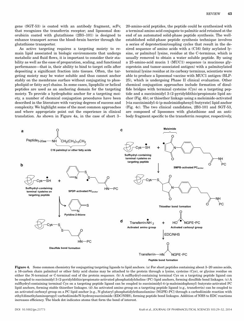
REVIEW 43
gene (SGT-53) is coated with an antibody fragment, scFv,that recognizes the transferrin receptor; and liposomal dox-orubicin coated with glutathione (2B3–101) is designed toenhance transport across the blood–brain barrier through theglutathione transporter.
As active targeting requires a targeting moiety to re-main lipid associated in biologic environments that undergometabolic and fluid flows, it is important to consider their sta-bility as well as the ease of preparation, scaling, and functionalperformance—that is, their ability to bind to target cells afterdepositing a significant fraction into tissues. Often, the tar-geting moiety may be water soluble and thus cannot anchorstably on the membrane surface without conjugating to phos-pholipid or fatty acyl chains. In some cases, lipophilic or helicalpeptides are used as an anchoring domain for the targetingmoiety. To provide a hydrophobic anchor for a targeting moi-ety, a number of chemical conjugation procedures have beendescribed in the literature with varying degrees of success andcomplexity. We highlight some of the most common approachesand where appropriate point out the experience in clinicaltranslation. As shown in Figure 4a, in the case of short 3–
20-amino-acid peptides, the peptide could be synthesized witha terminal amino acid conjugate to palmitic acid retained at theend of an automated solid-phase peptide synthesis. The well-established solid-phase peptide synthesis technique involvesa series of deprotection/coupling cycles that result in the de-sired sequence of amino acids with a (C16) fatty acylated ly-sine, or palmitoyl lysine, residue at the C-terminus, which isusually removed to obtain a water soluble peptide. By usinga 25-amino-acid mucin 1 (MUC1) sequence (a mucinous gly-coprotein and tumor-associated antigen) with a palmitoylatedterminal lysine residue at its carboxy terminus, scientists wereable to produce a liposomal vaccine with MUC1 antigen (BLP-25), which is undergoing Phase II clinical evaluation. Otherchemical conjugation approaches include formation of disul-fide bridges with terminal cysteine (Cys) on a targeting pep-tide and a succinimidyl 3-(2-pyridyldithio)propionate lipid an-chor (Fig. 4b); or thioether linkage using a meleimide-activated[via succinimidyl-4-(p-maleimidophenyl) butyrate] lipid anchor(Fig. 4c). The two clinical candidates, 2B3-101 and SGT-53,are composed of liposomes with glutathione and an anti-body fragment specific to the transferrin receptor, respectively,
Figure 4. Some common chemistry for conjugating targeting ligands to lipid anchors. (a) For short peptides containing about 3–20 amino acids,a 16-carbon chain palmitoyl or other fatty acid chains may be attached to the protein through a lysine, cysteine (Cys), or glycine residue oneither the N-terminal or C-terminal end of the protein sequence. (b) A sulfhydryl-containing terminal Cys on a targeting peptide ligand canbe coupled to succinimidyl 3-(2-pyridyldithio)propionate-activated phosphatidylcholine (PC) lipid anchors, forming disulfide bond linkages. (c) Asulfhydryl-containing terminal Cys on a targeting peptide ligand can be coupled to succinimidyl-4-(p-maleimidophenyl) butyrate-activated PClipid anchors, forming stable thioether linkages. (d) An activated amino group on a targeting peptide ligand (e.g., transferrin) can be coupled toan activated carboxyl group on a PC lipid anchor [e.g., N-glutaryl phosphatidylethanolamine (NGPE)-PC] through a carbodiimide reaction withethyl(dimethylaminopropyl) carbodiimide/N-hydroxysuccinimide (EDC/NHS), forming peptide bond linkages. Addition of NHS to EDC reactionsincreases efficiency. The black dot indicates atoms that form the bond of interest.
DOI 10.1002/jps.23773 Kraft et al., JOURNAL OF PHARMACEUTICAL SCIENCES 103:29–52, 2014

44 REVIEW
attached through a thioether linkage using maleimide-activated lipids and Cys–SH on the ligands. On the con-trary, MBP-426 conjugation is achieved by a peptide bond,formed between the activated carboxyl group on N-glutarylphosphatidylethanolamine and the activated amino groupon transferrin with reagents ethyl(dimethylaminopropyl)carbodiimide/N-hydroxysuccinimide (Fig. 4d).
There are three general approaches used for attaching lig-ands and incorporating them into liposomes: ligands are mixedwith lipid during liposome preparation, conjugated after lipo-some formation, or inserted into preformed liposomes. Lipidmixing involves carrying out the reaction between the ligandand lipidic anchor first, purifying the conjugate, then combin-ing the lipidic ligand with other lipids in the liposome prepara-tion. In theory, this allows stoichiometric control of the liganddensity, it is a single-step and efficient manufacturing proce-dure, and could help hydrophobic material embed better withinlamellae. For these reasons, mixing has significant advantagesfor scale-up preparation. However, targeting moieties are dis-tributed both in the inner and outer surfaces of the liposome.If a targeting moiety is a large protein, protein denaturationmight be an issue for preparation methods that include heat,shearing, or other energy sources to reduce particle size.
In the case of conjugating a ligand to preformed liposomesor lipid nanoparticles, the lipidic anchor is already included inthe liposome bilayer and the coupling reaction occurs on thesurface of preformed liposomes. This avoids potential denatu-ration that may occur during liposome synthesis. In addition,ligands are only attached to the outside surface, conserving ma-terial and maximizing encapsulation volume. However, chemi-cal coupling efficiency to preformed liposomes is not 100% andunconjugated ligand must be removed. Also, there may be arisk of altering the structure of the carrier particle or encapsu-lated drug. A variation to this is to insert the lipophilic target-ing moiety, only after formation of liposomes. This approach,so-called postinsertion, has some advantages but requires theconjugated ligand molecule to be in a micellar form or mixedwith surfactant. This process may not be 100% efficient. Ad-ditional methods and details of ligand coupling reactions andtheir strengths and challenges have been recently reviewed.162
In summary, active targeting of liposomes, particularly inlarge-scale preparation, is an emerging science and is in theearly stages of development. To encounter the least resistancewhen it comes time for a targeted nanotherapeutic to be scaled,the first step is to ensure product and content uniformity. Thatis, reproducible results, batch-to-batch consistency, and stabil-ity are crucial. The second step is the selectivity—whereby atargeted ligand enhances the localization of a therapeutic to abiological site, be it tissue, cells, or intracellular organelles. Theability of a targeted strategy to efficiently navigate physiolog-ical barriers and deliver a significant fraction of therapeuticsto specific cells and cellular organelles are major challenges.Not all target cell recognition ligands coated on lipid parti-cles enhance tumor tissue accumulation; some ligands are de-signed to promote cellular uptake. For ligand attachment, oneof the more stable and commonly used covalent linkages isthe thioether linkage (Fig. 4). This linkage is among the mostattractive for scale-up procedures because of its strength, invivo stability, and reaction efficiency. In addition, the final cou-pling method of choice must retain the binding selectivity andaffinity to target molecules. Ultimately, the choice of couplingreaction and ligand should be made based on the demands of
specific drugs and target applications. With recent advancesin understanding target molecule expression related to diseasecells and tissue phenotypes, along with a systems approachto identify the degree of background expression and link(s) todisease development, active drug targeting with liposomes andlipid nanoparticles is now in sight.
OTHER APPLICATIONS
Multifunctional Liposomes and Lipid Nanoparticles
There has been a recent increase in focus on the developmentof liposomal delivery systems that combine multiple drugsand functions into a single particle. Through combined inno-vations in targeting designs, selection of therapeutic agents,and imaging capabilities, there is great potential for the pro-duction of highly specialized, safe, and effective therapeuticand diagnostic nanoparticles. This section focuses on liposomesand lipid nanoparticles that combine several targeting methodsand different therapeutic or diagnostic modalities into a singlenanoparticle to promote safety and efficacy.
The term “multifunctional” has gained popularity in recentyears and is used here to describe a single nanoparticle thatexhibits multiple functions. These functions may include mul-tiple methods of targeting, targeting to multiple molecules, de-livery of multiple therapeutic agents, or a diagnostic functionwith more than one biologic capability. A similar term with amore ambiguous definition is “multivalent”. Simply put, the“valence” of a molecule refers to its relative binding capacity.In the context of liposomes and lipid nanoparticles, “multiva-lency” refers to displaying many copies of one or more bind-ing ligands capable of recognizing multiple target molecules onthe same or different cells.163,164 Some researchers have usedthe term “hetero-multivalent” to signify liposomes expressingmultiple and different binding peptides.165 The surface of a li-posome or lipid nanoparticle 50–100 nm in diameter can carrysignificantly more than the two valencies available on an im-munoglobulin G molecule. Thus, one can envision liposomeswith multiple binding epitopes for enhancing biological func-tions. For example, the lipid membrane could be coated with abinding moiety to recognize prostate-specific antigen (PSA) oncancer cells along with another binding moiety that recognizesactivated T or effector cell antigens such as CD64, CD8, andCD3. These lipid particles would bring together the cancer cellwith activated T cells, mediating cancer cell lysis. In theory,such an approach with multivalency and high ligand densityexpressed on liposomal surfaces may provide higher affinityand avidity than the bifunctional antibody approach; many bi-functional antibodies are undergoing clinical evaluation such asblinatumomab for B cell acute lymphoblastic leukemia (ALL),which targets to CD19 on ALL B cells and effector CD3+ Tcells.166,167 By bringing the effector T cells to leukemia B cells,blinatumomab (MT103) has progressed to Phase II studies.
If lipid particles are coated with a single targeting moiety,considerable off-target delivery is often detected in vivo becauseof the relatively high level of background expression in healthy,nontarget tissues. Folate receptor targeting is a good examplebecause, although many cancer cells express high-affinity re-ceptors and high receptor density, low-affinity receptors arealso expressed in macrophages, the proximal tubule of thekidney, and in some normal epithelial cells.168 To reduce off-target delivery and improve selectivity in vivo, a number of
Kraft et al., JOURNAL OF PHARMACEUTICAL SCIENCES 103:29–52, 2014 DOI 10.1002/jps.23773

REVIEW 45
modifications in the design of targeting ligand derivatives,binding structures, and environment-sensitive approacheshave been explored. Perche and Torchilin169 recently reviewedvarious strategies to enhance multifunctional liposome target-ing to cancer cells. Environmentally responsive PEGylated lipo-somes have been developed, which exhibit the prolonged bloodcirculation and reduced cellular uptake benefits of PEG con-jugation. They can release their protective PEG coating atthe target tissue to expose a previously hidden target recog-nition moiety. This environmentally responsive attribute isachieved through a pH-sensitive hydrazone link between thePEG chain and the PE lipid, which is cleaved at low pH, typ-ical of inflamed or neoplastic tissues.170 Using this method,off-site targeting could be decreased by exposing secondarytargeting proteins only in certain environmental conditions.Other environment-specific factors can be similarly exploitedto aid targeting specificity, such as a matrix metalloproteinase2 (MMP2)-cleavable linker that relies on the high local MMP2concentration in tumor tissues.171 Similarly, the enzymatic ac-tivity of PSA could be utilized to target prostate cancer. Folate-expressing liposomes carrying antitumor siRNA are coatedwith cell-penetrating peptides, but these peptides are shieldedby PSA-sensitive linkages. Upon binding via a folate receptor tocells expressing prostate-specific membrane antigen (PSMA),PSA cleaves the protective attachment and the cell-penetratingpeptides become exposed, allowing liposomal entry and deliv-ery of siRNA.172 By loading a therapeutic agent such as ananticancer drug into multifunctional environmentally respon-sive liposomes, targeting efficiency can be improved to increasedrug delivery to the region or cells of interest and decrease therisk of negative side effects.
The concept of an outside trigger, or “remote-controlled tar-geting,” has also been explored with multifunctional liposomes.Some methods include low-wavelength UV-light applicationto disrupt liposome membranes, application of laser light toinduce photosensitive release, and induction of local hyper-thermia by high-intensity focused ultrasound (HIFU) to trig-ger thermosensitive or pressure-sensitive release.173 These canalso be used in combination, such as the use of temperature-sensitive liposomes in conjunction with the ultrasound-inducedcavitation of coadministered microbubbles to enhance cellu-lar permeability. Mild local hyperthermia to induce drug re-lease combined with HIFU to enhance cellular permeabilityallows targeted intracellular accumulation of the encapsulateddrug.174 Remotely triggered drug release could provide anotherlayer of highly controllable targeting to improve the efficacyand safety profile of multifunctional lipid nanoparticles.
Finally, multifunctional liposomes may serve functionswithin multiple therapeutic or diagnostic modalities. For ex-ample, siRNA intended to silence the genes critical for cancergrowth can be combined with the anticancer drug doxorubicinin PEGylated liposomes to reduce drug resistance via a com-bination of pharmaceutical and gene therapeutics.175 Thera-peutic and diagnostic capabilities can even reside within thesame particle. These “theranostic” liposomes could deliver drugwhile providing real-time imaging using one or more modali-ties. In many cases, a definitive diagnosis is needed prior to theinitiation of therapy, and therefore it is not always practicalto implement theranostic procedures. However, in some situ-ations where both diagnostic imaging and therapeutic treat-ment coincide, such as a cancer patient whose tumor cellsmay become metastatic, theranostic liposomes could simulta-
neously provide quantitative information and treatment. Janibet al.176 have discussed the various classes of nanoparticlesand contrast agents for multiple imaging modalities includingmagnetic resonance imaging (MRI), nuclear imaging such aspositron emission tomography (PET) and single-positron emis-sion computed tomography (CT), X-ray-based CT, ultrasound,and optical imaging with fluorophores. Without the use of con-trast agents, ultrasound and X-ray-based CT are more conve-nient but provide lower resolution and detail compared withmore costly MRI. The use of liposomes and lipid particles withassociated contrast or PET agents has often been challenged byeither dissociation of contrast from the lipid particles or rapidclearance via the liver, lung, and spleen. This issue has beenrecently addressed in lipid particles carrying pentachelate di-ethylene triamine pentaacetic acid (DTPA) that not only stablybind the MRI contrast agent gadolinium in vivo, but also pro-duce contrast-enhanced resolution in the blood pool at low dosesand high vascular resolution in the liver and kidney withoutsignificant distribution outside of blood vessels.177 Having suc-cessfully produced high-performance particles with outstand-ing MR contrast properties, it expands the potential for multi-valent target visualization. Lipid particles carrying gadoliniumand/or PET agents (e.g., 111In) on the chelate DTPA expressedon their surface could be engineered with a target-seeking moi-ety such as PSMA or HER-2 to aid in the diagnosis and stagingof cancer.
Vaccines
Although researchers focus on exploring liposomes to enhanceweak antigenic or vaccine responses to one or more specificcomponents of a pathogen (i.e., virus, bacterial, or microbes),liposomes and lipid membrane preparations may also be usedas an adjuvant to boost the protective or therapeutic immuneresponses. As vaccine adjuvants, the primary role of liposomesis intended to induce antigen-dependent and specific humoraland cell-mediated immunity. Liposomal adjuvants are amongthe most promising candidates to replace the widely usedalum-based adjuvants, which elicit a weak T helper cell (TH1)response and inadequate cell-mediated immunity. Althoughthe antigen encapsulation or attachment strategy may vary,the antigen presented in liposomal or lipid nanoparticle formcould potentially stimulate both humoral and cell-mediated im-munity. Route of administration, antigen dose, and antigennature—as in size and density—may also modulate the typeand extent of immune response.
The mechanistic basis of how liposomes act as adjuvantsand enhance antigen-specific immune responses is not yet fullyunderstood. Currently, broad outlines of the adjuvanticity ofliposomes have been described. Larger (∼250–700 nm in diam-eter) and positively charged particles persist at subcutaneousor intramuscular injection sites and promote TH1 responses.178
In general, cationic liposome vaccines are more potent adju-vants and have superior immunogenicity than anionic or neu-trally charged vaccines. MLVs, with usually 10 lamellae, maypotentiate TH2 responses.179 Lipids that melt from a gel tofluid phase at higher temperatures (DSPC, 54◦C; DPPC, 42◦C;DMPC, 23◦C; 1,2-dilauroyl-sn-glycero-3-phosphocholine, −2◦C;DOPC, −17◦C) induce stronger antibody and T cell immuneresponses180; however, exceptions have been reported.181,182 Asdetails of the human immune system become clearer, con-founding factors such as immunologic variability among animal
DOI 10.1002/jps.23773 Kraft et al., JOURNAL OF PHARMACEUTICAL SCIENCES 103:29–52, 2014

46 REVIEW
strains and species, and differences among rodent, primate, andhuman immunity and physiology must be carefully consideredwhen designing studies and interpreting results.
The method of antigen attachment is essential to theimmunogenicity of liposome vaccines. As discussed, a vari-ety of antigens can be added to liposomes, including smallmolecule haptens, nucleic acids, carbohydrates, peptides, andproteins. Agonists for pattern recognition receptors (PRRs)such as toll-like receptors (TLRs), NOD-like receptors, and C-type lectin receptors are essential immunomodulators. For in-stance, the TLR agonist monophosphoryl lipid A is a potentimmunostimulant.183 Adding multiple PRR agonists to a sin-gle liposomal vaccine has been shown to be beneficial.184 Com-pared with antigens encapsulated inside liposomal vaccines,surface-associated antigens induce better antibody immuneresponses,185,186 likely as a result of increased exposure to B cellreceptors. However, T cell responses are equivalent for encap-sulated and surface-associated antigens.187,188 Processing andpresentation of antigens on antigen-presenting cells (APCs)may occur either by MHC II-containing organelles or throughendosomal escape and cytosolic delivery followed by loadingonto MHC I molecules. Liposome fusion with other cellular orendosomal bilayer membranes and lipid transfer is known topromote immunogenicity by increasing T cell responses.189,190
Fusogenicity has also been widely used to improve the transfec-tion efficiency in gene therapy. Cationic liposomes anneal withpDNA to form an electrostatic complex (lipoplex) and the pDNAserves as both an antigen and adjuvant.191 Following fusion,endosomal escape and cytosolic delivery may occur by cationicliposomes causing endosomal osmolysis192 or pH-sensitive li-posomes inducing acidosis and a lamellar-to-hexagonal phasetransition to disrupt endosomal compartments.193
Two marketed liposome-based vaccines are Inflexal R© V andEpaxal R©. The former is an influenza vaccine and the latter is avaccine against hepatitis A. Both are virosomes, which containfunctional influenza virus membranes (phospholipids, hemag-glutinin, and neuraminidase) and are unilamellar (mono or bi-layer) vesicles approximately 150 nm in diameter. Liposomalvaccines Stimuvax (BLP-25), for nonsmall cell lung carcinoma,and RTS,S/AS01, for malaria, are currently undergoing PhaseIII/II clinical trials (Table 2) and are progressing toward mar-keting approval.
Gene Therapeutics
Liposomal vaccines and nonviral vector gene therapeuticsshare similar principles as they both use cationic liposomesto deliver cargo to the cytoplasm upon endosomal disruptioncaused by the proton sponge effect.194 Although cytosolic deliv-ery is well studied, an insurmountable obstacle has been thechallenge of nonviral vector gene therapy because pDNA is as-sociated with low transgene expression and immunogenicity.For short-term expression of transgenes, gene therapeutics ap-pear to work in the case of vaccines that require a few days ofexpression.195,196 However, gene therapeutics for chronic pro-tein expression is a distance away from clinical translation. Todate, even the most novel and sophisticated gene delivery vehi-cles have not been successful in overcoming these problems. Analternative approach is using mRNA in place of pDNA to trans-late gene products without the need to enter the nucleus.197
Recently, mRNA was chemically modified and encapsulated inliposomes for successful systemic delivery to tumor sites.198
The chemical modification involved first structurally enhanc-ing mRNA transcripts and then substituting cytidine triphos-phate and uridine triphosphate with 5-methylcytidine triphos-phate and pseudouridine (�) triphosphate, respectively. Thesetwo nucleotide analogs reduced the activation of the innateimmune response through the TLR pathway. mRNA was con-densed with polycationic protamine into nanosized complexesto protect against nuclease degradation.
Antisense nucleic acid sequences—including RNA inhibitoror RNAi, siRNA, and miRNA—bind to target gene sequencesand inhibit or modulate gene expression. Lipid nanoparticlesare a leading delivery system for these gene therapeutics, butparticle structures have been debated. Recently, stable nu-cleic acid lipid particles for delivering siRNA were shown tohave an outer layer of PEG–lipid encapsulating immobilizedsiRNA bound to bilayer membrane surfaces that separate ir-regular water-filled cavities, which is counter to the view of abilayer vesicle with freely tumbling siRNA in the inner aque-ous compartment.199,200 About 10 years ago, siRNA and RNAiwere regarded as highly innovative therapeutic platforms formultiple disease targets. However, the small fraction of siRNAfound in target cells, the even smaller fractions found withintarget organelles or sites of action, and the challenge of rapidclearance (or excretion) from the body, have led to a lack of ther-apeutic efficacy and a broad divestment by biopharmaceuticaldevelopment programs.
Oral Drug Delivery
Oral dosing of liposomal drug delivery systems has advantagesover invasive routes because of the potential increase in patientcompliance and ease of use. However, bile salts, pH, and pancre-atic enzymes in the gut dismantle liposomal bilayers. To help re-sist these destabilizing factors, liposome formulations incorpo-rate protective polymeric coatings or cholesterol and saturatedphospholipids that increase membrane rigidity and decreaseenzymatic degradation.201 Other ways to achieve gastrointesti-nal (GI) stability include liposomes with a carrageenan andcollagen core202 or the addition of gangliosides GM1 and GMtype III.203 However, disruption of the liposomal vesicles be-cause of GI fluids, especially bile salts, remains a hindrance.In vitro lipolysis models are used to simulate human intestinaldigestion and are useful for testing the GI stability of liposomeformulations.204 Overall, successful oral liposomal delivery sys-tems must be stable and move from the gut into the circulatorysystem prior to releasing their cargo in the blood or at specifictarget sites. Lipid-based excipients (e.g., liposomes, micellularnanoemulsions and microemulsions) have indeed improved thebioavailability of oral drugs by solubilizing poorly water-solubledrugs and increasing intestinal membrane permeability. For ex-ample, oral paclitaxel avoids P-glycoprotein efflux transporterswhen loaded into liposomes.205,206 Mechanisms of oral absorp-tion of lipophilic drugs with lipid-based delivery systems havebeen thoroughly reviewed.207 Liposomal excipients also reduceGI side effects of nonsteroidal anti-inflammatory agents andeliminate the bitter taste of oral drugs.208 An oral PEG-coatedliposomal vaccine with ovalbumin and diameters of 1.7–3.3 :minduced a mucosal immune response in mice209 and oral lipo-somal $-sitosterol was shown to upregulate the host defense ofmetastatic cancer cells and may enhance mucosal immunity.210
Stimulating mucosal immune responses may enhance deliveryof antigen to APCs that actively take up particles in the GI
Kraft et al., JOURNAL OF PHARMACEUTICAL SCIENCES 103:29–52, 2014 DOI 10.1002/jps.23773

REVIEW 47
tract. Generally, some benefits of using liposomes for such ap-plications include biocompatibility, protection of antigen, flex-ibility in design, targeting of antigens to APCs, and improvedstimulation of immune responses by oral delivery of solubleantigens.211 In some cases, a lipid mixture may be used as a sol-ubilizing or suspension agent for highly insoluble or lipophilicdrugs to be delivered as a microemulsion in softgel capsules fororal dosage. A drug and lipid microemulsion encased in softgelcapsules was used to enhance reproducibility and bioavailabil-ity of cyclosporin A, originally formulated as a tablet.212
FUTURE DIRECTIONS
From a regulatory perspective, it is important to design adrug delivery strategy based on predefined desired attributesand to establish clinically meaningful specifications. In par-ticular, a lingering issue is the lack of in vitro/in vivo corre-lation of liposome drug release profiles. As a result, currentregulation relies heavily on time-consuming clinical studiesto establish bioequivalence and set regulatory specifications.To evaluate the quality, safety, and efficacy of liposomal prod-ucts, systematic understanding of the relationships betweenliposome physiochemical properties and its absorption, distri-bution, metabolism, and excretion (ADME) behaviors is crit-ical. Physiologically based pharmacokinetic (PBPK) modelingcould serve as a powerful tool to quantitatively describe andpredict the biodistribution of liposomal vesicles and drug sub-stances. Preclinical PBPK models can be readily extrapolatedto humans by substituting related physiologic and pharmacoki-netic parameters, making it possible to quantitatively predictthe effects of changes in liposome physiochemical properties onADME of liposomal drugs in humans.
Advances and continual growth in the identification of drugtargets have provided ever-expanding drug candidates. Yettranslating these compounds into therapeutic products contin-ues to face challenges because of the off-target distribution andlack of efficacy in late-stage clinical trials. Many highly potentdrugs often face limited solubility and target tissue exposure.Improved understanding of drug target distribution profileswithin the body along with pharmacokinetics, drug disposition,and elimination, as they relate to therapeutic outcome, point tothe need for consideration of drug delivery at a systems level.A systems approach not only considers the physiological con-text of target tissues and cells that link to disease symptomdevelopment, but also the ability and capacity of a drug de-livery platform to penetrate and localize at sufficient levelsnecessary to modify therapeutic impacts. To make a significanttherapeutic impact, drug delivery systems such as liposomesand lipid nanoparticles must serve as a drug carrier not onlyto improve drug stability and exposure, but also to enhancethe accumulation of a significant amount of drug in the targettissue. Without significant drug exposure in target tissues, en-hancing delivery of drug to cells or intracellular drug targets isunlikely. In this context, clinical insights in liposome and lipidnanoparticle disposition mechanisms have led to the design ofsmall (∼50 nm diameter) and sterically stabilized lipid parti-cles to increase their blood residence time required for clinicalapplications. Advances in molecular design and chemistry forexpression of ligand or receptor molecules on the liposome andlipid nanoparticle surface may further improve their interac-tion with target cells. Improved drug residence time (because
of the reduced clearance of liposomes and lipid nanoparticles)will provide carrier-associated drugs the sufficient time theyneed to eventually reach their intended target sites. A firststep for increasing the intracellular uptake of liposomal drugs(e.g., anticancer agents, antibiotics, and DNA) is to enhancetheir localization selectivity within the target tissue. As ad-ditional ligands with higher affinity and specificity continueto be developed, and progress is made in antibody and pep-tide engineering to mass-produce targeted lipid nanoparticlepreparations, lipid–drug complexes with extended therapeuticindices are now within reach.
ACKNOWLDGMENTS
This study was supported in part by NIH grants AI077390,AI071971, RR00166, RR025014, and UL1TR000423. R.J.Y.H.was also supported in part by the Milo Gibaldi endowment.
REFERENCES
1. Saunders L, Thomas IL. 1958. Diffusion studies with lysolecithin.J Chem Soc 483–485.2. Bangham AD, Horne RW. 1964. Negative staining of phospholipidsand their structural modification by surface-active agents as observedin the electron microscope. J Mol Biol 8:660–668.3. Torchilin VP. 2005. Recent advances with liposomes as pharmaceu-tical carriers. Nat Rev Drug Discov 4(2):145–160.4. Kinman L, Brodie SJ, Tsai CC, Bui T, Larsen K, Schmidt A, AndersonD, Morton WR, Hu SL, Ho RJ. 2003. Lipid–drug association enhancedHIV-1 protease inhibitor indinavir localization in lymphoid tissues andviral load reduction: A proof of concept study in HIV-2287-infectedmacaques. J Acquir Immune Defic Syndr 34(4):387–397.5. James JS. 1995. DOXIL approved for KS. AIDS treatment news(no. 236):6.6. James JS. 1995. DOXIL approved by FDA. AIDS patient care(no. 9):306.7. Safra T, Muggia F, Jeffers S, Tsao-Wei DD, Groshen S, Lyass O, Hen-derson R, Berry G, Gabizon A. 2000. Pegylated liposomal doxorubicin(doxil): Reduced clinical cardiotoxicity in patients reaching or exceedingcumulative doses of 500 mg/m2. Ann Oncol 11(8):1029–1033.8. Northfelt DW. 1994. Stealth liposomal doxorubicin (SLD) deliversmore doxorubicin (DOX) to AIDS-Kaposis sarcoma (AIDS-KS) lesionsthan to normal skin (abstract #5). Proc Am Soc Clin Oncol 13:51.9. NDA. 2004. Doxil: New drug application. Center for Drug Evaluation& Research—FDA.10. Lian T, Ho RJ. 2001. Trends and developments in liposome drugdelivery systems. J Pharm Sci 90(6):667–680.11. Ramishetti S, Huang L. 2012. Intelligent design of multifunc-tional lipid-coated nanoparticle platforms for cancer therapy. TherDeliv 3(12):1429–1445.12. Gregoriadis G. 2007. Liposome technology. 3rd ed., New York: In-forma Healthcare. Vol. I–III.13. Torchilin VP, Weissig V. 2003. Liposomes: A practical approach.2nd ed. Oxford; New York: Oxford University Press, p xxiii, 396.14. Allen TM, Cullis PR. 2012. Liposomal drug delivery systems: Fromconcept to clinical applications. Adv Drug Deliv Rev 65(1):36–48.15. Mabrey S, Mateo PL, Sturtevant JM. 1978. High-sensitivity scan-ning calorimetric study of mixtures of cholesterol with dimyristoyl- anddipalmitoylphosphatidylcholines. Biochemistry 17(12):2464–2468.16. Angst MS, Drover DR. 2006. Pharmacology of drugs formulatedwith DepoFoam: A sustained release drug delivery system for par-enteral administration using multivesicular liposome technology. ClinPharmacokinet 45(12):1153–1176.17. Katre NV. 2004. Liposomes-based depot injection technologies. AmJ Drug Deliv 2(4):213–227.
DOI 10.1002/jps.23773 Kraft et al., JOURNAL OF PHARMACEUTICAL SCIENCES 103:29–52, 2014

48 REVIEW
18. Cullis PR, de Kruijff B. 1979. Lipid polymorphism and the func-tional roles of lipids in biological membranes. Biochim Biophys Acta559(4):399–420.19. Edidin M. 2003. Lipids on the frontier: A century of cell-membranebilayers. Nat Rev Mol Cell Biol 4(5):414–418.20. Allen TM, Hansen C, Martin F, Redemann C, Yau-Young A. 1991.Liposomes containing synthetic lipid derivatives of poly(ethylene gly-col) show prolonged circulation half-lives in vivo. Biochim Biophys Acta1066(1):29–36.21. Lee RJ, Low PS. 1995. Folate-mediated tumor cell targetingof liposome-entrapped doxorubicin in vitro. Biochim Biophys Acta1233(2):134–144.22. Lee RJ, Low PS. 1994. Delivery of liposomes into cultured KB cellsvia folate receptor-mediated endocytosis. J Biol Chem 269(5):3198–3204.23. Drummond DC, Meyer O, Hong K, Kirpotin DB, PapahadjopoulosD. 1999. Optimizing liposomes for delivery of chemotherapeutic agentsto solid tumors. Pharmacol Rev 51(4):691–743.24. Plank C, Mechtler K, Szoka FC, Jr., Wagner E. 1996. Activation ofthe complement system by synthetic DNA complexes: A potential bar-rier for intravenous gene delivery. Hum Gene Ther 7(12):1437–1446.25. He C, Hu Y, Yin L, Tang C, Yin C. 2010. Effects of particle sizeand surface charge on cellular uptake and biodistribution of polymericnanoparticles. Biomaterials 31(13):3657–3666.26. Chonn A, Cullis PR, Devine DV. 1991. The role of surface charge inthe activation of the classical and alternative pathways of complementby liposomes. J Immunol 146(12):4234–4241.27. Szebeni J, Bedocs P, Rozsnyay Z, Weiszhar Z, Urbanics R, RosivallL, Cohen R, Garbuzenko O, Bathori G, Toth M, Bunger R, Barenholz Y.2012. Liposome-induced complement activation and related cardiopul-monary distress in pigs: Factors promoting reactogenicity of Doxil andAmBisome. Nanomedicine 8(2):176–184.28. Moghimi SM, Hamad I, Andresen TL, Jorgensen K, Szebeni J.2006. Methylation of the phosphate oxygen moiety of phospholipid-methoxy(polyethylene glycol) conjugate prevents PEGylated liposome-mediated complement activation and anaphylatoxin production.FASEB J 20(14):2591–2593.29. Sou K, Tsuchida E. 2008. Electrostatic interactions and comple-ment activation on the surface of phospholipid vesicle containing acidiclipids: Effect of the structure of acidic groups. Biochim Biophys Acta1778(4):1035–1041.30. Risbo J, Jorgensen K, Sperotto MM, Mouritsen OG. 1997. Phasebehavior and permeability properties of phospholipid bilayers contain-ing a short-chain phospholipid permeability enhancer. Biochim BiophysActa 1329(1):85–96.31. McMullen TP, Lewis RN, McElhaney RN. 1993. Differential scan-ning calorimetric study of the effect of cholesterol on the thermotropicphase behavior of a homologous series of linear saturated phosphatidyl-cholines. Biochemistry 32(2):516–522.32. Huang Z, Szoka FC, Jr. 2008. Sterol-modified phospholipids: choles-terol and phospholipid chimeras with improved biomembrane proper-ties. J Am Chem Soc 130(46):15702–15712.33. Jorgensen K, Ipsen JH, Mouritsen OG, Zuckermann MJ. 1993. Theeffect of anaesthetics on the dynamic heterogeneity of lipid membranes.Chem Phys Lipids 65(3):205–216.34. Krill SL, Lau KY, Plachy WZ, Rehfeld SJ. 1998. Penetrationof dimyristoylphosphatidylcholine monolayers and bilayers by modelbeta-blocker agents of varying lipophilicity. J Pharm Sci 87(6):751–756.35. Sullivan SM, Huang L. 1986. Enhanced delivery to target cells byheat-sensitive immunoliposomes. Proc Natl Acad Sci USA 83(16):6117–6121.36. Yanez AM, Wallace M, Ho R, Shen D, Yaksh TL. 1995. Touch-evoked agitation produced by spinally administered phospholipid emul-sion and liposomes in rats. Structure–activity relation. Anesthesiology82(5):1189–1198.37. Beauchamp C, Daddona PE, Menapace DP. 1984. Properties of anovel PEG derivative of calf adenosine deaminase. Adv Exp Med Biol165(Pt A):47–52.
38. Zhao XB, Muthusamy N, Byrd JC, Lee RJ. 2007. Cholesterol as abilayer anchor for PEGylation and targeting ligand in folate-receptor-targeted liposomes. J Pharm Sci 96(9):2424–2435.39. Allen TM, Chonn A. 1987. Large unilamellar liposomes with lowuptake into the reticuloendothelial system. FEBS Lett 223(1):42–46.40. Maruyama K, Yuda T, Okamoto A, Kojima S, Suginaka A, Iwat-suru M. 1992. Prolonged circulation time in vivo of large unilamelarliposomes composed of distearoyl phosphatidylcholine and cholesterolcontaining amphipathic poly(ethylene glycol). Biochim Biophys Acta1128(1):44–49.41. Woodle MC, Matthay KK, Newman MS, Hidayat JE, Collins LR,Redemann C, Martin FJ, Papahadjopoulos D. 1992. Versatility in lipidcompositions showing prolonged circulation with sterically stabilizedliposomes. Biochim Biophys Acta 1105(2):193–200.42. Huang SK, Lee KD, Hong K, Friend DS, Papahadjopoulos D.1992. Microscopic localization of sterically stabilized liposomes in coloncarcinoma-bearing mice. Cancer Res 52(19):5135–5143.43. Wu NZ, Da D, Rudoll TL, Needham D, Whorton AR, Dewhirst MW.1993. Increased microvascular permeability contributes to preferen-tial accumulation of Stealth liposomes in tumor tissue. Cancer Res53(16):3765–3770.44. Gabizon A, Catane R, Uziely B, Kaufman B, Safra T, Cohen R,Martin F, Huang A, Barenholz Y. 1994. Prolonged circulation time andenhanced accumulation in malignant exudates of doxorubicin encap-sulated in polyethylene-glycol coated liposomes. Cancer Res 54(4):987–992.45. Maeda H, Wu J, Sawa T, Matsumura Y, Hori K. 2000. Tumor vas-cular permeability and the EPR effect in macromolecular therapeutics:A review. J Control Release 65(1–2):271–284.46. Metselaar JM, Bruin P, de Boer LW, de Vringer T, Snel C, OussorenC, Wauben MH, Crommelin DJ, Storm G, Hennink WE. 2003. A novelfamily of L-amino acid-based biodegradable polymer-lipid conjugatesfor the development of long-circulating liposomes with effective drug-targeting capacity. Bioconjug Chem 14(6):1156–1164.47. Takeuchi H, Kojima H, Yamamoto H, Kawashima Y. 2001. Evalu-ation of circulation profiles of liposomes coated with hydrophilic poly-mers having different molecular weights in rats. J Control Release75(1–2):83–91.48. Torchilin VP, Levchenko TS, Whiteman KR, Yaroslavov AA, Tsat-sakis AM, Rizos AK, Michailova EV, Shtilman MI. 2001. Amphiphilicpoly-N-vinylpyrrolidones: Synthesis, properties and liposome surfacemodification. Biomaterials 22(22):3035–3044.49. Torchilin VP, Trubetskoy VS. 1995. Which polymers can makenanoparticuate drug carriers long-circulating? Adv Drug Deliv Rev16:141–155.50. Whiteman KR, Subr V, Ulbrich K, Torchilin VP. 2001. Poly(Hpma)-coated liposomes demonstrate prolonged circulation in mice. J Lipo-some Res 11(2–3):153–164.51. Woodle MC. 1998. Controlling liposome blood clearance by surface-grafted polymers. Adv Drug Deliv Rev 32(1–2):139–152.52. Roberts MJ, Bentley MD, Harris JM. 2002. Chemistry for peptideand protein PEGylation. Adv Drug Deliv Rev 54(4):459–476.53. Pasut G, Veronese FM. 2007. Polymer–drug conjugation, recentachievements and general strategies. Progr Polym Sci 32:933–961.54. Garay RP, El-Gewely R, Armstrong JK, Garratty G, Richette P.2012. Antibodies against polyethylene glycol in healthy subjects andin patients treated with PEG-conjugated agents. Expert Opin on DrugDeliv 9(11):1319–1323.55. Ishida T, Wang X, Shimizu T, Nawata K, Kiwada H. 2007. PEGy-lated liposomes elicit an anti-PEG IgM response in a T cell-independentmanner. J Control Release 122(3):349–355.56. Wang X, Ishida T, Kiwada H. 2007. Anti-PEG IgM elicited byinjection of liposomes is involved in the enhanced blood clearance of asubsequent dose of PEGylated liposomes. J Control Release 119(2):236–244.57. Schellekens H, Hennink WE, Brinks V. 2013. The immunogenic-ity of polyethylene glycol: Facts and fiction. Pharm Res 30(7):1729–1734.
Kraft et al., JOURNAL OF PHARMACEUTICAL SCIENCES 103:29–52, 2014 DOI 10.1002/jps.23773

REVIEW 49
58. Moghimi SM, Hunter AC, Murray JC. 2001. Long-circulatingand target-specific nanoparticles: Theory to practice. Pharmacol Rev53(2):283–318.59. Bertrand N, Leroux JC. 2012. The journey of a drug-carrier inthe body: An anatomo-physiological perspective. J Control Release161(2):152–163.60. Choi CH, Zuckerman JE, Webster P, Davis ME. 2011. Targetingkidney mesangium by nanoparticles of defined size. Proc Natl Acad SciU S A 108(16):6656–6661.61. Sarin H. 2010. Physiologic upper limits of pore size of differentblood capillary types and another perspective on the dual pore theoryof microvascular permeability. J Angiogenes Res 2:14.62. Marcus ML, Chilian WM, Kanatsuka H, Dellsperger KC, EasthamCL, Lamping KG. 1990. Understanding the coronary circulationthrough studies at the microvascular level. Circulation 82(1):1–7.63. Townsley MI, Parker JC, Longenecker GL, Perry ML, Pitt RM,Taylor AE. 1988. Pulmonary embolism: Analysis of endothelial poresizes in canine lung. Am J Physiol 255(5 Pt 2):H1075–1083.64. Wisse E, Jacobs F, Topal B, Frederik P, De Geest B. 2008. Thesize of endothelial fenestrae in human liver sinusoids: Implications forhepatocyte-directed gene transfer. Gene Ther 15(17):1193–1199.65. Luft FC, Aronoff GR, Evan AP, Connors BA, Blase DK, GattoneVH. 1982. Effects of moxalactam and cefotaxime on rabbit renal tissue.Antimicrobial Agents Chemother 21(5):830–835.66. Ogawa S, Ota Z, Shikata K, Hironaka K, Hayashi Y, Ota K, KushiroM, Miyatake N, Kishimoto N, Makino H. 1999. High-resolution ultra-structural comparison of renal glomerular and tubular basement mem-branes. Am J Nephrol 19(6):686–693.67. Lahdenkari AT, Lounatmaa K, Patrakka J, Holmberg C,Wartiovaara J, Kestila M, Koskimies O, Jalanko H. 2004. Podocytesare firmly attached to glomerular basement membrane in kidneys withheavy proteinuria. J Am Soc Nephrol 15(10):2611–2618.68. Conner SD, Schmid SL. 2003. Regulated portals of entry into thecell. Nature 422(6927):37–44.69. Rejman J, Oberle V, Zuhorn IS, Hoekstra D. 2004. Size-dependentinternalization of particles via the pathways of clathrin- and caveolae-mediated endocytosis. Biochem J 377(Pt 1):159–169.70. Hillaireau H, Couvreur P. 2009. Nanocarriers’ entry into the cell:Relevance to drug delivery. Cell Mol Life Sci 66(17):2873–2896.71. Wang J, Byrne JD, Napier ME, DeSimone JM. 2011. More effectivenanomedicines through particle design. Small 7(14):1919–1931.72. Venturoli D, Rippe B. 2005. Ficoll and dextran vs. globular proteinsas probes for testing glomerular permselectivity: Effects of molecularsize, shape, charge, and deformability. Am J Physiol 288(4):F605–F613.73. Beaumier PL, Hwang KJ. 1983. Effects of liposome size on thedegradation of bovine brain sphingomyelin/cholesterol liposomes in themouse liver. Biochim Biophys Acta 731(1):23–30.74. Proffitt RT, Williams LE, Presant CA, Tin GW, Uliana JA, GambleRC, Baldeschwieler JD. 1983. Tumor-imaging potential of liposomesloaded with In-111-NTA: Biodistribution in mice. J Nucl Med 24(1):45–51.75. Gabizon AA, Barenholz Y, Bialer M. 1993. Prolongation of the cir-culation time of doxorubicin encapsulated in liposomes containing apolyethylene glycol-derivatized phospholipid: Pharmacokinetic studiesin rodents and dogs. Pharm Res 10(5):703–708.76. Wagner A, Vorauer-Uhl K. 2011. Liposome technology for industrialpurposes. J Drug Deliv 2011:591325.77. Barenholz Y, Amselem S. 1993. Quality control assays in the devel-opment and clinical use of liposome-based formulations. In Liposometechnology; Gregoriadis G, Ed. 2nd ed. Boca Raton: CRC Press, pp527–616.78. Bangham AD, Standish MM, Watkins JC. 1965. Diffusion of uni-valent ions across the lamellae of swollen phospholipids. J Mol Biol13(1):238–252.79. Asmal M, Whitney JB, Luedemann C, Carville A, Steen R, LetvinNL, Geiben-Lynn R. 2012. In vivo anti-HIV activity of the heparin-activated serine protease inhibitor antithrombin III encapsulated inlymph-targeting immunoliposomes. PLoS One 7(11):e48234.
80. Batzri S, Korn ED. 1973. Single bilayer liposomes prepared withoutsonication. Biochim Biophys Acta 298(4):1015–1019.81. Tejera-Garcia R, Ranjan S, Zamotin V, Sood R, Kinnunen PK. 2011.Making unilamellar liposomes using focused ultrasound. Langmuir27(16):10088–10097.82. Mayhew E, Lazo R, Vail WJ, King J, Green AM. 1984. Characteri-zation of liposomes prepared using a microemulsifier. Biochim BiophysActa 775(2):169–174.83. Barnadas-Rodriguez R, Sabes M. 2001. Factors involved in the pro-duction of liposomes with a high-pressure homogenizer. Int J Pharm213(1–2):175–186.84. Dennison SM, Sutherland LL, Jaeger FH, Anasti KM, Parks R,Stewart S, Bowman C, Xia SM, Zhang R, Shen X, Scearce RM, OfekG, Yang Y, Kwong PD, Santra S, Liao HX, Tomaras G, Letvin NL,Chen B, Alam SM, Haynes BF. 2011. Induction of antibodies in rhesusmacaques that recognize a fusion-intermediate conformation of HIV-1gp41. PLoS One 6(11):e27824.85. Amselem S, Gabizon A, Barenholz Y. 1993. A large-scale methodfor the preparation of sterile and nonpyrogenic liposomal formulationsof defined size distributions for clinical use. In Liposome technology;Gregoriadis G, Ed. Boca Raton: CRC Press, pp 501–525.86. Rameez S, Bamba I, Palmer AF. 2010. Large scale production ofvesicles by hollow fiber extrusion: A novel method for generating poly-mersome encapsulated hemoglobin dispersions. Langmuir 26(7):5279–5285.87. Szoka F, Jr., Papahadjopoulos D. 1978. Procedure for preparationof liposomes with large internal aqueous space and high capture byreverse-phase evaporation. Proc Natl Acad Sci USA 75(9):4194–4198.88. Maitani Y, Soeda H, Junping W, Takayama K. 2001. Modifiedethanol injection method for liposomes containing beta-sitosterol beta-D-glucoside. J Liposome Res 11(1):115–125.89. Gentine P, Bourel-Bonnet L, Frisch B. 2012. Modified and derivedethanol injection toward liposomes: Development of the process. J Li-posome Res 23(1):11–19.90. Jaafar-Maalej C, Charcosset C, Fessi H. 2011. A new method forliposome preparation using a membrane contactor. J Liposome Res21(3):213–220.91. Wagner A, Platzgummer M, Kreismayr G, Quendler H, Stiegler G,Ferko B, Vecera G, Vorauer-Uhl K, Katinger H. 2006. GMP productionof liposomes—A new industrial approach. J Liposome Res 16(3):311–319.92. Meure LA, Foster NR, Dehghani F. 2008. Conventional and densegas techniques for the production of liposomes: a review. AAPS PharmSci Tech 9(3):798–809.93. Lasch J, Weissig V, Brandl M. 2003. Preparation of liposomes.In Liposomes: A practical approach; Torchilin V, Weissig V, Eds. Ox-ford/New York: Oxford University Press, pp 3–29.94. Schwendener RA. 1993. The preparation of large volumes of sterileliposomes for clinical applications. In Gregoriadis G, editor LiposomeTechnology, 2 ed., Boca Raton: CRC Press. p 487–500.95. Frederiksen L, Anton K, van Hoogevest P, Keller HR, LeuenbergerH. 1997. Preparation of liposomes encapsulating water-soluble com-pounds using supercritical carbon dioxide. J Pharm Sci 86(8):921–928.96. Spilimbergo S, Bertucco A. 2003. Non-thermal bacterial inactiva-tion with dense CO(2). Biotechnol Bioeng 84(6):627–638.97. Zhang J, Davis TA, Matthews MA, Drews MJ, LaBerge M. 2006.Sterilization using high-pressure carbon dioxide. J Supercritical Fluids38(3):354–372.98. Aggarwal P, Hall JB, McLeland CB, Dobrovolskaia MA, McNeilSE. 2009. Nanoparticle interaction with plasma proteins as it relatesto particle biodistribution, biocompatibility and therapeutic efficacy.Adv Drug Deliv Rev 61(6):428–437.99. Kim HR, Andrieux K, Delomenie C, Chacun H, Appel M, DesmaeleD, Taran F, Georgin D, Couvreur P, Taverna M. 2007. Analysis ofplasma protein adsorption onto PEGylated nanoparticles by comple-mentary methods: 2-DE, CE and Protein Lab-on-chip system. Elec-trophoresis 28(13):2252–2261.
DOI 10.1002/jps.23773 Kraft et al., JOURNAL OF PHARMACEUTICAL SCIENCES 103:29–52, 2014

50 REVIEW
100. Goppert TM, Muller RH. 2005. Protein adsorption patterns onpoloxamer- and poloxamine-stabilized solid lipid nanoparticles (SLN).Eur J Pharm Biopharm 60(3):361–372.101. Cedervall T, Lynch I, Foy M, Berggard T, Donnelly SC, CagneyG, Linse S, Dawson KA. 2007. Detailed identification of plasma pro-teins adsorbed on copolymer nanoparticles. Angew Chem Int Ed Engl46(30):5754–5756.102. Lynch I, Cedervall T, Lundqvist M, Cabaleiro-Lago C, Linse S,Dawson KA. 2007. The nanoparticle–protein complex as a biologicalentity; a complex fluids and surface science challenge for the 21st cen-tury. Adv Colloid Interface Sci 134–135:167–174.103. Scherphof G, Roerdink F, Waite M, Parks J. 1978. Disintegrationof phosphatidylcholine liposomes in plasma as a result of interactionwith high-density lipoproteins. Biochim Biophys Acta 542(2):296–307.104. Tall AR, Small DM. 1977. Solubilisation of phospholipidmembranes by human plasma high density lipoproteins. Nature265(5590):163–164.105. Mendez AJ, He JL, Huang HS, Wen SR, Hsia SL. 1988. Interactionof rabbit lipoproteins and red blood cells with liposomes of egg yolkphospholipids. Lipids 23(10):961–967.106. Papahadjopoulos D, Moscarello M, Eylar EH, Isac T. 1975. Effectsof proteins on thermotropic phase transitions of phospholipid mem-branes. Biochim Biophys Acta 401(3):317–335.107. Devine DV, Wong K, Serrano K, Chonn A, Cullis PR. 1994.Liposome–complement interactions in rat serum: Implications for li-posome survival studies. Biochim Biophys Acta 1191(1):43–51.108. Yan X, Scherphof GL, Kamps JA. 2005. Liposome opsonization.J Liposome Res 15(1–2):109–139.109. Maeda H. 2001. The enhanced permeability and retention (EPR)effect in tumor vasculature: The key role of tumor-selective macro-molecular drug targeting. Adv Enzyme Regul 41:189–207.110. Bui T, Faltynek C, Ho RJ. 1994. Differential disposition of solubleand liposome-formulated human recombinant interleukin-7: Effects onblood lymphocyte population in guinea pigs. Pharm Res 11(5):633–641.111. Patel HM. 1988. Fate of liposomes in the lymphatics. In Liposomesas drug carriers; Gregoriadis G, Ed. New York: John Wiley & Sons,p 51.112. Oussoren C, Storm G. 2001. Liposomes to target the lymphaticsby subcutaneous administration. Adv Drug Deliv Rev 50(1–2):143–156.113. Higuchi M, Fokin A, Masters TN, Robicsek F, Schmid-SchonbeinGW. 1999. Transport of colloidal particles in lymphatics and vascula-ture after subcutaneous injection. J Appl Physiol 86(4):1381–1387.114. Ikomi F, Hanna GK, Schmid-Schonbein GW. 1999. Size- andsurface-dependent uptake of colloid particles into the lymphatic sys-tem. Lymphology 32(3):90–102.115. Oussoren C, Velinova M, Scherphof G, van der Want JJ, van Rooi-jen N, Storm G. 1998. Lymphatic uptake and biodistribution of lipo-somes after subcutaneous injection. IV. Fate of liposomes in regionallymph nodes. Biochim Biophys Acta 1370(2):259–272.116. Oussoren C, Zuidema J, Crommelin DJ, Storm G. 1997. Lymphaticuptake and biodistribution of liposomes after subcutaneous injection.II. Influence of liposomal size, lipid compostion and lipid dose. BiochimBiophys Acta 1328(2):261–272.117. Gretz JE, Norbury CC, Anderson AO, Proudfoot AE, Shaw S. 2000.Lymph-borne chemokines and other low molecular weight moleculesreach high endothelial venules via specialized conduits while a func-tional barrier limits access to the lymphocyte microenvironments inlymph node cortex. J Exp Med 192(10):1425–1440.118. Fidler IJ, Sone S, Fogler WE, Barnes ZL. 1981. Eradication ofspontaneous metastases and activation of alveolar macrophages by in-travenous injection of liposomes containing muramyl dipeptide. ProcNatl Acad Sci USA 78(3):1680–1684.119. Li S, Goins B, Hrycushko BA, Phillips WT, Bao A. 2012. Feasibil-ity of eradication of breast cancer cells remaining in postlumpectomycavity and draining lymph nodes following intracavitary injection ofradioactive immunoliposomes. Mol Pharm 9(9):2513–2522.120. Wong JK, Hezareh M, Gunthard HF, Havlir DV, Ignacio CC, SpinaCA, Richman DD. 1997. Recovery of replication-competent HIV despite
prolonged suppression of plasma viremia. Science 278(5341):1291–1295.121. Wong JK, Gunthard HF, Havlir DV, Zhang ZQ, Haase AT, Ig-nacio CC, Kwok S, Emini E, Richman DD. 1997. Reduction of HIV-1in blood and lymph nodes following potent antiretroviral therapy andthe virologic correlates of treatment failure. Proc Natl Acad Sci USA94(23):12574–12579.122. Ruiz L, van Lunzen J, Arno A, Stellbrink HJ, Schneider C, Rull M,Castella E, Ojanguren I, Richman DD, Clotet B, Tenner-Racz K, RaczP. 1999. Protease inhibitor-containing regimens compared with nucleo-side analogues alone in the suppression of persistent HIV-1 replicationin lymphoid tissue. AIDS 13(1):F1–F8.123. Hockett RD, Kilby JM, Derdeyn CA, Saag MS, Sillers M, SquiresK, Chiz S, Nowak MA, Shaw GM, Bucy RP. 1999. Constant mean viralcopy number per infected cell in tissues regardless of high, low, orundetectable plasma HIV RNA. J Exp Med 189(10):1545–1554.124. Kinman L, Bui T, Larsen K, Tsai CC, Anderson D, Morton WR,Hu SL, Ho RJ. 2006. Optimization of lipid–indinavir complexes forlocalization in lymphoid tissues of HIV-infected macaques. J AcquirImmune Defic Syndr 42(2):155–161.125. Matsumura Y, Maeda H. 1986. A new concept for macromolecu-lar therapeutics in cancer chemotherapy: Mechanism of tumoritropicaccumulation of proteins and the antitumor agent smancs. Cancer Res46(12 Pt 1):6387–6392.126. Senger DR, Galli SJ, Dvorak AM, Perruzzi CA, Harvey VS,Dvorak HF. 1983. Tumor cells secrete a vascular permeability factorthat promotes accumulation of ascites fluid. Science 219(4587):983–985.127. Northfelt DW, Kaplan L, Russell J. 1995. Pharmacokinetics andtumor localization of DOX-SL (Stealth liposomal doxorubicin) by com-parison with Adriamycin in patients with AIDS and Kaposi’s sarcoma.In Stealth liposomes; Lasic DD, Martin FJ, Eds. Boca Raton, Florida:CRC Press, pp 257–266.128. Forssen EA, Coulter DM, Proffitt RT. 1992. Selective in vivo lo-calization of daunorubicin small unilamellar vesicles in solid tumors.Cancer Res 52(12):3255–3261.129. Adiseshaiah PP, Hall JB, McNeil SE. 2009. Nanomaterial stan-dards for efficacy and toxicity assessment. Wiley Interdiscip Rev2(1):99–112.130. Schroeder A, Heller DA, Winslow MM, Dahlman JE, Pratt GW,Langer R, Jacks T, Anderson DG. 2012. Treating metastatic cancerwith nanotechnology. Nat Rev 12(1):39–50.131. Prabhakar U, Maeda H, Jain RK, Sevick-Muraca EM, Zamboni W,Farokhzad OC, Barry ST, Gabizon A, Grodzinski P, Blakey DC. 2013.Challenges and key considerations of the enhanced permeability andretention effect for nanomedicine drug delivery in oncology. Cancer Res73(8):2412–2417.132. Strebhardt K, Ullrich A. 2008. Paul Ehrlich’s magic bullet concept:100 years of progress. Nat Rev 8(6):473–480.133. Kolishetti N, Dhar S, Valencia PM, Lin LQ, Karnik R, Lippard SJ,Langer R, Farokhzad OC. 2010. Engineering of self-assembled nanopar-ticle platform for precisely controlled combination drug therapy. ProcNatl Acad Sci USA 107(42):17939–17944.134. Heath TD, Fraley RT, Papahdjopoulos D. 1980. Antibody targetingof liposomes: Cell specificity obtained by conjugation of F(ab’)2 to vesiclesurface. Science 210(4469):539–541.135. Leserman LD, Barbet J, Kourilsky F, Weinstein JN. 1980. Target-ing to cells of fluorescent liposomes covalently coupled with monoclonalantibody or protein A. Nature 288(5791):602–604.136. Endsley AN, Ho RJ. 2012. Enhanced anti-HIV efficacy of indinavirafter inclusion in CD4-targeted lipid nanoparticles. J Acquir ImmuneDefici Syndromes 61(4):417–424.137. 2012. Comprehensive molecular characterization of human colonand rectal cancer. Nature 487(7407):330–337.138. 2012. Comprehensive genomic characterization of squamous celllung cancers. Nature 489(7417):519–525.139. 2012. Comprehensive molecular portraits of human breast tu-mours. Nature 490(7418):61–70.
Kraft et al., JOURNAL OF PHARMACEUTICAL SCIENCES 103:29–52, 2014 DOI 10.1002/jps.23773

REVIEW 51
140. Kandoth C, Schultz N, Cherniack AD, Akbani R, Liu Y, Shen H,Robertson AG, Pashtan I, Shen R, Benz CC, Yau C, Laird PW, DingL, Zhang W, Mills GB, Kucherlapati R, Mardis ER, Levine DA. 2013.Integrated genomic characterization of endometrial carcinoma. Nature497(7447):67–73.141. 2013. Genomic and epigenomic landscapes of adult de novo acutemyeloid leukemia. New Engl J Med 368(22):2059–2074.142. Rajendran L, Knolker HJ, Simons K. 2010. Subcellular targetingstrategies for drug design and delivery. Nat Rev Drug Discov 9(1):29–42.143. Mayor S, Rothberg KG, Maxfield FR. 1994. Sequestration ofGPI-anchored proteins in caveolae triggered by cross-linking. Science264(5167):1948–1951.144. Varma R, Mayor S. 1998. GPI-anchored proteins are organized insubmicron domains at the cell surface. Nature 394(6695):798–801.145. Schnitzer JE. 2001. Caveolae: From basic trafficking mechanismsto targeting transcytosis for tissue-specific drug and gene delivery invivo. Adv Drug Deliv Rev 49(3):265–280.146. Lv Q, Li LM, Han M, Tang XJ, Yao JN, Ying XY, Li FZ, GaoJQ. 2013. Characteristics of sequential targeting of brain glioma fortransferrin-modified cisplatin liposome. Int J Pharm 444(1–2):1–9.147. Miller K, Shipman M, Trowbridge IS, Hopkins CR. 1991. Transfer-rin receptors promote the formation of clathrin lattices. Cell 65(4):621–632.148. Altschuler Y, Kinlough CL, Poland PA, Bruns JB, Apodaca G,Weisz OA, Hughey RP. 2000. Clathrin-mediated endocytosis of MUC1is modulated by its glycosylation state. Mol Biol Cell 11(3):819–831.149. Wang CY, Huang L. 1987. Plasmid DNA adsorbed to pH-sensitiveliposomes efficiently transforms the target cells. Biochem Biophys ResCommun 147(3):980–985.150. Noble CO, Kirpotin DB, Hayes ME, Mamot C, Hong K, Park JW,Benz CC, Marks JD, Drummond DC. 2004. Development of ligand-targeted liposomes for cancer therapy. Expert Opin Ther Targets8(4):335–353.151. Ho RJ, Chien JY. 2013. Trends in translational medicine and drugtargeting and delivery: New insights on an old concept—Targeted drugdelivery with antibody–drug conjugates for cancers. J Pharm Sci [Epubahead of print].152. Alexis F, Pridgen EM, Langer R, Farokhzad OC. 2010. Nanopar-ticle technologies for cancer therapy. Handbook Exp Pharm (197):55–86.153. Torchilin VP. 2010. Passive and active drug targeting: Drug deliv-ery to tumors as an example. Handbook Exp Pharma (197):3–53.154. Jiang W, Kim BY, Rutka JT, Chan WC. 2008. Nanoparticle-mediated cellular response is size-dependent. Nat Nanotechnol3(3):145–150.155. Bhattacharyya S, Bhattacharya R, Curley S, McNiven MA,Mukherjee P. 2010. Nanoconjugation modulates the trafficking andmechanism of antibody induced receptor endocytosis. Proc Natl AcadSci USA 107(33):14541–14546.156. Mann AP, Bhavane RC, Somasunderam A, Liz Montalvo-Ortiz B,Ghaghada KB, Volk D, Nieves-Alicea R, Suh KS, Ferrari M, Annapra-gada A, Gorenstein DG, Tanaka T. 2011. Thioaptamer conjugated lipo-somes for tumor vasculature targeting. Oncotarget 2(4):298–304.157. Willis MC, Collins BD, Zhang T, Green LS, Sebesta DP, Bell C,Kellogg E, Gill SC, Magallanez A, Knauer S, Bendele RA, Gill PS,Janjic N. 1998. Liposome-anchored vascular endothelial growth factoraptamers. Bioconjug Chem 9(5):573–582.158. Cao Z, Tong R, Mishra A, Xu W, Wong GC, Cheng J, Lu Y. 2009.Reversible cell-specific drug delivery with aptamer-functionalized lipo-somes. Angew Chem Int Ed Engl 48(35):6494–6498.159. Li SD, Chen YC, Hackett MJ, Huang L. 2008. Tumor-targeteddelivery of siRNA by self-assembled nanoparticles. Mol Ther 16(1):163–169.160. Nakagawa O, Ming X, Huang L, Juliano RL. 2010. Targeted in-tracellular delivery of antisense oligonucleotides via conjugation withsmall-molecule ligands. J Am Chem Soc 132(26):8848–8849.
161. Reddy BS, Banerjee R. 2005. 17Beta-estradiol-associated stealth-liposomal delivery of anticancer gene to breast cancer cells. AngewChem Int Ed Engl 44(41):6723–6727.162. Sapsford KE, Algar WR, Berti L, Gemmill KB, Casey BJ, Oh E,Stewart MH, Medintz IL. 2013. Functionalizing nanoparticles with bi-ological molecules: Developing chemistries that facilitate nanotechnol-ogy. Chem Rev 113(3):1904–2074.163. Gray BP, Li S, Brown KC. 2013. From phage display to nanopar-ticle delivery: Functionalizing liposomes with multivalent peptides im-proves targeting to a cancer biomarker. Bioconjug Chem 24(1):85–96.164. Humphries HE, Williams JN, Blackstone R, Jolley KA, Yuen HM,Christodoulides M, Heckels JE. 2006. Multivalent liposome-based vac-cines containing different serosubtypes of PorA protein induce cross-protective bactericidal immune responses against Neisseria meningi-tidis. Vaccine 24(1):36–44.165. Ravikumar M, Modery CL, Wong TL, Gupta AS. 2012. Peptide-decorated liposomes promote arrest and aggregation of activatedplatelets under flow on vascular injury relevant protein surfaces invitro. Biomacromolecules 13(5):1495–1502.166. Portell CA, Advani AS. 2013. Novel targeted therapies in acutelymphoblastic leukemia. Leuk Lymphoma.167. Portell CA, Wenzell CM, Advani AS. 2013. Clinical and pharma-cologic aspects of blinatumomab in the treatment of B-cell acute lym-phoblastic leukemia. Clin Pharmacol 5(Suppl 1):5–11.168. Xia W, Low PS. 2010. Folate-targeted therapies for cancer. J MedChem 53(19):6811–6824.169. Perche F, Torchilin VP. 2013. Recent trends in multifunctionalliposomal nanocarriers for enhanced tumor targeting. J Drug Deliv2013:705265.170. Sawant RM, Hurley JP, Salmaso S, Kale A, Tolcheva E, LevchenkoTS, Torchilin VP. 2006. “SMART” drug delivery systems: Double-targeted pH-responsive pharmaceutical nanocarriers. Bioconjug Chem17(4):943–949.171. Zhu L, Kate P, Torchilin VP. 2012. Matrix metalloprotease 2-responsive multifunctional liposomal nanocarrier for enhanced tumortargeting. ACS Nano 6(4):3491–3498.172. Xiang B, Dong DW, Shi NQ, Gao W, Yang ZZ, Cui Y, CaoDY, Qi XR. 2013. PSA-responsive and PSMA-mediated multifunc-tional liposomes for targeted therapy of prostate cancer. Biomaterials34(28):6976–6991.173. Oude Blenke E, Mastrobattista E, Schiffelers RM. 2013. Strategiesfor triggered drug release from tumor targeted liposomes. Expert OpinDrug Deliv.174. Yudina A, de Smet M, Lepetit-Coiffe M, Langereis S, Van Ruijs-sevelt L, Smirnov P, Bouchaud V, Voisin P, Grull H, Moonen CT. 2011.Ultrasound-mediated intracellular drug delivery using microbubblesand temperature-sensitive liposomes. J Control Release 155(3):442–448.175. Gomes-da-Silva LC, Fonseca NA, Moura V, Pedroso de Lima MC,Simoes S, Moreira JN. 2012. Lipid-based nanoparticles for siRNA de-livery in cancer therapy: Paradigms and challenges. Acc Chem Res45(7):1163–1171.176. Janib SM, Moses AS, MacKay JA. 2010. Imaging and drug deliveryusing theranostic nanoparticles. Adv Drug Deliv Rev 62(11):1052–1063.177. Bui T, Stevenson J, Hoekman J, Zhang S, Maravilla K, Ho RJ.2010. Novel Gd nanoparticles enhance vascular contrast for high-resolution magnetic resonance imaging. PLoS One 5(9).178. Mann JF, Shakir E, Carter KC, Mullen AB, Alexander J, FerroVA. 2009. Lipid vesicle size of an oral influenza vaccine delivery vehicleinfluences the Th1/Th2 bias in the immune response and protectionagainst infection. Vaccine 27(27):3643–3649.179. Bhowmick S, Mazumdar T, Sinha R, Ali N. 2010. Comparison ofliposome based antigen delivery systems for protection against Leish-mania donovani. J Control Release 141(2):199–207.180. Badiee A, Jaafari MR, Khamesipour A, Samiei A, Soroush D,Kheiri MT, Barkhordari F, McMaster WR, Mahboudi F. 2009. Enhance-ment of immune response and protection in BALB/c mice immunized
DOI 10.1002/jps.23773 Kraft et al., JOURNAL OF PHARMACEUTICAL SCIENCES 103:29–52, 2014

52 REVIEW
with liposomal recombinant major surface glycoprotein of Leishmania(rgp63): The role of bilayer composition. Colloids Surf 74(1):37–44.181. Davis D, Gregoriadis G. 1987. Liposomes as adjuvants with im-munopurified tetanus toxoid: Influence of liposomal characteristics. Im-munology 61(2):229–234.182. Gregoriadis G, Davis D, Davies A. 1987. Liposomes as immuno-logical adjuvants: Antigen incorporation studies. Vaccine 5(2):145–151.183. Alving CR, Rao M. 2008. Lipid A and liposomes containing lipid Aas antigens and adjuvants. Vaccine 26(24):3036–3045.184. Kasturi SP, Skountzou I, Albrecht RA, Koutsonanos D, Hua T,Nakaya HI, Ravindran R, Stewart S, Alam M, Kwissa M, VillingerF, Murthy N, Steel J, Jacob J, Hogan RJ, Garcia-Sastre A, CompansR, Pulendran B. 2011. Programming the magnitude and persistenceof antibody responses with innate immunity. Nature 470(7335):543–547.185. Shahum E, Therien HM. 1994. Correlation between in vitro andin vivo behaviour of liposomal antigens. Vaccine 12(12):1125–1131.186. Therien HM, Lair D, Shahum E. 1990. Liposomal vaccine: Influ-ence of antigen association on the kinetics of the humoral response.Vaccine 8(6):558–562.187. Guan HH, Budzynski W, Koganty RR, Krantz MJ, Reddish MA,Rogers JA, Longenecker BM, Samuel J. 1998. Liposomal formulationsof synthetic MUC1 peptides: Effects of encapsulation versus surfacedisplay of peptides on immune responses. Bioconjug Chem 9(4):451–458.188. Engler OB, Schwendener RA, Dai WJ, Wolk B, Pichler W, Morad-pour D, Brunner T, Cerny A. 2004. A liposomal peptide vaccine inducingCD8+ T cells in HLA-A2.1 transgenic mice, which recognise humancells encoding hepatitis C virus (HCV) proteins. Vaccine 23(1):58–68.189. Nair S, Zhou F, Reddy R, Huang L, Rouse BT. 1992. Soluble pro-teins delivered to dendritic cells via pH-sensitive liposomes induce pri-mary cytotoxic T lymphocyte responses in vitro. J Exp Med 175(2):609–612.190. Perrie Y, Frederik PM, Gregoriadis G. 2001. Liposome-mediatedDNA vaccination: The effect of vesicle composition. Vaccine 19(23–24):3301–3310.191. Chen WC, Huang L. 2005. Non-viral vector as vaccine carrier.Adv Genet 54:315–337.192. Sonawane ND, Szoka FC, Jr., Verkman AS. 2003. Chloride ac-cumulation and swelling in endosomes enhances DNA transfer bypolyamine–DNA polyplexes. J Biol Chem 278(45):44826–44831.193. Legendre JY, Szoka Jr. FC. 1992. Delivery of plasmid DNA intomammalian cell lines using pH-sensitive liposomes: Comparison withcationic liposomes. Pharm Res 9(10):1235–1242.194. Nel AE, Madler L, Velegol D, Xia T, Hoek EM, SomasundaranP, Klaessig F, Castranova V, Thompson M. 2009. Understanding bio-physicochemical interactions at the nano–bio interface. Nat Mater8(7):543–557.195. Sullivan SM, Doukas J, Hartikka J, Smith L, Rolland A. 2010.Vaxfectin: A versatile adjuvant for plasmid DNA- and protein-basedvaccines. Expert Opin Drug Deliv 7(12):1433–1446.196. Chowdhery R, Gonzalez R. 2011. Immunologic therapy targetingmetastatic melanoma: Allovectin-7. Immunotherapy 3(1):17–21.197. Van Driessche A, Ponsaerts P, Van Bockstaele DR, Van TendelooVF, Berneman ZN. 2005. Messenger RNA electroporation: An efficienttool in immunotherapy and stem cell research. Folia Histochem Cyto-biol 43(4):213–216.
198. Wang Y, Su HH, Yang Y, Hu Y, Zhang L, Blancafort P, HuangL. 2013. Systemic delivery of modified mRNA encoding herpes simplexvirus 1 thymidine kinase for targeted cancer gene therapy. Mol Ther21(2):358–367.199. Leung AK, Hafez IM, Baoukina S, Belliveau NM, Zhigaltsev IV,Afshinmanesh E, Tieleman DP, Hansen CL, Hope MJ, Cullis PR. 2012.Lipid nanoparticles containing siRNA synthesized by microfluidic mix-ing exhibit an electron-dense nanostructured core. J Phys Chem C116(34):18440–18450.200. Semple SC, Akinc A, Chen J, Sandhu AP, Mui BL, Cho CK, SahDW, Stebbing D, Crosley EJ, Yaworski E, Hafez IM, Dorkin JR, QinJ, Lam K, Rajeev KG, Wong KF, Jeffs LB, Nechev L, Eisenhardt ML,Jayaraman M, Kazem M, Maier MA, Srinivasulu M, Weinstein MJ,Chen Q, Alvarez R, Barros SA, De S, Klimuk SK, Borland T, KosovrastiV, Cantley WL, Tam YK, Manoharan M, Ciufolini MA, Tracy MA, deFougerolles A, MacLachlan I, Cullis PR, Madden TD, Hope MJ. 2010.Rational design of cationic lipids for siRNA delivery. Nat Biotechnol28(2):172–176.201. Kokkona M, Kallinteri P, Fatouros D, Antimisiaris SG. 2000. Sta-bility of SUV liposomes in the presence of cholate salts and pancreaticlipases: Effect of lipid composition. Eur J Pharm Sci 9(3):245–252.202. Moutardier V, Tosini F, Vlieghe P, Cara L, Delpero JR, Clerc T.2003. Colloidal anticancer drugs bioavailabilities in oral administrationmodels. Int J Pharm 260(1):23–38.203. Taira MC, Chiaramoni NS, Pecuch KM, Alonso-Romanowski S.2004. Stability of liposomal formulations in physiological conditionsfor oral drug delivery. Drug Deliv 11(2):123–128.204. Parmentier J, Thomas N, Mullertz A, Fricker G, Rades T. 2012.Exploring the fate of liposomes in the intestine by dynamic in vitrolipolysis. Int J Pharm 437(1–2):253–263.205. Peltier S, Oger JM, Lagarce F, Couet W, Benoit JP. 2006. En-hanced oral paclitaxel bioavailability after administration of paclitaxel-loaded lipid nanocapsules. Pharm Res 23(6):1243–1250.206. Yang S, Gursoy RN, Lambert G, Benita S. 2004. Enhanced oralabsorption of paclitaxel in a novel self-microemulsifying drug deliverysystem with or without concomitant use of P-glycoprotein inhibitors.Pharm Res 21(2):261–270.207. Porter CJ, Trevaskis NL, Charman WN. 2007. Lipids and lipid-based formulations: Optimizing the oral delivery of lipophilic drugs.Nat Rev Drug Discov 6(3):231–248.208. Fricker G, Kromp T, Wendel A, Blume A, Zirkel J, Rebmann H,Setzer C, Quinkert RO, Martin F, Muller-Goymann C. 2010. Phospho-lipids and lipid-based formulations in oral drug delivery. Pharm Res27(8):1469–1486.209. Minato S, Iwanaga K, Kakemi M, Yamashita S, Oku N. 2003.Application of polyethyleneglycol (PEG)-modified liposomes for oralvaccine: Effect of lipid dose on systemic and mucosal immunity. J Con-trol Release 89(2):189–197.210. Imanaka H, Koide H, Shimizu K, Asai T, Kinouchi Shimizu N,Ishikado A, Makino T, Oku N. 2008. Chemoprevention of tumor metas-tasis by liposomal beta-sitosterol intake. Biol Pharm Bull 31(3):400–404.211. Rogers JA, Anderson KE. 1998. The potential of liposomes in oraldrug delivery. Crit Rev Ther Drug Carrier Syst 15(5):421–480.212. White M, Pelletier GB, Tan A, Jesina C, Carrier M. 1997. Pharma-cokinetic, hemodynamic, and metabolic effects of cyclosporine sandim-mune versus the microemulsion neoral in heart transplant recipients.J Heart Lung Transplant 16(8):787–794.
Kraft et al., JOURNAL OF PHARMACEUTICAL SCIENCES 103:29–52, 2014 DOI 10.1002/jps.23773