Embed Size (px)
Citation preview
Emergency Presentationsof Colorectal Cancer
Canaan Baer, MDa,*, Raman Menon, MDa, Sarah Bastawrous, DOb,Amir Bastawrous, MD, MBAa
KEYWORDS
� Emergency � Colorectal � Carcinoma � Obstruction � Perforation � Bleeding� Endoluminal stent
KEY POINTS
� Proximal large bowel obstructions are typically treated with resection and anastomosis,whereas distal obstructions have more treatment options and require more catering tothe individual situation.
� Obstructing rectal cancer is treated with proximal diversion, allowing for appropriate neo-adjuvant therapy before oncologic resection.
� The approach to perforated cancers depends on the degree of peritoneal contaminationand associated sepsis.
� Massive hemorrhage is uncommon in colorectal cancer and is treated similar to benignsources of colonic hemorrhage.
INTRODUCTION
Despite increased screening efforts, up to 33% of patients with colorectal cancer willpresent with symptoms requiring acute or emergent surgical intervention.1,2 Commonemergency presentations include large bowel obstruction, perforation, and hemorrhage.Rates ofmorbidity, mortality, and stoma formation are higher for patients requiring emer-gent intervention compared with those managed electively.3,4 Worse outcomes are feltto be not only related to the emergency itself but also to baseline differences in the 2 pa-tient populations, with emergency patients having more physiologic derangements,dehydration and electrolytes abnormalities, poor nutrition, and neglected comorbidities.Tumor biology may also play a role in their presentation and outcome. Cancers
resected emergently are typically of a more advanced T stage, higher histologic grade,
Disclosures: Dr A. Bastawrous received an honorarium from Intuitive Surgical and Cubist Phar-maceuticals. Dr C. Baer received a grant from the Foundation for Surgical Fellowships.a Colon and Rectal Clinic, Swedish Medical Center, 1101 Madison, Suite 500, Seattle, WA 98104,USA; b Department of Radiology, Puget Sound Veteran’s Affairs Administration Hospital, Uni-versity of Washington, 1660 South Columbian Way, Seattle, WA 98108, USA* Corresponding author.E-mail address: [email protected]
Surg Clin N Am 97 (2017) 529–545http://dx.doi.org/10.1016/j.suc.2017.01.004 surgical.theclinics.com0039-6109/17/ª 2017 Elsevier Inc. All rights reserved.
Baer et al530
and more likely to exhibit lymphovascular invasion.5–7 Concomitant liver metastasesare common as well.7–9 If forced to operate at the patient’s index presentation, thediagnosis and accurate staging information may be unavailable or incomplete.When initial findings suggest widely metastatic disease, the necessity for emergent in-terventions may have lasting implications on the eligibility for systemic chemotherapy.The complexities of patients presenting with limited information and suboptimal
physiology require individualization of surgical management. The tenets of oncologicresection for colorectal cancer surgery include wide radial, proximal, and distal mar-gins and high ligation of the lymphovascular pedicle for extended lymphadenectomy(>12 nodes). These oncologic principles should be upheld even in cases of emergencysurgery for symptomatic colorectal cancers.The Clinical Practice Guidelines Committee of the American Society of Colon and
Rectal Surgeons defines goals of treatment of colon cancer–related emergencies toinclude the following: (1) avert the immediate negative impact of the complication;(2) achieve the best possible tumor control; (3) ensure timely recovery to permitinitiation of appropriate adjuvant or systemic treatment.10 In this article, the authorslook at specific emergency scenarios and the surgical options to achieve thosegoals.
LARGE BOWEL OBSTRUCTION
Obstruction is a common symptom of colorectal cancer, with an incidence range of15% to 29%.11 Obstruction is also the most common indication for emergency sur-gery for colorectal cancer, making up 77% of emergencies in a recent series.3 Simi-larly, colonic malignancy is the most common cause of large bowel obstruction inadults.1,12,13 As such, surgery for large bowel obstruction presenting acutely shouldbe performed in an oncologic fashion, even if a formal diagnosis of malignancy hasnot yet been made. Patients presenting with obstruction and no evidence of metasta-tic disease should be operated on with curative intent.1
The presentation of complete bowel obstruction from a colon cancer is typicallydelayed by a gradual onset of symptoms. Patients may report increasing difficultywith bowel movements or self-medicating with over-the-counter laxatives. Theymay have developed significant abdominal distension before complete obstipation re-sults in a need for emergency medical attention. Such an insidious onset can result infairly stable physiology in patients presenting with malignant obstructions. Severedehydration and electrolyte abnormalities are typically late signs. In some cases,symptoms can be sudden in onset, with severe persistent colicky abdominal pain.14
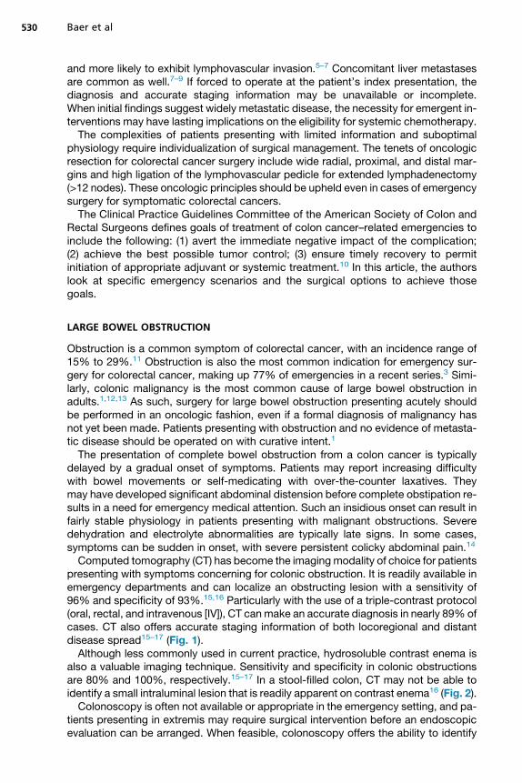
Computed tomography (CT) has become the imaging modality of choice for patientspresenting with symptoms concerning for colonic obstruction. It is readily available inemergency departments and can localize an obstructing lesion with a sensitivity of96% and specificity of 93%.15,16 Particularly with the use of a triple-contrast protocol(oral, rectal, and intravenous [IV]), CT can make an accurate diagnosis in nearly 89% ofcases. CT also offers accurate staging information of both locoregional and distantdisease spread15–17 (Fig. 1).Although less commonly used in current practice, hydrosoluble contrast enema is
also a valuable imaging technique. Sensitivity and specificity in colonic obstructionsare 80% and 100%, respectively.15–17 In a stool-filled colon, CT may not be able toidentify a small intraluminal lesion that is readily apparent on contrast enema16 (Fig. 2).Colonoscopy is often not available or appropriate in the emergency setting, and pa-
tients presenting in extremis may require surgical intervention before an endoscopicevaluation can be arranged. When feasible, colonoscopy offers the ability to identify
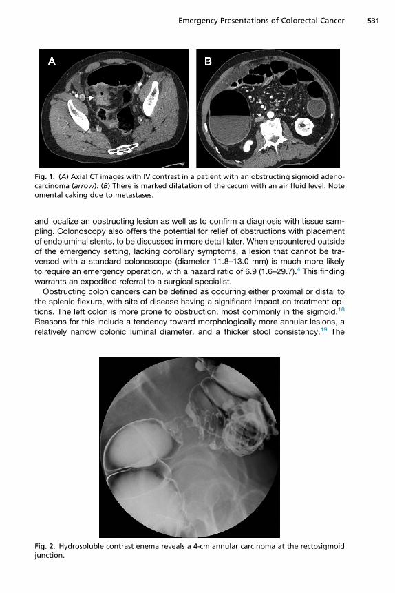
Fig. 1. (A) Axial CT images with IV contrast in a patient with an obstructing sigmoid adeno-carcinoma (arrow). (B) There is marked dilatation of the cecum with an air fluid level. Noteomental caking due to metastases.
Emergency Presentations of Colorectal Cancer 531
and localize an obstructing lesion as well as to confirm a diagnosis with tissue sam-pling. Colonoscopy also offers the potential for relief of obstructions with placementof endoluminal stents, to be discussed in more detail later. When encountered outsideof the emergency setting, lacking corollary symptoms, a lesion that cannot be tra-versed with a standard colonoscope (diameter 11.8–13.0 mm) is much more likelyto require an emergency operation, with a hazard ratio of 6.9 (1.6–29.7).4 This findingwarrants an expedited referral to a surgical specialist.Obstructing colon cancers can be defined as occurring either proximal or distal to
the splenic flexure, with site of disease having a significant impact on treatment op-tions. The left colon is more prone to obstruction, most commonly in the sigmoid.18
Reasons for this include a tendency toward morphologically more annular lesions, arelatively narrow colonic luminal diameter, and a thicker stool consistency.19 The
Fig. 2. Hydrosoluble contrast enema reveals a 4-cm annular carcinoma at the rectosigmoidjunction.
Baer et al532
larger diameter of the right colon means that obstructions are less common, typicallyinvolving very bulky tumors.
Proximal Obstructions
Because of the larger diameter of the cecum and ascending colon, right-sided ob-structions are less common and historically thought to represent bulky tumors ofmore advanced stage. Early studies found lower disease-free survival in proximalobstructing cancers compared with distal cancers, independent of perioperative com-plications or the presence of lymph node metastases.19 A more recent analysis, how-ever, looked at 377 patients undergoing colectomy for obstructing cancers, evenlysplit between proximal and distal sites, and found no difference in rates of recurrenceor 3-year survival.20
In general, proximal colonic obstructions have a simpler decision tree than distal ob-structions. Resection is often viewed as less technically demanding and most patientscan undergo an ileocolonic anastomosis, which is lower risk for complications whencompared to colocolonic or colorectal anastomoses. Reasons for this anastomosisbeing more favorable include a more reliable blood supply and a lower incidence ofsignificant proximal bowel dilation and size mismatch.Oncologic resection with primary anastomosis for right colon cancers has long been
advocated as safe and definitive surgical management in all but the frailest of patients,with fewer anastomotic complications compared with distal resections.8 The leak rateafter right hemicolectomy or extended right colectomy, even in emergency settings, isestimated at 2.8% to 4.6%, leading many to pursue this approach even in high-riskpatients.17 This rate is somewhat higher than the reported leak rate for an elective rightcolectomy in the range of 1% to 2%.Other studies, however, have documented higher rates of complications. A recent
review of 87 emergency colectomies included 43 proximal cancers. Anastomoticdehiscence after right hemicolectomy occurred in 12% (4 of 33). Two additional pa-tients had resection of the transverse colon only with primary colocolonic anasto-mosis; 1 leaked.3 In another large study, the leak rate after right colectomy was ashigh as 16.4% (28 of 173).20 The finding of a higher-than-expected leak rate afterright colectomies has led some investigators to advocate the benefits of a protectiveor terminal stoma in a subset of high-risk patients.21 Specific criteria have yet to bedefined. In these cases, attempt should still be made for definitive oncologicresection.The operative approach to an obstructing ascending colon tumor is typically a right
hemicolectomy with high ligation of the ileocolic artery and the right branch of the mid-dle colic artery, and an ileo-transverse anastomosis. When tumors are present in themid to distal transverse colon, a proper oncologic resection includes high ligation ofthe middle colic artery. When this is required, there is vascular compromise of the“watershed” splenic flexure, and the best approach is an “extended right colectomy,”including resection of the splenic flexure and an ileo-descending anastomosis. Ofnote, whenever the patient’s baseline condition or the intraoperative variables leadto a high risk of anastomotic leak, the safest approach is resection with an end ileos-tomy. For patients with more equivocal presentations, ileocolonic anastomosis with aproximal loop ileostomy may be appropriate.
Distal Obstructions
Because of narrow bowel diameter and thicker stool consistency, the descending andsigmoid colon are common sites for obstructing colon carcinomas. Compared withproximal lesions, there are considerably more options available to the surgeon
Emergency Presentations of Colorectal Cancer 533
addressing such a patient. Although it is widely acknowledged that the specificapproach must be tailored to each individual patient, surgeon expertise, and availableresources, there remains significant controversy on the optimal emergency manage-ment of obstructing distal colon cancers. These options are outlined and compared ina 2010 guideline statement from the World Society for Emergency Surgery (WSES)and Peritoneum and Surgery Society.22
Loop colostomyLoop colostomy is an established component of the surgical treatment options forobstructing distal carcinomas, with the intent of providing definitive oncologic resec-tion in a staged approach. The obstruction is thus managed in the first stage with cre-ation of a proximal loop colostomy. In the second stage, the tumor is resected and thestoma reversed. Alternatively, colostomy reversal can be performed as a third stage.Depending on patient- and tumors-specific factors, the transverse or descending co-lon can be used. In general, a loop ileostomy is discouraged, because the presence ofa competent ileocecal valve may prevent adequate alleviation of the distal obstruction.The appeal of this staged approach is that it minimizes operative time and surgical
trauma during the acute presentation when physiologic derangements and tissueintegrity are suboptimal. The initial colostomy may even be performed with only localanalgesia in some cases.15 It also reduces the risk of contamination from unpreparedbowel and allows for complete staging and multidisciplinary review before definitivetreatment.22 However, loop colostomies are often associated with high complicationrates, including stomal prolapse, hernia, and dehydration, and the approach doesnot allow for an oncologic resection.Loop colostomy is a safe option best suited to patients who are too frail to endure a
resectional procedure. Loop colostomy may also be appropriate when the cancer islocally advanced and invading adjacent organs, limiting the feasibility of a properoncologic resection in an emergency situation.
Hartmann resectionThe classic Hartmann procedure involves resection of the primary lesion with creationof an end colostomy and closing the distal colon/rectum. Large reviews have estab-lished the feasibility of emergency resection following standard oncologic principlesof high ligation of the vascular pedicle, retrieval of at least 12 regional lymph nodes,and en bloc resection of adjacent tissues for negative margins.3,23 Like a loop colos-tomy, this approach mitigates the risk of anastomotic leak. Hartmann resection iscurrently the most common operation performed for distal colon carcinomas present-ing emergently, especially by general surgeons.15,24,25
Despite the longer operative time for a formal resection, literature has not shown anyworse short- or long-term outcomes in patients undergoing formal Hartmann resec-tion compared with the staged approach. A randomized study by Kronborg26 showedno difference in mortality, recurrence rate, and cancer-specific survival between co-lostomy or Hartmann procedure in emergency presentations. The only differencefound in this study was a longer hospital stay in the staged approach due to theneed for multiple subsequent operations. Of note, this study has been criticized forits long accrual period, incomplete follow-up, and heterogenous underlying pathology.A Cochrane systematic review in 2004, which did not include the Kronborg study dueto methodological flaws, nevertheless made the same conclusions.27 WSES guide-lines conclude that colostomy (staged approach) should be reserved for “damagecontrol” situations, unresectable tumors, and cases where multimodal treatment isanticipated before formal resection.22
Baer et al534
A contradictory conclusion was made by another recent randomized controlled trial(RCT), which found no difference in outcomes, including transfusion rates or durationof hospitalization between a staged approach and Hartmann resection.28 The investi-gators of this study argue rather that colostomy for staged approach is ideal foryounger, healthier patients who will tolerate definitive surgery in as little as 2 to 3 weekswhen less bowel distention and inflammation may allow for a technically easier andmore oncologically sound resection.28 Nevertheless, most investigators agree thatHartmann resection is the procedure of choice for older patients with high AmericanSociety of Anesthesiologists (ASA) scores, advanced obstructions, and proximalbowel distension, and whose underlying medical comorbidities might preclude defin-itive surgery in a staged fashion.15,22,26–28
The main disadvantage of a Hartmann resection is the residual stoma. Among pa-tients with colon cancer, the rate of Hartmann reversal is only 20% for reasonsincluding advanced disease, complications from treatment, and poor performancestatus.29,30 Operations to restore intestinal continuity are also associated with signif-icant morbidity and mortality.15 Stomas are not without their own complications, andrates increase the longer they are in place, adversely affecting quality of life.31,32
Single-stage primary resection and anastomosisFor many years, a single-stage oncologic resection with primary anastomosis wasconsidered too high of a risk in the emergency setting. Concerns included furtherphysiologic derangement to a critically ill patient, increased extent of surgery andoperating room time, difficulty manipulating and mobilizing a distended colon, and po-tential for contamination of the peritoneal cavity. Patients may be severely malnour-ished due to reduced oral intake before presenting with obstruction, andproceeding with an operation before nutritional optimization may increase their riskfor postoperative complications, especially if the condition of proximal bowel isdilated, ischemic, or otherwise suboptimal for an anastomosis. Of utmost importanceare the complications from an anastomotic leak, which can be catastrophic and fore-stall adjuvant systemic chemotherapy when indicated.Large studies, however, have established the feasibility of primary resection and
anastomosis (PRA) in appropriately selected patients. Resection with primary anasto-mosis can reduce length of stay and reduce number of operations with similar rates ofmorbidity and mortality. Nonrandomized reviews and retrospective data have shownthe rate of anastomotic leak in emergency settings to be 2.2% to 12%, which is similarto rates in elective colon resection of 1.9% to 8%.22 Thus, even in acutely symptom-atic distal colon carcinomas, PRA is recommended in the position statement from theAssociation of Coloproctologists of Great Britain and Ireland.31,33
Appropriate patient selection is critical to success in this inherently high-riskenvironment. Specific factors that have been associated with poor outcomes inobstructing colon cancer operations include age greater than 70, ASA grades III–IV, preoperative renal failure, surgery within 24 hours of presentation, and advancedcancer stage.21,29,33 Any of these factors may argue for either a Hartmann resectionwith end colostomy or potentially a primary anastomosis with a protecting loopileostomy.
Total abdominal colectomyTotal abdominal colectomy with ileorectal anastomosis (TAC/IRA) is another option forselect patients. It also removes the distended and potentially ischemic proximal colon,resecting back to healthy terminal ileum for a primary anastomosis. This approach isparticularly appropriate in cases with suspected synchronous tumors or hereditary
Emergency Presentations of Colorectal Cancer 535
colorectal cancer syndromes. Another very important indication for TAC/IRA is cecalperforation or impending perforation, which is common in advanced distal obstruc-tions. In general, a double resection to remove the cecum and the distal tumor sepa-rately, leaving the transverse colon intact, is not recommended.A small randomized trial compared outcomes from subtotal colectomy to segmental
resection (PRA) and found no difference in hospital mortality or complication rates.Segmental colectomies in this study included intraoperative colonic irrigation. At4 months, however, the subtotal colectomy patients reported more frequent bowelmovements and more presentations with bowel problems than in the PRA group.The investigators concluded that subtotal colectomy should be reserved for casesof synchronous lesions or when the integrity and viability of proximal colon isquestioned.34
Self-expanding metal stentsSelf-expandable metal stents (SEMS) represent a nonoperative modality to addressdistal colonic malignant obstructions. These stents were first developed in the 1990sfor palliation of obstructions from unresectable tumors or in patients deemed poorcandidates for resectional surgery.24,35,36 Stents are also used as a temporizing“bridge to surgery” with the goal of enabling elective, possibly laparoscopic,resection.SEMS involve the endoscopic placement of a guide wire across the obstructing
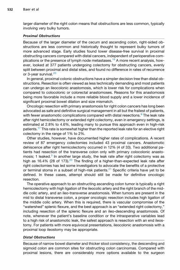
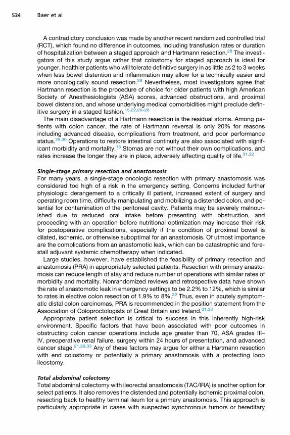
lesion, often with the assistance of fluoroscopy, followed by an uncovered, self-expanding metal stent. Balloon dilation is not typically necessary. Once the stenthas been deployed, success is confirmed by a rush of air and fluid past the obstruction(Fig. 3). If desired, the endoscope can typically be advanced through the stent to visu-alize the proximal colon. Although stenting is technically feasible for all areas of the co-lon, it has been more successful and better studied for left-sided lesions.Stenting is an attractive alternative to emergency surgery. Proponents argue that
stenting can allow for the managing team to stabilize the patient, correct dehydrationand other electrolyte imbalances, optimize medical comorbidities and nutritional sta-tus, complete oncologic staging, and involve a multidisciplinary team. Early studiessupporting the use of SEMS in this context argued that as a bridge to surgery, stentscould reduce morbidity and mortality and lower stoma rates compared with surgeryalone.35,37–41
Not all reported data, however, have supported these initial claims. For example, arecent observational study compared surgery to SEMS as a bridge to surgery.Despite high rates of technical success with stent placement (91%) and relativelylow rates of complications (microperforation rate 13%), there was no difference inperioperative mortality and no difference in rates of primary anastomosis or stomacreation.31
In a large systematic review and meta-analysis, the rate of clinical success relievingobstruction with SEMS was only 52.5% overall, compared with 99% with surgery.Morbidity and mortality were again similar between groups; however, the rates of pri-mary anastomosis were surprisingly low in the bridge-to-surgery group, only 64.9%compared with 55% in the surgery-first group, not statistically different. Anastomoticleak rates were slightly better in the stented patients, but also not significant.42
Stent deployment is not without risk. In fact, of the 6 RCTs comparing SEMS toupfront surgery in distal obstructing cancers, half closed enrollment early due tohigh rates of stent-related complications, most notably perforation during deploy-ment.15,35 Other complications include failure to relieve the obstruction, migration,and subsequent stent occlusion. Tumor perforation during stent deployment likely
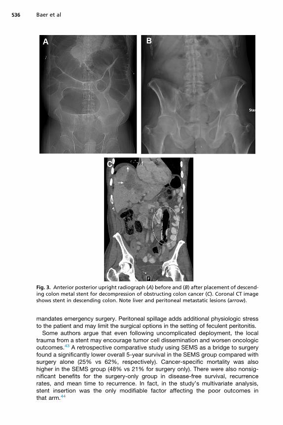
Fig. 3. Anterior posterior upright radiograph (A) before and (B) after placement of descend-ing colon metal stent for decompression of obstructing colon cancer (C). Coronal CT imageshows stent in descending colon. Note liver and peritoneal metastatic lesions (arrow).
Baer et al536
mandates emergency surgery. Peritoneal spillage adds additional physiologic stressto the patient and may limit the surgical options in the setting of feculent peritonitis.Some authors argue that even following uncomplicated deployment, the local
trauma from a stent may encourage tumor cell dissemination and worsen oncologicoutcomes.43 A retrospective comparative study using SEMS as a bridge to surgeryfound a significantly lower overall 5-year survival in the SEMS group compared withsurgery alone (25% vs 62%, respectively). Cancer-specific mortality was alsohigher in the SEMS group (48% vs 21% for surgery only). There were also nonsig-nificant benefits for the surgery-only group in disease-free survival, recurrencerates, and mean time to recurrence. In fact, in the study’s multivariate analysis,stent insertion was the only modifiable factor affecting the poor outcomes inthat arm.44
Emergency Presentations of Colorectal Cancer 537
In general, success rates are higher and complication rates lower in SEMS case se-ries involving experienced endoscopists. However, further studies are needed beforeSEMS is considered the standard for malignant bowel obstructions. In the presence ofmetastatic disease or short life expectancy, stents may prevent a morbid operationand allow quicker initiation or continuation of systemic chemotherapy. SEMS shouldonly be performed by endoscopists with adequate expertise to limit complicationrates.
Obstructing Rectal Cancer
Rectal bleeding and a change in stool appearance are the most common symptoms ofrectal cancer.45 Many early asymptomatic rectal cancers will be found on screeningendoscopy, but a rectal cancer presenting with acute obstructive symptoms is typi-cally of a locally advanced stage.The additional challenges and morbidity associated with pelvic surgery weigh in
the decision making for acutely symptomatic rectal cancers. Optimal oncologicresection should include total mesorectal excision. In the elective setting, neoadju-vant chemoradiation has become the standard of care for T3 or node-positive rectalcancers in the United States. Compared with the previous discussion of coloncancers, there is more enthusiasm for measures that safely temporize acute symp-toms of rectal cancer to allow for complete staging and initiation of neoadjuvanttreatment.
Loop ileostomy or colostomyIn patients with obstructing mid and low rectal cancers without findings ofmetastatic spread, simple diversion provides the opportunity to complete stagingand give neoadjuvant chemoradiation with a staged oncologic resection for curativeintent. Loop colostomy allows for decompression as well as access to the proximalcolon for assessment of proximal synchronous lesions. However, it may limit oppor-tunities for reconstructing bowel continuity with an eventual low anterior resection bysacrificing bowel length or blood supply to the future anastomosis. A loop ileostomyoften works better for these patients, although it is associated with a small risk of aclosed loop obstruction when a competent ileocecal valve exists. In the settingwhere sphincter preservation is clearly not an option, a loop colostomy is morefitting.
Hartmann resectionIn the case of obstructing carcinomas of the upper rectum, a Hartmann proceduremay be chosen, providing definitive resection without the added risks of an anasto-mosis. Indeed, this option may be appropriate for older patients with more comorbid-ities, even in the absence of acute obstruction. Patients should be aware thatcolostomy reversal in this setting is extremely uncommon.
Self-expandable metal stentsAs described above for distal colon cancers, the use of SEMS for obstructing rectalcancers is most appropriate in patients with widely metastatic disease who will benefitmost from systemic chemotherapy, or who are too physiologically stressed to toleratea low anterior resection or abdominal perineal resection. The risk of tumor perforationduring placement limits their use in treatment plans with curative intent.1 Importantly,placement of rectal stents carries significant risk of distal migration and severetenesmus from pressure on the upper anal sphincter mechanism. Therefore, SEMSis limited to lesions in the upper rectum.
Baer et al538
PERFORATION
Perforation is the second most common reason for urgent or emergent surgery asso-ciated with colorectal carcinoma, with an incidence of 2.6% to 12%.46,47 Perforationsmost commonly occur at the site of the primary tumor, due to necrosis and friable tis-sue. Depending on the location, these may progress to either free or contained perfo-rations. Perforation can also occur proximal to an obstructing carcinoma. Increasingpressure and distension from a complete distal obstruction follow the Law of Laplace,which can ultimately result in ischemia of the proximal bowel and perforations atremote proximal sites. The cecum is the most common site of this type of diastaticperforation.6 This clinical presentation has been recognized as an independent prog-nostic factor for morbidity and mortality.7
An obstructing cancer increases the risk of perforation, with rates of 12% to 19%.48
Perforation is reported to be the most lethal complication of colorectal carcinoma. Insome studies, mortality associated with secondary peritonitis from perforation is ashigh as 30% to 50%.1,49
Free Perforation
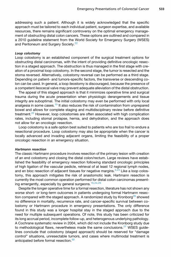
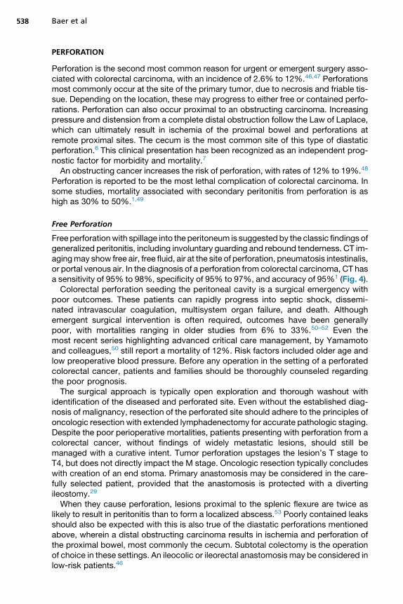
Free perforationwith spillage into the peritoneum is suggested by the classic findings ofgeneralized peritonitis, including involuntary guarding and rebound tenderness. CT im-agingmay show free air, free fluid, air at the site of perforation, pneumatosis intestinalis,or portal venous air. In the diagnosis of a perforation from colorectal carcinoma, CT hasa sensitivity of 95% to 98%, specificity of 95% to 97%, and accuracy of 95%1 (Fig. 4).Colorectal perforation seeding the peritoneal cavity is a surgical emergency with
poor outcomes. These patients can rapidly progress into septic shock, dissemi-nated intravascular coagulation, multisystem organ failure, and death. Althoughemergent surgical intervention is often required, outcomes have been generallypoor, with mortalities ranging in older studies from 6% to 33%.50–52 Even themost recent series highlighting advanced critical care management, by Yamamotoand colleagues,50 still report a mortality of 12%. Risk factors included older age andlow preoperative blood pressure. Before any operation in the setting of a perforatedcolorectal cancer, patients and families should be thoroughly counseled regardingthe poor prognosis.The surgical approach is typically open exploration and thorough washout with
identification of the diseased and perforated site. Even without the established diag-nosis of malignancy, resection of the perforated site should adhere to the principles ofoncologic resection with extended lymphadenectomy for accurate pathologic staging.Despite the poor perioperative mortalities, patients presenting with perforation from acolorectal cancer, without findings of widely metastatic lesions, should still bemanaged with a curative intent. Tumor perforation upstages the lesion’s T stage toT4, but does not directly impact the M stage. Oncologic resection typically concludeswith creation of an end stoma. Primary anastomosis may be considered in the care-fully selected patient, provided that the anastomosis is protected with a divertingileostomy.29
When they cause perforation, lesions proximal to the splenic flexure are twice aslikely to result in peritonitis than to form a localized abscess.53 Poorly contained leaksshould also be expected with this is also true of the diastatic perforations mentionedabove, wherein a distal obstructing carcinoma results in ischemia and perforation ofthe proximal bowel, most commonly the cecum. Subtotal colectomy is the operationof choice in these settings. An ileocolic or ileorectal anastomosis may be considered inlow-risk patients.46
Fig. 4. (A) Axial and (B) coronal CT images show a left colon soft tissue mass with markednarrowing of the lumen with circumferential wall thickening and infiltration of the sur-rounding pericolonic fat compatible with localized perforation. Note adjacent pericolonicabscess (arrow).
Emergency Presentations of Colorectal Cancer 539
Abscess
Contained perforations may present with localized tenderness. Imaging may reveal aphlegmon or abscess, which is more common than free perforation in descending andsigmoid colon lesions.53 Many cases of perforated colorectal cancer presenting as ab-scess are not diagnosed preoperatively and can mimic diverticulitis or appendicitis onCT imaging.46,53
Baer et al540
The role for percutaneous drainage of contained perforations from a carcinoma differsfrom that of benign diseases. In the presence of widely metastatic disease, treatmentwith antibiotics and percutaneous drainage avoids the morbidity of an operation. Insome cases, however, drawnout infectious complications can forestall systemic chemo-therapy. In the absenceofwidely disseminateddisease, percutaneousdrainageof a con-tainedperforationmay result in seeding tumorcells along thedrainage tract rendering thedisease metastatic.53 When a malignancy is suspected, drains should be placed in amanner where the skin and drain tract can be later resected en bloc with the cancer.Definitive surgical management involves en bloc resection of the mass and any invadedadjacent organs and/or percutaneous drains whenever technically feasible.1
BLEEDING
Gastrointestinal bleeding is reported in up to 50% of patients with colorectal can-cer.1,54 Most of this bleeding, however, is low volume, is self-limited, and does notrequire emergent surgical intervention. Bleeding is often an early symptom of a colo-rectal cancer associated with lower risk of advanced stage at diagnosis, and a shorterdelay in presentation. Unlike the insidious onset of an obstructing cancer, patientsoften remember to the day when bleeding began.45 Bleeding is complicated by thefact that most acute tumor bleeding is likely in the setting of chronic anemia of cancerand blood loss from the tumor.Acute massive gastrointestinal bleeding from a colorectal carcinoma is rare. The
initial management is aimed at resuscitation, establishing large-bore IV access, andstabilization with crystalloid and correction of underlying coagulopathy or other meta-bolic abnormalities.In the clinically stable patient, efforts to localize the source of bleeding should be
sought before surgical treatment whenever possible.1 Endoscopy will identify thesource in 74% to 89% of cases, although this technique may be limited in the unpre-pared colon.55,56 Tagged red blood cell scan is less sensitive, localizing the source in26% to 72%, but it does detect bleeding at rates as low as 0.1 mL/min, making it apotential screening test before angioembolization. Embolization has documented suc-cess rates of 42% to 86%; however, it carries the risk of worsening intestinalischemia.1,55 This option may be more attractive in the setting of metastatic diseaseto avoid laparotomy and associated delays in systemic chemotherapy.Surgery is the most effective and definitive approach for a hemorrhaging colorectal
cancer. Some general indications for surgical intervention include hemodynamicinstability despite transfusion of more than 6 units of blood products, slow bleedingrequiring more than 3 units of blood products per day, inability to stop hemorrhagewith endoscopic or endovascular techniques, or recurrent episodes of hemorrhagicshock.57
When the site has been localized, resection should adhere to oncologic principleswith curative intent. The decision to form a stoma or perform a PRA with or withoutproximal diversion should be carefully considered in light of any anemia, coagulop-athy, and unstable hemodynamics that often accompany the bleeding patient.
MINIMALLY INVASIVE PLATFORMS
Emergency laparoscopic colectomy for symptomatic colorectal cancer has beendescribed in several case series and case-control studies. Laparoscopy typically re-quires longer operative times, but is associated with lower blood loss, shorter hospitalstay, and similar morbidities and mortalities when compared with open surgery. Ratesof conversion to open surgery range from 0% to 17% in emergency colectomies.58
Emergency Presentations of Colorectal Cancer 541
Appropriate patient selection is central to the safety and feasibility of minimally inva-sive techniques in the emergency setting. Surgeon experience with elective laparo-scopic colectomy techniques is prerequisite.The first case report of emergency robotic colectomy was recently published for a
hemorrhagic right-sided colon cancer, with good postoperative and oncologicoutcomes.58
OUTCOMES
The feasibility of oncologic resections in the emergency setting has been well demon-strated. Teixeira and colleagues3 documented R0 resection possible in up to 92% ofemergency colectomies. Patients for whom R0 resection was not achieved had bulkyT4 lesions or were unable to tolerate more radical en bloc resections. Adequate lym-phadenectomy (>12 nodes) was documented in 71%.The long-term and oncologic outcomes for colorectal cancers presenting with
emergency complications are worse than their elective counterparts. A recent retro-spective review from Ireland included 34% of colon resections performed emergentlyand collected long-term follow-up to assess oncologic outcomes. Emergency resec-tions were more often T4 lesions (38% vs 13%) and more often lymph node positive(58% vs 38%). Perforation was the indication in 8%. Positive margins were found in10% of emergency colectomies compared with only 1% of elective cases. With upto 5 years follow-up, the median survival for emergency presentations was only59 months compared with 82 months for elective cases during the same time.6 Otherstudies have shown similar results,23,59 although exactly what is responsible for theseworse outcomes is still debated.60
High rates of complications have been associated with urgent or emergent colec-tomy. One institution’s retrospective review of 209 consecutive colectomies foundhigher rates of wound infections, wound dehiscence, and intra-abdominal abscessin emergency colectomies.61 The rates of perioperative mortality for emergency colo-rectal cancer resections range from 5% to 34%.62–64 The immediate threats to life willdictate how resources are allocated to the resuscitation and preoperative workup. Theliberal use of stomas is advocated and demonstrated in most series.
SUMMARY
The management of emergency complications of colorectal carcinomas has changedover the past few decades. For proximal lesions, general consensus is that hemicolec-tomy with primary anastomosis is safe with an acceptably low leak rate. For distal ob-structions, there is active investigation and controversy challenging practices bothnew and old. Single-stage resections and the use of endoluminal stents to temporizeemergency presentations have allowed some surgical specialists to reduce the mor-bidities of stomas and multiple operations. Ultimately, the best management must betailored to each specific scenario. In the treatment of emergency presentations ofcolorectal carcinoma, care must be individualized to the patient, the experience ofthe surgeon, and the resources available at the facility.
REFERENCES
1. Barnett A, Cedar A, Siddiqui F, et al. Colorectal cancer emergencies.J Gastrointest Cancer 2013;44(2):132–42.
2. Gunnarsson H, Holm T, Ekholm A, et al. Emergency presentation of colon canceris most frequent during summer. Colorectal Dis 2011;13(6):663–8.
Baer et al542
3. Teixeira F, Akaishi EH, Ushinohama AZ, et al. Can we respect the principles ofoncologic resection in an emergency surgery to treat colon cancer? World JEmerg Surg 2015;10:5.
4. Chalieopanyarwong V, Boonpipattanapong T, Prechawittayakul P, et al. Endo-scopic obstruction is associated with higher risk of acute events requiring emer-gency operation in colorectal cancer patients. World J Emerg Surg 2013;8:34.
5. Bayar B, Yilmaz KB, Akinci M, et al. An evaluation of treatment results of emer-gency versus elective surgery in colorectal cancer patients. Ulus Cerrahi Derg2016;32:11–7.
6. Bass G, Fleming C, Conneely J, et al. Emergency first presentation of colorectalcancer predicts significantly poorer outcomes: a review of 356 consecutive Irishpatients. Dis Colon Rectum 2009;52(4):678–84.
7. Alvarez JA, Baldonedo RF, Bear IG, et al. Presentation, treatment, and multivar-iate analysis of risk factors for obstructive and perforative colorectal carcinoma.Am J Surg 2005;190:376–82.
8. Phillips RK, Hittinger R, Fry JS, et al. Malignant large bowel obstruction. Br J Surg1985;72:296–302.
9. Garcia-Valdecasas JC, Llovera JM, deLacy AM, et al. Obstructing colorectal car-cinomas: prospective study. Dis Colon Rectum 1991;34(9):759–62.
10. Chang GJ, Kaiser AM, Mills S, et al. Practice parameters for the management ofcolon cancer. Dis Colon Rectum 2012;55:831–43.
11. Ohman U. Prognosis in patients with obstructing colorectal carcinoma. Am J Surg1982;143:742–7.
12. De Dombal FT, Matharu SS, Staniland JR, et al. Presentation of cancer to the hos-pital as “acute abdominal pain”. Br J Surg 1980;67:413–6.
13. Peterson M. Large intestine. In: Marx JA, Hockberger RS, Walls RM, editors.Rosen’s emergency medicine: concepts and clinical practice. 6th edition.Philadelphia: Elsevier; 2006. p. 1332–4.
14. Gordon PH. Malignant neoplasms of the colon. In: Gordon PH, Nivatvongs S,editors. Principles and practice of surgery for the colon, rectum and anus.3rd edition. New York: Informa Healthcare; 2007. p. 534–5.
15. Frago R, Ramirez E, Millan M, et al. Current management of acute malignant largebowel obstruction: a systematic review. Am J Surg 2014;207:127–38.
16. Frager D, Rovno HD, Baer JW, et al. Prospective evaluation of colonic obstructionwith computed tomography. Abdom Imaging 1998;23(2):141–6.
17. Gainant A. Emergency management of acute colonic cancer obstruction. J ViscSurg 2012;149:e3–10.
18. Kleespies A, Fuessl KE, Seeliger H, et al. Determinants of morbidity and survivalafter elective non-curative resection of stage IV colon and rectal cancer. Int JColorectal Dis 2009;24:1097–109.
19. Wolmark N, Wieand HS, Rockette HE, et al. The prognostic significance of tumourlocation and bowel obstruction in Dukes’ B and C colorectal cancer. Ann Surg1983;198:743–50.
20. Frago R, Biondo S, Millan M, et al. Differences between proximal and distal ob-structing colonic cancer after curative surgery. Colorectal Dis 2011;13:e116–22.
21. Biondo S, Pares D, Frago R, et al. Large bowel obstruction: predictive factors forpostoperative mortality. Dis Colon Rectum 2004;47:1889–97.
22. Ansaloni L, Andersson RE, Bazzoli F, et al. Guidelines in the management of ob-structing cancer of the left colon: consensus conference of the World Society ofEmergency Surgery (WSES) and Peritoneum and Surgery (PnS) Society. World JEmerg Surg 2010;5:29.
Emergency Presentations of Colorectal Cancer 543
23. McArdle CS, Hole DJ. Emergency presentation of colorectal cancer is associatedwith poor 5-year survival. Br J Surg 2004;91(5):605–9.
24. Trompetas V. Emergency management of malignant acute left-sided colonicobstruction. Ann R Coll Surg Engl 2008;90:181–6.
25. Meyer F, Marusch F, Koch A, et al. Emergency operation in carcinomas of the leftcolon: value of Hartmann’s procedure. Tech Coloproctol 2004;8(suppl):s226–9.
26. Kronborg O. Acute obstruction from tumour in the left colon without spread. Arandomized trial of emergency colostomy versus resection. Int J Colorectal Dis1995;10:1–5.
27. De Salvo GL, Gava C, Lise M, et al. Curative surgery for obstruction from primaryleft colorectal carcinoma: primary or staged resection? Cochrane Database Syst2004;(2):CD002101.
28. Krstic S, Resanovic V, Alempijevic T, et al. Hartmann’s procedure vs loop colos-tomy in the treatment of obstructive rectosigmoid cancer. World J Emerg Surg2014;9:52.
29. Zorcolo L, Covotta L, Carlomagno N, et al. Safety of primary anastomosis in emer-gency colorectal surgery. Colorectal Dis 2003;5:262–9.
30. Desai DC, Brennan EJ, Reilly JF, et al. The utility of the Hartmann procedure. Am JSurg 1998;175:152–4.
31. Kavanagh DO, Nolan B, Judge C, et al. A comparative study of short- andmedium-term outcomes comparing emergent surgery and stenting as a bridgeto surgery in patients with acute malignant colonic obstruction. Dis Colon Rectum2013;56:433–40.
32. Sprangers MA, Taal BG, Aaronson NK, et al. Quality of life in colorectal cancer.Stoma vs. nonstoma patients. Dis Colon Rectum 1995;38:361–9.
33. Tekkis PP, Kinsman R, Thompson MR, et al. The Association of Coloproctology ofGreat Britain and Ireland study of large bowel obstruction caused by colorectalcancer. Ann Surg 2004;204:76–81.
34. The SCOTIA Study Group. Single-stage treatment for malignant left-sided colonicobstruction: a prospective randomized clinical trial comparing subtotal colec-tomy with segmental resection following intraoperative irrigation. Br J Surg1995;82:1622–7.
35. Kwak MS, Kim WS, Lee JM, et al. Does stenting as a bridge to surgery in left-sided colorectal cancer obstruction really worsen oncological outcomes? Dis Co-lon Rectum 2016;59:725–32.
36. Dohmoto M, Hunerbein M, Schlag PM. Palliative endoscopic therapy of rectalcarcinoma. Eur J Cancer 1996;32a:25–9.
37. Khot UP, Lang AW, Murali K, et al. Systematic review of the efficacy and safety ofcolorectal stents. Br J Surg 2002;89:1096–102.
38. Cennamo V, Luigiano C, Coccolini F, et al. Meta-analysis of randomized trialscomparing endoscopic stenting and surgical decompression for colorectal can-cer obstruction. Int J Colorectal Dis 2013;28:855–63.
39. Sebastian S, Johnston S, Geoghegan T, et al. Pooled analysis of the efficacy andsafety of self-expanding metal stenting in malignant colorectal obstruction. Am JGastroenterol 2004;99:2051–7.
40. Matsuda A, Miyashita M, Matsumoto S, et al. Comparison of long-term outcomesof colonic stent as “bridge to surgery” and emergency surgery for malignantlarge-bowel obstruction: a meta-analysis. Ann Surg Oncol 2015;22:497–504.
41. Zhang Y, Shi J, Shi B, et al. Self-expanding metallic stent as a bridge to surgeryversus emergency surgery for obstructive colorectal cancer: a meta-analysis.Surg Endosc 2012;26:110–9.
Baer et al544
42. Cirocchi R, Farinella E, Trastulli S, et al. Safety and efficacy of endoscopic colonicstenting as a bridge to surgery in the management of intestinal obstruction due toleft colon and rectal cancer: a systematic review and meta-analysis. Surg Oncol2013;22(1):14–21.
43. Maruthachalam K, Lash GE, Shenton BK, et al. Tumour cell disseminationfollowing endoscopic stent insertion. Br J Surg 2007;94:1151–4.
44. Sabbagh C, Browet F, Diouf M, et al. Is stenting as “a bridge to surgery” an on-cologically safe strategy for the management of acute, left-sided, malignant,colonic obstruction? A comparative study with a propensity score analysis. AnnSurg 2013;258:107–15.
45. Korsgaard M, Pedersen L, Sorensen HT, et al. Reported symptoms, diagnosticdelay and stage of colorectal cancer: a population-based study in Denmark.Colorectal Dis 2006;8:688–95.
46. Tsai HL, Hsieh JS, Yu FJ, et al. Perforated colonic cancer presenting as intra-abdominal abscess. Int J Colorectal Dis 2007;22(1):15–9.
47. Saegesser F, Sandblom P. Ischemic lesions of the distended colon. A complica-tion of obstructive colorectal cancer. Am J Surg 1975;129:309–15.
48. Umpleby HC, Williamson RCN. Survival in acute obstructing colorectal carci-noma. Dis Colon Rectum 1984;27:299–304.
49. Langell JT, Mulvihill SJ. Gastrointestinal perforation and the acute abdomen. MedClin North Am 2008;92(3):599–625.
50. Yamamoto T, Kita R, Masui H, et al. Prediction of mortality in patients with colo-rectal perforation based on routinely available parameters: a retrospective study.World J Emerg Surg 2015;10:24.
51. Horiuchi A, Watanabe Y, Doi T, et al. Evaluation of prognostic factors and scoringsystem in colonic perforation. World J Gastroenterol 2007;13:3228–31.
52. Komatsu S, Shimomatsuya T, Nakajima M, et al. Prognostic factors and scoringsystem for survival in colonic perforation. Hepatogastroenterology 2005;52:761–4.
53. Yeo ES, Ng KH, Eu KW. Perforated colorectal cancer: an important differentialdiagnosis in all presumed diverticular abscesses. Ann Acad Med Singapore2011;40(8):375–8.
54. Adelstein BA, Macaskill P, Chan SF, et al. Most bowel cancer symptoms do notindicate colorectal cancer and polyps: a systematic review. BMC Gastroenterol2011;11:65.
55. Zuccaro G Jr. Management of the adult patient with acute lower gastrointestinalbleeding. Am J Gastroenterol 1998;93(8):1202–8.
56. Davila RE, Rajan E, Adler DG, et al. ASGE Guideline: the role of endoscopy in thepatient with lower-GI bleeding. Gastrointest Endosc 2005;62(5):656–60.
57. Tavakkolizadeh A, Ashley S. Acute gastrointestinal hemorrhage. In: Townsend CM,Beauchamp RD, Evers BM, et al, editors. Sabiston textbook of surgery: the biolog-ical basis of modern surgical practice. 19th edition. Philadelphia: Elsevier; 2012.p. 1139–59.
58. Felli E, Brunetti F, Disabato M, et al. Robotic right colectomy for hemorrhagic rightcolon cancer: a case report and review of the literature of minimally invasive ur-gent colectomy. World J Emerg Surg 2014;9:32.
59. Oliphant R, Mansouri D, Nicholson GA, et al. Emergency presentation of node-negative colorectal cancer treated with curative surgery is associated with poorershort and longer-term survival. Int J Colorectal Dis 2014;29:591–8.
60. Weixler B, Warschkow R, Ramser M, et al. Urgent surgery after emergency pre-sentation for colorectal cancer has no impact on overall and disease-free sur-vival: a propensity score analysis. BMC Cancer 2016;16:208.

Emergency Presentations of Colorectal Cancer 545
61. Kim J, Mittal R, Konyalian V, et al. Outcome analysis of patients undergoing colo-rectal resection for emergent and elective indications. Am Surg 2007;73:991–3.
62. Boyle DJ, Thorn C, Saini A, et al. Predictive factors for successful colonic stentingin acute large-bowel obstruction: a 15-year cohort analysis. Dis Colon Rectum2015;58:358–62.
63. Breitenstein S, Rickenbacher A, Berdajs D, et al. Systematic evaluation of surgi-cal strategies for acute malignant left-sided colonic obstruction. Br J Surg 2007;94(12):1451–60.
64. Tan CJ, Dasari BV, Gardiner K. Systematic review and meta-analysis of random-ized clinical trials of self-expanding metallic stents as a bridge to surgery versusemergency surgery for malignant left-sided large bowel obstruction. Br J Surg2012;99:469–76.