Embed Size (px)
Citation preview
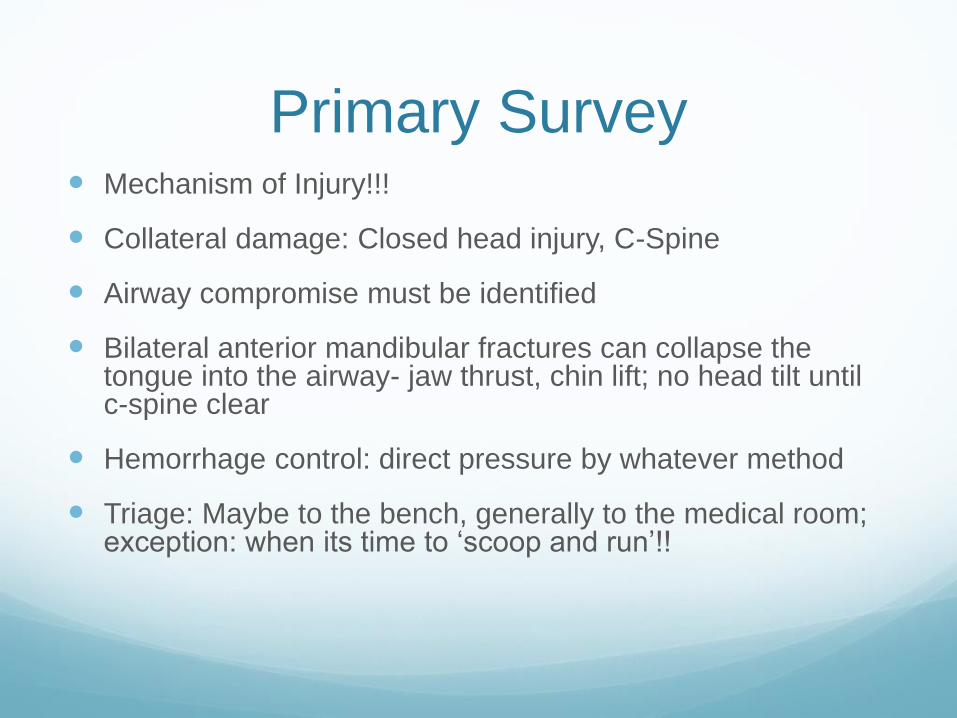
Primary Survey Mechanism of Injury!!!
Collateral damage: Closed head injury, C-Spine
Airway compromise must be identified
Bilateral anterior mandibular fractures can collapse the tongue into the airway- jaw thrust, chin lift; no head tilt until c-spine clear
Hemorrhage control: direct pressure by whatever method
Triage: Maybe to the bench, generally to the medical room; exception: when its time to ‘scoop and run’!!
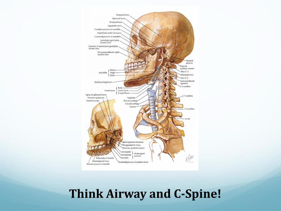
Think Airway and C-Spine!
Thorough Clinical Evaluation Inspect the face for symmetry, swelling, ecchymosis, mobility: bimanual
palpation
Orderly exam: top to bottom, inside out
Orbits, Zygomas, Nose, Maxilla and Mandible; Intra-oral exam
Things that move should move: eyes, tongue, TMJ; things that don’t move shouldn’t: bones, teeth
Look for fractures especially with higher pain report: crepitus, mobility, step discrepancy are good indicators
Raccoon eyes (periorbital ecchymosis): Middle or upper face fracture; Battle’s sign (mastoid ecchymosis): Basilar skull fracture: Subconjunctivalhemorrhage: Zygomatic or orbital fracture
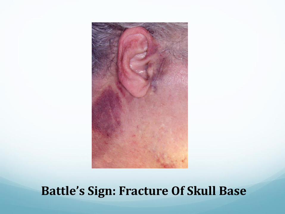
Battle’s Sign: Fracture Of Skull Base
Intra-Oral Exam Look for open wounds, foreign bodies, and loose teeth
or bony segments
Evaluate the occlusion- ‘How does your bite feel?’
Be wary of occult injuries that may lead to airway
compromise: sublingual hematoma
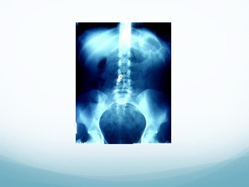
Appropriate x-rays if you suspect aspiration or
ingestion of tooth, dental appliance or foreign body.
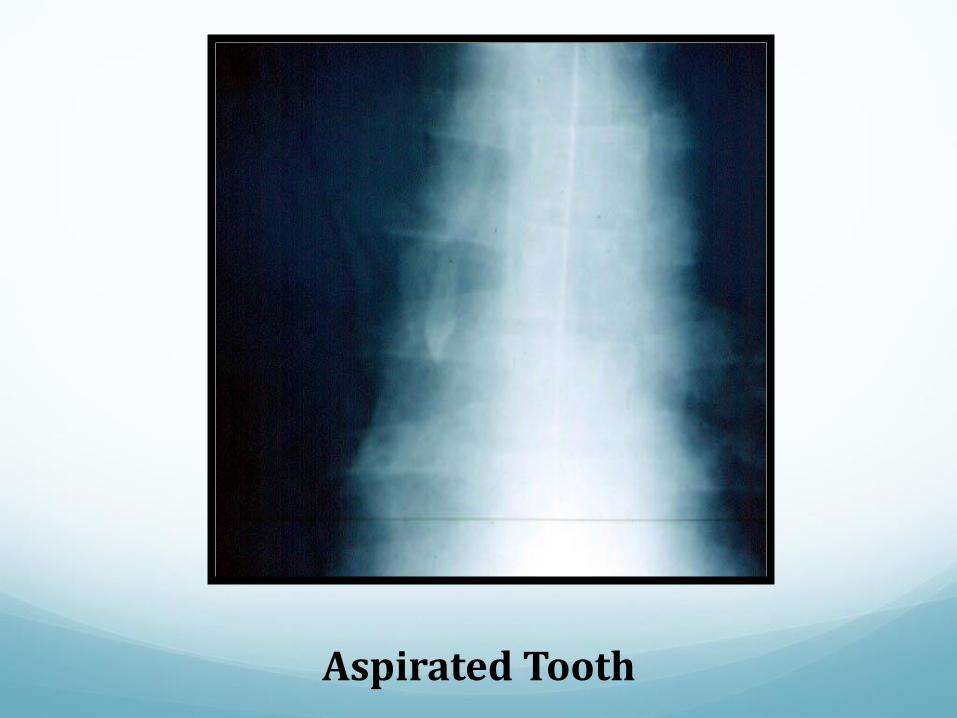
Aspirated Tooth
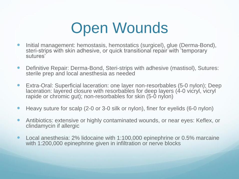
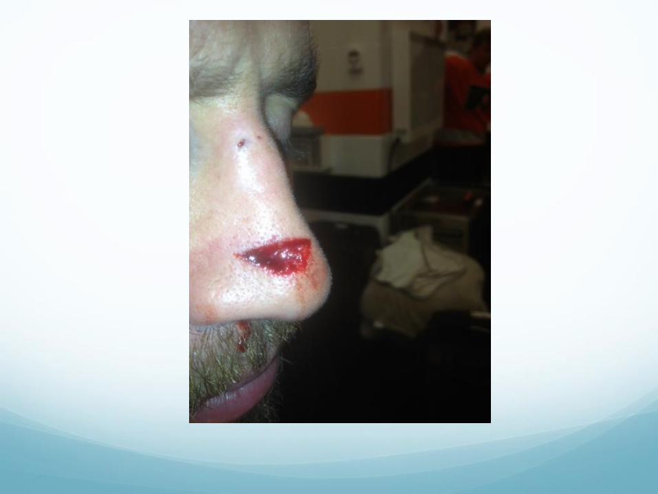
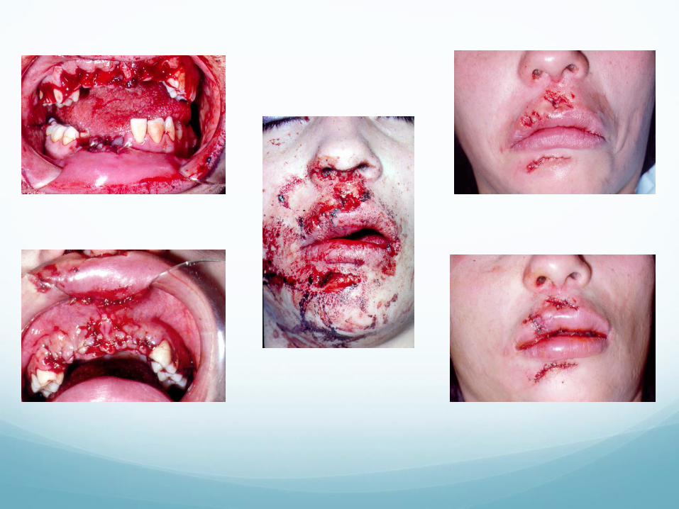
Open Wounds Initial management: hemostasis, hemostatics (surgicel), glue (Derma-Bond),
steri-strips with skin adhesive, or quick transitional repair with ‘temporary sutures’
Definitive Repair: Derma-Bond, Steri-strips with adhesive (mastisol), Sutures: sterile prep and local anesthesia as needed
Extra-Oral: Superficial laceration: one layer non-resorbables (5-0 nylon); Deep laceration: layered closure with resorbables for deep layers (4-0 vicryl, vicrylrapide or chromic gut); non-resorbables for skin (5-0 nylon)
Heavy suture for scalp (2-0 or 3-0 silk or nylon), finer for eyelids (6-0 nylon)
Antibiotics: extensive or highly contaminated wounds, or near eyes: Keflex, or clindamycin if allergic
Local anesthesia: 2% lidocaine with 1:100,000 epinephrine or 0.5% marcainewith 1:200,000 epinephrine given in infiltration or nerve blocks
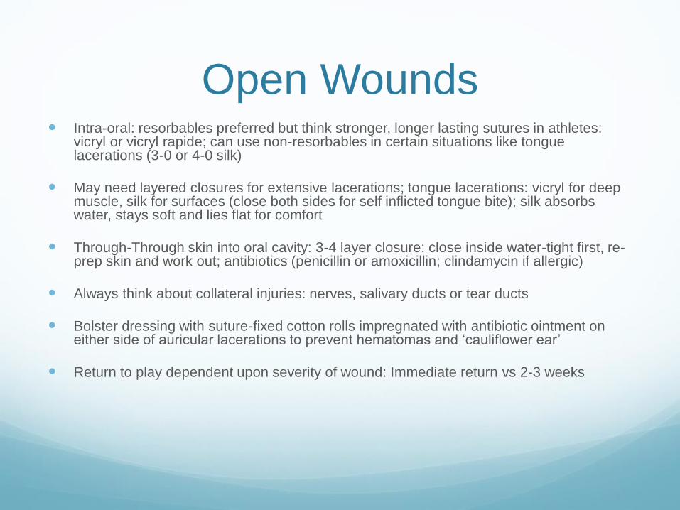
Open Wounds Intra-oral: resorbables preferred but think stronger, longer lasting sutures in athletes:
vicryl or vicryl rapide; can use non-resorbables in certain situations like tongue lacerations (3-0 or 4-0 silk)
May need layered closures for extensive lacerations; tongue lacerations: vicryl for deep muscle, silk for surfaces (close both sides for self inflicted tongue bite); silk absorbs water, stays soft and lies flat for comfort
Through-Through skin into oral cavity: 3-4 layer closure: close inside water-tight first, re-prep skin and work out; antibiotics (penicillin or amoxicillin; clindamycin if allergic)
Always think about collateral injuries: nerves, salivary ducts or tear ducts
Bolster dressing with suture-fixed cotton rolls impregnated with antibiotic ointment on either side of auricular lacerations to prevent hematomas and ‘cauliflower ear’
Return to play dependent upon severity of wound: Immediate return vs 2-3 weeks
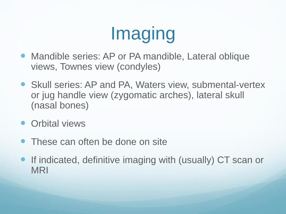
Imaging Mandible series: AP or PA mandible, Lateral oblique
views, Townes view (condyles)
Skull series: AP and PA, Waters view, submental-vertex or jug handle view (zygomatic arches), lateral skull (nasal bones)
Orbital views
These can often be done on site
If indicated, definitive imaging with (usually) CT scan or MRI
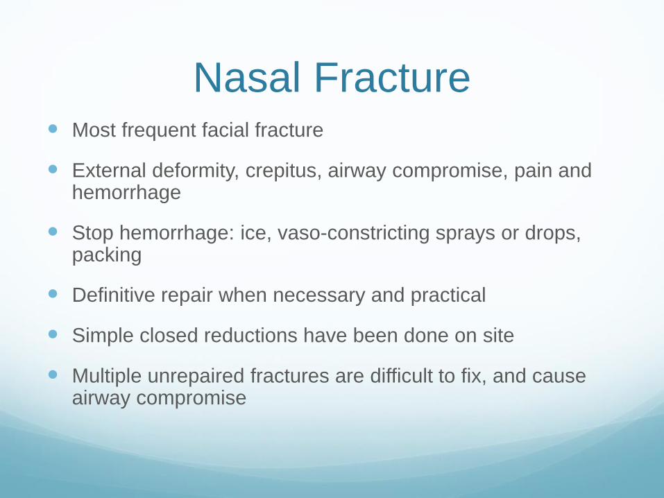
Nasal Fracture Most frequent facial fracture
External deformity, crepitus, airway compromise, pain and hemorrhage
Stop hemorrhage: ice, vaso-constricting sprays or drops, packing
Definitive repair when necessary and practical
Simple closed reductions have been done on site
Multiple unrepaired fractures are difficult to fix, and cause airway compromise
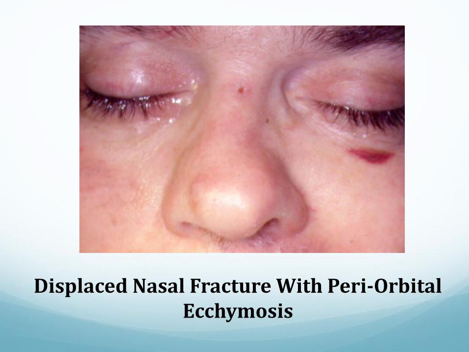
Displaced Nasal Fracture With Peri-Orbital Ecchymosis
Nasal Packing Dental roll
Rhino rocket or Nasal Doyle or Mericel (absorbent
expanding nasal tampons)
Inflatable balloons for severe bleeds (especially
posterior)
Septal hematomas must be drained to prevent
perforations
Treatment Nasal Fracture CT scan
Closed reduction of bones, closed reduction of septal
fracture if present
Intra-nasal packing for 3 days, Denver splint externally
for one week
Open reduction with plates if severe fracture
Return to play 4-6 weeks, 2-4 weeks with full face
protection
Mandibular Fractures Second most common facial fracture
‘See one fracture, look for two’
Can be associated with closed head injury because of
temporomandibular joint
Can cause airway compromise
Often occurs through impacted third molar site
Fracture sites: condylar, body/angle, symphysis,
parasymphysis, coronoid(rare)
Condylar Fracture Most common; can be unilateral or bilateral
Swelling pain crepitus, limited opening
Malocclusion with prematurity usually on fractured side
and anterior open bite
Deviation of jaw on opening toward fracture
May be open fracture with laceration of ear canal
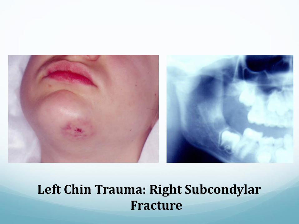
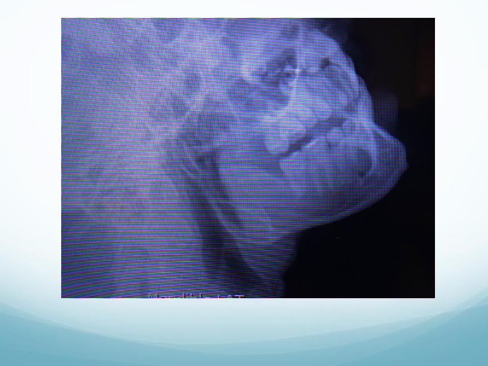
Left Chin Trauma: Right SubcondylarFracture
Body and Angle Fractures Movement and crepitus at site
Malocclusion, bleeding
Swelling and hematomas: intra and extra-oral
Pain and limited opening
Often through impacted third molars ( a good reason
for their prophylactic removal in athletes)
Symphysis or Parasymphysis
Fractures
More easily missed on x-rays
Often associated with condylar fracture
Sublingual hematoma!!
Avulsion of anterior teeth- chest x-ray if suspect
aspiration
Often a step deformity in teeth on either side of the
fracture
Bimanual palpation!!
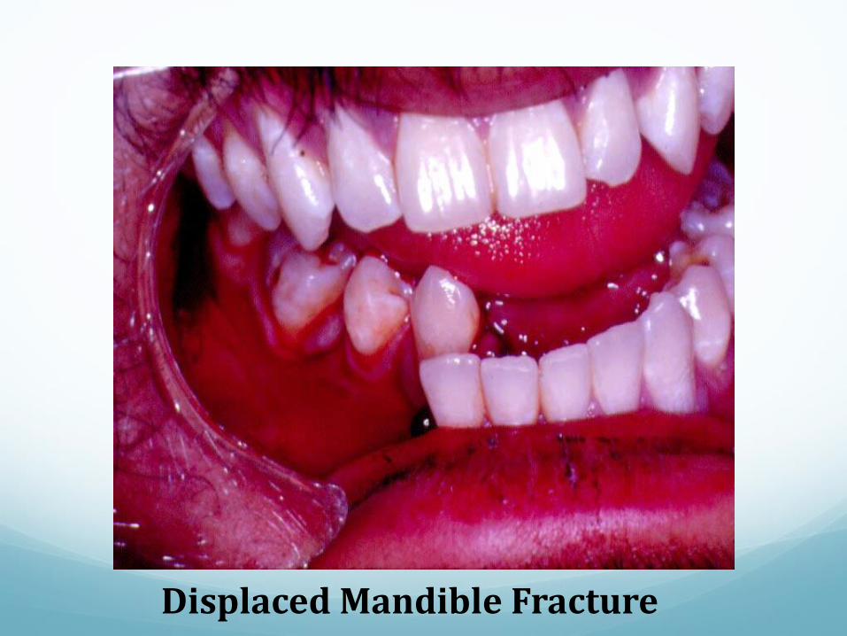
Displaced Mandible Fracture
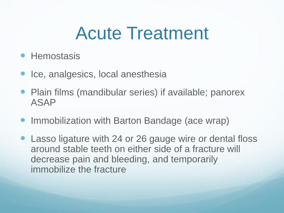
Acute Treatment Hemostasis
Ice, analgesics, local anesthesia
Plain films (mandibular series) if available; panorexASAP
Immobilization with Barton Bandage (ace wrap)
Lasso ligature with 24 or 26 gauge wire or dental floss around stable teeth on either side of a fracture will decrease pain and bleeding, and temporarily immobilize the fracture
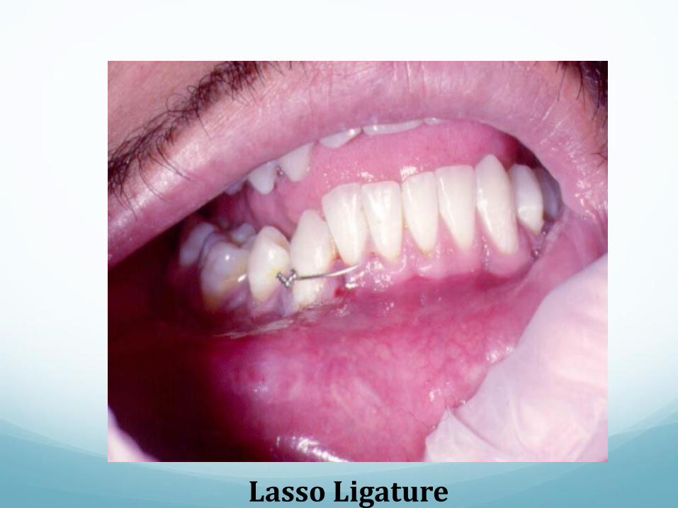
Lasso Ligature
Definitive Treatment of Jaw
Fractures CRMMF, ORIF
CRMMF: Arch bars, wires; slower return to play, liquid
diet, fixation for 4-6 weeks (less with some condylar
fractures); aerobic exercise fine, light weights possible
ORIF: Plate and screw fixation! Less MMF time
required, earlier return to solid food and play (with full
face protection)
What anatomic area of the mandible is
most likely to be fractured?
A. Angle
B. Parasymphysis
C. Condyle
D. Body
E. Coronoid
Maxillary Fractures Significant injury force injury
Malocclusion, swelling and pain
Mid-face instability: stabilize athlete’s head at forehead
or nose, grab the upper front teeth, and look for upper
jaw mobility
Bleeding, epistaxis
Mid-face elongation/shortening, or flattening
V2 paresthesia, concurrent injury
Principles of Treatment Re-establishment of proper occlusion is paramount
Anatomic reduction is secondary
Immobilize until bony union can occur
Earlier mobilization with elastic traction for condylar fractures
CRMMF (jaw wiring), ORIF (jaw wiring +plates and screws)
Out 2-6 weeks; return with facial protection for 2-4 additional weeks
Zygoma Fractures Fighting, object strike, or collision: requires significant force
Subconjunctival hemorrhage, paresthesia infra-orbital nerve, step discrepancy orbital rim or zygomatic arch
Depressed, indented cheek bone (compare both sides)
Can have altered bite
Trismus and decreased extra-ocular motion (especially upward gaze)
Plain films: Waters and Submental-Vertex views on site if available
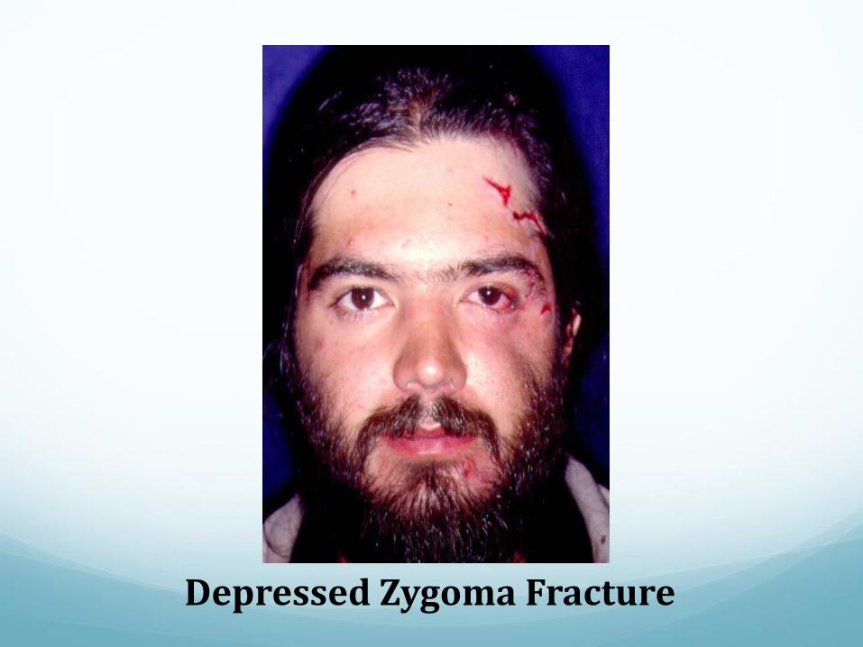
Depressed Zygoma Fracture
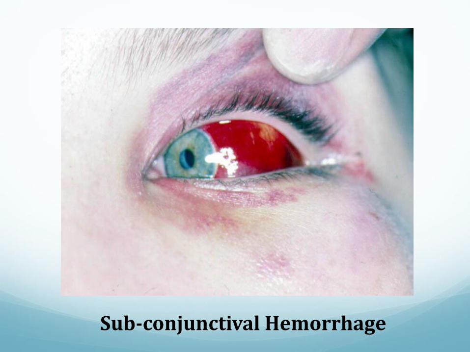
Sub-conjunctival Hemorrhage
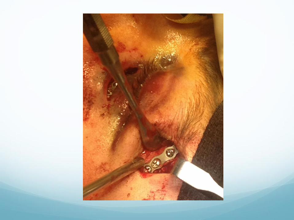
Treatment Zygoma Fractures CT scan recommended
Eye exam by Ophthalmologist before repair
ORIF with 2 or 3 point stabilization; plates at Frontal-
zygomatic suture, inferior orbital rim and possibly intra-
oral buttress
Orbital floor implant for blow-out fracture if needed
Return to play 4-6 weeks, 2-4 weeks with supplemental
full face protection if possible

Orbital Fractures Blow out fracture most common
Usually orbital floor or medial wall involved
Repair needed for functional or esthetic compromise
Often accompany zygoma fractures
Orbital rim fractures occur with more severe forces and
often accompany other facial bone fractures as well
Repair generally is required
Eye exam!!! Retinal injuries, globe trauma, hyphema
Orbital Signs Enophthalmos
Vertical and horizontal displacements: superior
(hematoma); inferior (blow out fracture); horizontal
(NOE fractures)
Diplopia, visual change
Decreased extra-ocular motion
Subconjunctival hemorrhage
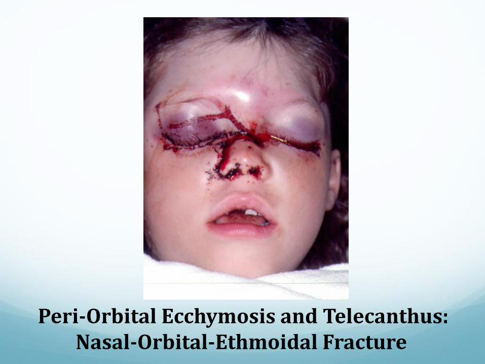
Peri-Orbital Ecchymosis and Telecanthus: Nasal-Orbital-Ethmoidal Fracture
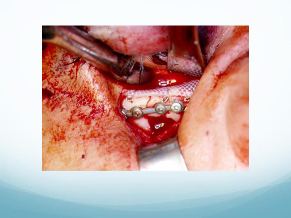
Treatment Orbital Fractures CT scan
Eye exam by ophthalmologist before repair
ORIF
Orbital floor implant if needed
Return to play 4-6 weeks, 2-4 weeks with full face
protection and normal eye exam
Subconjunctival hemorrhage after
facial trauma is suspicious for:
A. Maxillary Fracture
B. Zygoma Fracture
C. Concussion
D. Orbital Fracture
E. Both B and D
Injuries to the Teeth or
Supporting Structures Injuries to teeth may involve the tooth crown, the root,
or both
Supporting structures like alveolar bone and gingiva
are often involved
Mouthguards significantly reduce the number and
severity of dental injuries
Orthodontic appliances (braces) can add a level of
support to teeth, but without mouthguards they can
complicate soft tissue trauma
Avulsed Teeth May occur with tooth or root fracture, usually involves
alveolar process injury, and may result in alveolar
mucosal laceration
Teeth must be accounted for if possible; remember that
teeth can be swallowed or aspirated, and may even
lodge in the oral or hypo-pharynx
Monitor the event for signs of aspiration or
displacement into the pharynx or larynx (coughing,
speech changes, sore throat)
Avulsed or Subluxed Teeth In ideal situations replant immediately; if grossly contaminated
rinse tooth with water before replant; clot in socket may prevent re-implantation
If subluxed, can try to reposition manually but better for oral surgeon or dentist to do especially if tooth is intruded
If re-implant not possible but goal is to try to save tooth, place it in transport medium ( milk, saline, under tongue in adult), and get to dentist ASAP--dental x-rays are key
Don’t re-implant primary teeth
Questionable re-implant if socket damaged or if out for greater than 30 minutes
Dental implant success has shifted re-implant paradigm
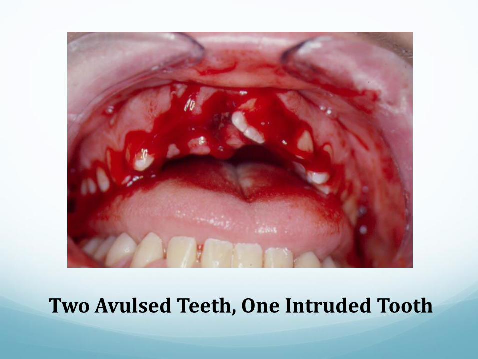
Two Avulsed Teeth, One Intruded Tooth
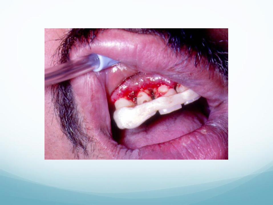
Definitive Management of
Avulsed or Subluxed Teeth Dental x-ray
Antibiotics
Splinting tooth to stable adjacent teeth with light wire or fishing line and dental composite, and left in place 2-3 weeks
Root canal therapy as needed
Or if the prognosis for the tooth is poor, extraction and dental implant
Mouthguards!!!!
Return to Play Issues with
Avulsed or Subluxed Teeth Return to play is largely dependent upon the athlete’s level
of comfort
Conditional factors may be concurrent soft tissue trauma, and relative danger of re-injury, including avulsion and aspiration
Dietary restrictions (soft chew or non-chew foods) are routinely necessary and can present nutritional challenges
Custom mouthguards made over splints may offer additional security
Added facial protection is recommended
Fractured Teeth Fractures may extend into enamel, dentin, pulp, or root
Enamel fractures are minor and usually not painful
Fractures into dentin cause pain and temperature sensitivity
Fractures exposing the pulp can cause severe pain and temperature sensitivity
Not all teeth are natural teeth: crowns, bridges, implants and dentures- be aware
Management of Fracture
Teeth Acutely: account for fragments (may be in soft tissue
wounds) and pain control: local anesthetics and analgesics
Dental x-rays
Extraction if fractured below gum line
1 year follow up and root canal therapy if tooth loses its vitality but is salvageable.
Splint if mobile
Dental restoration: bonding, veneers, or crowns as indicated
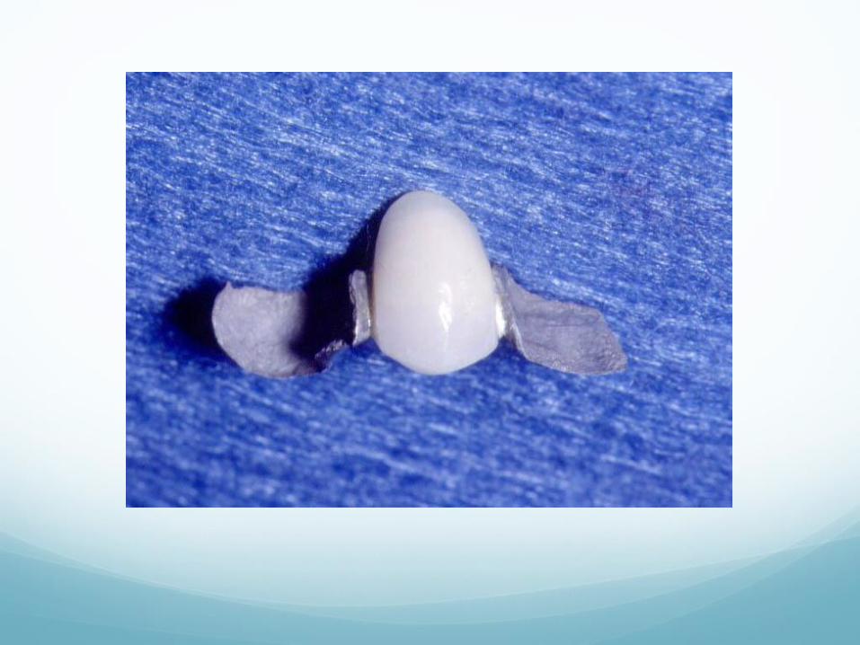
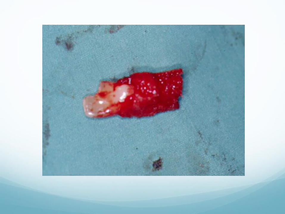
Alveolar Process Fractures Fractures of the tooth bearing portion of the upper and
lower jaws
Fracture segments may include multiple teeth
Soft tissue injury is common
Malocclusion is a common complaint so injury must be differentiated from jaw fracture
Segments may vary from minimally mobile and displaced, to grossly mobile and markedly displaced, to totally nonviable
Alveolar Process Fractures Minimally displace segment with normal occlusion can be
managed with antibiotics and soft diet
More mobile or displaced fractures must be repositioned and splinted
Repositioning can usually be done with digital manipulation under local anesthesia
Splinting is done with light cured acrylic with or without fine wire or heavy fishing line; severely displaced or larger segments are splinted with arch bars and wires; jaw wiring may be required in the most extensive cases
Nonviable segments may need to be debrided, but segments are usually splinted even if teeth are unsalvageable to try to preserve bone
Acute Treatment of Alveolar
Fractures Hemostasis, analgesics, local anesthesia
Reposition segment if possible
Soft tissue repair
Check clinically for jaw fracture
Plain radiographs if available
Definitive Treatment of
Alveolar Fractures Dental radiographs: Panorex, dental periapical films
Reposition and splint segment in proper anatomic and
occlusal relationship
Splint generally 2-4 weeks
Root canal evaluation and treatment as is needed
Return to play as is practical and comfortable, with
mouthguard and additional facial protection
Pearls for Treating Dental/ Oral
Injuries
Dental Kit: Dental rolls, Cavit (temporary filling material),
temporary cement (no super glue!)
Clove Oil
Magic Mouthwash/Viscous Lidocaine
Local anesthesia: Blocks or infiltration: marcaine 0.5%
1:200,000 epinephrine or lidocaine 2% 1:100,000
epinephrine; ½ and 1 ½ inch 25 gauge needles
Roll of 24 or 26 gauge stainless steel wire, dental floss
Obstructive Sleep Apnea 12 million people in US
1 in 25 men, 1 in 50 women; more than half are overweight
Snoring, poor sleep, daytime somnolence, accessory muscle breathing, apneic events
Apneic events can lead to reduced flow of blood to vital organs and cause irregular heart rhythms
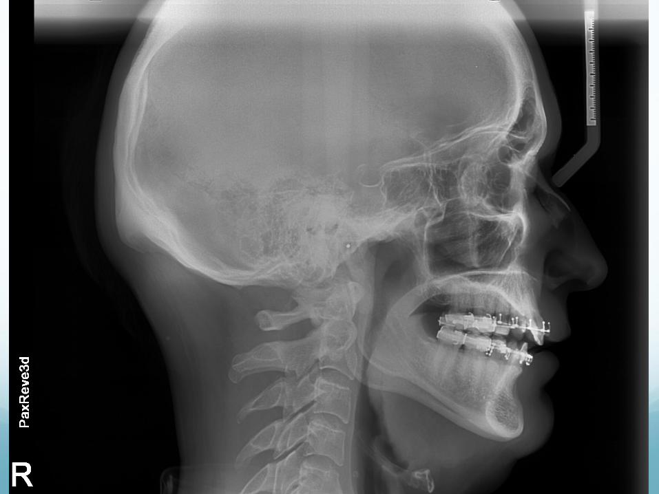
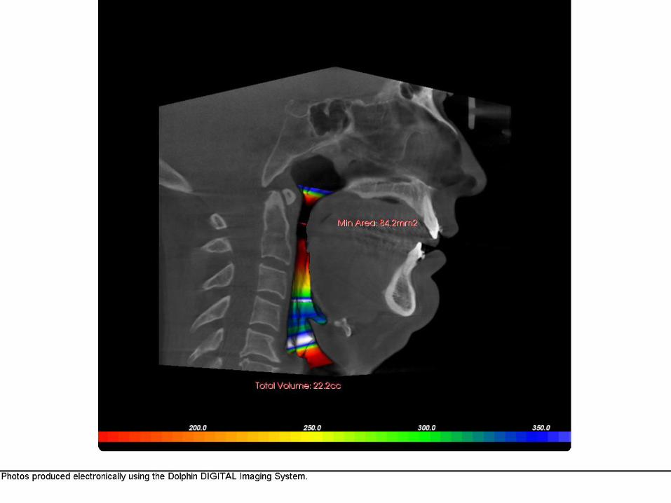
Diagnosis: physical exam, plain x-rays, 3D airway imaging
Definitive diagnosis: Sleep Study
Treatment Of OSA Mild: Weight loss, sleep position improvement with
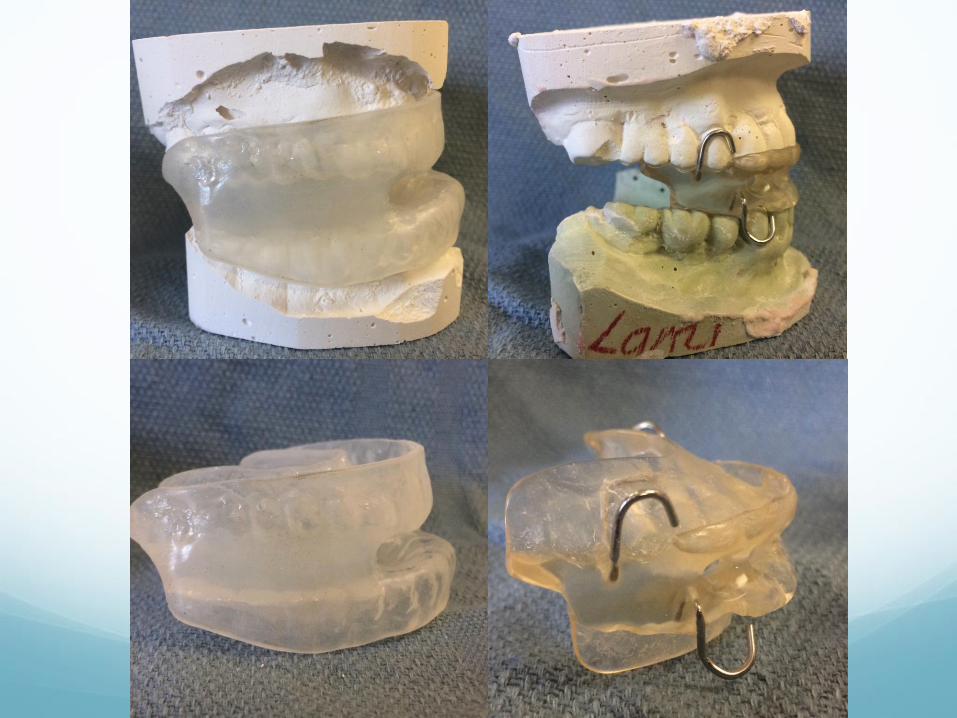
pillows or bed, avoid alcohol or sleeping pills, nasal sprays or strips, mandibular repositioning appliances
Mild to moderate: Appliances, CPAP, minor surgeries (ie., septoplasty, palatoplasty, tonsils and adenoids, chin advancement)
Severe: CPAP, minor surgeries, major surgery (Maxillo-Mandibular Advancement or MMA)
Mike Napoli
Maxillo-Mandibular
Advancement 54 yo dentist from Louisville
Snorer, fragmented sleep, daytime somnolence
6’3”, 203, otherwise healthy
Positive sleep study, decreased airway on plain film,
positive 3D volumetric airway study
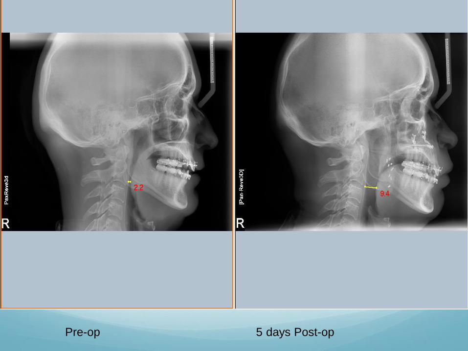
Pre-op 5 days Post-op
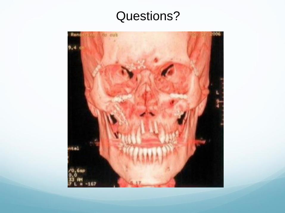
Questions?

Thank You!