Embed Size (px)
Citation preview

Hindawi Publishing CorporationJournal of Amino AcidsVolume 2012, Article ID 575180, 16 pagesdoi:10.1155/2012/575180
Research Article
Elucidation of the Rotavirus NSP4-Caveolin-1 and -CholesterolInteractions Using Synthetic Peptides
Megan E. Schroeder,1, 2 Heather A. Hostetler,3, 4 Friedhelm Schroeder,3 and Judith M. Ball1
1 Department of Veterinary Pathobiology, Texas A&M University, TVMC, College Station, TX 77843-4467, USA2 Molecular Diagnostics Texas Veterinary Medical Diagnostic Laboratory, College Station, TX 77843, USA3 Department of Pharmacology and Physiology, Texas A&M University, TVMC, College Station, TX 77843-4467, USA4 Department of Biochemistry and Molecular Biology, Boonshoft School of Medicine, Diggs 056, 3640 Colonel Glenn Hwy,Dayton, OH 45435, USA
Correspondence should be addressed to Judith M. Ball, [email protected]
Received 8 July 2011; Accepted 16 November 2011
Academic Editor: Jordi Bella
Copyright © 2012 Megan E. Schroeder et al. This is an open access article distributed under the Creative Commons AttributionLicense, which permits unrestricted use, distribution, and reproduction in any medium, provided the original work is properlycited.
Rotavirus (RV) NSP4, the first described viral enterotoxin, is a multifunctional glycoprotein that contributes to viral pathogenesis,morphogenesis, and replication. NSP4 binds both termini of caveolin-1 and is isolated from caveolae fractions that are rich inanionic phospholipids and cholesterol. These interactions indicate that cholesterol/caveolin-1 plays a role in NSP4 transport tothe cell surface, which is essential to its enterotoxic activity. Synthetic peptides were utilized to identify target(s) of interventionby exploring the NSP4-caveolin-1 and -cholesterol interactions. NSP4112−140 that overlaps the caveolin-1 binding domain anda cholesterol recognition amino acid consensus (CRAC) motif and both termini of caveolin-1 (N-caveolin-12−20, 19−40 andC-caveolin-1161−180) were synthesized. Direct fluorescence-binding assays were employed to determine binding affinities of theNSP4-caveolin-1 peptides and cholesterol. Intracellular cholesterol alteration revealed a redistribution of NSP4 and disintegrationof viroplasms. These data further imply interruption of NSP4112−140-N-caveolin-119−40 and cholesterol interactions may blockNSP4 intracellular transport, hence enterotoxicity.
1. Introduction
As the leading cause of gastroenteritis in young children un-der the age of five, rotavirus (RV) infections annually are res-ponsible for approximately 600,000 deaths worldwide [1, 2].During infection, the RV nonstructural protein 4 (NSP4)functions as a viral enterotoxin by binding an extracellularreceptor and activating a signal transduction pathway whichincreases intracellular calcium ([Ca2+]i) levels through therelease of ER calcium stores [3, 4]. This increase in [Ca2+]i
induces secretory chloride currents which result in diarrhea,but only when initiated from the exofacial leaflet of theplasma membrane (PM) [3]. The initiated increases in intra-cellular, calcium levels fail to induce the chloride secretoryresponse [4–6].
Traditionally defined as an ER glycoprotein, NSP4 con-tains a single transmembrane domain that serves to anchor
the protein into the ER membrane, such that a short N-terminal domain (amino acids [aa] 1–24) remains within thelumen of the ER while the longer C-terminus (aa 45–175)extends into the cytoplasm [7]. Interactions with numerousviral and cellular proteins occur within the extended C-terminal tail of NSP4 [8–12]. Also contained within the C-terminal cytoplasmic tail is an amphipathic α-helix (AAH),coiled-coil domain (aa 95–137) [13, 14]. Cross-linkingand crystallographic experiments reveal that this region ofNSP4 primarily oligomerizes into dimers and tetramers andcontains a cation-binding site [13–15]. Residing within theamphipathic α-helix, coiled-coil domain is the enterotoxicpeptide region (aa 114–135) as well as the caveolin-1 (cav-1)binding domain [16] and a putative cholesterol recognitionamino acid consensus (CRAC) sequence [17].
Many cholesterol binding proteins contain a CRACsequence characterized by—L/V-(X)(1−5)-Y-(X)(1−5)-R/K-,

2 Journal of Amino Acids
where (X)(1−5) represents between one and five unspecifiedresidues [17]. Numerous proteins involved in cholesteroltransport contain this sequence, including cav-1 [18]. How-ever, the presence of such a sequence does not assurean interaction with cholesterol, as this sequence is highlyvariable and likely is not the only requirement for cholesterolbinding [19]. Yet a number of viral proteins bind cholesterol,including the HIV-1 gp41 transmembrane glycoprotein [17,20], the influenza M2 protein [21], and the F1 subunit of thefusion protein of Sendai virus [22].
There is compelling evidence that the enterotoxic pep-tide, NSP4114−135, and the full-length NSP4 protein interactwith cav-1. In vivo laser scanning confocal microscopy(LSCM) colocalizations, fluorescent energy transfer (FRET)analyses, and co-immunoprecipitation data from our labo-ratory verify that NSP4 binds cav-1, the main constituentprotein of caveolae, at multiple sites within the cell, includingthe PM [23]. Yeast two-hybrid and in vitro binding assaysconfirm an interaction between NSP4 and cav-1 and map thecav-1 binding domain to NSP4114−135 [23]. Using deletionand site-directed mutagenesis, the binding site delineated tothree hydrophobic residues in the enterotoxic region indicat-ing that NSP4 and cav-1 associate via a hydrophobic interac-tion (unpublished). Additional studies reveal that the cav-1binding site for NSP4 maps, to both the N- and C-termini ofcav-1, residues 2–31 and 161–178, respectively [24].
Cav-1 is an intracellular, 21 kD protein bound by theinner leaflet of the PM in a hairpin-like structure such thatboth the N- and C-termini are oriented towards the cyto-plasm [23, 25]. Numerous signaling molecules, such as thoseinvolved in calcium signaling, are localized to caveolae andtheir function appears to be dependent on interaction withcav-1 [16, 26, 27]. Cav-1 also binds cholesterol, which hasbeen shown to be vital to caveolae biogenesis and the trans-port of cholesterol to caveolae at the PM [28–31].
Purified NSP4 and NSP4114−135 peptides interact withcaveolae-like model membranes, that is, those that have ahigh radius of curvature and are rich in anionic phospho-lipids and cholesterol [32, 33]. Upon interaction with modelmembranes, both the protein and the peptide undergo achange in secondary structure characterized by an increasein α-helix formation. Additional secondary structure studiesof other NSP4 peptides that overlap the enterotoxic peptideand cav-1 binding domain confirm that this region isimportant for membrane interactions. When caveolae areisolated from enriched PM fractions of RV-infected MDCKcells, the full-length, fully-glycosylated NSP4 is present inthe isolated caveolae fractions verifying transport of NSP4 tothe PM and caveolae [34]. It is possible that the transport ofNSP4 to caveolae occurs through an interaction with cav-1and cholesterol, although additional studies are neededto for verification. Our studies reveal the exposure of theNSP4 C-terminus and the enterotoxic region, but not the N-terminus, on the outer PM leaflet and subsequent release intoculture media at a time when the membrane is intact as verif-ied by a lack of exposure of cav-1 and other intracellularmarkers [34–36].
In this paper, we have extended these studies using syn-thetic peptides to address the following questions. (i) Does
NSP4 interact with both termini of cav-1 with equal affinity?(ii) Do the two termini of cav-1 interact with one anotherand thus facilitate binding of NSP4 to both termini? (iii)Is there a structural change when NSP4 and cav-1 peptidesinteract? (iv) Do the overlapping NSP4-cav-1 binding, CRACmotif, and enterotoxin regions interact with cholesterol?(v) Does alteration(s) of NSP4-cholesterol binding have abiological effect? To answer these questions, we employedpeptide-peptide and -cholesterol direct fluorescence-bindingassays to ascertain the interaction and determine the bindingaffinities (Kd). We also employed circular dichroism (CD) toinvestigate secondary structural changes upon interaction ofthe peptides in the absence or presence of model membranes.
2. Materials and Methods
2.1. Materials, Antibodies and Cells. Trifluoroacetic acid(TFA), 1-hydroxy-benzotriazole (HOBt), and O-Benzotria-zole-N,N,N′,N′-tetramethyluronium-hexafluoro-phosphate(HBTU) were purchased from American Bioanalytical (Nat-ick, MA). N,N-diisopropylethylamine (DIPEA) and N,N′-di-isopropylcarbodiimide (DIPCIDI) were purchased fromCreoSalus (Louisville, KY). Thioanisole, ethanedithiol, andanisole were purchased from EMD chemicals (Gibbstown,NJ). Cholesterol, Sephadex G-25, and trifluoroethanol (TFE)were purchased from Sigma (St. Louis, MO). PhospholipidsDOPS (1,2-dioleoyl-sn-glycero-3-[phosphor-L-serine]) andPOPC (1-palmitoyl-2-oleoyl-sn-glycero-3-phosphocholine)were purchased from Avanti Polar Lipids (Alabaster, AL).
Antibodies specific to the NSP4 peptide aa 150–175(NSP4150−175; deduced from the simian rotavirus SA11 NSP4sequence) were generated in rabbits by immunizing withpeptide cross-linked to keyhole limpet hemocyanin as pre-viously described [23]. Anti-NSP4150−175 was affinity puri-fied against the inoculating peptide linked to preactivatedcyanogen bromide Sepharose 4B beads according to themanufacturer (Amersham Pharmacia Biotech, Piscataway,NJ) [37]. Guinea pig anti-NSP5, a viroplasm (virus factoryin the cell) marker, was a gift from Dr. Oscar Burrone (Inter-national Center for Genetic Engineering and Biotechnology,Trieste, Italy). F(ab′)2 fragments of goat anti-guinea pigIgG-Cy3 and goat anti-rabbit IgG-Cy2 were purchased fromJackson Immuno Research (West Grove, PA).
HT29.f8 cells are a spontaneously polarizing cell line thatwas cloned from the human adenocarcinoma HT29 intesti-nal cell line [38]. Cells were maintained in Dulbecco’s Modi-fication of Eagle’s Media (DMEM) with 4.5 g/L glucose,2 mM L-glutamine, and 1 mM sodium pyruvate (Mediatech,Inc., Herndon, VA) and supplemented with 5% fetal bovinesera, 5% Serum supreme, and antibiotics (100 u/L penicillin,100 ug/L streptomycin, 0.25 ug/L amphotericin B) (Cam-brex, East Rutherford, NJ). For infection, cells were incu-bated in the absence of sera exactly as described and exposedto virus for 1 h (multiplicity of infection (MOI) = 2) [23, 35,36].
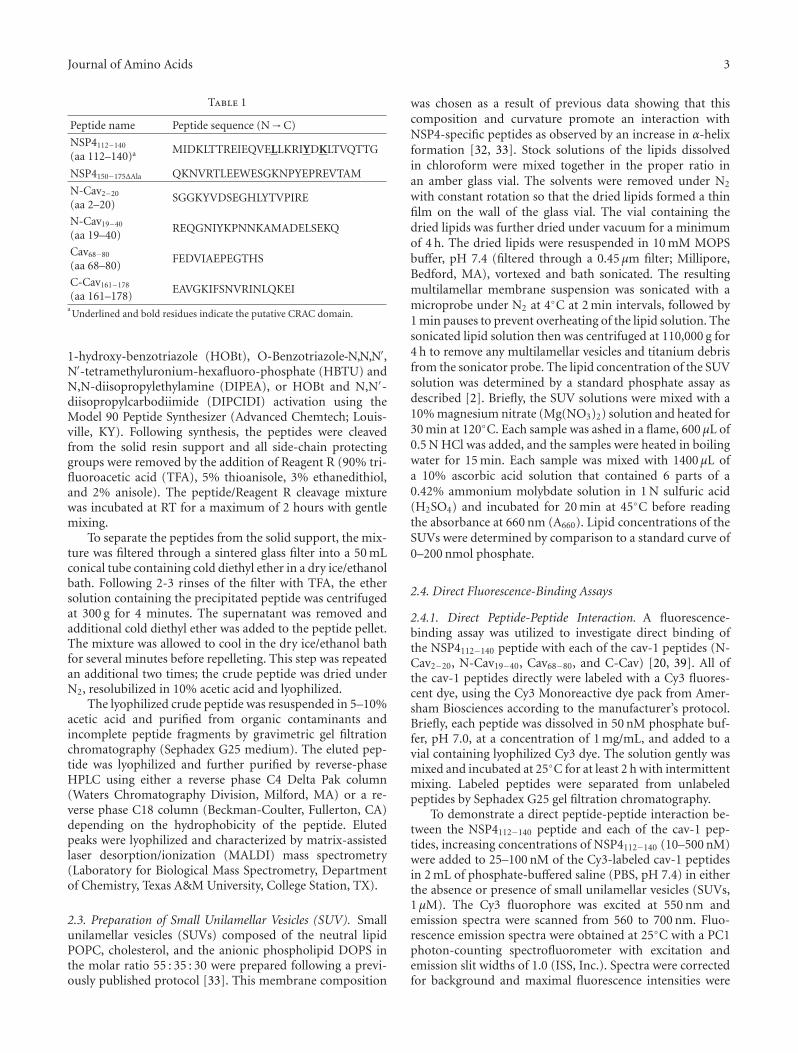
2.2. Synthesis and Purification of NSP4 and Cav-1 Peptides.All peptides (Table 1) were synthesized by fluorenylme-thoxycarbonyl (Fmoc) solid-phase chemistry with either
Journal of Amino Acids 3
Table 1
Peptide name Peptide sequence (N→C)
NSP4112−140
(aa 112–140)a MIDKLTTREIEQVELLKRIYDKLTVQTTG
NSP4150−175ΔAla QKNVRTLEEWESGKNPYEPREVTAM
N-Cav2−20
(aa 2–20)SGGKYVDSEGHLYTVPIRE
N-Cav19−40
(aa 19–40)REQGNIYKPNNKAMADELSEKQ
Cav68−80
(aa 68–80)FEDVIAEPEGTHS
C-Cav161−178
(aa 161–178)EAVGKIFSNVRINLQKEI
aUnderlined and bold residues indicate the putative CRAC domain.
1-hydroxy-benzotriazole (HOBt), O-Benzotriazole-N,N,N′,N′-tetramethyluronium-hexafluoro-phosphate (HBTU) andN,N-diisopropylethylamine (DIPEA), or HOBt and N,N′-diisopropylcarbodiimide (DIPCIDI) activation using theModel 90 Peptide Synthesizer (Advanced Chemtech; Louis-ville, KY). Following synthesis, the peptides were cleavedfrom the solid resin support and all side-chain protectinggroups were removed by the addition of Reagent R (90% tri-fluoroacetic acid (TFA), 5% thioanisole, 3% ethanedithiol,and 2% anisole). The peptide/Reagent R cleavage mixturewas incubated at RT for a maximum of 2 hours with gentlemixing.
To separate the peptides from the solid support, the mix-ture was filtered through a sintered glass filter into a 50 mLconical tube containing cold diethyl ether in a dry ice/ethanolbath. Following 2-3 rinses of the filter with TFA, the ethersolution containing the precipitated peptide was centrifugedat 300 g for 4 minutes. The supernatant was removed andadditional cold diethyl ether was added to the peptide pellet.The mixture was allowed to cool in the dry ice/ethanol bathfor several minutes before repelleting. This step was repeatedan additional two times; the crude peptide was dried underN2, resolubilized in 10% acetic acid and lyophilized.
The lyophilized crude peptide was resuspended in 5–10%acetic acid and purified from organic contaminants andincomplete peptide fragments by gravimetric gel filtrationchromatography (Sephadex G25 medium). The eluted pep-tide was lyophilized and further purified by reverse-phaseHPLC using either a reverse phase C4 Delta Pak column(Waters Chromatography Division, Milford, MA) or a re-verse phase C18 column (Beckman-Coulter, Fullerton, CA)depending on the hydrophobicity of the peptide. Elutedpeaks were lyophilized and characterized by matrix-assistedlaser desorption/ionization (MALDI) mass spectrometry(Laboratory for Biological Mass Spectrometry, Departmentof Chemistry, Texas A&M University, College Station, TX).
2.3. Preparation of Small Unilamellar Vesicles (SUV). Smallunilamellar vesicles (SUVs) composed of the neutral lipidPOPC, cholesterol, and the anionic phospholipid DOPS inthe molar ratio 55 : 35 : 30 were prepared following a previ-ously published protocol [33]. This membrane composition
was chosen as a result of previous data showing that thiscomposition and curvature promote an interaction withNSP4-specific peptides as observed by an increase in α-helixformation [32, 33]. Stock solutions of the lipids dissolvedin chloroform were mixed together in the proper ratio inan amber glass vial. The solvents were removed under N2
with constant rotation so that the dried lipids formed a thinfilm on the wall of the glass vial. The vial containing thedried lipids was further dried under vacuum for a minimumof 4 h. The dried lipids were resuspended in 10 mM MOPSbuffer, pH 7.4 (filtered through a 0.45 μm filter; Millipore,Bedford, MA), vortexed and bath sonicated. The resultingmultilamellar membrane suspension was sonicated with amicroprobe under N2 at 4◦C at 2 min intervals, followed by1 min pauses to prevent overheating of the lipid solution. Thesonicated lipid solution then was centrifuged at 110,000 g for4 h to remove any multilamellar vesicles and titanium debrisfrom the sonicator probe. The lipid concentration of the SUVsolution was determined by a standard phosphate assay asdescribed [2]. Briefly, the SUV solutions were mixed with a10% magnesium nitrate (Mg(NO3)2) solution and heated for30 min at 120◦C. Each sample was ashed in a flame, 600 μL of0.5 N HCl was added, and the samples were heated in boilingwater for 15 min. Each sample was mixed with 1400 μL ofa 10% ascorbic acid solution that contained 6 parts of a0.42% ammonium molybdate solution in 1 N sulfuric acid(H2SO4) and incubated for 20 min at 45◦C before readingthe absorbance at 660 nm (A660). Lipid concentrations of theSUVs were determined by comparison to a standard curve of0–200 nmol phosphate.
2.4. Direct Fluorescence-Binding Assays
2.4.1. Direct Peptide-Peptide Interaction. A fluorescence-binding assay was utilized to investigate direct binding ofthe NSP4112−140 peptide with each of the cav-1 peptides (N-Cav2−20, N-Cav19−40, Cav68−80, and C-Cav) [20, 39]. All ofthe cav-1 peptides directly were labeled with a Cy3 fluores-cent dye, using the Cy3 Monoreactive dye pack from Amer-sham Biosciences according to the manufacturer’s protocol.Briefly, each peptide was dissolved in 50 nM phosphate buf-fer, pH 7.0, at a concentration of 1 mg/mL, and added to avial containing lyophilized Cy3 dye. The solution gently wasmixed and incubated at 25◦C for at least 2 h with intermittentmixing. Labeled peptides were separated from unlabeledpeptides by Sephadex G25 gel filtration chromatography.
To demonstrate a direct peptide-peptide interaction be-tween the NSP4112−140 peptide and each of the cav-1 pep-tides, increasing concentrations of NSP4112−140 (10–500 nM)were added to 25–100 nM of the Cy3-labeled cav-1 peptidesin 2 mL of phosphate-buffered saline (PBS, pH 7.4) in eitherthe absence or presence of small unilamellar vesicles (SUVs,1 μM). The Cy3 fluorophore was excited at 550 nm andemission spectra were scanned from 560 to 700 nm. Fluo-rescence emission spectra were obtained at 25◦C with a PC1photon-counting spectrofluorometer with excitation andemission slit widths of 1.0 (ISS, Inc.). Spectra were correctedfor background and maximal fluorescence intensities were

4 Journal of Amino Acids
recorded. Calculation of the dissociation constants (Kd) wasperformed from the titration curves plotted as quenching inCy3-peptide fluorescence intensity (F0 − F, where F0 and Frepresented the Cy3-cav-1 peptide fluorescence intensities inthe absence and presence of NSP4112−140 or cav-1 peptide,resp., at each titration point) as a function of peptideconcentration. The Kd also was obtained by a reciprocal plotof 1/(1 − F/Fmax) and CL/F/Fmax according to the followingequation: y = bx + y0, where F is the fluorescence intensityat a given concentration of ligand, Fmax is the maximalfluorescence obtained, and CL is the ligand concentration.The slope of the line (b) is equal to 1/Kd.
2.4.2. Cholesterol-Peptide Interaction. Similarly, theNSP4112−140-cholesterol interaction and binding affinitywere determined by a direct fluorescent binding assay inwhich the peptide was labeled with a Cy5 fluorophoreusing the Cy5 Monoreactive dye pack from Amersham Bio-sciences (Piscataway, NJ). A concentrated cholesterol stocksolution (20 uM) was prepared by dissolving the cholesterolin ethanol. To analyze a binding interaction betweenNSP4112−140 and cholesterol, an increasing quantity of chole-sterol (5–35 nM) was added to 10 nM of the Cy5-labeledNSP4112−140 in 2 mL of PBS (pH 7.4). The Cy5 fluorophorewas excited at a wavelength of 649 nm, and the emission spec-tra were scanned from 655 to 720 nm using a PCI photon-counting spectrofluorometer (ISS, Inc., Champaign, IL). Flu-orescence emission spectra were obtained at 25◦C with exci-tation and emission slit widths of 1.0. As with the peptide-peptide interactions, spectra were corrected for backgroundand maximal fluorescence intensities were measured. Calcu-lation of the Kd was determined from the plotted titrationcurves whereby the Cy5-peptide fluorescence intensity wasquenched and measured as a function of cholesterol concen-tration (F0 − F, where F0 and F represented the Cy5-NSP4112−140 fluorescence intensities in the absence and pre-sence of cholesterol, resp., at each titration point). The sig-moidal curves also were fitted to a Hill plot according to thefollowing equation: y = axb/(cb + xb), where y and x cor-respond to (F0 − F) and the ligand concentration at eachpoint, while a, b, and c represent the maximum binding(Bmax), the number of binding sites (n), and the Kd value,respectively. Kd values were calculated with the sigmoidalfunction of Sigma Plot (SPSS, Chicago, IL) utilizing the Hillplot feature.
2.5. Circular Dichroism (CD) and Secondary Structure Estima-tions. The secondary structures of the NSP4 and cav-1 speci-fic peptides were determined by circular dichroism followinga previously published protocol [32, 33]. Briefly, each peptide(15–35 μM) was suspended in 10 mM potassium phosphatebuffer, pH 7.4, or 50% trifluoroethanol (TFE) to promote ahydrophobic environment and folding of the peptide, in thepresence or absence of lipid vesicles (1 mM). Samples con-taining buffer or SUVs without peptide were used for back-ground correction. Peptide concentrations were determinedby amino acid analysis (Protein Chemistry Laboratory,Department of Biochemistry, Texas A&M University, College
Station, TX). CD spectra were obtained in a 1 mm circularquartz cell using a Model J-710 JASCO spectropolarimeter(JASCO, Easton, MD) or in a 1 mm rectangular quartz cell ina Model 202 Aviv spectrometer (Aviv Biomedical, Lakewood,NJ). The spectra for each of the peptides were recorded from185 nm to 260 nm, with a step resolution of 1 nm, speedof 50 nm/min, response of 1 sec, bandwidth of 2.0 nm, andsensitivity of 10 mdeg. Data were averaged from 5 scans,background subtracted, smoothed, and converted into meanresidue molar ellipticity [θ] (deg cm2/dmol).
To determine the percent α-helix for each of the NSP4-and cav-1 specific peptides, the following equation was used:θ222 = ( fh − ικ/N)[θh 222∞] [12, 21, 22]. In this equation,θ222 is the mean residue molar ellipticity at 222 nm, fh is thefraction in α-helical form, ι is the number of helices, κ is awavelength-specific constant with a value of 2.6 at 222 nm,N is the number of residues in the peptide, and θh 222∞ is themolar ellipticity for a helix of infinite length at 222 nm, thatis, −39,500 deg cm2/dmol.
2.6. Treatment of RV-Infected MDCK and -HT29.f8 Cells withCholesterol-Altering Drugs. MDCK cells were infected withRV at a multiplicity of infection (MOI) of 2 and incubatedfor 12 hpi. The cells were washed and treated in triplicatewith the cholesterol altering drugs, fillipin [40], and nystatin[41] exactly as described. Chlorpromazine [40, 42] and noco-dazole [39] were utilized as negative and positive controls,respectively. Cells were fixed with methanol : acetone (1 : 1)at −20◦C for 10 min, blocked with 5% dry milk for 20 min,incubated with anti-NSP4150−175 and anti-rabbit-IgG-FITC,and examined under a UV microscope (Zeiss). The cellsurface carefully was observed and the presence or absenceof fluorescence carefully noted. Comparisons also were madeto untreated cells. Primary and secondary antibody controlswere negative. To ensure the lack of drug toxicity, a separateset of cells were treated and then stained with trypan blue.
2.7. Direct Fluorescence-Binding Assays
2.7.1. NSP4112−140 Binds the N-Terminus of cav-1 (Cav19−40)with Greater Affinity Than the C-Terminus. We previouslydemonstrated that NSP4114−135 interacts with both the N-and C-termini of cav-1 using yeast-2-hybrid and pull-downassays, which is unusual [24]. To further investigate this find-ing and determine if there was a preferential binding ofNSP4 to the N- or C-terminus of cav-1, the NSP4112−140
peptide that encompasses aa 114–135 and various cav-1 pep-tides (N-Cav2−20, N-Cav19−40, and C-Cav161−178) were uti-lized in a direct fluorescence-binding assay.
Upon titration with increasing concentrations ofNSP4112−140, each of the Cy-3-labled N- or C-terminal cav-1peptides showed an increase in fluorescence intensity at565 nm, indicative of an interaction. Plots of NSP4112−140
concentrations versus maximum fluorescence intensitiesrevealed saturated binding curves for each of the cav-1 pep-tides (Figure 1). Linear reciprocal plots were used tocalculate the Kd for each peptide-peptide pair (Table 2).The calculated Kd values indicated that NSP4112−140

Journal of Amino Acids 5
Wavelength (nm)
560 580 600 620 640 660 680 700
Rel
ativ
e fl
uor
esce
nce
inte
nsi
ty (
a.u
.)
0
1
2
3
4
5
12
3
4
567
×104
(a)
Rel
ativ
e fl
uor
esce
nce
inte
nsi
ty (
a.u
.)
Wavelength (nm)
560 580 600 620 640 660 680 700
0
2
4
6
8
×104
(b)
0 100 200 300 400 500
0
1
2
3
4
5
6
200 3000
30
60
×103
F−F
0(a
.u.)
1/(1−F/F
max
)
NSP4112–140 (nM)
(112–140)/F/Fmax
(c)
0 100 200 300
0
500
1000
1500
2000
2500
100 2000
10
20
F−F
0(a
.u.)
1/(1−F/F
max
)
NSP4112–140 (nM)
(112–140)/F/Fmax
(d)
0 200 400 600
0
1
2
3
400 5000
20
×104
F−F
0(a
.u.)
1/(1−F/F
max
)
NSP4112–140 (nM)
(112–140)/F/Fmax
(e)
Figure 1: Direct Fluorescence-Binding Assays of NSP4112−140 with Caveolin-1 Peptides. (a) Fluorescence spectra of Cy3-C-Cav161−178 titratedwith increasing concentrations of NSP4112−140. Spectrum 1 : 100 nM Cy3-C-Cav161−178 in buffer only. Spectra 2–11 : 100 nM Cy3-C-Cav161−178
in the presence of 10, 100, 150, 200, 250, 300, 350, 400, 450, and 500 nM of NSP4112−140, respectively. (b) Fluorescence spectra of Cy3-C-Cav161−178 titrated with increasing concentrations of NSP4150−175 (negative control). Plots (c)–(e) show maximal fluorescence emission(measured at 565 nm upon excitation at 550 nm) for NSP4112−140 in the presence of 25 nM Cy3-N-Cav2−20 (c), 50 nM Cy3-N- Cav19−40 (d),and 100 nM Cy3-C-Cav161−178. (e). Insets: linear plots of the binding curve of NSP4112−140.

6 Journal of Amino Acids
Table 2: Binding affinities (Kd) for NSP4112−140 + caveolin-1peptides in the absence and presence of SUV model membranesa.
SampleKd (nM) in
absence of SUVKd (nM) in
presence of SUV
Cy3-N-Cav2−20 +NSP4112−140
85± 6 26± 4
Cy3-N-Cav19−40 +NSP4112−140
40± 10 37± 7
Cy3-C-Cav161−178+NSP4112−140
217± 35 172± 22
aThe Kd values were calculated as outlined in Materials and Methods and
are presented as means ± SD, n = 4.
bound the N-Cav19−40 peptide with the strongest affinity(40 ± 10 nM), while it bound the C-Cav161−178 peptide withthe weakest affinity (217 ± 35 nM). The N-Cav2−20 peptidebound with intermediate affinity (85 ± 6 nM). None of thecav-1 peptides bound to the control peptide correspondingto the C-terminus of NSP4 (NSP4150−175ΔAla), which isknown to not bind cav-1 [23]. It is unusual for a proteinto interact with both termini of cav-1. We now show thatthere is a preferential interaction of NSP4112−140 with theN-terminus of cav-1, which, to our knowledge, is the firstreport of a preferential binding to one termini of cav-1.
Previous CD analyses of NSP4 peptides correspondingto the cav-1 binding domain show that aa 114–135 interactswith model membranes (SUVs) [32, 33]. Therefore, NSP4-cav-1 peptide-peptide interactions also were investigated inthe presence of the same SUV membranes. The SUVs weremixed with each of the Cy3-labeled cav-1 peptides in PBS andthen titrated with increasing concentrations of NSP4112−140
(Figure 2).The presence of lipid vesicles had no effect on the inter-
action of NSP4112−140 with either N-Cav19−40 or C-Cav161−178
(Figures 2(b) and 2(c)). Binding affinities in the presence ofmembranes were similar to those calculated in the absenceof membranes (Table 2). However, the presence of the SUVsresulted in an increased affinity between NSP4112−140 andN-Cav2−20. The calculated Kd value was determined to be26±4 nM, an approximate 3-fold increase over that observedin the absence of membranes (85 ± 6 nM) and about a1.5-fold increase over the Kd of N-Cav19−40 in the absence ofmembranes (40 ± 10 nM). These results strongly suggestthat the presence of lipid vesicles enhances binding betweenNSP4112−140 and the extreme N-terminus of cav-1 (aa 2-20),but not the adjacent N-terminal cav-1 peptide, N-Cav19−40.
2.7.2. The N-terminal cav-1 Peptides (aa 2–20 and 19–40)Failed to Bind the cav-1 C-Terminal Peptide (aa 161–178). Toevaluate the mechanism of the interaction of NSP4 with bothcav-1 termini, we examined whether the two cav-1 terminibound one another to facilitate the interaction with NSP4 byemploying the fluorescence-binding assay. C-Cav161−178 waslabeled with a Cy3-fluorophore and titrated with either theN-Cav2−20 or N-Cav19−40 peptide. Titration of C-Cav161−178
with increasing concentrations of either N-terminal peptideresulted in no change in fluorescence intensity at 565 nm,
Table 3: Percent α-helixa of NSP4112−140and Caveolin-1 Peptides inAqueous Buffer, 50% TFE and in the Presence of 1 mM SUV.
Peptide Aqueous Buffer 50% TFE 1 mM SUV
NSP4112−140 30.6± 2.4% 79.6± 4.6% 57.9± 1.6%
N-Cav2−20 18.9± 2.8% 16.5± 0.9% 18.5± 1.9%
Cav19−40 20.7± 0.6% 33.4± 0.5% 20.5± 2.7%
Cav68−80 23.4± 0.9% 24.4± 0.8% 21.4± 2.0%
C-Cav161−178 43.4± 3.8% 57.4± 4.4% 42.5± 10.6%aPercent α-helix for each peptide was calculated as described in Materials
and Methods. Data is presented as mean ± SD, n = 4.
indicative of a lack of association (Figure 3). Hence, wepropose that the N- and C- termini of cav-1 do not bindone another and the interaction between NSP4 and the cav-1 termini does not result from an initial binding betweenthe cav-1 N- and C-termini. To our knowledge, this is thefirst report of the cav-1 termini not interacting. Additionalstudies are needed to dissect the mechanism and implicationsof both cav-1 termini binding NSP4.
2.8. Circular Dichroism Analysis
2.8.1. Secondary Structure of NSP4112−140. The structure ofthe NSP4112−140 peptide was first determined in an aqueousbuffer (10 mM potassium phosphate buffer, pH = 7.4)(Figure 4, dark circles). The CD spectrum showed doubleminima at 208 and 222 nm and a single maximum peak at190 nm, indicative of some α-helical secondary structure.Using the molar ellipticity value at 222 nm [43], the α-helicalcontent of the NSP4112−140 peptide was calculated to be30.6± 2.4%.
To enhance innate peptide structure, the secondarystructure of NSP4112−140 was determined in 50% TFE, ahydrophobic solvent that promotes intramolecular hydrogenbonding (Figure 4, open circles). The CD spectrum showeda dramatic increase in α-helix formation of the peptide,as evidenced by an increase in both the negative molarellipticity at 208 and 222 nm and the positive molar ellipticityat 190 nm. The α-helical content was calculated using themolar ellipticity value at 222 nm and determined to be 79.6±4.6% (Table 3).
Previous CD experiments with the enterotoxic peptide,NSP4114−135 demonstrate that the α-helical content increasesin the presence of SUVs containing POPC, cholesterol, andDOPS at a 55 : 35 : 10 molar ratio [32, 33]. To determine thesecondary structural changes that occur in the presence ofmembranes, NSP4112−140 was mixed with SUV membranesand changes in α-helical content were noted by CD.
The CD spectrum of the NSP4112−140 peptide in thepresence of 1 mM SUV showed nearly a 2-fold increase inα-helix formation over that in aqueous buffer, as evidencedby the increase in both the negative molar ellipticity as 208and 222 nm and the positive molar ellipticity at 190 nm(Figure 5). The α-helical content was calculated using themolar ellipticity value at 222 nm and determined to be57.9± 1.6% (Table 3).

Journal of Amino Acids 7
0 100 200 300
0
200
400
600
800
1000
1200
1400
0 100 200 3000
5
10
15
F−F
0(a
.u.)
1/(1−F/F
max
)
NSP4112–140 (nM)
(112–140)/F/Fmax
(a)
0 100 200 300
0
200
400
600
800
100 200 300
01020304050
F−F
0(a
.u.)
1/(1−F/F
max
)
NSP4112–140 (nM)
(112–140)/F/Fmax
(b)
0 200 400 600
0
2
4
6
8
250 300 3500
2
4
6
8
F−F
0(a
.u.)
1/(1−F/F
max
)
NSP4112–140 (nM)
(112–140)/F/Fmax
×103
(c)
Figure 2: Direct Fluorescence-Binding Assays of NSP4112−140 with Caveolin-1 Peptides in the presence of 1 μM SUV model membranes.Direct binding based on dequenching of Cy3 fluorescence emission. (a–c) Plots of maximal fluorescence emission (measured at 565 nmupon excitation at 550 nm) for NSP4112−140 in the presence of 1 uM SUV and either 25 nM Cy3-N-Cav2−20 (a), 50 nM Cy3-Cav19−40 (b), or100 nM Cy3-C-Cav161−178 (c). Insets: linear plots of the binding curve of NSP4112−140.
2.8.2. Secondary Structure of Caveolin-1 Peptides. The sec-ondary structures of the cav-1 peptides were determinedand evaluated in the presence of 50% TFE and 1 mM SUVs(55 : 35 : 10; POPC : Cholesterol : DOPS) by CD (Table 3).In aqueous buffer, the α-helical content of N-Cav2−20, N-Cav19−40, and Cav68−80 was quite similar, at 18.9%, 20.7%,and 23.4%, respectively (Figures 6(a)–6(c), dark circles). Theα-helical contents of the C-terminal peptide (C-Cav161−178)was approximately twice (43.4%) that of the N-terminalpeptides (Table 3). When placed in 50% TFE, the α-helical content of N-Cav2−20, N-Cav19−40, Cav68−80, and C-Cav161−178 peptides were calculated to be 16.5%, 33.4%,23.1%, and 67.1% respectively (Figure 6, open circles). OnlyN-Cav19−40 and C-Cav161−178 showed an increase in α-helixformation in the presence of 50% TFE.
When mixed with the SUVs, none of the cav-1 peptidesdemonstrated a change in α-helix formation over that seenin aqueous buffer (Figure 6, dark triangles). This result was
anticipated as none of these peptides corresponded to a cav-1membrane interacting or transmembrane domain.
2.8.3. Secondary Structural Changes Observed upon Mix-ing NSP4112−140 with C-Cav161−178. Analyses by directfluorescence-binding assays revealed that N-Cav19−40 boundNSP4112−140 with a stronger affinity than C-Cav161−178. Wetherefore investigated whether structural changes occurredupon the association of the NSP4 and cav-1 peptides, in par-ticular with the amino terminus. Each of the cav-1 peptides(N-Cav2−20, N-Cav19−40, Cav68−80, and C-Cav161−178) wasmixed with the NSP4112−140 peptide and was analyzed by CD.Cav68−80 was used as a negative control, as previous studieshave shown that NSP4 does not interact with this regionof cav-1 [23, 24]. Analyses of the mixing experiments wereachieved by comparing the spectrum of each peptide-peptidemixture (observed) with the sum of the individual spectra

8 Journal of Amino Acids
Wavelength (nm)
560 580 600 620 640 660 680 700
Rel
ativ
e fl
uor
esce
nce
inte
nsi
ty (
a.u
.)
0
1
2
3
4
5
×103
(a)
0 200 400 600
1 2 3 40
2
4
6
8
10
N-Cav2–20 (nM)
1/(1−F/F
max
)
0
200
400
600
800
1000
F0−F
(a.u
.)
(N-Cav2–20)/F/Fmax
(b)
0 200 400 600
0
200
400
600
800
1000
100 200 3000
2
4
6
8
10
Cav19–40 (nM)
(Cav19–40)/F/Fmax
1/(1−F/F
max
)
F0−F
(a.u
.)
(c)
Figure 3: Direct Fluorescence-Binding Assays of N-Cav2−20 or N-Cav19−40 Peptide with C-Cav161−178 Peptide. (a) Fluorescence spectra ofCy3-C-Cav161−178 titrated with increasing concentrations of Cav19−40 (0–500 nM). (b-c) Plots of maximal fluorescence emission (measuredat 565 nm upon excitation at 550 nm) for (b) N-Cav2−20 and (c) N-Cav19−40 in the presence of 100 nM Cy3-C-Cav161−178. Insets: linear plotsof the binding curve of (b) N-Cav2−20 and (c) N-Cav19−40.
of each peptide (theoretical). The α-helical content wascalculated and significant differences (Student’s t-test, P <0.05) between the observed and theoretical values indicateda change in secondary structure and binding between the twopeptides. However, a lack of secondary structural alterationsupon mixing the peptides did not negate binding betweenthe NSP4112−140 and the cav-1 peptide being tested. Whiledirect fluorescent binding assays revealed the affinity of eachof the peptide-peptide interactions, the CD experimentsdisclosed the extent to which the interaction(s) caused achange in the secondary structure.
When each of the cav-1 peptides was mixed withNSP4112−140 in an aqueous buffer, only the NSP4112−140-C-Cav161−178 mixture showed a significant difference betweenthe observed and theoretical values (25.0 ± 1.7% versus35.3 ± 1.6% α-helix) (Figure 7(d); Table 4). No change insecondary structure was observed for the NSP4112−140-N-Cav2−20 (21.7±0.8% versus 23.6±2.6%), the NSP4112−140-N-Cav19−40 (23.8±1.2% versus 24.4±0.6%), or the NSP4112−140-Cav68−80 negative control (24.2 ± 0.3% versus 23.9 ± 0.7%)
mixtures even though N-Cav19−40 had the lowest Kd (Figures7(a)–7(c); Table 4). Similarly, when each peptide-peptidepair was placed in 50% TFE, only the NSP4112−140-C-Cav161−178 mixture showed a significant change in secondarystructure (44.2± 10.6% versus 67.8± 3.2%) (Table 4). Thesedata confirmed the weak binding between NSP4112−140 andC-Cav161−178 and verified that secondary structure alter-ations do not always occur upon an interaction.
2.9. Effect of SUV Model Membranes on the NSP4112−140-Caveolin-1 Peptide-Peptide Interactions. Cav-1 is critical toboth the structure and function of caveolae, is the majorconstituent of caveolae, and is controller of caveolae for-mation [44]. NSP4 is present in isolated caveolae from RV-infected cells [34], and together with NSP4114−135, prefer-entially interacts with caveolae-like model membranes [32].Therefore we evaluated the extent the SUVs influencedsecondary structure alterations of the NSP4-cav-1 peptide-peptide interactions. Each NSP4112−140-cav-1 peptide pairwas monitored by CD (Figure 8).

Journal of Amino Acids 9
Table 4: Observed and theoretical % helical content of NSP4112−140-caveolin-1 peptide-peptide interactions.
N-Cav2−20 Cav19−40 Cav68−80 C-Cav161−178
Observed Theoretical Observed Theoretical Observed Theoretical Observed Theoretical
Aqueous buffer 21.7± 0.8 23.6± 2.6 23.8± 1.2 24.4± 0.6 24.2± 0.3 23.9± 0.7 25.0± 1.7∗ 35.3± 1.6
50% TFE 46.3± 7.2 47.6± 1.8 58.7± 7.7 56.3± 2.3 45.9± 2.8 49.8± 1.8 44.2± 10.6∗ 67.8± 3.2
1 mM SUV 34.7± 6.0 37.7± 1.9 37.7± 3.6 39.0± 2.3 31.3± 0.2∗ 37.5± 1.9 51.9± 2.4 49.5± 6.2
NSP4112−140-Caveolin-1 Peptide-Peptide Interactions: Percent α-helical content of NSP4112−140 peptide + Caveolin-1 peptides, in aqueous buffer (10 mMpotassium phosphate buffer), 50% trifluoroethanol (TFE) and in the presence of 1 mM SUV (55 : 35 : 10, POPC/cholesterol/DOPS). Observed values werecompared with theoretical values, and those that were statistically significant by a Student’s t-test (P < 0.05; denoted by an asterisk) were consideredstructurally altered, indicative of an interaction. Results are presented as means ± S.D. (n = 3 or 4).
Wavelength (nm)
200 220 240 260
0
2
4
6
8
Buffer50% TFE
Mol
ar e
llipt
icit
y (d
eg c
m2
dmol−1
)
×104
−2
−4
Figure 4: CD spectra of NSP4112−140 in aqueous buffer (•) and 50%TFE (◦).
Wavelength (nm)
200 220 240 260
0
2
4
6
BufferSUV (55 : 35 : 10)
−2
−4
Mol
ar e
llipt
icit
y (d
eg c
m2
dmol−1
)
×104
Figure 5: CD spectra of NSP4112−140 in aqueous buffer (•) andin the presence of 1 mM SUV model membranes (55 : 35 : 10,POPC/cholesterol/DOPS) (◦).
Even though the NSP4112−140-C-Cav161−178 peptide pairshowed the most change in α-helical content in aqueousbuffer and 50% TFE, in the presence of SUVs there wasnot a significant conformational change indicating that thisinteraction was not enhanced by the presence of modelmembranes (Table 4). Similarly, the NSP4112−140-N-Cav19−40
showed the strongest affinity by direct fluorescent bindingassays (Table 2), yet this interaction failed to result in asignificant change in secondary structure when mixed withmembranes. The observed α-helical content did not varyfrom the expected theoretical value, regardless of whether thepeptide mixture was in aqueous buffer, 50% TFE, or mixedwith SUVs. These results suggest that the strong bindingbetween NSP4112−140 and N-Cav19−40 required neither achange in structure nor the presence of specific lipids.
Only the NSP4112−140-Cav68−80 (negative control) mixture showed a significant change in the α-helical content inthe presence of the SUVs (31.3 ± 0.2% versus 37.5 ± 1.9%)(Figure 8(c); Table 4). It is currently unclear why the controlpeptide mixture resulted in an alteration in helical structureand requires further study.
2.10. NSP4112−140 Peptide-Cholesterol Interaction. TheNSP4112−140 peptide, which contains the CRAC motif,was labeled with a Cy5-fluorophore and titrated with cho-lesterol in a direct fluorescence-binding assay. The maximumsolubility of cholesterol in water is 4.7 μM and the critical mi-cellar concentration (CMC) is 25–40 nM [18], so cholesterolconcentrations were kept below these limits in the bindingassays.
In the absence of cholesterol, the Cy5-NSP4112−140 peptide showed maximum fluorescence at 665 nm (Figure 9).Upon titration with increasing concentrations of cholesterol(5–35 nM), Cy5-NSP4112−140 showed a decrease in fluores-cence intensity, indicative of an interaction. Measurementsof fluorescent intensities at 665 nm of Cy5-NSP4112−140 inthe presence of increasing concentrations of cholesterolwere corrected for background (Cy5-NSP4112−140 in bufferalone) and plotted as a function of cholesterol concentration,demonstrating a saturable binding curve (Figure 9(b)). TheKd of cholesterol for NSP4112−140 was calculated to be 8 ±1 nM. A Hill plot revealed a single cholesterol binding site(n = 1) in the 28 residue peptide. A control peptide(NSP4150−175ΔAla), corresponding to the C-terminus of NSP4

10 Journal of Amino Acids
0
2
4
6
Wavelength (nm)
200 220 240 260
−2
−4
Mol
ar e
llipt
icit
y (d
eg c
m2
dmol−1
)×104
(a)
Wavelength (nm)
200 220 240 260
0
2
4
6
−2
−4
Mol
ar e
llipt
icit
y (d
eg c
m2
dmol−1
)
×104
(b)
Buffer50% TFE
Wavelength (nm)
200 220 240 260
0
2
4
6
−2
−4
Mol
ar e
llipt
icit
y (d
eg c
m2
dmol−1
)
×104
SUV (55 : 35 : 10)
(c)
Wavelength (nm)
200 220 240 260
0
2
4
6
−2
−4
Mol
ar e
llipt
icit
y (d
eg c
m2
dmol−1
)
×104
Buffer50% TFE
SUV (55 : 35 : 10)
(d)
Figure 6: CD Spectra of cav-1 peptides in aqueous buffer (•), 50% TFE (◦), and in the presence of 1 mM SUV (55 : 35 : 10,POPC/cholesterol/DOPS) (�). (a) N-Cav2−20; (b) N-Cav19−40; (c) Cav68−80; (d) C-Cav161−178.
and lacking the CRAC motif, failed to demonstrate a changein fluorescence intensity in the presence of cholesterol,indicative of a lack of binding (Figures 9(c) and 9(d)).
2.11. Cholesterol Altering Drugs Disrupt NSP4 Transport.Specific inhibitors aid in the dissection of biological pro-cesses. We evaluated the effect(s) of drugs known to disruptcholesterol (fillipin, nystatin, lovastatin) [40, 41, 45], hencecaveolin/caveolae transport properties [46], an inhibitor ofclathrin-coated pit processes (chloropromazine) [40, 42],and a disruptor of the cytoskeleton (nocodazole) [39]. Cellswere treated at the recommended effective dose, evaluatedwith trypan blue to ensure a lack of toxicity, and NSP4 trans-port to the cell periphery was noted. Whenever cholesteroltrafficking was disrupted, NSP4 likewise failed to transport tothe cell surface as evidenced by a lack of peripheral staining(Table 5).
3. Discussion
Several reports highlight the use of peptides as a means ofinvestigating the interactions of full-length proteins [3, 47–50]. This study similarly demonstrated the utility of syntheticpeptides by showing differential binding of NSP4112−140
to the cav-1 termini and disclosing secondary structuralchanges that were not necessary for the interaction betweenthe NSP4 and cav-1 binding domains. We also demonstrateda direct interaction between NSP4 and cholesterol. The Kd
was calculated as 8.0± 1 nM and a Hill plot revealed a singlebinding site for cholesterol when reacted with NSP4112−140.
An interaction between NSP4114−135 and the cellularprotein cav-1 [23–25] as well as caveolae is well estab-lished [34]. Additionally, we report the interaction of NSP4with both the N- and C-termini of cav-1 (aa 2–31 and161–178, resp.) based on yeast two-hybrid analyses and

Journal of Amino Acids 11
0
1
2
3
Wavelength (nm)
200 220 240 260−3
−2
−1
Mol
ar e
llipt
icit
y (d
eg c
m2
dmol−1
)×104
(a)
Wavelength (nm)
200 220 240 260
0
1
2
3
−3
−2
−1
Mol
ar e
llipt
icit
y (d
eg c
m2
dmol−1
)
×104
(b)
Cav-1 peptideTheoretical Observed
Wavelength (nm)
200 220 240 260
NSP4112–140
0
1
2
3
−3
−2
−1
Mol
ar e
llipt
icit
y (d
eg c
m2
dmol−1
)
×104
(c)
Wavelength (nm)
200 220 240 260
Cav-1 peptideTheoretical Observed
NSP4112–140
0
1
2
3
−3
−2
−1
Mol
ar e
llipt
icit
y (d
eg c
m2
dmol−1
)
×104
(d)
Figure 7: CD spectra of NSP4112−140-caveolin-1 peptide-peptide interactions in aqueous buffer. (a) NSP4112−140 + N-Cav2−20; (b)NSP4112−140 + Cav19−40; (c) NSP4112−140 + Cav68−80; (d) NSP4112−140 + C-Cav161−178. (•) NSP4112−140 spectra; (◦)-Cav-1 peptide spectra;(�) Theoretical spectra; (Δ) Observed spectra peptide-peptide combination was mixed with the SUVs, and any alteration in structure wasnoted.
Table 5: Treatment with cholesterol-altering drugsa.
Inhibitor Action Results
Fillipin Cholesterol-binding; disrupts caveolae and caveolar membrane transport. Disrupted NSP4 transport
Nystatin Cholesterol-binding; disrupts caveolae and caveolar membrane transport. Disrupted NSP4 transport
Chlorpromazine Inhibits clathrin-dependent endocytosis and coated pit processes. Normal NSP4 transport to the PM.
Nocodazole Disrupts microtubule polymerization Disrupted NSP4 transportaEach inhibitor was reacted with RV-infected MDCK cells. The cells were washed, stained for NSP4 (anti-NSP4150−175) and a fluorescent secondary antibody
(rabbit anti-IgG-FITC), and the intracellular location compared to untreated, infected cells. Chlorpromazine and nocodazole were included as controls.
in vitro peptide binding assays [24]. Herein, we confirmedthe interaction between NSP4 and the N- and C-terminiof cav-1 and calculated the binding affinities of thoseinteractions using peptides and direct fluorescence-bindingassays. NSP4112−140, which encompasses the cav-1 binding
domain and overlaps the AAH and enterotoxic peptide,was utilized with cav-1 peptides corresponding to the N-(N-Cav2−20 and N-Cav19−40) and C- (C-Cav161−178) terminiand a central region (Cav68−80). Calculations of the bindingaffinities revealed that NSP4112−140 bound the N-terminus

12 Journal of Amino Acids
0
2
4
6
8
×104
−2
−4
Mol
ar e
llipt
icit
y (d
eg c
m2
dmol−1
)
Wavelength (nm)
200 220 240 260
(a)
0
2
4
6
8
×104
−2
−4
Wavelength (nm)
200 220 240 260−
Mol
ar e
llipt
icit
y (d
eg c
m2
dmol−1
)
(b)
Theoretical Observed
Wavelength (nm)
200 220 240 260
0
2
4
6
8
×104
−2
−4
Mol
ar e
llipt
icit
y (d
eg c
m2
dmol−1
)
NSP4112–140 + SUV (55 : 35 : 10)Cav-1 peptide + SUV (55 : 35 : 10)
(c)
Wavelength (nm)
200 220 240 260
0
2
4
6
8
×104
−2
−4
Mol
ar e
llipt
icit
y (d
eg c
m2
dmol−1
)
Theoretical Observed
NSP4112–140 + SUV (55 : 35 : 10)Cav-1 peptide + SUV (55 : 35 : 10)
(d)
Figure 8: CD spectra of NSP4112−140-Cav-1 peptide-peptide interactions in the presence of 1 mM SUV (55 : 35 : 10, POPC/cholesterol/DOPS)in aqueous buffer. (a) NSP4112−140 + N-Cav2−20; (b) NSP4112−140 + Cav19−40; (c) NSP4112−140 + Cav68−80; (d) NSP4112−140 + C-Cav161−178. (•)NSP4112−140 spectra; (◦) Cav-1 peptide spectra; (�) Theoretical spectra; (Δ)-Observed spectra.
of cav-1 (N-cav19−40) with an affinity over 5 times strongerthan that of the C-terminus (C-cav161−178) (40 ± 10 nMversus 217 ± 35 nM, resp.). When mixed with SUVs, theKd values showed an increase in binding affinity for theNSP4112−140-N-Cav2−20 interaction such that this peptide-peptide interaction occurred with similar affinity (26±4 nM)as that observed between NSP4112−140 and N-Cav19−40 in theabsence of membranes (40 ± 10 nM). No change, however,was observed in the binding affinities between NSP4112−140
and N-Cav19−40 or C-Cav161−178 in the presence of the lipidvesicles indicating that lipids were not involved in theseinteractions. The functional significance of the preferentialbinding to N-terminal cav-1 currently is unknown.
While we did not specifically analyze the oligomerizationof the NSP4112−140 peptide in this study, our previous data
on a similar and overlapping peptide (NSP4114−135) showedoligomerization/aggregation at a peptide concentration of∼50 uM [32]. The peptide concentration utilized in thebinding assays stayed within the nM range, well below the50 uM concentration at which the oligomerization occurredwith the NSP4114−135 peptide.
Following confirmation of an interaction between NSP4and both termini of cav-1, we analyzed potential secondarystructural changes upon peptide-peptide association by CD.Previous secondary structure analysis of NSP4114−135 yieldedan estimate of 37% α-helix for the peptide [32, 33]. Analysesof the secondary structure of the full-length NSP4 proteinshowed ∼26% α-helix when analyzed by CD [32]. To ourknowledge, all other structural studies of NSP4 were com-pleted on specific fragments of NSP4 and not the full-length

Journal of Amino Acids 13
Wavelength (nm)
660 670 680 690 700 710 720
0
200
400
600
800
1000
12001
2
3
4
56 78
−200
Rel
ativ
e fl
uor
esce
nce
inte
nsi
ty (
a.u
.)
(a)
0 10 20 30 40
0
200
400
600
800
1000
1200
Cholesterol (nM)
F0−F
(b)
Rel
ativ
e fl
uor
esce
nce
inte
nsi
ty (
a.u
.)
Wavelength (nm)
660 670 680 690 700 710 720
0
200
400
600
800
1000
(c)
Cholesterol (nM)
0 10 20 30 40
0
200
400
600
800
1000
F0−F
(d)
Figure 9: Cholesterol directly binds NSP4112−140 peptide. (a) Fluorescence spectra of Cy5-NSP4112−140 titrated with increasing concentrationsof cholesterol. Spectrum 1 : 10 nM Cy5-NSP4112−140 in buffer only. Spectra 2–8 : 10 nM Cy5-NSP4112−140 in the presence of 5, 10, 15,20, 25, 20 and 35 nM of cholesterol, respectively. (b) Plot of maximal fluorescence emission (measured at 665 nm upon excitation at649 nm) for cholesterol in the presence of 10 nM Cy5-NSP4112−140. Values represent the mean ± S.E., n = 4. (c) Fluorescence spectra ofCy5-NSP4150−175ΔAla (negative control) titrated with increasing concentrations of cholesterol. (d) Plot of maximal fluorescence emission(measured at 665 nm upon excitation at 649 nm) for cholesterol in the presence of 10 nM Cy5-NSP4150−175ΔAla. Values represent the mean ±S.E., n = 4.
protein. The C-terminal region of NSP4 clearly is helical[13]. CD analysis of NSP4112−140 showed 30% α-helix, whichis within the range calculated for the enterotoxic peptideand the full-length protein. When placed in the presence ofSUVs, which mimics the more biologically relevant caveolaeenvironment, NSP4112−140 demonstrated an increase in α-helical content. This increase in α-helix formation indicatedthat this peptide behaved similarly to the NSP4114−135 peptideand the presence of membranes influenced the structure.
To date the crystallographic structure of cav-1 has notbeen resolved; however, limited secondary structural studiesand bioinformatics analyses have provided insight intoits folded conformation. CD analysis of aa 1–101 revealsthat the N-terminus contains 20% α-helix, with aa 79–96constituting the α-helix portion, while aa 1–78 likely lackssignificant secondary structure [51]. An additional study,which used a bioinformatics approach, notes that aa 95–101
contains α-helical structure, while aa 84–94 forms two β-strands [52]. Further, cav-1 is known to form high molecularweight oligomers that requires the cav-1 scaffolding domain(CSD, aa 82–101) [52–54]. Additionally, the C-terminus ispalmitoylated on three cysteines, which helps anchor the pro-tein to the membrane and may thus explain the preferentialbinding of NSP4 to the N-terminus of cav-1 [54–56].
The data presented herein expand on the currentlyreported structural information of cav-1. Peptides corre-sponding to the N-terminus of cav-1 displayed ∼20% α-helix, whereas the cav-1 C-terminal peptide unexpectedlyshowed 43% α-helix, twice that of the N-terminus whenpeptides were analyzed by CD. This was surprising as thebioinformatics study predicted that the C-terminus wouldcontain primarily random secondary structure. Hence, ourdata show that the cav-1 C-terminus contains helical struc-ture, suggesting that it may not exclusively be random, and

14 Journal of Amino Acids
supports the consensus structural predictions of cav-1 aa134–167 [52].
CD analysis of NSP4-cav-1 peptide-peptide interactionsrevealed that there was no significant change in α-helixformation upon binding of NSP4112−140 and the N-Cav19−40
peptide. However, a structural change was noted upon inter-action of NSP4112−140 with N-Cav2−20 and C-Cav161−178.Taken together with the binding assays, these results revealthat while the interaction between NSP4112−140 and N-Cav19−40 occurs with a strong affinity, binding does not re-quire a conformational change. However, the secondarystructure change observed when the NSP4112−140 and C-Cav161−178 peptides were mixed likely occurred to allow thetwo peptides to interact, albeit more weakly.
A statistically significant conformational change was ob-served when the NSP4112−140-Cav68−80 mixture was com-bined with model membranes. Since cav-168−80 is locatednear the N-terminal membrane attachment domain (N-MAD; aa 82–101), as well as the transmembrane domain (aa102–134) of cav-1, it was postulated that the observed con-formational change was due to the interaction of Cav68−80
with the lipid vesicles. However, when Cav68−80 was mixedwith SUVs in the absence of NSP4112−140, no change in sec-ondary structure was observed. Extensive studies are neededto dissect how the SUVs alter the structure of the NSP4112−140
and Cav68−80 mixture.The interaction of NSP4 with both termini of cav-1 is
unique, as most cav-1 binding proteins interact with theCSD [57, 58]. Since NSP4 (aa 112–140) interacted with theN-terminus of cav-1 more strongly than the C-terminus,it is possible that the interaction with the C-terminus istransitory. NSP4 activates signaling pathways that mobilize[Ca2+]i, requiring the protein to interact with membrane-associated signaling molecules [59, 60]. The weak bindingbetween NSP4 and the C-terminus of cav-1 may serve topresent and/or orient the viral protein for interactions withsignaling molecules or lipids, such as the phosphoinositides.Once NSP4 is properly presented to signaling moleculeswithin the membrane, the C-terminus of cav-1 may then qui-ckly disassociate thus allowing NSP4 to carry out its biolog-ical function(s). Since the C-terminus of the cav-1 proteinis closely associated with membranes and does not extendinto the cytoplasm to the same extent as the N-terminus,it likely has less flexibility and/or accessibility for interac-tions with other proteins. This lack of freedom is anotherpotential explanation for the weak binding observed betweenNSP4 and the C-terminus of cav-1.
It has been shown that NSP4 is released from RV-infectedcells in different forms dependent on the cell type, virusstrain, and MOI [36, 61, 62]. The differential binding ofNSP4 to the N- and C-termini of cav-1 could also contributeto the presentation and transport of NSP4 across the PMand provide a key target to block NSP4 from exiting the cell.Many models can be envisioned to explain how NSP4 inter-acts with both cav-1 termini. Results of the fluorescence-binding assay showed that the N- and C-termini of cav-1 donot interact, so this possibility can be ruled out. It is likelythat different molecules of NSP4 associate with the indivi-dual cav-1 termini.
Lastly, we demonstrated that an RV NSP4 peptidedirectly interacted with cholesterol and this interactionoverlapped the amphipathic α-helix, the enterotoxic peptide,and cav-1 binding domains (aa 112–140), as well as a puta-tive CRAC motif. The interaction between cholesterol andthe NSP4 CRAC sequence has implications for the traffickingof NSP4 in RV-infected cells. Given that caveolae are involvedin de novo cholesterol transport from the ER to PM caveolae[30, 39] and cav-1 assists in cholesterol transport [29, 30,63, 64], contains a CRAC motif, and binds cholesterol in a1 : 1 ratio, it is reasonable to propose that cav-1 and cho-lesterol function in NSP4 intracellular localization and trans-port [65, 66]. Indeed the interaction between NSP4 andcholesterol supports the hypothesis that NSP4 traffics via acholesterol transport pathway in RV-infected cells. Likewise,NSP4 interacts with cav-1, has a proposed CRAC motif, traf-fics to the cell surface via a Golgi-bypassing, unconventionalpathway [7, 34, 36, 67], and, as we show herein, interactswith cholesterol with a Kd of 8 ± 1 nM. We also showedthat the NSP4-cholesterol interaction was specific to aa 112–140, as there was no binding to a C-terminal NSP4150−175ΔAla
peptide.Based on these results using synthetic peptides, we hypo-
thesize that NSP4 traffics with cholesterol and cav-1 fromthe ER to the PM, and to viroplasms via association to cho-lesterol. Additional studies are needed to verify this hypothe-sis, dissect NSP4 transport to the PM, and resolve viroplasmlocalization, all of which may constitute novel targets ofreducing NSP4 effects.
Acknowledgments
The authors wish to acknowledge the technical expertise ofDr. Minglong Zhou (current address is BCCA, Vancouver,Canada). This work was supported by the Department ofHealth and Human Services-, National Institute of GeneralMedical Sciences, National Institutes of Health Grants GM62326 (J. M. Ball) and GM 131651 (F. Schroeder).
References
[1] T. K. Fischer, C. Viboud, U. Parashar et al., “Hospitalizationsand deaths from diarrhea and rotavirus among children <5years of age in the United States, 1993–2003,” Journal ofInfectious Diseases, vol. 195, no. 8, pp. 1117–1125, 2007.
[2] U. D. Parashar, J. P. Alexander, and R. I. Glass, “Preventionof rotavirus gastroenteritis among infants and children. Rec-ommendations of the Advisory Committee on ImmunizationPractices (ACIP),” MMWR, vol. 55, no. RR12, pp. 1–13, 2006.
[3] J. M. Ball, P. Tian, C. Q. Y. Zeng, A. P. Morris, and M. K. Estes,“Age-dependent diarrhea induced by a rotaviral nonstructuralglycoprotein,” Science, vol. 272, no. 5258, pp. 101–104, 1996.
[4] Y. Dong, C. Q. Y. Zeng, J. M. Ball, M. K. Estes, andA. P. Morris, “The rotavirus enterotoxin NSP4 mobilizesintracellular calcium in human intestinal cells by stimulatingphospholipase C-mediated inositol 1,4,5-trisphosphate pro-duction,” Proceedings of the National Academy of Sciences of theUnited States of America, vol. 94, no. 8, pp. 3960–3965, 1997.
[5] A. P. Morris, J. K. Scott, J. M. Ball, C. Q.-Y. Zeng, W. K. O’Neal,and M. K. Estes, “NSP4 elicits age-dependent diarrhea and

Journal of Amino Acids 15
Ca2+-mediated I- influx into intestinal crypts of CF mice,”American Journal of Physiology, vol. 277, pp. G431–G444,1999.
[6] P. Tian, M. K. Estes, Y. Hu, J. M. Ball, C. Q. Y. Zeng, and W.P. Schilling, “The rotavirus nonstructural glycoprotein NSP4mobilizes Ca2+ from the endoplasmic reticulum,” Journal ofVirology, vol. 69, no. 9, pp. 5763–5772, 1995.
[7] C. C. Bergmann, D. Maass, M. S. Poruchynsky, P. H. Atkinson,and A. R. Bellamy, “Topology of the non-structural rotavirusreceptor glycoprotein NS28 in the rough endoplasmic reticu-lum,” The EMBO Journal, vol. 8, no. 6, pp. 1695–1703, 1989.
[8] K. S. Au, E. Mavoungou, and M. K. Estes, “A subviral particlebinding domain on the rotavirus nonstructural glycoproteinNSP28,” Virology, vol. 194, pp. 165–173, 1993.
[9] J. A. Boshuizen, J. W. A. Rossen, C. K. Sitaram et al., “Rotavirusenterotoxin NSP4 binds to the extracellular matrix proteinslaminin-β3 and fibronectin,” Journal of Virology, vol. 78, no.18, pp. 10045–10053, 2004.
[10] J. A. O’Briek, J. A. Taylor, and A. R. Bellamy, “Probing thestructure of rotavirus NSP4: a short sequence at the extremeC terminus mediates binding to the inner capsid particle,”Journal of Virology, vol. 74, no. 11, pp. 5388–5394, 2000.
[11] N. S. Seo, C. Q. Y. Zeng, J. M. Hyser et al., “Integrins α1β1 andα2β1 are receptors for the rotavirus enterotoxin,” Proceedingsof the National Academy of Sciences of the United States ofAmerica, vol. 105, no. 26, pp. 8811–8818, 2008.
[12] A. Xu, A. R. Bellamy, and J. A. Taylor, “Immobilization of theearly secretory pathway by a virus glycoprotein that binds tomicrotubules,” The EMBO Journal, vol. 19, no. 23, pp. 6465–6474, 2000.
[13] G. D. Bowman, I. M. Nodelman, O. Levy et al., “Crystal struc-ture of the oligomerization domain of NSP4 from rotavirusreveals a core metal-binding site,” Journal of Molecular Biology,vol. 304, no. 5, pp. 861–871, 2000.
[14] J. M. Ball, R. D. Parr, and C. E. Schutt, “Genetic, structuraland functional analyses of rotavirus NSP4,” in Structure andMolecular Biology of Segmented Double-Standed RNA Viruses,N. A. Patton, Ed., pp. 307–332, Horizon Scientific Press, 2008.
[15] J. A. Taylor, J. A. O’Brien, and M. Yeager, “The cytoplasmic tailof NSP4, the endoplasmic reticulum-localized non-structuralglycoprotein of rotavirus, contains distinct virus binding andcoiled coil domains,” The EMBO Journal, vol. 15, no. 17, pp.4469–4476, 1996.
[16] L. Liu, J. Abramowitz, A. Askari, and J. C. Allen, “Role ofcaveolae in ouabain-induced proliferation of cultured vascularsmooth muscle cells of the synthetic phenotype,” AmericanJournal of Physiology, vol. 287, no. 5, pp. H2173–H2182, 2004.
[17] S. A. Vishwanathan, A. Thomas, R. Brasseur, R. F. Epand, E.Hunter, and R. M. Epand, “Hydrophobic substitutions in thefirst residue of the CRAC segment of the gp41 protein of HIV,”Biochemistry, vol. 47, no. 1, pp. 124–130, 2008.
[18] M. Murata, J. Peranen, R. Schreiner, F. Wieland, T. V.Kurzchalia, and K. Simons, “VIP21/caveolin is a cholesterol-binding protein,” Proceedings of the National Academy ofSciences of the United States of America, vol. 92, no. 22, pp.10339–10343, 1995.
[19] R. M. Epand, “Cholesterol and the interaction of proteins withmembrane domains,” Progress in Lipid Research, vol. 45, no. 4,pp. 279–294, 2006.
[20] N. Vincent, C. Genin, and E. Malvoisin, “Identification ofa conserved domain of the HIV-1 transmembrane proteingp41 which interacts with cholesteryl groups,” Biochimica etBiophysica Acta, vol. 1567, pp. 157–164, 2002.
[21] C. Schroeder, H. Heider, E. Moncke-Buchner, and T. I. Lin,“The influenza virus ion channel and maturation cofactor M2is a cholesterol-binding protein,” European Biophysics Journal,vol. 34, no. 1, pp. 52–66, 2005.
[22] K. Asano and A. Asano, “Binding of cholesterol and inhibitorypeptide derivatives with the fusogenic hydrophobic sequenceof F-glycoprotein of HVJ (Sendai virus): possible implicationin the fusion reaction,” Biochemistry, vol. 27, no. 4, pp. 1321–1329, 1988.
[23] R. D. Parr, S. M. Storey, D. M. Mitchell et al., “The rota-virus enterotoxin NSP4 directly interacts with the caveolarstructural protein caveolin-1,” Journal of Virology, vol. 80, no.6, pp. 2842–2854, 2006.
[24] K. D. Mir, R. D. Parr, F. Schroeder, and J. M. Ball, “RotavirusNSP4 interacts with both the amino- and carboxyl-terminiof caveolin-1,” Virus Research, vol. 126, no. 1-2, pp. 106–115,2007.
[25] J. M. Ball, D. M. Mitchell, T. F. Gibbons, and R. D. Parr, “Rota-virus NSP4: a multifunctional viral enterotoxin,” Viral Immu-nology, vol. 18, no. 1, pp. 27–40, 2005.
[26] M. Isshiki and R. G. W. Anderson, “Calcium signal transduc-tion from caveolae,” Cell Calcium, vol. 26, no. 5, pp. 201–208,1999.
[27] M. Isshiki and R. G. W. Anderson, “Function of caveolae inCa2+ entry and Ca2+ -dependent signal transduction,” Traffic,vol. 4, no. 11, pp. 717–723, 2003.
[28] E. Ikonen, “Cellular cholesterol trafficking and compartmen-talization,” Nature Reviews Molecular Cell Biology, vol. 9, no. 2,pp. 125–138, 2008.
[29] E. Ikonen and R. G. Parton, “Caveolins and cellular cholesterolbalance,” Traffic, vol. 1, no. 3, pp. 212–217, 2000.
[30] E. J. Smart, Y. S. Ying, W. C. Donzell, and R. G. W. Anderson,“A role for caveolin in transport of cholesterol from endo-plasmic reticulum to plasma membrane,” Journal of BiologicalChemistry, vol. 271, no. 46, pp. 29427–29435, 1996.
[31] R. G. Parton, M. Hanzal-Bayer, and J. F. Hancock, “Biogenesisof caveolae: a structural model for caveolin-induced domainformation,” Journal of Cell Science, vol. 119, no. 5, pp. 787–796,2006.
[32] H. Huang, F. Schroeder, M. K. Estes, T. McPherson, and J.M. Ball, “Interaction(s) of rotavirus non-structural protein4 (NSP4) C-terminal peptides with model membranes,”Biochemical Journal, vol. 380, no. 3, pp. 723–733, 2004.
[33] H. Huang, F. Schroeder, C. Zeng, M. K. Estes, J. K. Schoer,and J. M. Ball, “Membrane interactions of a novel viral entero-toxin: rotavirus nonstructural glycoprotein NSP4,” Biochemi-stry, vol. 40, no. 13, pp. 4169–4180, 2001.
[34] S. M. Storey, T. F. Gibbons, C. V. Williams, R. D. Parr, F. Schro-eder, and J. M. Ball, “Full-length, glycosylated NSP4 is local-ized to plasma membrane caveolae by a novel raft isolationtechnique,” Journal of Virology, vol. 81, no. 11, pp. 5472–5483,2007.
[35] T. F. Gibbons, Rotavirus NSP4 in extrareticular sites: support forits pathogenic role as an enterotoxin, Ph.D. thesis in VeterinaryPathobiology, Texas A&M University, College Station, 2007.
[36] T. F. Gibbons, S. M. Storey, C. V. Williams, M. Schroeder, F.Schroeder, and J. M. Ball, “278–297 full-length, fully-glycosy-lated rotavirus NSP4 is exposed on the plasma membraneexofacial surface and released from rotavirus-infected cells,”Virology Journal, vol. 8, pp. 278–297, 2011.
[37] R. Axen, J. Porath, and S. Ernback, “Chemical coupling of pep-tides and proteins to polysaccharides by means of cyanogenhalides,” Nature, vol. 214, no. 5095, pp. 1302–1304, 1967.

16 Journal of Amino Acids
[38] D. M. A. Mitchell and J. M. Ball, “Characterization of aspontaneously polarizing HT-29 cell line, HT-29/cl.f8,” InVitro Cellular & Developmental Biology, vol. 40, pp. 297–302,2005.
[39] P. A. Conrad, E. J. Smart, Y. S. Ying, R. G. W. Anderson,and G. S. Bloom, “Caveolin cycles between plasma membranecaveolae and the Golgi complex by microtubule-dependentand microtubule-independent steps,” Journal of Cell Biology,vol. 131, no. 6, pp. 1421–1433, 1995.
[40] P. A. Orlandi and P. H. Fishman, “Filipin-dependent inhi-bition of cholera toxin: evidence for toxin internalizationand activation through caveolae-like domains,” Journal of CellBiology, vol. 141, no. 4, pp. 905–915, 1998.
[41] L. Silva, A. Coutinho, A. Fedorov, and M. Prieto, “Competitivebinding of cholesterol and ergosterol to the polyene antibioticnystatin: a fluorescence study,” Biophysical Journal, vol. 90, no.10, pp. 3625–3631, 2006.
[42] J. E. Schnitzer, P. Oh, E. Pinney, and J. Allard, “Filipin-sensitivecaveolae-mediated transport in endothelium: reduced tran-scytosis, scavenger endocytosis, and capillary permeability ofselect macromolecules,” Journal of Cell Biology, vol. 127, no. 5,pp. 1217–1232, 1994.
[43] Y. H. Chen, J. T. Yang, and K. H. Chau, “Determination of thehelix and β form of proteins in aqueous solution by circulardichroism,” Biochemistry, vol. 13, no. 16, pp. 3350–3359, 1974.
[44] L. Campbell, A. J. Hollins, A. Al-Eid, G. R. Newman, C.Von Ruhland, and M. Gumbleton, “Caveolin-1 expression andcaveolae biogenesis during cell transdifferentiation in lungalveolar epithelial primary cultures,” Biochemical and Bio-physical Research Communications, vol. 262, no. 3, pp. 744–751, 1999.
[45] A. W. Alberts, “Discovery, biochemistry and biology of lova-statin,” The American Journal of Cardiology, vol. 62, no. 15, pp.10J–15J, 1988.
[46] F. Schroeder, A. Frolov, J. Schoer et al., “Intracellular choles-terol binding proteins, cholesterol transport, and membranedomains,” in Intracellular Cholesterol Trafficking, D. Freemanand T. Y. Chang, Eds., pp. 213–234, Kluwer AcademicPublishers, Boston, Mass, USA, 1998.
[47] C. F. Arias, M. A. Dector, L. Segovia et al., “RNA silencing ofrotavirus gene expression,” Virus Research, vol. 102, no. 1, pp.43–51, 2004.
[48] G. Cheng, A. Montero, P. Gastaminza et al., “A virocidal am-phipathic α-helical peptide that inhibits hepatitis C virus in-fection in vitro,” Proceedings of the National Academy of Sci-ences of the United States of America, vol. 105, no. 8, pp. 3088–3093, 2008.
[49] J. M. Ball, N. L. Henry, R. C. Montelaro, and M. J. Newman,“A versatile synthetic peptide-based ELISA for identifyingantibody epitopes,” Journal of Immunological Methods, vol.171, no. 1, pp. 37–44, 1994.
[50] D. W. Brighty and S. R. Jassal, “The synthetic peptide P-197inhibits human T-cell leukemia virus type 1 envelope-mediat-ed syncytium formation by a mechanism that is independentof Hsc70,” Journal of Virology, vol. 75, no. 21, pp. 10472–10478, 2001.
[51] I. Fernandez, Y. Ying, J. Albanesi, and R. G. W. Anderson,“Mechanism of caveolin filament assembly,” Proceedings of theNational Academy of Sciences of the United States of America,vol. 99, no. 17, pp. 11193–11198, 2002.
[52] E. Spisni, V. Tomasi, A. Cestaro, and S. C. Tosatto, “Structuralinsignts into the function of human caveolin 1,” Biochemicaland Biophysical Research Communications, vol. 338, pp. 1383–1390, 2005.
[53] M. Sargiacomo, P. E. Scherer, Z. Tang et al., “Oligomericstructure of caveolin: implications for caveolae membraneorganization,” Proceedings of the National Academy of Sciencesof the United States of America, vol. 92, no. 20, pp. 9407–9411,1995.
[54] J. Couet, S. Li, T. Okamoto, T. Ikezu, and M. P. Lisanti,“Identification of peptide and protein ligands for the caveolin-scaffolding domain: implications for the interaction of cave-olin with caveolae-associated proteins,” Journal of BiologicalChemistry, vol. 272, no. 10, pp. 6525–6533, 1997.
[55] R. G. W. Anderson, “The caveolae membrane system,” AnnualReview of Biochemistry, vol. 67, pp. 199–225, 1998.
[56] R. G. W. Anderson and K. Jacobson, “Cell biology: a role forlipid shells in targeting proteins to caveolae, rafts, and otherlipid domains,” Science, vol. 296, no. 5574, pp. 1821–1825,2002.
[57] S. Li, J. Couet, and M. P. Lisanti, “Src tyrosine kinases, G(α)subunits, and H-Ras share a common membrane-anchoredscaffolding protein, caveolin: caveolin binding negativelyregulates the auto-activation of Src tyrosine kinases,” Journal ofBiological Chemistry, vol. 271, no. 46, pp. 29182–29190, 1996.
[58] S. Li, T. Okamoto, M. Chun et al., “Evidence for a regulatedinteraction between heterotrimeric G proteins and caveolin,”Journal of Biological Chemistry, vol. 270, no. 26, pp. 15693–15701, 1995.
[59] M. C. Ruiz, J. Cohen, and F. Michelangeli, “Role of Ca2+ in thereplication and pathogenesis of rotavirus and other viral in-fections,” Cell Calcium, vol. 28, no. 3, pp. 137–149, 2000.
[60] M. C. Ruiz, Y. Dıaz, F. Pena, O. C. Aristimuno, M. E. Che-mello, and F. Michelangeli, “Ca2+ permeability of the plasmamembrane induced by rotavirus infection in cultured cells isinhibited by tunicamycin and brefeldin A,” Virology, vol. 333,no. 1, pp. 54–65, 2005.
[61] M. Zhang, C. Q. Y. Zeng, A. P. Morris, and M. K. Estes, “A fun-ctional NSP4 enterotoxin peptide secreted from rotavirus-infected cells,” Journal of Virology, vol. 74, no. 24, pp. 11663–11670, 2000.
[62] A. Bugarcic and J. A. Taylor, “Rotavirus nonstructural glyco-protein NSP4 is secreted from the apical surfaces of polarizedepithelial cells,” Journal of Virology, vol. 80, no. 24, pp. 12343–12349, 2006.
[63] F. J. Field, E. Born, S. Murthy, and S. N. Mathur, “Caveolin ispresent in intestinal cells: role in cholesterol trafficking?” Jour-nal of Lipid Research, vol. 39, no. 10, pp. 1938–1950, 1998.
[64] M. J. Robenek, K. Schlattmann, K.-P. Zimmer, G. Plenz, D.Troyer, and H. Robenek, “Cholesterol transporter caveolin-1 transits the lipid bilayer during intracellular cycling,” TheFASEB Journal, vol. 17, pp. 1940–1942, 2003.
[65] S. Martin and R. G. Parton, “Caveolin, cholesterol, and lipidbodies,” Seminars in Cell and Developmental Biology, vol. 16,no. 2, pp. 163–174, 2005.
[66] R. G. Parton, “Caveolae and caveolins,” Current Opinion in CellBiology, vol. 8, no. 4, pp. 542–548, 1996.
[67] C. Sapin, O. Colard, O. Delmas et al., “Rafts promote assemblyand atypical targeting of a nonenveloped virus, rotavirus, inCaco-2 cells,” Journal of Virology, vol. 76, no. 9, pp. 4591–4602,2002.

Submit your manuscripts athttp://www.hindawi.com
Hindawi Publishing Corporationhttp://www.hindawi.com Volume 2014
Anatomy Research International
PeptidesInternational Journal of
Hindawi Publishing Corporationhttp://www.hindawi.com Volume 2014
Hindawi Publishing Corporation http://www.hindawi.com
International Journal of
Volume 2014
Zoology
Hindawi Publishing Corporationhttp://www.hindawi.com Volume 2014
Molecular Biology International
GenomicsInternational Journal of
Hindawi Publishing Corporationhttp://www.hindawi.com Volume 2014
The Scientific World JournalHindawi Publishing Corporation http://www.hindawi.com Volume 2014
Hindawi Publishing Corporationhttp://www.hindawi.com Volume 2014
BioinformaticsAdvances in
Marine BiologyJournal of
Hindawi Publishing Corporationhttp://www.hindawi.com Volume 2014
Hindawi Publishing Corporationhttp://www.hindawi.com Volume 2014
Signal TransductionJournal of
Hindawi Publishing Corporationhttp://www.hindawi.com Volume 2014
BioMed Research International
Evolutionary BiologyInternational Journal of
Hindawi Publishing Corporationhttp://www.hindawi.com Volume 2014
Hindawi Publishing Corporationhttp://www.hindawi.com Volume 2014
Biochemistry Research International
ArchaeaHindawi Publishing Corporationhttp://www.hindawi.com Volume 2014
Hindawi Publishing Corporationhttp://www.hindawi.com Volume 2014
Genetics Research International
Hindawi Publishing Corporationhttp://www.hindawi.com Volume 2014
Advances in
Virolog y
Hindawi Publishing Corporationhttp://www.hindawi.com
Nucleic AcidsJournal of
Volume 2014
Stem CellsInternational
Hindawi Publishing Corporationhttp://www.hindawi.com Volume 2014
Hindawi Publishing Corporationhttp://www.hindawi.com Volume 2014
Enzyme Research
Hindawi Publishing Corporationhttp://www.hindawi.com Volume 2014
International Journal of
Microbiology