Embed Size (px)
Citation preview
ELR�-CXC Chemokines and Their Receptors in EarlyMetanephric Development
Zoia B. Levashova,* Nirmala Sharma,* Olga A. Timofeeva,* Jeffrey S. Dome,† andAlan O. Perantoni*
*Laboratory of Comparative Carcinogenesis, National Cancer Institute, National Institutes of Health, Frederick,Maryland; and †Division of Oncology, Children’s National Medical Center, Washington, DC
ABSTRACTAlthough originally identified as mediators of inflammation, it is now apparent that chemokines play afundamental role in tissue development. In this study, ELR�-CXC chemokine family members CXCL2 andCXCL7, along with their preferred receptor CXCR2, were expressed at the earliest stages of metaneph-ric development in the rat, and signaling through this receptor was required for the survival andmaintenance of the undifferentiated metanephric mesenchyme (MM). A specific antagonist of theCXCR2 receptor SB225002 induced apoptosis in this population but did not affect more maturestructures or cells in the ureteric bud. CXCL7 treatment of isolated MM elicited an angiogenic responseby upregulation of matrix metalloprotease 9 and endothelial and mesangial markers (platelet-endothelialcell adhesion molecule, Megsin, Thy-1, PDGF receptor �, and vascular �-actin) and induced SB225002-sensitive cell invasion through a matrix. Because Wilms’ tumor cells may similarly depend on CXCR2signaling for survival, primary tumor samples were analyzed, and 15 of 16 Wilms’ tumors were found tobe CXCR2 positive, whereas grossly normal kidney tissues from tumor patients or renal cell carcinomaswere CXCR2 negative. Furthermore, cell lines derived from Wilms’ tumors but not those from renal cellcarcinomas were sensitive to SB225002-induced apoptosis. These data provide evidence for a prosur-vival and proangiogenic role of ELR�-CXC chemokines and their receptor CXCR2 during metanephricdevelopment and suggest a novel mechanism for chemotherapeutic intervention in Wilms’ tumor.
J Am Soc Nephrol 18: 2359–2370, 2007. doi: 10.1681/ASN.2006040380
Metanephric development requires mutual interac-tions between the ureteric bud (UB) and the meta-nephric mesenchyme (MM). MM induces growthand branching of the UB, whereas survival and dif-ferentiation of the MM into nephronic epithelia de-pends on factors secreted by the UB.1 Nephron-in-ducing UB-secreted factors have been identifiedand include leukemia inhibitory factor and TGF-�2.2,3 In a search for new UB-secreted inductivemolecules, we applied microarray technology to arat UB-derived cell line4 and implicated chemo-kines as novel participants in kidney development.
Chemokines belong to one of four families ofsecreted polypeptides, initially identified for theirability to induce migration of leukocytes.5 The CXCfamily is defined by four conserved cysteine resi-dues; the first two are separated by one noncon-served residue (hence the CXC designation). Thisgroup can be further subdivided on the basis of the
presence of an N-terminal tripeptide motif gluta-mate-leucine-arginine (ELR) adjacent to the CXCmotif. All ELR�-CXC chemokines act throughCXC chemokine receptor type 1 or type 2 (CXCR1and CXCR2), which are rhodopsin-like seven-transmembrane G-protein– coupled receptors.CXCR1 exhibits a high affinity for CXCL8 (IL-8)but a 10- to 100-fold lower affinity for CXCL6(GCP-2), CXCL7 (NAP-2), or CXCL1 (GRO-�/MGSA-�).6,7 Rat species possess two homologousreceptors, CXCR1 and CXCR2, and four ELR�-
Received April 20, 2006. Accepted May 9, 2007.
Published online ahead of print. Publication date available atwww.jasn.org.
Correspondence: Dr. Alan O. Perantoni, NCI-Frederick, Building538, Room 205E, Frederick, MD 21702-1201. Phone: 301-846-6529; Fax: 301-846-5946; E-mail: [email protected]
Copyright © 2007 by the American Society of Nephrology
BASIC RESEARCH www.jasn.org
J Am Soc Nephrol 18: 2359–2370, 2007 ISSN : 1046-6673/1808-2359 2359
CXC chemokines (CXCL1, CXCL2, CXCL5, and CXCL7).However, they lack an equivalent for CXCL8, the key humanELR�-CXC chemokine.
Besides their “classical” function of providing migrationalsignals for leukocytes in adults, ELR�-CXC chemokines pro-mote angiogenesis, cell proliferation, and survival during de-velopment.8 –10 CXCL8 stimulated cell migration, prolifera-tion, and differentiation in the developing intestine and centralnervous system11,12 and enhanced endothelial cell survival andproliferation and the production of matrix metalloproteinasesfor matrix reconstruction and angiogenesis.10
Because they are expressed by UB cells, we hypothesizedthat ELR�-CXC chemokines may also function in metaneph-ric differentiation, cell proliferation, survival, angiogenesis, ormigration. In this study, we demonstrate that these chemo-kines are expressed in the metanephros and that they promotesurvival, angiogenesis, and cell migration. Furthermore, we re-port that Wilms’ tumors express CXCR2, suggesting that tu-morigenesis may depend in part on these factors.
RESULTS
Expression of CXC Chemokines and Their Receptors inRat Embryonic KidneyBecause metanephric inductive factors have heretofore beenidentified primarily through tedious protein purificationmethods, we sought to elucidate the majority of factorsthrough genomic analysis. We applied Affymetrix MicroarrayGene Chip technology (Affymetrix, Santa Clara, CA) to ourcell lines RUB1 and, for comparative purposes, RIMM-18.From these studies, we found that besides known UB cellmarkers (e.g., Claudin 3, Claudin 9, Bmp3, Bmp7, c-Met, Cyto-keratins 18 and 19 [data not shown]), RUB1 cells expressedmembers of the ELR� group of chemokines, namely, Cxcl1,Cxcl2, and Cxcl5. Expression of these chemokines in unin-duced RIMM-18 cells was negligible in comparison with RUB1cells, and both lines failed to express ELR� chemokines Cxcl4and Cxcl10 (Figure 1A).
Because immortalized RUB1 cell expression profiles maydiffer from those of primary tissues, we also evaluated expres-sion in freshly isolated 13-d postcoitus (dpc) UB or MM, 16- or19-dpc metanephroi, and adult kidneys from rats by reversetranscriptase–PCR (RT-PCR) for all members of the ELR�-CXC subfamily and their common receptors Cxcr1 and Cxcr2.RNA from RUB1 or RIMM-18 cells was included to confirmmicroarray findings. At 13 dpc, both MM and UB expressedCxcl2, Cxcl5, and Cxcl7 (Figure 1B), although levels by semi-quantitative RT-PCR were higher for all of these in the MM.Whereas the RIMM-18 cell line expressed Cxcl7 like its MMprogenitor, the RUB1 cell line differed significantly from theUB progenitor cells in that Cxcl1 (and not Cxcl7) was highlyexpressed, suggesting an adaptive change with culturing. Theexpression of CXCL7 in MM was confirmed by Western blot-ting (Figure 1C), and in situ hybridization demonstrated che-
mokine expression in both the UB and cortical MM (Figure 1E,purple staining). These studies demonstrate that ELR�-CXCfamily members are expressed at the earliest stages of meta-nephric development in both inductor UB and nephron pro-genitor MM, and they persist throughout renal development.
To assess renal cell competence to respond to ELR� chemo-kines, we evaluated kidney tissues at various stages of develop-ment by immunoblotting for CXCR1 and 2. By RT-PCR andimmunoblotting, both receptors were expressed in rat kidneyfrom 13 dpc through birth but not in adult kidney (Figure 1, Cand D). The data indicate that both UB and MM progenitorpopulations express Cxcr1 and 2, and this is supported by theobservation that CXCR2 is detectable by immunoblotting andRT-PCR (data not shown) in the RUB1 and RIMM-18 celllines. Immunohistochemistry with anti-CXCR2 antibody re-vealed a prominent staining in UB, cortical mesenchyme, andnewly formed epithelia such as S-shaped bodies (Figure 1F).These findings indicate that this receptor is widely expressed inmetanephric progenitors during development and suggest thatboth UB and MM progenitors are capable of responding toELR� chemokines.
CXC Chemokines Do Not Induce Differentiation in MMBecause CXC chemokines have been implicated in a number ofbiologic processes relevant to embryogenesis (progenitor celldifferentiation, cell survival and proliferation, cell migration/invasion, and angiogenesis), we assessed their effects on thesevarious processes. For this, we used an explant culture systemof isolated uninduced MM from 13-dpc rat kidneys or intactmetanephroi of the same age. Culture conditions require theaddition of fibroblast growth factor 2 (FGF2) and TGF-�,known survival factors for MM13,14 and endothelial cells.15,16
To minimize their inductive and angiogenic effects, we soughtto limit levels of these factors in the culture medium. We foundthat 30 ng/ml FGF2 and 20 ng/ml TGF-� maintained the sur-vival of explanted MM but did not induce morphologicchanges in the explants. However, such concentrations slightlyinduced expression of Cxcl1 and Cxcl2 (Figure 2, second col-umn versus first), so tissues were unavoidably exposed to thesechemokines as a result of endogenous production when culti-vated ex vivo. Conversely, Cxcl7 was downregulated in culturedMM under these conditions. In efforts to control for endoge-nous exposure, experiments included both uncultured and ex-plant cultured MM and the specific CXCR2 inhibitorSB22500217 in some studies.
For testing whether ELR�-CXC chemokines function in tu-bular development, explanted MM was treated with CXCL7and examined on subsequent days for markers of tubular dif-ferentiation, sFrp2, Lim1, and E-cadherin. Whereas treatmentof control cultures of MM with conditioned medium fromRUB1 cells induced expression of these markers (Figure 3A)and tubule formation, as previously demonstrated,1 CXCL7treatment failed to induce expression of markers (Figure 3) ortubular morphogenesis in MM even after 12 d in culture, sug-
BASIC RESEARCH www.jasn.org
2360 Journal of the American Society of Nephrology J Am Soc Nephrol 18: 2359–2370, 2007

gesting that ELR�-CXC chemokines do not function in tubuledevelopment.
ELR�-CXC Chemokines Function in Cell SurvivalELR�-CXC chemokines can enhance cell survival and prolif-eration of some cultured cell types (e.g., human umbilical veinendothelial cells, tumor cell lines). Using a 3-(4,5-dimethyl-thiazole-2-yl)-2,5-diphenyl tetrazolium bromide (MTT) cellviability test, we assessed the growth effects of CXCL2 orCXCL7 on RIMM-18 cells or primary MM explants. However,we observed no growth advantage for cells that were incubated
for 9 d with a wide range of chemokine concentrations (50 to2000 ng/ml; data not shown). Inhibition of CXCR2 activityusing the selective antagonistic compound SB225002 causedmassive cell death of both primary MM and RIMM-18 cells.Administration of SB225002 to 13-dpc metanephroi impairedrenal morphogenesis in a concentration-dependent manner.Both UB branching and MM tubulogenesis were greatly af-fected (Figure 4), but administration of SB225002 to embry-onic kidney at later stages of development (14- or 16-dpc kid-neys) was significantly less inhibitory as shown by staining withTO-PRO-1 reagent (Figure 5). This staining for nonviable cells
Figure 1. Expression of CXCR1 and CXCR2 receptors and their ligands in rat metanephric progenitors, metanephroi, and renal celllines. (A) Affymetrix data for GeneChip probes D11445 (rat gene Gro-�/CXCL1), U45965 (MIP-2/Gro-�/CXCL2), U90448 (LIX/CXCL5),rc_AI169104 (PF4/CXCL4), and U17035 (IP10/mob-1/CXCL10). RNA was purified from the RIMM-18 or the RUB1 cells and processedas described in the Concise Methods section. (B and D) Semiquantitative reverse transcriptase–PCR (RT-PCR). (C) Western blot analysisof protein extracts from 16-d postcoitus (dpc), 19-dpc, newborn, adult kidney, and 13-dpc metanephric mesenchyme (MM) with CXCR1(sc-988, Santa Cruz), CXCR2 (sc-683, Santa Cruz), or CXCL7 antibody (AF1116; R&D Systems, Minneapolis, MN). (E) In situ hybridizationof 16-dpc rat kidney probed with a CXCL7 antisense riboprobe. (F) Immunohistochemical staining of 16-dpc rat kidney for CXCR2.Bar � 100 �m.
BASIC RESEARCHwww.jasn.org
J Am Soc Nephrol 18: 2359–2370, 2007 Chemokines in Metanephric Development 2361

revealed that undifferentiated MM in the cortical nephrogeniczone is more sensitive to SB225002 treatment. For 13-dpc kid-ney, it involved almost the entire metanephros except the cen-tral area where the UB is located (Figure 5B). At 16 dpc, onlythe cortical nephrogenic zone of kidney was affected (Figure5F). At higher magnification, nephron formation was observeddespite SB225002 treatment, although there are many fewercondensates (as visualized by WT1 staining) in the periphery ofthe metanephros (Figure 6, A and B). UB branching was alsoinhibited in the periphery (Figure 6, D and F versus C and E),but staining with TO-PRO-1 does not indicate that cells in theUB are dying in greater numbers than in untreated cultures(Figures 5, E and F, and 6, G and H), suggesting that it is asecondary effect as a result of loss of the MM.
ELR�-CXC Chemokines Function in AngiogenesisAnother established property of ELR�-CXC chemokines isstimulation of angiogenesis. This process implies proteolyticdegradation of the basement membrane, proliferation of thetwo major cell types that populate the microvasculature (en-dothelial and vascular smooth muscle cells [i.e., the pericyte
and mesangial cell]), migration of endothelial cells, and tubeformation and fusion with other blood vessels.
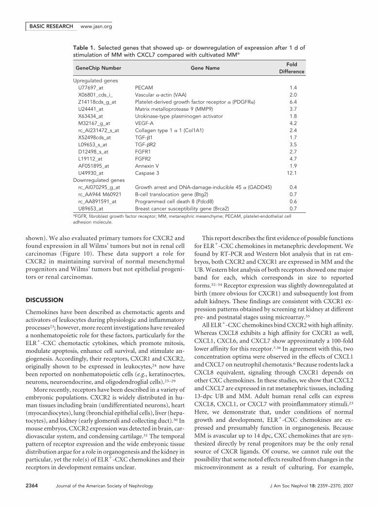
To evaluate directly the response of MM to chemokine sig-naling and determine genomic profiles after stimulation, wescreened Affymetrix GeneChips for expression of genes thatwere upregulated in explanted MM by treatment with CXCL7.Such analyses revealed genes that were associated with survivaland with angiogenesis, including endothelial and mesangialmarkers as well as metalloprotease Mmp9 (Table 1, Figure 7A),which functions in invasive growth, kidney organogenesis, andvasculogenesis.18,19 The upregulated genes included markers ofearly vascular development: Vegf, Fgfr1, Fgfr2, Tgf�1 and itsreceptor Tgf�RII, and urokinase-type Plasminogen Activator.Results were confirmed by semiquantitative RT-PCR as wereother markers of mesangial and endothelial cells, platelet-en-dothelial cell adhesion molecule (Pecam-1), Thy-1, and Megsin(Figure 7, B and C). Cultivation of MM for 1 d in the presenceof FGF2 and TGF-� resulted in upregulation of these markersin comparison with uncultivated MM, but addition of CXCL7induced much higher levels of expression. In addition, eleva-tion of PECAM-1 in CXCL7-treated MM explants was con-firmed by Western analysis (Figure 7D).
For assessment of a possible role of chemokine signaling in
Figure 3. CXCL7 does not induce epithelial markers in culture in explanted MM. (A) Semiquantitative RT-PCR of RNA purified from MMinduced by conditioned medium (CM; 20 �l/ml) or CXCL7 (200 ng/ml) for different time points. (B) RT-PCR analysis of expression ofnephron epithelial marker sFrp2 in MM induced with CM or CXCL7. Values are relative units determined by the LabWork software(mean � SD) based on triplicate samples.
Figure 4. Blocking of CXCR2 with a selective antagonistSB225002 impairs kidney development. SB225002 (Calbiochem)was applied at various concentrations to 13-dpc embryonic kid-neys 1 d after explantation to collagen-coated filters as describedin the Concise Methods section. After 24 h in SB225002, thetissues were fixed in methanol and immunostained with WT1antibody (green) and Dolichos biflorus agglutinin (DBA; red).Magnification, �100.
Figure 2. Cultivation of primary MM changes chemokine expres-sion profiles. Semiquantitative RT-PCR of RNA purified from un-cultivated MM (none) and from MM incubated for 24 h on colla-gen-coated filters in the presence of 30 ng/ml basic fibroblastgrowth factor (bFGF) and 20 ng/ml TGF-� (FTa) or the samefactors plus conditioned medium from the RUB1 cells (CFTa).
BASIC RESEARCH www.jasn.org
2362 Journal of the American Society of Nephrology J Am Soc Nephrol 18: 2359–2370, 2007

cell migration/invasion, isolated MM and RIMM-18 cells weretreated with CXCL7 and evaluated using Matrigel invasionchambers. In these experiments, addition of CXCL7 signifi-cantly increased the invasiveness of both MM and RIMM-18cells through a layer of Matrigel (Figure 8). Typically, we ob-served a 15 to 35% increase in invasion of MM (Figure 8B). Toconfirm that this activity was dependent on signaling throughCXCR2, we treated cultures with SB225002. In these studies,this CXCR2-specific inhibitor dramatically decreased MM andRIMM-18 cell invasiveness through a Matrigel layer withoutaffecting cell migration through a control membrane, suggest-ing that the inhibitor was applied at nontoxic levels. Thesestudies demonstrate that CXCL7 can elicit an angiogenic re-sponse in embryonic kidney by upregulation of Mmp9 andendothelial and mesangial markers and further suggest thatrenal progenitors may use CXCR2 signaling for tissue invasion.
Chemokine Signaling in Renal TumorsChemokine signaling has been reported to play a role in thepathogenesis of a variety of tumors, and CXCR1 or 2 specif-ically has been implicated in the growth or metastasis ofmelanomas and lung and colon tumor cells.20 –22 BecauseWilms’ tumors originate from MM and caricature meta-
nephric development, we speculated that they would ex-press CXCR and respond to the CXCR2 inhibitor SB225002.To evaluate the potential cell-selective toxicity of SB225002,we compared its effect on RUB1 and RIMM-18 cells as wellas on two Wilms’ tumor cell lines, SK-NEP-1 and WiT49,and three renal cell carcinoma lines, CRL, Caki-1, andCaki-2. All analyzed cell lines showed expression of theCXCR2 receptor, but SB225002 induced apoptosis only inblastemal/mesenchymal cell lines (RIMM-18, SK-NEP-1,and WiT49 lines) and not RUB1 cells, as demonstrated bycaspase-3 activity or poly(ADP-ribose) polymerase (PARP)cleavage (Figure 9). Administration of a high (2 �g/ml) con-centration of CXCL7 to SB225002-treated cells to activateCXCR1 did not rescue the cells from apoptosis (data not
Figure 5. The toxic effect of SB225002 depends on the stage ofkidney development. Metanephroi at ages 13 (A and B), 14 (C andD), or 16 dpc (E and F) were cultured for 20 h and then treated (B,D, and F) or not (A, C, and E) with 1.1 �M SB225002. After 24 hof incubation, explants were stained with 2% TO-PRO-1 reagent(Molecular Probes) for 30 min at 37°C. Magnification, �40.
Figure 6. Inhibition of CXCR2 signaling with SB225002 primarilyaffects the cortical nephrogenic zone of cultured metanephroi. (B,D, F, and H) One day of treatment with 1.1 �M SB225002. (A, C,E, and G) Untreated explants. (A and B) Immunostaining with WT1antibody (arrows show WT1-positive structures: condensed mes-enchyme and nephron epithelia). (C and D) DBA staining ofureteric bud (UB). (E and F) Skeletonized images of DBA-stainedUB. (G and H) TO-PRO-1 staining of nonviable cells (bracketsdenote peripheral blastemal area). Magnification, �100.
BASIC RESEARCHwww.jasn.org
J Am Soc Nephrol 18: 2359–2370, 2007 Chemokines in Metanephric Development 2363
shown). We also evaluated primary tumors for CXCR2 andfound expression in all Wilms’ tumors but not in renal cellcarcinomas (Figure 10). These data support a role forCXCR2 in maintaining survival of normal mesenchymalprogenitors and Wilms’ tumors but not epithelial progeni-tors or renal carcinomas.
DISCUSSION
Chemokines have been described as chemotactic agents andactivators of leukocytes during physiologic and inflammatoryprocesses23; however, more recent investigations have revealeda nonhematopoietic role for these factors, particularly for theELR�-CXC chemotactic cytokines, which promote mitosis,modulate apoptosis, enhance cell survival, and stimulate an-giogenesis. Accordingly, their receptors, CXCR1 and CXCR2,originally shown to be expressed in leukocytes,24 now havebeen reported on nonhematopoietic cells (e.g., keratinocytes,neurons, neuroendocrine, and oligodendroglial cells).25–29
More recently, receptors have been described in a variety ofembryonic populations. CXCR2 is widely distributed in hu-man tissues including brain (undifferentiated neurons), heart(myocardiocytes), lung (bronchial epithelial cells), liver (hepa-tocytes), and kidney (early glomeruli and collecting duct).30 Inmouse embryos, CXCR2 expression was detected in brain, car-diovascular system, and condensing cartilage.31 The temporalpattern of receptor expression and the wide embryonic tissuedistribution argue for a role in organogenesis and the kidney inparticular, yet the role(s) of ELR�-CXC chemokines and theirreceptors in development remains unclear.
This report describes the first evidence of possible functionsfor ELR�-CXC chemokines in metanephric development. Wefound by RT-PCR and Western blot analysis that in rat em-bryos, both CXCR2 and CXCR1 are expressed in MM and theUB. Western blot analysis of both receptors showed one majorband for each, which corresponds in size to reportedforms.32–34 Receptor expression was slightly downregulated atbirth (more obvious for CXCR1) and subsequently lost fromadult kidneys. These findings are consistent with CXCR1 ex-pression patterns obtained by screening rat kidney at differentpre- and postnatal stages using microarray.35
All ELR�-CXC chemokines bind CXCR2 with high affinity.Whereas CXCL8 exhibits a high affinity for CXCR1 as well,CXCL1, CXCL6, and CXCL7 show approximately a 100-foldlower affinity for this receptor.7,36 In agreement with this, twoconcentration optima were observed in the effects of CXCL1and CXCL7 on neutrophil chemotaxis.6 Because rodents lack aCXCL8 equivalent, signaling through CXCR1 depends onother CXC chemokines. In these studies, we show that CXCL2and CXCL7 are expressed in rat metanephric tissues, including13-dpc UB and MM. Adult human renal cells can expressCXCL8, CXCL1, or CXCL7 with proinflammatory stimuli.23
Here, we demonstrate that, under conditions of normalgrowth and development, ELR�-CXC chemokines are ex-pressed and presumably function in organogenesis. BecauseMM is avascular up to 14 dpc, CXC chemokines that are syn-thesized directly by renal progenitors may be the only renalsource of CXCR ligands. Of course, we cannot rule out thepossibility that some noted effects resulted from changes in themicroenvironment as a result of culturing. For example,
Table 1. Selected genes that showed up- or downregulation of expression after 1 d ofstimulation of MM with CXCL7 compared with cultivated MMa
GeneChip Number Gene NameFold
Difference
Upregulated genesU77697_at PECAM 1.4X06801_cds_i_ Vascular �-actin (VAA) 2.0Z14118cds_g_at Platelet-derived growth factor receptor � (PDGFR�) 6.4U24441_at Matrix metalloprotease 9 (MMP9) 3.7X63434_at Urokinase-type plasminogen activator 1.8M32167_g_at VEGF-A 4.2rc_AI231472_s_at Collagen type 1 � 1 (Col1A1) 2.4X52498cds_at TGF-�1 1.7L09653_s_at TGF-�R2 3.5D12498_s_at FGFR1 2.7L19112_at FGFR2 4.7AF051895_at Annexin V 1.9U49930_at Caspase 3 12.1
Downregulated genesrc_AI070295_g_at Growth arrest and DNA-damage-inducible 45 � (GADD45) 0.4rc_AA944 M60921 B-cell translocation gene (Btg2) 0.7rc_AA891591_at Programmed cell death 8 (Pdcd8) 0.6U89653_at Breast cancer susceptibility gene (Brca2) 0.7
aFGFR, fibroblast growth factor receptor; MM, metanephric mesenchyme; PECAM, platelet-endothelial celladhesion molecule.
BASIC RESEARCH www.jasn.org
2364 Journal of the American Society of Nephrology J Am Soc Nephrol 18: 2359–2370, 2007

CXCL1 expression increased in cultured rudiments, whereasCXCL7 expression was downregulated. The differences,though, were quantitative and not qualitative in nature andtherefore probably reflect either normal compensatory re-sponses or possible feedback regulatory relationships amongfamily members. The significance of such changes, however, isunclear, because ligand substitutions may not have a signifi-cant impact on CXCR signaling.
The angiogenic properties of ELR�-CXC chemokines arewell documented, so it is reasonable to hypothesize such a rolein the metanephros. Although the origin of endothelial ele-ments in the metanephros remains controversial and may arise
from more than one tissue source, there issufficient evidence to indicate that theyoriginate at least in part from the endoge-nous blastemal population.37 A comparisonof gene expression profiles of uninducedMM that were treated with CXCL7 revealeda group of genes related to angiogenesis. Wedetected elevated levels of endothelial andmesangial markers (Pecam-1 and Megsin)and other angiogenesis-related genes(Mmp9 and Vegf). We did not apply hy-poxic conditions for MM cultivation, whichare normal for embryonic development anda stimulus to angiogenesis. In cultured MM,the hypoxia-induced angiogenic stimulatorVegf38 was significantly downregulated rela-tive to primary tissue isolates (data notshown), so it is possible that exposure of ex-plants to hypoxia could further enhance theeffect of CXCL7 on these markers, includingVegf.
In these studies, the induction of somemarkers (e.g., Pecam-1, Vaa, Pdgfr�) wasgreatest using very high concentrations ofCXCL7 (2.0 �g/ml). This may reflect a con-tribution of CXCR1, which has a lower af-finity for CXCL7 and, unlike CXCR2, is rap-idly re-expressed upon internalization.39
Lower concentrations of CXCL7 still in-duced these markers but may be less effec-tive as a result of the upregulation of Mmp9,which inactivates the chemokine.40 Con-versely, MMP9 may contribute to angiogen-esis by degrading extracellular matrix andfacilitating the sprouting of growing bloodvessels.19 The findings on metalloproteaseupregulation obtained by Affymetrix andRT-PCR assays were supported by migra-tion/invasion studies. We detected a mea-surable basal rate of invasion by cells fromthe MM, presumably as a result of the pres-ence of FGF2 and TGF-�, which are neces-sary to sustain these cells and can induce
metalloprotease activity.41,42 Cultured MM had a six-foldgreater expression level for Mmp9 than freshly isolated MM. Inaddition, Matrigel itself contains multiple growth factors,which may also facilitate invasiveness. Despite this high basallevel, we observed a significant increase in invasion of MMupon stimulation with CXCL7. The CXCR2 antagonistSB225002 inhibited both basal and CXCL7-stimulated inva-sion of MM at nontoxic concentrations, supporting the role ofthe receptor in cell motility/invasion. RIMM-18 cells gave sim-ilar results, except cells were less dependent on added growthfactors, presumably as a result of E1A immortalization.
Genomic profiling of CXCL7-treated MM also revealed a
Figure 7. CXCL7 upregulates mesangial and endothelial markers and matrix metal-loprotease 9 (MMP9) expression. (A) Affymetrix data for GeneChip probes U77697_at(platelet-endothelial cell adhesion molecule 1 [PECAM-1]), X06801cds_i (VAA),Z14118cds_g_at (PDGFR�), and U24441_at (MMP9). (B) RT-PCR of RNA purified fromMM incubated for 24 h with 30 ng/ml bFGF and 20 ng/ml TGF-�, with or withoutCXCL7. (C) Densitometry of PCR gels. Values are relative units determined by theLabWork software. Data shown are the typical results of one of the four independentexperiments. In each set of experiments, four or five MM plated on a filter were usedas one experimental point for in vitro stimulation and consequent RNA preparation,RT, and PCR amplifications. As uninduced material, 15 to 45 freshly prepared MMwere used. (D and E) PECAM-1 expression in MM induced with 0.2 �g/ml CXCL7 for48 h. Immunostaining of MM plated on collagen-coated filters (two typical MM of 48total analyzed in two independent experiments are shown; D). (E) Western blotting.
BASIC RESEARCHwww.jasn.org
J Am Soc Nephrol 18: 2359–2370, 2007 Chemokines in Metanephric Development 2365

group of apoptosis- and growth-related genes. CXCL7-medi-ated downregulation of antiproliferative (Gadd45, Btg2), ap-optosis-inducing (Pdcd8), and tumor suppressor (Brca2)genes and upregulation of Fgfr1 and Fgfr2 genes argue for aprosurvival role of CXCL7. In fact, targeted loss of both Fgfr
genes in MM results in renal agenesis,43 so their expression isessential to MM survival. However, proapoptotic genes (An-nexin V and Caspase 3) were also upregulated with CXCL7administration. This may explain why an MTT test showed nogrowth advantage for CXCL7-treated MM. More probable,any proapoptotic effects may be offset by the observed upregu-lation of CXCL1 and CXCL2 in control MM cultures, whichmay be sufficient to maintain the survival of explants indepen-dent of CXCL7 in culture medium, but this will require furtherinvestigation.
It seems, however, that CXCR2 is critical for survival ofundifferentiated MM, because SB225002 caused its selectivedeath. MM cells from metanephroi or established mesenchy-mal lines, including two Wilms’ tumors, were sensitive toSB225002-induced apoptosis, unlike renal epithelial (UB)cells, suggesting that blastemal cells require signaling throughCXCR2. It is known that phosphatidylinositol-3 kinase/Aktand extracellular signal–regulated kinase prosurvival signalingcascades can be triggered via CXCR2.44,45 Furthermore, neu-
Figure 8. SB225002 (0.13 �M) inhibits CXCL7- stimulated invasion through a Matrigel layer. (A) The migration-invasion data for theRIMM-18 cells represent the number of cells per field of view (mean � SD, n � 4). (B) The migration-invasion data for the MM cellsrepresent the mean gray values (mean � SD, n � 3). Images of Giemsa-stained membranes were converted to gray mode, then colorinverted and evaluated using an ImageJ program. (C) Giemsa-stained membranes with primary MM.
Figure 9. SB225002 induces apoptosis selectively in mesenchy-mal/blastemal/Wilms’ tumor cells but not in epithelial/carcinomacells. (A) Image of RUB1 and RIMM-18 cells cultivated in serum-free conditions with or without 1.1 �M SB225002 for 24 h. (B)Caspase-3 activity is upregulated after 24 h of treatment with 1.1�M SB225002 in the RIMM-18 but not in the RUB1 cells. (C) Westernblot analysis of cell lines of renal progenitors (RUB1 and RIMM-18),renal cell carcinoma (CRL, Caki-1, and Caki-2), and Wilms’ tumors(SK-NEP-1 and WiT49) with CXCR2 rabbit polyclonal, a poly(ADP-ribose) polymerase (PARP) rabbit polyclonal (Cell Signaling Technol-ogy, Beverly, MA), or a glyceraldehyde-3-phosphate dehydroge-nase (GAPDH) mouse mAb (Ambion, Austin, TX).
Figure 10. CXCR2 receptor is expressed in Wilms’ tumors but notin renal cell carcinomas (RCC) or most normal counterparts. Kid-ney tumor samples and normal adjacent tissue were obtainedfrom the Children’s Oncology Group Renal Tumor Biologic Sam-ples Bank via the Cooperative Human Tissue Network. Eight of 16analyzed Wilms’ tumor samples are shown.
BASIC RESEARCH www.jasn.org
2366 Journal of the American Society of Nephrology J Am Soc Nephrol 18: 2359–2370, 2007
tralization of CXCR2 signaling was shown to modulate anti-and proapoptotic proteins in endothelial cells and negativelyaffect their survival.46 However, identification of the specificmechanism(s) responsible for caspase 3– dependent,SB225002-initiated apoptosis in MM cells is to be the subject offuture study. Impairment of UB branching after SB225002 ad-ministration seems to be a secondary event, because UB cells(both primary and from the RUB1 cell line) did not show signsof apoptosis or death. In all probability, the loss of branchingmay be due to the loss of MM-secreted branch-inducing fac-tors, such as glial cell line– derived neutrophic factor(GDNF).47 Participation of CXCR2 in the survival of blastemalcells in kidney may be critical, because levels of CXCL7 thatcould activate the CXCR1 receptor did not rescue the cellsfrom SB225002-induced apoptosis. In this regard, it is possiblethat the induction of angiogenic markers by CXCL7 is indirectand due instead to enhanced survival of angiogenic progeni-tors as a part of blastemal cell population. For nonblastemalcells, other signaling pathways may contribute to survivability,even though they express CXCR2.
Although ELR�-CXC chemokines have been implicated inthe control of cell differentiation,48,49 we did not detect mor-phologic changes that are characteristic of mesenchymal-to-epithelial conversion of MM or even CXCL7-induced expres-sion of specific epithelial markers. Although this may simplyreflect the inadequacies of cell culture conditions, it may alsobe attributable to the cooperative nature of inductive signalingas we have demonstrated.3 Therefore, CXCL7 may function intubulogenesis in combination with other factors.
The results obtained for CXCR2-deficient mice confirm theangiogenic role of the receptor. Decreased vascular density aswell as marked reduction of tumor growth and its metastaticpotential were shown in these animals.50,51 CXCR2 knockoutmice also have retarded wound healing, another process thatdepends on microvascularization.52 However, there is no re-ported evidence of kidney involvement in CXCR2 null mice.This may be explained by redundancy in receptor expression(i.e., CXCR1), which was recently discovered in mice53 andwhich may substitute for some CXCR2 functions.
These data provide evidence of a prosurvival and proangio-genic role for ELR�-CXC chemokines and their receptorCXCR2 during metanephric development. Expression of theCXCR2 receptor in blastemal tumors of the kidney and apop-tosis-promoting properties of its selective antagonist may pro-vide a novel approach for chemotherapeutic intervention inWilms’ tumors.
CONCISE METHODS
Cell CulturesRUB1 and RIMM-18 cell lines were established from rat UB and un-
differentiated MM, respectively, and were characterized previously
and grown as described.4,54 Wilms’ tumor line WiT49 was a gift of Dr.
Herman Yeger (Hospital for Sick Children, Toronto, Canada). Other
tumor lines were obtained from the ATCC (Rockville, MD).
Isolation and Cultivation of Rat Embryonic Kidneysand MMEmbryonic kidneys were excised from F344 rat embryos. MM were
enzymatically separated from embryonic day 13.5 UB. Metanephroi
or MM were cultured on polycarbonate filters (Whatman-Nuclepore,
Florham Park, NJ) coated with type IV collagen (BD Bioscience, Bed-
ford, MA) as described previously.1
Affymetrix GeneChip AnalysisRNA was extracted from cells using TRIzol reagent (Invitrogen, Carls-
bad, CA). Total RNA (5 �g) from duplicate samples was converted
into cDNA and purified by phenol/chloroform extraction. cDNA la-
beling, hybridization to U34A GeneChips, and scanning were per-
formed according to Affymetrix instructions, and data were analyzed
by GeneChip software.
Semiquantitative RT-PCR AnalysisRT reactions and PCR amplifications were performed using total
RNA (0.2 or 1 �g) as described previously.54 Primer sequences, an-
nealing temperatures, and numbers of cycles are shown in Table 2. No
RT controls were included to eliminate the possibility of DNA con-
tamination. All PCR products were sequenced to confirm identities.
Western Blot AnalysisProtein lysates were obtained from rat kidneys at various stages of
development (16 dpc, 19 dpc, newborn, or adult) or from isolated
13-dpc MM (20 to 30) and analyzed by Western blotting as described
previously.54
ImmunocytostainingMM or kidney explants cultivated on polycarbonate filters were
fixed in methanol, washed, preblocked with 10% sheep serum
(Sigma-Aldrich, St. Louis, MO), and stained for WT1 (rabbit poly-
clonal, 1:50; Santa Cruz Biotechnology, Santa Cruz, CA) or PE-
CAM-1 (mouse monoclonal, 1:50; Chemicon Int., Temecula, CA),
using Alexa488-conjugated secondary antibodies (1:100; Molecu-
lar Probes, Eugene, OR) in 1% sheep serum overnight at 4°C.
Filters were washed, stained if needed with 20 �g/ml Dolichos
biflorus agglutinin (Sigma), and mounted using a ProLong Anti-
fade Kit (Molecular Probes).
In Situ Hybridization and ImmunohistochemistryFrozen sections (20 �m) of 4% paraformaldehyde-fixed, 30% sucrose
permeabilized, and OCT-embedded 16-dpc rat kidneys were probed
with a 200-bp digoxigenin-labeled riboprobe according to Tuttle et
al.55 using a chromogenic alkaline phosphatase substrate BM Purple
(Roche, Palo Alto, CA). For immunohistochemistry, similarly pre-
pared frozen sections were probed with a rabbit polyclonal antibody
for CXCR2 (sc-683, Santa Cruz Biotechnology) or nonimmune rabbit
IgG at a 1:100 dilution. Staining was visualized using a Vectastain ABC
BASIC RESEARCHwww.jasn.org
J Am Soc Nephrol 18: 2359–2370, 2007 Chemokines in Metanephric Development 2367

kit (Vector Laboratories, Burlingame, CA), and sections were lightly
counterstained with hematoxylin.
Measurement of Caspase-3 Activity Using aFluorometric SubstrateCaspase-3 activity in cells was measured using 20 �M caspase-3 flu-
orometric substrate Ac-DEVD-amc (Upstate Biotech, Lake Placid,
NY) and 1 �M caspase-3 inhibitor IV Ac-VEID-CHO (for negative
control; Calbiochem, San Diego, CA), according to manufacturers’
instructions. Fluorescence was measured at excitation 380 nm and
emission 460 nm using a Luminescence Spectrometer (Perkin-Elmer
LS50B, Waltham, MA). Protein contents were quantified with a BCA
Protein Assay Kit (Pierce Biotechnology, Rockford, IL), and fluores-
cence was normalized to protein content.
Invasion AssayInvasiveness of RIMM-18 or MM cells was determined using BD Biocoat
Matrigel Invasion Chambers (BD Biosciences) according to the manu-
facturer’s recommendations. RIMM-18 cells (3 � 104 cells/well) or MM
(1 MM/well) were added to the invasion chambers in 500 �l of medium
with FGF2 (10 ng/ml); 700 �l of the same medium was added to the
culture wells. CXCL7 (200 ng/ml; Peprotech, Rocky Hill, NJ) was added
to the culture wells; inhibitors, when applied, were added to both the
invasion chambers and the culture wells. After 20 h of incubation, non-
invading cells were removed with cotton swabs, and membranes were
fixed in 100% methanol and stained with Giemsa solution.
ACKNOWLEDGMENTS
This research was supported in part by the Intramural Research Pro-
gram of the National Institutes of Health, National Cancer Institute,
Center for Cancer Research.
DISCLOSURESNone.
REFERENCES
1. Karavanova ID, Dove LF, Resau JH, Perantoni AO: Conditioned me-dium from a rat ureteric bud cell line in combination with bFGFinduces complete differentiation of isolated metanephric mesen-chyme. Development 122: 4159–4167, 1996
2. Barasch J, Yang J, Ware CB, Taga T, Yoshida K, Erdjument-Bromage
Table 2. PCR primers for amplification reactionsa
PrimerAccession
No.Sequence
ProductSize
MeltingTemperature
No. ofCycles
CXCL1 D11445 U 5�-CCA GCC ACA CTC CAA CAG 388 57.0 33D 5�-CCC TCA ATA GAA ATC GTA AAA TG
CXCL2 XM_346458 U 5�-ATC AGG GTA CAG GGG TTG TTG 238 57.0 33D 5�-GGT CAG TTA GCC TTG CCT TTG
CXCL5 U90488 U 5�-CGT CAT TCA CCC TGC TGG CAT 316 57.2 35D 5�-GCA AGT GCA TTC CGC TTT GTT TTC
CXCL7 AF349115 U 5�-ATG GGC TTC AGA CTC AGA CCT AC 209 57.0 33D 5�-AAC ACA TTC ACA CGG GAG ATA GAG
CXCR1 U71089 U 5�-TTG GAA ATA TCA CCC GAA TGC TG 480 59.5 32D 5�-AAG ATG GCA AAA GGC AGA GAC
CXCR2 D63584 U 5�-TTC TGA CCC GCC CTT TAC TCT GT 630 59.8 34D 5�-CGC AGT GTG AAC CCA TAG CAG
Megsin AF105329 U 5�-TGA ATG TGT TTC TCC CCC AGT TC 193 55.4 31D 5�-GCC TCG GTG CCT TCT TCT GAG
MMP9 U24441 U 5�-CCA CCG CCA ACT ATG ACC AG 877 59.8 35AGC CCC AAC TTA TCC AGA CTC CT
PECAM-1 U77697 U 5�-AGG CAT CGG CAA AGT GGT CAA GAG 693 59.4 28GCT GCA ACT ATC AAG GCG GCA ATG
PDGFR� Z14118 U 5�-TCA AAC TCC CGT CCA TCA AAC TG 747 58.8 27D 5�-CTC TGT TCC CAA TGC CAA GGT C
sFRP-2 U88567 U 5�-GCC TCG CTG CTG CTG CTA GTC 538 55.2 34D 5�-TGT CGT TGT CGT CCT CAT TCT TG
Thy-1 X03150 U 5�-ATA ACA CCA ACT TGC CCA TCC AG 371 58.2 30D 5�-CCC AAC CAG TCA CAG AGA AAT GAA
VAA X06801 U 5�-ATG CTC CCA GGG CTG TTT TC 159 57.6 24D 5�-TGG TGA TGA TGC CGT GTT CTA TC
GAPDH AB017801 U 5�-CCA TGC CAT CAC TGC CAC TCA GAA G 372 60.7 22D 5�-GCA ATG CCA GCC CCA GCA TCA AAG
aGAPDH, glyceraldehyde-3-phosphate dehydrogenase.
BASIC RESEARCH www.jasn.org
2368 Journal of the American Society of Nephrology J Am Soc Nephrol 18: 2359–2370, 2007

H, Tempst P, Parravicini E, Malach S, Aranoff T, Oliver JA: Mesenchy-mal to epithelial conversion in rat metanephros is induced by LIF. Cell99: 377–386, 1999
3. Plisov SY, Yoshino K, Dove LF, Higinbotham KG, Rubin JS, PerantoniAO: TGF beta 2, LIF and FGF2 cooperate to induce nephrogenesis.Development 128: 1045–1057, 2001
4. Perantoni A, Kan FW, Dove LF, Reed CD: Selective growth in cultureof fetal rat renal collecting duct anlagen. Morphologic and biochem-ical characterization. Lab Invest 53: 589–596, 1985
5. Baggiolini M: Novel aspects of inflammation: Interleukin-8 and relatedchemotactic cytokines. Clin Investig 71: 812–814, 1993
6. Ludwig A, Petersen F, Zahn S, Gotze O, Schroder JM, Flad HD, BrandtE: The CXC-chemokine neutrophil-activating peptide-2 induces twodistinct optima of neutrophil chemotaxis by differential interactionwith interleukin-8 receptors CXCR-1 and CXCR-2. Blood 90: 4588–4597, 1997
7. Wuyts A, Proost P, Lenaerts JP, Ben-Baruch A, Van Damme J, WangJM: Differential usage of the CXC chemokine receptors 1 and 2 byinterleukin-8, granulocyte chemotactic protein-2 and epithelial-cell-derived neutrophil attractant-78. Eur J Biochem 255: 67–73, 1998
8. Belperio JA, Keane MP, Arenberg DA, Addison CL, Ehlert JE, BurdickMD, Strieter RM: CXC chemokines in angiogenesis. J Leukoc Biol 68:1–8, 2000
9. Mockenhaupt M, Peters F, Schwenk-Davoine I, Herouy Y, Schraufstat-ter I, Elsner P, Norgauer J: Evidence of involvement of CXC-chemo-kines in proliferation of cultivated human melanocytes. Int J Mol Med12: 597–601, 2003
10. Li A, Dubey S, Varney ML, Dave BJ, Singh RK: IL-8 directly enhancedendothelial cell survival, proliferation, and matrix metalloproteinasesproduction and regulated angiogenesis. J Immunol 170: 3369–3376,2003
11. Maheshwari A, Lu W, Lacson A, Barleycorn AA, Nolan S, ChristensenRD, Calhoun DA: Effects of interleukin-8 on the developing humanintestine. Cytokine 20: 256–267, 2002
12. Ragozzino D: CXC chemokine receptors in the central nervous system:Role in cerebellar neuromodulation and development. J Neurovirol 8:559–572, 2002
13. Perantoni AO, Dove L, Karavanova I: Basic fibroblast growth factor canmediate the early inductive events in renal development. Proc NatlAcad Sci U S A 92: 4696–4700, 1995
14. Rogers SA, Ryan G, Hammerman MR: Metanephric transforminggrowth factor-alpha is required for renal organogenesis in vitro. Am JPhysiol 262: F533–F539, 1992
15. Montesano R, Vassalli JD, Baird A, Guillemin R, Orci L: Basic fibroblastgrowth factor induces angiogenesis in vitro. Proc Natl Acad Sci U S A83: 7297–7301, 1986
16. Vinals F, Pouyssegur J: Transforming growth factor beta1 (TGF-beta1)promotes endothelial cell survival during in vitro angiogenesis via anautocrine mechanism implicating TGF-alpha signaling. Mol Cell Biol21: 7218–7230, 2001
17. White JR, Lee JM, Young PR, Hertzberg RP, Jurewicz AJ, Chaikin MA,Widdowson K, Foley JJ, Martin LD, Griswold DE, Sarau HM: Identifi-cation of a potent, selective non-peptide CXCR2 antagonist thatinhibits interleukin-8-induced neutrophil migration. J Biol Chem 273:10095–10098, 1998
18. Lelongt B, Trugnan G, Murphy G, Ronco PM: Matrix metalloprotein-ases MMP2 and MMP9 are produced in early stages of kidney mor-phogenesis but only MMP9 is required for renal organogenesis invitro. J Cell Biol 136: 1363–1373, 1997
19. Lambert V, Munaut C, Jost M, Noel A, Werb Z, Foidart JM, Rakic JM:Matrix metalloproteinase-9 contributes to choroidal neovasculariza-tion. Am J Pathol 161: 1247–1253, 2002
20. Zhu YM, Webster SJ, Flower D, Woll PJ: Interleukin-8/CXCL8 is agrowth factor for human lung cancer cells. Br J Cancer 91: 1970–1976,2004
21. Li A, Varney ML, Singh RK: Constitutive expression of growth regu-
lated oncogene (gro) in human colon carcinoma cells with differentmetastatic potential and its role in regulating their metastatic pheno-type. Clin Exp Metastasis 21: 571–579, 2004
22. Varney ML, Johansson SL, Singh RK: Distinct expression of CXCL8 andits receptors CXCR1 and CXCR2 and their association with vesseldensity and aggressiveness in malignant melanoma. Am J Clin Pathol125: 209–216, 2006
23. Segerer S, Nelson PJ, Schlondorff D: Chemokines, chemokine recep-tors, and renal disease: From basic science to pathophysiologic andtherapeutic studies. J Am Soc Nephrol 11: 152–176, 2000
24. Chuntharapai A, Lee J, Hebert CA, Kim KJ: Monoclonal antibodiesdetect different distribution patterns of IL-8 receptor A and IL-8 re-ceptor B on human peripheral blood leukocytes. J Immunol 153:5682–5688, 1994
25. Schulz BS, Michel G, Wagner S, Suss R, Beetz A, Peter RU, Kemeny L,Ruzicka T: Increased expression of epidermal IL-8 receptor in psoria-sis. Down-regulation by FK-506 in vitro. J Immunol 151: 4399–4406,1993
26. Kondo S, Yoneta A, Yazawa H, Kamada A, Jimbow K: Downregulationof CXCR-2 but not CXCR-1 expression by human keratinocytes byUVB. J Cell Physiol 182: 366–370, 2000
27. Horuk R, Martin AW, Wang Z, Schweitzer L, Gerassimides A, Guo H, LuZ, Hesselgesser J, Perez HD, Kim J, Parker J, Hadley TJ, Peiper SC:Expression of chemokine receptors by subsets of neurons in thecentral nervous system. J Immunol 158: 2882–2890, 1997
28. Tecimer T, Dlott J, Chuntharapai A, Martin AW, Peiper SC: Expressionof the chemokine receptor CXCR2 in normal and neoplastic neuroen-docrine cells. Arch Pathol Lab Med 124: 520–525, 2000
29. Nguyen D, Stangel M: Expression of the chemokine receptors CXCR1and CXCR2 in rat oligodendroglial cells. Brain Res Dev Brain Res 128:77–81, 2001
30. Dame JB, Juul SE: The distribution of receptors for the pro-inflamma-tory cytokines interleukin (IL)-6 and IL-8 in the developing humanfetus. Early Hum Dev 58: 25–39, 2000
31. Luan J, Furuta Y, Du J, Richmond A: Developmental expression of twoCXC chemokines, MIP-2 and KC, and their receptors. Cytokine 14:253–263, 2001
32. Samanta AK, Oppenheim JJ, Matsushima K: Identification and char-acterization of specific receptors for monocyte-derived neutrophilchemotactic factor (MDNCF) on human neutrophils. J Exp Med 169:1185–1189, 1989
33. Tikhonov II, Fomin IK, Doroshenko TM, Chalyi IuV, Voitenok NN:Expression of cDNA for human interleukin-8 type one receptor inBALB 3T3 fibroblasts and characteristics of products of expression [inRussian]. Mol Biol (Mosk) 30: 1014–1021, 1996
34. Ludwig A, Ehlert JE, Flad HD, Brandt E: Identification of distinctsurface-expressed and intracellular CXC-chemokine receptor 2 glyco-forms in neutrophils: N-glycosylation is essential for maintenance ofreceptor surface expression. J Immunol 165: 1044–1052, 2000
35. Stuart RO, Bush KT, Nigam SK: Changes in gene expression patternsin the ureteric bud and metanephric mesenchyme in models of kidneydevelopment. Kidney Int 64: 1997–2008, 2003
36. Schumacher C, Clark-Lewis I, Baggiolini M, Moser B: High- and low-affinity binding of GRO alpha and neutrophil-activating peptide 2 tointerleukin 8 receptors on human neutrophils. Proc Natl Acad SciU S A 89: 10542–10546, 1992
37. Hyink DP, Tucker DC, St John PL, Leardkamolkarn V, Accavitti MA,Abrass CK, Abrahamson DR: Endogenous origin of glomerular endo-thelial and mesangial cells in grafts of embryonic kidneys. Am J Physiol270: F886–F899, 1996
38. Shweiki D, Itin A, Soffer D, Keshet E: Vascular endothelial growthfactor induced by hypoxia may mediate hypoxia-initiated angiogene-sis. Nature 359: 843–845, 1992
39. A Chuntharapai A, Kim KJ: Regulation of the expression of IL-8 recep-tor A/B by IL-8: Possible functions of each receptor. J Immunol 155:2587–2594, 1995
BASIC RESEARCHwww.jasn.org
J Am Soc Nephrol 18: 2359–2370, 2007 Chemokines in Metanephric Development 2369
40. Van den Steen PE, Proost P, Wuyts A, Van Damme J, Opdenakker G:Neutrophil gelatinase B potentiates interleukin-8 tenfold by aminot-erminal processing, whereas it degrades CTAP-III, PF-4, and GRO-alpha and leaves RANTES and MCP-2 intact. Blood 96: 2673–2681,2000
41. Liu JF, Crepin M, Liu JM, Barritault D, Ledoux D: FGF-2 and TPAinduce matrix metalloproteinase-9 secretion in MCF-7 cells throughPKC activation of the Ras/ERK pathway. Biochem Biophys Res Com-mun 293: 1174–1182, 2002
42. Ganser GL, Stricklin GP, Matrisian LM: EGF and TGF alpha influence invitro lung development by the induction of matrix-degrading metal-loproteinases. Int J Dev Biol 35: 453–461, 1991
43. Poladia DP, Kish K, Kutay B, Hains D, Kegg H, Zhao H, Bates CM: Roleof fibroblast growth factor receptors 1 and 2 in the metanephricmesenchyme. Dev Biol 291: 325–339, 2006
44. Knall C, Young S, Nick JA, Buhl AM, Worthen GS, Johnson GL:Interleukin-8 regulation of the Ras/Raf/mitogen-activated protein ki-nase pathway in human neutrophils. J Biol Chem 271: 2832–2838,1996
45. Limatola C, Ciotti MT, Mercanti D, Santoni A, Eusebi F: Signalingpathways activated by chemokine receptor CXCR2 and AMPA-typeglutamate receptors and involvement in granule cells survival. J Neu-roimmunol 123: 9–17, 2002
46. Li A, Varney ML, Valasek J, Godfrey M, Dave BJ, Singh RK: Autocrinerole of interleukin-8 in induction of endothelial cell proliferation, sur-vival, migration and MMP-2 production and angiogenesis. Angiogen-esis 8: 63–71, 2005
47. Basson MA, Watson-Johnson J, Shakya R, Akbulut S, Hyink D, Costan-tini FD, Wilson PD, Mason IJ, Licht JD: Branching morphogenesis of
the ureteric epithelium during kidney development is coordinated bythe opposing functions of GDNF and Sprouty1. Dev Biol 299: 466–477, 2006
48. Merz D, Liu R, Johnson K, Terkeltaub R: IL-8/CXCL8 and growth-related oncogene alpha/CXCL1 induce chondrocyte hypertrophic dif-ferentiation. J Immunol 171: 4406–4415, 2003
49. Emadi S, Clay D, Desterke C, Guerton B, Maquarre E, Charpentier A,Jasmin C, Le Bousse-Kerdiles MC; French INSERM Research Networkon MMM: IL-8 and its CXCR1 and CXCR2 receptors participate in thecontrol of megakaryocytic proliferation, differentiation, and ploidy inmyeloid metaplasia with myelofibrosis. Blood 105: 464–473, 2005
50. Keane MP, Belperio JA, Xue YY, Burdick MD, Strieter RM: Depletion ofCXCR2 inhibits tumor growth and angiogenesis in a murine model oflung cancer. J Immunol 172: 2853–2860, 2004
51. Mestas J, Burdick MD, Reckamp K, Pantuck A, Figlin RA, Strieter RM:The role of CXCR2/CXCR2 ligand biological axis in renal cell carci-noma. J Immunol 175: 5351–5357, 2005
52. Devalaraja RM, Nanney LB, Du J, Qian Q, Yu Y, Devalaraja MN,Richmond A: Delayed wound healing in CXCR2 knockout mice. J In-vest Dermatol 115: 234–244, 2000
53. Moepps B, Nuesseler E, Braun M, Gierschik P: A homolog of thehuman chemokine receptor CXCR1 is expressed in the mouse. MolImmunol 43: 897–914, 2006
54. Levashova ZB, Plisov SY, Perantoni AO: Conditionally immortalizedcell line of inducible metanephric mesenchyme. Kidney Int 63: 2075–2087, 2003
55. Tuttle R, Nakagawa Y, Johnson JE, O’Leary DDM: Defects in thalamo-cortical axon pathfinding correlate with altered cell domains in Mash-1-deficient mice. Development 126: 1903–1916, 1999
BASIC RESEARCH www.jasn.org
2370 Journal of the American Society of Nephrology J Am Soc Nephrol 18: 2359–2370, 2007