Embed Size (px)
Citation preview
Intensive Care Med (2008) 34:1216–1223DOI 10.1007/s00134-008-1042-7 O R I G I N A L
Barry DixonDuncan J. CampbellJohn D. Santamaria
Elevated pulmonary dead spaceand coagulation abnormalities suggest lungmicrovascular thrombosis in patientsundergoing cardiac surgery
Received: 18 October 2007Accepted: 30 December 2007Published online: 27 February 2008© Springer-Verlag 2008
Electronic supplementary materialThe online version of this article(doi:10.1007/s00134-008-1042-7) containssupplementary material, which is availableto authorized users.
This article is discussed in the editorialavailable at: http://dx.doi.org/10.1007/s00134-008-1043-6.
The study was registered with theAustralian Clinical Trials RegistryNo. 12605000133639.URL: http://www.actr.org.au/.
B. Dixon (�) · J. D. SantamariaSt. Vincent’s Hospital, Intensive Care Unit,Melbourne, 3065, Victoria, Australiae-mail: [email protected].: +61-3-92884488Fax: +61-3-92884487
D. J. CampbellSt. Vincent’s Institute of Medical Research,Melbourne, 3065, Victoria, Australia
Abstract Objective: Inflammationhas been shown to trigger microvas-cular thrombosis. Patients undergoingcardiac surgery sustain significantinflammatory insults to the lungsand in addition are routinely givenanti-fibrinolytic agents to promotethrombosis. In view of these riskfactors we investigated if evidence ofpulmonary microvascular thrombosisoccurs following cardiac surgery and,if so, whether a pre-operative heparininfusion may limit this. Design:Double-blind randomised controlledtrial. Setting: Tertiary universityaffiliated hospital. Patients: Twentypatients undergoing elective cardiacsurgery. Interventions: Patients wererandomised to receive a pre-operativeheparin infusion or placebo. Allpatients were administered apro-tinin. Measurements and results:Pulmonary microvascular obstruc-tion was estimated by measuringthe alveolar dead-space fraction.Pulmonary coagulation activationwas estimated by measuring the ratioof prothrombin fragment levels in
radial and pulmonary arterial blood.Systemic tissue plasminogen acti-vator (t-PA) levels were alsoassessed. In the placebo groupcardiac surgery triggered increasedalveolar dead-space fraction levelsand the onset of prothrombin frag-ment production in the pulmonarycirculation. Administration of pre-operative heparin was associatedwith a lower alveolar dead-spacefraction (p < 0.05) and reduced pro-thrombin fragment production in thepulmonary circulation (p < 0.05).Pre-operative heparin also increasedbaseline t-PA levels (p < 0.05). Con-clusion: The changes in the alveolardead-space fraction and pulmonarycoagulation activation suggest thatpulmonary microvascular thrombosisdevelops during cardiac surgery andthis may be limited by a pre-opera-tive heparin infusion.
Keywords Cardiopulmonary by-pass · Coagulation · Fibrinolysis ·Inflammation · Thrombosis
Introduction
In patients undergoing cardiac surgery a range of in-flammatory insults contribute to post-operative lunginjury [1, 2]. Inflammatory insults include lung ischaemiasustained during cardiopulmonary bypass (CPB), surgicaltrauma and contact of blood with the foreign surface ofthe CPB circuit [3–7]. In other inflammatory conditions,such as sepsis, microvascular thrombosis has been shown
to be a mechanism of organ injury [8, 9]. Stimulation ofmicrovascular endothelial cells by inflammatory media-tors is the initial process that gives rise to microvascularthrombosis [10]. In response, endothelial cells expressplatelet and white cell ligands, such as vascular celladhesion molecule-1 (VCAM-1) [11–13]. Within min-utes platelets and white cells adhere to the endotheliumforming cellular aggregates. The endothelium and aggre-gated cells, in turn, express tissue factor, which triggers
1217
coagulation activation [8]. These responses result in fibrindeposition and microvascular thrombosis [14–18]. Inpatients undergoing cardiac surgery the risk of developingmicrovascular thrombosis may be further increased by theroutine use of anti-fibrinolytic agents to promote thromb-osis [19, 20].
Case reports have demonstrated microvascular thrombiin the lungs, heart and kidneys of patients that diedfollowing cardiac surgery [21–25]. In a case series of ninepatients that developed acute post-operative pulmonaryhypertension, multiple microvascular thrombi weredemonstrated in the lungs [25].
In this study we sought to assess if a pre-operative hep-arin infusion limited evidence of pulmonary microvascularthrombosis. Pulmonary microvascular thrombosis wasestimated by measuring the alveolar dead-space fraction(the alveolar dead space to tidal volume ratio) [26] andthe extent of coagulation activation in the pulmonarycirculation was estimated by measuring the ratio of pro-thrombin fragment levels in radial and pulmonary arterialblood.
Materials and methods
Subjects
We studied patients undergoing elective coronary arterybypass grafting (CABG) with CPB. Patients were excludedif they had had previous CABG or required a surgical inter-vention in addition to CABG, a creatinine level greaterthan 200 umol/l or age greater than 85 years. The study wasapproved by the St. Vincent’s Hospital Human ResearchEthics Committee. All patients gave written informed con-sent before participation in the study.
Interventions
We undertook a double-blind placebo-controlled trial.Patients were randomised (computer-generated blocks offour) to a continuous pre-operative infusion of heparinor placebo. The infusion bags (500 ml of 5% glucose)and rates of infusion were identical in both groups.The pre-operative heparin group had 25,000 units (U)of heparin (Porcine Heparin Sodium, Pharmacia, Mel-bourne, Australia) added to the bag. Drug preparationwas performed by nurses not involved with data col-lection. The infusion commenced with a bolus of100 ml of fluid (5000 U of heparin) over 30 min, andwas then continued at 0.36 ml/kg h–1 (18 U of hep-arin/kg h–1). The infusion commenced on average 10 hbefore surgery and was continued until the intra-operativeheparin bolus – given just before commencement ofCPB.
Data collection
The alveolar dead-space fraction, the alveolar arterial(A-a) oxygen gradient, lactate, Hb and haemodynamicvariables were measured following anaesthetic induction,following sternotomy and at 0, 1, 2, 3 and 4 h post-CPB.At the same time points blood was aspirated from thedistal port of the pulmonary artery catheter and thenimmediately from the radial arterial line for prothrombinfragments, blood gas, white cell and platelet levels.The ratio of prothrombin fragment levels in radial andpulmonary arterial blood was calculated. A ratio greaterthan 1 suggests coagulation activation in the pulmonarycirculation. Tissue plasminogen activator, s-VCAM-1 andtroponin I levels were sampled at the same time pointsfrom the radial arterial line. The ventilator and ventilatorsettings were standardised for all measurements. Thealveolar dead space was measured using the Cosmo PlusRespironics monitor (Novametrix Medical Systems, Con-necticut). The A-a gradient was calculated using standardformulae. The left ventricular (LV) ejection fraction wasgraded by intra-operative trans-oesophageal echocardio-graphy, where severe dysfunction represents a ejectionfraction < 30%, moderate 30–44%, mild 45–54% andnormal ≥ 55%. Haemodynamic variables were meas-ured with a pulmonary artery catheter using standardtechniques. Automated laboratory analyses of Hb, whitecell and platelet levels were undertaken. Prothrombinfragments, t-PA, s-VCAM-1 and troponin-I levels wereassayed by enzyme-linked immunoassays. (For moredetails regarding data collection see the ESM.)
Anaesthesia and surgical management
Patients were administered 2 million units IV aprotininfollowing induction followed by 0.5 million U/h for theduration of the operation. An additional 2 million unitswas added to the pump prime. All patients were hep-
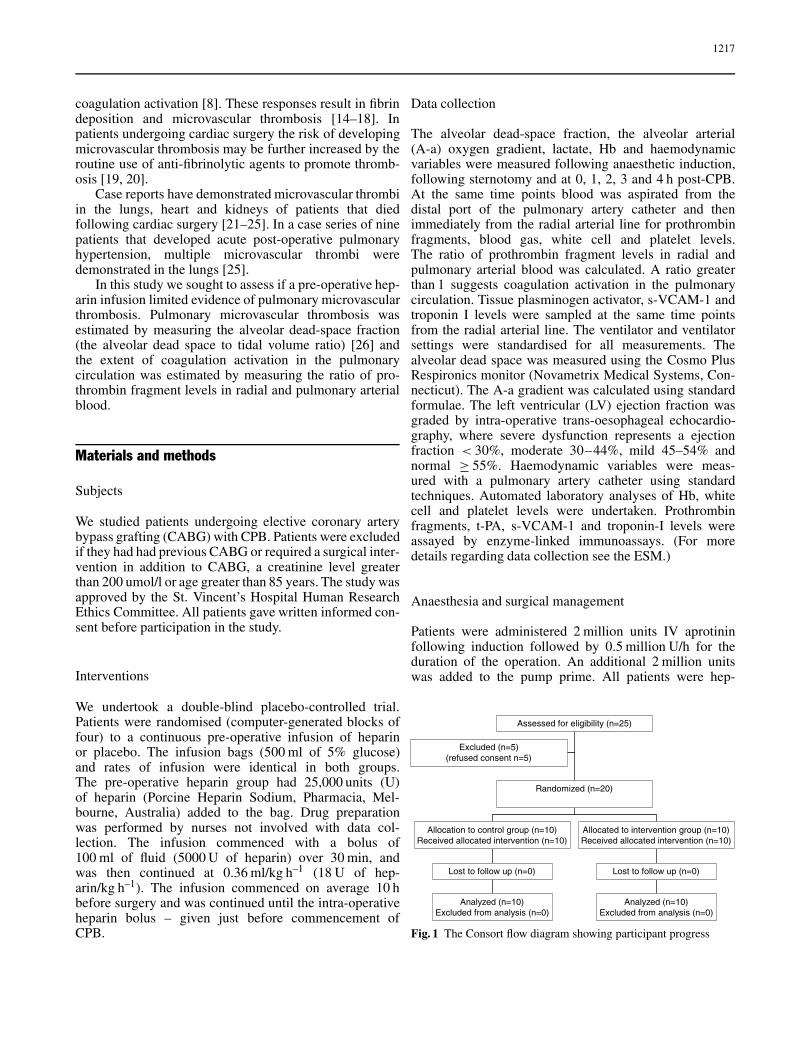
Fig. 1 The Consort flow diagram showing participant progress
1218
arinised just before commencement of CPB (initial dose~ 300 U/kg) to maintain an activated clotting time (usingkaolin) above 400 s. (For more details see the ESM.)
Statistical analysis
The study was powered to demonstrate a 30% differencein the alveolar dead-space fraction, with 80% powerat a significance level of 0.05. Fisher’s exact test com-pared categorical variables. Student’s t-test comparednormally distributed variables and the Mann–Whitney testnon-normally distributed variables. Repeated measuresanalysis of variance (ANOVA) was used to compare vari-ables repeatedly measured over time. Statistical analysiswas performed with Statview (SAS Institute, Cary, NC,USA).
Results
The Consort flow diagram presents participant progress(Fig. 1). Twenty patients were studied, 10 receivedpre-operative heparin and 10 placebo. The baseline andoperative characteristics for the two groups were similar(Tables 1, 2). The APTT level at anaesthesia inductionwas significantly higher in the heparin group (126 vs.37 s, p < 0.0001). One patient in the pre-operative heparingroup died from acute heart failure, as a result of graftfailure, on the second post-operative day. Operative
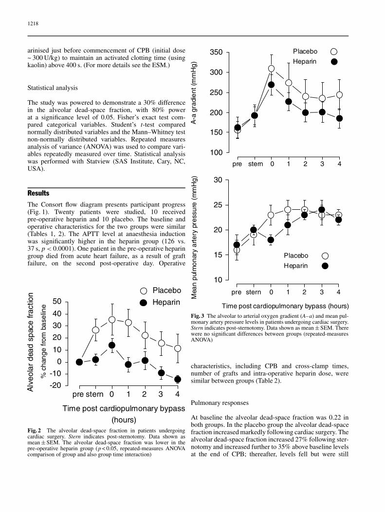
Fig. 2 The alveolar dead-space fraction in patients undergoingcardiac surgery. Stern indicates post-sternotomy. Data shown asmean ± SEM. The alveolar dead-space fraction was lower in thepre-operative heparin group (p < 0.05, repeated-measures ANOVAcomparison of group and also group time interaction)
Fig. 3 The alveolar to arterial oxygen gradient (A–a) and mean pul-monary artery pressure levels in patients undergoing cardiac surgery.Stern indicates post-sternotomy. Data shown as mean ± SEM. Therewere no significant differences between groups (repeated-measuresANOVA)
characteristics, including CPB and cross-clamp times,number of grafts and intra-operative heparin dose, weresimilar between groups (Table 2).
Pulmonary responses
At baseline the alveolar dead-space fraction was 0.22 inboth groups. In the placebo group the alveolar dead-spacefraction increased markedly following cardiac surgery. Thealveolar dead-space fraction increased 27% following ster-notomy and increased further to 35% above baseline levelsat the end of CPB; thereafter, levels fell but were still
1219
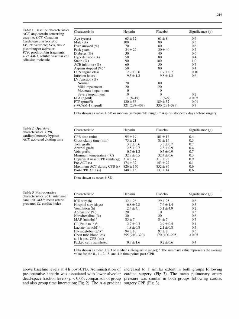
Characteristic Heparin Placebo Significance (p)
Age (years) 63 ± 12 61 ± 8 0.6Male (%) 100 80 0.5Ever smoked (%) 70 80 0.6Pack years 24 ± 22 30 ± 40 0.7Diabetes (%) 30 40 0.6Hypertension (%) 50 80 0.4Statin (%) 90 100 1.0ACE inhibitor (%) 60 50 0.7Aspirin stopped (%) a 50 80 0.4CCS angina class 2.2 ± 0.6 1.7 ± 0.7 0.10Infusion hours 9.5 ± 1.2 9.8 ± 1.3 0.6LV function (%)
Normal 70 80Mild impairment 20 20Moderate impairment 0 0Severe impairment 10 0 0.2
t-PA (ng/ml) 11 (8–15) 7 (6–9) < 0.05PTF (pmol/l) 120 ± 56 189 ± 57 0.01s-VCAM-1 (ng/ml) 321 (297–403) 330 (291–389) 0.7
Data shown as mean ± SD or median (interquartile range); a Aspirin stopped 7 days before surgery
Table 1 Baseline characteristics.ACE, angiotensin convertingenzyme; CCS, CanadianCardiovascular Society;LV, left ventricle; t-PA, tissueplasminogen activator;PTF, prothrombin fragments;s-VCAM-1, soluble vascular celladhesion molecule
Characteristic Heparin Placebo Significance (p)
CPB time (min) 95 ± 19 101 ± 16 0.4Cross-clamp time (min) 73 ± 21 81 ± 14 0.3Total grafts 3.2 ± 0.6 3.3 ± 0.7 0.7Arterial grafts 2.5 ± 0.7 2.8 ± 0.9 0.4Vein grafts 0.7 ± 1.2 0.5 ± 0.9 0.7Minimum temperature (◦C) 32.7 ± 0.5 32.4 ± 0.6 0.3Heparin at onset CPB (units/kg) 314 ± 47 317 ± 28 0.9Pre-ACT (s) 174 ± 32 153 ± 22 0.1Maximum ACT during CPB (s) 826 ± 150 852 ± 86 0.6Post-CPB ACT (s) 140 ± 15 137 ± 14 0.6
Data shown as mean ± SD
Table 2 Operativecharacteristics. CPB,cardiopulmonary bypass;ACT, activated clotting time
Characteristic Heparin Placebo Significance (p)
ICU stay (h) 32 ± 26 29 ± 25 0.8Hospital stay (days) 6.8 ± 2.8 7.6 ± 1.4 0.5Ventilation (h) 12.4 ± 4.1 15.1 ± 4.9 0.2Adrenaline (%) 20 10 0.5Noradrenaline (%) 30 20 0.6MAP (mmHg) a 85 ± 7 84 ± 7 0.7CI (l/min m−2) a 2.7 ± 0.3 2.9 ± 0.5 0.4Lactate (mmol/l) a 1.8 ± 0.8 2.1 ± 0.8 0.3Haemoglobin (g/l) a 94 ± 10 97 ± 8 0.5Chest tube blood loss 255 (210–320) 170 (100–205) < 0.05at 4 h post-CPB (ml)Packed cells transfused 0.7 ± 1.6 0.2 ± 0.6 0.4
Data shown as mean ± SD or median (interquartile range); a The summary value represents the averagevalue for the 0-, 1-, 2-, 3- and 4-h time points post-CPB
Table 3 Post-operativecharacteristics. ICU, intensivecare unit; MAP, mean arterialpressure; CI, cardiac index
above baseline levels at 4 h post-CPB. Administration ofpre-operative heparin was associated with lower alveolardead-space fraction levels (p < 0.05, comparison of groupand also group time interaction; Fig. 2). The A-a gradient
increased to a similar extent in both groups followingcardiac surgery (Fig. 3). The mean pulmonary arterypressure was similar in both groups following cardiacsurgery CPB (Fig. 3).
1220
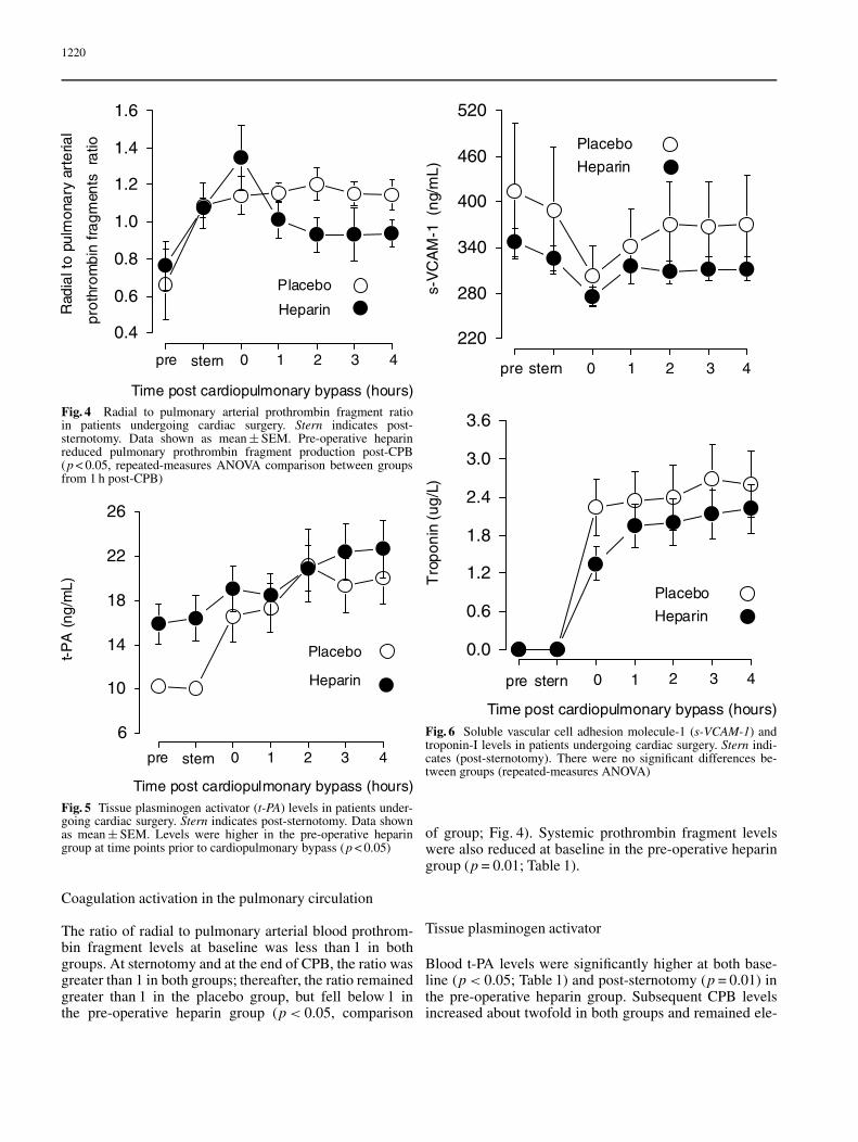
Fig. 4 Radial to pulmonary arterial prothrombin fragment ratioin patients undergoing cardiac surgery. Stern indicates post-sternotomy. Data shown as mean ± SEM. Pre-operative heparinreduced pulmonary prothrombin fragment production post-CPB(p < 0.05, repeated-measures ANOVA comparison between groupsfrom 1 h post-CPB)
Fig. 5 Tissue plasminogen activator (t-PA) levels in patients under-going cardiac surgery. Stern indicates post-sternotomy. Data shownas mean ± SEM. Levels were higher in the pre-operative heparingroup at time points prior to cardiopulmonary bypass (p < 0.05)
Coagulation activation in the pulmonary circulation
The ratio of radial to pulmonary arterial blood prothrom-bin fragment levels at baseline was less than 1 in bothgroups. At sternotomy and at the end of CPB, the ratio wasgreater than 1 in both groups; thereafter, the ratio remainedgreater than 1 in the placebo group, but fell below 1 inthe pre-operative heparin group (p < 0.05, comparison
Fig. 6 Soluble vascular cell adhesion molecule-1 (s-VCAM-1) andtroponin-I levels in patients undergoing cardiac surgery. Stern indi-cates (post-sternotomy). There were no significant differences be-tween groups (repeated-measures ANOVA)
of group; Fig. 4). Systemic prothrombin fragment levelswere also reduced at baseline in the pre-operative heparingroup (p = 0.01; Table 1).
Tissue plasminogen activator
Blood t-PA levels were significantly higher at both base-line (p < 0.05; Table 1) and post-sternotomy (p = 0.01) inthe pre-operative heparin group. Subsequent CPB levelsincreased about twofold in both groups and remained ele-
1221
vated thereafter. The levels reached were similar in bothgroups post-CPB (Fig. 5).
Vascular cell adhesion molecule-1 and troponin-I levels
Blood s-VCAM-1 levels were similar in both groupsthroughout the study period (Fig. 6). Troponin-I levelswere initially lower at the end of CPB, in the pre-operative heparin group, and thereafter levels were similar(Fig. 6).
Pulmonary white cell and platelet retention
The changes in the radial to pulmonary arterial platelet andwhite cell ratios were similar in both groups. We did notfind significant evidence of pulmonary platelet or whitecell retention.
Post-operative characteristics
Post-operative characteristics, including hours of mechan-ical ventilation, intensive care unit and hospital length ofstays, haemodynamic characteristics, inotrope use, Hb andlactate levels, were similar between groups. Chest-drainblood loss at 4 h was greater in the pre-operative heparingroup, but transfusion requirements were similar betweengroups (Table 3).
Discussion
Our study found evidence suggesting that pulmonarymicrovascular thrombosis occurs during cardiac surgery.Firstly, in the placebo group we found that the alveolardead-space fraction increased by 35% compared withbaseline levels following cardiac surgery (a findingconsistent with reduced alveolar perfusion). Secondly,we demonstrated that cardiac surgery triggered coagu-lation activation in the pulmonary circulation. Finally,we demonstrated that a pre-operative heparin infusionwas associated with reduced alveolar dead-space fractionlevels and reduced coagulation activation in the pulmonarycirculation.
This effect of pre-operative heparin in limiting co-agulation activation in the pulmonary circulation mayappear surprising in view of the fact that both groups wereadministered 300 U/kg of heparin just before commence-ment of CPB; however, a number of the anti-coagulantactions of heparin manifest through pathways requiringprotein synthesis; hence, these actions peak some hoursafter heparin administration. The addition of a pre-operative heparin infusion to the standard intra-operativebolus of heparin may theoretically therefore enhance
heparin’s anti-thrombotic properties during cardiacsurgery. These anti-coagulant actions include endothelialexpression of heparan sulfate [27], endothelial and plateletsecretion of tissue factor pathway inhibitor (TFPI) andinhibition of endothelial and monocyte expression oftissue factor [28, 29]. Our finding that post-operative chesttube drainage was significantly greater in the pre-operativeheparin group also supports this contention.
Heparin has also previously been shown to trigger in-creased endothelial expression of t-PA [30]. As expected,therefore, t-PA levels were significantly increased beforeCPB in the pre-operative heparin group. Following theintra-operative heparin bolus levels increased in bothgroups to a similar extent. Enhancement of fibrinolysismay also be a mechanism by which pre-operative heparinlimited the increase in the alveolar dead-space fraction.Pre-operative heparin had no effect on s-VCAM-1 levelsor the extent of white cell or platelet retention in the lungs.Pre-operative heparin did not significantly improve theA-a gradient, mean pulmonary artery pressure levels ortroponin I levels.
Our finding that cardiac surgery was associated witha marked increase in the alveolar dead-space fraction isconsistent with other forms of acute inflammatory lunginjury, such as the acute respiratory distress syndrome(ARDS). A recent study of patients presenting to the emer-gency department with ARDS found that the extent of theincrease in the alveolar dead space was an independentpredictor of death [31].
The administration of aprotinin may have played a rolein our finding of evidence of pulmonary microvascularthrombosis. Case reports have demonstrated an asso-ciation between aprotinin and histological evidence ofmicrovascular thrombosis [21–25]. In addition, aprotininhas been implicated in the development of multi-organfailure following cardiac surgery [19, 20].
Potential limitations
The major limitations of our study were the indirectmethods used to assess evidence of pulmonary micro-vascular thrombosis. Factors other than microvascularobstruction may increase the alveolar dead-space fraction.Alveolar blood flow may fall due to low blood pressureor a poor cardiac output [32]. Blood pressure and cardiacoutput levels were, however, adequate and equivalent inboth groups. Variations in ventilation parameters, suchas tidal volume and respiratory rate, may also increasethe alveolar dead-space fraction [33]. The ventilationparameters were, however, kept constant throughout thestudy period.
Atelectasis may also contribute to an increase in thealveolar dead space through high V/Q mismatch. Ourinterpretation of the changes in the ratio of radial topulmonary arterial prothrombin fragment levels may be
1222
questioned. Our interpretation is supported by a study ofpatients undergoing cardiac surgery that demonstratedincreased intravascular fibrin formation following reper-fusion of the lungs and heart [34], and also by animalmodels of CPB and pulmonary ischaemic-reperfusioninjury, which demonstrated pulmonary microvascularthrombosis and beneficial outcomes associated withanti-coagulants [14–17].
The major implications of our study are that microvas-cular thrombosis may be a mechanism of lung injury inpatients undergoing cardiac surgery, and that this may belimited by a pre-operative heparin infusion. Further work,however, is required to establish this.
Acknowledgements. This study was supported by the St. Vincent’sHospital Research Endowment Fund, The Intensive Care Foundationand by departmental funds.
References
1. Ng CS, Wan S, Yim AP, Arifi AA(2002) Pulmonary dysfunction aftercardiac surgery. Chest 121:1269–1277
2. Massoudy P, Zahler S, Becker BF,Braun SL, Barankay A, Meisner H(2001) Evidence for inflammatoryresponses of the lungs during coronaryartery bypass grafting with cardiopul-monary bypass. Chest 119:31–36
3. Serraf A, Robotin M, Bonnet N, De-truit H, Baudet B, Mazmanian MG,Herve P, Planche C (1997) Alter-ation of the neonatal pulmonaryphysiology after total cardiopul-monary bypass. J Thorac CardiovascSurg 114:1061–1069
4. Schlensak C, Doenst T, Preusser S,Wunderlich M, Kleinschmidt M,Beyersdorf F (2001) Bronchial arteryperfusion during cardiopulmonarybypass does not prevent ischemia of thelung in piglets: assessment of bronchialartery blood flow with fluorescentmicrospheres. Eur J CardiothoracSurg 19:326–331
5. Chai PJ, Williamson JA, Lodge AJ,Daggett CW, Scarborough JE, Me-liones JN, Cheifetz IM, Jaggers JJ,Ungerleider RM (1999) Effects ofischemia on pulmonary dysfunctionafter cardiopulmonary bypass. AnnThorac Surg 67:731–735
6. Suzuki T, Ito T, Kashima I, Teruya K,Fukuda T (2001) Continuous perfusionof pulmonary arteries during totalcardiopulmonary bypass favorablyaffects levels of circulating adhesionmolecules and lung function. J ThoracCardiovasc Surg 122:242–248
7. Wan S, LeClerc JL, Vincent JL (1997)Inflammatory response to cardiopul-monary bypass: mechanisms involvedand possible therapeutic strategies.Chest 112:676–692
8. Dixon B (2004) The role of microvas-cular thrombosis in sepsis. AnaesthIntensive Care 32:619–629
9. Sapru A, Wiemels JL, Witte JS,Ware LB, Matthay MA (2006)Acute lung injury and the coagula-tion pathway: potential role of genepolymorphisms in the protein C andfibrinolytic pathways. Intensive CareMed 32:1293–1303
10. Beck G, Habicht GS, Benach JL,Miller F (1986) Interleukin 1: a com-mon endogenous mediator of inflamma-tion and the local Shwartzman reaction.J Immunol 136:3025–3031
11. Dosquet C, Weill D, Wautier JL (1995)Cytokines and thrombosis. J CardiovascPharmacol Suppl 25(2):S13–S19
12. Blume ED, Nelson DP, Gauvreau K,Walsh AZ, Plumb C, Neufeld EJ,Hickey PR, Mayer JE, Newburger JW(1997) Soluble adhesion moleculesin infants and children under-going cardiopulmonary bypass.Circulation 96:II-352–357
13. Massoudy P, Zahler Sea, Becker BF,Braun SL, Barankay A, Richter JA,Meisner H (1999) Significant leuko-cyte and platelet retention duringpulmonary passage after declamping ofthe aorta in CABG patients. Eur J MedRes 4:178–182
14. Tanaka K (2001) Specific inhibi-tion of thrombin activity duringcardiopulmonary bypass reducesischemia-reperfusion injury of the lung.Fukuoka Igaku Zasshi 92:7–20
15. Okada K, Fujita T, Minamoto K,Liao H, Naka Y, Pinsky DJ (2000)Potentiation of endogenous fibri-nolysis and rescue from lung is-chemia/reperfusion injury in interleukin(IL)-10-reconstituted IL-10 null mice.J Biol Chem 275:21468–21476
16. Pinsky DJ, Liao H, Lawson CA,Yan SF, Chen J, Carmeliet P, Loskut-off DJ, Stern DM (1998) Coordinatedinduction of plasminogen activatorinhibitor-1 (PAI-1) and inhibitionof plasminogen activator gene ex-pression by hypoxia promotes pul-monary vascular fibrin deposition.J Clin Invest 102:919–928
17. Lawson CA, Yan SD, Yan SF, Liao H,Zhou YS, Sobel J, Kisiel W, Stern DM,Pinsky DJ (1997) Monocytes and tissuefactor promote thrombosis in a murinemodel of oxygen deprivation. J ClinInvest 99:1729–1738
18. Argenbright LW, Barton RW (1992)Interactions of leukocyte integrins withintercellular adhesion molecule 1 in theproduction of inflammatory vascularinjury in vivo. The Shwartzman reactionrevisited. J Clin Invest 89:259–272
19. Mangano DT (2002) Aspirin andmortality from coronary bypass surgery.N Engl J Med 347:1309–1317
20. Mangano DT, Tudor IC, Dietzel C(2006) The risk associated withaprotinin in cardiac surgery. N EnglJ Med 354:353–365
21. Saffitz JE, Stahl DJ, Sundt TM,Wareing TH, Kouchoukos NT(1993) Disseminated intravascu-lar coagulation after administrationof aprotinin in combination withdeep hypothermic circulatory arrest.Am J Cardiol 72:1080–1082
22. Sundt TM III, Kouchoukos NT, Saf-fitz JE, Murphy SF, Wareing TH,Stahl DJ (1993) Renal dysfunction andintravascular coagulation with aprotininand hypothermic circulatory arrest. AnnThorac Surg 55:1418–1424
23. Blaisdell FW, Lim RC Jr, Amberg JR,Choy SH, Hall AD, Thomas AN(1966) Pulmonary microembolism.A cause of morbidity and deathafter major vascular surgery. ArchSurg 93:776–786
24. Gregoric ID, Patel V, Radovance-vic R, Bracey AW, Radovancevic B,Frazier OH (2005) Pulmonary micro-thrombi during left ventricular as-sist device implantation. Tex HeartInst J 32:228–231
25. Cooper JR Jr, Abrams J, Frazier OH,Radovancevic R, Radovancevic B,Bracey AW, Kindo MJ, Gregoric ID(2006) Fatal pulmonary microthrombiduring surgical therapy for end-stageheart failure: possible associationwith antifibrinolytic therapy. J ThoracCardiovasc Surg 131:963–968
26. Severinghaus JW, Stupfel M (1957)Alveolar dead space as an index ofdistribution of blood flow in pulmonarycapillaries. J Appl Physiol 10:335–348
1223
27. Cadroy Y, Gaspin D, Dupouy D,Lormeau JC, Boneu B, Sie P (1996)Heparin reverses the procoagulantproperties of stimulated endothelialcells. Thromb Haemost 75:190–195
28. Gori AM, Pepe G, Attanasio M, Fal-ciani M, Abbate R, Prisco D, Fedi S,Giusti B, Brunelli T, Comeglio P,Gensini GF, Neri Serneri GG (1999)Tissue factor reduction and tissuefactor pathway inhibitor releaseafter heparin administration. ThrombHaemost 81:589–593
29. Pepe G, Giusti B, Attanasio M,Gori AM, Comeglio P, Martini F,Gensini G, Abbate R, Neri Serneri GG(1997) Tissue factor and plasminogenactivator inhibitor type 2 expressionin human stimulated monocytes isinhibited by heparin. Semin ThrombHemost 23:135–141
30. Marsh NA, Minter AJ, Chesterman CN(1990) The effect of heparin and otherglycosaminoglycans on levels of tissueplasminogen activator and plasminogenactivator inhibitor in cultured humanumbilical vein endothelial cells. BloodCoagul Fibrinolysis 1:133–138
31. Nuckton TJ, Alonso JA, Kallet RH,Daniel BM, Pittet JF, Eisner MD,Matthay MA (2002) Pulmonary dead-space fraction as a risk factor for deathin the acute respiratory distress syn-drome. N Engl J Med 346:1281–1286
32. Askrog V, Pender J, Eckenhoff J (1964)Changes in the physiological deadspace during deliberate hypotension.Anesthesiology 25:744–751
33. Nunn JF (1977) Respiratory dead space.In: Applied respiratory physiology, 2ndedn. Butterworths, London
34. Chandler WL, Velan T (2003) Esti-mating the rate of thrombin and fibringeneration in vivo during cardiopul-monary bypass. Blood 101:4355–4362