Embed Size (px)
Citation preview

General Pathology
Hemostasis
(Web)
Paul Hanna Feb 2015
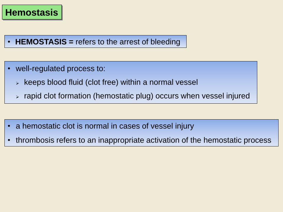
• well-regulated process to:
keeps blood fluid (clot free) within a normal vessel
rapid clot formation (hemostatic plug) occurs when vessel injured
• a hemostatic clot is normal in cases of vessel injury
• thrombosis refers to an inappropriate activation of the hemostatic process
• HEMOSTASIS = refers to the arrest of bleeding
Hemostasis
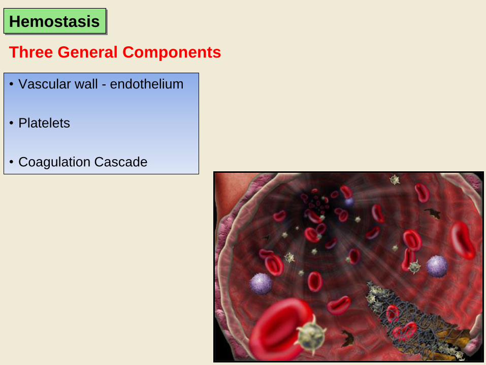
Three General Components
• Vascular wall - endothelium
• Platelets
• Coagulation Cascade
Hemostasis
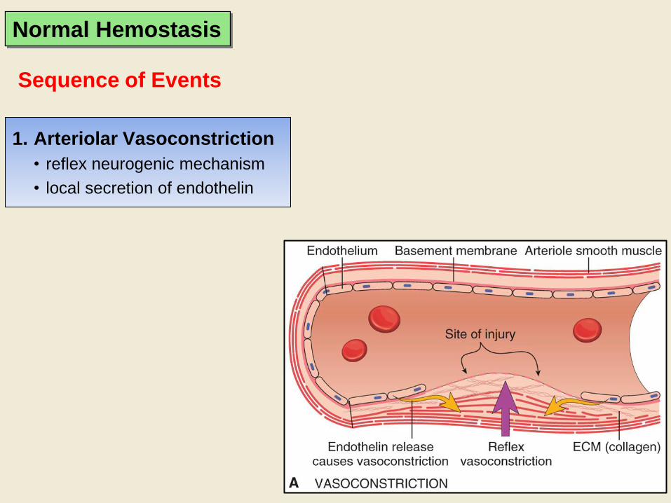
1. Arteriolar Vasoconstriction
• reflex neurogenic mechanism
• local secretion of endothelin
Sequence of Events
Normal Hemostasis

1. Arteriolar Vasoconstriction
2. Primary Hemostasis -
PLATELET
• platelets respond to exposure
of subendothelial ECM by:
Adhesion
Shape Change
Granule Release
Recruitment
• Platelet aggregation
(1o hemostatic plug)
Sequence of Events
Normal Hemostasis
1. Arteriolar Vasoconstriction
2. Primary Hemostasis -
PLATELET
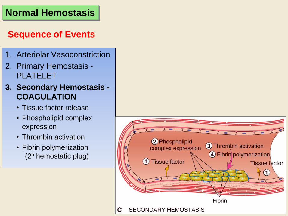
3. Secondary Hemostasis -
COAGULATION
• Tissue factor release
• Phospholipid complex
expression
• Thrombin activation
• Fibrin polymerization
(2o hemostatic plug)
Sequence of Events
Normal Hemostasis

1. Arteriolar Vasoconstriction
2. Primary Hemostasis - PLATELET
3. Secondary Hemostasis - Coagulation
4. Antithrombotic Counter-Regulation
• Factors released to limit the size of the hemostatic plug
Sequence of Events
Normal Hemostasis
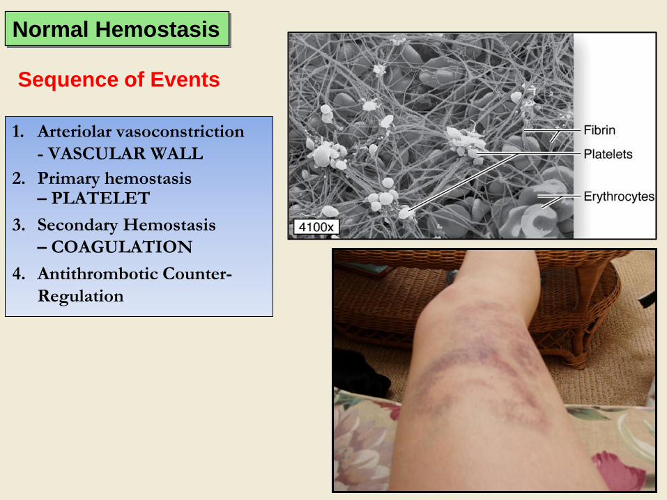
1. Arteriolar vasoconstriction
- VASCULAR WALL
2. Primary hemostasis – PLATELET
3. Secondary Hemostasis
– COAGULATION
4. Antithrombotic Counter-
Regulation
Sequence of Events
Normal Hemostasis
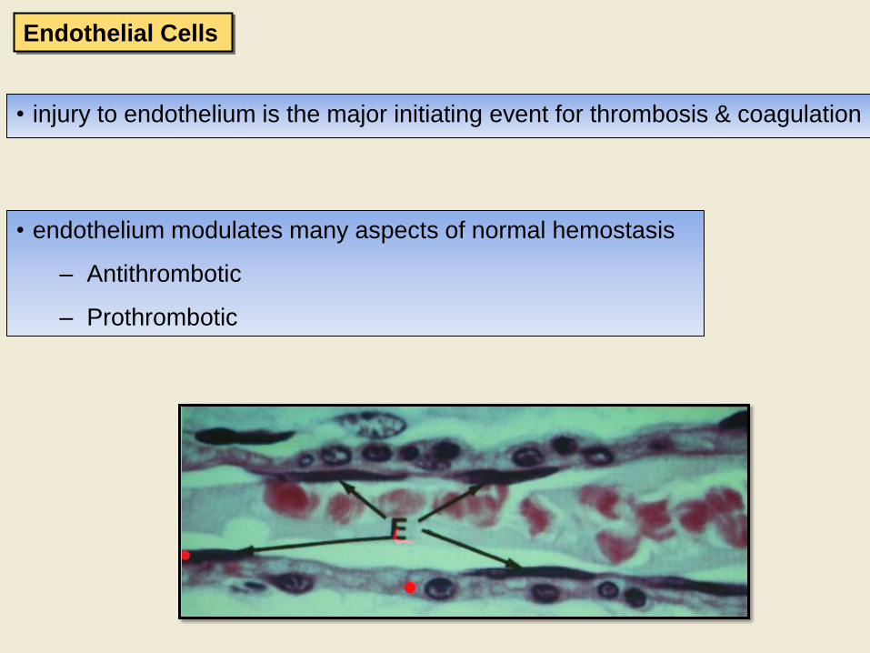
• injury to endothelium is the major initiating event for thrombosis & coagulation
• endothelium modulates many aspects of normal hemostasis
– Antithrombotic
– Prothrombotic
Endothelial Cells

Antithrombotic Properties
• Antiplatelet
– barrier to ECM
– PGI2 and NO
– ADPase
• Fibrinolytic
– Plasminogen activators (tPA)
• Anticoagulant
– Heparin-like molecules (- ATIII)
– Thrombomodulin
– Tissue factor pathway inhibitor
Endothelial Cells
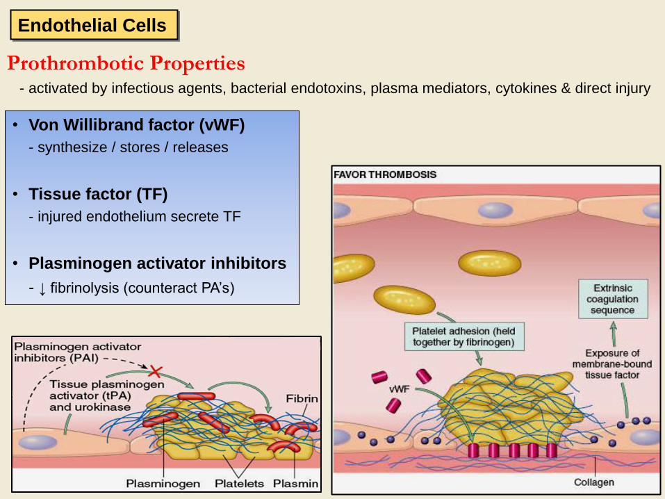
Prothrombotic Properties - activated by infectious agents, bacterial endotoxins, plasma mediators, cytokines & direct injury
Endothelial Cells
• Von Willibrand factor (vWF)
- synthesize / stores / releases
• Tissue factor (TF)
- injured endothelium secrete TF
• Plasminogen activator inhibitors
- ↓ fibrinolysis (counteract PA’s)

• Numerous growth factors are secreted by endothelial cells
– Platelet Derived Growth Factor (PDGF)
• stimulates smooth muscles and fibroblasts
– Fibroblast Growth Factor (FGF)
• stimulates fibroblasts and angiogenesis
– Transforming Growth Factor β (TGF- β)
• modulates vascular (& fibrous) repair
Vascular Repair
Endothelial Cells

Figure 12–13.(Mescher) Platelets. Platelets are cell fragments 2–4 µm in diameter
derived from megakaryocytes of bone marrow. Their primary function is to rapidly
release the content of their granules upon contact with collagen (or other materials
outside of the endothelium) to begin the process of clot formation and reduce blood
loss from the vasculature. (a): In a blood smear, platelets (arrows) are often found as
aggregates. Individually they show a lightly stained hyalomere region surrounding a
more darkly stained central granulomere containing membrane—enclosed granules.
X1500. Wright. (b): Ultrastructurally a platelet typically shows a system of
microtubules and actin filaments near the periphery to help maintain its shape and an
open canalicular system of vesicles continuous with the plasmalemma. The central
granulomere region contains glycogen and secretory granules of different types.
• derived from megakaryocytes; circulate as round, smooth discs
• play central role in hemostasis contain mostly procoagulants
• form the 1o hemostatic plug covers and seals a small damaged area
Platelets

For information only
Platelets

Vascular injury
Exposure of ECM
VWF–bridge for
platelets to ECM
Adhesion and Shape Change
Platelet Response
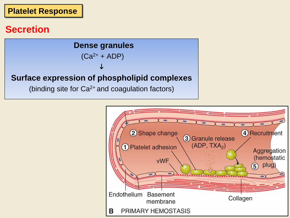
Secretion
Dense granules
(Ca2+ + ADP)
Surface expression of phospholipid complexes
(binding site for Ca2+ and coagulation factors)
Platelet Response

Aggregation
• TXA2 + ADP enlarging platelet aggregate 1o haemostatic plug
Platelet Response

THROMBOCYTOPENIA
Definition: platelet numbers are low
(<100 x 109/L is a thrombocytopenia in many species)
Diagnosis: history of bleeding
low platelet counts
Mechanisms: Deficient formation of platelets (eg, estrogen toxicoses)
Excessive utilization (eg, consumptive coagulopathies)
Premature destruction (eg, antibodies to platelets)
THROMBOCYTOPATHY
Definition: Defective platelet function
Mechanisms: Defect in adhesion (eg, von WIllebrand’s disease)
aggregation
release of granules
Platelet Disorders

• a reaction pathway:
enzyme (previously activated coagulation factor)
+
substrate (next non-activated coagulation factor)
activated coagulation factor
• 3rd arm of hemostasis an enzymatic cascade
• reaction typically occurs on platelet phospholipid complex held together by Ca2+
Coagulation Cascade
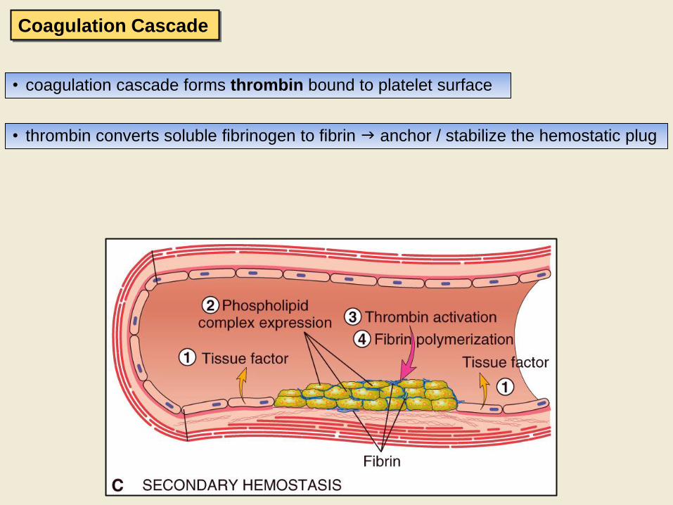
• coagulation cascade forms thrombin bound to platelet surface
• thrombin converts soluble fibrinogen to fibrin anchor / stabilize the hemostatic plug
Coagulation Cascade
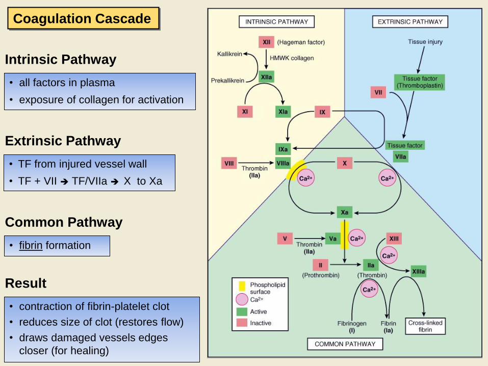
Intrinsic Pathway
• all factors in plasma
• exposure of collagen for activation
Extrinsic Pathway
• TF from injured vessel wall
• TF + VII TF/VIIa X to Xa
Common Pathway
• fibrin formation
Result
• contraction of fibrin-platelet clot
• reduces size of clot (restores flow)
• draws damaged vessels edges
closer (for healing)
Coagulation Cascade
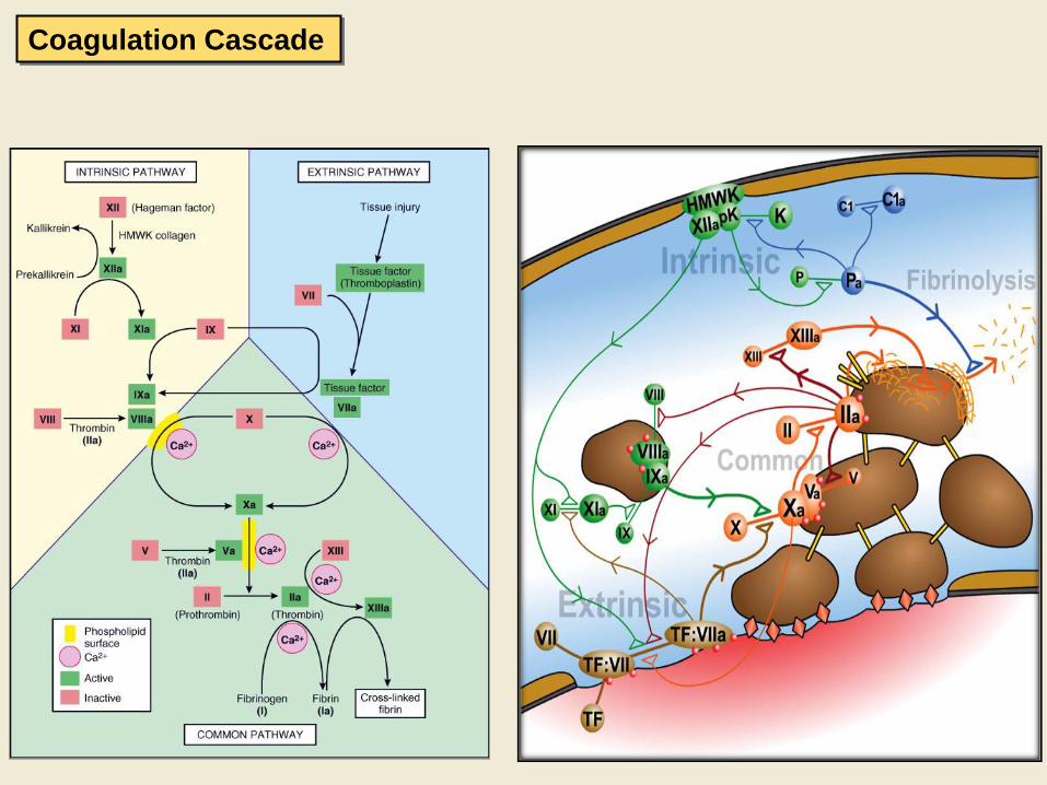
Coagulation Cascade
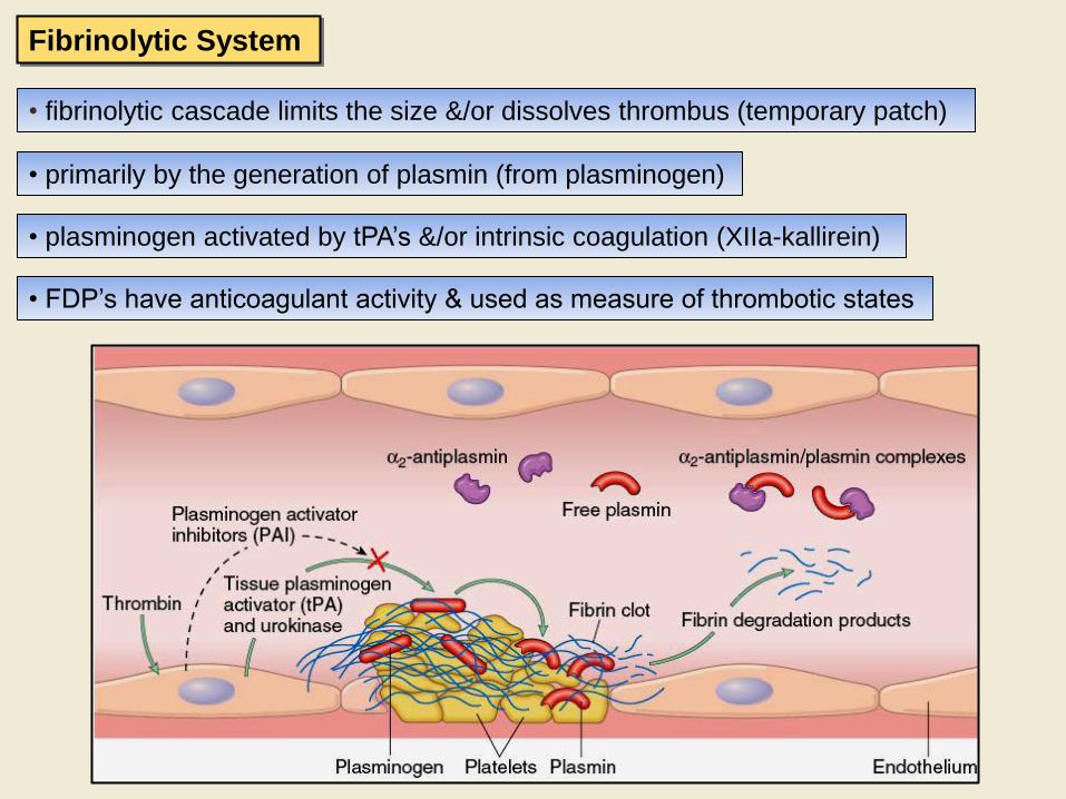
• fibrinolytic cascade limits the size &/or dissolves thrombus (temporary patch)
• primarily by the generation of plasmin (from plasminogen)
• plasminogen activated by tPA’s &/or intrinsic coagulation (XIIa-kallirein)
• FDP’s have anticoagulant activity & used as measure of thrombotic states
Fibrinolytic System

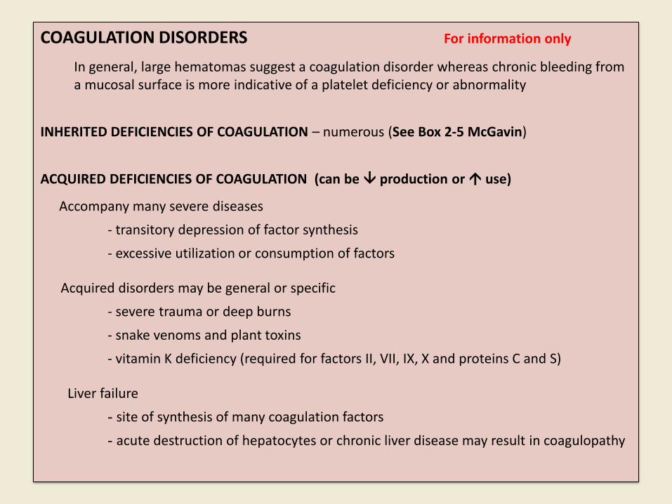
COAGULATION DISORDERS For information only
In general, large hematomas suggest a coagulation disorder whereas chronic bleeding from a mucosal surface is more indicative of a platelet deficiency or abnormality

COAGULATION DISORDERS For information only
In general, large hematomas suggest a coagulation disorder whereas chronic bleeding from a mucosal surface is more indicative of a platelet deficiency or abnormality
INHERITED DEFICIENCIES OF COAGULATION – numerous (See Box 2-5 McGavin)
COAGULATION DISORDERS For information only
In general, large hematomas suggest a coagulation disorder whereas chronic bleeding from a mucosal surface is more indicative of a platelet deficiency or abnormality
INHERITED DEFICIENCIES OF COAGULATION – numerous (See Box 2-5 McGavin)
ACQUIRED DEFICIENCIES OF COAGULATION (can be production or use)
Accompany many severe diseases
- transitory depression of factor synthesis
- excessive utilization or consumption of factors
Acquired disorders may be general or specific
- severe trauma or deep burns
- snake venoms and plant toxins
- vitamin K deficiency (required for factors II, VII, IX, X and proteins C and S)
Liver failure
- site of synthesis of many coagulation factors
- acute destruction of hepatocytes or chronic liver disease may result in coagulopathy