Embed Size (px)
Citation preview
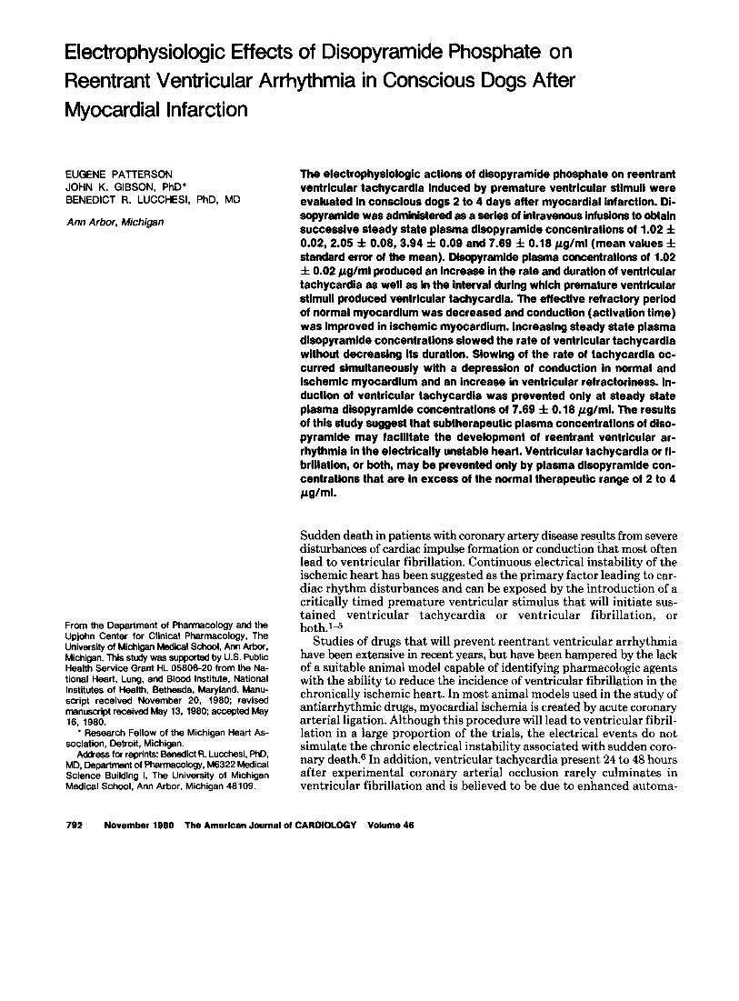
Electrophysiologic Effects of Disopyramide Phosphate on
Reentrant Ventricular Arrhythmia in Conscious Dogs After
Myocardial Infarction
EUGENE PATTERSON JOHN K. GIBSON, PhD” BENEDICT R. LUCCHESI, PhD, MD
Ann Arbor, Michigan
From the Department of Pharmacology and the Upjohn Center for Clinical Pharmacology, The University of Michigan Medical School, Ann Arbor, Michigan. This study was suppcrted by U.S. Public Health Service Grant HL 05806-20 from the Na- tional Heart, Lung, and Blood Institute, National Institutes of Health, Bethesda, Maryland. Manu- script received November 20, 1980; revised manuscript received May 13.1980; accepted May 16, 1980.
l Research Fellow of the Michigan Heart As- sociation, Detroit, Michigan.
Address for reprints: Benedict R. Lucchesi, PhD, MD, Department of Pharmacology, M6322 Medical Science Building I, The University of Michigan Medical School, Ann Arbor, Michigan 48109.
The electrophysiologic actions of dtsopyramide phosphate on reentrant ventricular tachycardia Induced by premature ventricular stimuli were evaluated in conscious dogs 2 to 4 days after myocardial infarction. Di- sopyramlde was administered as a series of intravenous infusions to obtain successive steady state plasma dtsopyramide concentrations of 1.02 f 0.02,2.05 f 0.08,3.94 f 0.09 and 7.69 f 0.18 pg/ml (mean values f standard error of the mean). Dtsopyramide plasma concentratffns of 1.02 f 0.02 pg/ml produced an increase in the rate and duration of ventricular tachycardia as well as in the interval during which premature ventricular stimull produced ventricular tachycardia. The effective refractory period of normal myocardium was decreased and conduction (activation time) was improved in ischemic myocardium. Increasing steady state plasma disopyramide concentrations slowed the rate of ventricular tachycardta without decreasing its duration. Slowing of the rate of tachycardia oc- curred simultaneously with a depression of conduction in normal and ischemic myocardium and an increase in ventricular refractoriness. In- duction of ventricular tachycardla was prevented only at steady state plasma disopyramide concentrations of 7.69 f 0.18 pg/ml. The results of this study suggest that subtherapeutic plasma concentrattons of diso- pyramide may facilitate the development of reentrant ventricular ar- rhythmia in the electrically unstable heart. Ventricular tachycardia or ft- brlllation, or both, may be prevented only by plasma disopyramide con- centrations that are in excess of the normal therapeutic range of 2 to 4 pg/ml.
Sudden death in patients with coronary artery disease results from severe disturbances of cardiac impulse formation or conduction that most often lead to ventricular fibrillation. Continuous electrical instability of the ischemic heart has been suggested as the primary factor leading to car- diac rhythm disturbances and can be exposed by the introduction of a critically timed premature ventricular stimulus that will initiate sus- tained ventricular tachycardia or ventricular fibrillation, or both.1-5
Studies of drugs that will prevent reentrant ventricular arrhythmia have been extensive in recent years, but have been hampered by the lack of a suitable animal model capable of identifying pharmacologic agents with the ability to reduce the incidence of ventricular fibrillation in the chronically ischemic heart. In most animal models used in the study of antiarrhythmic drugs, myocardial ischemia is created by acute coronary arterial ligation. Although this procedure will lead to ventricular fibril- lation in a large proportion of the trials, the electrical events do not simulate the chronic electrical instability associated with sudden coro- nary death.6 In addition, ventricular tachycardia present 24 to 48 hours after experimental coronary arterial occlusion rarely culminates in ventricular fibrillation and is believed to be due to enhanced automa-
792 November 1990 The American Journal ol CARDIOLOGY Volume 46
ticity in Purkinje fibers. 6,7 Recently, it was demon- strated that a critically timed electrically induced pre- mature ventricular stimulus could initiate reentrant ventricular tachyarrythmia and ventricular fibrillation in dogs 2 to 9 days after acute myocardial infarction. This reentrant electrical activity also simulated that observed in patients in whom reentrant rhythms could be induced by programmed electrical stimulation of the heart.8rg
This study was undertaken for the purpose of eval- uating the ability of disopyramide phosphate to prevent reentrant arrhythmia resulting from programmed electrical stimulation in the presence of chronic myo- cardial ischemia. The electrophysiologic effects of di- sopyramide phosphate were evaluated in relation to drug plasma concentrations. The results of these studies in the conscious dog with experimentally induced chronic myocardial ischemia demonstrate that diso- pyramide in clinically recommended plasma concen- trations (2 to 4 pg/ml) is unlikely to be beneficial in preventing reentrant ventricular tachyarrhythmia; this prevention can only be accomplished with plasma concentrations in excess of 4 pg/ml.
Methods Animal preparation: Male mongrel dogs weighing between
14.2 and 18.0 kg were anesthetized with intravenous sodium pentobarbital, 30 mg/kg body weight. Each animal was intu- bated with a cuffed endotracheal tube and ventilated with room air using a Harvard respirator. With use of aseptic techniques, sterile catheters were implanted in the left com- mon carotid artery and left external jugular vein and passed subcutaneously to the surface through an incision in the back of the neck. A left thoracotomy was performed in the fourth intercostal space and the heart was suspended in a pericardial cradle. A 5 mm section of the left anterior descending coronary artery was dissected free from the surrounding myocardium at the tip of the left atria1 appendage. A length of suture was passed around the artery and a 20 gauge needle, then securely tied and the needle withdrawn. Left anterior descending coronary arterial flow was monitored using an electromagnetic flow probe (Carolina Medical Electronics) before and after stenosis to ensure that coronary flow at rest was undisturbed. Silastic@ tubing was used to occlude coronary flow for a period of 90 minutes and then flow was allowed to return through the stenosed artery. Lidocaine hydrochloride, 5 mg/kg intrave- nously followed by 5 mg/kg intramuscularly, was given to those animals manifesting severe ventricular arrhythmia on reinstitution of coronary flow.
Electrical placements: A bipolar electrode was sewn onto the surface of the left atria1 appendage. A second bipolar plunge electrode (28 gauge stainless steel, 5 to 7 mm in length) was placed on the right ventricular outflow tract with the exposed electrode tips placed in the interventricular septum.
Bipolar composite electrodes were placed on the epicardial surface of the left ventricle. One bipolar composite electrode was placed over normal myocardium and the second was placed over ischemic and infarcted myocardium. Correct placement of the bipolar composite electrodes was established at autopsy. The electrode wires were exited through the chest and passed subcutaneously to the back of the neck. The chest was closed in layers and procaine penicillin G, 1.2 million units, was administered intramuscularly.
Electrophysiologic studies: Forty-eight hours after myocardial infarction, the animals were conscious and resting
XX’PYRANiDE AND VENTRICULAR TACHYCARDIA-PATTERSON ET AL.
comfortably, supported in a sling. Arterial pressure was monitored with a Statham P23DC pressure transducer and recorded on a Grass model 7 polygraph. A lead II electrocar- diogram was monitored continuously and recorded. Bipolar composite electrograms, an atria1 bipolar electrogram and a lead II electrocardiogram were recorded on a Honeywell model 1858 Visicorder filtered at 15 to 2,500 hertz. If the ventricular ectopic rate was greater than 10 beats/min, the animal was returned to its quarters and monitored 24 hours later. If the rate was 10 beats/min or less, the following procedure was used: A Grass model SD5 stimulator was used to deliver square wave impulses, 3 ms in duration, at a voltage twice the dia- stolic threshold for atria1 or ventricular pacing (S1 stimuli). A Tektronix model 565 oscilloscope and differential amplifier were used to trigger the output of a Grass model S88 stimu- lator at the peak of the R wave of the lead 11 electrocardio- gram. A Grass model S88 stimulator and SIU5 stimulus iso- lation unit delivered 3 ms square wave pulses (Sa and S:j stimuli) at selected delays after the peak of the R wave to the bipolar electrode located in the interventricular septum. Di- astolic threshold was determined 200 ms after the peak of the R wave. All subsequent premature stimuli delivered to the interventricular septum were 1.5 times diastolic threshold.
The effective refractory period of the left ventricular myocardium was determined using square wave pulses (1.5 times threshold, 3 ms in duration) and decreasing the R to S2 interval until a conducted beat was not generated. Activation times (Q-EG intervals), the delay from the Q wave of the lead II electrocardiogram to the major positive spike of the normal zone or ischemic zone epicardial bipolar composite electro- grams, were determined during atria1 pacing at a rate of 175 beats/min. All electrophysiologic measurements were made from recordings performed at a paper speed of 100 to 400 mm/s.
Administration of disopyramide: Disopyramide phos- phate was administered by a series of two stage intravenous infusions as described by Wagner.‘O The values for apparent volume of distribution (2.01 liters/kg), alpha phase elimination constant (44.4 hour-l), and beta phase elimination constant (0.55 hour-l) were obtained from earlier pharmacokinetic studies.11J2 The infusion protocol was designed to reach and maintain successive steady state disopyramide plasma levels of 1,2,3,4, and 8 pg/ml. An infusion of disopyramide at a rate of 23.3 mg/kg per hour for 4 minutes was followed by a second infusion of 1.09 mg/kg per hour designed to maintain plasma disopyramide concentrations of 1 pg/ml. The infusion rate was increased to 24.4 mg/kg per hour for 4.0 minutes followed by an infusion at a rate of 2.18 mg/kg per hour to maintain plasma concentrations of 2 pg/ml. The infusion rate was increased again to 24.4 mg/kg per hour for 4.8 minutes followed by an infusion at a rate of 3.28 mg/kg per hour to maintain plasma concentrations of 3 pg/ml. To increase the steady state plasma concentrations to 4 p&/ml, the infusion rate was increased to 24.4 mg/kg per hour for 5.3 minutes followed by an infusion at a rate of 4.37 mg/kg per hour to maintain steady state concentrations. Steady state plasma disopyramide concen- trations of 8 pg/ml were achieved by increasing the infusion rate to 101.9 mg/kg per hour for 5 minutes followed by an infusion at a rate of 8.72 mg/kg per hour. A period of 20 min- utes was allowed to pass at each steady state plasma concen- tration before electrophysiologic testing was initiated. Diso- pyramide phosphate was administered using a Harvard model 975 infusion pump.
Determination of plasma disopyramide concentration: Blood samples (3 ml) were collected in 5 ml tubes containing 500 units of sodium heparin. Plasma disopyramide concen- trations were determined with the gas chromatographic method of Patterson et al.‘” Plasma samples were taken just
November 1990 The American Journal of CARDIOLOGY Volume 46 793
DISDPYRAMIDE AND VENTRICULAR TACHYCARDIA-PATTERSON ET AL.
before and after electrophysiologic testing at each steady state plasma level. Plasma potassium concentrations were deter- mined from these samples with use of flame spectrophotom- etry.
Determination of infarct size: The animals were anes- thetized with sodium pentobarbital, 20 mg/kg intravenously. The heart was excised, rinsed briefly in cold water and sliced from apex to base into 1 cm thick sections. The slices were incubated in 0.2 percent triphenyltetrazolium in 0.1 M phosphate buffer, pH 7.4, at 40°C for 15 minutes. Infarct size was determined gravimetrically and expressed as a percent of total left ventricular mass.
Materials: Disopyramide phosphate and p-chlorodiso- pyramide phosphate were obtained from the G.D. Searle Company. Triphenyltetrazolium was purchased from the Sigma Chemical Company. All other chemicals were reagent grade.
Statistical methods: Numerical values are expressed as the mean f standard error of the mean. Differences between groups were analyzed by analysis of variance.
Results
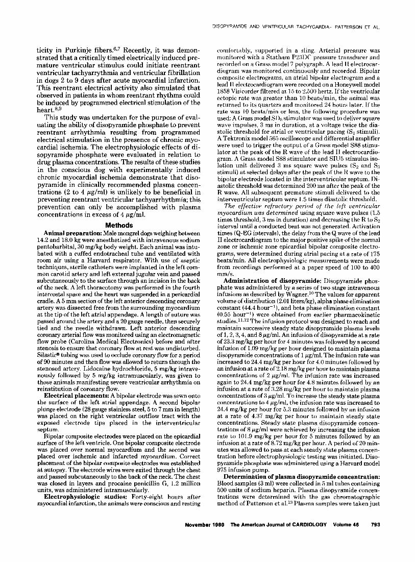
Rate-dependent conduction disorders: Two to 4 days after myocardial infarction, the ventricular ectopic rate had decreased to 3 f 1 complexes/min, and the animals were entered into the study protocol. Com- posite bipolar electrodes were used to record left ven- tricular myocardial activation. Activation of ischemic myocardium was prolonged (82 f 13 ms) compared with activation of normal myocardium (49 f 11 ms) (Table I). Left atria1 pacing was used to increase sequentially the ventricular rate to 300 beats/min or until the de- velopment of atrioventricular dissociation. As the ventricular rate increased, there was a significant in- crease in the duration of activation of ischemic myo- cardium whereas no change was seen in the duration of electrical activity in normal left ventricular myocardium (Table I). A further increase in the ventricular rate produced fractionation of the ischemic zone bipolar composite electrogram (Fig. 1).
Induction of ventricular tachycardia: Two to 4 days after myocardial infarction, programmed electrical stimulation was used to initiate reentrant rhythm in the conscious dogs. Premature ventricular stimuli were introduced during both normal sinus rhythm (cycle lengths 357 to 501 ms) and atria1 pacing (cycle lengths 284 to 454 ms). No differences were observed between the duration or rate of ventricular tachycardia induced
TABLE I
Duratlon of Electrical Activation of Ventricular Myocardium 48 Hours After Myocardial lnfarctlon
Cycle Length Duration of Activation (ms) (ms) Normal Zone lschemic Zone
::: 2 :; 49f 11 82 f 13+ 46f 8 106 f 19’
l ,+ Probability [p] values: l <0.05, compared with ischemic zone electrical activation duration at a cycle length of 430 f 23 ms, + <0.05 compared with normal zone.
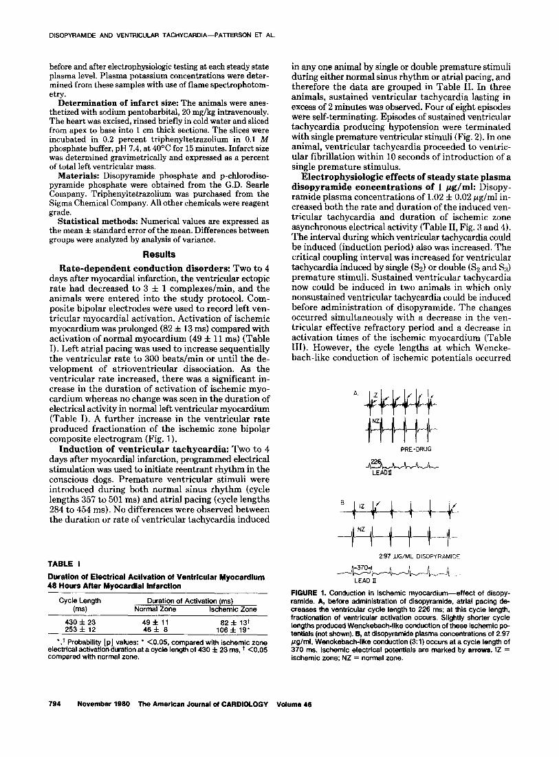
in any one animal by single or double premature stimuli during either normal sinus rhythm or atria1 pacing, and therefore the data are grouped in Table II. In three animals, sustained ventricular tachycardia lasting in excess of 2 minutes was observed. Four of eight episodes were self-terminating. Episodes of sustained ventricular tachycardia producing hypotension were terminated with single premature ventricular stimuli (Fig. 2). In one animal, ventricular tachycardia proceeded to ventric- ular fibrillation within 10 seconds of introduction of a single premature stimulus.
Electrophysiologic effects of steady state plasma disopyramide concentrations of 1 pg/ml: Disopy- ramide plasma concentrations of 1.02 f 0.02 pg/ml in- creased both the rate and duration of the induced ven- tricular tachycardia and duration of ischemic zone asynchronous electrical activity (Table II, Fig. 3 and 4). The interval during which ventricular tachycardia could be induced (induction period) also was increased. The critical coupling interval was increased for ventricular tachycardia induced by single (Sz) or double (Ss and Ss) premature stimuli. Sustained ventricular tachycardia now could be induced in two animals in which only nonsustained ventricular tachycardia could be induced before administration of disopyramide. The changes occurred simultaneously with a decrease in the ven- tricular effective refractory period and a decrease in activation times of the ischemic myocardium (Table III). However, the cycle lengths at which Wencke- bath-like conduction of ischemic potentials occurred
PRE-DRUG
LEADU
2.97 JJG/ML DISOPYRAMIDE
LEAD II
FIGURE 1. Conduction in ischemic myocardium-effect of disopy- ramide. A, before administration of disopyramide, atrial pacing de- creases the ventricular cycle length to 226 ms; at this cycle length, fractionation of ventricular activation occurs. Slightly shorter cycle lengths produced Wenckebach-like conduction of these ischemic po- tentials (not shown). B, at disopyramide plasma concentrations of 2.97 pglml. Wenckebach-like conduction (3:l) occurs at a cycle length of 370 ms. lschemic electrical potentials are marked by arrows. IZ = ischemic zone; NZ = normal zone.
794 November 1980 The Ametlcan Journal of CARDIOLDGY Volume 46
C)ISOPYFAWDE AND VENTRICULAR TACHYCARDIA-P.hTTERSON ET AL.
TABLE II
Electrophyslologic Variables
Plasma Disopyramide Reentrant Concentration Beats
@g/ml) (n)
Cycle Length of Ventricular Tachycardia
(ms)
_____.~____~_ ..-- ~.___ __..___ Duration of
Asynchronous induction Activity Period (ms)
(ms) S7 SR
(before drug administration) 1.02 f 0.02 2.05 f 0.08 2.99 f 0.03 3.94 f 0.09 7.69 f 0.18
6.2 f 0.8 163f 6 1005f 83 25 f 2 37 f 4
8.4 f 0.8‘ 155f 2‘ 1195f 87’ 42 f 4’ 48 f 4’ 5.5 f 0.8 193 f 8‘ 990f 81 24 f 6 34 f 4 4.3 f 1.1’
;:z ; ‘“8: 968 f 123 20 f 5 26 f 7’
3.6 f 0.4+ 882 f 102’ 16 f 5’ 23f9’ 0.6 f 0.5t 315 f lo+ - - -
l p <0.05, + p <O.Ol compared with values before drug administration.
were not altered from the values before drug adminis- tration.
Electrophysiologic effects of increasing plasma disopyramide concentrations: Plasma disopyramide concentrations were increased sequentially to steady state concentrations of 2.05 f 0.08,2.99 f 0.03,3.94 f 0.09 and 7.69 f 0.18 pg/ml. These increases produced a concentration-dependent increase in the cycle length of ventricular tachycardia without shortening the du- ration of the tachycardia until plasma levels of 7.69 f 0.18 pg/ml were attained (Table II, Fig. 3 and 4). The induction periods for the premature stimuli Ss and Ss were reduced sequentially by increasing plasma diso- pyramide concentrations, although ventricular tachy- cardia could be induced in all animals at plasma diso- pyramide concentrations of 3.94 f 0.09 pg/ml. The slowing of the rate of tachycardia occurred simulta- neously with the depression of cardiac conduction, as reflected in the concentration-dependent increase in the
cycle length of the ventricular arrhythmia and the in- crease in the activation times of normal and ischemic myocardium. Increasing plasma disopyramide con- centrations also produced sequential increases in ven- tricular refractoriness (Table III).
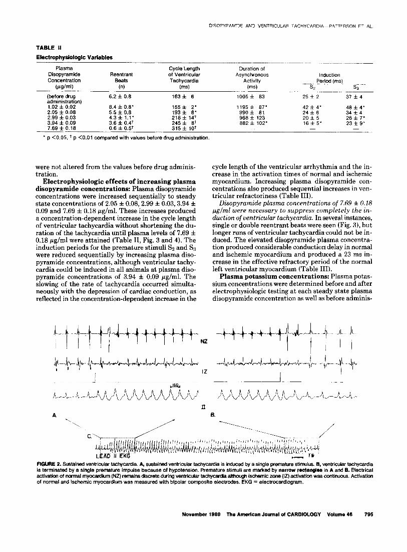
Disopyramide plasma concentrations of 7.69 f 0.18 pglml were necessary to suppress completely the in- duction of ventricular tachycardia. In several instances, single or double reentrant beats were seen (Fig. 31, but longer runs of ventricular tachycardia could not be in- duced. The elevated disopyramide plasma concentra- tion produced considerable conduction delay in normal and ischemic myocardium and produced a 23 ms in- crease in the effective refractory period of the normal left ventricular myocardium (Table III).
Plasma potassium concentrations: Plasma potas- sium concentrations were determined before and after electrophysiologic testing at each steady state plasma disopyramide concentration as well as before adminis-
FIGURE 2. Sustained ventricular tachycardff. A, sustained ventricufar tachycardii is induced by a single premature stimulus. B, ventricular tachycardia is terminated by a single premature impulse because of hypotension. Premature stimuli are marked by narrow rectangles in A and B. Electrical activation of ncrmal myocardium (NZ) remains discrete.dving ventricular tachycardii aftho@ ischemic zone (12) activation was continuous. Activation of normal and ischemic myocardium was measured with bipolar composite electrodes. EKG = electrocardiogram.
November 1990 The American Journal of CARDIOLOGY Volume 46 795
DISOPYRAMIDE AND VENTRICULAR TACHYCARDIA-PATTERSON ET AL.
A. PRE - DRUG 6.1.02 IJG/ML
NZ
C.3.97 l_lG/Ml_
FIGURE 3. Induced ventricular tachycardia-effect of disopyramide. A, before administration of disopyramide, a single premature stimulus elicits a nine beat ventricular tachycardia with a cycle length of 225 ms. B, at a plasma disopyramide concentration of 1.02 Wg/ml, a single premature stimulus produces sustained ventricular tachycardia at a cycle length of 216 ms. The ventricular tachycardia was sustained for 2 minutes and 45 seconds, after which spontaneous slowing and termination were seen. C, at a plasma disopyramide concentration of 3.97 pg/ml, a single premature stimulus elicits a ventricular tachycardia of three beats having a cycle length of 419 ms. D. at a plasma disopyramide concentration of 7.72 pg/ml, two premature stimuli produce a single reentrant beat. Activation of normal myocardium (NZ) remained discrete in the composite electrogram whereas ischemic zone activation (Ii!) was continuous during ventricular tachycardia.
tration of disopyramide. Before administration of di- sopyramide, the plasma potassium concentration was 3.8 f 0.1 mEq/liter and did not change significantly throughout the course of the experiment.
Infarct size: Occlusion of the left anterior descending coronary artery for a period of 90 minutes followed by reperfusion of the ischemic myocardium produced in- farct masses of 3.2 to 34.6 g, representing 3.0 to 34.0
TABLE III
Effective Refractory Periods and Changes In Q-EG Interval (mean values f standard error of the mean)
Plasma Disopyramide Concentration
(ualml)
Effective Refractory
Periods (ms)
AQ-EG (ms) Normal Zone lschemic Zone
(before drug 142 f 8 . administration)
1.02 f 0.02 138 f 9’ -0.2 f 0.3 -3.9 f 1.6’ 4.2 f 0.9’ 2.05 f 0.08 146 f 8’ 0.2 f 0.3
2.99 f 0.03 147 f 9‘ 9; ; 8.26’ 7.6 f 1.2’ 3.94 f 0.09 l 9.8 f 0.9+ 7.89 f 0.18 4.6 f 017’ 18.5 f 2.9+
l p <0.05, + p <O.Ol compared with values before drug adminis- tration.
Q-EG = interval from the initiation of the Q wave of lead II to the major deflection in the bipolar electrogram; AMG = change from the value obtained before drug administration (value before drug administration = 0).
percent of the left ventricular mass (mean 19.4 f 3.8 percent). The infarcts were located subendocardially and did not extend to the epicardial surface.
Discussion
Improved activation in ischemic myocardium and induction of reentrant tachyarrhythmia with subtherapeutic concentrations: Steady state plasma disopyramide concentrations of 1.02 f 0.02 pg/ml in- creased the duration and decreased the cycle length of reentrant ventricular tachycardia. This action is not unique to disopyramide: El-Sherif and Lazzara14 have shown verapamil to improve conduction in ischemic canine myocardium by improving depressed phase 0 maximal upstroke velocity of Purkinje fibers. In addi- tion, verapamil has been shown to decrease the cycle length of reentrant ventricular tachycardia and to in- crease the incidence of ventricular fibrillation in dogs 3 to 5 days after myocardial ischemic injury.15 Although there are few similarities in the mechanisms of action of disopyramide and verapamil, these data suggest that improvement of depressed conduction in ischemic myocardium may result in increased rates of ventricular tachycardia and an increased incidence of ventricular fibrillation.
Reddy and co-workenG6J7 demonstrated that sub- therapeutic concentrations of procainamide facilitated the induction of tachyarrhythmia in human subjects. Nonsustained ventricular tachycardia became sustained
796 November 1980 The American Journal of CARDIOLOGY Volume 46
DISOPYRAMIDE AND VENTRICULAR TACHYCARDIA-PATTERSON ET AL.
200 D
M( HG
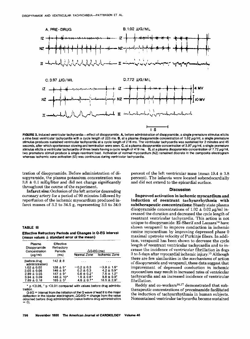
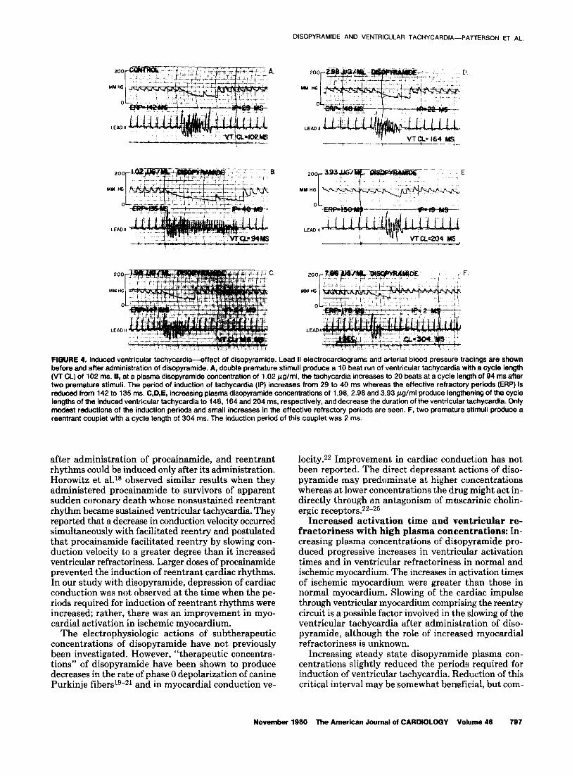
FfBURE 4. Induced ventricular tachycardia-effect of disopyramide. Lead II electrocardiograms and arterial blood pressure tracings are shown before and after administration of disopyramide. A, double premature stimuli produce a 10 beat run of ventricular tachycardia with a cycle length (VT CL) of 102 ms. B, at a plasma disopyramide concentration of 1.02 pg/ml. the tachycardia increases to 20 beats at a cycle length of 94 ms after two premature stimuli. The period of induction of tachycardia (IP) increases from 29 to 40 ms whereas the effective refractory periods (ERP) is reduced from 142 to 135 ms. C,D,E, increasing plasma disopyramide concentrations of 1.98. 2.98 and 3.93 pg/ml produce lengthening of tha cycle lengths of the induced ventricular tachycardia to 146, 164 and 2b4 ms, respectively, and decrease the duration of the ventricular tachycardia. Only modest reductions of the induction periods and small increases in the effective refractory periods are seen. F, two premature stimuli produce a reentrant couplet with a cycle length of 304 ms. The induction period of this couplet was 2 ms.
after administration of procainamide, and reentrant rhythms could be induced only after its administration. Horowitz et al.rs observed similar results when they administered procainamide to survivors of apparent sudden coronary death whose nonsustained reentrant rhythm became sustained ventricular tachycardia. They reported that a decrease in conduction velocity occurred simultaneously with facilitated reentry and postulated that procainamide facilitated reentry by slowing con- duction velocity to a greater degree than it increased ventricular refractoriness. Larger doses of procainamide prevented the induction of reentrant cardiac rhythms. In our study with disopyramide, depression of cardiac conduction was not observed at the time when the pe- riods required for induction of reentrant rhythms were increased; rather, there was an improvement in myo- cardial activation in ischemic myocardium.
The electrophysiologic actions of subtherapeutic concentrations of disopyramide have not previously been investigated. However, “therapeutic concentra- tions” of disopyramide have been shown to produce decreases in the rate of phase 0 depolarization of canine Purkinje fibers1g-21 and in myocardial conduction ve-
locity. Improvement in cardiac conduction has not been reported. The direct depressant actions of diso- pyramide may predominate at higher concentrations whereas at lower concentrations the drug might act in- directly through an antagonism of muscarinic cholin- ergic receptors.22-25
Increased activation time and ventricular re- fractoriness with high plasma concentrations: In- creasing plasma concentrations of disopyramide pro- duced progressive increases in ventricular activation times and in ventricular refractoriness in normal and ischemic myocardium. The increases in activation times of ischemic myocardium were greater than those in normal myocardium. Slowing of the cardiac impulse through ventricular myocardium comprising the reentry circuit is a possible factor involved in the slowing of the ventricular tachycardia after administration of diso- pyramide, although the role of increased myocardial refractoriness is unknown.
Increasing steady state disopyramide plasma con- centrations slightly reduced the periods required for induction of ventricular tachycardia. Reduction of this critical interval may be somewhat beneficial, but com-
November 1980 The American Journal of CARDfOLOGY Volume 48 797
DISOPYRAMIDE AND VENTRICULAR TACHYCARDIA-PATTERSON ET AL.
plete prevention of the induction of ventricular tachy- cardia may be necessary for prevention of sudden cor- onary death. Ruskin and Garan26 demonstrated that antiarrhythmic drug therapy that renders ventricular tachycardia noninducible by programmed cardiac stimulation constitutes effective prophylaxis against spontaneous recurrent sustained ventricular tachy- cardia in the majority of patients studied. Josephson et a1.27 showed that failure to maintain plasma concen- trations of antiarrhythmic drugs at levels previously shown to be effective in preventing the induction of sustained ventricular tachycardia can lead to the re- currence of sustained ventricular tachycardia or sudden coronary death. The 25 to 40 percent reduction in the period required for induction of sustained tachycardia, seen in our study at plasma disopyramide concentra- tions of 3.94 f 0.09 pg/ml, may be ineffective in pre- venting sudden coronary death because only concen- trations of 7.69 f 0.18 pg/ml prevented electrical in- duction of ventricular tachyarrhythmia (Table II).
It is not surprising that high plasma disopyramide concentrations are needed for suppression of induction of ventricular tachycardia. Benditt et a1.28 reported that disopyramide plasma concentrations that prevented the induction of ventricular tachycardia in human subjects were in excess of 3 pg/ml (mean nearly 5 pg/ml). Simi- larly, procainamide’s effectiveness was reported to re- quire plasma concentrations in excess of 9 pg/ml whereas quinidine plasma concentrations necessary for prevention of induction of ventricular tachycardia were in excess of 3 pg/ml. 25-27 Thus, the plasma concentra- tions of antiarrhythmic agents necessary for prevention of sudden coronary death may be higher than currently accepted “therapeutic concentrations.” However the “high” plasma concentrations of antiarrhythmic drugs may prove to have deleterious actions on cardiac elec- trophysiology and myocardial function.2g-31
Clinical implications: The majority of cardiac deaths are sudden and most likely due to a “primary” arrhythmic event most often characterized by ventric- ular fibrillation. Endeavors to prevent sudden coronary death with pharmacologic means have resulted in nu- merous reports in which drugs have been claimed to be
“effective” solely because of their ability to reduce the frequency of premature ventricular complexes. This study, although conducted in the experimental animal, might provide some important insights into an under- standing of why there often exists a lack of a predictable relation between the ability to suppress chronic asymptomatic complex ventricular arrhythmia and ventricular fibrillation or sudden coronary death. As demonstrated in the canine model using programmed cardiac stimulation in the presence of latent electrical instability, concentrations (2 to 4 pg/ml) of disopy- ramide known to reduce or prevent complex premature depolarizations were unable to prevent the induction of sustained reentrant ventricular tachyarrhythmia. The suggestion that recurrent episodes of ventricular tachycardia or ventricular fibrillation, or both, might be prevented by relatively high plasma concentrations of disopyramide has important implications with re- spect to future clinical trials designed to explore the prophylactic use of this drug in the patient at risk of sudden coronary death. A beneficial effect might not be realized if such trials follow current “therapeutic” guidelines in suggesting maintenance of plasma con- centrations of 2 to 4 pg/ml. However, the negative ino- tropic effect of disopyramide may preclude the use of higher dosage regimens in patients with compromised left ventricular function.
We are entering an era in which the pharmacologic prevention of sudden coronary death may soon become a reality. The task of identifying the proper agent or agents will not be a simple one; a single approach to the prevention of ventricular fibrillation is probably not attainable because the arrhythmia probably results from the interplay of many mechanisms, the most im- portant of which is an acute or chronic state of electrical instability. The animal model used in our study has the advantage over previous models of more closely repre- senting the clinical state associated with sudden coro- nary death. It may be useful in exploring the patho- physiologic processes that render the myocardium vulnerable to ventricular fibrillation and in studying how pharmacologic agents act to prevent a fatal elec- trical event.
References
1. Wellens HJ, Schuilenburg RM, Durrier D. Electrical stimulation rhythmias and electrophysiological consequences of myocardial of the heart in patients with ventricular tachycardia. Circulation ischemia and infarction. Circ Res 1978;42:740-8. 1972;46:216-26. 7. Harris AS. Delayed development of ventricular ectopic rhythms
2. Mason JW, Winkle RA. Electrode-catheter arrhythmia induction following experimental coronary occlusion. Circulation 195O;l: in the selection and assessment of antiarrhythmic drug therapy for 1318-28. recurrent ventricular tachycardia. Am J Cardiol 1977:40:579- 8. Karagueuzlan HS, Feneglio JJ, Weiss MB, Wft AL. Protracted 85. ventricular tachycardia induced by premature stimulation of the
3. Weflens HJJ, Duren DR, Lie KI. Observations on mechanisms of canine heart after coronary artery occlusion and reperfusion. Circ ventricular tachycardia in man. Circulation 1976;54:237-44. Res 1979;44:833-46.
4. Spielman SR, Famhkli A, Horowitz LN, Josephmn ME. Ventricular 9. El-Sherlf N, Scherlag BJ, Larzara R, Hope RR. Reentrant ven- fibrillation during programmed ventricular stimulation: incidence tricular arrhythmias in the late myocardial infarction period. 4. and clinical implications. Am J Cardiol 1978;42:913-8. Mechanism of action of lidocaine. Circulation 1977;56:395-
5. Josephson ME, Horowitz LN, Farshidi A, Splefman SR, Mlchefson 402. EL, Greenspan AM. Sustained ventricular tachycardia: evidence 10. Wagner J. Fundamentals of Clinical Pharmacokinetics. Hamilton, for protected localized reentry. Am J Cardiol 1978;42:416-24. IL: Drug Intelligence Publications, 1975:91-7.
6. Lazrara R, El-Sherif N, Hope RR, Scherlag BJ. Ventricular ar- 11. Ranney RE, Dean RR, Karlm A, Radzfalowskl FM. Disopyramide
798 November 1980 The American Journal of CARDIOLOGY Volume 48
DISOPYRAMIOE AND VENTRICULAR TACHYCARDIA-PATTERSON ET AL.
12.
13.
14.
15.
16.
17.
18.
19.
20.
phosphate: pharmacokinetics and pharmacological relationships of a new antiarrhythmic agent. Arch Int Pharmacodyn Ther 1972;191:162-88. Karim A, Kook C, Novotney RL, Zagarella J, Camplon J. Phar- macokinetics and steady state myocardial uptake of disopyramide. Drug Metab Dispos 1978;6:338-45. Patterson E, Stetson P, Lucchesi BR. Disopyramide plasma and myocardial tissue concentrations as they relate to antiarrhythmic activity. J Cardiovasc Pharmacol 1979;1:541-50. El-Sherif N, Lazzara R. Reentrant ventricular arrhythmias in the late myocardial infarction period. 7. Effect of verapamil and D-600 and the role of the “slow channel.” Circulation 1979;60:605- 15. Glassman RD, Davis JC, Wit AL. Effects of antiarrhythmic drugs on sustained ventricular tachycardia induced by a premature stimulus in dogs after coronary artery occulsion and reperfusion (abstr). Fed Proc 1978;37:730A. Reddy CP, Damato AN, Akhtar M, Dhatf MS, Gornes JAC, Calon AH. Effect of procainamide on reentry within the His-Purkinje system in man. Am J Cardiol 1977;4:957-64. Reddy CP, Lynch M. Abolition and modification of m-entry within the His-Purkinje system by procainamide in man. Circulation 1978;58:1010-22. Horowftz LN, Josephson ME, Far&Ml A, Spfeknan SR, Michelson EL, Greenspan AM. Recurrent sustained ventricular tachycardia. 3. Role of ths electrophysiologic study in selection of antiarrhyfhmic regimens. Circulation 1978;58:968-97. Danilo P, Hordof AJ, Rosen MR. Effects of disopyramide on electrophysiologic properties of canine cardiac Purkinje fibers. J Pharmacol Exp Ther 1977;201:701-10. Kus T, Sasynluk BI. The electrophysiological effects of disopy- ramide phosphate on canine ventricular muscle and Purkinje fibers in normal and low potassium. Can J Physiol Pharmacol 197855: 139-49.
21.
22.
23.
24.
25.
26.
27.
28.
29.
30.
31.
KUS T, Sasynluk BI. Electrophysiological effects of disopyramide phosphate on canine ventricular muscle and Purkinje fibers. Circ Res 1.975;37:844-54. KUS T, Sasyniuk BI. Effects of disopyramide phosphate on ven- tricular arrhythmias in experimental myocardial infarction. J Pharmacol Exp Ther 1974;196:665-75. Mirro MJ, Watanabe AM, Bally JC. Anticholinergic effects of quinidine and disopyramide on canine cardiac Purkinje fibers (abstr). Clin Res 1979;27:189A. Bally JC, Watanabe AM, Besch HR, Lathrpp DA. Acetylcholine antagonism of the electrophysiological effects of isoproterenol on canine Purkinje fibers. Circulation 1979:44:378-83. Gadsby DC, Wit AL, Cranefield PF. The effects of acetylcholine on the electrical activity of canine cardiac Purkinje fibers. Circ Res 1978;43:29-35. Rusfdn JN, Garan H. Chronic electrophysiologic testing in patients with recurrent sustained ventricular tachycardia (abstr). Am J Cardiol 1979;43:400. Josephson ME, Horowitz LN. Electrophysiologic approach to therapy of recurrent sustained ventricular tachycardia. Am J Cardiol 1979;43:631-42. Bendfff DG, Pritcheff ELC, Wallace AG, Gallagher JJ. Recurrent ventricular tachycardia in man: evaluation of disopyramide therapy by intracardiac electrical stimulation. Eur J Cardiol 1979;9: 225-76. Meltzer RS, Robert EW, YcMorrow M, Maitln RP. Atypical ven- tricular tachycardia as a manifestation of disopyramide toxicity. Am J Cardiol 1978;42:1049-53. Seizer A, Wray HW. Quinidine syncope: paroxysmal ventricular fibrillation occurring during treatment of chrunic atrial arrhythmias. Circulation 1964; 30:17-23. El-Sherff N. Electrophysiologic basis of procainamide therapeutic and toxic effects on ischemia-related ventricular arrhythmias (abstr). Am J Cardiol 1979;43:429.
November 1980 The American Journal of CARDIOLOGY Volume 46 799