Embed Size (px)
Citation preview
Bioehimica et Biophysica Acta, 426 (1976) 38-45 ,'C~ Elsevier Scientific Publishing Company, Amsterdam - Printed in The Netherlands
BBA 77199
ELECTRON SPIN RESONANCE ANALYSIS OF IRREVERSIBLE CHANGES INDUCED BY CALCIUM PERTURBATION OF ERYTHROCYTE MEM-
BRANES*
DOROTHY ADAMS, MARK E. MARKES, WILLIAM J. LEIVO and KERMIT L. CARRAWAY
Departments of Biochemistry and Physies, Oklahoma State University, Stillwater, Okla. 74074 ( U.S.A.)
(Received June 16th, 1975) (Revised manuscript received October 7th, 1975)
SUMMARY
Introduction of calcium during hemolysis of erythrocytes causes irreversible membrane changes, including protein aggregation. These changes have been investi- gated by incorporation of one protein and three fatty acid spin label probes into washed membranes from erythrocytes hemolyzed with a range of Ca 2 + concentra- tions. Electron spirt resonance spectra of the lipid probes were analyzed for changes in the order parameters, isotropic coupling constants and mean angular deviations of the lipid hydrocarbon chains. The results generally indicated an increased freedom of mobility of the probes with increased Ca 2 + concentration during hemolysis, but the response of each probe showed a different concentration dependence. The maximal response was obtained with the I(5, 10) probe. Variations in the responses were interpreted to reflect different modes of protein-lipid or protein-probe interactions arising from Ca 2 +-induced membrane protein alterations.
Spectra from membranes treated with the protein spin label showed an in- creased ratio of immobilized to mobile label with increased Ca 2+ concentrations at hemolysis. This is consistent with the membrane protein aggregation phenomena previously observed. It is suggested that the increased protein-protein interactions formed as a result of calcium treatment permit an increased lipid mobility in the membrane regions monitored by the fatty acid probes.
INTRODUCTION
Ca 2+ has pronounced effects on erythrocyte morphology [1 ]. Introduction of the cation into resealed ghosts causes decreases in cell volume [2] and in deformability [3]. At concentrations above about 1 mM, irreversible changes occur in the mem- brane, including protein aggregation [4, 5]. The membrane deformability changes are
* Journal Article J-3013 of the Agricultural Experiment Station, Oklahoma State University, Stillwater, Oklahoma.
39
reminiscent of those which occur in pathological conditions such as hereditary spherocytosis [6] and sickle cell anemia [7]. In fact, increased cellular Ca 2+ concen- trations are found in sickled cells [8]. Thus the studies of Ca 2 + effects on erythrocyte membranes serve two functions: (1) as a model system for certain pathological condi- tions and (2) as a perturbation technique for studying membrane structural relation- ships, e.g. protein-lipid interactions.
The extensiveness of the membrane structural changes with Ca 2 + suggests that both protein and lipid moieties are involved. We have previously reported some of the protein changes that occur [5]. The present study has concentrated primarily on changes in the lipid regions of the membrane, using spin label probes to monitor these alterations. Spin labeling has been used previously with lipid bilayers [9, 10], isolated membranes [11, 12] and intact cells [13, 14] to examine membrane properties. The technique is particularly well suited to studying membrane perturbations by membrane-active agents where differences in spin label parameters can be observed. Both the fluidity and the polarity of the environments in the locale of the spin probe can be monitored using lipid probes.
In this study both lipid and protein spin probes show changes when incorporat- ted into erythrocyte membranes from cells hemolyzed in the presence of Ca 2 +. The Ca z+ concentration dependence of the changes was similar to that observed for aggregation of membrane proteins.
EXPERIMENTAL PROCEDURES
Materials All spin labels were obtained from Syva Associates, Palo Alto, California. The
structures of the three stearic acid spin labels I(1, 14), I(5, 10) and I(12, 3) used in these experiments are shown in Fig. 1. The protein spin label was the 4-maleimido- 2,2,6,6-tetramethylpiperidoxyl derivative. Bovine serum albumin was obtained from Sigma.
Preparation of spin label The stearic acid labels were prepared for incorporation by a variation of the
method of Landsberger et al. [15], which consists of dissolving 10 mg of each label in a small amount of CC14 and evaporating it in a gentle stream of Na to leave a thin film coating the surface of the flask. 10 ml of a 5 ~ aqueous solution of lipid-free serum albumin was added to the flask, covered and allowed to stir overnight (12-14 h) at room temperature to insure adsorption of label to the protein. Sample spectra were run to assure that the label in each case had been adsorbed by the serum albumin.
Membrane preparation and spin label incorporation Erythrocytes were hemolyzed in the presence and absence of Ca z + as previous-
ly described [4]. In some experiments the ghosts were resealed in 0.17 M salt before washing with 10 mM Tris (pH 7.4) to remove hemoglobin [4]. Protein [16] and cholesterol [17] analyses were run to determine membrane concentrations. Ghosts were labeled in either 10 mM Tris or isotonic phosphate (pH 7.4) by incubation of 0.5 ml packed membranes with 0.5 ml serum albumin spin label solution for 6 h at room temperature. Erythrocytes, from both fresh and aged blood, were labeled in the
40
same manner. The samples were centrifuged, and the supernatants carefully removed and examined for free label by spin resonance spectrometry. The cells or ghosts were carefully washed free of excess label with buffer and resuspended for spectrometry. Landsberger et al. [15] have shown that serum albumin-spin label complex is present on the spin labeled membranes. However, the presence of the complex does not significantly alter the spectral splittings arising from the membrane-bound label.
Membrane samples (1.5 mg protein/ml) were treated with 1.8 mM maleimide spin label for 3 h at room temperature, using the procedure of Schneider and Smith [18]. Membranes were washed free of excess label and spectra were recorded for both membranes and supernatants.
The ESR spectra were obtained on an electron spin resonance spectrometer constructed in the Physics Department of Oklahoma State University from Varian components. It is an X-band spectrometer utilizing 100 kHz modulation and phase- sensitive detection. The magnetic field was provided by a Varian six-inch current- regulated electromagnet. Magnetic field modulation was obtained from a Varian V-4560 100 kHz field modulation and control unit. This unit also contained a phase sensitive detector, which selected and amplified only signals of 100 kHz frequency, which were in phase with the 100 kHz magnetic field modulation. Radiation was concentrated on the sample by a Varian V4531 rectangular cavity.
The magnetic field scan rate was determined using an NMR marginal oscilla- tor. The standard chosen was pure white glycerin, C3HsO 3. A Varian E-248 aqueous solution sample cell was used to accurately position the sample, reducing power loss by water and increasing sensitivity.
RESULTS
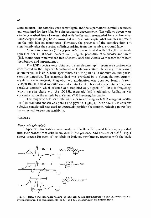
Fatty acid spin labels Spectral observations were made on the three fatty acid labels incorporated
into membranes from cells hemolyzed in the presence and absence of Ca 2+. Fig. 1 shows spectra for each of the labels in isolated membranes, together with the basic
/ •_• 1{12,31
~ OOH
K ~ _ 2 TI, )~ I1[,141
IEOG~
Fig. ]. Electron spin resonance spectra for fatty acid spin labels incorporated into untreated erythro- cyte membranes. The measurements for 2Tli and 2Tz are shown on the bottom trace.
41
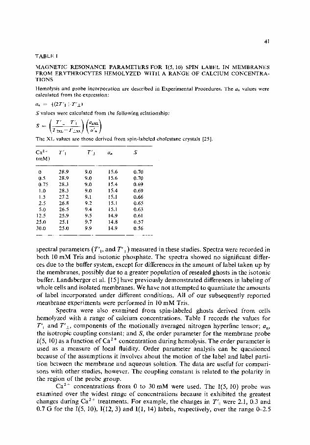
T A B L E I
M A G N E T I C R E S O N A N C E P A R A M E T E R S F O R 1(5, 10) SPIN L A B E L IN M E M B R A N E S F R O M E R Y T H R O C Y T E S H E M O L Y Z E D W I T H A R A N G E O F C A L C I U M C O N C E N T R A - T I O N S
Hemolys is and probe incorpora t ion are described in Exper imenta l Procedures. The an values were calculated f rom the expression:
an : ~ (2T ' t I+T ' I . )
S values were calculated f rom the following relat ionship:
\rlrxL-ra×L! ~ The X L values are those derived f rom spin-labeled cholestane crystals [25].
Ca 2 + T'II T '± an S (mM)
0 28.9 9.0 15.6 0.70 0.5 28.9 9.0 15.6 0.70 0.75 28.3 9.0 15.4 0.69 1.0 28.3 9.0 15.4 0.69 1.5 27.2 9.1 15.1 0.66 2.5 26.8 9.2 15.1 0.65 5.0 26.5 9.4 15.t 0.63
12.5 25.9 9.5 14.9 0.61 25.0 25.1 9.7 14.8 0.57 30.0 25.0 9.9 14.9 0.56
spectral parameters (T'~, and T'±) measured in these studies. Spectra were recorded in both 10 mM Tris and isotonic phosphate. The spectra showed no significant differ- ces due to the buffer system, except for differences in the amount of label taken up by the membranes, possibly due to a greater population of resealed ghosts in the isotonic buffer. Landsberger et al. [15] have previously demonstrated differences in labeling of whole cells and isolated membranes. We have not attempted to quantitate the amounts of label incorporated under different conditions. All of our subsequently reported membrane experiments were performed in 10 mM Tris.
Spectra were also examined from spin-labeled ghosts derived from cells hemolyzed with a range of calcium concentrations. Table I records the values for T'~ and T'±, components of the motionally averaged nitrogen hyperfine tensor; an, the isotropic coupling constant; and S, the order parameter for the membrane probe I(5, 10) as a function of Ca" ÷ concentration during hemolysis. The order parameter is used as a measure of local fluidity. Order parameter analysis can be questioned because of the assumptions it involves about the motion of the label and label parti- tion between the membrane and aqueous solution. The data are useful for compari- sons with other studies, however. The coupling constant is related to the polarity in the region of the probe group.
Ca 2+ concentrations from 0 to 30 mM were used. The 1(5, 10) probe was examined over the widest range of concentrations because it exhibited the greatest changes during Ca z+ treatments. For example, the changes in T', were 2.1, 0.3 and 0.7 G for the 1(5, 10), I(12, 3) and I(1, 14) labels, respectively, over the range 0-2.5
42
17
~u5 ~ 1 4
(
13
i i i
A
0 ~, 8 12 I~5 [Cff'*] ,ram
5 0 - -
45
4(
3~
30
B
25 -o
15
0 I 2 3 4 [Co"], ~M
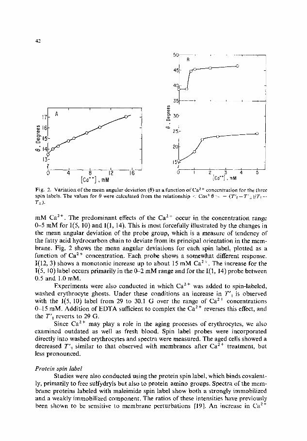
Fig. 2. Variation of the mean angular deviation (0) as a function of Ca 2 + concentration for the three spin labels. The values for 0 were calculated from the relationship <i Cos z 0 ~ ~ (T'll--T'~)/T~I-- T±).
mM Ca 2+. The predominant effects of the Ca 2+ occur in the concentration range 0-5 m M for I(5, 10) and I(1, 14). This is most forcefully illustrated by the changes in the mean angular deviation of the probe group, which is a measure of tendency of the fatty acid hydrocarbon chain to deviate from its principal orientation in the mem- brane. Fig. 2 shows the mean angular deviations for each spin label, plotted as a function of Ca 2+ concentration. Each probe shows a somewhat different response. I(12, 3) shows a monotonic increase up to about 15 mM Ca 2+. The increase for the I(5, 10) label occurs primarily in the 0-2 mM range and for the I(1, 14) probe between 0.5 and 1.0 mM.
Experiments were also conducted in which Ca z + was added to spin-labeled, washed erythrocyte ghosts. Under these conditions an increase in T'~I is observed with the I(5, 10) label f rom 29 to 30.1 G over the range of Ca 2+ concentrations 0-15 mM. Addition of EDTA sufficient to complex the Ca 2+ reverses this effect, and the T' , reverts to 29 G.
Since Ca 2 + may play a role in the aging processes of erythrocytes, we also examined outdated as well as fresh blood. Spin label probes were incorporated directly into washed erythrocytes and spectra were measured. The aged cells showed a decreased T ' , similar to that observed with membranes after Ca 2+ treatment, but less pronounced.
Prote in spin label
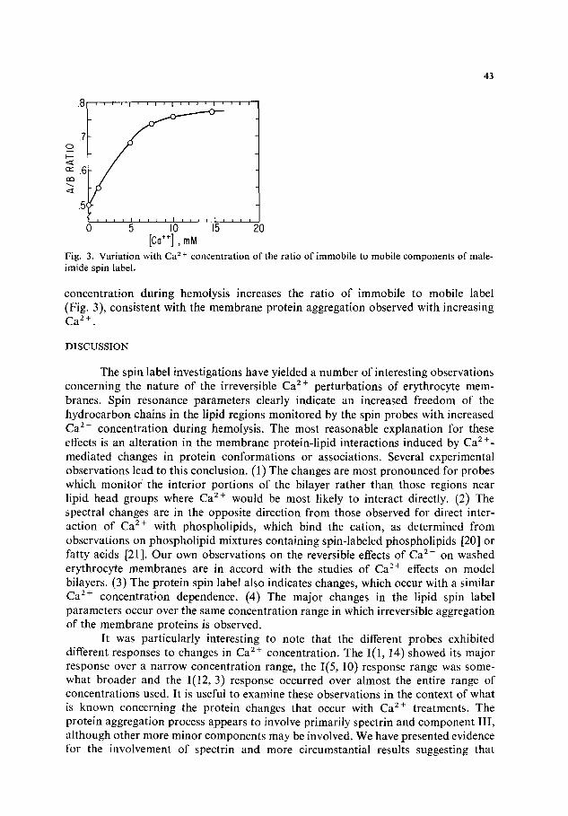
Studies were also conducted using the protein spin label, which binds covalent- ly, primarily to free sulfydryls but also to protein amino groups. Spectra of the mem- brane proteins labeled with maleimide spin label show both a strongly immobilized and a weakly immobilized component. The ratios of these intensities have previously been shown to be sensitive to membrane perturbations [19]. An increase in Ca 2÷
43
o . O i i , i I ' i i J I i , J , I , i i i
...,..-<y--
°--'7.~ ~
n- ' . 6
.5c
. . . . ~ . . . . b . . . . 5 . . . . 0 I I 20 mM
Fig. 3. Variation with Ca 2 + concentrat ion of the ratio o f immobile to mobile components o f male- imide spin label.
concentration during hemolysis increases the ratio of immobile to mobile label (Fig. 3), consistent with the membrane protein aggregation observed with increasing C a 2+"
DISCUSSION
The spin label investigations have yielded a number of interesting observations concerning the nature of the irreversible Ca 2 ÷ perturbations of erythrocyte mem- branes. Spin resonance parameters clearly indicate an increased freedom of the hydrocarbon chains in the lipid regions monitored by the spin probes with increased Ca 2÷ concentration during hemolysis. The most reasonable explanation for these effects is an alteration in the membrane protein-lipid interactions induced by Ca 2÷- mediated changes in protein conformations or associations. Several experimental observations lead to this conclusion. (1) The changes are most pronounced for probes which monitor the interior portions of the bilayer rather than those regions near lipid head groups where Ca 2÷ would be most likely to interact directly. (2) The spectral changes are in the opposite direction from those observed for direct inter- action of Ca 2+ with phospholipids, which bind the cation, as determined from observations on phospholipid mixtures containing spin-labeled phospholipids [20] or fatty acids [21]. Our own observations on the reversible effects of Ca 2÷ on washed erythrocyte membranes are in accord with the studies of Ca 2 ÷ effects on model bilayers. (3) The protein spin label also indicates changes, which occur with a similar Ca z+ concentration dependence. (4) The major changes in the lipid spin label parameters occur over the same concentration range in which irreversible aggregation of the membrane proteins is observed.
It was particularly interesting to note that the different probes exhibited different responses to changes in Ca z+ concentration. The I(1, 14) showed its major response over a narrow concentration range, the 1(5, 10) response range was some- what broader and the I(12, 3) response occurred over almost the entire range of concentrations used. It is useful to examine these observations in the context of what is known concerning the protein changes that occur with Ca 2+ treatments. The protein aggregation process appears to involve primarily spectrin and component III, although other more minor components may be involved. We have presented evidence for the involvement of spectrin and more circumstantial results suggesting that
44
component III is involved [5]. The latter suggestion is supported by recent results of Riggs and Ingram* [22] using lactoperoxidase labeling. In order to explain the spin label results, one might postulate that the aggregation process decreases the internal membrane volume required for the proteins, thus increasing the volume available for lipid mobility. Alternatively, protein-protein interactions formed by aggregation might release a fraction of lipid from lipid-protein interactions. It is difficult to determine the extent to which these possibilities might contribute without further knowledge of the mode and regions of association of the probes with the membrane.
The limited range of the Ca 2 ÷ concentration dependence of the I(! , 14) probe would thus suggest that it is responsive only to proteins such as component III which extend deep into the bilayer or through it [23]. This probe may even reflect a particular phase or segment of the aggregation process or Ca 2 ÷ effects. The response of the 1(5, 10) probe is the most extensive and most nearly corresponds to the concentration dependence of the primary protein changes, including aggregation. This might suggest that the interactions of the proteins, which are involved with the Ca 2 + effects and extend into the bilayer, are strongest within this region of the hydrocarbon chain of the lipids. The broad concentration range of the response of the 1(12, 3) probe probably indicates that it reflects interactions which are in part different from those seen by the other probes. This would suggest that a broader range or completely different set of proteins interacts with the lipids at or near the region of their head groups. These proteins may include those affected by the apparent proteolytic diges- tions [4] or those that appear to be adsorbed to the membrane from the cytoplasm. Ca 2 ÷ interactions with the phospholipid head groups may also influence the response to the 1(12, 3) probe. Since the effects of these interactions tend to restrict phospho- lipid mobility [20, 21], they would act to reduce the Ca 2 ÷ effects mediated through protein. It must be stressed, however, that all of these changes reflect only the ir- reversible Ca 2 + effects, since the membranes were well washed to remove excess Ca 2 + before spin label incorporation. Therefore these findings relate only to the thermo- dynamics of the process of stabilization of the membrane in a new structure after Ca 2+ perturbation and not to the kinetics of the processes involved at the time of Ca 2+ treatment. This is an important distinction, since Ca 2÷ may not be required for maintenance of the new state [5].
The concentration dependence of the immol;ilization of protein spin label probably most closely reflects the aggregation phenomenon. Deviations of the lipid probes from this behavior presumably result from contributions of lipid-protein or probe-protein interactions to the response [24]. Any extensive analysis of membrane changes by spin label investigations must be treated with caution because of the possibility of nonrandom association of the spin probe groups with the membrane. If the probes were associated with specific proteins or lipid regions of the membrane, the conclusions outlined in the preceding paragraph might have to be qualified. However, the basic premise that the irreversible Ca 2÷ effects are predominantly mediated through the effects of the cation on membrane proteins appears to be well founded. In addition, the results suggest that there is not a strong correlation between
* These results were presented at the FASEB meeting, Atlantic City, N.J., April 1975, but are not included in the abstract of the paper (ref. 22).
45
membrane deformability and lipid fluidity, but rather that the protein network supporting the membrane is most important to the deformability properties.
ACKNOWLEDGEMENTS
We wish to thank Mr. Darrell Anderson and Ms. C. Trecia Markes for com- ments and assistance with the manuscript preparation. We also thank Drs. Bob Floyd, Larry Scott and Olin Spivey for helpful discussion. This research was supported by NIH HL 15687 and the Oklahoma Agricultural Experiment Station, and conducted in cooperation with the USDA, Agricultural Research Service, Southern Region.
REFERENCES
1 LaCelle, P. L. (1970) Seminars Hematol. 7, 355-371 2 Palek, J., Curby, W. A. and Lionetti, F. J. (1971) Am. J. Physiol. 220, 19-26 3 LaCelle, P. L. (1969) Transfusion 9, 238-245 4 Triplett, R. B., Wingate, J. M. and Carraway, K. L. (1972) Biochem. Biophys. Res. Commun.
49, 1014-1020 5 Carraway, K. L., Triplett, R. B. and Anderson, D. R. (1975) Biochim. Biophys. Acta 379, 571-
581 6 Jacob, H., Amsden, T. and White, J. (1972) Proc. Natl. Acad. Sci., U.S. 69, 471-474 7 Harris, J. W., Brewster, H. A., Ham, T. H. and Castle, W. B. (1956) Arch. Int. Med. 97, 145-168 8 Eaton, J. W., Skelton, T. D., Swofford, H. S., Kolpin, C. E. and Jacob, H. S. (1973) Nature 246,
105-106 9 Libertini, L. J., Waggoner, A. S., Jost, P. C. and Griffith, O. H. (1969) Proc. Natl. Acad. Sci.
U.S. 64, 13-19 10 McFarland, B. G. and McConnell, H. M. (1971) Proc. Natl. Acad. Sci. U.S. 68, 1274-1278 11 Tourtellotte, M. E., Branton, D. and Keith, A. (1970)Proc. Natl. Acad. Sci. U.S. 66, 909-916 12 Hubbell, W. L., Metcalfe, J. C., Metcalfe, S. M. and McConnell, H. M. (1970) Biochim. Biophys.
Acta 219, 415-427 13 Rottem, S., Hubbell, W. L., Hayflick, L. and McConnell, H. M. (1970) Biochim. Biophys. Acta
219, 104-113 14 Kaplan, J., Canonico, P. G. and Caspary, W. J. (1973) Proc. Natl. Acad. Sci. U.S. 70, 66-70 15 Landsberger, F. R., Paxton, J. and Lenard, J. (1971) Biochim. Biophys. Acta 266, 1-6 16 Lowry, O. H., Rosebrough, N. J., Farr, A. L. and Randall, R. J. (1951) J. Biol. Chem. 193,265-
275 17 Zlatkis, A., Zak, B. and Boyle, A. J. (1953) J. Lab. Clin. Med. 41,486-492 18 Schneider, H. and Smith, I. C. P. (1970) Biochim. Biophys. Acta 219, 73-80 19 Holmes, D. E. and Piette, L. H. (1970) J. Pharmacol. Exp. Therap. 173, 78-84 20 Ohnishi, S. and Ito, T. (1974) Biochemistry 13, 881-887 21 Schnepel, G. H., Hegner, D. and Schummer, U. (1974) Biochim. Biophys. Acta 367, 67-74 22 Riggs, M. G. and Ingram, V. M. (1975) Fed. Proc. 34, 552 Abstract 23 Shin, B. C. and Carraway, K. L. (1974) Biochim. Biophys. Acta 345, 141-153 24 Wallach, D. F. H., Verma, S. P., Weidekamm, E. and Bieri, V. (1974) Biochim. Biophys. Acta
356, 68-81 25 Gaffney, B. J. and McConnell, H. M. (1974) J. Magn. Resonance 16, 1-28