Embed Size (px)
Citation preview
PLASMID 4, 139-147(1980)
Electron Microscopic Mapping of Deletions on a Streptococcal Plasmid Carrying Extraordinarily Long Inverted Repeats
DETLEV BEHNKE,'PAUL K. TOMICH,~ AND DON B. CLEWELL
Dental Research Institute, Departments of Oral Biology and Microbiology, Schools of Dentistry and Medicine. The University of Michigan, Ann Arbor, Michigan 48109
Received April 2, 1980
Deletions AlOl, A102, and Al03 which occurred within the extraordinarily long inverted repeats of the self-ligated large EcoRI fragment of the streptococcal MLS (macrolides, lincosamides, streptogramin B)-resistance plasmid pSMl9035 led to the formation of plasmids pDBlO1, pDBl02, and pDB103. Their molecular lengths were determined by contour length measurements to be 17.8, 17.4, and 13.9 kb, respectively. Electron micro- scopic examination of self-annealed molecules revealed stem-loop structures with inverted repeats comprising 41 to 91% of the mass of plasmids. Two unique sequences (US1 and US,) separated the inverted repeats in the case of pDBlO1 and pDBl03, while in pDB102 the repeats were joined at one end and separated at the other by a unique sequence (US,). The size of the unique sequence US, was identical for all three plasmids, and the location of the resistance determinant was determined by electron microscopic examination of self- annealed molecules of the recombinant plasmid pDB201. Mapping of the deletion termini, accomplished by combining electron microscopic and Hind111 restriction data, suggested that deletions may occur at preferential sites.
The l&MdaP streptococcal plasmid pSM- 19035 (Behnke et al., 1979a) specifies in- ducible resistance to macrolides, lincosa- mides, and streptogramin B-type antibiotics (MLS) (Malke, 1974). It has been shown by restriction enzyme analyses (Behnke et al., 1979a) and electron microscopy (Boitsov et al., 1979) that the plasmid has extra- ordinarily long inverted repeat sequences that comprise about 80% of the molecule. These sequences are separated by two non- repeated segments equivalent to 14 and 5% of the pSM19035 genome. Cleavage of pSM19035 with EcoRI gave rise to two fragments of about 14 and 4 Mdal; with HindIII, 15 fragments were obtained, 12 of which represented six pairs of identical
’ Present address: Academy of Sciences of the GDR, Central Institute of Microbiology and Experimental Therapy, DDR-69 Jena, PSF 73, German Democratic Republic.
’ Present address: The Upjohn Company, Kalama- zoo, Mich. 49001.
3 Abbreviations used: Mdal, megadalton; MLS, macrolides, lincosamides, and streptogramin B-type antibiotics; kb, kilobase.
fragments derived from the inverted repeats (Behnke et al., 1979a). In order to construct a potential streptococcal vector plasmid featuring a single EcoRI cleavage site, the purified large EcoRI fragment of pSM19035 was self-ligated and afterward introduced into the Challis strain of Streptococcus sang& by transformation (Behnke et al., 1979b). Subsequent analyses of plasmid DNA isolated from three such transformants revealed that the ligated fragment had suffered deletions of various extents, as a result of sequence rearrangements during or following the transformation process. As a result of deletions designated AlOl, A102, and A103, three smaller plasmids were obtained. Only one of these plasmids, pDB 101, retained the single EcoRI cleavage site while the other two pDB102 and pDB- 103, were devoid of this site. Hind111 analyses of the deletions of the three plas- mids, together with HirId cleavage pat- terns of the purified EcoRI fragments of pSM19035, allowed the construction of EcoRIIHindIII restriction site maps for pSM-
139 0147-619X/80/050139-09$02.00/0 Copyright 0 1980 by Academic Press, Inc. All rights of reproduction in any form reserved.

140 BEHNKE. TOMICH, AND CLEWELL
xdl EcoRI tmgmmt
FIG. 1. Derivation of plasmids pDB 101, pDB 102, and pDB 103 and HindllIiEcoRI cleavage site maps of the plasmids involved. Inverted repeats are indicated by heavy lines; their extension into the terminal Hind111 fragments are marked by dotted lines. Light lines represent unique sequences. Arrows label the positions ofEcoR1 cleavage sites while the letters refer to Hind111 fragments. Within the self-ligated large EcoRI fragment, the extent of deletions AlOl, A102, and At03 leading to the formation of the respective plasmids are indicated.
19035 and its derivative plasmids (Behnke In this paper we report on electron micro- et al., 1979b; Behnke and Ferretti, 1980a). scopic analyses of the deletion-containing The results are summarized in Fig. 1. The molecules, and combine the data with deletions included portions of one or both restriction enzyme analyses to map pre- of the repeated sequences, and all of them cisely the boundaries of the deletions. The overlap. (No difference in either the type data suggest that the deletions may involve or level of MLS resistance was detectable preferential sites on the plasmid. In addi- in the case of the molecules with deletions). tion, the MLS resistance determinant was
TABLE 1
STREPTOCOCCAL STRAINS USED AS SOURCES FOR PLASMID DNA
Strain
Challis (pDB101)
Challis (pDB102)
Challis (pDB103)
Challis (pDB201)
V318
Description of the plasmid
pDB 101, a deletion mutant of the MLS-resistance plasmid pSM19035
pDB102, a deletion mutant of the MLS-resistance plasmid pSM19035
pDB103, a deletion mutant of the MLS-resistance plasmid pSM19035
pDB201, a recombinant plasmid between pVA3 18 and the MLS- resistance determinant of pDB102
pVA318, a 5.4-kb cryptic plasmid having a single Hind111 restriction site
Reference
Behnke et al., 1979b; Behnke and Ferretti, 1980a
Behnke et al., 1979b
Behnke et al., 1979b
Behnke and Ferretti, 1980b
Macrina and Scott, 1978
MAPPING OF DELETIONS IN A STREPTOCOCCAL PLASMID 141
observed to be located on one of the unique tion-renaturation procedure as the plasmid sequences of pSM 19035. DNAs. All measurements were made after
projection on a Nikon profile projector MATERIALS AND METHODS (Model 60).
Bacteria. All bacterial strains which served as sources for the isolation of plasmid DNA are listed in Table 1. The Streptococcus mutans strain V318 was kindly provided by F. Macrina. Brain heart infusion broth from Difco was used to grow all strains.
Isolation of plasmid DNA. Growth in the presence of DL-threonine (Chassy, 1976) or glycine (Reider and Macrina, 1976) facili- tated lysis of S. sanguis or S. mutans; respectively. Deproteinized cleared lysates were subjected to dye-buoyant density gradient centrifugation to isolate and purify plasmid DNA (Behnke and Ferretti, 1980a). All plasmid DNAs were kept at 4°C in 10 mM Tris-HCl (pH 7.4).
Electron microscopy. The techniques used for electron microscopy of native and self-annealed plasmid molecules were as described elsewhere (Davis et al., 1971; Sharp et al., 1972; Yagi and Clewell, 1976). An Hitachi HS-8 electron microscope was used throughout these experiments. Bac- teriophage $X174 RF11 DNA (5.386 kb) (Sanger et al., 1977) served as a molecular length standard for both double- and single- stranded DNA. For the latter +X174 RF11 DNA was subjected to the same denatura-
RESULTS
Molecular Length of Native Plasmids and Their Different Regions after Self-Annealing of Denatured Molecules
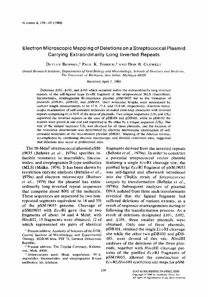
The molecular length of native plasmid DNA was determined by contour length measurement of open circular molecules [generated by heating at 95°C for 10 min in buffer (Yagi and Clewell, 1976)] to be 17.9 kb for pDBlO1, 17.4 kb for pDB102, and 14.0 kb for pDB 103 (Table 2). These results corresponded to molecular lengths of 17.8, 16.9, and 14.5 kb for the respective plas- mids, as calculated from restriction enzyme analyses (Behnke et al., 1979b). The slight discrepancies between the two values for pDB102 and pDB103) are within experi- mental error.
Denaturation of open circular plasmid molecules followed by a short renaturation period allowed the formation of intrastrand duplex regions as a result of reannealing of inverted repeat sequences. As expected from restriction data stem-loop structures were observed for all three plasmids indicat- ing the presence of inverted repeat se-
TABLE 2
MOLECULAR LENGTH OF NATIVE PLASMIDS AND THEIR DIFFERENT REGIONS AFTER DENATURATION AND INTRASTRAND ANNEALING”
Plasmid Molecular length of
native molecules us,
Self-annealed molecules
us* IR Total
pDB101 17850 i 890 (lo)* 3050 k 270 1440 2 200 6870 + 530 18230 2 1530 (9)b pDB 102 17400 e 540 (10) - 1460 k 170 7760 k 560 16980 k 1290 (12) pDB103 13950 k 1020 (8) 6320 + 560 1310 -c 180 3420 k 390 14470 + 1520 (6) pDB201 7160 k 570 (6) 5280 f 240 1530 -e 180 240 + 30 7290 2 480 (12)
G The molecular lengths are given in nucleotides or nucleotide pairs (bp). US, unique sequences; IR, inverted repeat.
b The figures in parentheses refer to the number of molecules measured.
142 BEHNKE, TOMICH, AND CLEWELL
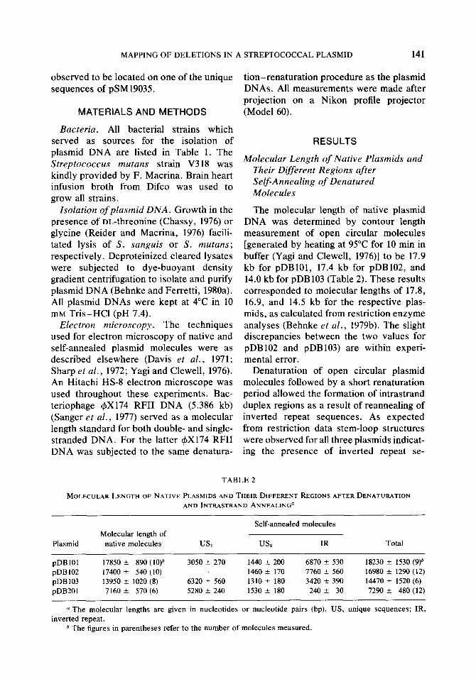
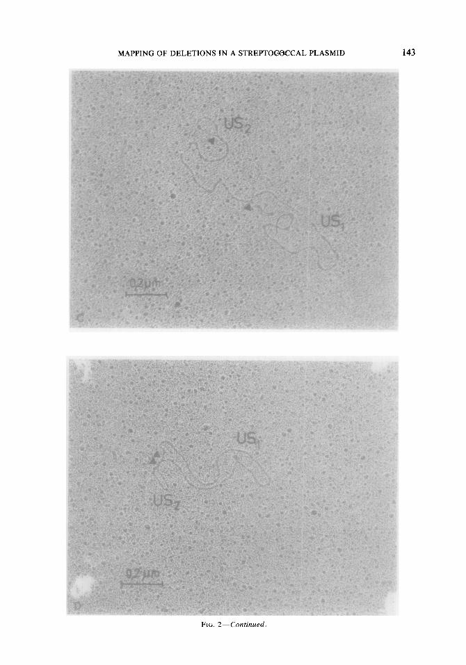
FIG. 2. Electron micrographs of denatured and self-annealed plasmid molecules. Junction points between double-stranded regions and single-stranded loops are marked by arrows. (A) pDBlO1; (B) pDB102; (C) pDB103; (D) pDB201.
MAPPING OF DELETIONS IN A STREPTOtXWCAL PLASMID 143
FIG. 2-Continued.
144 BEHNKE, TOMICH. AND CLEWELL
quences of different lengths for pDB101, pDB102, and pDB103. Electron micro- graphs of typical molecules are shown in Figs. 2A-C. In self-annealed molecules of plasmids pDB101 and pDB103 single- stranded loops were observed on both ends of the continuous central double-stranded region (Figs. 2A and C), thus indicating the presence of two interstitial unique sequences (US, and US,) that separate the inverted repeat stretches in these plasmids. In contrast, only one terminal single-stranded DNA loop was detectable in self-annealed molecules of pDB102 (Fig. 2B). Plasmid pDB102, therefore, consisted of two in- verted repeats which were connected with each other at one end and separated at the other by a short intervening sequence (US,). (A second interstitial sequence shorter than 50-75 base pairs may have escaped detec- tion by electron microscopic techniques.)
The molecular length of inverted repeats and nonrepeated sequences were deter- mined by measuring the different regions of denatured and self-annealed plasmid molecules (Table 2). Plasmid pDB103 was the result of the largest deletion (A103) reducing the inverted repeat regions to only 47% of the plasmid genome and generating a large unique sequence (US,) that com- prised 44% of the molecule. The sym- metrical deletion A102 led to the formation of pDB102 which had an extreme of more than 91% of duplicated DNA in its genome. Relations of inverted repeat to nonrepeated sequences of pDB 101 were similar to those observed for the parental plasmid pSM- 19035. For all three plasmids the smaller of the two unique sequences (US,) was similar in size, averaging a length of 1.4 kb (Table 2). This value was consistent with the one determined previously for US, in self- annealed pSM19035 molecules (Boitsov er al., 1979).
Localization of the Resistance Determinant on US2 Based on restriction data it was deduced
that US, consisted of the Hind111 fragment
D into which part of the inverted repeats extended (Fig. 1). Conclusive evidence for this was derived from electron microscopic examination of pDB201 -a recombinant plasmid consisting of the cryptic S. mutans plasmid pVA3 18, which has a single Hind111 site, and the inserted pSM19035 Hind111 fragment D which carried the resistance functions (Behnke and Ferretti, 1980b). Self-annealed pDB201 molecules showed a short double-stranded region (-240 base pairs) and two single-stranded loops (Fig. 2D), the smaller of which was identical in size with US, of pSM19035 and its deriva- tives (Table 2). The larger loop corre- sponded to the molecular length of pVA3 18 (5.4 kb) (Macrina and Scott, 1978). No stem- loop structures were observed when de- natured pVA3 18 molecules were allowed to self-anneal (data not shown). Therefore, the small loop (resembling US,) together with the short double-stranded region of -240 base pairs found with pDB201 originated from the cloned Hind111 fragment D-the location of the resistance determinant of pSMl9035 and its derivatives.
Mapping of the Deletion Termini in pDBIO1, pDB102, and pDBIO3
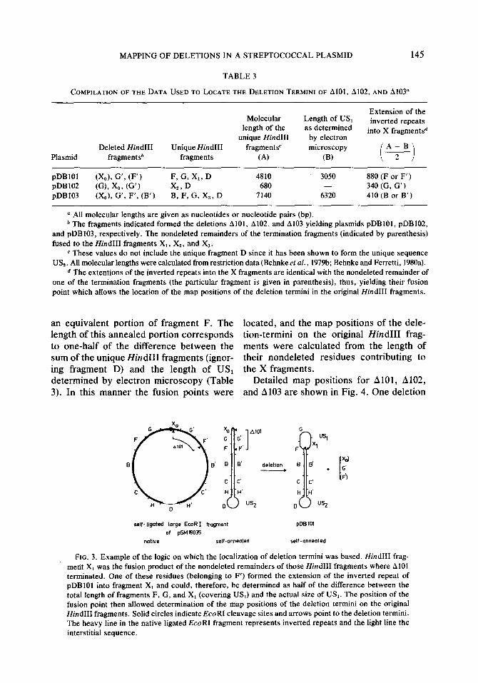
Mapping of the deletion termini was accomplished by combining Hind111 restric- tion data with results obtained from electron microscopic examination of self-annealed plasmid molecules. To facilitate compre- hension, Table 3 and Fig. 3 give a compila- tion of the data and an example of the logic on which the location of the deletion termini was based. Plasmids pDB 101, pDB 102, and pDB103 contained the new Hind111 frag- ments X1, Xz, and X, that were fusion products of the nondeleted remainders of those two fragments in which the deletions terminated (Figs. 1 and 3). Since all of the deletions include part of at least one of the inverted repeats, this fusion point can be located by determining the length of that portion of the X fragment that contains repeated sequences. In the case of pDB101 (see Fig. 3), this portion should anneal with
MAPPING OF DELETIONS IN A STREPTOCOCCAL PLASMID 145
TABLE 3
COMPILATION OF THE DATA USED TO LOCATE THE DELETION TERMINI OF 8101, A102, AND Al03”
Plasmid
Extension of the Molecular Length of US,
length of the as determined inverted repeats
unique Hind111 by electron into X fragment&
Deleted Hind111 Unique Hind111 fragmentsc microscopy A-B fragmentsb fragments (4 (W t i 2
pDB101 &A G’, (F’) F, G, X,, D 4810 3050 880 (F or F’) pDB102 (G), x09 (G’) X,, D 680 - 340 (G, G’) pDB103 W,), G’, F’, (B’) B, F, G, X,, D 7140 6320 410 (B or B’)
(1 All molecular lengths are given as nucleotides or nucleotide pairs (bp). * The fragments indicated formed the deletions AlOl, A102, and Al03 yielding plasmids pDBlO1, pDB102,
and pDB103, respectively. The nondeleted remainders of the termination fragments (indicated by parenthesis) fused to the Hind111 fragments X,, X,, and Xs.
c These values do not include the unique fragment D since it has been shown to form the unique sequence US,. All molecular lengths were calculated from restriction data (Behnke et al., 1979b; Behnke and Ferretti, 1980a).
d The extentions of the inverted repeats into the X fragments are identical with the nondeleted remainder of one of the termination fragments (the particular fragment is given in parenthesis), thus, yielding their fusion point which allows the location of the map positions of the deletion termini in the original Hind111 fragments.
an equivalent portion of fragment F. The located, and the map positions of the dele- length of this annealed portion corresponds tion-termini on the original Hind111 frag- to one-half of the difference between the ments were calculated from the length of sum of the unique Hind111 fragments (ignor- their nondeleted residues contributing to ing fragment D) and the length of US, the X fragments. determined by electron microscopy (Table Detailed map positions for AlOl, 8102, 3). In this manner the fusion points were and A103 are shown in Fig. 4. One deletion
Al01
deletion .
“Sz
t
u G' f)
self- ligated large Ed?1 fragment pDElol
of psM19035
native self-annealed self-mneoled
FIG. 3. Example of the logic on which the localization of deletion termini was based. Hind111 frag- ment X, was the fusion product of the nondeleted remainders of those Hind111 fragments where A101 terminated. One of these residues (belonging to F’) formed the extension of the inverted repeat of pDBlO1 into fragment X, and could, therefore, be determined as half of the difference between the total length of fragments F, G, and X, (covering US,) and the actual size of US,. The position of the fusion point then allowed determination of the map positions of the deletion termini on the original Hind111 fragments. Solid circles indicate EcoRI cleavage sites and arrows point to the deletion termini. The heavy line in the native ligated EcoRI fragment represents inverted repeats and the light line the interstitial sequence.
146 BEHNKE, TOMICH, AND CLEWELL
DHC B F G X, G’ F’ Et C’ H’ b I’ 1
9.55 11.78 12.15 141 16,95 lb,53 19.96 0 0,87 2.3 3.88 673 8.68 9,s lkbl
L.-..--A- AlO1 17,83 2478 o,os 235
4 A 102 18.87 I,96
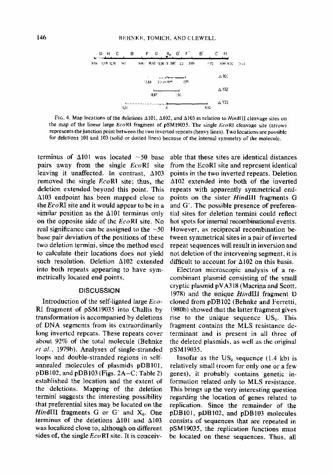
~.-mm.-e-----se-~ A.03 IL,51 0 932
FIG. 4. Map locations of the deletions AlOl, A102, and Al03 in relation to Hind111 cleavage sites on the map of the linear large EcoRI fragment of pSMl9035. The single EcoRI cleavage site (arrow) represents the junction point between the two inverted repeats (heavy lines). Two locations are possible for deletions 101 and 103 (solid or dotted lines) because of the internal symmetry of the molecule.
terminus of A101 was located -50 base pairs away from the single EcoRI site leaving it unaffected. In contrast, A103 removed the single EcoRI site; thus, the deletion extended beyond this point. This A103 endpoint has been mapped close to the EcoRI site and it would appear to be in a similar position as the Al01 terminus only on the opposite side of the EcoRI site. No real significance can be assigned to the -50 base pair deviation of the positions of these two deletion termini, since the method used to calculate their locations does not yield such resolution. Deletion A102 extended into both repeats appearing to have sym- metrically located end points.
DISCUSSION
Introduction of the self-ligated large Eco- RI fragment of pSM19035 into Challis by transformation is accompanied by deletions of DNA segments from its extraordinarily long inverted repeats. These repeats cover about 92% of the total molecule (Behnke et al., 1979b). Analyses of single-stranded loops and double-stranded regions in self- annealed molecules of plasmids pDB101, pDB 102, and pDB 103 (Figs. 2A-C; Table 2) established the location and the extent of the deletions. Mapping of the deletion termini suggests the interesting possibility that preferential sites may be located on the Hind111 fragments G or G’ and X,. One terminus of the deletions A101 and Al03 was localized close to, although on different sides of, the single EcoRI site. It is conceiv-
able that these sites are identical distances from the EcoRI site and represent identical points in the two inverted repeats. Deletion A102 extended into both of the inverted repeats with apparently symmetrical end- points on the sister Hind111 fragments G and G’. The possible presence of preferen- tial sites for deletion termini could reflect hot spots for internal recombinational events. However, as reciprocal recombination be- tween symmetrical sites in a pair of inverted repeat sequences will result in inversion and not deletion of the intervening segment, it is difficult to account for A102 on this basis.
Electron microscopic analysis of a re- combinant plasmid consisting of the small cryptic plasmid pVA3 18 (Macrina and Scott, 1978) and the unique Hind111 fragment D cloned from pDB102 (Behnke and Ferretti, 1980b) showed that the latter fragment gives rise to the unique sequence US,. This fragment contains the MLS resistance de- terminant and is present in all three of the deleted plasmids, as well as the original pSM19035.
Insofar as the US, sequence (1.4 kb) is relatively small (room for only one or a few genes), it probably contains genetic in- formation related only to MLS resistance. This brings up the very interesting question regarding the location of genes related to replication. Since the remainder of the pDB101, pDB102, and pDB103 molecules consists of sequences that are repeated in pSM19035, the replication functions must be located on these sequences. Thus, all
MAPPING OF DELETIONS IN A STREPTOCOCCAL PLASMID 147
replication genes must be present in dupli- cate and arranged in reverse orientation with respect to each other. It also follows that there are probably two origins of replication. Two replication origins have been reported in the case of the R6K (Crosa et al., 1975; Lovett ef al., 1975) and NRl (Perlman and Rownd, 1976). There is no evidence, however, for the involvement of extensive duplicate segments of origin- containing DNA in these plasmids. Analy- ses of the replication of pSM19035 should yield information on whether two origins are in fact used, and whether replication from the origin(s) is unidirectional or bi- directional. Analysis of replication might also shed some light on why pSM19035 stably maintains such extraordinarily long duplicate sequences.
ACKNOWLEDGMENTS
We wish to thank Dr. J. Ferretti for his help in supporting certain aspects of this study. Support also was provided by Public Health Research Grant DE02731 from the National Institute of Dental Research Grants AI103 18 and K04 AI00061 (Research Career Development Award to DBC) from the National Institute of Allergy and Infectious Diseases.
REFERENCES
BEHNKE, D., GOLUBKOV, V. I., MALKE, H., BOITSOV, A. S., AND TOTOLIAN, A. A. (1979a). Restriction enzyme analysis of group A streptococcal ptasmids determining resistance to macrolides, lincosamides and streptogramin-B antibiotics. FEMS Micro&l. Lett 6, 5-9.
BEHNKE, D., MALKE, H., HARTMANN, M., AND WALTER, F. (1979b). Post-transformationa rear- rangement of an in vitro reconstructed group A streptococcal erythromycin resistance plasmid. Plas- mid 2, 605-616.
BEHNKE, D., AND FERRETTI, J. J. (1980a). Physical mapping of plasmid pDB101: a potential vector plasmid for molecular cloning in streptococci. Plasmid 2, 130-138.
BEHNKE, D., AND FERRETTI, J. J. (1980b). Molecular cloning of an erythromycin resistance determinant in streptococci. J. Bacterial., in press.
BOITSOV, A. S., GOLUBKOV, V. I., IONTOVA, I. M., ZAITSEV, E. N., MALKE, H., AND TOTOLIAN, A. A. (1979). Inverted repeats on plasmids determining resistance to MLS antibiotics in group A strep- tococci. FEMS Microbial. Lett. 6, 1 l- 14.
CHASSY, B. M. (1976). A gentle method for the lysis of oral streptococci. Biochem. Biophys. Res. Com- mun. 68, 603-608.
CROSA, J. H., LUTTROPP, L. K., HEFFRON, F., AND FALKOW, S. (1975). Two replication initiation sites on R plasmid DNA. Mol. Gen. Genet. 140, 39-50.
DAVIS, R. W., SIMON, M., AND DAVIDSON, N. (1971). In “Methods in Enzymology” (L. G. Grossman, and K. Moldave, eds.), Vol. 21D, pp. 413-428. Academic Press, New York.
LOVETT, M. A., SPARKS, R. B., AND HELINSKI, D. R. (1975). Bidirectional replication of plasmid R6K DNA in Escherichia coli. Correspondence between origin of replication and position of single-strand break in relaxed complex. Proc. Nat. Acad. Sci. USA 12, 2905-2909.
MACRINA, F. L., AND SCOTT, C. L. (1978). Evidence for a disseminated plasmid in Streptococcus mutans. Infect. Immunol. 20, 296-302.
MALKE, H. (1974). Genetics of resistance to macrolide antibiotics and lincomycin in natural isolates of Streptococcus pyogenes. Mol. Gen. Genet. 135, 349-367.
PERLMAN, D., AND ROWND, R. H. (1976). Two origins of replication in composite R plasmid DNA. Nature (London) 259, 281-284.
REIDER, J. L., AND MACRINA, F. L. (1976). Plasmid DNA isolation in S. mutans: glycine enhanced cell lysis. Spec. Suppl. Microbial. Abstr. 3, 725-736.
SANGER, F., AIR, G. M., BARRELL, B. G., BROWN, N. L., COULSON, A. R., FIELDER, J. C., HUTCHI- SON, C. A., III, SLOCOMBE, P. M., AND SMITH, M. (1977). Nucleotide sequence of bacteriophage 4X 174 DNA. Nature (London) 265, 681-695.
SHARP, Z. A., Hsu, M., OHTSUBO, E., AND DAVIDSON, N. (1972). Electron microscope heteroduplex studies of sequence relations among plasmids of Escherichia co/i. I. Structure of F prime factors. J. Mol. Biol. 71, 471-497.
YAGI, Y., AND CLEWELL, D. B. (1976). Plasmid- determined tetracycline resistance in Streptococcus faeca[is: tandemly repeated resistance determinants in amplified forms of pAMa DNA. J. Mol. Biol. 102, 583-600.